Note: Descriptions are shown in the official language in which they were submitted.
CA 02606623 2007-10-31
WO 2006/118863 PCT/US2006/015561
1
SYSTEM FOR THE CONTROLLED DELIVERY OF STENTS AND GRAFTS
Background Of The Invention
1. Field of the Invention
This invention relates generally to percutaneous transluminal vascular
procedures and more particularly to delivery apparatus for placing a stent, a
stent graft
or a tubular graft at a desired target location witliin a subject's vascular
system.
II. Discussion of the Prior Art
In the field of interventional cardiology, it is now becoming routine to treat
stenotic lesions in the vascular system using balloon angioplasty to render
more patent
a partially occluded blood vessel and to atteinpt to thwart restenosis by
placement of a
stent at the site of the treated lesion.
Stents used in these procedures must be capable of assuming a reduced
diameter configuration for delivery through a guide catheter, but which are
either self-
expanding upon exit of the distal end of the guide catheter or "balloon
expandable".
In carrying out a balloon angioplasty procedure with stenting, the Seldinger
technique is frequently used to gain access to the vascular system and a
tubular
introducer having a hemostatic valve for preventing blood loss is inserted and
typically, a puncture wound is made in the artery. A guide catheter is then
inserted
through the introducer and routed through the vascular system until the distal
end
portion of the guide catheter is disposed at an ostium of a selected artery
having the
stenotic lesion.
Next, an angioplasty catheter may be advanced over a guide wire sufficiently
far so that an expandable balloon on the distal end of the delivery catheter
is
juxtaposed relative to the stenotic lesion. Upon inflation of the balloon, the
stenotic
lesion is compressed relative to the wall of the blood vessel being treated.
If the
balloon also carries a radially collapsed stent in surrounding relation to the
balloon, as
the balloon is expanded, so is the stent which becomes pressed against the
vessel wall.
Now, upon deflation of the balloon, it can be extracted leaving the stent in
place.
Stents intended for use in percutaneous transluminal angioplasty applications
come in various sizes depending on the vessel being treated.
CA 02606623 2007-10-31
WO 2006/118863 PCT/US2006/015561
2
Grafts are used for the treatment of aneurysms and commonly involve a
tubular metal or polymeric scaffold having a fabric covering preventing blood
leakage
there through. Because of this construction, such grafts could not be
compressed
sufficiently to pass through an introducer like those used in executing the
Seldinger
procedure. As such, the medical team involved required a surgeon to perform a
cut-
down procedure. Because of the radial size of most prior art vascular grafts
of the
covered scaffold variety typically would require a 24 Fr delivery sheath.
Moreover,
once the graft is delivered from the distal end of the delivery sheath, it is
incapable of
being retracted back into the sheath should repositioning be required.
What is needed, then, is an apparatus that will allow the controlled delivery
if
stents and grafts using percutaneous translumenal delivery thereby obviating
the need
for a surgeon. Further, a need exists for a delivery system for stents, stent
grafts and
grafts wherein the device to be delivered remains affixed to the delivery
device, thus
allowing the stent, stent graft or graft to be extended from and retracted
into a delivery
sheath repeatedly until such device is precisely positioned and deemed to be
of the
appropriate size to address the particular lesion or aneurysm involved. As
used
herein, a stent is a tubular scaffold for bridging a stenotic lesion in a
blood vessel, a
stent graft is a stent having a fabric, blood impervious covering and a graft
is a
scaffold for bridging a true aneurysm, a false aneurysm or a berry aneurysm.
Such
devices are collectively referred to herein as a vascular prosthesis or simply
a
prosthesis.
SUMMARY OF THE INVENTION
The foregoing desired objects are achieved in accordance with the present
invention by providing an apparatus for percutaneously delivering a self-
expanding
stent or graft to a target site within a patient's vascular system. The
apparatus
comprises an outer tubular guide catheter having a proximal end, a distal end
and a
lumen extending there between along with an inner tubular pusher catheter also
having a proximal end, a distal end and a lumen and where the inner pusher
catheter
has an outer diameter sized to slidingly fit within the lumen of the guide
catheter. An
elongate, flexible member is coaxially inserted through the lumen of the inner
pusher
CA 02606623 2007-10-31
WO 2006/118863 PCT/US2006/015561
3
catheter and it has a first bead member affixed to its distal end where the
bead is sized
to at least partially fit within the lumen of the inner pusher catheter at the
distal end of
the pusher catheter when a proximally directed tension force is applied to the
proximal
end of the elongated flexible member with respect to the inner pusher
catheter.
Completing the apparatus is a compression spring that is operatively coupled
between
the proximal end of the imler pusher catheter and a clamp member that is
releasably
affixed to the elongate member near the proximal end of the elongate member.
The stent, stent graft or graft deployed using the apparatus of the present
invention comprises a large plurality of very fine braided metal strands
exhibiting a
memory property and which is radially collapsible to a relatively small size
for
passage through the outer tubular guide catheter but which, when released from
the
guide catheter, self-expands to a relatively large diameter. The number of
strands, the
diameter of each strand, the pitch and pick of the braid are such that the
pore size of
the resulting tubular graft is sufficiently small that fibrin present in the
blood will
close such pores, rendering the graft leak-proof. The braided tubular graft is
installed
on the delivery system by capturing the free ends of the strands comprising
the braided
graft at its proximal end between the bead member affixed to the elongate
flexible
member and the wall defining the lumen of the inner tubular pusher catheter at
its
distal end. The compression spring is used to maintain the requisite tension
force on
the elongate member to maintain the ends of the strands pinched between the
bead
member and the wall of the inner tubular pusher catheter proximate its distal
end.
DESCRIPTION OF THE DRAWINGS
The foregoing features, objects and advantages of the invention will become
apparent to those skilled in the art from the following detailed description
of a
preferred embodiment, especially when considered in conjunction with the
accompanying drawings in which like numerals in the several views refer to
corresponding parts.
Fig. 1 is a partial side elevation view illustrating the percutaneous delivery
system for stents and grafts configured in accordance with the present
invention;
CA 02606623 2007-10-31
WO 2006/118863 PCT/US2006/015561
4
Fig. 2 is a greatly enlarged view of the distal end portion of the assembly of
Fig. 1 showing the proximal ends of the wires comprising the braided stent or
graft
captured at the distal end of the delivery catheter; and
Fig. 3 is a view like that of Fig. 2 showing the stent or graft released from
the
distal end of the delivery catheter.
DESCRIPTION OF THE PREFERRED EMBODIMENT
Referring first to Fig. 1, the percutaneous translumenal stent or graft
delivery
system is identified generally by numeral 10 and, as already indicated, is
used to
deliver a stent or graft menlber 12 to a target site within the vascular
system such as at
the location of an abdominal aortic aneurysm for the purpose of exclusion of
the
aneurysm to prevent fizrther bulging and possible rupture thereof.
The vascular prosthesis 12 is preferably formed of a metal fabric exhibiting
an
expanded configuration and a collapsed configuration. The prosthesis, when
collapsed, can be deployed through the lumen of a catheter and, upon exiting
the distal
end of the catheter at a target site in a patient's vascular system, will
substantially
return to its expanded configuration.
As is described in U.S. Patent 5,725,552 to Curtis Amplatz, the metal fabric
comprising the prosthesis may comprise a plurality of braided metal strands
where the
metal is preferably a shape memory alloy such as NITINOL . In accordance with
the
present invention, the metal fabric is braided in the form of a tube that can
be fitted
onto a cylindrical mandrel and then heat-treated so that in its expanded
configuration,
the prosthesis will have an internal diameter substantially equal to the outer
diameter
of the mandrel on which it is heat-treated.
Without limitation, the graft may comprise a 72, a 144, or a 288-strand
tubular
wire braid using wires of selected diameters dependent on the number of wires
employed in the braiding process Using a tubular braid of about 20-30 mm in
diameter with a. predetennined pitch and pick such that the graft exhibits a
pore size
less than 100 microns, the graft can be longitudinally stretched to a reduced
diameter
permitting it to be passed through the lumen of a 7 French guiding catheter
that can
readily be inserted into the vascular system using the Seldinger technique.
Upon exit
CA 02606623 2007-10-31
WO 2006/118863 PCT/US2006/015561
from the distal end of the delivery catheter at the desired target site, the
graft 12 will
self-expand to a limit defined by the vessel wall in which it is disposed.
Using a metal fabric braided from 288 or even 144 strands or wires whose
diameters may be about 0.00075 inch yields a fabric that is rather blood-
impervious
5 and within a relatively short time following placement becomes
endothelialized.
Blood trapped between the outer surface of the graft and the bulge comprising
the
aneurysm rapidly clots to fill the bulge space with a congealed mass. The
lumen of
the graft, however, remains patent, allowing continuous blood flow through the
treated area of the blood vessel.
Those skilled in the art interested in obtaining more information concerning
the fabrication of occluding devices using braided structures of the type
contemplated
herein for the prosthesis 12 are referred to the following patents, each of
which is
assigned to AGA Medical Corporation, the assignee of the present application,
the
teachings of which are hereby incorporated by reference.
5,725,552
5,944,738
6,468,303
6,402,772
6,468,301
6,368,339
6,506,204
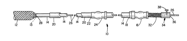
Turning next to the construction of the graft delivery device 10, it is seen
to
comprise a pusher catheter 14 having a male Luer coupler 16 of a standard
variety
affixed to its proximal end 18. The delivery catheter may be of various
lengths and
may have an outer diameter of from about 50 to 10 French, depending on the
location
of the vessel segment to be treated, allowing it to pass through an internal
lumen of an
outer guide catheter 20.
The guide catheter 20 has a lumen of a size to receive the pusher catheter 14
therethrough with a close tolerance so that blood flow between the two is
substantially
blocked. Affixed to its proximal end 22 of the guide is a female Luer fitting
24 that is
CA 02606623 2007-10-31
WO 2006/118863 PCT/US2006/015561
6
adapted to mate with the male Leur fitting 16 affixed to the proximal end 18
of the
delivery catlieter 14.
Disposed within the lumen of the pusher catheter 14 is a wire or cable 26
whose length allows it to extend beyond the full length of the delivery
catheter 14
when pushed from its proximal end portion. Laser welded to the distal end of
the
cable or wire 26 is a bead that is spherical or frusto-conically shaped clamp
member
28 and a short, predetermined distance proximal of the clamp member 28 is an
annular washer-like member 30 that is also welded or otherwise fixedly
attached to
the cable or wire 26.
A helically-wound compression spring 32 slips over and surrounds the cable or
wire 26 and is operatively disposed between the proximal end of the male Luer
fitting
16 and a releasable clamp 34 here shown as a tubular sleeve 36 having a
transversely
extending threaded bore leading to the lumen of the tubular sleeve 36. Fitted
into this
threaded bore is a thumbscrew 38 that when tightened down against the wire or
cable
26 serves to lock the sleeve 36 to that cable or wire.
To ready the delivery system for use, the free ends of the strands comprising
the braid at the proximal end 13 fed into the lumen of the pusher catheter 14
and are
captured between the outer surface of the bead member 28 and the distal end 15
of the
pusher catheter 14, as best seen in the greatly enlarged partial view of Fig.
2. To
achieve this result, the prosthesis 12 in its expanded configuration is
slipped over the
tapered clamp member 28 and the proximal end of the cable or wire 26 is fitted
through a disposable, tear-away funnel member (not shown) before being
inserted into
the distal end 15 of the pusher catheter 14 and fed down its length. As the
prosthesis
is pushed through the funnel, the proximal ends of the strands are made to
feed into
the lumen of the pusher catheter 14 and now, as the cable or wire 26 is pulled
in the
proximal direction, the proximal ends of the wire strands 13 become captured
between
the bead member 28 and the lumen wall of the pusher catheter 14. So long as
the
tension is maintained, the free ends 13 of the braided prosthesis 12 will
remain
captured.
CA 02606623 2007-10-31
WO 2006/118863 PCT/US2006/015561
7
To maintain the prosthesis clamped to the distal end of the pusher catheter
14,
tension is applied at the proximal end of the wire or cable 26 as the sleeve
36 is
pushed in the distal direction to thereby compress the coil spring 32 between
the
sleeve 36 and the Luer fitting 16. With the spring 32 so compressed, the
thuinbscrew
38 will be tightened to thereby hold the sleeve 36 in position relative to the
coil or
wire 26, thus maintaining the tension force on the cable or wire 26.
The assenibly comprising the pusher catheter 14, the compression spring 32,
the clamping member 34 can be drawn in the proximal direction while holding
the
female Luer fitting 24 stationary, thus drawing the distal end 15 of the
pusher catheter
along with the prosthesis 12 into the lumen of the outer guide catheter 20.
All of these
steps of clamping the braided device to the pusher catheter 14 and drawing the
prosthesis 12 within the lumen of the outer guiding catheter 20 may be
performed at a
manufacturer's facility prior to packaging and sterilization of the assembly.
At the
time of use with a patient, a cardiologist may first gain percutaneous entry
of the guide
catheter 20 containing the stent or a stent/graft or a graft (the prosthesis)
and route the
distal end thereof under fluoroscopic viewing to the target site of an
aneurysm to be
reinforced. While keeping the outer guide catheter 20 stationary, the pusher
catheter
14 is advanced in the distal direction until its distal end 15 to which the
prosthesis 12
is clamped exits the distal end of the guide catheter 20. So long as the
compression
spring is providing the tension force on the cable, the prosthesis remains
coupled to
the distal end of the pusher catheter allowing it to be again retracted into
the lumen of
the outer guide catheter should it become necessary to reposition the device
before it
is released.
To release the prosthesis from the distal end 15 of the pusher catheter 14,
the
physician merely has to loosen the thumbscrew 38 and then move the cable or
wire 26
in the distal direction sufficiently far so that the washer 30 pushes against
the
proximal end surfaces of the wires 13 to move the prosthesis free of the end
of the
pusher catheter. At this point, and as shown in Fig. 3, the prosthesis 12 has
self-
expanded to a larger diameter so that the bead 28 can readily be withdrawn
from the
interior of the tubular prosthesis. The delivery system 10 can then be
withdrawn from
CA 02606623 2007-10-31
WO 2006/118863 PCT/US2006/015561
8
the vascular system.
While a preferred embodiment of the present invention has been described, it
should be understood that various changes, adaptations and modifications may
be
made therein without departing from the spirit of the invention and the scope
of the
appended claims. For example rather than front loading the pusher catheter 14
carrying the elongate member 26 and the prosthesis 12 by feeding the proximal
end of
the pusher catheter through the distal end of the delivery sheath 20 and then
along the
length of the delivery sheath, it is also contemplated that a loader tube
containing the
prosthesis be coupled to the Luer fitting 24 and the pusher wire 26 be used to
advance
the prosthesis down the delivery sheath until it approaches the distal end of
the
delivery sheath 20.
1 claim: