Note: Descriptions are shown in the official language in which they were submitted.
CA 02606665 2007-10-31
WO 2006/119034 PCT/US2006/016292
METHOD AND APPARATUS FOR THE TREATMENT
OF THE INTERVERTEBRAL DISC ANNULUS
Cross-Reference to a Related Applications
[001] This application claims priority to U.S. Patent Application No.
11/120,750 filed May 3, 2005, which is a continuation-in-part of U.S. Patent
Application No. 10/352,981 filed January 29, 2003 and a continuation-in-part
of
U.S. Patent Application No. 10/327,106 filed December 24, 2002, each of which
are continuations-in-part of U.S. Patent Application No. 10/133,339 filed
April 29,
2002, which is a continuation-in-part of US Patent Application No. 09/947,078,
filed September 5, 2001, now U.S. Patent 6,592,695, issued July 15, 2003,
which.
is a continuation of U.S. Patent Application No. 09/484,706, filed January 18,
2000, which claims the benefit of U.S. Provisional Application No. 60/160,710,
filed October 20, 1999. This application also claims, through Application
10/133,339 the benefit of U.S. Provisional Application No. 60/309,105, filed
July
31, 2001. This application is also related to, and claims the benefit of, U.S.
Patent Application 10/075,615, filed on February 15, 2002. All are
incorporated
herein by reference in their entirety.
Field of the Invention
[002] The invention generally relates to methods and devices for the
closure, sealing, repair and/or reconstruction of an intervertebral disc
annulus,
and accompanying delivery devices and tools, and their methods of use. The
repair can be of an aperture in the disc wall, or a weakened or thin portion.
The
term "aperture" refers to a hole in the annulus that is a result of a surgical
incision
or dissection into the intervertebral disc annulus, or the consequence of a
naturally occurring tear (rent). The invention generally relates to surgical
devices
and methods for the treatment of intervertebral disc wall repair or
reconstruction.
The invention further relates to an annular repair device, or stent, for
annular disc
repair. These stents can be of natural or synthetic materials. The effects of
said
reconstruction is restoration of disc wall integrity, which may reduce the
failure
CA 02606665 2007-10-31
WO 2006/119034 PCT/US2006/016292
rate (3-21 %) of a common surgical procedure (disc fragment removal or
discectomy), or advantageously provide a barrier to intradiscal material
migration.
Background of the Invention
[003] The spinal column is formed from a number of bony vertebrae,
which in their normal state are separated from each other by intervertebral
discs.
These discs are comprised of the annulus fibrosus, and the nucleus pulposus,
both of which are soft tissue. The intervertebral disc acts in the spine as a
crucial
stabilizer, and as a mechanism for force distribution between adjacent
vertebral
bodies. Without a competent disc, collapse of the intervertebral disc may
occur,
contributing to abnormal joint mechanics and premature development of
degenerative and/or arthritic changes.
[004] The normal intervertebral disc has an outer ligamentous ring called
the annulus surrounding the nucleus pulposus. The annulus binds the adjacent
vertebrae together and is constituted of collagen fibers that are attached to
the
vertebrae and cross each other so that half of the individual fibers will
tighten as
the vertebrae are rotated in either direction, thus resisting twisting or
torsional
motion. The nucleus pulposus is constituted of soft tissue, having about 85%
water content, which moves about during bending from front to back and from
side to side.
[005] The aging process contributes to gradual changes in the
intervertebral discs. The annulus loses much of its flexibility and
resilience,
becoming more dense and solid in composition. The aging annulus may also be
marked by the appearance or propagation of cracks or fissures in the annular
wall. Similarly, the nucleus desiccates, increasing viscosity and thus losing
its
fluidity. In combination, these features of the aged intervertebral discs
result in
less dynamic stress distribution because of the more viscous nucleus pulposus,
and less ability to withstand localized stresses by the annulus fibrosus due
to its
desiccation, loss of flexibility and the presence of fissures. Fissures can
also
occur due to disease or other pathological conditions. Occasionally fissures
may
form rents through the annular wall. In these instances, the nucleus pulposus
is
urged outwardly from the subannular space through a rent, often into the
spinal
-2-
CA 02606665 2007-10-31
WO 2006/119034 PCT/US2006/016292
column. Extruded nucleus pulposus can, and often does, mechanically press on
the spinal cord or spinal nerve rootlet. This painful condition is clinically
referred
to as a ruptured or herniated disc.
[006] In the event of annulus rupture, the subannular nucleus pulposus
migrates along the path of least resistance forcing the fissure to open
further,
allowing migration of the nucleus pulposus through the wall of the disc, with
resultant nerve compression and leakage of chemicals of inflammation into the
space around the adjacent nerve roots supplying the extremities, bladder,
bowel
and genitalia. The usual effect of nerve compression and infiammation is
intolerable back or neck pain, radiating into the extremities, with
accompanying
numbness, weakness, and in late stages, paralysis and muscle atrophy, and/or
bladder and bowel incontinence. Additionally, injury, disease or other
degenerative disorders may cause one or more of the intervertebral discs to
shrink, collapse, deteriorate or become displaced, herniated, or otherwise
damaged and compromised.
[007] Surgical repairs or replacements of displaced or herniated discs are
attempted approximately 390,000 times in the USA each year. Historically,
there
has been no known way to repair or reconstruct the annulus. Instead, surgical
procedures to date are designed to relieve symptoms by removing unwanted disc
fragments and relieving nerve compression. While results are currently
acceptable, they are not optimal. Various authors report 3.1- 21 % recurrent
disc
herniation, representing a failure of the primary procedure and requiring re-
operation for the same condition. An estimated 10% recurrence rate results in
39,000 re-operations in the United States each year.
[008] An additional method of relieving the symptoms is thermal
annuloplasty, involving the heating of sub-annular zones in the non-herniated
painful disc, seeking pain relief, but making no claim of reconstruction of
the
ruptured, discontinuous annulus wall.
[009] Some have also suggested that the repair of a damaged
intervertebral disc might include the augmentation of the nucleus pulposus,
and
various efforts at nucleus pulposus replacement have been reported. The
-3-
CA 02606665 2007-10-31
WO 2006/119034 PCT/US2006/016292
present invention is directed at the repair of the annulus, whether or not a
nuclear
augmentation is also warranted.
[010] In addition, there has been experimentation in animals to assess
various surgical incisions with and without the direct surgical repair of the
annulus.
These studies were performed on otherwise healthy animals and involved no
removal or augmentation of nucleus pulposus. The authors of these experiments
conclude that direct repair of the annulus does not influence the healing of
the
disc.
[011] The present inventors have found, advantageously and contrary to
accepted practice, that the annulus tissue may be sutured and that annular
healing may be facilitated by reapproximation, reinforcement, and/or support
of
annular tissue. Methods and devices for carrying out annular repair and/or
reconstruction are a subject of the present invention.
Brief Summary of the Invention
[012] The present inventions provide methods and related devices for
reconstruction of the disc wall in cases of displaced, herniated, thinned,
ruptured,
or otherwise damaged or infirm intervertebral discs. In accordance with the
invention, a method is disclosed for intervertebral disc reconstruction for
treating a
disc having an aperture, weakened or thin portion in the wall of the annulus
fibrosis of the intervertebral disc. Repair, reconstruction, sealing,
occluding an
aperture, weakened or thin portion in the wall of the annulus may prevent or
avoid
migration of intradiscal material from the subannular space.
[013] The method of the invention includes, in one embodiment, the steps
of providing a first delivery tool having a proximal end and a distal end, the
distal
end carrying a treatment device; providing at least one second delivery tool
having a proximal end and a distal end, the distal end carrying a fixation
element;
introducing the distal end of the first delivery tool at least partially into
subannular
intervertebral disc space; deploying said treatment device; introducing the
distal
end of said at least one second delivery tool at lest partially into
subannular
intervertebral disc space; and deploying at least one fixation device into, or
-4-
CA 02606665 2007-10-31
WO 2006/119034 PCT/US2006/016292
through, the wall of an annuius to hold said treatment device at least
partially
within the subannular intervertebral disc space; and removing the delivery
tools.
[014] A fixation device useful for intervertebral disc reconstruction for
treating a disc having an aperture, weakened, or thin portion in the wall of
the
annulus fibrosis of said intervertebral disc, said device, in one embodiment
comprises at least one anchor portion and at least one band.
[015] A treatment device, according to one embodiment, comprises a
mesh patch that radially expands in the subannular space.
[016] The invention also comprises delivery tools for delivering fixation
devices and treatement devices, as well as kits comprising devices and tools.
[017] The objects and various advantages of the invention will be
apparent from the description which follows. In general, the impiantable
medical
treatment devices are placed, positioned, and subsequently affixed in the
annulus
to reduce re-extrusion of the nucleus or other indtradiscal material through
an
aperture by: establishing a barrier or otherwise closing or partially closing
the
aperture; and/or helping to restore the natural integrity of the wall of the
annulus;
and/or promoting healing of the annulus. Increased integrity and faster and/or
more thorough healing of the aperture may reduce future recurrence of
herniation
of the disc nucleus, or intradiscal material, from the intervertebral disc,
and the
recurrence of resulting back pain. In addition, it is believed that the repair
of the
annular tissue could promote enhanced biomechanics and reduce the possibility
of intervertebral disc height collapse and segmental instability, thus
possibiy
avoiding back pain after a surgical procedure.
[018] Moreover, the repair of an aperture (after for example, a discectomy
procedure) with the reduction of the re-extrusion of the nucleus may also
advantageously reduce adhesion formation surrounding the nerve roots. The
nuclear material of the disc is toxic to the nerves and is believed to cause
increased inflammation surrounding the nerves, which in turn can cause
increased scar formation (adhesions or epidural fibrosis) upon healing.
Adhesions created around the nerve roots can cause continued back pain. Any
reduction in adhesion formation is believed to reduce future recurrence of
pain.
-5-
CA 02606665 2007-10-31
WO 2006/119034 PCT/US2006/016292
[019] The methods and devices of the present inventions may create a
mechanical barrier to the extrusion of intradiscal material (i.e., nucleus
pulposus,
or nuclear augmentation materials) from the disc space, add mechanical
integrity
to the annulus and the tissue surrounding an aperture, weakened, or thin
portion
of the wall of the annulus, and promote faster and more complete healing of
the
aperture, weakened or thin portion
[020] Although much of the discussion is directed toward the repair of the
intervertebral disc after a surgical procedure, such as discectomy (a surgical
procedure performed to remove herniated fragments of the disc nucleus), it is
contemplated that the devices of the present invention may be used in other
procedures that involve access (whether induced or naturally occurring)
through
the annulus of the intervertebral disc, or prophylactic application to the
annulus.
An example of another procedure that could require a repair technique involves
the replacement of the nucleus (nucleus replacement) with an implantable
nucleus material to replace the functioning of the natural nucleus when it is
degenerated. The object of the invention in this case would be similar in that
the
repair would maintain the replacement nucleus within the disc space.
[021] According to an embodiment of the invention, a sub-annular device
can be employed to repair an aperture, degenerated, weakened, or thin portion
in
an intervertebral disc annulus. The device can be secured in place with one or
more fixation elements, such as sutures or anchors which may also be used to
re-
approximate the tissues surrounding the aperture, degenerated, weakened, or
thin portion. The invention, through invoivement of the sub-annular space and
wall for the repair of the aperture has several advantages. The first
advantage of
a repair that involves a sub-annular surface derives itself from the physical
nature
of a circular (or an elliptical) compressed chamber with a radius, like an
intervertebral disc. Sealing the inside wall has the inherent advantage of
being at
a smaller radius of curvature versus the outer wall and thus, according to
LaPlace's Law, the patch would be subjected to lower stresses at any given
pressure, all else held equal.
-6-
CA 02606665 2007-10-31
WO 2006/119034 PCT/US2006/016292
[022] Another advantage of utilizing the inner surface to accomplish
sealing is that the natural pressure within the disc can enhance the seating
of the
device against the inner wall of the disc space. Conversely, if the repair is
performed on the outer surface of the annulus there is an inherent risk of
leakage
around the periphery of the device, with the constant exposure to the pressure
of
the disc.
[023] Another advantage of the present invention in utilizingan inner
surface of the annulus is the reduction of the risk of having a portion of the
device
protruding from the exterior surface of the annulus. Device materials
protruding
from the exterior of the annulus pose a risk of damaging the nerve root and/or
spinal canal which are in close proximity. Damage to these structures can
result
in continued pain, incontinence, bowel dysfunction and paralysis.
[024] Some embodiments of the present invention may also incorporate
the concept of pulling the tissues together that surround the aperture, the
inner
surface, and the outer surface of the annulus to help close the aperture,
increase
the integrity of the repair, and promote healing.
[025] An example of the technique and placement of the device according
to one embodiment of the invention is as follows:
[026] 1. A treatment device is actuated into a delivery configuration by
delivery device and passed through an aperture in the wall of the annulus,
positioning a treatment device in the subannular disc space, as shown in FIG.
3A
- 3C.
1027] 2. The delivery device is actuated to deploy the treatment
device, as shown in FIGs. 3D.
[028] 3. Holding the treatment device in the deployed configuration, a
fixation instrument is employed to introduce one or more fixation elements
into, or
through, the annulus to secure the treatment device and subsequently removed,
as shown in FIGs. 2, 5 and 6.
[029] 4. The delivery device is disengaged from the treatment device.
[030] Several devices according to the present invention can be used to
practice the above illustrative inventive steps to seal, reconstruct and/or
repair the
-7-
CA 02606665 2007-10-31
WO 2006/119034 PCT/US2006/016292
intervertebral disc. In some of the representative devices described herein,
there
is: a reconfigurable device (note: patch, stent, device, mesh, barrier, and
treatment device are here used interchangeably) that has, in use, at least a
portion of the device in the sub-annular space of the intervertebral disc
annulus; a
means to affix the at least a portion of the device to or within at least a
portion of
the annulus; a means to draw the patch or fixation device into engagement with
the annular tissue in tension to thereby help reduce the relative motion of
the
surfaces of the aperture and/or annulus after fixation, and thus promote
healing.
Reducing motion of the annular surfaces may provide the optimal environment
for
healing.
[031] Some of the concepts disclosed hereinbelow accomplish these
objectives, as well as may advantageously additionally incorporate design
elements to reduce the number of steps (and time), and/or simplify the
surgical
technique, and/or reduce the risk of causing complications during the repair
of the
intervertebral disc annulus. In addition, the following devices may become
incorporated by the surrounding tissues, or to act as a scaffold in the short-
term
(3 - 6 months) for tissue incorporation.
[032] In an exemplary embodiment, one or more mild biodegradable
surgical sutures can be placed at about equal distances along the sides of a
pathologic aperture in the ruptured disc wall (annulus) or along the sides of
a
surgical dissection or incision in the annular wall, which may be weakened or
thinned. The sutures hold an expandable device to a subannular surface of the
annulus.
[033] Sutures are then drawn in tension and secured in such a fashion as
to draw the expandable device into engagement with the annular tissue, and
also
to help effect closure of the aperture, to enhance natural healing and
subsequent
reconstruction by natural tissue (fibroblasts) crossing the gap now bridged by
the
device in the disc annulus.
[034] In an exemplary embodiment, the method can be augmented by
creating a subannular barrier in and across the aperture by placement of a
patch
of biocompatible material 'acting as a bridge or a scaffold, providing a
platform for
-3-
CA 02606665 2007-10-31
WO 2006/119034 PCT/US2006/016292
traverse of fibroblasts or other normal cells of repair existing in and around
the
various layers of the disc annulus.
[035] Such biocompatible materials may be, for example, medical grade
biocompatible fabrics or fibers, biodegradable polymeric sheets, or form
fitting or
non-form fitting fillers for the cavity created by removal of a portion of the
disc
nucleus pulposus in the course of the disc fragment removal or discectomy. The
prosthetic material can be placed in and around the intervertebral space,
created
by removal of the degenerated disc fragments.
[036] Additional objects and advantages of the invention will be set forth in
part in the description which follows, and in part will be obvious from the
description, or may be learned by practice of the invention. The objects and
advantages of the invention will be realized and attained by means of the
elements and combinations particularly pointed out in the appended claims.
[037] It is to be understood that both the foregoing general description
and the following detailed description are exemplary and explanatory only and
are
not restrictive of the invention, as claimed.
Brief Description of the Drawings
[038] The accompanying drawings, which are incorporated in and
constitute a part of this specification, illustrate illustrative embodiments
of the
invention and, together with the description, serve to explain the principles
of the
invention.
[039] FIG. 1 shows a primary closure of an opening in the disc annulus.
[040] FIGs. 2A-2B show a primary closure with a stent.
[041] FIGs. 3A-3D show an annulus stent being inserted into and
expanded within the disc annulus.
[042] FIGs. 4A-4C shows a perspective view of a further illustrative
embodiment of an annulus stent, and coliapsed views thereof.
[043] FIGs. 5A-5C show the annulus stent of FIG. 4A being inserted into
the disc annulus.
[044] FIGs. 6A-6C show a method of inserting the annulus stent of FIG.
4A into the disc annulus.
-9-
CA 02606665 2007-10-31
WO 2006/119034 PCT/US2006/016292
[045] FIG. 7 shows an illustrative embodiment of an introduction device
for an annulus stent.
[046] FIG. 8 shows a variation of the device depicted in FIG. 7.
[047] FIGs. 9A-9C show an exemplary introduction tool for use with the
devices of FIGs. 7 and 8 with a stent deflected.
[048] FIGs. 10A-10B show a still further illustrative embodiment of an
annulus stent employing secondary barbed fixation devices.
[049] FIG. 11A shows a herniated disc in perspective view, and FIG. 11 B
shows the same disc after discectomy.
[050] FIGs. 12A-12G show a still further illustrative embodiment of an
introduced and expanded annulus stent/patch being fixated and the aperture
reapproximated.
[051] FIGs. 13A-13C schematically depict a still further embodiment of the
invention where an expandable stent/patch is tethered in situ using a cinch
line.
[052] FIGs. 14A-14C schematically depict the patch of FIG. 13 being
fixated through use of a barbed surgical staple device and a cinch line.
[053] FIGs. 15A-15C schematically depict a still further embodiment of the
invention where an expandable stent/patch is tethered in situ using a cinch
line.
[054] FIGs. 16A-16C schematically depict the stnet/patch of FIG. 15 being
fixated through use of a barbed surgical staple device that penetrates the
patch/stent and a cinch line.
[055] FIG. 17 depicts an exemplary use of filler material within the
aperture during placement of a patch/stent tethered by a cinch line.
[056] FIGs. 18A-18E show exemplary embodiments of various additional
patch/stent fixation techniques.
[057] FIGs. 19 shows a still further illustrative embodiment of a
stent/patch having a frame.
[058] FIGs. 20A-20C show a still further exemplary embodiment of the
invention having external fixation anchors.
[059] FIGs. 21A-21 C show still further embodiments of the invention
having external fixation anchors.
-10-
CA 02606665 2007-10-31
WO 2006/119034 PCT/US2006/016292
[060] FIGs. 22A-22C show still further embodiments of the invention
having external fixation anchors.
[061] FIG. 23 shows a delivered configuration of fixation means that may
result from the use of a single, or multiple, devices to deliver multiple
barbs,
anchor, or T-anchors sequentially or simultaneously.
[062] FIGs 24A-24B show an illustrative configuration of an anchor band
delivery device.
[063] FIGs 25A-25D show an anchor band delivery device comprising two
devices, each with at least one T-anchor (barbs) and band with pre-tied knot
and
optional knot pusher according to illustrative embodiments of the invention.
[064] FIG 26 shows an anchor and band delivery device according to one
embodiment of the invention.
[065] FIGs. 27A-27B show, respectively, a lateral view of a still further
exemplary embodiment of the present invention having a braided arrangement in
a collapsed configuration and an axial view of the exemplary embodiment in an
expanded configuration.
[066] FIG. 28shows a lateral view of the exemplary embodiment of FIG.
27A in a collapsed configuration mounted on an illustrative delivery device.
[067] FIG. 29 shows a lateral cutaway view of the exemplary embodiment
of FIG. 27A in a collapsed configuration.
[068] FIG. 30 shows a lateral cutaway view of the exemplary embodiment
of FIG. 27B in an expanded configuration.
[069] FIG. 31 shows a lateral view of an illustrative delivery member as
shown in the exemplary embodiment of FIGs. 29 and 30.
[070] FIG. 32 shows a lateral view of an exemplary embodiment of the
invention in an expanded configuration subannularly.
[071] FIG. 33 shows a transverse view of a treatment device mounted on
a delivery tool in an unexpanded configuration in the subannular cavity.
[072] FIG. 34 shows a transverse view of the treatment device being
deployed into an expanded configuration in the subannular cavity.
- 11 -
CA 02606665 2007-10-31
WO 2006/119034 PCT/US2006/016292
[073] FIG. 35 shows a transverse view of the treatment device fully
deployed and adjacent the annular wall.
[074] FIG. 36 shows a transverse view of the placement of a fixation
element delivery device into the deployed treatment device.
[075] FIG. 37 shows a transverse view of the placement of a fixation
element through the treatment device and the annular wall.
[076] FIG. 38 shows a transverse view of after affixing a fixation element
delivered in FIG. 37 and partial removal of the fixation element delivery
device.
[077] FIG. 39 shows a transverse view of the fixation element after
removal of the fixation element delivery tool.
[078] FIG. 40 shows a transverse view of an additional fixation element
locked in place on the opposite side of the treatment device.
[079] FIG. 41 shows a transverse view of the removal of the treatment
device delivery tool.
[080] FIG. 42 shows an transverse view of an illustrative embodiment of a
treatment device mounted on a delivery tool in an unexpanded configuration in
the subannular cavity.
[0811 FIG. 43 shows a transverse view of after affixing a fixation element
to the treatment device of FIG. 42.
[082] FIG. 44 shows a transverse view of the placement of a fixation
element delivery tool through the treatment device and the annular wall.
[083] FIG. 45 shows a transverse view of the placement of an additional
fixation element through the treatment device and the annular wall.
[084] FIG. 46 shows a transverse view after the removal of the fixation
element delivery tool.
[085] FIG. 47 is a view of the anchor band delivery tool pre-deployment in
cross section.
[086] FIG. 48 shows a detail of the distal end of the anchor band (fixation
element) delivery tool in cross section.
[087] FIG. 49 shows a detail of the slide body and cannula anchor of an
exemplary fixation element delivery tool in cross section.
-12-
CA 02606665 2007-10-31
WO 2006/119034 PCT/US2006/016292
[088] FIG. 50 is a view of the anchor band delivery tool in cross section
during a deployment cycle.
[089] FIG. 51 is a detail pf the distal end of the anchor band delivery tool
depicted in FIG. 50.
[090] FIG. 52 shows a detail of the slide body and cannula anchor of an
exemplary fixation element delivery tool in cross section during a deployment
cycie.
[091] FIG. 53 shows a detail of the suture retention block and blade
assembly of the anchor band delivery tool.
[092] FIG. 54 is a view of the anchor band delivery tool in cross section
during the cutting of the suture tether and release of the anchor band.
[093] FIG. 55 shows a detail of the distal end of the anchor band delivery
tool during release of the anchor band.
[094] FIG. 56 shows a detail of the shows a detail of the suture retention
block and blade assembly of the anchor band delivery tool during the cutting
of
the tether.
[095] FIG 57 depicts an illustrative embodiments of a therapeutic device
delivery tool (TDDT)
[096] FIG 58 shows a detail of the distal end of the threapetutic device
delivery tool with a therapeutic device mounted thereon.
[097] FIG. 59 depicts the deployment of a therapeutic device using the
TDDT.
[098] FIG. 60 depicts a detail of the distal end fo the TDDT during
deployment of a therapeutic device.
[099] FIG. 61 depicts the TDDT during release of the therapeutic device.
[0100] FIG. 62 is a detail view of the distal end of the TDDT during release
of the therapeutic device.
[0101] Fig. 63 is a pian view along the axis of an expanded exemplary
therapeutic device, showing the engagement of the TDDT latch.
[0102] Fig. 64 is a plan view along the axis of an expanded exemplary
therapeutic device, showing the disengagement of the TDDT latch.
-13-
CA 02606665 2007-10-31
WO 2006/119034 PCT/US2006/016292
[0103] FIG. 65 illustrates an exemplary sizing tool used to assess the
treatment site prior to delivery of patch, anchor bands or other treatment
devices.
[0104] FIGs. 66A-66C depict various configurations of an exemplary
therapeutic device.
[0105] FIG. 67 shows an illustrative embodiment of a pre-set heat formed
therapeutic device in cross section through a longitudinal axis.
[0106] FIGs. 68A-68B show a therapeutic device with a single rib of
material affixed to or formed on the device.
[0107] FIGs. 69A-69D show a therapeutic device with multiple ribs of
material affixed to or formed on the device.
[0108] FIGs. 70A-70C illustratively show means that may be attached to
the anchor band or anchor band delivery tool for providing perceptible
feedback.
[0109] FIG. 71A-71G depict illustrative means for latching, locking or
otherwise securing the treatment device in its final configuration.
[0110] FIGs. 72A-72B show a treatment device constructed of two bodies.
[0111] FIGs. 73A-73C show an illustrative example of multiple members
forming inner and outer members of a treatment device.
[0112] FIGs. 74A-74B show alternative illustrative mechanisms of drawing
together locking elements/anchors.
[0113] FIGs. 75A-75B show alternative illustrative attachment mechanisms
where a pledget element that initially resides on outer annular surface.
[0114] FIGs. 76A-76B show a cross section view of the delivery of a
flowable material from the distal end of delivery device.
[0115] FIG. 77 illustrates an alternative method and device for the
introduction of material into the disc space via the fixation element delivery
tool.
[0116] FIGs. 78A-78B depicts an anchor band assembly used to repair a
circumferential tear in the annulus.
[0117] FIG 79A-79B illustrate illustrative embodiments of the invention
used to treat meniscal tissue of the knee.
[0118] FIGs. 80A-80B illustrate an illustrative general surgical application
of the invention for treating hernia.
-14-
CA 02606665 2007-10-31
WO 2006/119034 PCT/US2006/016292
Detailed Description of Illustrative Embodiments of the Invention
[0119] Reference will now be made in detail to selected illustrative
embodiments of the invention, with occasional reference to the accompanying
drawings. Wherever possible, the same reference numbers will be used
throughout the drawings to refer to the same or like parts.
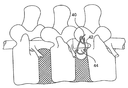
[0120] In the surgical repair of an aperture in the annulus , as shown in
FIG. 1 and as described in related commonly-assigned U.S. Patent 6,592,625 to
Cauthen, a damaged annulus 42 is repaired by use of surgical sutures 40. One
or more surgical sutures 40 are placed at about equal distances along the
sides
of a pathologic aperture 44 in the annulus 42. Reapproximation or closure of
the
aperture 44 is accomplished by tying the sutures 40 so that the sides of the
aperture 44 are drawn together. The reapproximation or closure of the aperture
44 enhances the natural healing and subsequent reconstruction by the natural
tissue (e.g., fibroblasts) crossing the now surgically narrowed gap in the
annulus
42. Preferably, the surgical sutures 40 are biodegradable, but permanent non-
biodegradable may be utilized. In all embodiments where biodegradable
materials are indicated, suitable biodegradable materials may include, but are
not
limited to, biodegradable polyglycolic acid, swine submucosal intestine,
collagen,
or polylactic acid. Other suitable suturing (and band) materials include,
e.g.,
polymeric materials such as polyethylene teraphthalate (PET), polyester (e.g.,
DacronT"'), polypropylene, polyethylene, polycarbonate urethane or metallic
material include, e.g., titanium, nickel titanium alloy, stainless steel,
surgical steels
or any combinations thereof.
[0121] Additionally, to repair a weakened or thinned wall of a disc annulus
42, a surgical incision or dissection can be made along the weakened or
thinned
region of the annulus 42 and one or more surgical sutures 40 can be placed at
about equal distances laterally from the incision. Reapproximation or closure
of
the incision is accomplished by tying the sutures 40 so that the sides of the
incision are drawn together. The reapproximation or closure of the
incision/dissection enhances the natural healing and subsequent reconstruction
by the natural tissue crossing the now surgically narrowed gap in the annulus
42.
-15-
CA 02606665 2007-10-31
WO 2006/119034 PCT/US2006/016292
Preferably, the surgical sutures 40 are biodegradable, but permanent non-
biodegradable materials may be utilized.
[0122] Where necessary or desirable, the method can be augmented by
placing a patch in and across the aperture 44. The patch acts as a bridge in
and
across the aperture 44, providing a platform for traverse of fibroblasts or
other
normal cells of repair existing in and around the various layers of the disc
annulus
42, prior to closure of the aperture 44. FIGs. 2A-B, for example, show a
biocompatible device employed as an annulus stent 10, being piaced in and
across the aperture 44. The annulus stent 10 acts as a bridge in and across
the
aperture 44, providing a platform for a traverse of fibroblasts or other
normal cells
of repair existing in and around the various layers of the disc annulus 42,
prior to
closure of the aperture 44. In some embodiments the device, stent or patch can
act as a scaffold to assist in tissue growth that healingly scars the annulus.
[0123] In an illustrative embodiment, the annulus stent 10 is a solid unit,
formed from one or more of the flexible resilient biocompatible or
bioresorbable
materials well know in the art. The selection of appropriate stent materials
may
be partially predicated on specific stent construction and the relative
properties of
the material such that, after fixed placement of the stent, the repair may act
to
enhance the healing process at the aperture by relatively stabilizing the
tissue and
reducing movement of the tissue surrounding the aperture.
[0124] For example, the annulus stent 10 may be made from:
[0125] A porous matrix or mesh of biocompatible and bioresorbable fibers
acting as a scaffold to regenerate disc tissue and replace annulus fibrosus as
disclosed in, for example, U, S. Patent Nos. 5,108,438 (Stone) and 5,258,043
(Stone), a strong network of inert fibers intermingled with a bioresorbable
(or
bioabsorbable) material which attracts tissue ingrowth as disclosed in, for
example, U.S. Patent No, 4,904,260 (Ray et al.).
[0126] a biodegradable substrate as disclosed in, for example, U.S. Patent
No. 5,964,807 (Gan at al.); or
[0127] an expandable polytetrafluoroethylene (ePTFE), as used for
conventional vascular grafts, such as those sold by W.L. Gore and Associates,
-16-
CA 02606665 2007-10-31
WO 2006/119034 PCT/US2006/016292
Inc. under the trademarks GORE-TEX and PRECLUDE, or by Impra, Inc. under
the trademark IMPRA.
[0128] Furthermore, the annulus, stent 10, may contain hygroscopic
material for a controlled limited expansion of the annulus stent 10 to fill
the
evacuated disc space cavity.
[0129] Additionally, the annulus stent 10 may comprise materials to
facilitate regeneration of disc tissue, such as bioactive silica-based
materials that
assist in regeneration of disc tissue as disclosed in U.S. Patent No.
5,849,331
(Ducheyne, et al.), or other tissue growth factors well known in the art.
[0130] Many of the materials disclosed and described above represent
embodiments where the device actively promotes the healing process. It is also
possible that the selection of alternative materials or treatments may
modulate the
role in the healing process, and thus promote or prevent healing as may be
required. It is also contemplated that these modulating factors could be
applied to
material substrates of the device as a coating, or similar covering, to evoke
a
different tissue response than the substrate without the coating.
[0131] Materials of the patch couid include a metallic material (e.g., NiTi
alloy, Stainless steel, Titanium), or a polymeric material (e.g.,
polypropylene,
polyethylene, polyurethane, polycarbonate urethane, Polyetheretherketone
(PEEK), polyester, PET, poly olefin copolymer, polypropylene, polyethylene),
or a
biodegradable or bioresorbable material (e.g., collagen, cellulose,
polysaccharide,
polyglycolic acid (PGA), a polylevolactic acid (PPLA), a polydioxanone (PDA)
or
for example a racemic polylactic acid (PDLLA), or a combination of these
materials.
[0132] In an illustrative method of use, as shown in FIGs. 3A-3D, lateral
extensions 20 and 22 of a stent 10 are compressed together for insertion into
the
aperture 44 of the disc annulus 42. The annulus stent, 10 is then inserted
into the
aperture 44, where the lateral extensions 20, 22 expand. In an expanded
configuration, the upper surface 28 can substantially conform to the contour
of the
inside surface of the disc annulus 42. The upper section 14 is positioned
within
-17-
CA 02606665 2007-10-31
WO 2006/119034 PCT/US2006/016292
the aperture 44 so that the annulus stent 10 may be secured to the disc
annulus
42, using means well known in the art.
,[0133] In an alternative method, where the length of the aperture 44 is less
than the length of the outside edge 26 of the annulus stent 10, the annulus
stent
can be inserted laterally into the aperture 44. The lateral extensions 20 and
22
are compressed, and the annulus stent 10 can then be laterally inserted into
the
aperture 44. The annulus stent 10 can then be rotated inside the disc annulus
42, such that the upper section 14 can be held back through the aperture 44.
The
lateral extensions 20 and 22 are then allowed to expand, with the upper
surface
28 contouring to the inside surface of the disc annulus 42. The upper section
14
can be positioned within, or proximate to, the aperture 44 in the subannular
space
such that the annulus stent 10 may be secured to the disc annulus, using means
well known in the art.
[0134] It is anticipated that fibroblasts will engage the fibers of the
polymer
or fabric of the intervertebral disc stent 10, forming a strong wall
duplicating the
currently existing condition of healing seen in the normal reparative process.
[0135] In an alternative embodiment, as shown in FIG. 4A, the annulus
stent 10 is substantially umbrella shaped, having a central hub 66 with
radially
extending struts 67. Each of the struts 67 is joined to the adjacent struts 67
by a
webbing material 65, forming a radial extension 76 about the central hub 66.
The
radial extension 76 has an upper surface 68 and a lower surface 70, where the
upper surface 68 contours to the shape of the disc annulus' 42 inner wall when
inserted as shown in FIG. 6A-6C, and where the lower surface 70 contours to
the
shape of the disc annulus' 42 inner wall when inserted as shown in FIG. 5A-5C.
The radial extension 76 may be substantially circular, elliptical, or
rectangular in
plan shape.
[0136] As shown in FIGs. 4B and 4C, the struts 67 are formed from flexible
material, allowing the radial extension 76 to be collapsed for insertion into
aperture 44, then the expand conforming to the shape of the inner wall of disc
annulus 42. In the collapsed position, the annulus stent 10 is substantially
-18-
CA 02606665 2007-10-31
WO 2006/119034 PCT/US2006/016292
frustoconical or shuttlecock shaped, and having a first end 72, comprising the
central hub 66, and a second end 74.
[0137] In an alternative embodiment, the radial extension 76 has a greater
thickness at the central hub 66 edge than at the outside edge.
[0138] In a method of use, as shown in FIGs. 5A-5C, the radial extension
76 is collapsed together, for insertion into the aperture 44 of the disc
annulus 42.
The radial extension 76 is folded such the upper surface 68 forms the outer
surface of the cylinder. The annulus stent 10 is then inserted into the
aperture
44, inserting the leading end 72 though the aperture 44 until the entire
annulus
stent 10 is within the disc annulus 42. The radial extension 76 is released,
expanding within the disc 44. The lower surface 70 of the annulus stent 10
contours to the inner wall of disc annulus 42. The central hub 66 is
positioned
within the aperture 44 so that the annulus stent 10 may be secured to the disc
annulus 42 using means well known in the art.
[0139] It is anticipated that fibroblasts will engage the fibers of the
polymer
or fabric of the annulus stent 10, forming a strong wall duplicating the
currently
existing condition of healing seen in the normal reparative process.
[0140] In an alternative method of use, as shown in FIGs. 6A-6C, the radial
extension 76 is collapsed together for insertion into the aperture 44 of the
disc
annulus 42. The radial extension 76 is folded such that the upper surface 68
forms the outer surface of the stent, for example in a frustoconical
configuration
as illustrated. The annulus stent 10 is then inserted into the aperture 44,
inserting
the tail end 74 through the aperture 44 until the entire annulus stent 10 is
in the
disc. The radial extension 76 is released, expanding within the disc. The
upper
surface 68 of the annulus stent 10 contours to the disc annulus' 42 inner
wall.
The central hub 66 is positioned within the aperture 44 so that the annulus
stent
may be secured to the disc annulus 42, using means well known in the art.
[0141] In one illustrative embodiment, the barbs 82 on the upper surface 68
of one or more strut 67 or other feature of the radial extension 76, engage
the
disc annulus' 42 inner wall, holding the annulus stent 10 in position.
-19-
CA 02606665 2007-10-31
WO 2006/119034 PCT/US2006/016292
[0142] FIG. 7 shows a further aspect of the present invention. According to
a further illustrative embodiment, a simplified schematic cross section of a
vertebral pair is depicted including an upper vertebral body 110, a lower
vertebral
body 112 and an intervertebral disc 114. An aperture or rent 116 in the
annulus
fibrosus (AF) is approached by a tube 118, which is used to deliver a device
120
according to a further aspect of the present invention. The device 120 may be
captured by a delivery tool 122 through the use of a ring or other fixation
feature
124 mounted on the repair device 120.
[0143] FIG. 8 shows a delivery method similar to that depicted in FIG. 7,
with the exception that the tube 11 8A has a reduced diameter so that it may
enter
into the sub-annular space of the disc 114 through the aperture or rent.
[0144] Turning to FIGs. 9A-9C, according to a further aspect of the present
invention, the delivery of the device 120 through the delivery tube 118 or
118A
may be facilitated by folding the arms or lateral extensions 128, 130 of the
device
to fit within the lumen of the tube 118 or 118A so that the stent or device
120 is
introduced in a collapsed configuration. The device 120 is moved through the
lumen of the tubes 118 or 118A through the use of delivery tool 122. FIG. 9B
shows the arms deflected in a distal, or forward direction for insertion into
the
delivery tube 118 or 118A whiie FIG. 9A shows the arms 128, 130 deflected into
a
proximal position. FIG. 9C shows the device 120 curled so that one arm 128 is
projecting distally, or in a forward direction, and the other arm 130 is
projecting
proximally, or in a rearward direction. Because the lateral extent of the
device is
relatively flexible, whether the device is of natural or synthetic material,
other
collapsible configurations consistent with the intent of this invention are
also
possible, including twisting, balling, crushing, folding, bending, etc.
[0145] FIG. 10A shows an alternative fixation strategy where a pair of
barbs 134 and 136 are plunged into the annulus fibrosus from the exterior of
the
annulus while the device 120 is retained in the sub-annular space by means of
a
tether 142. Although there are a wide variety of fixation devices in this
particular
example, a tether 142 may be knotted 145 with the band 144 holding the barbs
134 and 136 together to fix the device in the sub-annular space. The knot is
-20-
CA 02606665 2007-10-31
WO 2006/119034 PCT/US2006/016292
shown in an uncinched position to clarify the relationship between the tether
142
and the bands 144. Using this approach, the device can be maintained in a
subannular position by the barbed bands while the tether knot is cinched,
advantageously simultaneously reapproximating the annulus to close the
aperture
while drawing the device into sealing, bridging engagement with the subannuiar
wall of the annulus fibrosus. ,
[0146] FIG. 10B shows an alternative fixation strategy where the barbs 148
and 150 are sufficiently long that they can pierce the body of the device 120
and
extend all the way through the annulus fibrosus into the device 120. In this
configuration, the band 144 connecting the barbs 148 and 150 may be tightened
to gently restrain and position the device 120 in the sub-annular space, or
tightened with greater force to reapproximate the aperture or rent.
[0147] Patches can be folded and expanded in a single plane or in three
dimensions. As shown in FIGs. 9A-9C for example, collapsing the patch can be
accomplished laterally, whether the device is a single material or composite.
Others can collapse in three dimensions, such as those shown in FIGs. 4, 5,
27,
30 and 34. Devices which expand in three dimensions can optionally be
packaged in a restraining sheath, jacket, gelatin shell or "gelcap", or a mesh
of
biosorbable or dissolvable material, that would allow for facile placement and
subsequent expansion.
[0148] It is understood that there can be a variety of device designs of
patches/stents/meshes/devices/treatment devices to accomplish the expansion of
a device from a first configuration, to a second configuration to occupy at
least a
portion of the sub-annular space and reduce re-extrusion of the nucleus, or
otherwise facilitate maintaining other intradiscal materials within the disc
space.
These devices can be constructed of single components or multiple components,
with a variety of different materials, whether synthetic, naturally occurring,
recombinated (genetically engineered) to achieve various objectives in the
delivery, deployment and fixation of a device to repair or reconstruct the
annulus.
The following device concepts are further discussed for additional embodiments
of a device and/or system for the repair of an intervertebral disc annulus.
The
-21 -
CA 02606665 2007-10-31
WO 2006/119034 PCT/US2006/016292
following descriptions will illustratively depict and describe methods,
devices, and
tools to deliver a treatment to an intervertebral disc after a, lumbar
discectomy
procedure; although, it is anticipated that these methods, devices, and tools
may
be similarly used in a variety of applications. As an example, the embodiments
described herein may also advantageously maintain materials within the disc
space other than natural disc tissue (nucleus, annulus, cartilage, etc.), such
as
implants and materials that may be used to replace and/or augment the nucleus
pulposus or other parts of disc's tissues. These procedures may be performed
to
treat, for example, degenerative disc disease. Whether these materials are
intended to replace the natural functioning of the nucleus pulposus (i.e.,
implantable prosthetics or injectable, in-situ curable polymer protein, or the
like) or
provide a fusion between vertebral bodies (i.e., implantable bony or synthetic
prosthetics with materials to facilitate fusion, such as growth factors like
bone
morphogenic proteins) one skilled in the art would realize that variations to
the
embodiments described herein may be employed to better address characteristic
differences in the various materials and/or implants that could be placed
within
the subannular space, and that these variations would be within the scope of
the
invention.
[0149] Furthermore, it should be noted that surgeons differ in their
techniques and methods in performing an intervention on a spinal disc, and the
inventive descriptions and depictions of methods, devices and delivery tools
to
repair annular tissue could be employed with a variety of surgical techniques;
such as, but not limited to: open surgical, microsurgical discectomy (using a
magnifying scope or loupes), minimally invasive surgical (through, for
example, a
METRxTM system available from Medtronic, Inc.), and percutaneous access.
Surgeons may also employ a variety of techniques for intra-operative
assessment
and/or visualization of the procedure, which may include: intra-operative
probing,
radiography (e.g., C-arm, flat plate), and endoscopy. It is contemplated that
the
inventive embodiments described are not limited by the various techniques that
may be employed by the surgeon.
-22-
CA 02606665 2007-10-31
WO 2006/119034 PCT/US2006/016292
[0150] In addition, the surgical approach to the intervertebral disc
throughout the figures and descriptions depict a common approach, with related
structures, to a lumbar discectomy; although, it is possible that surgeons may
prefer alternative approaches to the intervertebral disc for various
applications (for
example, different intervertebral disc levels such as the cervical or thoracic
region,
or for nucleus augmentation), which may include, but is not limited to:
posterior-
lateral, anterior, anterior-lateral, transforaminal, extra-foraminal, extra-
pedicular,
axial (i.e., through the vertebral bodies), retroperitoneal, trans psoas
(through the
Psoas muscle), contralateral. The approach to the intervertebral disc space
should not be interpreted to limit the use of the invention for the repair or
reconstruction of the an aperture, weakened or thin portion of the annulus, as
described herein.
[0151] It is also important to note that the boundary in the intervertebral
disc space between the annulus fibrosus and the nucleus pulposus as depicted
herein may be demarked or otherwise highlighted; however, it is important to
recognize that these tissues are not as precisely demarked in human tissues,
and
may be even less so as the patient ages or evinces degeneration of the
intervertebral disc. This demarcation may be especially difficult to discern
during
an operative procedure, using for example; available surgical tools (i.e.,
probes),
fluoroscopic guidance (x-ray), or visual (endoscope) guidance. However, in
general, the layers of the annulus have more structural integrity (and
strength)
than the nucleus, and this integrity varies from the outer most layers of the
annulus being of higher structural integrity than the inner most layers of the
annulus.
[0152] Moreover, the drawings and descriptions herein are necessarily
simplified to depict the operation of the devices and illustrate various steps
in the
method. In use, the tissues may be manipulated by, and are frequently in
contact
with, the various tools and devices; however, for clarity of construction and
operation, the figures may not show intimate contact between the tissues the
tools and the devices.
-23-
CA 02606665 2007-10-31
WO 2006/119034 PCT/US2006/016292
[0153] As depicted in FIG. 11 A, a herniated disc occurs when disc nucleus
material emerges from the subannular region and outside of the disc. Herniated
disc nucleus material then impinges on nerve tissue, causing pain. A
discectomy
attempts to relieve pressure on the nerve tissue through surgical removal of
disc
material, the result usually being an aperture in the disc annulus wall, and
usually "
a void in the subannular space where disc nucleus was removed, as shown in
FIG. 11 B. Figure 11 B typifies a disc after the discectomy procedure has been
performed, as do most of the drawings and descriptions contained herein.
However, it should be understood that in order to perform a discectomy
procedure, there are a variety of instruments and tools readily available to
the
surgeon during spine surgery, or other surgical procedures, to obtain the
outcome
as shown in Fig. 11, or other outcomes intended by the surgeon and the
surgical
procedure. These tools and instruments may be used to: incise, resect,
dissect,
remove, manipulate, elevate, retract, probe, cut, curette, measure or
otherwise
effect a surgical outcome. Tools and instruments that may be used to perform
these functions may include: scalpels, Cobb elevators, Kerrison punch, various
elevators (straight, angled, for example a Penfield), nerve probe hook, nerve
retractor, curettes (angled, straight, ringed), rongeurs (straight or
angulated, for
example a Peapod), forceps, needle holders, nerve root retractors, scissors.
This
list is illustrative, but is not intended to be exhaustive or interpreted as
limiting. It
is anticipated that some of these tools and/or instruments could be used
before,
during, or after the use of the inventive methods, devices and tools described
herein in order to access, probe (e.g., Penfield elevator), prepare (e.g.,
angled or
ringed curette, rongeur, forceps), and/or geherally assess (e.g., angled
probe)
treatment site or facilitate the manipulation (e.g., forceps, needle holder),
introduction (e.g., forceps, needle holder, angled probe), or deployment
(e.g.,
forceps, needle holder, angled probe) of the treatment device and/or it's
components.
[0154] The are a variety of ways to affix a device to the sub-annular wall of
the annulus in addition to those discussed hereinabove. The following
exemplary
embodiments are introduced here to provide inventive illustrations of the
types of
-24-
CA 02606665 2007-10-31
WO 2006/119034 PCT/US2006/016292
techniques that can be employed to reduce the time and skill required to affix
the
patch to the annulus, versus suturing and tying a knot. Discussed hereinabove
is
the use of sutures, staples and other fixation devices to affix the patch to
the
annulus. In a simple example, a patch/stent could be compressed, passed
through a guide tube such as tubes 18, 18A shown in FIGs. 7 and 8, and
expanded within the sub-annular space.
[0155] Another fixation means includes the passing of "anchoring bands"
into the wall of the annulus, vertebral bodies (superior, inferior, or both),
or the
Sharpey's Fibers (collagenous fibers between the junction of the annular
fibers
and vertebral bodies). In the following example of anchors, the barbs or bands
are affixed to the annulus/vertebral bodies/Sharpey's fibers. Another element,
for
example a suture, cinch line, or a staple is utilized to attach the anchor
bands to
the patch, and thus hold the patch in proximity to the inner wall of the
annulus. In
addition, these bands may re-approximate the tissues at the aperture.
[0156] Another example of fixating the device to inner wall of the annulus is
further illustrated by FIGs 12-14. As discussed hereinabove, with reference to
FIGs. 7-10, a patch 120 is placed with a delivery tool 122, through the inner
lumen of a guide tube 118, into the sub-annular space and then expanded. This
step can also be seen in FIGs. 13 and 14, where a patch 702 is folded and
passed through a guide tube 706 and is held by a delivery tool 704. Also shown
is a anchor band or staple 709 and an anchor band delivery device 708. Within
the guide tube, or within the delivery tool, there is a suture line or cinch
line 710
that is attached to the center of the patch 702. This can be seen in FIG. 12A
with
the guide tube 706 removed. As seen in FIGs. 13C and 14A, the guide tube 706
is retracted after the patch 702 has been expanded and deployed. Next, as
shown in FIGs. 12 and 14, an anchor band delivery tool 708 is used to deliver
one
or more "bands" 709 onto the outer surface of the annuius. These are intended
to
be anchored into the wall of the annulus with barb shapes that do not allow
for the
barbs to be pulled back through the annulus. The anchor bands resemble a
construction of a "staple". The bands could actually be constructed by
connecting
-25-
CA 02606665 2007-10-31
WO 2006/119034 PCT/US2006/016292
two barbed elements with, for example, a suture between the two barbed
elements.
[0157] The barbs and the connection band between the barbs could be
constructed of the same material or of different materials. For example, the
barbed part of the anchor band could be a biodegradable/bioabsorbable material
(such as, for example, collagen, cellulose, polysaccharides, carbohydrates,
polygiycolic acid, polylevolactic acid, polydioxanone, racemic polylactic
acid) or
could be constructed of a metallic or polymeric biocompatible material (e.g.,
titanium, NiTi alloy, stainless steel, platinum, gold, polyurethane,
polycarbonate
urethane, polyimide, polyamide, polypropylene, polyethylene, polypropylene,
polyester, PET, PEEK). The anchors could also be constructed of a combination
of these materials. In addition, the band that connects these barbs can be
constructed of materiais that are similar to the barbs, or different
materials. For
example, the connection band could be a biodegradable/bioabsorbable suture,
such as Vicryl, or a biocompatible material such as polypropylene,
polyethylene,
silk, stainless steel, PET. In addition, it is possible that these eiements
are
constructed from multiple materials to accomplish the objective of anchoring
into
the annulus and providing for a fixation site to draw the tissues together.
[0158] FIGs. 12B and 12C show the placement of the anchor bands 709
into the annulus 712 with the anchor band delivery tool 708. FIGs. 14A and 14B
schematically show the placement of the anchor bands 709 into the wall of the
annulus 712 and the retraction of the anchor band delivery device 708, with
the
patch delivery tool 704 still in place. FIG. 12D depicts a representative
anchor
band 709, having a pair of stainless steel barbs 709" connected by a suture
709'.
FIG. 12E shows the patch 702, anchor bands 709, and cinch line or suture 710
with the delivery tools removed, prior to drawing the patch and the tissues of
the
annulus together. In this embodiment there is a pre-fabricated slip knot 714
on
the cinch line, although other locking elements or knots are possible. Suture
loops can connect to the barbs directly, as in FIG. 12, or loop to surgical
staples,
or are placed directly into the annulus. The presence of a pre-fabricated knot
on
the cinch line makes the process of repairing quicker since there is no need
to tie
-26-
CA 02606665 2007-10-31
WO 2006/119034 PCT/US2006/016292
a knot. It also facilitates drawing the tissues together. The use of the cinch
line
and a pre-fabricated knot can be placed by, for example, an external tube such
as
a knot pusher. FIG. 12E is similar to FIG. 10 described hereinabove prior to
"tying" the knot 714. FIG. 12F shows the drawing of the patch and the annular
tissues together by pulling on the suture in the direction "A" indicated by
the
arrow. In this case, the Knot Pusher has been removed from the cinch line 710.
The suture 710 is drawn proximally to draw the patch 702 into engagement with
the inner wall of the annulus to seal the aperture from within, as well as
draw the
walls of the annulus together to reapproximate the annular aperture. FIG. 14C
and FIG. 12G show the cinch line suture 710 tied and drawing the annular
tissues
together, after the excess suture line has been cut. It is also apparent from
this
device, fixation and delivery system that the outer surfaces of the aperture
may
be drawn together for re-approximation.
[0159] The cinching of the anchor bands and the patch also allows for
taking-up the slack that allows for the accommodation of varying sizes. For
example, the thickness of the annular wall surrounding the aperture can vary
from
1 mm up to 10 mm. Therefore, if the anchor bands have a set length, this
design
with a cinch line accommodates different dimensions of the thickness of the
wall
of the annulus by drawing the "slack" of the bands together within the
aperture.
[0160] Although it has been described here as patch placement that
involves two lateral anchor bands with a suture to draw the patch, bands and
tissues together, one or two or more bands could be used and two bands is only
an example. Furthermore, the anchor bands were placed with the barbs in a
superior-inferior fashion. One skilled in the art would recognize that these
could
be placed at different locations surrounding the aperture, vertebral bodies or
into
the Sharpey's fibers
[0161] Although the patch depicted in the example above does not have
barbs attached to the patch, it is also possible to have the barbs as
described
hereinabove to further promote the fixation of the patch to the inner wall of
the
annulus.
-27-
CA 02606665 2007-10-31
WO 2006/119034 PCT/US2006/016292
[0162] Finally, although the drawings depict an aperture that lends itself to
re-approximating the tissues, it is conceivable that some apertures, whether
natural or surgically made, may be relatively large and therefore might
require the
placement of additional material within the aperture to act as a scaffold for
tissue
in growth, between the patch on the inner wall of the annulus and the anchor
bands located on the outer wall. An example of material to fill the aperture
might
include autograft para-spinal fascial tissue, xenograft, allograft, or other
natural
collagenous materials. The filler material could also be of a biocompatible
material such as a Dacron (polyester, or PET), polypropylene, polyethylene
material. FIG. 17 shows the illustrative filling of an aperture with implant
material
716 prior to cinching the suture 710.
[0163] As an alternative embodiment of the present invention, the anchor
bands 709 as described previously (anchor bands into annulus) could be
sufficiently long enough to pass through the annulus and then through the
patch.
The barbs in this embodiment have an engaging involvement with the patch. This
concept was previously discussed hereinabove in connection with FIG 10.
Further illustration of such a system is schematically shown in FIGs. 15 and
16.
Passing the barbs through the patch, in this embodiment, provides additional
security and safety by reducing the possibility that the barbs may migrate
after
implantation. In this application of the invention, the suture cinch line may
(FIG.
16C) or may not (FIG. 10B) be used in addition to the anchor bands to draw the
tissues together and reduce tissue movement surrounding the aperture.
[0164] In addition, although the bands shown in FIG. 12 through 16 take
the form of a "barb", they could as easily take a form of a simple T-barb 720,
as
shown in FIG. 18E, or a C- type element wherein the object is to have
irrevocable
engagement with the patch device 702 after the penetration through the patch.
A
T-type attachment, when aligned longitudinally with the suture, passes through
the patch. The T section then rotates to prevent the suture anchor from being
pulled back through the patch. In another embodiment a "C' retainer made of a
superelastic material may be attached to the end of the suture band. The C
retainer is loaded into a needle wherein it is held straight. The needle is
used to
-28-
CA 02606665 2007-10-31
WO 2006/119034 PCT/US2006/016292
pass the C retainer and suture through the patch and deploy the retainer in a
second configuration in the shape of a "C".
[0165] It is also foreseen within the scope of the invention that there may
be patch designs which will accommodate the placement and securement of the
anchor to the fabric that covers the frame of the patch. For example, a frame
for
a patch that is made out of metal such as Nitinol can provide for "windows".
The
device, covered with a mesh fabric, for example silicone or Dacron, would
therefore allow the anchoring barbs to be passed through the "windows" in the
frame of the patch. In this case, the barb can be secured to the patch in the
fabric covering the frame.
[0166] Alternatively, the patch can be secured by passing barbs that
engage the lattice of the patch frame. These embodiments of the invention
illustrate designs in which the barbs engage with the vertical, horizontal or
criss-
crossed structures/members of the frame. In this case, the barbs would pass
through the mesh or lattice of the frame and they would be unable to pass back
out of the structure.
[0167] Although this discussion refers to "anchor bands" that are shown to
be two anchors connected by a suture, it is also contemplated that single
barbs
with sutures could be placed and the sutures' ends, at the outer surface of
the
annulus, are tied after placement through the patch. It is also possible that
these
"single anchors" could be retained by a suture "pledget" on the outer wall of
the
annulus to better hold the outer surface, or could include a suture (or band)
locking device.
[0168] One objective in the designs discussed hereinabove is to provide a
way to "pull up the slack" in a system to adjust the length of sutures and for
anchor bands. According to the present invention, a technique referred to as
the
"Lasso Cinch Knot" was developed as a means to draw the anchor bands
together with a suture cinch line that is incorporated into the patch design.
FIG.
19 gives further description of the use of the Lasso embodiment. In essence,
patch and frame constructs are used that incorporate the "barbs through the
patch" design. Once the barbs have passed through the patch, an internal lasso
-29-
CA 02606665 2007-10-31
WO 2006/119034 PCT/US2006/016292
722 is drawn tight around the sutures of the anchor bands and thus draws the
extra suture material within the patch. The internal lasso gathers the sutures
of
the bands, and as the lasso is tightened, it cinches together the sutures of
the
bands and therefore tightens them and eliminates slack, bringing the
patch/stent
into closer or tighter engagement with the annulus wall. The patch in FIG. 19
additionally provides for a diamond shape grid pattern, which advantageously
provides a grid which will while allowing a probe or similar instrument to
pass
through with little resistance, provides resistance to a barb or other
restraining
feature on the instrument. The frame shown can be made from nitinol, and the
locking and holding windows shown at the center of the FIG. 19 would allow for
rotation about the z-axis during placement. A slipknot technique using, for
example a knot pusher, would aid in the loop pulling process by the lasso. The
internal loop (lasso) can be tacked to the outside corners of the patch/stent,
in
order to hold the loop at the outer edges of the patch frame. When cinching
the
lasso knot, the loop can be pulled free from some or all of its tacked
attachment
points to the frame, to prevent deformation of the planar shape of the frame
when
cinching the lasso. As above, the frame can be a composite structure or
sandwich formed with some type of mesh fabric. The proximal mesh fabric can
be bonded fully to the patch frame, for example through the use of an
adhesive,
for instance a silicone. Adhesive, advantageously, can fill the interstices of
the
grid pattern while allowing for easy probe penetration and protection of the
suture
lines. Protection of the suture lines is advantageous when the lasso is used
to
pull and bunch a group of band sutures together.
[0169] It is also contemplated within the scope of the present invention that
sutures or bands 710' can be preattached directly to a stent/patch. As shown
in
FIG. 18A several separate barbs 709" into the annulus 712 can be directly
attached to the patch 702. Each "barb" of FIG. 18A can be independently placed
into the annulus after the patch is deployed.
[0170] An alternative embodiment for securing a patch 902 and
reapproximating a rent or incision is to provide each of the separate barbs
with
sutures having variable lengths as shown in FIG. 20. Each independent suture
-30-
CA 02606665 2007-10-31
WO 2006/119034 PCT/US2006/016292
barb 904 is placed into the annulus 906 or into the patch 902 with the barb
delivery tool 908. After the placement, all of the suture lines 910 are drawn
taught, by drawing on the free ends that exit the patch delivery tool 912. A
locking
element (which may be referred to as a locking clamp, or band locking device,
or
band retention device) 914 that uses a gasket 915 and threading mechanism
within is attached to the patch 902 and is used to tighten the gasket 915
around
the distal ends of the sutures 910. The patch delivery tool 912 is removed and
the extra suture length is cut. It is also possible that the gasket mechanism
could
be a press-fit to accommodate the tightening of the sutures to the patch.
[0171] Alternatively, the locking mechanism can be as shown in FIG. 21,
although in this case the engagement of the locking element 914' takes part on
the anchor. Pulling the tether 910 in the direction of arrow B will tighten
and
lockingly hold in tension to aid in securement and tissue approximation. The
adjustable length band between the two anchors allows slack to be taken up
between the anchors 916. Two T-type anchors are illustratively shown in this
example, but multiple anchors of differing configurations could be used. The
locking features can be included on the feature band, as depicted here, and
allow
for substantially one-way locking engagement with the anchor members. This
adjustability advantageously promotes for the accommodation of varying
thickness of the annulus from patient to patient. The suture/band slack in
this
embodiment may be taken up to close the defect in the annulus and/or to
shorten
the band between anchors for a secondary cinching of multiple tensioned suture
bands as described herein.
[0172] Fig. 22 shows alternative embodiments for tightening "anchoring
barbs" with different configurations of sutures and cinch lines. For example
in Fig.
58B each independent barb'has a looped suture attached to it. Through each of
these loops is passed a cinch line, which contains a knot. After placement of
the
barbs within the annulus, and possibly through the patch, the cinch line draws
the
loops of the barbs together. The advantage of this embodiment is that it
allows
for the independent placement of multiple barbs and the ability to draw all of
them together.
-31-
CA 02606665 2007-10-31
WO 2006/119034 PCT/US2006/016292
[0173] Although cinch lines have been described as using a knot to "lock"
the length of the suture, other mechanisms could also lock the length, as
shown
in Figure 21. The locking of the suture length is accomplished through a
mechanical element located on the barb which engages with three dimensional
elements attached to the suture line which mechanically press fit through the
engagement element on the barb, thus locking the length of the suture line
into
place.
[0174] Although the embodiments of Fig. 21 and Fig. 22 depict the use of a
single locking mechanism (e.g., knot on cinch line), ft is conceivable that
various
designs could use more than one locking element to achieve the re-
approximation
and drawing together the tissue surrounding an aperture.
[0175] Similarly, an alternative embodiment to cause tension within the
device and draw the tissues together after placement of the anchor bands might
include an elastic band or band with a spring which one end can be attached to
the anchor bands and the other end attached to the patch. Alternatively, the
anchor bands might, in and of themselves may be made of an elastic band
between the barbs, or may contain a spring element between the barbs. Such an
embodiment can be made to resemble a so-called "Bobber Spring." Again, it is
contemplated that the elastic or resilient element could be made from a wide
variety of metals, polymeric, or biodegradable/bioabsorbable material.
[0176] As previously mentioned, the present invention also encompasses
delivery devices or tools of the following description. The delivery devices
of the
present invention are configured to deliver at least one, or a portion
thereof,
device into (or through) the annulus or other surface or tissue. The delivery
tools
(or devices) will typically comprise devices or shafts having proximal and
distal
ends. As referred to herein, the proximal portion of a device or tool or
component
will generally refer to the portion of the device/tool/component that is
located
furthest away from the patient (and closest to the surgeon); whereas, the
distal
portion will generally refer to the portion that is within (in use), or
closest to the
patient (and therefore furthest away from the surgeon). Although some of the
device descriptions may refer to some fixation element embodiments as being
-32-
CA 02606665 2007-10-31
WO 2006/119034 PCT/US2006/016292
"fixation" or "anchor/anchor band/barb", this is done for clarity reasons and
should
not be misconstrued to suggest that the device is not capabie of also
performing a
treatment and/or a repair.
[0177] In addition, the following descriptions of delivery devices/toois are
generally intended to be single-use and disposable; however, it is clear that
these
tools could as easily be constructed to be partially, or wholly, re-usable and
re-
sterilizable.
[0178] An illustrative delivery device as depicted in FIGs. 24-26 may be
configured to accommodate and deploy at least one fixation device, such as a
barb or T-anchor with one or more associated bands. Advantageously, the distal
end of the delivery device will comprise a hollow needle or cannula 711,
having a
circular, elliptical, triangular, hexagonal or other inner cross sectional
area,
suitable to accommodate the cross-sectional shape of the fixation device
within.
The distal point of the cannula 711 is advantageously sharpened, as a needle,
to
accommodate insertion. The cannula 711 is advantageously cut obliquely as
shown in FIG. 26 to form a sharp leading surface or point for ease of
insertion.
The cannula 711 may contain a cut or groove 718 along its side to accommodate
one or more anchors 709 as shown (or barbs, not shown), e.g., in FIGs. 24B or
26. In one embodiment, the at least one fixation device (including band and
barb
or T-anchor), or portion thereof, is disposed within the cannula 711 as shown
in
FIGs. 24a, 24b, and/or 26. Alternatively, the T-anchor 709 (or barb, not
shown),
or other fixation device may be hollow and disposed in a manner surrounding
the
device of the delivery device.
[0179] The delivery device 708 will also advantageously contain within it an
ejection rod 715. The proximal end of the ejection rod 715 typically will
contain an
end portion 713 to function as a stopper, e.g., having a diameter larger than
the
remaining portion of the rod, such as is shown in FIG. 24a. The diameter of
the
remaining portion of the ejection rod 715 will be small enough for insertion
within
the shaft of the device 708. Upon insertion of the cannula 711 into the
location of
choice, the ejection rod is pushed to deliver the fixation device. The
delivery
device is then removed.
-33-
CA 02606665 2007-10-31
WO 2006/119034 PCT/US2006/016292
[0180] Advantageously, the ejection rod 715 and delivery device may be
configured to deliver multiple fixation devices, sequentially or
simultaneously.
Thus, if multiple fixation devices are contained within the device, the
ejection rod
715 and delivery device may be configured such that the rod 715 be pushed a
first distance, sufficient to deliver a first fixation device. The device is
then
removed from the first insertion point and inserted into a second insertion
point,
where the ejection rod is then pushed a second distance for delivery of a
second
fixation device, and so-on as desired. For simultaneous delivery of multiple
fixation devices, multiple delivery devices may be arranged in parallel (or
substantially parallel). The distance between (or among) the delivery devices
may be fixed or adjustable, as desired.
[0181] The distance the ejection rod 715 is pushed to define a first, second,
and subsequent distances may be regulated by feel. Alternatively, the distance
can be regulated by the architecture of the device. For example, the shaft and
ejection rod may be fitted with a notch-and-groove configuration,
respectively. In
such configuration, the notch in the outer surface of the ejection rod may be
aligned with a groove in the inner surface of the device. The length of the
groove
defines a first distance. The ejection rod 715 would be then turned or rotated
within the device, aligning the notch within the device to a second groove
defining
a second distance, and so-on. In an alternative embodiment, the ejection rod
and
anchor portion of the fixation device (e.g., barb or T-anchor) may surround
the
shaft of the device, as a sleeve surrounds an arm. In such a configuration,
the
delivery tool would comprise a solid shaft and the ejection rod and fixation
device
would be at least partially hollow and disposed over the distal portion of the
delivery device. Pushing the ejection rod in a proximal to distal direction
would
deploy the anchor portion of the fixation device.
[0182] FIGs. 24A and 24B describe one embodiment of an anchor band
delivery device 708 and fixation means. FIG. 24A shows a general drawing of a
delivery device. FIG. 24b further depicts the distal end of the delivery
device.
Anchor band delivery device 708 contains two pointed needles or cannulas 711.
Each cannula 711 contains an anchoring T-type anchor 709 (or barb) positioned
-34-
CA 02606665 2007-10-31
WO 2006/119034 PCT/US2006/016292
within the distal end of the cannula 711. A band 709' links the two anchors
709
(or barbs) together and a cinch knot 714 secures the anchors (or barbs). Cinch
line 710 is pulled to decrease the length of the band 709' that attaches the
anchors 709.
[0183] Referring to FIG. 25A, anchor band delivery device 708 is inserted
into the annulus 712 sufficiently to engage the inner layers of the annulus
712,
and preferably located at the inner wall of the annulus 712. The anchors 709
are
ejected from the delivery device by pressing the ejection rod 715 in a fashion
to
expel the T-anchors 709 (or barbs, not shown) from the device. For example,
pressing on the proximal end of ejection rod 715 as shown in FIG. 24A drives
the
ejection rod 715 in a distal direction, thus expelling the anchor from the
device.
FIG. 25B shows the anchors 709 (or barbs) after being ejected. FIG. 25C shows
a knot pusher 716 attached to the delivery tooI708 that can be used to tighten
the
knot 714 once the fixation device is secured into the annular tissue. FIG. 25C
shows the placement of two anchors 709, or fixation devices (anchors and
bands), after they have been delivered to the annulus and before the bands 709
have been tightened. The knot pushers 716 of both devices are still in contact
with the knots and the delivery needles have been pulled back, away from the
annulus. FIG. 25D shows the final placement of the two anchor bands after
drawing together the tissues surrounding the aperture 717, the inner wall of
the
annulus 712, and/or the outer wall of the annuius; and, after tightening and
cutting
the knot located on each anchor band. Although this FIG.25shows the passage of
the bands superior and inferior to the aperture, these bands could also be
placed
in a multitude of locations to effect desired or equivalent outcomes.
[0184] In addition, as previously described, one could use barbs having a
multitude of configurations. One could also configure delivery devices to
deliver
one (as in FIG. 26), two (as in FIG. 24A), or more barbs simultaneously,.and
according to predetermined or variable distances or patterns. The delivery
devices may also be configured to eject one, two, or more barbs sequentially.
Further, the barbs could be delivered by a delivery device that does not
require a
cannula to cover the barb. In such a configuration, the barb may be disposed
on
-35-
CA 02606665 2007-10-31
WO 2006/119034 PCT/US2006/016292
the tip or outside of the delivery device's shaft, and removed therefrom upon
injection into the desired location of the annulus or other tissue. Bands and
knots
may be pre-tied to accommodate each configuration, as previously discussed.
[0185] For example, although FIGs. 24 and 25A-B depict a device that
places two anchors 709 banded together with one device, one could accomplish
an equivalent or other desired result with a single device that delivers
multiple
barbs at the same time.
[0186] FIG. 26 shows an alternative delivery device that delivers two or
more anchors (or barbs) from a single cannula 711. In this embodiment, a first
single anchor 709 is ejected from the cannula 711 by pushing the ejection rod
715
a first distance sufficient to eject the first anchor 709, but insufficient to
eject the
second. Then the delivery device is removed from the first site and passed
into
another annular location. The second anchor (or barb), not shown, connected to
the first anchor or barb by band, is ejected out of the cannula 711 by pushing
the
ejection rod 715 an additional distance sufficient to eject the second anchor
709"(or barb) into a second fixation point in the annulus.
[0187] Although much of this description has described placement of the
anchors into the annulus (or soft tissue) of the disc, one could perform
anchoring
into other tissues surrounding the aperture, including the bone or Sharpey
fibers,
it is also contemplated that, given the delivery device construction, a bone
drill or
similar device may be necessary to facilitate the placement of the delivery
device
through the bony or similar tissue.
[0188] The band 709' connecting the thus implanted anchors (or barbs)
advantageously contains a moveable knot 714 between the anchors. Suitable
knots include, but are not limited to, the Roeder knot and its functional
equivalents, and are advantageously, but not necessarily, pre-tied. After
insertion
of both anchors 709 (or barbs), the band 709' is advantageously tightened by
hand or by pushing on the knot with a knot-pusher or similar device. Although
not
shown in FIG. 26, the knot pusher may be integral to the delivery device.
After
drawing together the tissues surrounding the aperture, inner wall, and outer
wall
of the annulus, the excess suture line can be cut. It is also possible to use
a
-36-
CA 02606665 2007-10-31
WO 2006/119034 PCT/US2006/016292
cutting device integral to the delivery device to cut the band after cinching.
Although the device shown in FIG. 26 depicts two anchors being delivered from
a
single device, multiple anchors or barbs could be delivered from the same or a
similar type of device. Additionally, a delivered configuration of fixation
means
may result from the use of a single device to deliver multiple anchors
sequentially.
[0189] The shaft of the device may be of any convenient length, typically
from, e.g., 1 inch to 10 inches. Materials of which to make the delivery
device
include, but are not limited to: metals, such as stainless steel, nickel,
titanium
alloy, and titanium; plastics, such as PTFE, polypropylene, PEEK,
polyethylene,
and polyurethane, acrylic, polycarbonate, engineering plastics; and/or
composites. ,
[0190] Advantageously, the shaft of the device will have a cross-sectional
shape suitable to accommodate an ejection rod and at least one fixation
element,
or portion thereof. In one embodiment, at least a portion of the shaft of the
device
may be hollow, having a circular, elliptical, triangular, trapezoidal or other
suitable
cross-sectional area sufficient to accommodate an ejection rod.
[0191] The delivery device may also contain a handle or raised surface
configured to accommodate the shape of surgeon's hands or fingers for easier
handling. Such raised or configured portion may be made of the same or
different material as the tube or shaft. Suitable materials known in the art
include,
among others, polymers, such as acrylic polymers, polyurethane, polycarbonate,
engineering plastics; and metals, such as stainless steel and titanium.
[0192] Much of the previous discussion relates to the use of a patch (or
stent) for annular repair and/or reconstruction. In some clinical instances,
the
method of the invention may be accomplished without the use of a patch,
however. For example, a patch may be unnecessary to repair small apertures or
apertures of certain shapes, or certain weakened or thin portion(s) of an
annulus.
The invention therefore also encompasses methods for repairing or
reconstructing annular tissue that do not necessarily involve the use of a
patch,
and to fixation devices and tools useful in carrying out these methods, as
exemplified in FIG. 25. Accordingly, an additional embodiment of the invention
-37-
CA 02606665 2007-10-31
WO 2006/119034 PCT/US2006/016292
also provides fixation devices that may be used to reapproximate and hold
annular tissue. Such fixation devices, as described herein, may contain an
anchor portion and a band portion. The anchor portion serves to fix the
fixation
device in the annular tissue. The band portion, attached to the anchor
portion,
serves to draw together annular tissue when tightened and secured. At least
one
fixation device is placed into, or through, the wall of an annulus surrounding
an
aperture, weakened, or thin portion of the annulus. The device is then drawn
in,
tension to pull together, wholly or partially, the surrounding annular tissue.
[0193] The band and the barbs may be separate elements or comprise one
continuous element. Bands and barbs may be made of the same or different
materials.
[0194] The bands may be string-like, made from suture or similar material,
or of any construction or dimension that is amenable to the delivery and
engagement of the fixation device. For example, the band may have a width
greater than, in some embodiments far greater than, its thickness. The suture
material may in some embodiments have a width:height ratio of 1.25:1. In some
embodiments, bands may be constructed, wholly or partially, of a mesh tube.
Moreover, different segments along the length of the band may have different
dimensions and constructions. For example, the band may be constructed of thin
material, such as nickel titanium alloy or stainless steel wire, close to the
anchor
barbs, while the middle portion that spans the aperture may comprise a much
wider band made of optionally softer material.
[0195] Figures 21, 22, and 23 show additional examples of embodiments of
the invention for repair or reconstruction of the annulus that could be
utilized
without the additional use of a patch. For instance, in FIGs. 21A-21C, in lieu
of
(or optionally in addition to) a patch, two anchors are shown having passed
through the annulus to the subannular space. By drawing on band 910, the inner
and outer walls of the annulus may be drawn together in tension, and may also
reapproximate the tissue surrounding the aperture. FIG 57C shows a single
anchor band being placed along an incision or tear in the annulus.
-38-
CA 02606665 2007-10-31
WO 2006/119034 PCT/US2006/016292
[0196] The fixation devices of the invention could be delivered as a pair of
barbs attached by a single band, or each barb could be delivered individually
Alternatively, multiple barbs (anchors) may be pre-attached to a single or
multiple
bands for ease and speed of deiivery. For example, FIG. 23 shows a fixation
device that has multiple anchors 916 (or barbs, not shown) connected together
in
a configuration similar to FIGs. 22B and 22C, with each anchor 916 being
delivered individually into, or through the nucleus or annulus. The anchors,
if
present, may be shown as in FIG. 23. By drawing on the cinch line, the tissues
surrounding the aperture and/or the inner wall of the annulus and/or the outer
wall
of the annulus are drawn together. Although a knot 914 is shown to affix the
suture lines together, other means to lock, fasten clip, retain, or secure the
sutures together may also be used.
[0197] An additional exemplary depiction of fixation devices that may be
used to reapproximate and hold annular tissue as previously described in FIGs.
21-23 can be seen, for example, in FIG. 78. In FIG. 78, an anchor band
assembly 308 and its complementary delivery tool 400, as exemplarily depicted
in
FIGs. 42 and FIGs. 47-56, are used to repair a damaged, degenerated,
weakened, or thin portion in an intervertebral disc annulus having, for
example, a
circumferential tear 216 compromising the integrity of the annulus. Anchor
band
assembly 308 may draw in tension the annular tissue surrounding the tear or
delamination of the annular laminae, helping to fortify, reconstruct, augment,
repair, or otherwise reinforce the annular tissue.
[0198] A further exemplary embodiment of the invention in the form of a
braided patch 1100 such as depicted in FIGs. 24-32, is a further illustrative
embodiment of the present invention that can be deployed into the subannular
space to act as a barrier to the extrusion of the nucleus pulposus, or other
intradiscal material.
[0199] The "patch" 1100 is constructed from a braided tube of filaments
1102. The ends 1104 of the braided tube are heat-sealed to keep the braid from
unraveling and the seals also provide structural integrity to the patch when
deployed. Although the devices described herein principally utilize heat
sealing
-39-
CA 02606665 2007-10-31
WO 2006/119034 PCT/US2006/016292
for forming the ends of the device, there may be a variety of ways to fixate,
secure or otherwise form the ends of the device through the addition of other
materials to add structure to the filaments, to include, but not limited to,
the
addition of collars or sleeves, dipping the ends in a material to fixate
(i.e., heated
polymer, adhesive). These added materials could be metallic or polymeric.
[0200] The braided patch 1100 is woven on a braiding machine with
multiple filaments 1102 to create the structure. For example, the patch can be
woven with 72 polyester filaments in order to create the construct that
readily.
deploys into the annular defect, promotes tissue or matrix ingrowth into the
device, and retains an anchor after it has been placed through the wall of the
annuius and through the patch. Changing the number of filaments 1102 in the
patch, the material of the filaments, the dimension of the filaments (e.g.,
diameter), as well as the configuration of the filaments (e.g., cross-
sectional
area), or changing the braid pattern, can create differences in the
characteristics
of the patch. The braided patch can be made on a standard Steeger braider, or
similar type braiding machine, that can handle braiding from anywhere from 16
filaments at a time, to up to 196 filaments. Preferably the patch is braided
with
between 32 to 144 filaments. In one exemplary embodiment of the present
invention; the device is braided with 72 filaments of polyester filaments,
with every
other braid filament being approximately 0.012" diameter, alternating with
yarn
(e.g., approximately 64 microfilaments, each approximately 17 microns in
diameter, bundied) or alternating with a polyester braid monofilament
approximately 0.004" in diameter.
[0201] In addition, much of the description herein depicts devices that
generally have a tubular form, although it is also anticipated that these
devices
could be woven on the braider (i.e., by changing the configuration of the
braiding
mandrel), or re-formed in processing (i.e., heat forming) to obtain a patch
construct that deviates from a "circular" cross section, in order to obtain
different
characteristics of the patch pre, during or post deployment to accommodate
anatomical, physical, and biological considerations of the patient or the
delivery of
the implant. These device configurations may include square, rectangular,
-40-
CA 02606665 2007-10-31
WO 2006/119034 PCT/US2006/016292
oblong, symmetrical, non-symmetrical, triangular, "clover leaf', or other
cross-
sectional constructions that may be partially (i.e., only in a portion of the
device
body, and/or only in a portion of the device ends), or completely present
throughout the device.
[0202] The filaments 1102 of the patch can be made of different materials
or dimensions, or all of the filaments in a patch can be of like material and
dimensions. The filaments can be biocompatible metallic material, such as a
stainless steel, a nickel titanium alloy, or other metallic materials. The
patch 1100
can also be made from biocompatible polymeric material such as
polyethyleneteraphthalate (PET), polyester, polyethylene, polycarbonate
urethane, polymethylmethacrylate, or polypropylene, for example. It is also
conceivable that the patch can be braided from biodegradable materials, such
as
polyglycolic acid (PGA), polylactic acid (PLA), collagen (or its derivatives),
fibrin
(or its derivatives), cellulose (or its derivatives), polysaccharides (or its
derivatives)
or other biocompatible material that may degrade and/or be re-absorbed by the
body over time.
[0203] It is also possible to braid the patch 1100 with multiple materials
and/or multiple dimensions of the filaments. For example, the patch can be
braided with 32 filaments of a polymeric PET material and 32 filaments of
polyester yarn material to create a patch that may be optimal for sealing an
annulus. The combination of different filament materials, sizes, cross-
sectional
configuration, number of filaments, and braiding pattern can be used to
construct
a braided patch that can be delivered into the sub-annular space, while acting
as
a scaffold to induce healing of the aperture.
[0204] The braided patch has advantages in that it can be placed through
an aperture in the wall of the annulus that is relatively small, but then
expand to a
dimension that is substantially greater than the aperture. For example, it is
possible to construct the braided tube to be less than 5 mm in diameter,
whereas
in its fully deployed state it could be greater than, for example, 20 mm. This
is
exemplary and is not intended to be construed as limiting in the actual
dimensions
of the device pre and post deployment
1 -41-
CA 02606665 2007-10-31
WO 2006/119034 PCT/US2006/016292
[0205] Referring to FIG. 28, the non-deployed braided patch 1100 is affixed
on the distal end of the patch delivery tool 1200. It is situated in a fashion
that is
co-axial 1208 with the delivery tool's delivery members, which include inner
delivery member 1202. A finger grip 1206 can be formed onto the proximal end
of the delivery tool body to assist in manipulation. Further detail of the
deployment mechanism can be seen in FIG. 29. The braided patch 1100 is
placed on the distal end of the inner delivery member 1202. The heat-set
distal
cuff 1104 of the patch is situated within a depressed region on the distal
region of
the inner delivery member 1216. The distal portion of the delivery member 1216
is slotted as shown in FIG. 31, and, in the non-deployed state, contains a co-
axial
retention member 1208 that acts to press the slotted potions of the inner
delivery
member apart, and thus securing the distal cuff of the patch 1104 on the
distal
region of the inner delivery member 1202. The proximal portion of the patch
abuts and is in contact with an outer pusher member 1204. In the non-deployed
state, the delivery device is passed into the aperture of the annulus. Once
inside
the annular aperture, the outer pusher member 1204 of the delivery device 1200
is pushed toward the distal end of the device, while the inner d i elivery
member
1202 is pulled proximally. This action of moving these members in such a
fashion
results in the braided patch expanding perpendicular to tube's axis, as shown
in
FIGs. 27 and 30.
[0206] Once the patch 1100 has been expanded to its fully expanded state,
a cinch line 1212 that is connected to the distal and proximal ends of the
patch
can be tightened and a knot, such as a Roeder knot, can be used to hold the
braided patch in its expanded configuration. Although, the device is shown
with a
cinch knot 1214, it is possible that a locking element may not be needed,
depending on the means used to fixate the patch into the annulus. It is
possible
that no locking means is necessary . It is also possible that alternative
locking
means can be contemplated to keep the braided patch in its expanded form. A
knot pusher 1210 can also be employed to manipulate the knot locking device
1214.
-42-
CA 02606665 2007-10-31
WO 2006/119034 PCT/US2006/016292
[0207] Once the device patch has been expanded into its final
configuration in the aperture and subannular space, the retention member 1208
can be removed from the distal portion of the inner member by slidabiy pulling
the
proximal end of the retention member in a proximal direction. Removing the
retention member relieves the stress holding the distal cuff of the patch in
place
and allows the patch to be slidably removed from the distal end of the
delivery
device, and thus deployed into the subannular space.
[0208] As depicted in FIG. 32, the patch 1100 can be affixed to the inner
surface either before or after the deployment of the patch from the delivery
device. It is also contemplated that this patch can be affixed to the inner
surface
of the annulus by the various fixation means described in other parts of this
application. For example, anchor bands as shown in FIG. 32 could be used to
penetrate the annulus 1306, shown between vertebrae 1302, and the patch to
anchor the patch into the sub-annular space. It is also conceivable that
single T-
anchors 1310 with a band 1314 (e.g., suture) could be delivered through the
annulus 1306 and patch 1100 with the portion of the suture on the outer
surface
of the annulus locked to the outer surface with a knot, pledget, or other
locking
device 1316. Path 1312 illustrates another possible suture path through the
bone
of the vertebra to penetrate and hold a T-anchor member 1310 in the patch. It
is
also conceivable that the patch could be affixed to the inner surface of the
annulus through the use of adhesives, such as cyanoacrylate, fibrin glue,
polymer
protein, polyurethane, compounds that mimic mussel shell adhesive proteins
(manufactured by Nerites Corp.), adhesive materials that may be used as
adhesives for dural or dermal wound repairs/sealing, or other material used to
cure, or adhesively affix the patch in the subannular space in situ. The
delivery of
these adhesive fixation materials could be delivered through the patch
delivery
tool, as depicted in FIG. 76 or through the anchor band delivery tool, FIG.
77, or
both. FIGs. 76 and 77 are illustratively intended to depict an alternative
embodiment (further described below) of the invention in which a nuclear
replacement material 218 may be delivered to the intervertebral disc, although
they may also exemplify methods and devices to delivery adhesive materials. It
is
-43-
CA 02606665 2007-10-31
WO 2006/119034 PCT/US2006/016292
also contemplated that if an adhesive were used to affix the patch to the
annulus
that an additional membrane material may be added to the patch device to
further
help restrict fluidic extravasation of the material out of the disc during
adhesive
delivery, if required. Conversely, the patch construction may be altered to
reduce
the patch porosity in order to accomplish a similar objective. Furthermore, it
is
anticipated that materials maybe added to, or changed, on portions the
delivery
tools to reduce the possibility of the tools being adhesively bonded to the
instruments during delivery. For example, cannula 526 in FIG. 76 may be coated
with, or be constructed of, PTFE, FEP, polypropylene, polyethylene or other
lubriocious materials or coatings. Similarily, portions of delivery device 400
in
FIG. 77 may have similar material treatments to accomplish the same objective.
[0209] The advantages of the braided design, given the right selection of
filament dimension, configuration, material, braid pattern, and number of
filaments
is that it can be easily delivered to the annular repair site, have the
flexibility to
take the shape of the annular defect while maintaining the mechanical
integrity
needed to remain within the disc space upon loading. Another advantage, again
with the appropriate selection of material, filament configuration, braiding,
dimensional considerations, and multiple filament weaves, is that one can
construct a patch that is conducive, in its deployed state, for incorporation
of
fibrosis and the fibrotic healing of the annular defect. Finally, the patch
can be
designed so that when it is in its delivered state, it can easily receive one
or more
anchor bands through the braided filaments while retaining the T-anchor or
other
similar type fixation device, after passing the fixation device through the
patch.
[0210] FIGs 33-41 depict an illustrative method for the deployment of a
treatment device into the intervertebral disc 200. As described previousiy,
there
are a variety of applications, approaches, techniques, tools, and methods for
accessing and performing spinal disc surgery which may be dependent on
physician preferences and could be arbitrary. Therefore, the following
description
and depiction of the method should be considered illustrative and not
limiting. In
the illustrative scenario which is used in the following descriptions, and
with
reference to FIG. 33, the disc 200, which is comprised of the annulus fibrosus
202
-44-
CA 02606665 2007-10-31
WO 2006/119034 PCT/US2006/016292
and the nucleus pulposus 204, is shown in a transverse cross section. The disc
200, as described above, is disposed anatomically between caudal and cephalad
vertebral bodies, which a portion of a vertebral body (spinous process206)
seen in
FIG. 30. The disc 200 may be accessed for treatment via -a surgical incision
208
made in the paramedian region lateral of the spinal canal 210. A
microdiscectomy procedure may precede the placement of a treatment device in
order to remove disc fragments and to provide a subannular cavity 212. The
subannular cavity 212, however, may be preexisting or may be created for the
purpose of performing a nuclear augmentation An aperture 214 in the annulus
provides a path for the mesh or treatment device delivery tool 500 to place
treatment device 600. The treatment device 600 can take the form as described
in the embodiments above, or as additionally described below with reference to
FIGs. 63-64, as described in commonly-assigned copending U.S. Patent
Application 10/352,981, filed on January 29, 2003 and incorporated herein by
reference, or any other appropriate form. Likewise, the anchor band delivery
device 400 can take the form as described in the embodiments above, or as
additionally described below with reference to FIGs. 47-52, , as described in
commonly-assigned copending U.S. Patent Application 10/327, 106, filed on
December 24, 2002 and incorporated herein by reference or any other
appropriate form.
[0211] As shown in FIG. 33, a delivery device 500 is introduced through
surgical incision 208 to traverse aperture 214 and position treatment device
600
in subannular cavity 212. As depicted, treatment device 600 is in a first
configuration sized to permit its passage to the subannular cavity 212. FIG.
39
shows a detail, sagittal view of mesh device 600 mounted on the distal portion
602 of delivery tool 500, introduced to the cavity. Also shown are sections of
intervertebral disc tissues. As illustrated, treatment device 600 may have
element
608 to latch the mesh device once deployed into its final deployed
configuration.
If required, there may be a variety of ways to latch, lock or otherwise secure
the
device in its final configuration, as described previously, or additionally
depicted
and described below in FIGs. 71A-E below
-45-
CA 02606665 2007-10-31
WO 2006/119034 PCT/US2006/016292
[0212] As depicted in FIG. 34, the treatment device delivery tool 500 can
be manipulated by, for example, pulling a finger grip 502 in the direction of
arrow
300 to deploy treatment device 600 in the subannular cavity 212. As
illustrated
here, this deployment involves a longitudinal shortening of the treatment
device,
drawing end 606 toward end 604, resulting in a lateral expansion of the
treatment
device 600. The pulling of the finger grip 502 may be preceded by the release
of
a safety lock 504 preventing deployment of the treatment device until intended
by
the surgeon. As illustrated here, the lock is released through rotation of
handle
member 504 in the direction of arrow 302. Also shown is a marking 538 on the
delivery tool 500 that may visually assist the surgeon in assessing the degree
to
which the device has been placed in subannular space.
[0213] FIG. 35 shows the finger grip 502 reaching its intended limit, and the
concomitant full intended deployment of treatment device 600, where end 606
reaches its intended design position for the deployed configuration of the
device
600. In this illustrative depiction, end 606 is pulled adjacent to end 604,
and
device 600 has reached its maximum intended lateral expansion. As shown, the
deployed device 600 may be pulled to internally engage and at least partially
conform to the cavity 212. Naturally, the full travel of the finger grip 502
can be
determined by the design of the delivery device, or informed by the judgment
of
the surgeon through visualization, tactile realization, or the like. Once the
intended limit has been achieved and the device fully deployed, the delivery
device 500 can lock finger pull 502 in piace so as to maintain the treatment
device
600 in the deployed configuration. It may also be advantageous for the
delivery
tool 500 to have a perceptible (i.e., audible, tactile, visual) indication
that the
treatment device has been fully deployed. The mesh/patch delivery tool 500 may
be of the type described hereinabove, or as additionally described in FIGs. 57-
62
below, or in other sections of this disclosure.
[0214] FIG. 36 next depicts a fixation element or anchor band delivery
device 400 introduced through surgical incision 208, where the distal end 402
is
passed through the annulus fibrosus 202 adjacent to the aperture 214, and
subsequently through treatment device 600, as illustrated by arrow 190.
Fixation
-46-
CA 02606665 2007-10-31
WO 2006/119034 PCT/US2006/016292
element delivery tool 400 may have features to provide tactile feedback once
the
delivery tool has been introduced into tissue to an acceptable extent, for
example
a feature like tissue-stop 432. As illustrated, delivery device 400 is passed
distally
until stop 432 and pledget member 309 of the fixation device 308 come in
contact
with the outer surface of the annulus. Alternatively, and without tissue stop
432
use, pledget member 309 could be of construction to similarly resist, or
otherwise
visually or tactilely indicate ceasing the passage of delivery device 400
through
annular tissue. FIG. 44 shows a detail, sagittal view of a distal end of a
fixation
eiement delivery tool 400 introduced into disc tissue and through treatment
patch
600. As shown in FIG. 44, one fixation element has been deployed and fixated.
FIG. 44 also depicts an exemplary treatment device detection feature 442 on
the
outer surface of needle cannula 428, as more clearly illustrated in FIG. 48.
The
patch detection feature 442 on the distal end of needle cannula 428 may
advantageously provide perceptible feedback (tactile and/or audible) to the
surgeon that the anchor band delivery tool has accessed and penetrated the
patch and it is therefore acceptable to deliver the band. Feature 442 is
discussed
in more detail below. In operation as illustrated in FIG. 36 and in FIG. 37,
the
delivery device 400 can be manipulated similarly to the treatment device
delivery
tool. For example, moving finger grip 404 in the direction of arrow 304 will
withdraw a portion (for example, the slotted needle cannula 428) of distal end
402
of the device 400 and deploy a fixation element 308, as more described below,
in
the subannular cavity 212 to secure the treatment device 600. The pulling of
the
finger grip 404 may be preceded by the release of a safety lock 406 preventing
deployment of the fixation element until intended by the surgeon. As
illustrated
here, the safety 406 is released through rotation of safety 406 in the
direction of
arrow 306. The fixation element delivery tool 400 may be of the type described
hereinabove, or as additionally described in FIGs. 47-56 below, or in other
areas
of this disclosure
[0215] FIG. 37 depicts the deployment of a fixation element, 308 into disc
tissue following the deployment of FIG. 36. The fixation device may be as
described above, for instance a T-anchor, suture, tether, knot, pledget or
barb.
- 47 -
CA 02606665 2007-10-31
WO 2006/119034 PCT/US2006/016292
As illustrated here, the fixation element 308 is a T-anchor with suture
bodies,
knot, and pledget as more fully described below. During the pulling of finger
grip
404 and retraction of slotted needle cannula 428, a knot pusher end 406 of
inner
cannula 426 is shown holding a proximal portion of the fixation device's 308
slip
knot 440, while T-anchor 316 is drawn in tension proximally by tether or
suture
line 310, to adjust the length of the fixation element 308 to provide the
proper
tension to securely hold the treatment device 600 in situ. A proximal end of
the
fixation element, such as a pledget 309, is held or urged into engagement with
a
bearing surface on the exterior of the annulus. The proximal end of the
fixation
device can also include a T-anchor or knot or similar tissue locking element.
FIG.
48 is a cross sectional view of the distal end of delivery tool 400 as it may
be
introduced in disc tissue. FIG. 55 shows the distal end of the delivery tool
400
after retraction of the slotted needle cannula 428 and tensioning and drawing
T-
anchor 316 proximally to a potential final state. The proximal drawing of T-
anchor
316 is also illustrated in a detail, sagittal view in FIG. 45, with arrows 324
illustrating motion of the T-anchor. The construction of the locking element
316 is
exemplary and is not intended to be limiting of alternative constructions of
316,
such as one or more pledgets, knots, barbs or other forms to effect the same
function.
[0216] FIG. 38 shows the partial withdrawal of the fixation element delivery
device once the fixation element has been deployed. In the illustrations
shown,
the final step during the pulling of finger grip 404 proximally results in the
release
of the fixation element in situ. The release may be accompanied by visual or
tactile or auditory confirmation, such as a click. Once released, the fixation
element delivery tool can be completely withdrawn as shown in FIG. 39, leaving
the suture body 310 of a fixation element extending through the surgical
incision
208. The proximal portion of suture body 310 may be cut to a suitable length
with
readily available surgical tools such as a scalpel or surgical scissors and
removed
from the surgical site. FIG. 43 shows a detail, sagittal view of a single
deployed
anchor band assembly 308 with T-anchor 316, pledget 309, slip knot 440 and
associated tether components 318 and 310 (after it has been cut in the epi-
-48-
CA 02606665 2007-10-31
WO 2006/119034 PCT/US2006/016292
annular space). Also shown are portions or sections of intervertebral disc
tissues.
As shown, fixation element 308 is fixedly engaged with the disc tissue and the
patch 600. FIG. 40 depicts the treatment device 600 after placement of 2
fixation
devices 308, as does FIG. 46 shown in a detail, sagittal view Of course, any
number of fixation devices appropriate to secure the treatment device 600 can
be
used. It is also anticipated that device 600 may be of a construction and
design,
as described herein, that does not necessitate anchor bands to effect
securement
of device 600 within the disc space and therefore, illustrations using
fixation
eiements are to be exemplary, and not limiting. Once secured, the treatment
device 600 is released from the delivery tool 500. As illustrated here, this
is
accomplished in a two-step process. First the release mechanism is enabled by
rotating knob 506 in the direction of arrows 312. An indicator may then be
activated as shown by arrow 320 of indicator 508 in FIG. 41, such as spring-
loaded release indicator 508 to notify the surgeon that the treatment device
has
been released from the delivery tool 500. Accompanying the deployment of
indicator 508 is the uncoupling of the treatment device 600 at the distal end
602,
as will be described in greater detail below. The delivery tool 500 can then
be
withdrawn as depicted in the transverse view of FIG. 41, leaving treatment
device
600 in situ.
[0217] FIGs. 47-53 depict illustrative embodiments of an fixation element
delivery tool (or FEDT) as discussed above, which may be referred to
alternatively as an anchor band delivery tool (or ABDT). The fixation element
308
is depicted as loaded in the distal end 402 of the ABDT, which will be
discussed
in greater detail with reference to FIG. 48. The ABDT 400 is comprised of a
main
body member 410 which may be fixediy attached distally to outer cannula 422,
and also to inner cannula 426 at inner cannula anchor 438. Distally, inner
cannula 426, as better illustrated in detail in FIG. 48, may comprise a knot
pusher
(or other means to effect securement of suture tethers 310 and 318 with
locking
element 440) and T-anchor stand-off 434.. Proximally, main body 410 has
disposed safety member 406 with an outside diameter telescopically and
rotatably
received in the inner diameter of a knob 408. Knob 408 and main body member
- 49 -
CA 02606665 2007-10-31
WO 2006/119034 PCT/US2006/016292
410 are rigidly attached to one another Slidably disposed within the lumen of
the
main body member 410 is suture retention block 414, depicted with suture body
310 threaded through its center hole. A spring 316 is also slidably disposed
within the lumen of the main body member and can abut either suture retention
block 414 or slider member 418. Slider member 418 can be integral with finger
grip 404 (not shown) as depicted in FIGs. 36-38. Attached to the proximal end
of
slider member 418 is a suture cutting blade assembly 420.. The blade assembly,
as will be discussed in greater detail below, serves to sever the suture body
after
deployment of the fixation elements as described herein. A slot in the slider
member 418 allows the slider member 418 to slide past the outer cannula anchor
426 and, as described previously, 426 may be stationary with respect to main
body 410. A slotted needle cannula 428, slidably disposed in the lumen of the
outer cannula 422, is secured the distal end of slider member 418 by needle
cannula anchor 430, such that the translation of the slider member 418 within
main body member 410 concomitantly translates the slotted hypotube 428 within
the outer cannula 422.
[0218] FIG. 48 is a detailed view of the distal end 402 of the ABDT 400. As
described above, the slotted hypotube 428 is slidably received in the outer
cannula 422. A tether, consisting of a suture line 318 and a pledget body 309
is
located in proximity to an optional tissue stop 432on the outer cannula 422.
It is
also possible for pledget 309 to be held by an optional outer cannula pledget
holder 433 until release of the anchor band. The suture line 318 is slidably
knotted to suture body 310. The distal end of suture body 310 is attached to T-
anchor 316, which is held by T-anchor stand-off 434. As described above, T-
anchor stand-off 434 and knot pusher 436 may be components of inner cannula
426. In the initial configuration, needle hypotube 428 extends distally of
outer
cannula 422 and allows the point of slotted hypotube 428 to extend distally of
the
T-anchor holder 434.
[0219] FIGs. 47 and 48 depict the ABDT in its initial delivery configuration.
The ABDT is locked in this configuration by the distal end of safety 406
engaging
-50-
CA 02606665 2007-10-31
WO 2006/119034 PCT/US2006/016292
the finger grip 404 (not shown) as depicted in FIGs. 36-38. Turning now to
FIG.
36, the rotation of handle member 406 in the direction of arrow 306 allows the
finger grip 404 (not shown) to engage a slot on safety 406, and permits the
surgeon to pull finger grip 404 proximally toward the proximal knob 408. Doing
so
results in the translation of the slider member 418 proximally, and
concomitantly,
the proximal translation of the slotted needle cannula 426 (as a result of
slotted
needle cannula anchor 430) in the direction of arrow 326 (illustrated in FIG.
45).
The result, as discussed above, is the unsheathing by the needle 428 of T-
anchor
316 held by T-anchor holder 434. The translation of the slide body 418
proximally also urges the spring 416 and suture retention block 414
proximally.
The suture retention block 414 is attached to suture body 310, and therefore
tension is leveraged onto the suture body 310 to hold it taught and, when
appropriate, draw T-anchor 316 from within the delivery tool to a position
proximally
[0220] FIGs. 50 and 51 illustrate the partial deployment of anchor band
assembly from ABDT, wherein slotted needle cannula 428 has been partially
retracted to expose T-anchor 316. FIG. 49 is a detail, cross sectional view of
the
distal end of the handle of ABDT 400, illustratively showing the inter-
relationships
of delivery tool components in the initial configuration and FIG. 52 is a
similar
detail, cross sectional view showing the inter-relationships after at least a
partial
deployment of device 400.FIG. 53 is a detail of the suture retention body 414,
suture body 310, spring 316 and cutting assembly blade 420, during partial
deployment of delivery tool 400, as discussed above.
As depicted in FIG. 54 and detail drawings of FIGs. 55 and 56, as slider body
418 continues to slide proximally, in addition to continuing to draw T-anchor
as
shown in FIG. 55 with arrows, the tether retention block 414 reaches the limit
of
it's proximal translation (discussed further below), and the slider member
engages
and compresses spring 316. As the spring is compressed, the blade assembly
420, which is aligned with the hole of suture retention body 414 through which
suture body 310 passes, comes into engagement with the suture body 310. FIG.
-51-
CA 02606665 2007-10-31
WO 2006/119034 PCT/US2006/016292
56 is a detail view of the blade 420 severing the suture body 310. Up to the
limit
of travel of the suture block 414 and the severing of tether 310, the suture
body
310 continues to apply tension to the T-anchor, as shown in greater detail in
FIG.
55. With knot pusher holding knot 440, pledget 309, and suture 318 in
apposition, and in distally exerted fashion, to the tensioning of suture body
310,
anchor band assembly 308 is advantageously cinched into a fixing and/or
compressive relationship between ends 309 and 316, as well as any structures
(e.g., nucleus, annulus, treatment device) between elements 309 and 316. After
severing suture body 310, suture body 310 is still attached, to the anchor
band,
but has at this point been severed proximally. The suture body 310 will
therefore
be unthreaded from the interior of the ABDT as the ABDT is withdrawn. As
discussed above the suture line 310 may be further cut to length with readily
available surgical scissors. Alternatively, a severing mechanism similar to
those
described herein in the distal portion of tool 400 may be employed to avoid an
additional step of trimming the end of body 310.
[0221] FIG. 53 is a detail of the suture retention body 414, suture body
310, spring 316 and cutting assembly blade 420, during partial deployment of
delivery tool 400, as discussed above
[0222] Additionally inventive of the anchor band device (and its delivery
and deployment tools) is the unique inter-relationship of the slide body,
spring,
and the tension delivered to the T-anchor and tissue during deployment. For
example, T-anchor assembly can be designed to pass through softer, or
otherwise more pliable tissues (e.g., nucleus pulposus, softer annular layers)
while resisting, under the same tension, passage through tougher tissues
and/or
substrates (e.g., outer annular layers, treatment device construct). In
further
illustrative description, tension delivered to the suture line 310 can be
limited by
the interface between the slide body member 318 and the suture retention biock
414, through spring 316 such that tension is exerted on T-anchor body 316
which
may sufficiently allow movement of T-anchor 316 through softer tissue, but
alternatively requires a greater force to pull T-anchor body through other
materials
or substrates such as the treatment device 600 or outer layers of the annulus
202.
-52-
CA 02606665 2007-10-31
WO 2006/119034 PCT/US2006/016292
Spring 316 can be designed to sufficiently draw tissues and/or the patch
together,
while not overloading suture line 310 when the fixation has been effected.
Spring
316 may also be advantageously designed to allow blade assembly 420, upon
reaching an appropriate loading to effect the delivery, to sever the suture
line 310.
As illustrative example, but not intended to be limiting, T-anchor body and
suture
line may be constructed to require approximately 5 pounds of force to draw the
T-
anchor assembly through nuclear tissue, but substantially greater load to draw
T-
anchor through annular tissue and/or patch device. Spring may be designed to
exert approximately 5 pounds, sufficiently pulling anchor through nuclear
tissue,
and in proximity to treatment device, as intended. Once sufficient load has
been
applied to move T-anchor to engage patch, the loading on the suture line is
not
allowed to substantially increase. Advantageously, additional loading would
cause the final compression of spring between suture retention block and blade
assembly to sever suture line. Preferably, the severing and the design of the
tether elements are such that the ultimate strength of the suture line is
greater
than the load required to draw T-anchor through soft tissue, or the like, and
less
than the load inflicted to cause the severing by blade assembly. The
description
herein is intended to be illustrative and not limiting, in that other device
and
delivery tools could be derived to employ the inventive embodiments.
[0223] FIGs. 57-62 depict illustrative embodiments of a therapeutic device
delivery tool (TDDT), or mesh delivery tool (or MDT) as discussed above. The
treatment device (or mesh or patch) 600 is depicted as loaded in the distal
end of
the TDDT 500, which will be discussed in greater detail with reference to FIG.
58.
The TDDT 500 is comprised of a main body housing 510 which may be fixedly
attached distally to outer cannula 522, which in a lumen thereof slidably
receives
a holding tube assembly 526. Distally, holding tube 526, as better illustrated
in
detail in FIG. 58, may comprise a slotted end and accommodate an actuator rod
or stylet 514 in an inner lumen. Proximally, main body 510 has disposed
thereon
safety member 504, and has an outside diameter telescopically and rotatably
received in the inner diameter of cap 506. Cap 506 forms part of end cap
assembly 524, which also comprises ball plunger assembly 536, which will be
-53-
CA 02606665 2007-10-31
WO 2006/119034 PCT/US2006/016292
described in greater detail below. Slidably disposed within the lumen of the
main
body member 510 is actuator body assembly 518, which abuts at its distal end,
optionally in mating fashion or via detents, against a proximal end of finger
grip
member 502, which his also slidably disposed in the lumen of main body 510. At
the proximal end of the actuator body assembly 518 is formed device release
indicator 508, which will be described in greater detail below. A spring 516
is also
slidably disposed within the lumen of the main body member and can abut either
actuator body assembly 518 or finger grip member 502. The finger grip member
can optionally comprise finger members at a distal end, carrying detents to
engage with tabs, slots, or other cooperative structure on the inner lumen of
main
body 510 to lock the finger grip member, aggressively or gently, in the
undeployed
(unused) or deployed (used) configuration. A holding tube assembly, in the
form
of a slotted hypotube needle cannula 526, is slidably disposed in the lumen of
the
outer cannula 522, and is secured to the distal end of actuator body assembly
518, such that the translation of the finger grip member 502 proximally within
main body member 510 concomitantly translates the actuator body assembly 518,
and thus holding tube assembly 526 within the outer cannula 522.
[0224] FIG. 58 is a detailed view of the distal end 602 of the TDDT 500. As
described above, the holding tube assembly 526 is slidably received in the
outer
cannula 522. The TDDT is designed to releasably deploy the treatment device
600 after the distal end 602 is navigated by the surgeon to the intended
deployment site. The treatment device 600, shown in cross section and
discussed further below, comprises a a proximal end , forming a collar or cuff
604,
and a distal end, also forming a collar or cuff 606. The proximal end 604 is
slidably disposed on holding tube assembly 526, and abuts and is held
stationary
by outer cannula 522. The distal end of the holding tube assembly 526 can be
formed to carry treatment device latch 608. The device latch 608 is formed
with a
flange or other detent to engage the distal end of treatment device 600,
preferable the distalmost end of distal collar 606. The slotted end of holding
tube
-54-
CA 02606665 2007-10-31
WO 2006/119034 PCT/US2006/016292
assembly 526 is held radially rigid by actuation rod 514, such that the
treatment
device 600 is held firmly on the distal end 602 of the TDDT 500.
[0225] FIGs. 57 and 58 depict the TDDT in its initial delivery configuration.
The TDDT is locked in this configuration by the distal end of safety 506
engaging
the finger grip 502. Turning now to FIG. 59, the rotation of safety 506 in the
direction of arrow 302 allows the finger grip 502 to engage a slot on safety
506,
and permits the surgeon to pull finger grip 502 proximally in the direction of
arrow
300 toward the proximal cap 506. Doing so results in the translation of the
slider
member 518 proximally, and concomitantly, the proximal translation of the
holding
tube assembly 526. The result, as further illustrated in FIG. 60, is the
movement
of the distal end 606 of treatment device 600 moving toward the proximal end
604, resulting in a bulging or lateral expansion of the treatment device 600.
The
translation of the actuator body assembly 518 proximally also urges the device
release indicator 508 proximally, as will be discussed further below.
[0226] FIGs. 60 depicts the distal end of the TDDT 500 after fully
withdrawing the finger grip member 502 proximally, as discussed above. When
the finger grip has reached the limit of its intended travel upon being pulled
by a
surgeon, the treatment device 600 will be in it's deployed configuration. In
this
configuration, detents on the proximal end of treatment device latch 608 will
be
poised to engage the proximal end 604 of treatment device 600 to hold it in
the
deployed state. As illustrated in FIG. 60, the actuation rod 514 can be seen
to
hold the distal end of the holding tube assembly 526 engaged with the distal
end
606 of the treatment device 600, providing for maneuverability or removal
until
released.
[0227] FIGs. 61 and 62 illustrate the final deployment of the treatment
device 600 just prior to withdrawal of the TDDT. As shown in FIG. 61, the
rotation
of cap 506 in the direction of arrow 312 reieases actuator body assembly 518
from ball plunger 536, permitting its translation proximally under the bias of
spring
516. Translation of the actuator body assembly 518 withdraws actuator rod 514
in the proximal direction, which permits the release of the treatment device
600
-55-
CA 02606665 2007-10-31
WO 2006/119034 PCT/US2006/016292
from the distal end of the TDDT, as further described with reference to figure
62.
The translation proximally of actuator body assembly 518 permits indicator 508
to
emerge from a hole in the cap 506, providing a perceptible indication to the
surgeon that the TDDT can be removed and will leave the treatment device in
situ. Turning to FIG. 62, the withdrawal of the actuation rod 514 is
illustrated,
which allows for inward radial compression of the tip of the holding tube
assembly
526. Once the distal end of the holding tube assembly 526 is compressed
radially
inwardly, it can then pass through the inner diameter of the treatment device
Iatch
608, and allow withdrawal of the entire TDDT from the treatment device 600.
The
final disengagement of the distal end of the outer cannula 522 can
advantageously permit the engagement of detents on the treatment device latch
608 to engage the proximal collar 604 of the treatment device 600, locking it
in a
deployed configuration.
[0228] Additionally inventive of the treatment device (and its delivery and
deployment tools) is the unique inter-relationship of the actuator body,
spring, and
the holder tube assembly, allowing the device to be deployed while still
holding
the device firmly during deployment. The use of the actuator rod to stiffen
the
distal end of the small diameter outer cannula, and the use of a radially
compact
treatment device offers additional advantages, such as the ability to pass
through
softer, or otherwise more pliable tissues (e.g., nucleus pulposus, softer
annular
layers) while resisting columnar bending during navigation. As an illustrative
embodiment, a mesh patch as described in FIGS. 63 and 64 can be employed,
but such a device configuration is not intended to be limiting. Other devices
that
expand radially through linear actuation can also be used.
[0229] The spring may be designed to exert approximately 5 pounds,
sufficient to provide tactile control while preventing inadvertent release of
the
treatment device. By requiring actuation of the device in a different
direction for
release (i.e., rotation of the proximal cap) than that required for initial
deployment
(i.e., proximal translation of the finger grip), each with tactile, auditory
or visually
perceptible confirmation, safe an affirmative deployment can be achieved.
-56-
CA 02606665 2007-10-31
WO 2006/119034 PCT/US2006/016292
[0230] FIGs. 63 and 64 depict anterior views of the distal end 602 of the
TDDT and treatment device 600 following deployment. FIG. 63 shows the distal
end of holding tube assembly 526 engaging the treatment device latch 608. FIG.
64 shows the distal end of 526' disengaged, following withdrawal of the
actuation
rod 514 as discussed hereinabove.
[0231 ] Additional embodiments of treatment device 600 might include
constructions that can be "inverted" or "non-inverted" at the either, or both,
ends
of the device. As illustratively depicted in FIGs. 66A-66C, device 600 is
shown in
its deployed state, and FIG. 66C may be exemplary of devices described
previously wherein distal device portion 606 has a "non-inverted"
configuration,
shown with distal cuffed portion extending distally, and away, from the body
of
device 600. Similarly, FIG, 66C reveals a proximal portion 604 of device 600
extending proximally, and away, from the body of device 600. Conversely, FIG.
66B illustratively demonstrates a proximal portion of device 600 in an
"inverted"
construction, shown with proximal cuffed portion extending distally, and
towards,
the body of device 600. Advantageously, a deployed configuration with inverted
proximal portion of device depicted in FIG. 66 may allow less material of the
device to project proximally, in use, and possibly reduce risks of causing
injury to
elements outside of the disc, including if a device were to expulse from the
disc
subannular space. FIG. 66A depicts a device construction in which both end
portions of the device are "inverted". Changing constructions on ends of the
device may also, advantageously, facilitate device 600 deployment and/or
device
function in the repair of an aperture, weakened, or thin portion in the
annulus
fibrosus.
[0232] In alternative embodiment in the construction of device, such as, for
example implant 600, devices may be pre-set in a heat forming process to
induce
the device to have different structural or physical characteristics during the
introduction, delivery, deployment, fixation or otherwise use of the device to
treat
a treatment site. An illustrative example of a pre-set heat forming process is
shown in FIG. 67 wherein device 600 is shown in cross section through a
longitudinal axis. As shown, device 600 is folded along four axial dimensions
in
-57-
CA 02606665 2007-10-31
WO 2006/119034 PCT/US2006/016292
its undeployed state A configuration, such as shown, could, for example,
advantageously reduce the radial profile of the device in order to facilitate
introduction into the subannular cavity or provide other benefits in the
functioning
of the device and its delivery. Such a benefit is illustrative, is not
intended to limit
the many other beneficial effects of altering configurations of devices, or
their
components as described herein. After deforming the device into a desired
configuration, the device may be held in a mold or similar retention apparatus
and
subjected to a heating process to "heat-set" the material of the device.
Naturally,
temperature and time duration of heat-setting process are dependent on a
device's (and/or components of a device) materials. As an example, but not
intending to limit the scope of the invention, if device were comprised of
polyethyleneteraphalate (PET) the heat-set process might be accomplished in a
temperature range above the material's glass transition temperature (typical
glass
transition temperatures of various PET's range from approximately 160 degrees
Fahrenheit to approximately 180 degrees Fahrenheit) and below the material's
melting temperature (typical melting temperatures of various PET's range from
approximately 450 degrees Fahrenheit to approximately 550 degrees
Fahrenheit)). One skilled in the art would realize that this process is
illustrative
and will vary depending on the material and the intended characteristics of
the
device., and therefore should not be limiting in the scope of the invention.
[0233] Although FIG. 66A exemplifies a device that is folded along the axis
in four equidistant creases, it is clear that the device'could be constructed
with a
single fold or multiple folds, and that four folds is only an example. It is
also
possible that a single fold or more could be located in a fashion that does
not
result in a geometrically spaced folds along the axis, for example, a device
might
have two folds located on generally one side of the device. In addition, it is
also
possible that one might beneficially construct a configuration in which only a
portion of the device has a fold along its longitudinal axis. Further, it is
also
possible to obtain alternative characteristics of the device by creating one
or more
circumferential folds (partially, or wholly) along longitudinal axis of a
device.. It is
also possible that the device may have a combination of axial, radial, or
otherwise
-58-
CA 02606665 2007-10-31
WO 2006/119034 PCT/US2006/016292
special heat set folds or configurations to beneficially affect the
characteristics of
the implant.
[0234] In additional alternative embodiments, it may be possible to perform
alternative processes on the implant that may alter the structural or physical
characteristics of the device, advantageously inducing the device to evince
beneficial characteristics during the introduction, delivery, deployment,
fixation or
otherwise use of the device to treat a treatment site.For example, it is
possible to
provide heat to selected regions of a device to heatmelt, heat seal, or
otherwise
flow and/or fixate various components of a device in selected regions, thus
causing the device to have different characteristics during the pre-
deployment,
deployment or post-deployment of the device. Inclusive in the inventive
embodiments are elements that are fixedly attached to at least an element of a
device to also effect beneficial characteristic changes of an otherwise
unaltered
device. The elements may, or may not be of similar materials as the treatment
device. FIG. 68A shows a side view of device 600 with a single rib 640 of
material affixed to the device, or conversely a rib 640 created by heat
setting the
material of the device. It should be understood in the description and
depictions
herein that the rib 640 can be constructed of either form, and the
descriptions is
intended to be illustrative and not limiting. FIG 68A exemplifies a single rib
640
element placed along the longitudinal axis of a device 600 having end portions
604 and 606. FIG. 68B shows an expanded configuration of device 600 of FIG.
68A as seen from the proximal portion 604 of device 600. As can be seen, the
rib
640 may advantageously change the configuration of a device to induce the
device to have different structural or physical characteristics during the
introduction, delivery, deployment, fixation or otherwise use of the device to
treat
a treatment site. Applying heat sealing to a treatment device may be
accomplished by a process similar to heat setting, although the temperatures
and
duration of time of the process would most likely differ. Naturally,
temperature
and time duration of heat-sealing processes are dependent on a device's
(and/or
components of a device) materials. As an example, but not intending to limit
the
scope of the invention, if device were comprised of polyethyleneteraphalate
(PET)
-59-
CA 02606665 2007-10-31
WO 2006/119034 PCT/US2006/016292
the heat-seal process might be accomplished in a temperature range slightly
below, egual to, or above the material's meiting temperature (typical melting
temperatures of various PET's range from approximately 450 degrees Fahrenheit
to approximately 550 degrees Fahrenheit). One skilled in the art would realize
that
this process is illustrative and will vary depending on the material and the
intended characteristics of the device., and therefore should not be limiting
in the
scope of the invention. There are a variety of methods that may be used to
apply
heat-sealing processes, welding, or otherwise melting the material of a
device,
such as laser, heat iron, RF, and ultrasound. Although FIG. 68 shows as single
rib along the device, it is anticipated that the device could have any
combination
of radial, axial, or otherwise special ribs or rib configurations to
beneficially affect
the characteristics of the implant, as are further illustrated in FIGs. 69A-D.
FIGs
69A, 69C, 69C, 69D illustrate multiple ribs on device 600. FIG. 69A
illustrates ribs
aligned a longitudinal dimension of the device. FIG 69B is exemplary of a
circumferential rib, with 69C illustrating partially applied ribs in a
circumferential
fashion. FIG. 69D is illustrative of ribs that may be applied to a device that
are
neither circumferential nor longitudinal and illustrative that alternative rib
patterns
may be employed.
[0235] Changing characteristics of the treatment device with various ribbing
effects may be performed for a variety of reasons, as an example (and not to
be
limited in scope of the invention), one might prefer to change the base (e.g.,
unaltered) characteristics of a device to accomplish, for example, a lower
profile
of the device while mounted on its delivery tool, or during its deployment.
Alternatively, one might want to change the symmetry of the deployed device to
open with different configurations to address different anatomical
considerations
of the treatment site. For example, one might prefer that a deployed device be
placed predominantly, more medially of an aperture of a annulus fibrosus.
Alternatively, it is possible that one might want to increase the structural
integrity
of the patch during delivery to provide more radially directed force into soft
tissue,
for example nuclear tissue, in order to better deliver the device into the
intervertebral disc. The drawings and descriptions of ribs and heat sealing
-60-
CA 02606665 2007-10-31
WO 2006/119034 PCT/US2006/016292
elements, and the resulting beneficial outcomes, above and herein are intended
to serve as examples to altering the configuration through various processes
applied to a device, although , they are only intended to serve as examples,
and
should not be considered exhaustive of the advantages of changing the
characteristics through various heat sealing altering processes, or
alternative
procedures that apply rib elements to a device.
[0236] Heat sealing, in an embodiment of the invention may be used to
heat melt, fasten, or otherwise secure the device ends 606 and 604 of the
filaments of device 600, as discussed previously. Some of the devices
described
herein may iliustrate end portions as radial (or circular) in cross section.
However,
heat sealing may advantageously allow end portions of devices to have a
multitude of configurations which may address various needs of a device and
its
delivery tools. For example, end portions of the device may be square, oblong,
rectangular, triangular, or multi-sided. Heat sealing may readily be performed
by
heat sealing and otherwise flowing material of the ends of a device over
mandrels
having the shape of the intended configurations. The heat sealing method may
advantageously facilitate construction of end portions in such a fashion. The
various shape,s are intended to be exemplary, and not limiting in the scope of
the
invention. Similarly, it is possible that alternative methods to fixate,
secure or
otherwise form the ends of the device may also incorporate the different
shaped
end portions configurations.
[0237] Treatment devices throughout the descriptions and illustrations
contained herein may be described as comprising a single braided or woven body
like member, However, it is anticipated that treatment devices may contain
multiple braided, woven, otherwise patch-like structures, as illustratively
shown in
FIGs. 72 and 73. FIG. 72 A shows a treatment device 600' which may be
constructed of two bodies 600, each of which may appear similar to a device
shown elsewhere in the description. The form of treatment device 600' may
advantageously allow a treatment device to occupy, in use, a dimension which
is
greater along a plane than another plane, as shown in FIG. 72B were device
600'
is depicted in its deployed configuration. In another illustrative example of
-61-
CA 02606665 2007-10-31
WO 2006/119034 PCT/US2006/016292
multiple braided, woven or otherwise patch-like members comprising a treatment
device, inner 642 and outer members 644 of braided, woven or patch-like
materials are affixed 646 in the form as shown in FIG. 73A-73C. FIG. 73A shows
a side view of device 600. FIG 73B shows a cross sectional view of device 600,
comprising inner body 642 and outer body 644 . FIG 73C illustratively depicts
a
form of device 600 as constructed in FIG 73A and 73B in its deployed
configuration, advantageously achieving a larger radial profile than device
body
642 alone. The devices bodies exemplified in FIGs. 72 and 73 may be affixed
through heat sealing, adhesively bonding, sewing, stitching or other various
means to attach multiple braided, woven, otherwise patch-like structures
together.
The use of multiple body members, and any resulting beneficial outcomes, above
and herein are intended to serve as examples to altering the construction a
treatment device, although, they are only intended to serve as examples, and
should not be considered exhaustive of the advantages of employing multiple
braided, woven, patch, otherwise mesh-like structures to treatment device.
[0238] With regards to introduction, delivery, deployment and/or fixation of
fixation element 308 as described previously and in particular, with regards
to
FIGs. 47-56, for example, anchor band assembly 308 and its associated delivery
tool 400 may be described as effecting a fixation as shown in FIG. 75A and B.
FIG. 75A shows a pledget element 309 that, initially, may be placed on outer
annular surface. As depicted, tether 318 is attached to pledget 309, and
pledget
and tether are secured to suture line 310 via a slip knot 440, for example
During
deployment, T-anchor is drawn toward, and engaged with, treatment device 600
as illustrated in FIG. 75B. There may be alternative methods and mechanisms of
drawing together locking elements/anchors 309 and 316, as exemplified in FIG.
74. FIG. 74 illustrates a T-anchor member 316 that may be positioned,
initially, in
proximity of patch 600. As depicted, tether 318 is attached to T-anchor, and T-
anchor and tether are secured to suture line 310 via a slip knot 440, for
example.
During deployment, pledget 309 may be drawn to, and engage with, the surface
of outer annulus tissue, as illustrated in FIG. 75B. The description of
methods of
drawing members together and effecting a fixation of an fixation element with
its
-62-
CA 02606665 2007-10-31
WO 2006/119034 PCT/US2006/016292
fixation element delivery tools are intended to be illustrative, and not
limiting in the
scope of the invention.
[0239] Since the surgeon's visualization of during discectomy procedures is
typically limited to the epi-annular space and the aperture at the outside
surface
of the annulus, any tactile, visual or audible signals to assist, or otherwise
enhance, the surgeon's ability to reliably deliver and deploy treatment
devices
and/or anchor bands may be advantageous. The anchor band delivery tool 400,
may have a patch detection feature 442 on the distal end of slotted needle
cannula 428 which may provide perceptible feedback (tactile and/or audible) to
the surgeon that the anchor band delivery tool has accessed and penetrated the
patch and it is therefore acceptable to deliver the band. As shown, detection
feature 442 is composed of muitiple bands or ribs although the outer surface
of
needle 428. The movement of the ribs of 442 against the patch structure (e.g.,
the
filaments of treatment device 600) may produce a clicking sound and feel, and
the interface of the components of the devices and tools may be optimally
designed to enhance such feedback features. One, or multiple, ribs or tabs may
be utilized to achieve the perceptible features. The feed back may be
perceived
on or with the patch and/or patch delivery tool or through the anchor band
and/or
anchor band delivery tool, or both. FIGs. 70A-70C illustratively shows
additional
means that may be attached to the anchor band or anchor band delivery tool
which might also provide perceptible feedback. These depictions are meant to
be
illustrative and not limiting in scope of the invention. FIG. 70A shows a tab
442
attached to needle cannula 428 which may be laser cut from the distal end of
needle 428. Detection tab 442 may be designed to readily pass through soft
tissue and the patch 600 without causing significant disruption, but may be
capable due to its design construction to produce tactile and/or audible
sensation
as it engages the patch lattice or structure. Lateral extent of tab 442 of
FIG. 70A
may advantageously deflect, or otherwise deform or bend toward the distal end
of
needle cannula upon removal of the delivery tool so as not to be restricted by
the
lattice or structure of treatment device 600 upon its removal. Alternatively,
detection tab 442 of FIG 70B is affixed to, or integral with, T-anchor 316.
-63-
CA 02606665 2007-10-31
WO 2006/119034 PCT/US2006/016292
Similarly, detection tab 442 may be designed to readily pass through soft
tissue
and treatment device 600 without causing significant disruption, but may be
capable of producing tactile and/or audible sensation as it engages the patch
lattice or structure. In this embodiment, tab 442 advantageously remains with
T-
anchor 316 after removal of delivery tool 400. Moreover, it is possible to
have a
detection feature 442 as depicted in FIG. 70C wherein the feature is wholly,
or
partially, coaxial disposed on the delivery tool and feature 442 may be of a
construction that does not readily pass through patch 600, but it is capable
of
passing through soft tissue of the disc and produce a tactile and/or audible
sensation as it engages the patch lattice or structure. Although some of the
embodiments illustrate a single tab or rib, it is possible to use more than a
single
element. Detection features described herein may be of a variety of shapes and
affixed to the devices or delivery tools (for example, welding 'ribs onto the
surface
of the delivery tool, affixing a flexible filament member to the T-anchor) or
be
incorporated as an integral component thereof (for example, laser cutting or
stamping tabs out of a portion of needle 428, injection molding tabs as part
of t-
anchor 316). Exemplary materials that could be used to construct the various
detection features include, but are not limited to: biocompatible polymeric
materials (polyester, polypropylene, polyethylene, polyimides and derivatives
thereof (e.g., polyetherimide) , polyamide and derivatives thereof (e.g.,
polyphthalamide), polyketones and derivatives thereof (e.g., PEEK, PAEK,
PEKK), PET, polycarbonate, acrylic, poiyurethane, polycarbonate urethane,
acetates and derivatives thereof (e.g., acetal copolymer), Polysulfones and
derivatives thereof (e.g., polyphenylsulfone) , or biocompatible metallic
materials
(stainless steel, nickel titanium, titanium, cobalt chromium, platinum and its
alloys,
gold and it alloys).
[0240] As discussed previously, a patch/stent/mesh/treatment device such
as treatment device 600 may have element 608 to facilitate the fixing of the
patch
or its ends in a final deployed configuration, if required. In one exemplary
embodiment as shown in FIGs. 29 and 30, a suture with a knot is used to secure
ends together after deployment. There may be a variety of ways to latch, lock
or
-64-
,
CA 02606665 2007-10-31
WO 2006/119034 PCT/US2006/016292
otherwise secure the treatment device in its final configuration, as further
depicted
in FIG. 71A-G. The drawings and description are intended to be illustrative
and
not limiting in scope of the invention. It is also anticipated that delivery
device
tools may need to be altered or otherwise reconfigured in order to accommodate
the various latches described. FIG. 71A depicts a latching mechanism that
principally involves "press fitting" distal portion/end 606 of treatment
device 600
and proximal portion/end 604 of treatment device 600 onto latching element
608.
FIG. 71 B shows a "barbed" latching element, wherein proximal end 604 passes
over the barb to fixate treatment device in its final configuration. FIG. 71 C
shows
an additional slot 618 placed in barbed latch of FIG. 71 B to facilitate the
flexing of
latch 608, to accommodate passing end portion 604 over the proximal end of the
latch and latching. FIG. 71 D shows an embodiment of a latching device that
incorporates a washer 610 to pass over the barbed 616 portion of latch 608 to
effect the latching and securing the device in its deployed configuration. In
addition, FIG. 71 E shows an exemplary embodiment in which a "flared" region
620 is created by the patch delivery tool, or other tool, after the proximal
end of
the patch 604 has been passed onto the latch. FIG. 71 G shows an alternative
method and device for latching the patch together incorporating a deformable
latching mechanism. In step one of FIG 71 G, inner latching member is drawn
out
as indicated by the arrow 622 to readily allow mesh proximal end 604 to be
slidable moved along the latching member 608. Once patch is advantageously
positioned in its final configuration, latch member tension is released,
lockingly
fixing 604 in closer proximity to distal end 606. Optionally, a latch member
cutting
tool may be integral to the patch delivery tool to cut latch member 608 to
size as
shown in step three in FIG. 71 G(i.e., resect proximal portion of the latch
member
in close proximity to proximal end), or if present may be cut with other
available
tools after delivery of the treatment device.
[0241] The latching or securing embodiments described above principally
show the locking effected at, around, or near the proximal portion of the
treatment
device. However, it is equally anticipated that these locking embodiments
could
be effected on the distal portion of the treatment device, or at both ends
-65-
CA 02606665 2007-10-31
WO 2006/119034 PCT/US2006/016292
simultaneously, as shown in FIG. 71 F wherein both ends of the device are
drawn
together and locked with separate parts of a two part, or multiple piece,
locking
mechanism 608.
[0242] Latching elements could be constructed from various biocompatible
materials, such as polymeric materials (for example, polypropylene, polyimide,
polyamide, PEEK, polyester, PET polycarbonate urethane, PTFE, polyethylene,
engineering plastics), metallic materials (for example, stainless steel,
titanium,
chromium cobalt alloy, nickel titanium alloy, platinum, gold), bioresorbable
of
biodegradable materials (for example, material discussed previously for patch
or
suture materials), natural materials (for example, silk, cotton), or a
combination of
above materials.
[0243] Figures 76 and 77 further illustrate an additional embodiment of the
invention wherein the device may be used to repair an aperture within the
intervertebral disc in conjunction with a nuclear augmentation procedure. The
augmentation of the intervertebral disc nucleus may include partial or
complete
removal of the disc nucleus and possibly potions of the annulus and other
spinal
tissues, depending on the design of the implant. The prophylactic and/or
therapeutic objective of these nuclear augmentation implants is to help
maintain
and/or restore the normal or natural function of the spinal disc, as described
for
example in U.S. Patent No. 5,976,186 (Bao et al.). The surgical repair of an
intervertebral disc may require the removal of the entire disc nucleus being
replaced with an implant or a portion of the disc nucleus thereby leaving a
void in
the intervertebral disc cavity. The volume of nuclear replacement added to the
subannular cavity may be less than, equal to, or larger than disc tissue
volume
removed. The volume of the implant may vary over time, depending on the type
of replacement or augmentation device implanted. Although previous
embodiments of the invention described herein could be used to contain implant
materials within the disc space, FIG. 76 and FIG. 77 illustrate embodiments
that
may more readily accommodate the introduction of injectable, or otherwise
flowable, materials into the disc space. Preferably, the materials would be in
situ
curable (by way of a number of methods, for example, chemical, thermal, or
-66-
CA 02606665 2007-10-31
WO 2006/119034 PCT/US2006/016292
photoinitiated), although it is not necessary that they are. For example, one
could
introduce a nucleus augmentation implant comprising small hydrogel particles
in
an aqueous suspension that could be delivered to the disc space, with the
annular repair patch 600 retaining the intradiscal material within the disc
space.
Figures 76B and 77 are depicted with nuclear augmentation material 218 being
introduced into the disc space. Figure 76A shows a cross section view of the
delivery of a flowable material 218 from the distal end of delivery device 500
as
shown by arrow 220. The nuclear replacement may be introduced through the
cannula 526 (as shown in FIG. 58 with rod 514 removed) or another cannula of
500, such as 522. As further illustrated in Figure 76B, the nuclear
augmentation
material flows, in use, from the distal end of the delivery tool as shown with
arrow
220 and into the intervertebral disc space. FIG. 77 illustrates an alternative
method and device for the introduction of material 218 into the disc space via
the
fixation element delivery tool 400. The nuclear augmentation material is
injected
and flows from the needle cannula of tool 400 as shown by arrow 222, although,
other cannulae of tool 400 might also be advantageously used to deliver
material
218, such as cannula 426. As an alternative embodiment, using cannula 426 as
a delivery conduit, needle cannula 428 may be advantageously be retracted as
described above, prior to, during, or after the delivery of material through
conduit
426. In the embodiments described, flowable and/or injectable material is
introduced into the disc space and the annular treatment device acts to avoid,
restrict or other wise reduce the possibility of the implanted material from
extruding or migrating from the subannular space. It is contemplated that one
could adapt the delivery tools' construction to accommodate specific
characteristics of a variety of augmentation materials, therefore the
construction
of the delivery tools as depicted are meant to be illustrative, and not
limiting.
[0244] Flowable, injectable, or otherwise insertable nuclear augmentation
materials might include natural or synthetic materials comprising: hydrogels,
polymers, polymeric precipitates, polymeric emulsions, collagen, fibrin,
polymeric
protein compositions, poly vinyl alcohols, polysaccharides, cellulose or any
derivations of these materials.
-67-
CA 02606665 2007-10-31
WO 2006/119034 PCT/US2006/016292
[0245] As discussed previously, FIGs. 76 and 77 are illustratively intended
to depict the introduction of nuclear replacement material 218 to the
intervertebral disc, although they may also exemplify methods and devices to
delivery adhesive materials. It is also contemplated that additional membrane
material may be added to the treatment device to further help restrict
extravasation of material out of the disc during augmentation delivery, if
required.
Conversely, the treatment device construction may be altered to reduce the
mesh
porosity in order to accomplish a similar objective. Furthermore, it is
anticipated
that materials maybe added to, or changed, on portions of the delivery tools
to
facilitate delivery of the material and removal of the delivery devices after
implant
introduction. For example, cannula 526 in FIG. 76 may be coated with, or be
constructed of, PTFE, FEP, polypropylene, polyethylene or other lubricious
materials or coatings. Similarily, portions of delivery device 400 in FIG. 77
may
have similar material treatments to accomplish the same objective.
[0246] As described above, there are a variety of instruments and tools
readily available to the surgeon during spine surgery, or other surgical
procedures, to obtain outcomes intended by the surgeon and the surgical
procedure. These tools and instruments may be used to: incise, resect,
dissect,
remove, manipulate, elevate, retract, probe, cut, curette, measure or
otherwise
effect a surgical outcome. It is anticipated that some of these tools and/or
instruments may be used before, during, or after the use of the inventive
methods, devices and tools described herein in order to access, prepare,
and/or
generally assess treatment, treatment site, or facilitate the manipulation,
introduction, or deployment of the treatment device and/or it's components.
Additional tools and instruments may also be provided to the surgeon to
address
some of these functions. FIG. 65 illustrates one such tool, wherein sizing
tool
626, with handle 628 used for tool manipulation, is placed within the
subannular
cavity to probe the subannular space and generally assess the treatment site
prior
to delivery of patch, anchor bands or other treatment devices. Generally the
length of the device from the tissue stop 634 to the distal end 630 of tool
626
could allow a physician to measure, or otherwise assess the depth of the
cavity
-68-
CA 02606665 2007-10-31
WO 2006/119034 PCT/US2006/016292
and assure sufficient space available as not to cause any untoward events as a
result of introduction of the treatment device or its delivery tools.
Additional
markers 636, preferably radiographically enhanced, may be placed along distal
end of tool to provide indication of the relative components of the treatment
device and/or its delivery tools. For example, as shown, two markers may
identify
where the distal and proximal ends of treatment device may be situated, after
deployment, of the patch. This may be performed prior to introduction of
treatment device to the cavity. Proximal end may advantageously contain other
functional instruments, helping the surgeon to otherwise manipulate, resect,
probe and/or assess the cavity or surrounding tissues, such as a angled
curette,
as shown in FIG. 65. It is anticipated that instrument 626 could be provided
as a
disposable tool, or a re-sterilizable instrument. The tool may be
advantageously
constructed of biocompatible metallic materials or polymeric materials, or a
combination of both. As an example, the handle and general construction, of
the
instrument may be formed from a polymer such as: polyester, polypropylene,
polyethylene, polyimides and derivatives thereof (e.g., polyetherimide) ,
polyamide and derivatives thereof (e.g., polyphthalamide), polyketones and
derivatives thereof (e.g., PEEK, PAEK, PEKK), PET, polycarbonate, acrylic,
polyurethane, polycarbonate urethane, acetates and derivatives thereof (e.g.,
acetal copolymer), polysuifones and derivatives thereof (e.g.,
polyphenyisulfone),
or the like, whereas markers 636, tissue stop 634, distal 630 and proximal
ends
632 could be made of metallic materials such as stainless steel, platinum
iridium
alloys, platinum, titanium, gold, or the like. It is also contemplated that
the
radiographic components could be deposited (e.g., vapor deposition) or affixed
(e.g., tubular bands attached circumferentially) onto the instrument 626. The
description and depiction of tool is intend to illustrative and not limiting
in scope.
[0247] Finally, the description and illustrations described previously may be
directed and illustrative of various spinal applications of the invention, it
is
possible that the inventive methods, devices and delivery tools could be
applied to
the repair, fixation, augmentation, reinforcement, support or otherwise
generally
prophylactically or therapeutically treating other tissues. As an example, but
not
-69-
CA 02606665 2007-10-31
WO 2006/119034 PCT/US2006/016292
to be limiting the scope of use in other tissues, FIG 79A-79B illustrate a
knee joint
in transverse section, containing a meniscus of the knee, anatomically
positioned
above the Tibia bone 652. The meniscus may be damaged, torn, weakened,
delaminated, degenerated, thin or otherwise in need of treatment. Fixation
elements and/or treatment devices as described herein, may be deployed within
the meniscal tissue to effect a repair as shown in FIG 79B, for example, with
two
anchor band assemblies 308 drawing together, and repairing defect 750. It is
anticipated within the scope of the invention that methods, devices and/or
delivery
tools described herein, and their associated materials, may be adapted to
accommodate anatomical and physiological characteristics involved in repair of
the meniscus.
[0248] An additional example of using the inventive methods, devices and
delivery tools described herein for the repair, fixation, reinforcement,
augmentation, support or otherwise generally prophylactically or
therapeutically
treating other tissues is exemplified in FIGs. 80A-80B. FIG. 80A illustrates a
general surgical application wherein the contents of the abdomen may urge the
peritoneal cavity to pass out of, or through, the femoral ring 660 in
proximity of the
Inguinal 656 and Lacunar 658 ligaments, and otherwise present with symptoms
associated with a femoral herniation. In FIG. 80A, External Iliac Artery 662
and
External Iliac Vein 664 are identified for reference purposes. Treatment
devices,
as described herein, may be delivered and applied to repair the femoral ring
area
by placing a treatment device just below the femoral ring, and the associated
Inguinal and Lacunar ligaments surrounding the femoral ring area. As
illustratively shown in FIG. 80B, once treatment device 600 is deployed, one
or
more fixation elements 308 may be affixed to the patch and the Lacunar and/or
Inguinal ligaments to secure the treatment device, thereby reducing the
tendency
of the abdominal contents to urge through the femoral ring area. It is
anticipated
within the scope of the invention that methods, devices and/or deiivery tools
described herein, and their associated materials, may be adapted to
accommodate anatomical and physiological characteristics involved in the
-70-
CA 02606665 2007-10-31
WO 2006/119034 PCT/US2006/016292
augmentation, reinforcement, or otherwise reparation of various femoral,
inguinal,
umbilical, and incisional hernia.
[0249] All patents referred to or cited herein are incorporated by reference
in their entirety to the extent they are not inconsistent with the explicit
teachings of
this specification, including; U.S. Patent No. 5,108,438 (Stone), U.S. Patent
No.
5,258,043 (Stone), U.S. Patent No. 4,904,260 (Ray et al.), U.S. Patent No.
5,964,807 (Gan et al.), U.S. Patent No. 5,849,331 (Ducheyne et al.), U.S.
Patent
No. 5,122,154 (Rhodes), U.S. Patent No. 5,204,106 (Schepers at al.), U.S.
Patent
No. 5,888,220 (Felt et al.) U.S. Patent No. 5,376,120 (Sarver et al.) and U.S.
Patent No. 5,976,186 (Bao et al.).
[0250] Various materials know to those skilled in the art can be employed
in practicing the present invention. By means of example only, the body
portions
of the stent could be made of NiTi alloy, plastics including polypropyiene and
polyethylene, polymethylmethacrylate, stainless steel and other biocompatible
metals, chromium cobalt alloy, or collagen. Webbing materials can include
silicone, collagen, ePTFE, DACRON, polyester, polypropylene, polyethylene, and
other biocompatible materials and can be woven or non-woven. Membranes
might be fashioned of silicone, polypropylene, polyester, SURLYN, PEBAX,
polyethylene, polyurethane or other biocompatible materials. Inflation fluids
for
membranes can include gases, liquids, foams, emulsions, and can be or contain
bioactive materials and can also be for mechanical, biochemical and medicinal
purposes. The stent body, webbing and/or membrane can be drug eluting or
bioabsorbable, as known in the medical implant arts.
[0251] Further, any of the devices or delivery tools described herein, or
portions thereof, could be rendered visible or more visible via fluoroscopy,
if
desired, through the incorporation of radioopaque materials or markers.
Preferably implantable devices are constructed with MRI compatible materials.
In
particular, devices and/or their components could be wholly or partially
radiopaque, as result of, for example: compounding various radiopaque
materials
(e.g., barium sulphate) into device materials; affixing radiopaque materials
to
device structures (e.g., bands of platinum, gold, or their derivative alloys);
-71-
CA 02606665 2007-10-31
WO 2006/119034 PCT/US2006/016292
deposition of radiopaque materials onto device structures (e.g., deposition of
platinum, gold of their derivative alloys); processing radiopaque materials
into
device structures (e.g., braiding/weaving platinum or gold wires or its alloy
derivatives). One inventive way to achieve radiopacity of a device described
herein, for example treatment device 600, is placing one or more radiopaque
marker bands onto filaments of braided device 600 before (or possibly after)
creating end potions of the device.
[0252] Other embodiments of the invention will be apparent to those skilled
in the art from consideration of the specification and practice of the
invention
disclosed herein. It is intended that the specification and examples be
considered
as exemplary only, with a true scope and spirit of the invention being
indicated by
the following claims.
-72-