Note: Descriptions are shown in the official language in which they were submitted.
DEMANDE OU BREVET VOLUMINEUX
LA PRESENTE PARTIE DE CETTE DEMANDE OU CE BREVET COMPREND
PLUS D'UN TOME.
CECI EST LE TOME 1 DE 2
CONTENANT LES PAGES 1 A 24
NOTE : Pour les tomes additionels, veuillez contacter le Bureau canadien des
brevets
JUMBO APPLICATIONS/PATENTS
THIS SECTION OF THE APPLICATION/PATENT CONTAINS MORE THAN ONE
VOLUME
THIS IS VOLUME 1 OF 2
CONTAINING PAGES 1 TO 24
NOTE: For additional volumes, please contact the Canadian Patent Office
NOM DU FICHIER / FILE NAME:
NOTE POUR LE TOME / VOLUME NOTE:
CA 02607868 2007-11-05
WO 2006/121656 PCT/US2006/016481
TITLE OF THE INVENTION
PEPTIDE CONJUGATE COMPOSITIONS AND METHODS FOR THE PREVENTION AND
TREATMENT OF ALZHEIMER' S DISEASE
FIELD OF THE INVENTION
The present invention relates to compositions and methods for the prevention
and
treatment of amyloidogenic diseases and, in particular, Alzheimer's disease.
BACKGROUND OF THE INVENTION
Alzheimer's disease (AD) is characterized by progressive memory iinpairment
and
cognitive decline. Its hallmark pathological lesions are amyloid deposits
(senile plaques), neurofibrillary
tangles and neuronal loss in specific brain regions. The amyloid deposits are
composed of amyloid beta
peptides (A(3) of 40 to 43 amino acid residues, which are the proteolytic
products of the amyloid
precursor protein (APP). Neurofibrillary tangles are the intracellular
filamentous aggregates of
hyperphosphorylated tau proteins (Selkoe, Science, 275: 630-631, 1997).
The pathogenesis of AD has not been fully understood, but it is expected to be
a multi-
factored event. Accumulation and aggregation of A(3 in brain tissue is
believed to play a pivotal role in
the disease process, also know as the amyloid cascade hypothesis (Golde, Brain
Pathol., 15: 84-87,
1995). According to this hypothesis, A(3, particularly AP42, is prone to form
various forms of aggregates,
ranging from small oligomers to large, elongated profibrile structures. These
aggregates are neurotoxic
and are responsible for the synaptic pathology associated with the niemory
loss and cognition decline in
the early stage of the disease (Klein et al., Neurobiol. Aging, 25: 569-580,
2004). A recent publication
suggests that reduction of A(3 in a triple transgenic mouse model also
prevents intracellular tau
deposition (Oddo et al., Proc. Neuron, 43:321-332, 2004). This finding
suggests that the extracellular
amyloid deposition may be causative for subsequent neurofibrillary tangle
formation, which may in turn
lead to neuronal loss.
Immunization of APP transgenic mice with A(3 antigen can reduce the brain A(3
deposits
and mitigate disease progression. This was first reported by Shenk et al.,
Nature, 400: 173-177, 1999,
and has now been corroborated by a large number of studies involving different
transgenic animal
models, various active vaccines as well as passive immunization with A(3
specific monoclonal antibodies
(Bard et al., Nature Med, 6: 916-919, 2000; Janus et al., Nature, 408: 979-
982, 2000; Morgan et al.,
Nature, 408: 982-985, 2000; DeMattos et al., Proc. Natl. Acad. Sci., 98: 8850-
8855, 2001; Bacskai et al.,
J. Neurosci., 22: 7873-7878, 2002; Wilcock et al., J. Neurosci., 23: 3745-
3751, 2003). Consistent with
these animal data, three published evaluations of postmortem human brain
tissues from patients who had
previously received active immunization with a pre-aggregated A(3 (1-42)
immunogen (AN1792,
Betabloc) showed regional clearance of senile plaques (Nicoll et al., Nature
Med., 9: 448-452, 2003;
Ferrer et al., Brain Pathol., 14: 11-20, 2004; Masliah et al., Neurology, 64:
129-131, 2005). This data
-1-
CA 02607868 2007-11-05
WO 2006/121656 PCT/US2006/016481
collectively indicates that vaccines that effectively elicit antibody
responses to A(3 antigens are
efficacious against the pathological senile plaques found in AD. However, the
mechanism of vaccine or
antibody efficacy remains to be defined.
The most advanced immunotherapy-based AD program in the public domain had been
an
active immunization Phase II vaccine trial using AN 1792 (Betabloc), a vaccine
composed of pre-
aggregated A(3 (1-42) co-administered with the adjuvant, QS-21Tm (Antigenics,
New York, NY). In
January 2002, this study was terminated when four patients showed symptoms
consistent with
meningoencephalitis (Senior, Lancet Neurol., 1: 3, 2002). Ultimately, 18 of
298 treated patients
developed signs of menigoencephalitis (Orgogozo et al., Neurology, 61: 46-54,
2003). There was no
correlation between encephalitis and antibody titer and it has been reported
that the likely causative
mechanism for this effect was activation of T-cells to the self-immunogen,
particularly the mid- and
carboxy-terminal portion of the A042 peptide (Monsonego et al., J. Clin.
Invest., 112: 415-422, 2003).
In support of this conclusion, postmortem examination of brain tissues from
two vaccine recipients that
developed encephalitis revealed substantial meningeal infiltration of CD4+ T
cells in one patient (Nicoll
et al., Nature Med., 9: 448-452, 2003) and CD4+, CD8+, CD3+, CD5+, CD7+ T
cells in the other (Ferrer
et al., Brain Pathol., 14: 11-20, 2004).
Current evidence suggests that increases in plasma AJ3 levels following
passive or active
immunization reflect the initiation of a peripheral sink as a precursor to
subsequent decreases in brain
A(3. The peripheral sink refers to a change in the equilibrium of brain and
plasma Ap stores resulting in
a net efflux of central Ap to the periphery (see, for example, Deane et al.,
J. Neurosci., 25: 11495-11503,
2005; DeMattos et al., Pro. Natl. Acad. Sci. USA, 98: 8931-8932, 2001). Other
studies suggest that this
increase in plasma A(3 observed following anti-A(3 immunotherapy is necessary
for subsequent decreases
in central A(3 to be realized (Cribbs et al., 7th International Conference on
AD/PD, Sorrento, Italy, 2005).
Thus, when two amino acids within A(3 are substituted (for example, such as
occurs with the Dutch and
Iowa mutations) the peptide is no longer able to cross from central to
peripheral compartments (Davis et
al., Neurobiol. Aging, in press, available on line 18 August 2005). When mice
expressing this mutant
form of A(3 and the Swedish mutation were inununized, no elevations in plasma
A(3 were found and no
subsequent lowering of brain A(3 was noted. By contrast, mice expressing the
wild-type human A(3
sequence plus the Swedish mutation responded to active immunization with both
increases in plasma A(3
and subsequent decreases in central A(3 (Cribbs et al., 7th International
Conference on AD/PD, Sorrento,
Italy, 2005). Accordingly, it is expected that any active vaccine immunogen
capable of generating an
immune response that results in the elevation of plasma A(3 levels will be
useful for the treatment of
Alzheimer's disease and related disorders characterized by elevated brain A(3
levels.
Applicants herein have surprisingly found that an antigen which eliminated T-
cell
epitopes, to avoid a self T-cell response, is immunogenic and elevates plasma
A(3 levels. This represents
a potential means to produce a safe and effective AD vaccine. Applicants
herein provide such an antigen
and a formulation for use as an AD vaccine.
-2-
CA 02607868 2007-11-05
WO 2006/121656 PCT/US2006/016481
SUMMARY OF THE INVENTION
In one embodiment, the invention provides a pharmaceutical composition
comprising an
immunogenic fragment of A(3, lacking a T-cell epitope, capable of inducing an
immune response in the
form of antibodies to A(3. In one aspect, this composition comprises linear 8
amino acid peptides (8-
mers) of A(3. In still another aspect, this composition comprises multivalent
linear 8-mers interspersed
with at least one spacer or a multivalent branched multiple antigenic peptide
(MAP). The pharmaceutical
composition can be used as a vaccine for AD and related amyloid diseases.
In another embodiment of the invention, the phanmaceutical composition is an
Ap
plasma elevating agent comprising an immunogenic fragment of A(3, lacking a T-
cell epitope, capable of
inducing an immune response in the form of antibodies to A(3 that elevate
plasma AP levels. The
pharmaceutical composition can be used as a vaccine for AD and related amyloid
diseases characterized
by elevated brain Ap levels.
In still another embodiment of the invention, the pharmaceutical composition
is linked to
a carrier molecule to form a conjugate, wherein the carrier helps to elicit an
immune response comprising
antibodies to the Ap fragment. In a preferred embodiment of the invention, the
carrier is the outer
membrane protein complex of Neisseria meningitides (OMPC).
In a further embodiment of the invention, the pharmaceutical composition is
administered with a pharmaceutically acceptable adjuvant. In a preferred
embodiment the adjuvant is an
aluminum adjuvant (Merck alum adjuvant, MAA) or a saponin-based adjuvant
(ISCOMATRIX , CSL
Ltd., Parkville, Australia).
In still another embodiment, the invention provides methods for preventing or
treating a
disease associated with amyloid deposits of Ap in the brain of a patient. Such
diseases include
Alzheimer's disease, Down's syndrome, cognitive impairment or other forms of
senile dementia. The
method comprises administering an immunogenic fragment of Aj3, lacking a T-
cell epitope, selected from
the group consisting of linear 8 amino acid peptides (8-mers), a multivalent
linear peptides interspersed
with at least one spacer and a multivalent branched multiple antigenic peptide
(MAP). In a preferred
embodiment the immunogenic fragment comprises a multivalent linear peptide
with a polyethylene
glycol (PEG) spacer. In a more preferred embodiment the immunogenic fragment
comprises a
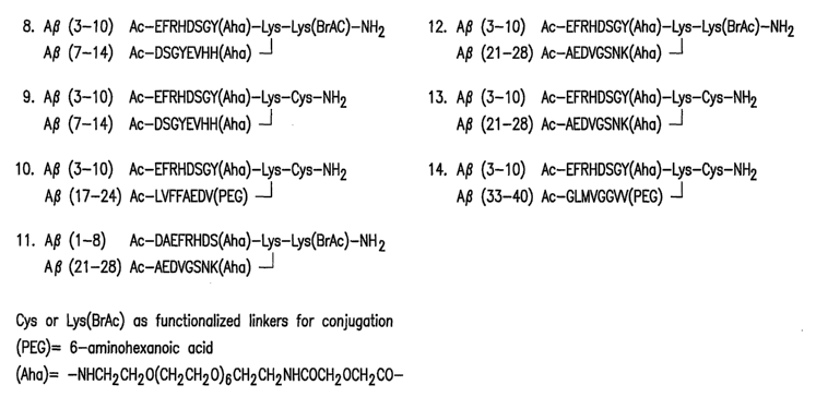
multivalent branched MAP, A(3 (3-10)/(21-28) conjugate, Construct No. 12,
Figure 6A, conjugated to
OMPC.
Such methods entail the administration of an effective dose of an immunogenic
fragment
of A(3, lacking a T-cell epitope, to patients in need of such treatment that
will induce an immune response
in the form of antibodies to A(3. Said antibody response is capable of
elevating plasma A(3levels. In
another aspect of this embodiment, the immunogenic fragment to be administered
is linked to a carrier
molecule. In yet another aspect of this embodiment, the immunogenic fragment
is administered with an
adjuvant.
-3-
CA 02607868 2007-11-05
WO 2006/121656 PCT/US2006/016481
BRIEF DESCRIPTION OF THE DRAWINGS
Figure 1 represents synthetic 8-amino acid peptides (8-mers) (SEQ ID NOS: 2-
36)
derived from A(3 (1-42) (SEQ ID NO: 1) from which peptides were selected to
conduct a linear peptide
scan to identify the epitopes of A(3.
Figure 2 represents the 8-mers selected for conjugation to KLH (Figure 2A) and
OMPC
(Figure 2B).
Figure 3 represents the inununogenicity of selected A(3 conjugates, described
in Figure 2,
after the first (PD1), second (PD2) and third dose (PD3).
Figure 4 represents the cross-reactivity of sera extracted from a guinea pig
previously
immunized with an A(3 (3-10)-KLH conjugate (SEQ ID NO: 40) on human AD brain
tissue. Figure 4A
shows immunoreactivity of the anti-A(3 monoclonal antibody 6F3D (which
recognizes amino acids 8-17
of A(3). The staining pattern reveals extensive amyloid pathology in this
human brain. Figure 4B
demonstrates a lack of immunoreactivity of this same brain to the pre-immune
sera from the immunized
guinea pig prior to immunization. Figure 4C shows the immunoreactivity of the
sera from an immunized
guinea pig following immunization
Figure 5 shows representative multivalent linear 8-mer peptides, which were
selected
based on the immunogenicity of the separate 8-mers in guinea pig studies
(Example 3). These conjugates
were synthesized as described and conjugated to OMPC (Example 1.J and 1.K).
Figure 6 shows representative multivalent branched MAP conjugates, which were
selected based on the immunogenicity of the separate 8-mers in guinea pig
studies (Example 3). Figure
6A shows representative divalent MAPs and Figure 6B shows representative
bromoacetyl-cysteine
MAPs. These conjugates were synthesized as described and conjugated to OMPC
(Example 2).
Figure 7 represents the anti-A(340 titer from sera collected from rhesus
monkeys
following 1(PD1) or 2 (PD2) injections with an A(3 (1-18) peptide conjugated
to OMPC formulated in
Merck alum alone or Merck alum plus IMX (ISCOMATRIX CSL, Ltd., Parkville,
Australia) as an
adjuvant.
Figure 8 represents the increase in plasma A(3 levels following administration
of a A(3
conjugate. Figure 8A shows a greater than three-fold elevation following
administration of a MAP
construct comprising A(3 (3-10)/(21-28) (Construct No. 12, Figure 6A)
conjugated to OMPC versus the
monomeric constructs, A(3 (3-10) (SEQ ID NO: 69) and A(3 (21-28) (SEQ ID NO:
73) (o , Construct No.
12, Figure 6A; =, Aj3 (3-10) (SEQ ID NO 69), =, AJ3 (21-28) (SEQ ID NO: 73).
Figure 8B shows that
plasma A+3 levels are independent of titer levels (o , Construct No. 12,
Figure 6A; =, A(3 (3-10) (SEQ ID
NO 69), =, A(3 (21-28) (SEQ ID NO: 73).
-4-
CA 02607868 2007-11-05
WO 2006/121656 PCT/US2006/016481
DEFINITIONS
The term "8-mer" refers to an eight amino acid peptide which corresponds to a
fragment
of A(3, an analog of a natural Ap peptide or a peptide mimetic. One or inore 8-
mers may be combined
with at least one spacer to form a multivalent linear peptide or to form a
multivalent branched MAP.
The term "A(3 conjugate" means an 8-mer or immunogenic fragment of A(3 that is
chemically or biologically linked to a carrier, such as keyhole limpet
hemocyanin or the outer membrane
protein complex of Nesseria meningitidies (OMPC).
The term "Ap peptide" means any of the A(3 peptides described herein,
including, but not
limited to, linear 8-mers, multivalent linear peptides with at least one
spacer and multivalent branched
multiple antigenic peptides (MAPs).
The term "epitope" refers to a site on an antigen to which B and/or T cells
respond. B-
cell epitopes can be formed both from contiguous amino acids or noncontiguous
amino acids juxtaposed
by tertiary folding of a protein. Epitopes formed from contiguous amino acids
are typically retained on
exposure to denaturing solvents whereas epitopes formed by tertiary folding
are typically lost on
treatment with denaturing solvents. T-cell epitopes consist of peptides which
are capable of forming
complexes with host MHC molecules. T-cell epitopes for a human MHC class I
molecules, which are
responsible for induction of CD8+ T-cell responses, generally comprise 9 to 11
amino acid residues,
while epitopes for human MHC class II molecules, which are responsible for
CD4+ T-cell responses,
typically comprise 12 or more amino acid residues (Bjorkman et al. Nature
329:506-512, 1987; Madden
et al. Cell 75:693-708; Batalia and Collins; Engelhard Annu Rev Iiiununol.,
12: 181-207-622. 1995;
Madden, Annu Rev Immunol., 13:587-622. 1995). Unlike T cells, B cells are
capable of recognizing
peptides as small as 4 amino acids in length. It is the T-cell epitope/MHC
complexes that are recognized
by T-cell receptors leading to T cell activation.
The term "immunogenic fragment of A(3" or "immunogenic fragment of A(3,
lacking a
T-cell response," as used herein refers to an 8-mer or an AJ3 fragment that is
capable of inducing an
immune response in the form of antibodies to A(3, but which response does not
include a T-cell response
to the self antigen, A(3.
The term "immunological" or "immune" or "immunogenic" response refers to the
development of a humoral (antibody mediated) and/or a cellular (mediated by
antigen-specific T cells or
their secretion products) response directed against an antigen in a vertebrate
individual. Such a response
can be an active response induced by administration of an immunogen or a
passive response induced by
administration of an antibody.
The term "multivalent peptide" refers to peptides having more than one
antigenic
determinant.
-5-
CA 02607868 2007-11-05
WO 2006/121656 PCT/US2006/016481
The term "pharmaceutical composition" means a chemical or biological
composition
suitable for administration to a mammalian individual. As used herein, it
refers to a composition
comprising 8-mers, immunogenic fragments of A(3 and A(3 conjugates described
herein to be
adniinistered optionally with or without an adjuvant.
DETAILED DESCRIPTION OF THE INVENTION
As previously described, preclinical studies suggest that active immunization
resulting in
an anti-A(3 polyclonal antibody response provides efficacy against the
pathological and cognitive
symptoms associated with AD in transgenic mice that overexpress the amyloid
precursor protein (Bard et
al., Nature Med., 6: 916-919, 2000; Janus et al., Nature, 408: 979-982, 2000;
Morgan et al., Nature, 408:
982-985, 2000; DeMattos et al., Proc. Natl. Acad. Sci., 98: 8850-8855, 2001;
Bacskai et al., J. Neurosci.,
22: 7873-7878, 2002; Wilcoc, et al., J. Neurosci., 23: 3745-3751, 2003). These
preclinical studies are
supported by a single clinical trial where an aggregate form of A042 was used
as an active immunogen.
Preliminary evidence from this study suggests that pathological (Nicoll et
al., Nature Med., 9: 448-452,
2003; Ferrer et al., Brain Pathol., 14: 11-20, 2004; Masliah et al.,
Neurology, 64:129-131, 2005) and
cognitive improvements (Gilman et al., Neurology, 64 (9): 1553-1562, 2005)
were found following
treatment. While these findings are encouraging and consistent with
preclinical studies, the treatment
proved unsafe and was terminated following the appearance of
meningoencephalitis in approximately 6%
of the treated patients (Orgogozo et al., Neurology, 61: 46-54, 2003). Thus,
there exists a need for active
immunization procedures capable of an efficacious immune response and devoid
of adverse safety issues.
Progress in understanding the nature of the adverse events in this preliminary
clinical
trial has been made. Several investigators have now reported the presence of
CD4+ and CD8+ positive
meningeal infiltrates on post-mortem evaluation (Nicoll, et al., Nature Med.,
9: 448-452, 2003; Ferrer et
al., Brain Pathol., 14: 11-20, 2004) suggestive of a T-cell response directed
at the self-peptide A(342=
However, while those skilled in the art would recognize the need to avoid a
self-directed T-cell response
while maintaining an appreciable antibody response (B-cell mediated), the
means to produce an agent
having this property is not known. This difficulty is compounded by a lack of
predictive animal models
or other preclinical assays with predictive validity for these activities.
To this end, Applicants herein used the differing nature of T and B cell
epitopes to
design the peptides used for the invention. The vaccine constructs were
designed, by restricting the
linear peptide size to eight amino acids and, if necessary, removing any
potential C-terminal T-cell
epitope anchor residues.
Accordingly, one aspect of the present invention was the identification of A(3
fragments
that are immunogenic, but lack a T-cell epitope, for use as an AD vaccine.
Prior to the present
application, it was not definitively known which amino acid fragments of the
A(3 peptide would produce
an immunogenic response that would also be deficient in a T-cell epitope.
Those skilled in the art would
appreciate that previous teachings in the field did not predict, for example,
that an 8-mer would produce
-6-
CA 02607868 2007-11-05
WO 2006/121656 PCT/US2006/016481
an immunogenic response and did not distinguish the usefulness of fragments
from different regions of
the A(3 peptide. See, for example, U.S. Pat. Nos. 6,808,712 and 6,787,144.
An additional aspect of the invention herein includes the identification of
A(3 plasma
elevating agents comprising an immunogenic fragment of A(i, lacking a T-cell
epitope, that induce an
immune response in the form of antibodies to A(3 and that elevate plasma A3
levels. Such agents can be
used as an AD vaccine and for related amyloid diseases characterized by
elevated brain Ap levels. Prior
to Applicants' invention, it was not known or predictable which immunogenic
fragments of A(3 would
result in elevated plasma A(3levels. Without wishing to be bound by any
theory, it is believed that the
A(3 plasma elevating agents described herein act to induce an immune response
in the form of antibodies
to A(3 that, according to the peripheral sink theory of A(3 clearance, produce
elevated levels of plasma A(3
that leads to subsequent decreases in brain A(3. Moreover, while individual 8-
mers or immunogenic
fragments of Ap may be capable of inducing an immune response such that plasma
A(3 levels are
elevated, Applicants found that a multivalent branched MAP, A(3 (3-10)/(21-28)
(Construct No. 12,
Figure 6A), conjugated to OMPC, was particularly effective in elevating plasma
Ap levels relative to
those of its constituent monomeric constructs, A(3 (3-10) (SEQ ID NO: 69) or
A(3 (21-28) (SEQ ID NO:
73).
Amyloid Diseases
The invention provides compositions and methods for prophylactic and
therapeutic
treatment of disease characterized by accumulation of amyloid deposits.
Amyloid deposits comprise a
peptide aggregated to an insoluble mass. The nature of the peptide varies in
different disease but in most
cases, the aggregate has aP-pleated sheet structure and stains with Congo Red
dye. Diseases
characterized by amyloid deposits include Alzheimer's disease (AD), both late
and early onset. In both
diseases, the amyloid deposit comprises a peptide termed amyloid beta (A(3),
which accumulates in the
brain of affected individuals. Thus, the term "amyloid disease" also refers to
disease characterized by
elevated brain A(3levels.
Therapeutic Agents
Therapeutic agents for use in the present invention induce an immune response
in the
form of antibodies to A(3. Induction of an immune response can be active as
when an innnunogen is
administered to induce antibodies or T cells reactive with A(3 in an
individual or passive, as when an
antibody is administered that itself binds to A(3 in the individual.
The therapeutic agent to be used in preventing or treating amyloid diseases,
such as AD,
include peptide fragments of A(3, which can be any of the naturally occurring
forms (i.e. A039, AP40,
A(342, A(342, or A(343). These sequences are known in the art, see, for
example, Hardy et al., TINS 20:
155-158, 1997.
-7-
CA 02607868 2007-11-05
WO 2006/121656 PCT/US2006/016481
As used herein, the therapeutic agent is, in a preferred embodiment, an
immunogenic
fragnient, lacking a T-cell epitope, capable of inducing an immune response in
the form of antibodies to
A(3. The immunogenic fragment of A(3 can be in the form of an 8-mer, a
multivalent linear A(3 conjugate
having at least one PEG spacer or a multivalent branched MAP A(3 conjugate.
The therapeutic agent can
be adniinistered in the form of a pharmaceutical composition. In an another
embodiment, the therapeutic
agent is an A(3 plasma elevating agent capable of inducing an immune response
in the form of antibodies
to Ap and that elevate plasma A(3levels in an individual. Such agents can
comprise a naturally occurring
peptide fragment or may include one or more substitutions, additions or
deletions, and may include
synthetic or non-naturally occurring amino acids. Fragments and constructs can
be screened for
prophylactic and therapeutic efficacy in the assays described in the examples
herein.
While in a preferred embodiment the therapeutic agents comprise a peptide
fragment of
AJ3, such agents may also include peptides and other compounds that do not
necessarily have a
significant amino acid sequence similarity with A(3, but that nevertheless can
serve as mimetics of A(3
and induce a similar immune response. For example, peptides and proteins
forming (3-pleated sheets can
be screened for suitability for the invention herein. Siniilarly,
combinatorial libraries and other
compounds can be screened for suitability for the invention herein.
Such identified therapeutic agents can be linked either chemically or
biologically to a
carrier to facilitate their use as an immunogen. Such carriers include serum
albumins, keyhole limpet
hemocyanin (KLH), immunoglobulin molecules, ovalbumin, tetanus toxoid protein,
or a toxoid from
other pathogenic bacteria, such as diphtheria, E. coli, cholera, or H. pylori,
or an attenuated toxin
derivative. In a preferred embodiment of the invention the carrier is the
outer membrane protein complex
of Neisseria meniragitides (OMPC).
The invention herein also contemplates the use of such therapeutic agents in a
pharmaceutical coinposition comprising an 8-mer or immunogenic fragment of
A(3, which may be linked
to a carrier, to be administered optionally with an adjuvant. Suitable
adjuvants include aluminum salts
(alum), a lipid, such as 3 De-O-acylated monophosphoryl lipid A (MPL) or a
saponin-based adjuvant. In
a preferred embodiment the adjuvant is an aluminum adjuvant (Merck alum
adjuvant, MAA) or a
saponin-based adjuvant (ISCOMATRIX , CSL Ltd, Parkville, Australia.
Treatment Regimes
Effective doses of the compositions of the invention herein for the
prophylactic or
therapeutic treatment of AD and other amyloid diseases will vary depending
upon many factors
including, but not limited to, means of administration, target site,
physiological state of the patient, other
medications administered and whether treatment is a therapeutic, i.e. after on-
set of disease symptoms, or
prophylactic, i.e. to prevent the on-set of disease symptoms. In a preferred
embodiment the patient is
human and the therapeutic agent is to be administered by injection.
-8-
CA 02607868 2007-11-05
WO 2006/121656 PCT/US2006/016481
The amount of immunogen or therapeutic agent to be employed will also depend
on
whether an adjuvant is to be administered either concomitantly or
sequentially, with higher doses being
employed in the absence of an adjuvant.
The amount of an immunogen or therapeutic agent to be administered will vary,
but
amounts ranging from 0.5-50 g of peptide (based on the A(3 peptide content)
per injection are
considered for human use. Those skilled in the art would know how to formulate
compositions
comprising antigens of the type described herein.
The administration regimen would consist of a primary immunization followed by
booster injections at set intervals. The intervals between the primary
immunization and the booster
immunization, the intervals between the booster injections, and the number of
booster immunizations
will depend on the antibody titers and duration elicited by the vaccine. It
will also depend on the
functional efficacy of the antibody responses, namely, levels of antibody
titers required to prevent AD
development or exerting therapeutic effects in AD patients. A typical regimen
will consist of an initial
set of injections at 1, 2 and 6 months. Another regimen will consist of
initial injections at 1 and 2 months.
For either regimen, booster injections will be given either every six months
or yearly, depending on the
antibody titers and durations. An administration regimen can also be on an as-
needed basis as determined
by the monitoring of immune responses in the patient.
Identification of AD vaccine epitopes.
In order to determine which 8-amino acid fragments ("8-mers") of the A(3
peptide were
sufficient to produce an immunogenic response, Applicants systematically
scanned the entire length of
A(342 with small (8 amino acids) overlapping synthetic peptides derived from
the naturally occurring A(3
sequence (SEQ ID NO. 1) as shown in Figure 1(SEQ ID NOS: 2-37). Twenty nine
overlapping eight
amino acid peptides, spanning the entire length of A(342, were synthesized
(Figure 2A) for use as
antigens. To improve solubility, several of the peptides were modified by the
addition of triple lysine
(KKK) (SEQ ID NOS: 52, 53, 54, 56, 59, 60, 62, 64 and 65) or glutamine (EEE)
(SEQ ID NOS: 50, 51
and 61) residues or the use of a polyethelyene glycol (PEG) (SEQ ID NOS: 55
and 63) spacer. For this
reason, peptides spanning the sequences of A(3 corresponding to residues (11-
18) and (13-20) were made
in multiple forms, the first with a 6-aminohexanoic acid (Aha) spacer plus a
functional group for
chemical cross-linking at N-terminus and the other form with Aha and the
functional group at C-
terminus. As a control, Applicants included a longer peptide, A(3 (1-18).
As used herein, the immunogenic fragments may be 8-mer peptides (eight amino
acid
residues) derived from the naturally occurring, i.e. wild type, or synthetic
Ap (SEQ ID NO: 1) or any
mutation or variation thereof. Such mutation or variant can be produced by
synthetic or recombinant
means known to those of ordinary skill in the art. One example of such a
variant is the EV substrate
(EVEFRHDSGYEVHHQKLVFFAEDVGSNKGAIIGLMVGGVVIA) (SEQ ID NO: 66) a peptide
corresponding to A(3 (1-42) in which positions 1 and 2 of wild type A(3 have
been varied.
-9-
CA 02607868 2007-11-05
WO 2006/121656 PCT/US2006/016481
Ap conjugates for use in vaccine formulation
Selection of A(3 conjugates for use in formulating a vaccine was based on the
immunogenicity of the 8-mers. In order to determine the immunogenicity of the
8-mer in a species with
a sequence identical to the human A(i sequence, the 29 peptides (Figure 2A)
were conjugated to KLH to
form an Aj3 conjugate and tested in guinea pigs (Figure 3). As a control
immunogen, A(3 (1-18)-KLH
(SEQ ID NO: 37) was included in this analysis.
Guinea pigs were immunized as described in Example 3.B with conjugated
immunogens
formulated in alum plus 50 g of ISCOMATRIX (CSL, Ltd., Parkville,
Australia). In order to
distinguish immunogenic from non-immunogenic A042 fragments, guinea pigs were
immunized three
times at four week intervals. Three weeks after each immunization, blood
samples were collected and
tested by ELISA for antibody titers against A(340 peptide. These titers are
shown in Figure 3 as post-
dose 1(PD1), post-dose 2 (PD2) and post-dose 3 (PD3), respectively.
Following the first injection (PD1) some peptide regions elicited appreciable
antibody
titers as did the 18-mer control. In particular, A(3 conjugates corresponding
to A(3 amino acids 1-8, 2-9,
3-10, 17-24, 21-28, and 33-40 all produced titers in excess of 1:800. After
the second injection (PD2), 15
of the Aj3 conjugates elicited antibody titers in excess of 1:1000. Analysis
at post-dose 3 (PD3) further
confirmed that certain regions of A(3 are more immunogenic relative to others.
Eleven regions
demonstrated titers greater than 1:6000. These included regions corresponding
to A(3 amino acids 1-8, 3-
10, 7-14, 11-18, 13-20, 15-22, 19-26, 21-28, 23-30, 27-34 and 29-36. Of these
regions, five regions were
highly immunogenic (>1:10000) including: regions 1-8, 15-22, 21-28, 23-30 and
29-36. This data
suggests that certain 8-amino acid regions of A(3 are highly immunogenic,
while other regions (e.g., 5-12,
25-32, 31-38 and 35-42) are non-immunogenic (titers < 1:300). The results also
demonstrate that while
the A(3 conjugates were capable of eliciting an A(340 peptide-specific
antibody response, not all
fragments of A(3 were equally immunogenic.
Immunoreactivity of A(3 peptide-KLH conjugates
In order to demonstrate that the immune sera generated from the guinea pigs
following
immunization with the A(3 peptide-KLH conjugates is relevant to human AD, a
study was performed to
evaluate the immunoreactivity of polyclonal sera from a guinea pig immunized
with an A(3 (3-10)-KLH
(SEQ ID NO: 40) conjugate. The serum sample collected four weeks following the
second injection of
A(3 (3-10)-KLH (SEQ ID NO: 40) conjugate from a guinea pig was tested for
reactivity with human AD
brain tissues by immunohistochernistry (Example 4).
As depicted in Figure 4 the immunogenic response produced by the A(3 (3-10)-
KLH
(SEQ ID NO: 40) conjugate produced an antibody response that was directed
against human AD brain
tissue. Figure 4A deinonstrates immunoreactivity of the monoclonal anti-A(3
antibody 6F3D (Vector
Laboratories). As shown, this brain has extensive A(3 deposits in a manner
expected to be typical for
liuman AD. Figure 4B demonstrates a lack of immunoreactivity of sera from a
pre-immunized guinea
-10-
CA 02607868 2007-11-05
WO 2006/121656 PCT/US2006/016481
pig. Figure 4C shows positive immunoreactivity of sera from this same guinea
pig following two
injections of the A(3 (3-10)-KLH (SEQ ID NO: 40) conjugate. Collectively, this
data demonstrates that
the immunogenicity found by ELISA contains a significant antibody response
directed against human A(3
found in this AD tissue. These results confirm and extend the unexpected
finding of the differential
immunogenicity imparted by particular fragments of Ap to further demonstrate
that this response is
directed in a manner consistent with a therapeutic application.
Generation of OMPC conjugates and multiple antigenic conjugates
On the basis of immunogenicity in guinea pigs, the relative location of the
peptide
fragment within the A(342 amino acid sequence, the solubility of the A(3
fragments and the feasibility of
using OMPC as a carrier protein, Applicants selected seven 8-mers (Figure 2B)
for OMPC conjugation.
These peptide fragments correspond to the following amino acid regions of A(3:
1-8, 2-9, 3-10, 7-14, 17-
24, 21-28 and 33-40.
The invention described herein includes multivalent peptide conjugates such as
those
shown in Figures 5, 6A and 6B. Multivalent branched MAP-OMPC conjugates
(Figures 6A and 6B)
were generated by using a lysine-based scaffold, whereas multivalent linear 8-
mer-OMPC conjugates
(Figure 5) were prepared using a PEG linker. Those skilled in the art will
appreciate that a PEG linker,
compared to conventional amino acid linkers that can also be used herein,
offers the advantage of lower
immunogenicity and greater peptide solubility. In a preferred embodiment of
the invention, the
immunogenic fragment is a multivalent MAP conjugated to OMPC. It should be
understood by those
skilled in the art that such a conjugation is not a 1:1 ratio of peptide to
carrier. Rather, a plurality of
peptides is attached in a spherical manner to each OMPC molecule. It will be
further appreciated by
those skilled in the art that the use of multivalent linear constructs and
MAPs will enhance solubility,
formulation stability, immunogenicity and the diversity of the polyclonal
response.
Immunogenicity of OMPC conjugate vaccines
In an effort to evaluate the immunogenicity of an Ap peptide - OMPC conjugate
and to
further evaluate the benefit of an adjuvant with this vaccine construct,
Applicants initiated a study in
rhesus monkeys. Rhesus monkeys were vaccinated with an A(3 (1-18)-OMPC
conjugate (dose based on
the A(3 peptide conntent), which was formulated either in Merck alum adjuvant
(MAA) or MAA and
ISCOMATRIX (CSL, Ltd., Parkville, Australia). Blood samples were collected
and used to determine
the antibody titers against A(340. Interim analysis of this ongoing study
demonstrated that at post-dose 1
(PD1) the monkeys receiving 5 g vaccine in alum failed to develop any
detectable titers, while those
receiving 30 gg vaccine in alum developed low A(340 specific titers. All
monkeys that received the alum
plus ISCOMATRIX formulation developed significant antibody titers. At post-
dose 2 (PD2) both
doses of the A(3 conjugate in alum alone produced similar titer levels,
whereas the cohorts receiving the
alum plus ISCOMATRIX@ developed 10-fold higher antibody titers relative to the
alum alone cohorts.
-11-
CA 02607868 2007-11-05
WO 2006/121656 PCT/US2006/016481
The results of this study confirmed that the A(3-OMPC conjugate is immunogenic
in non-human
primates. The data further demonstrated that the efficacy of such a conjugate
vaccine is significantly
enhaiiced by a saponin-based adjuvant such as ISCOMATRIX@.
EXAMPLES
EXAMPLE 1
Preparation of A(3 Conjugates
This example describes the preparation of A(3 peptide fragments subsequently
used for
the A(3 conjugates to induce an immune response in the form of antibodies to
A(3.
A. Preparation of A(3 (8-mers) peptides (SEQ ID NOS.: 37-65; Figure 2A)
The peptides intended for conjugation to maleimide derivatized carrier
proteins were
synthesized with a cysteine residue at the carboxy terminus. The spacer, Aha
(6-aminohexanoic acid)
was incorporated between the primary peptide sequence and the carboxy terminal
cysteine as a structural
element for minimizing steric accessibility to carrier protein during
conjugation. Additionally,
solubilizing residues represented by EEE, KKK or PEG were introduced at the C-
terminus in sequences
14,15,16 17,18,19,20,23,24,25,26,27,28,29. The PEG unit was introduced as, O-
(N-Fmoc-2-
aminoethyl)-O'-(2-carboxyethyl)-undecaethyleneglycol [Fmoc-
NHCHzCHZO(CHZCH2O)10
CH2CH2OCH2CH2CO2H].
Starting with Rink Amide MBHA resin the A(3 peptides were prepared by solid-
phase
synthesis on an automated peptide synthesizer using Fmoc chemistry protocols
as supplied by the
manufacturer (Applied Biosystems, Foster City, CA). Following assembly the
resin bound peptide was
deprotected and cleaved from the resin using a cocktail of 94.5%
trifluoroacetic acid, 2.5% 1,2-
ethanedithiol, 1% triisopropylsilane and 2.5% H20. Following a two hour
treatment the reaction was
filtered, concentrated and the resulting oil triturated with ethyl ether. The
solid product was filtered,
dissolved in 50% acetic acid/H20 and freeze-dried. Purification of the semi-
pure product was achieved
by RPHPLC using a 0.1% TFA/H20/acetonitrile gradient on a C-18 support.
Fractions were evaluated
by analytical HPLC. Pure fractions (>98%) were pooled and freeze-dried.
Identity was confirmed by
amino acid analysis and mass spectral analysis.
B. Preparation of AJ3 peptide-KLH conjugates (SEQ ID NOS.: 37-65; Figure 2A)
For preparing the KLH conjugates, the A(3 peptides (8-mers), 2 mg, containing
a C-
terminal cysteine was suspended in 1 ml of commercial maleimide conjugation
buffer (83 mM sodium
phosphate, 0.1 M EDTA, 0.9 M NaCl, 0.02% sodium azide, pH 7.2 (Pierce
Biotechnology, Rockford,
IL). A 2 mg sample of commercial maleimide-activated KLH (Pierce
Biotechnology, Rockford, IL) was
-12-
CA 02607868 2007-11-05
WO 2006/121656 PCT/US2006/016481
added to the peptide and allowed to react at 25 C for four hours. The
conjugate was separated from
unreacted peptide and reagents by exhaustive dialysis versus PBS buffer using
100,000 Da dialysis
tubing. The amount of peptide incorporated into the conjugate was estimated by
amino acid analysis
following a 70 hour acid hydrolysis. Peptide concentrations were determined to
be between 0.24 and
0.03 ing/ml.
C. Synthesis of bromoacetylated A(3 peptides (SEQ ID NOS.: 67-77; Figure 2B)
Bromoacetylated peptides were prepared by standard t-Boc solid-phase
synthesis, using a
double coupling protocol for the introduction of amino acids on the Applied
Biosystems mode1430A
automated synthesizer. Starting with p-methylbenzhydrylamine resin the carboxy
terminal amino acid t-
Boc-Lys (Fmoc)-OH was introduced followed by the subsequent amino acids in the
sequence. Aha was
introduced as a spacer to all of these sequences and a PEG unit in sequences
35 and 37 to aid in aqueous
solubility. The PEG unit was introduced as O-(N-Boc-2-aminoethyl)-O'-(N-
diglycolyl-2-aminoethyl)
hexaethyleneglycol [Boc-NHCH2CH2O(CH2CH2O)6CH2CH2NHCOCH2OCH2CO2H]. The amino
terminous was capped by the coupling of acetic acid. After assembly of the
primary sequence the Fmoc
protecting group on the epsilon amino group of the carboxy terminal lysine was
removed by treatment
with piperidine. Subsequently the NE amino group was reacted with Bromoacetic
anhydride in methylene
chloride as the solvent for 30 minutes. Deprotection and removal of the
peptide from the resin support
were achieved by treatment with liquid hydrofluoric acid and 10% anisole as a
scavenger. The peptides
were purified by preparative HPLC on reverse phase C-18 silica columns using a
0.1% TFA/acetonitrile
gradient. Identity and homogeneity of the peptides were confirmed by
analytical HPLC and mass
spectral analysis.
D. Synthesis of bromoacetylated divalent MAP, Construct No. 8, Figure 6A
The synthesis of bromoacetylated branched multiple antigenic peptides (MAPs)
is
similar to that described in Example I.C. Following coupling of the
carboxyterminal Fmoc-Lys(ivDde)-
OH [ivDde = 1, (4,4-Dimethyl-2, 6-dioxo-cyclohexylidene)-3-methyl-butyl] to
MBHA resin the a-amino
Fmoc protecting group was removed using piperidine and the synthesis continued
with the introduction
of t-Boc-Lys(Fmoc)-OH. After deprotection of the t-Boc group the sequence was
extended with the
following t-Boc protected amino acids: Aha, Y, G, S, D, H, R, F, E and the
arnino terminous capped by
coupling acetic acid on the ABI synthesizer. The side chain lysine Fmoc
protecting group was removed
with piperidine and the NE arm of lysine extended on the ABI synthesizer with
the introduction of the
following protected arnino acids: Aha, H, H, V, E, Y, G, S, D and the amino
ternlinous capped by
coupling acetic acid. Removal of the ivDde protecting group was by treatment
with 5% hydrazine in
dimethylformamide for 5 minutes providing the unblocked N~ anuno group on the
carboxy terminal
lysine which was further elaborated with bromoacetic anhydride as described in
Example I.C. Cleavage
-13-
CA 02607868 2007-11-05
WO 2006/121656 PCT/US2006/016481
of the peptide from the resin, its subsequent purification and
characterization are as described in Example
I.C.
E. Synthesis of bromoacetylated MAPs, Construct Nos. 11 and 12, Figure 6A
MAP Constructs Nos. 11 and 12 were prepared as described in Example I.D.
F. Synthesis of cysteine multivalent MAP, Construct No. 9, Figure 6A
Starting with MBHA resin the following t-Boc protected amino acids were
assembled on
the ABI automated synthesizer C, Lys(Fmoc), Aha, Y, G, S, D, H, R, F, E
followed by coupling with
acetic acid. The N~ amino Fmoc protecting group of lysine was removed and the
synthesis continued
with the introduction of the following t-Boc protected amino acids: Aha, H, H,
V, E, Y, G, S, D followed
by coupling with acetic acid. The resin bound peptide was isolated, purified
and characterized as in
Example 1.C. Note: Instead of 10% anisole as in Example I.C, a 1:1 mixture of
p-cresol: p-thiocresol
was used as a scavenger during HF cleavage.
G. Synthesis of cysteine divalent MAPs, Construct Nos. 10, 13 and 14, Figure
6A
Divalent MAPs, Construct Nos. 10, 13 and 14, Figure 6A, were prepared as
described in
Example 6.F. The PEG unit was introduced as O-(N-Boc-2-aminoethyl)-O'-(N-
diglycolyl-2-aminoethyl)
hexaethyleneglycol (t-Boc-NHCH2CH2O(CH2CH2O)6 CH2CH2NHCOCH2OCH2CO2H).
H. Synthesis of bromoacetylated multivalent MAP, Construct No. 16, Figure 6B
Using the ABI automated synthesizer Fmoc-Lys (t-Boc)-OH was coupled to MBHA
resin. Following removal of the t-Boc protecting group on the NE amino group
of lysine the sequence
was extended with the introduction of the following t-Boc protected amino
acids: Aha, Y,G, S, D, H, R,
F, E, followed by coupling of acetic acid. The Na Fmoc protecting group on
lysine was removed by
manual treatment with piperidine. The sequence was further elaborated (on ABI
synthesizer) with the
introduction of Fmoc-Lys (t-Boc)-OH followed by the following t-Boc protected
amino acids: Aha, H,
H, V, E, Y, G, S, D and coupling of acetic acid. The lysine Fmoc Na amino
protecting group was
removed as previously described and the synthesis continued with the
introduction of Fmoc-Lys(t-Boc)-
OH followed by the t-Boc protected amino acids: Aha, K, N, S, G, V, D, E, A
and acetic acid coupling.
The Na Fmoc protecting on lysine was removed and the synthesis continued with
the introduction Fmoc-
Lys(t-Boc)-OH followed by the following t-Boc protected amino acids: Aha, V,
V, G, G, V, M, L, G and
acetic acid coupling. Following removal of the NaFmoc protecting group of
lysine the resin bound
peptide was reacted with bromoacetic anhydride as in Example 1.C. Isolation
and characterization of the
final product was as in Example I.C.
I. Synthesis of multivalent MAPs, Construct Nos. 15 and 17, Figure 6B
-14-
CA 02607868 2007-11-05
WO 2006/121656 PCT/US2006/016481
The synthesis of MAP A(3 conjugates, Construct Nos. 15 and 17, Figure 6B, are
as
described in Example 1.F and I.H.
J. Synthesis of bromoacetylated multivalent linear peptide, Construct No. 1,
Figure 5
Starting with MBHA resin the primary sequence was synthesized using t-Boc
chemistry
on the ABI automated synthesizer as described in Example 6.A. The interspaced
PEG units were
manually introduced as the Fmoc-l-amino-4, 7, 10-trioxa 13-tridecanamine
succinic acid [Fmoc-
NHCH2CH2CH2O(CH2 CH2O)2 CH2CH2CH2 NHCOCH2CH2CO2H] using BOP reagent as the
coupling
agent. Piperidine was used for deprotection of the Fmoc group.
Bromoacetylation of the amino terminus
was as described in Example I.C. Isolation and characterization of the desired
product was as in
Example 1.C.
K. Synthesis of multivalent linear A(3 peptides, Construct Nos. 2, 5, 6 and 7,
Figure 5
The synthesis of multivalent linear AJ3 peptides, Construct Nos. 2, 5, 6 and 7
are as
described in Example I.J.
EXAMPLE 2
Chemical conjugation of A(3 peptides to OMPC
This example presents the chemical conjugation of peptides derived from human
AP42
to purified Outer Membrane Protein Complex (OMPC) of Neisseria rneizingitidis,
type B. The chemical
nature of the coupling is reaction between haloacetyl-derivatized peptide and
thiol-derivatized protein of
the membrane complex. Amyloid peptides were synthesized as described above
with a bromoacetyl
functionality on the N-terminus for divalent linear epitope peptides or on the
C-terminus or attached
through the epsilon amino group of a lysine residue for monovalent linear and
branched MAP forms. The
BrAc group was separated from the mature peptide by a spacer consisting of 6-
aminohexanoic acid
(Aha). Refer to sequences described above. Conjugation will be described for
the representative
peptide, Ap (3-10). All manipulation of OMPC-containing solutions was
performed in a laminar flow
environment following standard aseptic techniques.
A. Thiolation of OMPC
Purified, sterile OMPC, obtainable from a process such as that described in
Fu, U.S. Pat.
No. 5,494,808 used for the production of PedvaxHlB and pneumococcal conjugate
vaccines, was
thiolated on a portion of its surface-accessible lysine residues using the
reagent N-
acetylhomocysteinethiolactone (NAHT, Aldrich, St. Louis, MO). OMPC in water,
117mg, was pelleted
by centrifugation at 289,000 x g for 60 minutes at 4 C and the supernatant was
discarded. N2-sparged
activation buffer (0.11 M sodium borate, pH 11) was added to the centrifuge
tube and the pellet was
dislodged with a glass stir rod. The suspension was transferred to a glass
Dounce homogenizer and
resuspended with 30 strokes. The centrifuge tube was washed and the wash
dounced with 30 strokes.
-15-
CA 02607868 2007-11-05
WO 2006/121656 PCT/US2006/016481
Re-suspended pellet and wash were combined in a clean vessel to give a OMPC
concentration of 10
mg/mL. Solid DTT and EDTA were dissolved in N2-sparged activation buffer and
charged to the
reaction vessel at a ratio of 0.106 mg DTT/mg OMPC and 0.57 mg EDTA/mg OMPC.
After gentle
mixing, NAHT was dissolved in N2-sparged water and charged to the reaction at
the ratio of 0.89 mg
NAHT/mg OMPC. Reaction proceeded for three hours at ambient temperature,
protected from light in a
N2 hood. At completion, OMPC was pelleted as described above and re-suspended
at 6 mg/mL by
Dounce homogenization in N2-sparged conjugation buffer (25 mM sodium borate,
pH 8.5, 0.15 M NaCI)
to wash the pellet. For final re-suspension, the OMPC was pelleted as above
and re-suspended at 10
mg/mL by Dounce homogenization in N2-sparged conjugation buffer. An aliquot
was removed for free
thiol determination by Ellman assay and the bulk product was stored on ice in
dark until use. Measured
thiol content was between 0.2 to 0.3 mol/mL.
B. Conjugation of A(3 peptide to OMPC
Functional BrAc content of peptide was assumed to be 1:1 on a molar basis.
Sufficient
peptide was weighed to give a 1.6 molar excess of BrAc over total thiol. The
targeted total OMPC
protein for each conjugation was 15 mg. Peptides were re-suspended in N2-
sparged conjugation buffer at
2.6 mg/mL and slowly added to thiolated OMPC solution. The reactions were
protected from light and
incubated at ambient temperature for about 22 hours. Residual free OMPC thiol
groups were quenched
with a 5-fold molar excess of N-ethylmaleimide for 18 hours at ambient
temperature. A thiolated
OMPC-only control was carried through the conjugation protocol in parallel.
Upon completion of
quenching, conjugate and control were transferred to 100,000 Da molecular
weight cut-off dialysis units
and dialyzed exhaustively against at least five changes of conjugation buffer.
Upon completion of
dialysis, samples were transferred to 15 ml polypropylene centrifuge tubes and
centrifuged at 2,280 x g
for five minutes at 4 C to remove any aggregated material. Aliquots were
removed for analysis and the
bulk was stored at 4 C.
C. Analysis of A(3 peptide-OMPC conjugates
Total protein was determined by the modified Lowry assay and samples of
conjugate and
control were analyzed by quantitative amino acid analysis (AAA). Peptide to
OMPC molar ratios were
determined from quantitation of the unique residue S-carboxymethylhomocysteine
which was released
upon acid hydrolysis of the nascent peptide-OMPC bond. The OMPC-specific
concentration was
determined from hydrolysis-stable residues which were absent from the peptide
sequence and thus
unique to OMPC protein. Assuming 1 mol of peptide for every mol SCMHC, the
ratio of
SCMHC/OMPC was thus equivalent to the peptide/OMPC content. The mass loading
of peptide could be
calculated from this ratio using the peptide molecular weight and an average
OMPC mass' of 40,000,000
Da.
-16-
CA 02607868 2007-11-05
WO 2006/121656 PCT/US2006/016481
The covalent nature of the conjugation was qualitatively confirmed by SDS-PAGE
analysis using 4-20% Tris-glycine gels (Invitrogen, Carlsbad, CA) where an
upward shift in mobility was
observed for the Coomassie-stained conjugate bands relative to control.
The calculated molar loading ratios (mol peptide/mol OMPC) for all conjugated
BrAc
peptides were:
Peptide/OMPC
Peptide Peptide Mw (mol/moI)
AR (3-10) - BrAc 1,412 2,793
Ab (7-14) - BrAc 1,344 2,283
Ab (21-28) - BrAc 1,222 2,126
Ab (17-24) - BrAc 1,809 1,795
Ab (33-40) - BrAc 1,601 2,139
A-D-MAP-BrAc 2,498 2,173
A-B-MAP-BrAc 2,622 2,147
BrAc-linear-D-A 2,649 2,263
BrAc-linear-B-A 2,773 2,178
Ab (1-8) - BrAc 1,378 2,759
F-D-MAP-BrAc 2,463 1,318
BrAc-Iinear-D-F 2,615 1,812
F-G-A-D-MAP-BrAc 5,111 636
EXAMPLE 3
Immunogenicity of A(3 conjugates
This example describes the formulation and administration of the A(3
conjugates capable
of inducing an immune response in the form of antibodies to Ap.
A. Formulation of vaccine conjugates
The A(3 peptide-KLH conjugate vaccines were formulated in ISCOMATRIX (CSL
Ltd., Parkville, Australia). All A(3 peptide-OMPC conjugate vaccines were
formulated in alum, either
with or without a second adjuvant, such as the saponin-based adjuvant,
ISCOMATRIX (CSL Ltd.,
Parkville, Australia). All the sample manipulations were performed under
sterile conditions.
For the alum formulations, conjugates are diluted one times saline at a
designated
peptide concentration and mixed with two times alum (Merck, Product No.
39943), which corresponds to
900 g/mL Merck alum prepared in sterile saline (150 mM sterile sodium
chloride solution). Thus, target
concentration in the vaccine is 450 g/mL Merck alum or one time Merck alum.
Target peptide
(antigen) concentrations for animal studies were as follows: for mice - 12.1
g/mL (Dose 0.1 mL); for
monkeys - 10 g/niL or 60 g/mL (Dose 0.5 mL) and for guinea pigs - 12.5 g/mL
(Dose 0.4 mL). The
mix is incubated for two hours at room temperature. To obtain the injection
dose, the alum-absorbed
conjugates are diluted with one time alum to reach the target peptide
concentration. Where a second
-17-
CA 02607868 2007-11-05
WO 2006/121656 PCT/US2006/016481
adjuvant is needed, i.e. ISCOMATRIX, the target concentration was 10 g/ML
for mice studies, 0, 100
or 200 g/mL for monkey studies and 125 g/mL for guinea pigs.
1. ISCOMATRIX preparation
Using a cassette membrane (Slide-A-Lyzer R Dialysis Cassett,lOK MWCO, Pierce,
Rockford, IL), ISCOMATRIX is dialyzed into sterile saline solution at 2-8 C.
Sterile saline solution is
changed 2-3 tiines during dialysis. After completion of dialysis, ISCOMATRIX
is filter sterilized
using a syringe filter (0.22 M Millex-GV syringe filter, Millipore,
Billerica, MA). The concentration of
sterile, dialyzed ISCOMATRIX is determined by RP-HPLC. ISCOMATRIX is stored
sterile at 2-8 C
until use.
2. A(3 peptide-OMPC conjugateand Merck alum preparation
A(3 peptide-OMPC conjugate stocks are diluted into sterile 1X saline solution.
The
diluted AD peptide-OMPC conjugate stocks are then added to 2X Merck alum in 1X
sterile saline
solution and mixed for one hour on a rotating wheel at room temperature: The
mixture is allowed to
settle on the bench top for 15 minutes at room temperature and is then
centrifuged at 1500 rpm for ten
minutes. The supernatant is decanted off gently (UV analysis of supernatant is
perforzned to determine
% AJ3 peptide-OMPC conjugate bound to alum) and the pellet is resuspended in
sterile 1X saline. The
mixture is aliquoted into sterile 3 mL tubing glass vials and then stored at 2-
8 C until final formulation
with ISCOMATRIX .
3. Formulation of Af3 peptide-OMPC/alum and ISCOMATRIX vaccine
Prior to final formulation with ISCOMATRIX, the particle size of the A(3
peptide-
OMPC/alum in saline is determined by static light scattering to confirm
binding and monitor particle
stability. The sterile, dialyzed ISCOMATRIX R in 1X saline is added to A(3
peptide-OMPC/alum in
sterile 150 mM NaCl while vortexing. Vials are stoppered, capped and crimped
to completely seal.
Vaccine is stored at 2-8 C prior to injection. Prior to injection, each
vaccine is vortexed for 3-5 minutes.
B. Iinmunogenicity of conjugate vaccines in guinea pigs
Six to ten week old female guinea pigs were obtained froin Harlan Inc.,
Indianapolis, IA
and maintained in the animal facilities of Merck research Laboratories in
accordance with institutional
guidelines. All animal experiments were approved by Merck Research
Laboratories Institutional Animal
Care and Use Committee (IACUC). Antigens were prepared in phosphate-buffered
saline and
formulated in the designated adjuvant.
Two animals per group were immunized with the A(3 peptide - KLH conjugates
shown in
Figure 2A intramuscularly with 400 1tl of a conjugate vaccine (8 g by peptide
content or 50 g by total
conjugate) in the presence of 40 g of ISCOMATRIX. The immunizations were
performed three times
in four-week intervals. Serum samples were collected before first immunization
(pre-bleeds) and three
weeks after each immunization and stored at 4 C prior to antibody titer
determinations. The antibody
titers were determined by ELISA according to the protocol that follows using
A(340 as the target antigen.
-18-
CA 02607868 2007-11-05
WO 2006/121656 PCT/US2006/016481
The ELISA based analysis is as follows: Ninety six-well plates were coated
with 50 l
per well of A(3 at a concentration of 4 g/ml in 50 mM bicarbonate buffer, pH
9.6, at 4 C overnight.
Plates were washed with phosphate buffered saline (PBS) and blocked with 3%
skim milk in PBS
containing 0.05% Tween-20 (milk-PBST). Testing samples were diluted in a 4-
fold series in PBST. One
hundred l of a diluted sample was added to each well, and the plates were
incubated at 24 C for two
hours and then washed six times with PBST. Fifty l of HRP-conjugated
secondary antibodies at 1:5000
dilution in milk-PBST was added per well and the plates were incubated at 24 C
for one hour. The
plates were washed three times and 100 l of 1 mg/ml o-phenylenediamine
dihydrochloride in 100 mM
sodium citrate, pH 4.5 was added per well. After 30 minutes incubation at 24
C, the reaction was
stopped by adding 100 l of 1N H2S04 per well, and the plates were read at 490
nm using an ELISA
plate reader. The antibody titer was defined as the reciprocal of the highest
dilution that gave an OD490
nm value above the mean plus two standard deviations of the conjugate control
wells.
The results of this analysis, shown in Figure 3, demonstrated that following
the first
injection (PD1) some peptide regions elicited appreciable antibody titers as
did the 18-mer control. In
particular, A(3 peptide fragments corresponding to Ao amino acids 1-8, 2-9, 3-
10, 17-24, 21-28, and 33-
40 all produced titers in excess of 1:800. After the second injection (PD2),
15 of the 8-mer conjugates
elicited antibody titers in excess of 1:1000. Analysis at post-dose 3 (PD3)
further confirmed that certain
regions of the A(3 peptide were more immunogenic relative to others. Eleven
regions demonstrated titers
greater than 1:6000. These included regions corresponding to A(3 amino acids 1-
8, 3-10, 7-14, 11-18, 13-
20, 15-22, 19-26, 21-28, 23-30, 27-34 and 29-36. Of these regions, five
regions were highly
immunogenic (>1:10000) including: regions 1-8, 15-22, 21-28, 23-30 and 29-36.
The results demonstrate
that 8-mer conjugates are capable of eliciting an A(340 specific antibody
response. Unexpectedly, and
contrary to previous teachings, not all fragments of A(3 were equally
immunogenic. In fact, these data
suggest that certain 8-mers are highly immunogenic, while other regions of A(3
(e.g., 5-12, 25-32, 31-38
and 35-42) are non-immunogenic (titers < 1:300).
C. Immunogenicity of conjugate vaccines in rhesus monkeys
A study was conducted in non-human primates, i.e. rhesus monkeys, comparable
to that
done with guinea pigs to determine whether A(3 peptide-OMPC conjugates and an
alum and
ISCOMATRIX adjuvant provided an immune response. Rhesus monkeys (Macaca
inulatta) were
maintained in accordance with the institutional animal care protocols of Merck
Research Laboratories
and New Iberia Research Center (The University of Louisiana at Lafayette, New
Iberia, LA).
Applicants used A(3 peptides conjugated to OMPC as the model antigens,
including, the
8-mers shown in Figure 2B: A(3 (1-8) (SEQ. ID NO: 67), A(3 (3-10) (SEQ. ID NO:
69), A(3 (7-14) (SEQ
ID NO: 70), A(3 (17-24) (SEQ ID NO. 72), A(3 (21-28) (SEQ ID NO: 73) and A(3
(33-40) (SEQ ID NO.
74); the divalent linear peptides shown in Figure 5: A(3 (3-10) (7-14)
(Construct No. 1), A(3 (3-10) (21-
28) (Construct No. 2), Ap (1-8)(21-28) (Construct No. 5); and the multivalent
branched MAPs shown in
-19-
CA 02607868 2007-11-05
WO 2006/121656 PCT/US2006/016481
Figure 6A: A(3 (3-10)(7-14) (Construct No. 8), AJ3 (1-8)(21-28) (Construct No.
11), A(3 (3-10) (21-28)
(Construct No. 12).
Rhesus macaques (N=3) were ixnmunized with 5 g of each of the vaccine
formulated in
Merck alum adjuvant (MAA) plus 100 ug of ISCOMATRIX every four weeks. Serum
samples were
collected four weeks following each injection and determined for A(3 specific
antibody responses by
ELISA. Consistent with the results from the guinea pig studies, all conjugates
were found to be
immunogenic in monkeys. A(3 specific antibody titers were detectable after
single injections and further
boosted after the subsequent injections. Generally for the conjugates tested,
the peak titers were reached
after the second or third immunization where geometric mean titers ranged from
25,000 to 500,000.
These results confirm the finding that the A(3 conjugates described herein are
capable of eliciting an A(3
specific antibody response.
D. Adjuvant effect on immunogenicity of conjugate vaccines in rhesus monkeys
An additional study was conducted in non-human primates, i.e. rhesus monkeys,
to
deterniine whether an A(3 peptide-OMPC conjugate and a saponin-based adjuvant,
such as
ISCOMATRIX , can provide an improved immune response. Applicants used an A(3
(1-18) peptide
conjugated to OMPC as the model antigen. Rhesus monkeys (Macaca mulatta) were
maintained in
accordance with the institutional animal care protocols of Merck Research
Laboratories and New Iberia
Research Center (The University of Louisiana at Lafayette, New Iberia, LA).
Five groups of monkeys, three per group, were given the following A(3 (1-18)-
OMPC
conjugates: (1) 5 g conjugate (based on peptide content) in alum, (2) 5 g
conjugate in alum + 100 g
ISCOMATRIX , (3) 5 g conjugate in alum + 50 mg ISCOMATRIX , (4), 30 g
conjugate in alum,
(2) 30 g conjugate in alum + 100 g ISCOMATRIX . The immunizations were
carried out by
intramuscular injections in 0.5 ml aliquots at weeks 0, 8 and 24 using
tuberculin syringes (Becton-
Dickinson, Franklin Lakes, NJ). Serum samples were collected at four week
intervals starting from week
0 (pre-bleed) and the tested for antibody titers against AD40 by ELISA,
performed as described in the
preceding example.
Interium analysis of this ongoing study demonstrated that at PDl the monkeys
receiving
5 mcg conjugate vaccine in alum failed to develop any detectable titers, while
those receiving 30 g
conjugate vaccine in alum developed low AP40 specific titers. All monkeys that
received the alum plus
ISCOMATRIX formulation developed significant antibody titers. At PD2, both
doses of immunogen
in alum alone produced similar titer levels, whereas the cohorts receiving the
alum plus ISCOMATRIX@
developed 10-fold higher antibody titers relative to the alum alone condition.
The results of this study
confirmed that this A(3 peptide-OMPC conjugate is immunogenic in non-human
primates. The data
further demonstrate that the efficacy of such a conjugate vaccine is
significantly enhanced by a saponin-
based adjuvant such as ISCOMATRIX .
-20-
CA 02607868 2007-11-05
WO 2006/121656 PCT/US2006/016481
EXAMPLE 4
Immunoreactivity of guinea pig polyclonal sera
In order to demonstrate that the immune sera generated from the guinea pigs
above
(Example 3.B) following immunization with 8-mer KLH conjugates is relevant to
human AD, a study
was performed to evaluate the immunoreactivity of polyclonal sera from a
guinea pig immunized with an
A(3 (3-10)-KLH immunogen. Four weeks following a second injection of this
construct blood was
collected from a representative guinea pig according to the following
methodology.
Reactivity of the polyclonal sera was evaluated on human AD brain sections
(BioChain
Institute, Inc., Hayward, CA). Human brain sections were prepared by
incubating at 60 C for thirty
minutes followed by two five minute xylene washes at room temperature.
Sections were subsequently
immersed in 100% EtOH twice for five minute each followed by a five minute
immersion in ddH2O.
Sections were immersed for three minutes in 99% formic acid followed by a
brief wash in ddH2O and a
five minute immersion in phosphate buffered solution (PBS). Sections were then
incubated with a
peroxidase blocker for ten minutes followed by a five minute PBS wash.
Sections were blocked by a ten
minute exposure to 10% goat serum followed by a five minute wash with PBS.
Sections were then
incubated with diluted guinea pig sera at 4 C overnight or for one hour at
room temperature. Following
a five minute PBS wash, sections were incubated for ten minutes with diluted
biotinylated goat anti-
guinea pig IgG or biotinylated horse anti-mouse antibody (1 drop in 5 ml PBS).
Sections were washed
for five minutes in PBS and subsequently incubated with ABC solution
(Vectorstain ABC kit; Vector
Laboratories, Inc.) for thirty minutes. Sections were washed with PBS for five
minutes. Sections were
then stained with DAB (DakoCytomation) for five minutes and washed with dd
H20. Sections were then
counterstained in hematoxylin for thirty seconds and dehydrated in graded EtOH
and Xylenes (70%
EtOH for five minutes, 80% EtOH for five minutes, 100% EtOH for five minutes
and xylenes for five
minutes). Sections were then cover-slipped and evaluated by liglit microscopy.
The immunogenic response produced by the A(3 (3-10)-KLH conjugate produced an
antibody response that was directed against human AD brain tissue. As shown in
Figure 4, this liuman
brain section has extensive Aj3 deposition in a mamler typical to that
expected for human AD. While
pre-immunized guinea pig sera demonstrates a lack of immunoreactivity when
exposed to this tissue,
positive immunoreactivity of sera from this same guinea pig is noted following
two injections of the
A(3 (3-10)= KLH construct. These data demonstrate that the inununogenicity
found by ELISA, and
illustrated in Figure 3, contains a significant antibody response directed
against human A~ found in this
AD tissue. Thus, the results extend the unexpected finding of differential
immunogenicity by some Aj3
fragments to further demonstrate that this response is directed in a manner
consistent with therapeutic
application.
-21-
CA 02607868 2007-11-05
WO 2006/121656 PCT/US2006/016481
EXAMPLE 5
Identification of immunogenic fragments lacking T-cell epitopes
To identify immunogenic fragments lacking a T-cell epitope for use in the
invention
herein, the following Enzyme-Linked ImmunoSpot (ELISpot) assay can be used as
a method to assess T-
cell responses to a particular antigen. Immunogen fragments possessing T-cell
epitopes are identified by
the presence of a dark spot on the surface of a white membrane; each spot
indicates the presence of a T-
cell that has secreted interferon gamma (1FN-'y) in response to the antigen
(i.e. immunogenic fragment).
Those skilled in the art of vaccines and immunology are familiar with this
assay, see, for example,
Larsson et al., AIDS 3: 767-777, 1999, and Mwau et al., AIDS Research and
Human Retroviruses 18:
611-618, 2002. A recent review can be found in A.E. Kalyuzhny, Methods Mol
Biol. 302: 15-31, 2005.
Applicants used peripheral blood monocytes (PBMCs) from rhesus macaques (New
Iberia Research Center, The University of Louisiana at Lafayette, New Iberia,
LA) for response to the
peptides A(31-40 (American Peptide Co., Sunnyvale, CA) (amino acid sequence
DAEF.RHDSGYEVHHQKLVFFAEDVG SNKGAIIGLMVGGVV) (SEQ ID NO: 78) and A(31-20
(Synpep, Dublin, CA) (amino acid sequence DAEFRHDSG YEVHHQKLVFF) (SEQ ID NO:
79).
Purified monoclonal mouse anti-monkey IFN-y (clone MD-l, Cat No. CT 525,
U-CyTech biosciences, Utrecht, The Netherlands) was diluted in phosphate
buffered saline (PBS) with
1% penicillin and streptomycin sulfate (GIBCO Penicillin-Streptomycin, Cat.
No. 15140-122,
Invitrogen,Carlsbad, CA), then added to 96-well HTS IP sterile plates (Cat.
No. MSIPS4W 10, Millipore,
Billerica, MA), and incubated for greater than twelve hours at 4 C. Plates
were washed and R10 [RPMI
medium 1640 (GIBCO Cat. No. 11875-093), 10% Fetal bovine serum (HyClone
SH30070.03, Logan,
UT), 0.1% 50 mM 2-Mercaptoethanol (GIBCO Cat. No. 21985-023), 1% 1M HEPES
Buffer (GIBCO
15630-080), 1% 200mM L-glutamine (GIBCO Cat. No. 25030-08 1), 1% 100mM MEM
sodium
pyruvate solution (GIBCO@ Cat. No. 11360-070), 1% penicillin-streptomycin
solution (GIBCO Cat.
No. 15140-122)] was added before incubation for at least two hours at 37 C.
PBMCs were centrifuged
and re-suspended in R10. PBMCs were counted on a Z2 Coulter counter (Beckman
Coulter, Fullerton,
CA). Each well of the aspirated plate received either 0.4 g of A(3 1-40, Aj3 1-
20, PHA
(phytohemagglutinin, Cat No. L-8902, Sigma, St. Louis, MO, positive control),
or diluted DMSO
(Sigma, negative control); 400000 PBMCs were then added to each well. Plates
were incubated for 18-
24 hours at 37 C in a humid CO2 incubator. Plates were washed in PBS with 5%
FBS and 0.005%
Tween; biotin-conjugated anti-monkey IFN-y polyclonal antibodies (U-CyTech
biosciences, Utrecht, The
Netherlands) were diluted in the same media and added to each plate; plates
were then incubated at 4 C
for 18-24 hours. Streptavidin-AP (Cat. No. 13043E, BD PharMingen, Franklin
Lakes, NJ) was diluted in
the same media and added to washed plates; plates were incubated at room
temperature and in the dark
for two hours. Filtered 1-Step NBT/BCIP Substrate (Pierce, Rockford, IL, Cat.
No. 34042) was added
and a further incubation at room temperature in the dark for ten minutes was
performed. After washing,
-22-
CA 02607868 2007-11-05
WO 2006/121656 PCT/US2006/016481
plates were allowed to dry before being imaged with a CCD camera and the spots
within each well were
automatically counted by computer.
Applicants have established that spot forming cells per million PBMCs (SFC/106
PBMCs) must exceed 55 and must exceed 4-fold the negative control to be
defined as a positive result;
failing to meet both these criteria defines a negative result. Rhesus macaques
were vaccinated with
either a MAP construct comprising A(3 (3-10)/(21-28) (Construct No. 12, Figure
6A) conjugated to
OMPC or with both of two monomeric constructs, Ap (3-10) (SEQ ID NO: 69) and
A(3 (21-28) (SEQ ID
NO: 73) conjugated to OMPC. Each macaque was assayed during the vaccination
regimen at monthly
intervals for three or four months; the highest signal ever recorded against
either A(3 1-40 or A(3 1-20 is
only 18 SFC/106 PBMCs, significantly below the 55 SFC/1 06 PBMCs criterion.
Thus, all resulted in a
negative score, providing strong evidence that the vaccines did not elicit T-
cell responses and, as such,
did not include a T-cell epitope.
EXAMPLE 6
Elevation of plasma AJ3
Rhesus macaque non-human primates (N=3) were immunized with 5 gg of the MAP
Aj3
(3-10)/(21-28) conjugate (Construct No. 12, Figure 6A) or its monomeric
constituent conjugate, A(3 (3-
10) (SEQ ID NO: 69) and Ap (21-28) (SEQ ID NO: 73) linked to OMPC as the
carrier and formulated in
MAA plus 100 g ISCOMATRIX The rhesus primates received vaccinations every
four weeks with
bleeds collected and analyzed four weeks following each injection. Anti-A(340
titers and total A(31-40
levels were deterinined.
Plasma AJ31-40 levels were determined in these immunized animals using a
6E10/G210
ELISA. This assay measures A(31-40 using a sandwich ELISA comprising an N-
terminal capture
antibody 6E10 (A(3 1-8) (Signet Laboratories, Dedham, MA) and a C-terminal
A(340 neo-epitope
antibody (G210) (The Genetics Company, Inc., Zurich, Switzerland) conjugated
with alkaline
phosphatase. The antibody, 6E10, was coated onto plates at a concentration of
5ug/ml. Diluted plasma
samples (1:3) were used at 50 1/well in triplicates. A(31-40 standards were
prepared from 400 pM - 3pM
in 6E10 immuno-depleted rhesus plasma matrix. This assay has a signal-to-noise
ratio of about 5-20.
The CDP-star alkaline phosphatase substrate was obtained from Applied
Biosystems, Foster City, CA.
R
SuperBlock, a pre-formulated blocking buffer, was obtained from Pierce
Biotechnology, Rockford, IL
(Cat#37515). Counts from individual samples, run in triplicate, were converted
to actual analyte
concentrations using a third order spline fit to the standards. QC samples
were run to evaluate plate to
plate variability of the signal.
As depicted in Figure 8A, the results of these analyses demonstrated a greater
than 3-fold
increase in plasma A040 at PD3 following inununization with the MAP AJ3 (3-
10)/(21-28) construct
(Construct No.12, Figure 6A). This increase in plasma A040 was not observed in
animals immunized
- 23 -
CA 02607868 2007-11-05
WO 2006/121656 PCT/US2006/016481
with the monomeric Ap conjugate/OMPC vaccine constructs. Specifically,
immunization using either
A(3 (3-10) (SEQ ID NO: 69) or A(3 (21-28) (SEQ ID NO: 73) produced a lack of,
or appreciably lower,
response on this measure. It was notable that these differences were
independent of titer levels as
depicted in Figure 8B.
Collectively, these data demonstrate that some constructs have an advantage
relative to
other immunogenic constructs with respect to their ability to elevate plasma
A(3 levels. Those skilled in
the art would appreciate that this selectivity of immunogenic fragments, i.e.
the ability to elevate plasma
A(3 levels, has not been shown prior to the invention herein and was not
predictable from the prior art.
As such, the identification of immunogens, either 8-mers or MAPs, lacking a T-
cell epitope, that elevate
plasma A(3 following immunization, provides a method for selecting said 8-mers
or MAPs for use in a
vaccine construct. As a result of the invention herein, those skilled in the
art are now able to
characterize said vaccine constructs both quantitatively (i.e.,
immunogenicity) and qualitatively (i.e.,
nature of the polyclonal antibody response - ability to elevate plasma AB
levels). It will be further
appreciated by those skilled in the art that the invention herein is not
limited to 8-amino acid A(3
fragments, but is inclusive of any antigen capable of producing a polyclonal
antibody response in the host
organism that is reactive to A(3.
-24-
DEMANDE OU BREVET VOLUMINEUX
LA PRESENTE PARTIE DE CETTE DEMANDE OU CE BREVET COMPREND
PLUS D'UN TOME.
CECI EST LE TOME 1 DE 2
CONTENANT LES PAGES 1 A 24
NOTE : Pour les tomes additionels, veuillez contacter le Bureau canadien des
brevets
JUMBO APPLICATIONS/PATENTS
THIS SECTION OF THE APPLICATION/PATENT CONTAINS MORE THAN ONE
VOLUME
THIS IS VOLUME 1 OF 2
CONTAINING PAGES 1 TO 24
NOTE: For additional volumes, please contact the Canadian Patent Office
NOM DU FICHIER / FILE NAME:
NOTE POUR LE TOME / VOLUME NOTE: