Note: Descriptions are shown in the official language in which they were submitted.
CA 02607889 2007-11-07
WO 2006/121446 PCT/US2005/016964
METHOD AND SYSTEM TO REGUI.ATE BODY ORGAN FUNCTION
CROSS-REFERENCE TO RELATED APPLICATIONS
[0001] This application is a continuation-in-part of U.S. Application No.
10/000,005, filed
November 20, 2001.
FIELD OF THE PRESENT INVENTION
[0002] The present invention relates generally to medical methods and systems
for the
treatment and/or management of body organs. More particularly, the invention
relates to
a method and system for recording, storing and transmitting wavefonn signals
to regulate
body organ function.
BACKGROUND OF THE INVENTION
[0003] As is well known in the art, the brain modulates (or controls) body
organ
function via electrical signals (i.e., action potentials or waveform signals),
which are
transmitted through the nervous system. The nervous system includes two
components:
the central nervous system, which comprises the brain and the spinal cord, and
the
peripheral nervous system, which generally comprises groups of nerve cells
(i.e.,
neurons) and peripheral nerves that lie outside the brain and spinal cord. The
two
systems are anatomically separate, but functionally interconnected.
[0004] Referring to Fig. 1, the nervous system comprises seven anatomical
regions:
(i) the spinal cord, (ii) the medulla, (iii) the pons, (iv) the cerebellum,
(v) the midbrain,
(vi) the diencephalon, and (vii) the cerebral hemisphere.
[0005] The spinal cord, which is subdivided into cervical, thoracic, lumbar,
and sacral
regions, is the most caudal part of the central nervous system. The spinal
cord receives
and processes sensory information from the skin, joints and muscles of the
limbs and
trunk. The spinal cord also controls movement of the limbs and trunk.
[0006] The spinal cord continues rostrally as the brain stem, which conveys
information
to and from the spinal cord and brain. The brain stem contains several
distinct clusters
CA 02607889 2007-11-07
WO 2006/121446 PCT/US2005/016964
of cell bodies, referred to as the cranial nerve nuclei. Some of the cranial
nerve nuclei
receive information from the skin and muscles of the head; others control
motor output
to muscles of the face, neck, and eyes. Still others are specialized for
information from
the special senses, e.g., hearing and taste. The brain stem also regulates
levels of arousal
and awareness through the diffusely organized reticular formation.
[0007] As illustrated in Fig. 1, the brain stem includes three regions: the
medulla, pons
and midbrain. The medulla oblongata, which lies directly above the spinal
cord,
includes several centers responsible for such vital automatic functions as
digestion,
breathing, and the control of heart rate. The pons, which lies above the
medulla,
conveys information about movement from the cerebral hemisphere to the
cerebellum.
[0008] The cerebellum, which lies behind the pons, is connected to the brain
stem by
several major fiber tracts referred to as peduncles. The cerebellum modulates
the force
and range of movement.
[0009] The diencephalon, which lies rostral to the midbrain, contains two
structures: the
thalamus, which processes most of the information reaching the cerebral cortex
from the
rest of the central nervous system, and the hypothalamus, which regulates
autonomic,
endocrine and visceral function.
[00010] The cerebral hemisphere comprises the cerebral cortex and three deep-
lying
structures: the basal ganglia, the hippocampus and the amygdaloid nucleus. The
basal
ganglia are operative in regulating motor performance; the hippocampus is
operative in
various aspects of memory storage; and the amygdaloid nucleus coordinates
autonomic
and endocrine responses in conjunction with emotional states.
[00011] The peripheral nervous system includes somatic and autonomic
divisions. The
somatic division provides the central nervous system with sensory information
relating
to muscle and limb position and the external environment. The somatic division
includes sensory neurons of the dorsal root and cranial ganglia that innervate
the skin,
muscles and joints.
2
CA 02607889 2007-11-07
WO 2006/121446 PCT/US2005/016964
[00012] Somatic motor neurons, which innervate skeletal muscle, have axons
that
project to the periphery. These axons are often considered part of the somatic
division,
even though the cell bodies are located in the central nervous system.
[00013] The autonomic division, which is often referred to as the autonomic
motor
system, is the motor system for the viscera, the smooth muscles of the body
and the
exocrine glands. The autonomic divisions comprise three spatially segregated
subdivisions: the sympathetic, the parasympathetic and enteric nervous
systems. The
sympathetic system participates in the response of the body to stress, whereas
the
parasympathetic system acts to conserve the body's resources, e.g., restore
the body to
the resting state.
[00014] As indicated, the nervous system is constructed of nerve cells (or
neurons) and
glial cells (or glia), which support the neurons. Operative neuron units that
carry signals
from the brain are referred to as "efferent" nerves. "Afferent" nerves are
those that carry
sensor or status information to the brain.
[00015] Referring now to Fig. 2, there is shown an illustration of the links
effected by
the long nerves outside the central nervous system. As illustrated in Fig. 2,
a typical
neuron includes four morphologically defmed regions: (i) cell body, (ii)
dendrites, (iii)
axon and (iv) presynaptic terminals. The cell body (soma) is the metabolic
center of the
cell. The cell body contains the nucleus, which stores the genes of the cell,
and the
rough and smooth endoplasmic reticulum, which synthesizes the proteins of the
cell.
[00016] The cell body typically includes two types of outgrowths (or
processes); the
dendrites and the axon. Most neurons have several dendrites; these branch out
in tree-
like fashion and serve as the main apparatus for receiving signals from other
nerve cells.
[00017] The axon is the main conducting unit of the neuron. The axon is
capable of
conveying electrical signals along distances that range from as short as 0.1
mm to as
long as 2 m. Many axons split into several branches, thereby conveying
information to
different targets.
3
CA 02607889 2007-11-07
WO 2006/121446 PCT/US2005/016964
[00018] Near the end of the axon, the axon is divided into fine branches that
make
contact with other neurons. The point of contact is referred to as a synapse.
The cell
transmitting a signal is called the presynaptic cell, and the cell receiving
the signal is
referred to as the postsynaptic cell. Specialized swellings on the axon's
branches (i.e.,
presynaptic terminals) serve as the transmitting site in the presynaptic cell.
[00019] Most axons terminate near a postsynaptic neuron's dendrites. However,
communication can also occur at the cell body or, less often, at the initial
segment or
terminal portion of the axon of the postsynaptic cell.
[00020] The electrical signals transmitted along the axon, referred to as
action
potentials, are rapid and transient "all-or-none" nerve impulses.. Action
potentials
typically have an amplitude of approximately 100 millivolts (mV) and a
duration of
approximately 1 msec. Action potentials are conducted along the axon, without
failure
or distortion, at rates in the range of approximately 1- 100 meters/sec. The
amplitude
of the action potential remains constant throughout the axon, since the
impulse is
continually regenerated as it traverses the axon.
[00021] To ensure high-speed conduction of action potentials, large axons are
surrounded by a fatty insulating sheath referred to as myelin. The myelin is
interrupted
at regular intervals by the nodes of Ranvier. It is at these nodes that the
action potentials
are regenerated.
[00022] A "neurosignal" is a composite signal that includes many action
potentials.
The neurosignal also includes an instruction set for proper organ funetion. By
way of
example, an instruction set for the diaphragm to perform an efficient
ventilation will
include information regarding frequency, initial muscle tension, degree (or
depth) of
muscle movement, etc.
[00023] Neurosignals are thus codes that contain complete sets of information
for
complete organ function. These codes must be "decoded" to be understood or
executed
by a target organ. The present technology, described in detail herein,
establishes that the
neurosignals contain more accurate and complete infonnation than previously
accepted.
4
CA 02607889 2007-11-07
WO 2006/121446 PCT/US2005/016964
[00024] Once these neurosignals, which are embodied in the "waveform signals"
referred to herein, have been isolated, recorded, standardized and transmitted
to a subject
(or patient), the generated nerve-specific waveform instruction (i.e.,
waveform
signal(s)) can be employed to, for example, restore breathing, restart hearts,
eliminate
pain, reduce blood pressure, restore sexual function, regulate bladder and
bowel
functions, reduce weight, move appendages, such as legs and arms, and wet dry
eyes,
via implants or transdermally, without harinful additional voltage or current.
[00025] In a recent study, phrernic neurosignals were collected from a rat and
stored in a
Neuriac system. The neurosignals were subsequently transmitted to a dog
(i.e., beagle)
to control the diaphragm muscles, without added voltage, current, or modifying
the
signals.
[00026] The noted study thus establishes that neurocode similarity exists
between
various, and most likely all, common mammalian species. It is thus reasonable
to
conclude that neurosignals (and, hence, waveform signals embodying same) can
be used
to control the human respiratory system and, inferentially, other body
functions.
[00027] Applicants have further found that existing models of nervous system
communication are incomplete with regards to the description of functions
which appear
to be performed peripheral to the central nervous system. The operation of the
long
nerves has also been simplistically described as a physically mapped
communication
system. Further, the role served by ganglia, wherein nerve bodies are found in
clumps
along nerves, has not been clearly described.
[00028] It has been found that neural codes do, in fact, exist. The existence
of neural
codes thus requires the existence of decoders to ensure that peripheral
function
commands are interpreted and directed to the proper effectors. A model which
explains
this decoding function is shown in Fig. 24.
[00029] Figure 24 shows a classical serial digital decoder formed by a delay
line (a), an
input "and" gate (b) and two inverters(c). As digital data, represented by "1"
or "0", is
sent down the delay line, the conditions necessary for the "and" gate to have
all input
CA 02607889 2007-11-07
WO 2006/121446 PCT/US2005/016964
values "1" exist only when the sequence I 1010 is sent into the delay line.
Only this
condition will result in a " 1 " being generated by the "and" gate; that is
the gate has
decoded the digital sequence required. An analog of each of these elements
exists within
a ganglion, wherein lie axons and terminal dendrites (delay line), excitatory
and
inhibitory terminal fibers (non-inverting and inverting inputs), and inter-
neurons (and
gates).
[00030] Accordingly, by simple mapping of inhibitory and excitatory synapses,
the
inter-neuron can be "programmed" to be either a serial or parallel decoder -
sending a
functional signal only when the digital pulses (axon potential pulses) arrive
at the inter-
neuron inputs simultaneously in the proper quantity and spacing.
[00031 ] Various apparatus, systems and methods have been developed, which
include
an apparatus for or step of recording action potentials or signals, to
regulate body organ
function. The signals are, however, typically subjected to extensive
processing and are
subsequently employed to regulate a"mechanicaP' device or system, such as a
ventilator
or prosthesis. Illustrative are the systems disclosed in U.S. Pat. Nos.
6,360,740 and
6,522,926.
[00032] In U.S. Pat. No. 6,360,740, a system and method for providing
respiratory
assistance is disclosed. The noted method includes the step of recording
"breathing
signals", which are generated in the respiratory center of a patient. The
"breathing
signals" are processed and employed to control a muscle stimulation apparatus
or
ventilator.
[00033] In U.S. Pat. No. 6,522,926, a system and method for regulating
cardiovascular
function is disclosed. The noted system includes a sensor adapted to record a
signal
indicative of a cardiovascular function. The system then generates a control
signal (as a
function of the recorded signal), which activates, deactivates or otherwise
modulates a
baroreceptor activation device.
[00034] A major drawback associated with the systems and methods disclosed in
the
noted patents, as well as most known systems, is that the control signals that
are
6
CA 02607889 2007-11-07
WO 2006/121446 PCT/US2005/016964
generated and transmitted are "user determined" and "device determinative".
The noted
"control signals" are thus not related to or representative of the signals
that are generated
in the body and, hence, would not be operative in the control or modulation of
a body
organ function if transmitted directly thereto.
[00035] It would thus be desirable to provide a method and system for
regulating body
organ function that includes means for recording waveform signals that are
generated in
the body, means for storing the collected waveform signals, and means for
providing and
transmitting waveform signals directly to the body that substantially
correspond to the
recorded waveform signals and are operative in the control of body organ
function.
[00036] It is therefore an object of the present invention to provide a method
and
system for regulating body organ function that overcomes the drawbacks
associated with
prior art methods and systems for regulating body organ function.
[00037] It is another object of the invention to provide a method and system
for
regulating body organ function that includes means for recording waveform
signals that
are generated in the body.
[00038] It is another object of the invention to provide a method and system
for
regulating body organ function that includes means for generating signals that
substantially correspond to waveform signals that are generated in the body
and are
operative in the control of body organ function.
[00039] It is another object of the invention to provide a method and system
for
regulating body organ function that includes processing means adapted to
generate a
base-line signal that is representative of at least one waveform signal
generated in the
body from recorded waveform signals.
[00040] It is another object of the invention to provide a method and system
for
regulating body organ function that includes processing means adapted to
compare
recorded waveform signals to baseline signals and generate a modified base-
line signal
as a function of the recorded waveform signal.
7
CA 02607889 2007-11-07
WO 2006/121446 PCT/US2005/016964
[00041] It is another object of the invention to provide a method and system
for
regulating body organ function that can be readily employed in the assessment
and/or
treatment of multiple disorders, including, but not limited to, sleep apnea,
respiratory
distress, asthma, acute low blood pressure, abnormal heart beat, paralysis,
spinal chord
injuries, acid reflux, obesity, erectile dysfunction, a stroke, tension
headaches, a
weakened immune system, imtable bowl syndrome, low sperm count, sexual
unresponsiveness, muscle cramps, insomnia, incontinence, constipation, nausea,
spasticity, dry eyes syndrome, dry mouth syndrome, depression, epilepsy, low
levels of
growth hormone and insulin, abnormal levels of thyroid hormone, melatonin,
adrenocorticotropic hormone, ADH, parathyroid hormone, epinephrine, glucagon
and
sex hormones, pain block and/or abatement, physical therapy and deep tissue
injury.
[00042] It is another object of the invention to provide a method and system
for
regulating body organ function that includes means for transmitting signals
directly to
the body that substantially correspond to waveform signals that are generated
in the body
and are operative in the control of body organ function.
[00043] It is another object of the present invention to provide a method and
system for
regulating body organ function that includes means for transmitting signals
directly to
the nervous system in the body that substantially correspond to waveform
signals that
are generated in the body and are operative in the control of body organ
function.
SUMMARY OF THE INVENTION
[00044] In accordance with the above objects and those that will be mentioned
and will
become apparent below, the method to record, store and transmit waveform
signals to
regulate body organ fanction generally comprises (i) capturing waveform
signals that are
generated in a subject's body and are operative in the regulation of body
organ fiuiction
and (ii) transmitting at least a first waveform signal to the body that is
recognizable by at
least one body organ as a modulation signal.
[00045] In one embodiment of the invention, the first waveform signal includes
at least
a second waveform signal that substantially corresponds to at least one of the
captured
waveform signals and is operative in the regulation of the body organ.
8
CA 02607889 2007-11-07
WO 2006/121446 PCT/US2005/016964
[00046] In one embodiment of the invention, the first waveform signal is
transmitted to
the subject's nervous system.
[00047] In another embodiment, the first waveform signal is transmitted
proximate to
the body organ.
[00048] In another embodiment of the invention, the method to record, store
and
transmit waveform signals to regulate body organ function generally comprises
(i)
capturing waveform signals that are generated in the body and are operative in
the
regulation of body organ function and (ii) storing the captured waveform
signals in a
storage medium, the storage medium being adapted to store the captured
waveform
signals according to the organ regulated by the captured waveform signals, and
(iii)
transmitting at least a first waveform signal to the body that substantially
corresponds to
at least one of the captured waveform signals and is operative in the
regulation of at least
one body organ.
[00049] In one embodiment of the invention, the storage medium is further
adapted to
store the captured waveform signals according to the function performed by the
captured
waveform signals.
[00050] In another embodiment of the invention, the method to record, store
and
transmit waveform signals to regulate body organ function generally comprises
(i)
capturing a first plurality of waveform signals generated in a first subject's
body, the
first plurality of wavefonn signals including first waveform signals that are
operative in
the control of a first body organ, (ii) generating a base-line waveform signal
from the
first waveform signals, (iii) capturing a second plurality of waveforms
signals generated
in the first subject's body, the second plurality of waveform signals
including at least a
second waveform signal that is operative in the control of the first body
organ, (iv)
comparing the base-line waveform signal to the second waveform signal, (v)
generating
a third waveform signal based on the comparison of the base-line and second
waveform
signals, and (vi) transmitting the third waveform signal proximate to the
first body
9
CA 02607889 2007-11-07
WO 2006/121446 PCT/US2005/016964
organ, the third waveform signal being operative in the regulation of the
first body organ
function.
[00051] In one embodiment of the invention, the first plurality of waveform
signals is
captured from a plurality of subjects.
[00052] Preferably, the third waveform signal is transmitted to said subject's
nervous
system.
[00053] In an alternative embodiment, the third waveform signal is transmitted
proximate to the first body organ.
[00054] The system to record, store and transmit waveform signals to regulate
body
organ function in accordance with one embodiment of the invention generally
comprises
(i) at least a first signal probe adapted to capture waveform signals from a
subject's
body, the waveform signals being representative of waveform signals naturally
generated in the body and indicative of body organ function, (ii) a processor
in
communication with the signal probe and adapted to receive the waveform
signals, the
processor being further adapted to generate at least a first waveform signal
based on the
captured waveform signals, the first waveform signal being recognizable by at
least one
body organ as a modulation signal and (iii) at least a second signal probe
adapted to be
in communication with the subject's body for transmitting the first waveform
signal
proximate to the body organ to regulate organ function.
[00055] In an alternative embodiment, the signal probe is positioned and
adapted to
transmit the first waveform signal to the subject's nervous system.
[00056] In one embodiment, the processor includes a pulse rate detector for
sampling
the captured waveform signals and a pulse rate generator for generating the
first
waveform signal.
[00057] Preferably, the processor includes a storage medium adapted to store
the
captured waveform signals.
CA 02607889 2007-11-07
WO 2006/121446 PCT/US2005/016964
[00058] Preferably, the storage medium is adapted to store the captured
waveform
signals according to the organ regulated by the captured waveform signals.
[00059] In one embodiment of the invention, the storage medium is further
adapted to
store the captured waveform signals according to the function performed by the
captured
waveform signals.
BRIEF DESCRIPTION OF THE DRAWINGS
[00060] Further features and advantages will become apparent from the
following and
more particular description of the preferred embodiments of the invention, as
illustrated in
the accompanying drawings, and in which like referenced characters generally
refer to the
same parts or elements throughout the views, and in which:
[00061] FIGURE 1 is an illustration of the central nervous system;
[00062] FIGURE 2 is an illustration of the links effected by the long nerves
outside the
central nervous system;
[00063] FIGURES 3A and 3B are illustrations of waveform signals captured from
the
body that are operative in the control of the respiratory system;
[00064] FIGURES 4A through 4D are illustrations of waveform signals captured
from
the body that are operative in the control of the skeletal muscles of the arm,
forearm,
hands and fingers;
[00065] FIGURE 5 is a perspective view of one embodiment of a signal probe,
according to the invention;
[00066] FIGURE 6A is a side elevation view of another embodiment of a signal
probe,
according to the invention;
[00067] FIGURE 6 B is a perspective view of the signal probe shown in FIGURE
6A;
11
CA 02607889 2007-11-07
WO 2006/121446 PCT/US2005/016964
[00068] FIGURE 7A is an illustration showing one embodiment of the engagement
of
the signal probes of the invention to a target nerve;
[00069] FIGURE 7B is an illustration showing an alternative embodiment of the
engagement of the a single signal probe of the invention to a target nerve;
[00070] FIGURE 8 is a further illustration of the chest and diaphragm regions
of a subject
showing the engagement of the signal probes of the invention to the phrenic
nerves;
[00071] FIGURE 9 is a schematic illustration of one embodiment of the body
organ
regulation system, according to the invention;
[00072] FIGURES IOA - lOB and 11A - 11B are illustrations of waveform signals
captured from the body that are operative in the control of the cardiovascular
system;
[00073] FIGURES 12A and 12B are illustrations of waveform signals captured
from
the diaphragm muscle that are operative in the control of the respiratory
system;
[00074] FIGURES 13A - 13B and 14A - 14B are illustrations of waveform signals
captured from the phrenic nerve that are operative in the control of the
respiratory
system;
[00075] FIGURES 15A - 15B and 16A - 16B are illustrations of waveform signals
captured from the body that are operative in the control of the shoulder
muscle;
[00076] FIGURES 17A - 17B and 18A - 18B are illustrations of waveform signals
captured from the radial nerve that are operative in the control of the
muscles of the arm,
wrist and fingers;
[00077] FIGURES 19A - 19B and 20A - 20B are illustrations of waveform signals
captured from the sciatic nerve that are operative in the control of muscles
in the leg,
ankle and toes;
12
CA 02607889 2007-11-07
WO 2006/121446 PCT/US2005/016964
[00078] FIGURES 21A and 21B are illustrations of waveform signals captured
from
the ulnar nerve that are operative in the control of muscles in the arm, wrist
and fingers;
[00079] FIGURE 22 is a schematic illustration of the storage means of the
invention;
[00080] FIGU.RES 23A and 23B are illustrations of waveform signals that have
been
generated by the process means of the invention; and
[00081] FIGURE 24 is a schematic illustration of a prior art serial digital
decoder.
DETAILED DESCRIPTION OF THE INVENTION
[00082] Before describing the present invention in detail, it is to be
understood that this
invention is not limited to particularly exemplified apparatus, systems,
structures or
methods as such may, of course, vary. Thus, although a number of apparatus,
systems
and methods similar or equivalent to those described herein can be used in the
practice
of the present invention, the preferred materials and methods are described
herein.
[00083] It is also to be understood that the terminology used herein is for
the purpose of
describing particular embodiments of the invention only and is not intended to
be
limiting.
[00084] Unless defined otherwise, all technical and scientific terms used
herein have
the same meaning as commonly understood by one having ordinary skill in the
art to
which the invention pertains.
[00085] Further, all publications, patents and patent applications cited
herein, whether
supra or infra, are hereby incorporated by reference in their entirety.
[00086] Finally, as used in this specification and the appended claims, the
singular
forms "a, "an" and "the" include plural referents unless the content clearly
dictates
otherwise. Thus, for example, reference to "a waveform signal" includes two or
more
such signals; reference to "a neuron" includes two or more such neurons and
the like.
13
CA 02607889 2007-11-07
WO 2006/121446 PCT/US2005/016964
Definitions
[00087] The term "nervous system", as used herein, means and includes the
central
nervous system, including the spinal cord, medulla, pons, cerebellum,
midbrain,
diencephalon and cerebral hemisphere, and the peripheral nervous system,
including the
neurons and glia.
[00088] The terms "waveform" and "waveform signal", as used herein, mean and
include a composite electrical signal that is generated in the body and
carried by neurons
in the body, including neurocodes and components and segments thereof.
[00089] The term "body organ", as used herein, means and includes, without
limitation,
skin, bones, cartilage, tendons, ligaments, skeletal muscles, smooth muscles,
heart, blood
vessels, brain, spinal cord, peripheral nerves, nose, eyes, ears, mouth,
tongue, pharynx,
larynx, trachea, bronchus, lungs, esophagus, stomach, liver, pancreas, gall
bladder, small
intestines, large intestines, rectum, anus, kidneys, ureter, bladder, urethra,
hypothalamus,
pituitary, thyroid, adrenal glands, parathyroid, pineal gland, ovaries,
oviducts, uterus,
vagina, mammary glands, testes, seminal vesicles, prostate, penis, lymph
nodes, spleen,
thymus and bone marrow.
[00090] The terms "patient" and "subject", as used herein, mean and includes
humans
and animals.
[00091] The term "plexus", as used herein, means and includes a branching or
tangle of
nerve fibers outside the central nervous system.
[00092] The term "ganglion", as used herein, means and includes a group or
groups of
nerve cell bodies located outside the central nervous system.
[00093] The present invention substantially reduces or eliminates the
disadvantages and
drawbacks associated with prior art methods and systems for regulating body
organ
function. In one embodiment of the invention, the method and system for
regulating
body organ function generally comprises means for recording (or capturing)
waveform
signals that are generated in the body, means for storing the recorded
waveform signals,
14
CA 02607889 2007-11-07
WO 2006/121446 PCT/US2005/016964
means for generating at least one signal that substantially corresponds to at
least one,
recorded waveform signal and is operative in the regulation of at least one
body organ,
and means for transmitting the signal to the body organ. Each of the noted
components
(or modules) is described in detail below.
[00094] Referring first to Figs. 3A - 3B and 4A - 4D, there are shown exemplar
waveform signals operative in the regulation of the respiratory system and
skeletal
muscles, respectively.
[00095] Figures 3A and 3B represent actual waveform signals that are operative
in the
efferent operation of the human (and animal) diaphragm; Fig. 3A showing three
(3)
signals 10A, lOB, l OC, having rest periods 12A, 12B therebetween, and Fig. 3B
showing an expanded view of signal 10B. The noted signals traverse the phrenic
nerve,
which runs between the cervical spine and the diaphragm.
[00096] As will be appreciated by one having skill in the art, signals 10A, l
OB, l OC
will vary as a function of various factors, such as physical exertion,
reaction to changes
in the environment, etc. As will also be appreciated by one having skill in
the art, the
presence, shape and number of pulses of signal segment 14 can similarly vary
from
muscle (or muscle group) signal-to-signal.
[00097] Figures 4A and 4B represent waveform signals that are operative in the
control
of the skeletal muscles of the arm, forearm, hands and fmgers. The signals 16,
17 shown
in Figs. 4A and 4B bring the arm upward and pull the hand back with the fmgers
spread.
The signals 28, 30 shown in Figs. 4C and 4D provide the same movement as the
signals
shown in Figs. 4A and 4B with less intensity (i.e., moderate movement).
[00098] As discussed in detail herein, each signal 16, 28 includes a negative
segment
18, which is believed to reflect the muscle and/or nerve setting up for
movement.
Following the negative segment 18 is a large positive segment 20, 32, which
produces
the desired muscle movement, and a negative segment 22, 34 thereafter,
reflecting the
rest and evaluation segment of the signal.
CA 02607889 2007-11-07
WO 2006/121446 PCT/US2005/016964
Signal Acquistion
[00099] Various apparatus and methods have been described in the art and
employed to
capture waveform signals from the body. The conventional apparatus and methods
typically communicate with the nerves via direct attachment of the apparatus
(e.g.,
probe) to a target nerve. Illustrative are the probes manufactured by World
Precision
Instruments, and Harvard Apparatus, sold under the trade names Metal
Electrodes
Tungsten Profile B and Reusable Probe Point 28 gauge 9.5 mm length,
respectively.
[000100] Conventional probes are, however, too large for certain mammalian
applications, particularly, the nerves of a rat. As is known in the art, a rat
phrenic nerve
has a diameter of approximately 0.254 mm.
[000101] Novel nerve probes were thus developed and employed (in one
embodiment
of the invention) to capture signals directly from small diameter nerves. The
noted
probes are shown in Figs. 5, 6A and 6B.
[000102] Referring first to Fig. 5, there is shown a "needle" probe 50, which
is adapted
to cradle a small target nerve. As illustrated in Fig. 5, the probe 50
includes an electrode
52, which is preferably encased in an insulated head 54, an electrical lead 56
and a
hooked connecting member 58, which extends from the electrode 52.
[000103] In a preferred embodiment of the invention, the connecting member 58
comprises a fme wire having a diameter in the range of 0.02 - 0.4, more
preferably, in
the range of 0.03 - 0.26 mm. Preferably, the wire comprises silver, platinum
or gold, or
like material.
[000104] According to the invention, the connecting member 58 can be coated
with
various materials, such as non-conductive plastic, rubber or silicon rubber,
to insulate the
probe from the surrounding tissue. In a preferred embodiment, the connecting
member
58 is coated with a non-conductive polymeric material.
[000105] Preferably, the connecting member 58 has a length in the range of 6.0
-
26 mm, more preferably, in the range of 7.5 - 15.25 mm. The hooked region 59
of the
16
CA 02607889 2007-11-07
WO 2006/121446 PCT/US2005/016964
connecting member 58 preferably has a radius in the range of approximately 0.5
-
1.25 mm, more preferably, in the range of approximately 0.51 - 0.77 mm.
[000106] Referring now to Figs. 6A and 6B, there is shown a further probe,
designated
generally 60, adapted to acquire signals from small, target nerves. As
illustrated in Figs.
6A and 6B, the probe 60 includes an electrical lead 61, a planar bottom
section 62 and a
planar top section 64, which is hingedly connected to the bottom section 62
via pin 66.
[000107] The top and bottom sections 62, 64 include nose regions 63, 65,
respectively,
which are designed and adapted to be proximate each other when the top and
bottom
sections 62, 64 are in a closed position. Disposed proximate the edge region
of nose
region 63 is a nerve channe167a adapted to receive the target nerve.
[000108] The probe 60 further includes a force member 68 adapted to provide a
closing
force to return the top and bottom sections 62, 64 to the closed position. In
a preferred
embodiment, the force member 68 comprises a silicon rubber drop.
[000109] In operation, a force (designated Fo) is applied to the top and
bottom sections
62, 64 proximate the end opposite the nose regions 63, 65 to open the probe
60. The
target nerve is then placed in nerve channe167a and the force (Fo) is
released, whereby a
closing force (Fj is provided by the silicon rubber drop 68 and the nerve
channe167a
seats the target nerve. Preferably, the closing force (Fc) is less than 0.5
kg, more
preferably, approximately 0 kg, when the probe 60 is in a closed position.
[000110] As is well known in the art, direct attachment to a nerve typically
requires
preparation of the nerve to facilitate communication by and between the nerve
and the
probe. For example, in some techniques, all or a portion of the myelin is
removed to
expose the axon and, hence, provide an engagement region for attachment of the
probe.
[000111] Applicants have, however, developed a technique to capture signals
directly
from a nerve that does not require damaging or altering the tissues of the
nerve. As
illustrated in Figs. 7A and 7B, in a preferred embodiment of the invention,
the target
17
CA 02607889 2007-11-07
WO 2006/121446 PCT/US2005/016964
nerve (5) is merely separated from the surrounding tissue 7 (e.g., muscle,
veins,
connective tissue) and elevated slightly.
[000112] In one embodiment of the invention, a dual signal probe system, such
as
shown in Figs. 7A and 8 is employed. In an alternative embodiment, shown in
Fig. 7B,
a single probe system is employed.
[000113] Referring now to Figs. 7A and 8, in a preferred embodiment of the
invention,
a positive signal probe 70 and a negative signal probe 72 are secured to the
target nerve
proximate the raised nerve area 6. Preferably, the positive probe 70 and
negative
probe 72 have a space therebetween in the range of 0.5 - 25 mm, more
preferably, in the
range of approximately 0.75 - 20 mm. It is also preferred that the probes 70,
72 are not
in contact with any surrounding tissue.
[000114] As illustrated in Fig. 7A, a ground probe 74 is attached to an
interior muscle.
This creates an electrostatic shield using the subject's interior muscles.
[000115] Referring now to Fig 7B, in an alternative embodiment, a single probe
73 is
connected to the target nerve 5. The ground probe 74 is similarly attached to
an interior
muscle.
[000116] In an alternative embodiment of the invention, the target nerve is
dissected to
expose the afferent and efferent nerve bundles prior the placement of the
probe (e.g.,
probe 60) or probes (e.g., probes 70, 72, 73) thereon. While this technique
can, and in
many instances will, damage the nerve, it would provide more definite afferent
and
efferent signals.
[000117] In further envisioned embodiments of the invention, the nerve is
stimulated
either directly or indirectly by electromagnetic, laser or sound waves,
wherein the signal
is captured by a receiving antenna that is in communication with a target
nerve.
[000118] Refemng now to Fig. 9, there is shown one embodiment of a system (or
processor) for regulating body organ function. As illustrated in Fig. 9, the
electrical
18
CA 02607889 2007-11-07
WO 2006/121446 PCT/US2005/016964
leads 71a, 71b of the positive and negative "high speed" signal probes 70, 72,
respectively, are preferably connected to a high impedance head-stage
preamplifier 200.
As will be appreciated as one having ordinary skill in the art, various pre-
amps can be
employed within the scope of the invention. In a preferred embodiment of the
invention,
the pre-amp 200 comprises a Super-Z high-impedance preamplifier manufactured
by
CWE, Inc.
[000119] As is known in the art, the noted preamplifier has a very high
impedance, low
drift, differential input amplifier and a built in DC off-set adjustment. The
unit is
preferably set to the AC (alternating current) mode, which eliminates any DC
(direct
current) off-sets. The amplifications of the unit are also preferably set to
0.
[000120] As illustrated in Fig. 9, the signal is routed from the high
impedance head-
stage pre-amplifier 200 to the bioamplifier 210 via leads 202a, 202b. The
ground probe
74 is also in communication with the bioamplifier 210 via lead 75. In one
embodiment,
the bioamplifier 210 is preferably set to magnify the wavefonn signal X 50 to
produce a
desirable signal.
[000121] As will be appreciated by one skilled in the art, the captured
signal(s) will
include the waveform signal representative of the signal produced in the body
as well as
background noise and extraneous material. The captured signal is thus filtered
to
substantially reduce, more preferably, eliminate, the background noise and
extraneous
material.
[000122] According to the invention, various conventional apparatus and
techniques can
be employed to filter the captured signals. In a preferred embodiment, the
signals are
filtered by a 4 pole Butterworth filter with resultant attenuation of -12
dB/octave for
frequencies outside of the selected cutoff frequencies.
[000123] Preferably, the high pass filter cutoff frequency is set to 1 Hz and
the low pass
filter cutoff frequency is preferably set to 10,000 Hz.
19
CA 02607889 2007-11-07
WO 2006/121446 PCT/US2005/016964
[000124] In one embodiment of the invention, the magnified signal is then
transmitted
(or routed) from the bioamplifier 210 to the analog to digital conversion unit
220, which
is adapted to convert the signal from an analog format to a digital format.
This
conversion makes the waveform signal easy for the computer to display, read,
and store
by changing the wave of information into a stream of data points.
[000125] According to the invention, various analog to digital converters can
be
employed to provide the noted conversion. In a preferred embodiment of the
invention,
the conversion apparatus comprises a National Instruments Corporation unit
(Part number DAQ Pad 6070E).
[000126] Referring back to Fig. 9, in an alternative embodiment of the
invention,
wherein a low speed input probe 73 is employed, the signal captured by the low
speed
probe 73 is routed directly to the analog digital converter 220 via lead 77.
The ground
probe 74 is similarly routed to the analog digital converter 220 via lead 75.
[000127] Referring now to Figs 10A - l OB through Figs. 21A - 21B, there are
shown
various waveform signals that were captured (or recorded) from a subject
(i.e., rat) using
the apparatus and methods of the invention. Referring first to Figs. 10A - l
OB and
11A -11B, there are shown signals 100, 102, 104, 106 acquired from the phrenic
nerves
that are operative in the control of the cardiovascular system (i.e., heart).
[000128] Signals 100, 102 reflect the normal heart rate of a rat. Signals 104,
106 reflect
the heart rate of the rat under stress. The sample rate for the signals 100,
102, 104, 106
shown in Figs. 10A - l OB and 11A - 11B were 10,000 point/sec. and 250,000
point/sec,
respectively.
[000129] Referring now to Figs. 12A - 12B, there are shown signals 108 and 110
that
were acquired directly from the diaphragm muscle that are operative in the
control of the
respiratory system. Referring to Fig. 12B, which is an expanded segment of
signal 108,
it can be seen that the signal 110 reflects a common muscle signal pattern. It
can also be
seen that the signal 110 has an initial negative region or segment (designated
generally
CA 02607889 2007-11-07
WO 2006/121446 PCT/US2005/016964
112) followed by a sharp positive spike (designated generally 114), and a
longer
segment (designated generally 116) thereafter.
[000130] It is believed that the first negative segment 112 reflects the nerve
and/or
muscle setting up for the contraction. The large positive spike 114 is the
signal segment
that causes the muscle to contract. Changes in the ainplitude of the positive
segment 114
determine how much the muscle contracts. The longer negative segment 116 is
believed
to be the rest and evaluation portion of the signal.
[000131 ] Referring now to Figs. 13A - 13B, and 14A - 14B, there are shown
traces
118, 120 having waveform signals 122A, 122B acquired from the phrenic nerve
that are
operative in the control of the respiratory system. Figure 13A shows the two
signals
122A, 122B having a rest period 124 therebetween. Figure 13B shows an expanded
view of signal 122B.
[000132] Referring now to Figs. 14A - 14B, there are shown signals 126, 128,
which
reflect a rat in distress (i.e., going into shock). Refemng to Fig. 13A, it
can be seen that
the pattern of the signal 126 has changed greatly as the rat tries to breathe
rapidly. In
segment 130 of signal 126 it can be seen that the initial segment is longer
and the
number of pulses is greater.
[000133] Referring now to Figs. 15A - 15B and 16A - 16B, there are shown
signals
132, 134, 136 and 138 acquired from the suprascapular nerve that are operative
in the
control of a shoulder muscle. The noted signals 132, 134, 136, 138 similarly
reflect the
common signal pattern for muscle movement.
[000134] Each signal 132, 134, 136, 138 includes a sharp negative segment 140,
which
is believed to reflect the muscle and/or nerve setting up for movement.
Following the
negative segment 140, there is a large positive segment 142. The shape of this
segment
142 will change based upon how fast or smoothly the muscle is to move. The
last
segment 146 is believed to be the rest and evaluation portion of the signal.
21
CA 02607889 2007-11-07
WO 2006/121446 PCT/US2005/016964
[000135] As with most signals, the longer the positive segment 142, the longer
and
more pronounced the muscle movement. The shorter the segment 142, the quicker
and
shorter the muscle movement. The strength of the muscle movement is also
dependent
on the amplitude of the signal, i.e., higher voltages cause stronger movement.
[000136] Referring now to Figs. 17A - 17B and 1 8A -18B, there are shown
signals
148, 150, 152, 154 that were acquired from the radial nerve and are operative
in the
control of several muscles in the arm, wrist and fmgers.
[000137] As illustrated in Figs. 17A - 17B and 18A - 18B, each signal 148 -
154
includes a negative segment 156. It is believed that the negative segment 156
similarly
reflects the nerve and/or muscle setting up for movement.
[000138] The spike or second segment 158 is the signal segment that moves the
muscle. A longer spike segment 158 reflects a more pronounced muscle movement.
The shorter segment 158 reflects quicker muscle moveinent. The higher the
voltage
during this segment 158, the stronger the muscle movement.
[000139] Following the spike or positive segment 158, is a negative segment
160. It is
believed that this segment 160 reflects the rest and evaluation portion of the
signal.
[000140] Referring now to Figs 18A - 18B, there are shown signals 152, 154
that
reflect the muscle responding to an environmental condition. More
particularly, it is
believed that the signals 152, 154 reflect the muscle moving in response to a
sudden
sharp pain. It can be seen that the second segment 158 is very strong. The
third segment
160 is also more pronounced since the muscle had a greater movement and would
require more rest.
[000141] Referring now to Figs. 19A -19B and 20A - 20B, there are shown
signals
162 - 168 acquired from the sciatic nerve that are operative in the control of
several
muscles in the leg, ankle and toes. Signal 164 reflects three movements. It
can also be
seen that the signals 162 - 168 similarly include a negative segment 172, and
second
22
CA 02607889 2007-11-07
WO 2006/121446 PCT/US2005/016964
positive segment 174, which produces movement in the muscle, and a third
negative
segment 176, which is believed to be the rest and evaluation portion of the
signal.
[000142] Referring now to Fig. 20A, the signal 166 shows multiple leg
movements.
Segment 178 reflects a single leg movement, which is depicted in an expanded
format in
signal 168.
[000143] Referring now to Figs 21A and 21B there are shown signals 180, 182
acquired from the ulnar nerve that are operative in the control of several
muscles in the
arm, wrist and fingers. The noted signals 180, 182 similarly include a first
negative
segment 182, followed by a positive segment 184, which produces the required
movement in the muscles and a third negative segment 186, reflecting the rest
and
evaluation portion of the signal.
Storage
[000144] Referring to Fig. 9, the converted signal is routed to the processing
means of
the invention. In a preferred embodiment of the invention, the processing
means
comprises a computer 240.
[000145] According to the invention, the computer 240 can include various
operating
systems. In a preferred embodiment, the computer includes a Windows operating
system.
[000146] Prior to capturing signal information, a unique directory is created
on one of
the computer disk drives to store the information to be captured. The
directory name is
then employed on the system configuration window in the directory field, which
instructs the software where to store the captured data.
[000147] Referring now to Fig. 22, there is shown one embodiment of a storage
module 300 of the programming means. As illustrated in Fig. 22, the storage
module
300 includes a plurality of cells 302 (or files) that are adapted to receive
at least one
captured signal that is operative in the control of a target organ or muscle.
By way of
example, storage cell A can comprise captured signals operative in the control
of the
23
CA 02607889 2007-11-07
WO 2006/121446 PCT/US2005/016964
respiratory system; storage cell B can comprise captured signals operative in
the control
of the cardiovascular system, etc.
[000148] Preferably, the programming means of the invention is further adapted
to
store the captured signals according to the fiinction performed by the signal.
According
to the invention, the noted signals can be stored separately within a
designated storage
cell 302 (e.g., storage cell A) or in a separate sub-cell.
[000149] According to the invention, the stored signals of each cell (e.g., A)
and/or
sub-cell can subsequently be employed to establish a base-line signal for each
body
function or organ. The computer can then be programmed to receive a plurality
of
signals from one or more probes, compare the signals to the golden signals to
identify
specific signals and store the identified signals in the appropriate cell 302.
[000150] In further envisioned embodiments of the invention, the computer is
fiirther
programmed to compare "abnormal" signals captured from a subject and generate
a
modified base-line signal for transmission back to the subject. Such
modification can
include, for example, increasing the amplitude of a respiratory signal,
increasing the rate
of the signals, etc.
Ti-ansfer of Signals from Storage to Transmitting Means
[000151] Referring back to Fig. 9, to access a desired signal for transmission
to a
subject, one merely opens the file in the system. Once the desired signal is
accessed, the
user determines if frequency modulation (i.e., changes in amplitude/voltage)
is
necessary. If frequency modulation is desired or necessary, the user sets the
modulation
(e.g., 500Hz) to provide the necessary signal modification.
[000152] In one embodiment of the invention, the modified (or unmodified)
signal is
then routed to a digital to analog converter 250 via lead 208 to convert the
signals to an
analog format. According to the invention, various digital to analog
converters can be
employed within the scope of the invention to provide the desired conversion.
In a
preferred embodiment, the converter 250 comprises a National Instruments DAQ
Pad-
6070E converter.
24
CA 02607889 2007-11-07
WO 2006/121446 PCT/US2005/016964
Tratismission of Signals to the Subject
[000153] A key feature of the present invention is that the signals generated
by the
apparatus and methods described herein and transmitted to a subject are
representative of
the signals generated in the body. More particularly, the signal(s)
transmitted to the
subject substantially correspond to at least one waveform signal generated by
the body
and are operative in the control of at least one body organ (i.e., recognized
by the brain
or a selected organ as a modulation signal).
[000154] According to the invention, the signals generated by the processing
means
can be transmitted (or broadcast) to the subject by various conventional means
(discussed in detail below). In a preferred embodiment, the signals are
transmitted to the
nervous system of the subject by direct conduction, i.e., direct engagement of
a signal
probe (or probes) to a target nerve. In alternative embodiments of the
invention, the
signals are transmitted externally via a signal probe (or probes) that is
adapted to be in
communication with the body (e.g., in contact with the body) and disposed
proximate to
a target nerve or selected organ.
[000155] Referring now to Fig. 9, in one embodiment of the invention, the
converted
waveform signal is routed from the digital to analog converter 250 to a
biphasic stimulus
isolator 260. The isolator unit 260 is adapted to isolate the signal sent to
the body from
the rest of the electronics.
[000156] The biphasic stimulus isolator 260 is preferably set to provide a
constant
current throughout the waveform signal. In a preferred embodiment, the varying
voltages are preferably converted to percentages of + and - 10 volts
throughout the
signal.
[000157] By way of example, if a specific point in the analog waveform signal
equals 6
volts, then the percentage equals 60%. This percentage, i.e., 60%, is then
used to
calculate the current to be sent out. If the isolator 260 is set to an output
range of 10
milliamps, then 60% results in 6 milliamps of output at that point in the
analog
waveform.
CA 02607889 2007-11-07
WO 2006/121446 PCT/US2005/016964
[000158] As the voltage of the analog waveform signal changes from zero to the
maximum peak, the output from the isolator 260 will preferably have varying
levels of
current from zero to the corresponding percentage of the output range. The
isolator 260
will thiis ensure that the current being supplied is constant regardless of
the changing
resistance of the body.
[000159] In one embodiment of the invention, an oscilloscope is used to
display the
waveform signal transmitted from the isolator 260. The waveform signal shape
should
match what was displayed on the output window's graph. Indeed, the only
possible
change should be the amplitude or voltage of the waveform signals coming from
the
isolator 260.
[000160] Referring now to Figs. 23A and 23B, there are shown signals 190, 191
that
were generated by the apparatus and methods of the invention. The noted
signals are
merely representative of the signals that can be generated by the apparatus
and methods
of the invention and should not be interpreted as limiting the scope of the
invention in
,any way.
[000161] Referring first to Fig. 23A, there is shown the exemplar phrenic
waveform
signal 190 showing only the positive half of the transmitted signal. The
signal 190
comprises only two segments, the initial segment 192 and the spike segment
193.
[000162] Referring now to Fig. 23B, there is shown the exemplar phrenic
waveform
signal 191 that has been fully modulated at 500 Hz. The signal 191 includes
the same
two segments, the initial segment 194 and the spike segment 195.
[000163] As is known in the art, the parameters for stimulating a nerve will
change
from nerve to nerve, organ to organ, and from human to human and animal to
animal.
Applicants have, however, found that a DC (direct current) voltage of over 2.5
volts can,
and in many instances will, damage the phrenic nerve and an AC (alternating
current)
voltage of over 5 volts can, and in many instances will, contract the
diaphragm muscle
too much and cause pain and/or damage.
26
CA 02607889 2007-11-07
WO 2006/121446 PCT/US2005/016964
[000164] For proper stimulation of the target nerve of a human, in accordance
with the
invention, the amount of voltage of the waveform signal is thus preferably set
to a low
value. Preferably, the maximum transmitted voltage is in the range of 100
milli-volts -
50 volts, more preferably, in the range of 100 milli-volts - 5.0 volts, even
more
preferably, in the range of approxiinately 100 - 500 milli-volts (peak AC). In
a preferred
embodiment, the maximum transmitted voltage is less than 2 volts.
[000165] Preferably, the amperage is less than 2 amps, more preferably, in the
range of
1 micro-amp - 24 milli-amps, even more preferably, in the range of 1 - 1000
micro-
amps. In a preferred embodiment, the amperage is in the range of 1 - 100 micro-
amps.
[000166] As will be appreciated by one having ordinary skill in the art, it is
also
possible to develop a digital to analog conversion unit, which would provide
enough
electrical power to eliminate the need of the isolator 260. Care would however
have to
be exercised to ensure that this modified digital to analog conversion unit
could also
perform the function of isolating the body from the rest of the electronics.
'[000167] In alternative embodiments of the invention, the analog to digital
and digital
to analog converters 220, 250 are eliminated. This is achieved by employing a
pulse rate
detector for input sampling and a pulse rate generator for output signal
generation. The
threshold for detection of pulses and the amplitude of generated pulses will
be readily
observed to be a direct function of the size of the nerve and the contact area
of the
electrodes employed.
[000168] In alternative embodiments, the functions described in the existing
preferred
embodiment of a laptop computer may be performed by utilizing discreet logic
circuits,
programmable logic arrays, microprocessors or microcontrollers, or Application
Specific
Integrated Circuits designed for the nerve detection and stimulus generation.
[000169] Referring back to Fig. 9, the waveform signal transmitted from the
biphasic
stimulus isolator 260 is routed to probes 270, 272. While probes 70, 72, which
were
employed to capture the signal, comprised simple hook probes, the probes for
27
CA 02607889 2007-11-07
WO 2006/121446 PCT/US2005/016964
transmitting the signals of the invention can be varied depending on the size
of the
nerve.
[000170] For rat nerves, the hook probes are preferably still employed (with
the signal
probe cradling the target nerve and the ground probe attached to an interior
muscle).
The surgeon must however exercise extreme care when isolating the target
nerve. The
target nerve cannot be frayed, stretched too much, or twisted. Even slight
damage will
diminish the effect of the transmitted waveform signal.
[000171] For larger nerves (e.g., dog, pig, human), there are a variety of
nerve probes
that can be employed to transmit the signal(s) to the subject. By way of
example, needle
probes (e.g., World Precision Instruments PTM23BO5) can be inserted into the
target
nerve. Nerve cuffs or spiral cuffs, which wrap around nerve forcing the
electrodes to
make contact with the target nerve, can also be employed.
[000172] Magnetic stimulation of nerves is also possible (e.g., Magstim
Magstim 200).
Transcutaneous electrical nerve stimulators (TENS) units, e.g., Bio Medical
BioMed
2000, which magnetically stimulate the nerve through the skin, can also be
employed. A
laser can also be employed to stimulate the target nerve; or electromagnetic
stimulation
may be employed. Finally, ultrasonic and broadband transmission of the signals
is also
possible.
[000173] According to the invention, delivery of the waveform signal to the
subject is
not based upon a particular probe or probe design. Thus, a user can select a
specific
probe for a specific procedure.
[000174] Further, the transmitted signal can be transmitted to virtually any
target nerve
in the nervous system. Preferably, the signal is transmitted to a branch of
the effector
nerve proximal to divisional glanglia which branch to various portions of the
target
muscle or organ. In the case of the phrenic nerve, a preferred location is
between the
plexi in the neck and the diaphragm (shown generally as reference "79" in Fig.
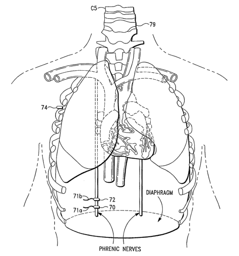
8):
28
CA 02607889 2007-11-07
WO 2006/121446 PCT/US2005/016964
EXAMPLES
[000175] The following examples are given to enable those skilled in the art
to more
clearly understand and practice the present invention. They should not be
considered as
limiting the scope of the invention, but merely as being illustrated as
representative
thereof.
Example 1
[000176] A study was performed to locate the phrenic nerve in the neck and
stimulate
the diaphragm. A .58kg rat was anesthetized; the neck, the back of the neck
and chest
were shaved. A tracheotomy was performed and the rat was intubated using a 14g
catheter. An incision was made at the back of the neck to locate the spine. A
dremmel
tool was used to perform a laminectomy and sever the spinal cord at C-2, C-3.
Diaphragm and intercostal movement stopped.
[000177] The tracheotomy incision was extended to locate the right phrenic in
the
neck. The isoflurane was then reduced from 1 to 0.25% and the oxygen flow was
then
reduced to 0.3L/min.
[000178] A hook probe was attached to the right phrenic nerve in the neck. The
red
'(signal) lead was attached to the hook probe and the black (ground) lead was
attached to
an exposed muscle in the neck.
[000179] Stimulation began at 2:35 pm with strong diaphragm movement, and
stopped
at 9:35 pm. Throughout the seven hours, the rat was "breathing" using the
input signal.
As reflected in Table I, vital signs were within normal limits.
29
CA 02607889 2007-11-07
WO 2006/121446 PCT/US2005/016964
Table I
Time Heart Rate Blood Pressure SPO2 TEMP 02 Level Degree of
BPM
L/min Movement
2:37pm 246 98 96.4 0.3 Strong
2:45pm 246 93 95.9 0.3 Strong
2:55pm 212 84/54 93 94.8 0.3 Strong
3:05pm 248 94/61 91 94.3 0.3 Strong
3:35pm 238 57/27 89 94.5 0.3 Strong
4:15pm 212 77/45 94 94.8 0.3 Strong
4:35pm 2hrs 216 69/49 94 95 0.3 Strong
4:55pm 230 86/57 94 95.7 0.3 Strong
5:15pm 212 89/47 93 95.4 0.3 Strong
5:40pm 3hrs 220 74/40 95 93.6 0.3 Strong
6:00pm 204 69/44 95 93.6 0.3 Strong
6:20pm 192 67/40 96 91 0.3 Strong
6:30pm 4hrs 192 64/34 100 91.8 0.3 Strong
6:50pm 218 55/24 96 94.3 0.3 Strong
7:10pm 208 69/35 98 93.9 0.3 Strong
SQ fluids to rat
7:30pm 5hrs 210 74/40 98 94.5 0.3 Strong
7:50pm 208 76/42 98 95.5 0.3 Strong
8:10pm 220 74/40 99 94.8 0.3 Strong
8:30pm 6hrs 226 72/40 98 95 0.3 Strong
8:50pm 222 71/39 97 95.5 0.3 Strong
9:10pm 224 72/40 96 96.4 0.3 Strong
9:20pm 200 77/53 94 96.3 0.3 Strong
9:35pm 7hrs 218 Signal stopped 87 96.3 0.3 Strong
CA 02607889 2007-11-07
WO 2006/121446 PCT/US2005/016964
Example 2
[000180] A study was performed to locate the phrenic nerve in the neck and
stimulate
the diaphragm. A .74 kg rat was anesthetized; the neck, the back of the neck,
and chest
were shaved, a tracheotomy was performed. The rat was intubated using a 14g
catheter.
[000181] An incision was made at the back of the neck to locate the spine. A
dremmel
tool was used to perform a laminectomy and sever the spinal cord at C-2, C-3.
Diaphragm and intercostals movement stopped.
[000182] The tracheotomy incision was extended to locate the right phrenic in
the
neck. The isoflurane was then reduced from 1 to 0.25% and the oxygen flow was
reduced to 0.3L/min.
[000183] A hook probe was attached to the riglit phrenic nerve in the neck.
The red
(signal) lead was attached to the hook probe and the black (ground) lead was
attached to
an exposed muscle in the neck. Stimulation began at 3:50 pm with strong
diaphragm
movement. At 4:05 pm the intercostals muscles began moving on their own again.
Stimulation was stopped and another attempt was made to completely sever the
spinal
cord. Intercostal movement stopped. The probe was reattached to the right
phrenic but
no movement resulted when stimulated. The left phrenic was then located and
the hook
probe was attached.
[000184] Stimulation started at 4:30 pm with good strong diaphragm movement
and
continued unti17:30 pm when the study was ended. As reflected in Table II,
vital signs
were within the normal limits throughout the three hours that the rat was
"breathing".
31
CA 02607889 2007-11-07
WO 2006/121446 PCT/US2005/016964
Table II
Time Heart Rate SPO2 TEMP 02 Level Degree of
BPM L/min Movement
3:50pm 3.32 98 96.3 0.3 Strong
4:00pm 284 92 93.6 0.3 Strong
4:40pm 266 99 97 0.3 Strong
4:50pm 260 100 97.5 0.3 Strong
5:00pm 254 99 97.7 0.3 Strong
5:10pm 250 99 98.2 0.3 Strong
5:20pm 246 99 98.1 0.3 Strong
5:30pm lhr 250 99 98.2 0.3 Strong
5:40pm 236 98 98.1 0.3 Strong
5:50pm 242 100 97.7 0.25 Strong
6:00pm 242 98 97.5 0.25 Strong
6:20pm 234 97 97.9 0.25 Strong
6:30pm 2hrs 238 98 97.5 0.25 Strong
6:40pm 242 98 97.5 0.25 Strong
6:50pm 240 98 97.5 0.25 Strong
7:00pm 232 97.7 0.25 Strong
7:10pm 234 100 97.2 0.25 Strong
7:20pm 232 100 97 0.25 Strong
7:30pm 3hrs 238 99 97 0.25 Strong
[000185] As will be appreciated by one having ordinary skill in the art, the
method and
system for recording, storing and transmitting waveform signals described
above
provides numerous advantages.
[000186] The method and systems of the invention can also be employed in
numerous
applications to control one or more body functions. Among the envisioned
applications
are the following:
32
CA 02607889 2007-11-07
WO 2006/121446 PCT/US2005/016964
a) Sleep Apnea
A patient is diagnosed with sleep apnea. A first sensor is employed to monitor
diaphragm
contractions, neck muscle tension, and/or airway pressure, and a second sensor
is
employed to capture signals from phrenic nerve or hypoglossal nerve. The
signal(s) from
the first sensor are routed to a processing unit (e.g., computer), where they
are analyzed.
If the signal indicates that a breath needs to be taken, a signal generated by
the processing
unit (as described herein) is transmitted to the subject to open the pharynx
and/or contract
the diaphragm.
b) Respiratory Distress
A patient is suffering from an inability to contract the diaphragm, e.g. from
a high spinal
cord injury. A first sensor is employed to monitor blood gas levels and a
second sensor is
employed to capture signals from the phrenic nerve. The signal(s) from the
first sensor are
routed to a processing unit (e.g., computer), where they are analyzed. If the
signal
indicates low blood oxygen levels, a signal generated by the processing unit
(as described
herein) is transmitted to the subject to contract the diaphragm.
c) Asthma
A patient is diagnosed with asthma. A first sensor is employed to monitor
airway
constriction and a second sensor is employed to capture signals from nerves
innervating
the bronchi and bronchioles. The signal(s) from the first sensor are routed to
a processing
unit (e.g., computer), where they are analyzed. If the signal indicates
constricted airways,
a signal generated by the processing unit (as described herein) is transmitted
to the subject
to open the constricted airways.
d) Low Blood Pressure
A patient is diagnosed with suffering from acute low blood pressure, e.g. as a
result of
traumatic blood loss or septic shock syndrome. A first sensor is employed to
monitor
blood pressure and a second sensor is employed to capture signals from the
carotid sinus.
The signal(s) from the first sensor are routed to a processing unit (e.g.,
computer), where
they are analyzed. If the signal indicates low blood pressure, a signal
generated by the
processing unit (as described herein) is transmitted to the subject to
increase blood
pressure by constricting blood vessels.
33
CA 02607889 2007-11-07
WO 2006/121446 PCT/US2005/016964
e) Abnormal Heart Beat
A patient is diagnosed with an abnormal heart beat, e.g. atrial fibrillation,
ventricular
fibrillation, or tachycardia. A first sensor is employed to monitor the heart
rate and a
second sensor is employed to capture signals from nerves innervating the
heart. The
signal(s) from the first sensor are routed to a processing unit (e.g.,
computer), where they
are analyzed. If the signal indicates an abnormal heart beat, a signal
generated by the
processing unit (as described herein) is transmitted to the subject to restore
the heart to
normal sinus rhythm.
f) Acid Reflux
A patient is diagnosed with acid reflux. A first sensor is employed to monitor
acid levels
in the lower esophagus and a second sensor is employed to capture muscle
contraction
signals from the lower esophageal sphincter. The signal(s) from the first
sensor are routed
to a processing unit (e.g., computer), where they are analyzed. If the signal
indicates
excess acid reflux, a signal generated by the processing unit (as described
herein) is
transmitted to the subject to tense the muscles of the lower esophageal
sphincter.
g) Obesity
A patient is diagnosed with obesity. A first sensor is employed to monitor
blood sugar
levels and stomach contents and a second sensor is employed to capture signals
from the
vagus nerve. The signal(s) from the first sensor are routed to a processing
unit (e.g.,
computer), where they are analyzed. If the signal indicates sufficient levels
of blood sugar
or that the stomach is sufficiently distended, a signal generated by the
processing unit (as
described herein) is transmitted to the subject to give a sensation of
fullness and suppress
the appetite.
h) Erectile Dysfunction
A patient is diagnosed with erectile dysfunction. A first sensor is employed
to monitor
penile tumescence and a second sensor is employed to capture signals from the
dorsal
penile nerve. The signal(s) from the first sensor are routed to a processing
unit (e.g.,
computer), where they are analyzed. If the signal indicates erectile
dysfunction, a signal
generated by the processing unit (as described herein) is transmitted to the
subject to
achieve an erection.
34
CA 02607889 2007-11-07
WO 2006/121446 PCT/US2005/016964
Alternatively, a first sensor is employed to capture signals from the dorsal
penile nerve.
When an erection is desired but cannot be obtained naturally, a signal
generated by the
processing unit (as described herein) is transmitted to the subject to achieve
an erection.
i) Stroke
A patient is diagnosed with a stroke that has affected motor control. A first
sensor is
employed to monitor muscle movement and a second sensor is employed to capture
signals from the nerves innervating those muscles. The signal(s) from the
first sensor are
routed to a processing unit (e.g., computer), where they are analyzed. If the
signal
indicates inability to move, a signal generated by the processing unit (as
described herein)
is transmitted to the subject to move the muscles in order to maintain muscle
tone.
Alternatively, a first sensor is employed to capture signals from the nerves
innervating
those muscles. If the patient is unable to move the desired muscles, a signal
generated by
the processing unit (as described herein) is transmitted to the subject to
move the muscles
in order to maintain muscle tone.
j) Tension Headaches
A patient is diagnosed with tension headaches. A first sensor is employed to
monitor
headache pain and a second sensor is employed to capture signals from the
nerves
innervating the muscles of the neck. The signal(s) from the first sensor are
routed to a
processing unit (e.g., computer), where they are analyzed. If the signal
indicates a
headache, a signal generated by the processing unit (as described herein) is
transmitted to
the subject to relax the muscles of the neck.
Alternatively, a first sensor is employed to capture signals from the nerves
innervating the
muscles of the neck. When the patient experiences a headache, a signal
generated by the
processing unit (as described herein) is transmitted to the subject to relax
the muscles of
the neck.
CA 02607889 2007-11-07
WO 2006/121446 PCT/US2005/016964
k) Weakened Immune System
A patient is immuno-compromised or is being immunized with a weak immunogen. A
first
sensor is employed to monitor immune fanction and a second sensor is employed
to
capture signals from thymus, lymph nodes, and/or spleen. The signal(s) from
the first
sensor are routed to a processing unit (e.g., computer), where they are
analyzed. If the
signal indicates a weakened immune system, a signal generated by the
processing unit (as
described herein) is transmitted to the subject to stimulate the immune
response.
Alternatively, a first sensor is employed to capture signals from thymus,
lymph nodes,
and/or spleen. When the patient is being immunized with a weak immunogen or
has a
weakened immune system that needs to be bolstered, a signal generated by the
processing
unit (as described herein) is transmitted to the subject to stimulate the
immune system.
1) Irritable Bowl Syndrome
A patient is diagnosed with irritable bowel syndrome. A first sensor is
employed to
monitor bowel contractions and a second sensor is employed to capture signals
from the
nerves innervating the bowel. The signal(s) from the first sensor are routed
to a
processing unit (e.g., computer), where they are analyzed. If the signal
indicates abnormal
bowel function, a signal generated by the processing unit (as described
herein) is
transmitted to the subject to restore normal bowel functions.
m) Low Sperm Count
A patient is diagnosed with low sperm count. A first sensor is employed to
monitor sperm
levels and a second sensor is employed to capture signals from nerves
innervating the
testes. The signal(s) from the first sensor are routed to a processing unit
(e.g., computer),
where they are analyzed. If the signal indicates low sperm count, a signal
generated by the
processing unit (as described herein) is transmitted to the subject to
increase production of
sperm.
Alternatively, a first sensor is employed to capture signals from nerves
innervating the
testes. In order to increase the sperm count, a signal generated by the
processing unit (as
described herein) is transmitted to the subject to increase production of
sperm.
36
CA 02607889 2007-11-07
WO 2006/121446 PCT/US2005/016964
n) Muscle Cramps
A patient is diagnosed with muscle cramps. A first sensor is employed to
monitor muscle
conditions and a second sensor is employed to capture signals from the nerves
innervating
those muscles. The signal(s) from the first sensor are routed to a processing
unit (e.g.,
computer), where they are analyzed. If the signal indicates a muscle cramp, a
signal
generated by the processing unit (as described herein) is transmitted to the
subject to relax
the cramped muscle.
Alternatively, a first sensor is employed to capture signals from the nerves
innervating
those muscles. When the patient experiences a muscle cramp, a signal generated
by the
processing unit (as described herein) is transmitted to the subject to relax
the cramped
muscle.
o) Sexual Unresponsiveness
A patient is suffering from an inability to achieve orgasm. A first sensor is
employed to
monitor whether an orgasm has been achieved and a second sensor is employed to
capture
signals from the external genitalia responsible for orgasm. The signal(s) from
the first
sensor are routed to a processing unit (e.g., computer), where they are
analyzed. If the
signal indicates a lack of orgasm after an appropriate length of time, a
signal generated by
the processing unit (as described herein) is transmitted to the subject to
achieve orgasm.
p) Insonuzia
A patient is diagnosed with insomnia. A first sensor is employed to monitor
fatigue and a
second sensor is employed to capture signals from large muscle groups. The
signal(s)
from the first sensor are routed to a processing unit (e.g., computer), where
they are
analyzed. If the signal indicates a lack of sleep, a signal generated by the
processing unit
(as described herein) is transmitted to the subject to relax their large
muscle groups and aid
in falling asleep.
Alternatively, a first sensor is employed to capture signals from large muscle
groups. If the
patient is unable to fall asleep, a signal generated by the processing unit
(as described
herein) is transmitted to the subject to relax his large muscle groups and aid
in falling
asleep.
37
CA 02607889 2007-11-07
WO 2006/121446 PCT/US2005/016964
q) Restless Legs Syndrome
A patient is diagnosed with restless legs syndrome. A first sensor is employed
to monitor
leg movement and a second sensor is employed to capture signals from the
nerves
innervating the muscles of the legs. The signal(s) from the first sensor are
routed to a
processing unit (e.g., computer), where they are analyzed. If the signal
indicates restless
legs, a signal generated by the processing unit (as described herein) is
transmitted to the
subject to relax the muscles of the legs.
r) Incontinence
A patient is diagnosed with urinary incontinence. A first sensor is employed
to monitor
fullness of the bladder and a second sensor is employed to capture signals
from the
urethral sphincter. The signal(s) from the first sensor are routed to a
processing unit (e.g.,
computer), where they are analyzed. If the signal indicates a less than full
bladder, a
signal generated by the processing unit (as described herein) is transmitted
to the subject
to keep the sphincter closed. When the bladder needs to be emptied, at the
appropriate
moment, a signal is transmitted to open the sphincter.
s) Constipation
A patient is suffering from constipation. A first sensor is employed to
monitor bowel
movement and a second sensor is employed to capture signals from nerves
innervating the
bowel. The signal(s) from the first sensor are routed to a processing unit
(e.g., computer),
where they are analyzed. If the signal indicates constipation, a signal
generated by the
processing unit (as described herein) is transmitted to the subject to
increase peristaltic
motion.
t) Nausea
A patient is suffering from frequent nausea. A first sensor is employed to
monitor levels of
nausea and a second sensor is employed to capture signals from the vagus
nerve. The
signal(s) from the first sensor are routed to a processing unit (e.g.,
computer), where they
are analyzed. If the signal indicates nausea, a signal generated by the
processing unit (as
described herein) is transmitted to the subject to counteract the signals of
nausea.
38
CA 02607889 2007-11-07
WO 2006/121446 PCT/US2005/016964
u} Spasticity
A patient is diagnosed with spasticity. A first sensor is employed to monitor
muscle
tension and a second sensor is employed to capture signals from the nerves
that innervate
the muscles. The signal(s) from the first sensor are routed to a processing
unit (e.g.,
computer), where they are analyzed. If the signal indicates continuously
contracted
muscles, a signal generated by the processing unit (as described herein) is
transmitted to
the subject to relax the muscles.
v) Dry Eyes Syndrome
A patient is diagnosed with dry eyes syndrome. A first sensor is employed to
monitor
levels of tears and a second sensor is employed to capture signals from the
nerves
innervating the lacrimal glands. The signal(s) from the first sensor are
routed to a
processing unit (e.g., computer), where they are analyzed. If the signal
indicates dry eyes,
a signal generated by the processing unit (as described herein) is transmitted
to the subject
to increase tear production.
w) Dry Mouth Syndrome
.A patient is diagnosed with dry mouth syndrome. A first sensor is employed to
monitor
levels of saliva and a second sensor is employed to capture signals from the
nerves
innervating the salivary glands. The signal(s) from the first sensor are
routed to a
processing unit (e.g., computer), where they are analyzed. If the signal
indicates dry
mouth, a signal generated by the processing unit (as described herein) is
transmitted to the
subject to increase saliva production and secretion.
x) Depression
A patient is diagnosed with depression. A first sensor is employed to monitor
signals from
the limbic system and a second sensor is employed to capture signals from the
vagus
nerve. The signal(s) from the first sensor are routed to a processing unit
(e.g., computer),
where they are analyzed. If the signal indicates a depressed mood, a signal
generated by
the processing unit (as described herein) is transmitted to the subject to
produce a state of
euphoria.
39
CA 02607889 2007-11-07
WO 2006/121446 PCT/US2005/016964
y) Epilepsy
A patient is diagnosed with epilepsy. A first sensor is employed to monitor
brain wave
activity and a second sensor is employed to capture signals from the vagus
nerve. The
signal(s) from the first sensor are routed to a processing unit (e.g.,
computer), where they
are analyzed. If the signal indicates an epileptic attack is imminent, a
signal generated by
the processing unit (as described herein) is transmitted to the subject to
counteract the
epileptic attack.
z) Overactive Bladder
A patient is diagnosed with an overactive bladder. A first sensor is employed
to monitor
the status of the bladder and a second sensor is employed to capture signals
from nerves
that innervate the bladder. The signal(s) from the first sensor are routed to
a processing
unit (e.g., computer), where they are analyzed. If the signal indicates an
overactive
bladder, a signal generated by the processing unit (as described herein) is
transmitted to
the subject to relax the muscles of the bladder.
aa) Low Levels of Growth Honnone
A patient is diagnosed with low levels of growth hormone. A first sensor is
employed to
monitor growth hormone levels and a second sensor is employed to capture
signals from
the pituitary gland. The signal(s) from the first sensor are routed to a
processing unit (e.g.,
computer), where they are analyzed. If the signal indicates low levels of
growth hormone,
a signal generated by the processing unit (as described herein) is transmitted
to the subject
to increase growth hormone levels.
bb) Low Levels of Insulin
A patient is diagnosed with low levels of insulin. A first sensor is employed
to monitor
insulin and blood sugar levels and a second sensor is employed to capture
signals from the
pancreas. The signal(s) from the first sensor are routed to a processing unit
(e.g.,
computer), where they are analyzed. If the signal indicates low levels of
insulin and high
levels of blood sugar, a signal generated by the processing unit (as described
herein) is
transmitted to the subject to increase insulin secretion.
CA 02607889 2007-11-07
WO 2006/121446 PCT/US2005/016964
cc) Abnormal Levels of Thyroid Hormone
A patient is diagnosed with abnormal levels of thyroid hormone. A first sensor
is
employed to monitor thyroid hormone levels and a second sensor is employed to
capture
signals from the thyroid and/or the pituitary gland. The signal(s) from the
first sensor are
routed to a processing unit (e.g., computer), where they are analyzed. If the
signal
indicates abnormal levels of thyroid hormone, a signal generated by the
processing unit (as
described herein) is transmitted to the subject to restore the thyroid hormone
levels to
normal, either by increasing or decreasing secretion of thyroid hormone or
thyroid
stimulating hormone, as appropriate.
dd) Abnormal Levels of Melatonin
A patient is diagnosed with abnormal levels of melatonin. A first sensor is
employed to
monitor melatonin levels and a second sensor is employed to capture signals
from the
pineal gland. The signal(s) from the first sensor are routed to a processing
unit (e.g.,
computer), where they are analyzed. If the signal indicates abnormal levels of
melatonin,
a signal generated by the processing unit (as described herein) is transmitted
to the subject
to restore the melatonin levels to normal, either by increasing or decreasing
secretion of
the horrnone, as appropriate.
ee) Abnormal Levels of Adrenocorticotrophic Hormone
A patient is diagnosed with abnormal levels of adrenocorticotrophic hormone
(ACTH). A
first sensor is employed to monitor levels of ACTH and a second sensor is
employed to
capture signals from the pituitary gland. The signal(s) from the first sensor
are routed to a
processing unit (e.g., computer), where they are analyzed. If the signal
indicates an
abnormal level of ACTH, a signal generated by the processing unit (as
described herein) is
transmitted to the subject to restore the ACTH levels to normal, either by
increasing or
decreasing secretion of the hormone, as appropriate.
ff) Abnormal Levels of Antidiuretic Hormone (ADH)
A patient is diagnosed with abnormal levels of antidiuretic hormone (ADH). A
first sensor
is employed to monitor levels of ADH and a second sensor is employed to
capture signals
from the pituitary gland. The signal(s) from the first sensor are routed to a
processing unit
(e.g., computer), where they are analyzed. If the signal indicates abnormal
levels of ADH,
41
CA 02607889 2007-11-07
WO 2006/121446 PCT/US2005/016964
a signal generated by the processing unit (as described herein) is transmitted
to the subject
to restore the ADH levels to normal, either by increasing or decreasing
secretion of the
hormone, as appropriate.
gg) Abnormal Levels of Parathyroid Hormone
A patient is diagnosed with abnormal levels of parathyroid hormone. A first
sensor is
employed to monitor levels of parathyroid hormone and a second sensor is
employed to
capture signals from the parathyroid glands. The signal(s) from the first
sensor are routed
to a processing unit (e.g., computer), where they are analyzed. If the signal
indicates
abnormal levels of parathyroid hormone, a signal generated by the processing
unit (as
described herein) is transmitted to the subject to restore the parathyroid
hormone levels to
normal, either by increasing or decreasing secretion of the hormone, as
appropriate.
hh) Abnormal Levels of Epinephrine or Norepinephrine
A patient is diagnosed with abnormal levels of epinephrine or norepinephrine.
A first
sensor is employed to monitor levels of epinephrine or norepinephrine and a
second sensor
is employed to capture signals from the adrenal glands. The signal(s) from the
first sensor
are routed to a processing unit (e.g., computer), where they are analyzed. If
the signal
indicates abnormal levels of epinephrine or norepinephrine, a signal generated
by the
processing unit (as described herein) is transmitted to the subject to restore
the epinephrine
or norepinephrine levels to normal, either by increasing or decreasing
secretion of the
hormone, as appropriate.
ii) Abnormal Levels of Glucagon
A patient is diagnosed with abnormal levels of glucagon. A first sensor is
employed to
monitor levels of glucagon and a second sensor is employed to capture signals
from the
pancreas. The signal(s) from the first sensor are routed to a processing unit
(e.g.,
computer), where they are analyzed. If the signal indicates abnormal levels of
glucagon, a
signal generated by the processing unit (as described herein) is transmitted
to the subject
to restore glucagon levels to normal, either by increasing or decreasing
secretion of the
hormone, as appropriate.
42
CA 02607889 2007-11-07
WO 2006/121446 PCT/US2005/016964
jj) Abnormal Levels of Sex Hormones
A patient is diagnosed with abnormal levels of sex hormones, e.g.,
testosterone or
estrogen. A first sensor is employed to monitor levels of sex hormones and a
second
sensor is employed to capture signals from the gonads. The signal(s) from the
first sensor
are routed to a processing unit (e.g., computer), where they are analyzed. If
the signal
indicates abnormal levels of sex hormones, a signal generated by the
processing unit (as
described herein) is transmitted to the subject to restore sex hormone levels
to normal,
either by increasing or decreasing secretion of the hormone, as appropriate.
kk) Pain Abatement
A patient is suffering from chronic pain. A first sensor is employed to
monitor pain signals
and a second sensor is employed to capture signals from the relevant nerve.
The signal(s)
from the first sensor are routed to a processing unit (e.g., computer), where
they are
analyzed. If the signal indicates pain, a signal generated by the processing
unit (as
described herein) is transmitted to the subject to counteract the pain signal.
A patient is preparing to undergo a procedure that will produce pain, e.g.,
surgery, a dental
extraction or childbirth. A first sensor is employed to capture signals from
the relevant
nerve, e.g. the trigeminal nerve. A signal generated by the processing unit
(as described
herein) is transmitted to the subject to block the sensation of pain from the
procedure.
11) Organ Transplant
A patient is undergoing heart or liver transplant, and ice is placed inside
the body during
the procedure. Following the procedure, a first sensor is employed to capture
signals from
the phrenic nerve. The signals are compared to those of normal phrenic nerves
to diagnose
any damage done to the nerve during the procedure.
mm) Paralysis
A patient has suffered a stroke and some muscles are not moveable. A first
sensor is
employed to capture signals from the nerves that innervate the paralyzed
muscle. The
signals are compared to those from normal nerves to diagnose any damage done
to the
nerve from the stroke.
43
CA 02607889 2007-11-07
WO 2006/121446 PCT/US2005/016964
nn) Cardiac Irregularity
A patient suffers a cardiac iuregularity. A first sensor is employed to
capture signals from
the heart. The signals are compared to those from normal heart to diagnose any
damage
done to the heart and evaluate its condition.
oo) Spinal Chord Injury
A patient suffers a spinal cord injury. A first sensor is employed to capture
signals from
various nerves that emerge from the spinal cord. The signals are compared to
those from
normal nerves to diagnose any damage done to the nerves.
pp) Physical Therapy
A patient has undergone surgery, e.g., hip replacement, knee surgery, etc. A
first sensor is
employed to capture signals to the affected muscles. A signal generated by the
processing
unit (as described herein) is transmitted to the subject to move the affected
muscles and
provide physical therapy.
qq) Deep Tissue Injury
A patient has suffered a deep tissue injury. A first sensor is employed to
capture signals to
the affected deep tissue. A signal generated by the processing unit (as
described herein) is
transmitted to the subject to provide increased blood flow to the affected
deep tissue.
rr) Military Interrogation
A military, government, or law enforcement agency desires a non-lethal weapon
to subdue
or interrogate an opponent. A signal generated by the processing unit (as
described herein)
is transmitted to the subject to subdue said subject. This signal may include,
but is not
limited to, causing the bladder or bowel to evacuate, causing temporary
blindness, causing
a temporary ringing in the ears, causing hyperventilation to the point of
losing
consciousness, or causing temporary severe pain.
ss) Traumatic Injury
A patient has suffered a traumatic injury, and trained emergency medical
personnel need
to provide immediate treatment to manage the patient. A signal generated by
the
processing unit (as herein described) is transmitted to the subject to
stabilize vital signs or
44
CA 02607889 2007-11-07
WO 2006/121446 PCT/US2005/016964
provide support. Examples of transmitted signals include a signal to control
breathing
frequency and volume, a signal to control heart rate, a signal to regulate
blood pressure, a
signal to reduce pain, or a signal to induce unconsciousness.
tt) Alternative to "chemical castration"
A patient requiring suppression of sexual desire to affect re-entry to society
following
sexual abuse treatment may have the regulation of sexual hormones controlled
by signals
applied to nerve connections to the hormone secretion glands to reduce
testosterone levels
without excessive generation of estrogen.
uu) Muscle Atrophy
A patient is in a coma. A first sensor is employed to capture signals from
relevant muscle
groups. If it is desired that the patient receive muscle stimulation, a signal
generated by the
processing unit (as described herein) is transmitted to the subject to
contract his muscles
regularly to aid in maintaining muscle tone.
vv) Acupuncture
A patient is undergoing acupuncture treatment. A first sensor is employed to
capture
signals from the relevant body organ being treated. If it is desired that the
patient receive
electrical stimulation through the acupuncture needles, a signal generated by
the
processing unit (as described herein) is transmitted to the subject to achieve
the desired
acupuncture treatment.
ww) Chiropractic
A patient is undergoing chiropractic treatments. A first sensor is employed to
capture
signals from the relevant body organ being treated. If it is desired that the
patient receive
electrical stimulation in conjunction with chiropractic treatment, a signal
generated by the
processing unit (as described herein) is transmitted to the subject to achieve
the desired
chiropractic treatment.
CA 02607889 2007-11-07
WO 2006/121446 PCT/US2005/016964
[0001 87] Without departing from the spirit and scope of this invention, one
of ordinary
skill can make various changes and modifications to the invention to adapt it
to various
usages and conditions. As such, these changes and modifications are properly,
equitably, and intended to be, within the full range of equivalence of the
following
claims.
46