Note: Descriptions are shown in the official language in which they were submitted.
CA 02607959 2007-11-06
WO 2006/124819 PCT/US2006/018806
MULTI-STAGE SYRINGE AND METHODS OF USING THE SAME
CROSS REFERENCE TO RELATED APPLICATIONS
[0001] This application claims the benefit of U.S. Provisional Application No.
60/681394, filed on
16 May 2006.
FIELD OF THE INVENTION
[0002] The present invention relates to syringes, and more particularly to a
syringe equipped with
an intermediate plunger for enabling at least generally sequential delivery of
first and second medical
fluids.
BACKGROUND
[0003] This section is intended to introduce the reader to various aspects of
art that may be
related to various aspects of the present invention, which are described
and/or claimed below. This
discussion is believed to be helpful in providing the reader with background
information to facilitate a
better understanding of the various aspects of the present invention.
Accordingly, it should be
understood that these statements are to be read in this light, and not as
admissions of prior art.
[0004] Nuclear medicine utilizes radioactive material for diagnostic and
therapeutic purposes by
injecting a patient with a small dose of the radioactive material, which
concentrates in certain organs or
biological regions of the patient. Radioactive materials typically used for
nuclear medicine include
Technetium-99m, Indium-113m, and Strontium-87m among others. Some radioactive
materials
naturally concentrate toward a particular tissue, for example, iodine
concentrates toward the thyroid.
However, radioactive materials are often combined with a tagging or organ-
seeking agent, which
targets the radioactive material for the desired organ or biologic region of
the patient. These radioactive
materials alone or in combination with a tagging agent are typically referred
to as radiopharmaceuticals
in the field of nuclear medicine. At relatively lower doses of the
radiopharmaceutical, a radiation
imaging system (e.g., a gamma camera) provides an image of the organ or
biological region that
collects the radiopharmaceutical. Irregularities in the image are often
indicative of a pathologic
condition, such as cancer. Higher doses of the radiopharmaceutical may be used
to deliver a
therapeutic dose of radiation directly to the pathologic tissue, such as
cancer cells.
[0005] In certain applications, multiple medical fluids may be injected into a
patient. In positron
emission tomography (PET) or single photon emission computed tomography
(SPECT), a syringe may
intake, contain, and subsequently inject a radioactive substance, such as a
radiopharmaceutical. In
magnetic resonance imaging (MRI), computed tomography (CT), radiography (e.g.,
x-ray), or
ultrasound, a syringe may intake, contain, and subsequently inject a contrast
agent. These applications
also may utilize other medical fluids in combination, prior to, or after
injecting the radiopharmaceutical
or contrast agent. Unfortunately, these applications generally utilize
multiple syringes or independent
1
CA 02607959 2007-11-06
WO 2006/124819 PCT/US2006/018806
injection mechanisms, which can lead to time delays, dosing inaccuracies, a
greater potential for
contamination, a greater potential for fluid wastage, and other problems. For
example, a significant
quantity of the radiopharmaceutical may be left in a conventional syringe. In
addition, the syringe
utilized to administer the radiopharmaceutical may contain more residual
radiopharmaceutical than
desired, posing potential safety and/or disposal concerns.
SUMMARY
[0006] Certain aspects commensurate in scope with the originally claimed
invention are set forth
below. It should be understood that these aspects are presented merely to
provide the reader with a
brief summary of certain forms the invention might take and that these aspects
are not intended to limit
the scope of the invention. Indeed, the invention may encompass a variety of
aspects that may not be
set forth below.
[0007] A first aspect of the present invention is directed to a syringe having
a plunger. This
plunger includes a one-way valve having a fluid passage defined through an
interior of the plunger to a
downstream side of the plunger.
[0008] A second aspect of the present invention is directed to a flow control
plunger having a
check-valve disposed between an upstream fluid side and a downstream fluid
side thereof. This check-
valve includes an interior passage fluidly coupling the upstream and
downstream fluid sides when the
check-valve is in an open position.
[0009] Still third aspect of the invention is directed to a syringe barrel
having a plunger check-
valve actuator disposed inside the syringe barrel at a front portion of the
syringe barrel.
[0010] Yet a fourth aspect of the invention is directed to a method of using a
syringe. In particular,
a flow control plunger disposed inside a syringe is actuated to enable fluid
flow through an interior of the
flow control plunger to a downstream side of the flow control plunger.
[0011] Still yet a fifth aspect of the invention is directed to a method of
using a syringe. In
particular, a plunger of the syringe is biased toward a terminus of the
syringe to discharge a first
medical fluid between the terminus and an intermediate plunger. The terminus
of the syringe is
contacted with the intermediate plunger, and a second medical fluid is
discharged through the
intermediate plunger while the terminus and the intermediate plunger are in
contact.
[0012] Various refinements exist of the features noted above in relation to
the various aspects of
the present invention. Further features may also be incorporated in these
various aspects as well.
These refinements and additional features may exist individually or in any
combination. For instance,
various features discussed below in relation to one or more of the illustrated
embodiments may be
incorporated into any of the above-described aspects of the present invention
alone or in any
combination. Again, the brief summary presented above is intended only to
familiarize the reader with
certain aspects and contexts of the present invention without limitation to
the claimed subject matter.
2
CA 02607959 2007-11-06
WO 2006/124819 PCT/US2006/018806
BRIEF DESCRIPTION OF THE FIGURES
[0013] The accompanying figures, which are included to provide further
understanding of various
aspects of the invention, illustrate exemplary embodiments of the present
invention and, together with
the description, serve to explain various principles of the invention.
[0014] FIG. 1 is a perspective view of an embodiment of what may be
characterized as a multi-
chamber, multi-stage, or sequential injection syringe having a first medical
fluid in a front chamber
thereof and a second medical fluid in a rear chamber thereof, the chambers
separated by a
intermediate flow control plunger of the syringe;
[0015] FIG. 2 is an enlarged side view of an embodiment of a body of the
intermediate flow control
plunger of FIG. 1;
[0016] FIG. 3 is an enlarged exploded view of an embodiment of the
intermediate flow control
plunger of FIGS. 1 and 2, illustrating an elastomeric piston cap exploded from
the body;
[0017] FIG. 4 is an enlarged cross-sectional view of an embodiment of a
terminal end portion of
the multi-chamber, multi-stage, or sequential injection syringe and the
intermediate flow control plunger
of FIGS. 1-3, illustrating a check valve of the intermediate flow control
plunger in a closed position;
[0018] FIG. 5 is an enlarged cross-sectional view of an embodiment of a
terminal end portion of an
embodiment of the multi-chamber, multi-stage, or sequential injection syringe
and the intermediate flow
control plunger of FIGS. 1-3, illustrating a check valve of the intermediate
flow control plunger in an
open position;
[0019] FIG. 6 is a cross-sectional view of an embodiment of a multi-chamber,
multi-stage, or
sequential injection syringe, illustrating the terminal end oriented
substantially downward and the rear
chamber being filled from an open end of a barrel of the syringe, with a
pushrod withdrawn;
[0020] FIG. 7 is a cross-sectional view of an embodiment of the filled multi-
chamber, multi-stage,
or sequential injection syringe of FIG. 6, illustrating the terminal end
oriented substantially upward to
purge unwanted air from the rear chamber;
[0021] FIG. 8 is a cross-sectional view of an embodiment of a multi-chamber,
multi-stage, or
sequential injection syringe, illustrating a terminal end oriented
substantially downward and the rear
chamber being filled through a fill port in the barrel;
[0022] FIG. 9 is a cross-sectional view of an embodiment of the filled multi-
chamber, multi-stage,
or sequential injection syringe of FIG. 8, illustrating the terminal end
oriented substantially upward to
purge unwanted air from the rear chamber;
[0023] FIG. 10 is a cross-sectional view of an embodiment of a multi-chamber,
multi-stage, or
sequential injection syringe, illustrating a terminal end thereof pointing up
and a needle inserted through
a pushrod to fill the rear chamber with the second medical fluid;
3
CA 02607959 2007-11-06
WO 2006/124819 PCT/US2006/018806
[0024] FIG. 11 is a cross-sectional view of an embodiment of a multi-chamber,
multi-stage, or
sequential injection syringe, illustrating a check valve on the plunger of the
pushrod and a check valve
on the intermediate piston, the rear chamber being filled with the second
medical fluid;
[0025] FIG. 12 is a cross-sectional view of an embodiment of a multi-chamber,
multi-stage, or
sequential injection syringe, illustrating an axial passageway through the
pushrod with an open plunger,
the rear chamber being filled with the medical fluid;
[0026] FIG. 13 is a cross-sectional view of an embodiment of a multi-chamber,
multi-stage, or
sequential injection syringe, illustrating another embodiment of the
intermediate flow control plunger
between first and second chambers;
[0027] FIG. 14 is a partial cross-sectional view of the multi-chamber, multi-
stage, or sequential
injection syringe of FIG. 13, further illustrating a first injection from the
first chamber immediately prior to
an injection transition or intermediate position of the intermediate flow
control plunger between multiple
injections of substances;
[0028] FIG. 15 is a partial cross-sectional view of an embodiment of the multi-
chamber, multi-
stage, or sequential injection syringe of FIG. 13, further illustrating a
second injection from the second
chamber directly through the intermediate flow control plunger immediately
after the injection transition
or intermediate position;
[0029] FIG. 16 is a flowchart illustrating an embodiment of a method of use or
syringe preparation
process utilizing one or more of the multi-chamber, multi-stage, or sequential
injection syringes of FIGS.
1-15;
[0030] FIG. 17 is a flowchart illustrating an embodiment of a method of
operation or imaging
process utilizing one or more of the multi-chamber, multi-stage, or sequential
injection syringes of FIGS.
1-15;
[0031] FIG. 18 is a flowchart illustrating an embodiment of a nuclear medicine
process utilizing
one or more of the multi-chamber, multi-stage, or sequential injection
syringes of FIGS. 1-15;
[0032] FIG. 19 is a block diagram illustrating an embodiment of a
radiopharmacy or system
utilizing one or more of the multi-chamber, multi-stage, or sequential
injection syringes of FIGS. 1-15;
and
[0033] FIG. 20 is a block diagram illustrating an embodiment of a nuclear
imaging system utilizing
one or more of the multi-chamber, multi-stage, or sequential injection
syringes of FIGS. 1-15.
DETAILED DESCRIPTION OF SPECIFIC EMBODIMENTS
[0034] One or more specific embodiments of the present invention will be
described below. In an
effort to provide a concise description of these embodiments, all features of
an actual implementation
may not be described in the specification. It should be appreciated that in
the development of any such
actual implementation, as in any engineering or design project, numerous
implementation-specific
decisions must be made to achieve the developers' specific goals, such as
compliance with system-
4
CA 02607959 2007-11-06
WO 2006/124819 PCT/US2006/018806
related and business-related constraints, which may vary from one
implementation to another.
Moreover, it should be appreciated that such a development effort might be
complex and time
consuming, but would nevertheless be a routine undertaking of design,
fabrication, and manufacture for
those of ordinary skill having the benefit of this disclosure.
[0035] As discussed in detail below, various embodiments of the present
invention include an
intermediate flow control plunger for separating two medical fluids in a
syringe, and also enabling
sequential administration of the two fluids. Some disclosed embodiments of the
intermediate flow
control plunger substantially reduce or may even virtually eliminate the
possibility of the two medical
fluids mixing while within the syringe. A first medical fluid may be disposed
in a first chamber generally
downstream of the intermediate flow control plunger, while a second medical
fluid may be disposed in a
second chamber generally upstream of the intermediate flow control plunger.
Thus, the first medical
fluid may be injected downstream from the intermediate flow control plunger,
and the second medical
fluid may be sequentially injected by directly passing through the
intermediate flow control plunger. For
example, the intermediate flow control plunger may include a check valve, or
one-way valve, or
automatic valve mechanism that enables flow of the second medical fluid
therethrough after the first
medical fluid is at least substantially or entirely output from the syringe.
Thus, the intermediate flow
control plunger may generally prevent or substantially reduce the possibility
of backflow of the first
medical fluid from the first chamber to the second chamber, thereby
substantially or entirely reducing a
likelihood of internal mixing of the first and second medical fluids.
[0036] In one embodiment, an intermediate flow control plunger is used in a
syringe for sequential
delivery of two medical fluids. The intermediate flow control plunger may
separate the syringe barrel
into a front chamber that may contain a first medical fluid and a rear chamber
that may contain a
second medical fluid which may or may not be different from the first medical
fluid. In one regard, the
intermediate flow control plunger may be characterized as a check valve that
substantially prohibits
backflow from the front chamber to the rear chamber. In use, force from a
pushrod of the syringe on
the second medical fluid in the rear chamber causes the intermediate plunger
to slide forward in the
syringe barrel causing the first medical fluid in the front chamber to be
discharged (e.g., out a nozzle of
the syringe). The check valve may be designed to exhibit a high enough opening
pressure to
substantially reduce or prevent mixture of the two fluids during discharge of
the first medical fluid. After
the first medical fluid is discharged (e.g., administered to the patient), the
force from the pushrod being
biased toward the nozzle of the syringe may cause the intermediate plunger to
contact a conical or at
least generally tapered end of the syringe (by the nozzle), and force from the
pushrod causes the check
valve to open and allow the second medical fluid to pass through the
intermediate plunger and be
discharged from the syringe.
[0037] The various embodiments of the disclosed syringes, though not limited
to nuclear medicine,
may be particularly useful in some nuclear medicine procedures where a
biocompatible flush (e.g.,
saline) may be used to flush a nozzle of the syringe, extension tubing
interconnected with the syringe,
and/or an injection site. Incidentally, a biocompatible flush may generally
refer to any biocompatible
fluid that does not significantly detrimentally affect the function of other
compositions being
CA 02607959 2007-11-06
WO 2006/124819 PCT/US2006/018806
administered by way of a syringe of the invention. Examples of appropriate
biocompatible flushes
include, but are not limited to, saline, sterilized water, heparin solution,
and glucose solution.
[0038] For example, a single syringe can contain both a radiopharmaceutical
and a biocompatible
flush. Based on desired dosing parameters, a 5 mL syringe may be a suitable
size for multi-stage
syringes in nuclear medicine, although other sizes may be used for various
injection applications. In
general, the intermediate flow control plunger separates the first and second
fluids in corresponding first
and second chambers of a multi-stage syringe until injection. A syringe
pushrod can be safely retracted
before the injection to check for vein patency. With a single continuous push,
the radiopharmaceutical
may be injected first, and then the biocompatible flush (e.g., saline) may be
injected afterwards. The
biocompatible flush may be utilized to flush the radiopharmaceutical from the
syringe and/or the
injection site in one step (if desired) and/or may reduce the residual
radiation in the syringe.
[0039] The benefits potentially provided by various embodiments of the
invention may be
numerous. For example, in some cases, there may be little or no need to
purchase or stock
biocompatible flush or an extra syringe and needle. In some cases, there may
be no need to prepare a
separate biocompatible flush syringe and needle. Another potential benefit is
that various embodiments
of the present invention may effectively enable only one injection to be
performed that substantively
includes what was previously two or more separate and distinct injections.
Certain aspects of the
invention may at least generally reduce chances of accidental needle sticks
(e.g., due to utilizing one
syringe instead of two). Other benefits of various aspects of the invention
may potentially include one
or more of the following: reduce need to dispose of saline vial and/or second
syringe and needle;
relieve need to draw saline into the syringe after the radiopharmaceutical
injection to perform the
syringe flush; reduced radiation exposure (e.g., due to the greater distance
between the
radiopharmaceutical and the user's hand and/or due to the flushing of the
front end of the
radiopharmaceutical syringe); fewer occurrences of drips and spills due to the
handling of one syringe
instead of two; and flushing may become so convenient that it may be used for
procedures that
normally do not get flushed.
[0040] FIG. 1 is a perspective view of a multi-chamber, multi-stage, or
sequential injection syringe
20 having a first medical fluid 22 disposed in a front chamber 24 of the
syringe 20 and a second
medical fluid 26 disposed in a rear chamber 28 of the syringe 20. The first
medical fluid 22 may be any
medical fluid appropriate for administration to a patient. Further, the second
medical fluid may be the
same as or different from the first medical fluid and may be any medical fluid
appropriate for
administration to a patient. For instance, in some embodiments, the first
medical fluid may be a
radiopharmaceutical or imaging contrast agent, and the second medical fluid
may be a biocompatible
flush. In addition, the embodiment of FIG. 1 may include a radioisotope
generator, a fluid dispensing
system, a power injector (e.g., motor, worm drive, radiation shield, etc.), a
support structure, a rotatable
arm (e.g., manual or robotic arm), a stand, an electronic control unit, a
computer, an imaging system, a
diagnostic system, or a combination thereof coupled to or generally associated
with the syringe 20. In
fact, each of the disclosed syringes may include one or more of these systems
or components as
discussed further below.
6
CA 02607959 2007-11-06
WO 2006/124819 PCT/US2006/018806
[0041] The front and rear chambers 24, 28 may be separated by an intermediate
plunger 30,
which includes a pressure activated check-valve 31. A pushrod 32 of the
syringe 20 has an integral
thumb tab 34 on one end and a plunger 36 (sometimes referred to as a proximal
plunger) on the other
end. The plunger 36 forms a seal with an inside wall 38 of a barrel 40 of the
syringe 20. The
intermediate plunger 30 may slide back and forth along the inside wall 38 of
the barrel 40 in response to
pressure from the front chamber 24 and/or pressure from the rear chamber 28
and therefore may be
said to be "slidably positioned" in the barrel. The plunger 36 may also slide
back and forth along the
inside wall 38 of the barrel 40 as the pushrod 32 may be urged in and out
(e.g., by a user or power
injector). Accordingly, the plunger 36 may also be said to be "slidably
positioned" in the barrel. It should
be noted that some embodiments may not include an elongate pushrod 32 that is
interconnected with
the plunger 36. For instance, some embodiments for use with power injectors
may include a plunger
without an associated elongate pushrod 32. Further, the pushrod 32 may be
generally utilized to bias
or move the plunger 36; accordingly, any of a wide range of sizes, shapes, and
designs of pushrods
may be appropriate depending on the desired use of the syringe.
[0042] Finger grips 42 of the syringe 20 may be defined at an open end 44 of
the barrel 40. At the
end of the barrel 40 opposite the open end 44 is a terminus 46 of the barrel
40, which is sometimes
referred to in the industry as a "conical end," as shown, or it may be other
shapes. A main passageway
48 may be defined in the terminus 46 of the syringe 20. The main passageway 48
may be bidirectional.
In other words, the main passageway 48 may be designed to enable medical fluid
to be both drawn into
and discharged from the barrel 40 of the syringe 20 (e.g., in response to
movement of the pushrod 32
and plunger 36). A luer fitting 50 or other appropriate interconnection device
may also be formed on or
attached to the syringe 20 (e.g., on or near the terminus 46).
[0043] To discharge the medical fluids 22, 26 from the syringe 20, pressure
may be applied to the
thumb tab 34 of the pushrod 32 causing the plunger 36 to slide down the inside
wall 38 of the barrel 40,
at least generally pressurizing the second fluid 26 in the rear chamber 28.
The pressure from the
second fluid 28 may act on the intermediate plunger 30 causing the
intermediate plunger 30 to slide
down the inside wall 38 of the barrel 40 to apply pressure on the first
medical fluid 22. As the plunger
36 on the pushrod 32 and the intermediate plunger 30 slide down the barrel 40,
the check-valve 31 in
the intermediate plunger 30 may be closed which keeps the second medical fluid
26 in the rear
chamber 28 separate from the first medical fluid 22 in the front chamber 24
during discharge of the first
medical fluid 22. As the plunger 36 and the intermediate plunger 30 continue
sliding down the inside
wall 38 of the barrel 40, the first medical fluid 22 may be discharged through
the main passageway 48
(e.g., for administration to a patient). After substantially all of the first
medical fluid 22 has been
discharged from the syringe 20, the intermediate plunger 30 makes contact with
the terminus 46 of the
syringe 20. When the intermediate plunger 30 contacts the terminus 46,
continued pressure applied to
the tab 34 of the pushrod 32 causes the plunger 36 (which is attached to the
pushrod 32) to slide down
the inside wall 38 of the barrel 30, increasing the pressure on the second
medical fluid 26, which
causes the check valve 31 of the intermediate plunger 30 to open allowing the
second medical fluid 26
to pass through the check valve 31 into the main passageway 48 for discharge
from the syringe 20.
7
CA 02607959 2007-11-06
WO 2006/124819 PCT/US2006/018806
[0044] Referring now to FIGS. 2 and 3, the intermediate plunger 30 includes a
body 52 and a
flexible, resilient, or elastomeric piston cap 54. The body 52 of the
intermediate plunger 30 includes a
rear flange 56, a forward flange 58, a circumferential seat 60, and a
protruding nose 62. The rear
flange 56 and the forward flange 58 define a retention channel 64 therebetween
to accommodate a
retention ring 72 (FIGS. 4 and 5) of the elastomeric piston cap 54. The
retention ring 72, when
disposed between the rear and forward flanges 56, 58 of the body 52, may
function to at least assist in
holding the elastomeric piston cap 54 on the body 52 of the intermediate
plunger 30. Incidentally, the
flanges 56, 58 and retention ring 72 may be one manner of providing an
appropriate interconnection
between the body 52 and the elastomeric piston cap 54; other manners of
providing an appropriate
interconnection may be utilized. Further, the intermediate plunger 30
illustrates one embodiment of
intermediate plungers. It should be noted that other intermediate plungers
than enable fluid to flow
through the intermediate plunger upon the intermediate plunger contacting the
terminus of the syringe
are within the scope of the disclosed embodiments.
[0045] Referring to FIG. 3, an intermediate passageway 66 may be defined in
each of the rear
flange 56 and the forward flange 58 of the body 52 of the intermediate plunger
30. Further, a nose
passageway 68 may be defined in the nose 62 of the body 52. The elastomeric
piston cap 54 includes
a flexible lip 74 which has an aperture 76 (e.g., centrally located) defined
therein. Around an outer
circumference of the elastomeric piston cap 54 is a first circumferential seal
78 and a second
circumferential seal 80 which seal against the inside wall 38 of the barrel 40
of the syringe 20. While
the elastomeric piston cap 54 is shown as having first and second seals 78,
80, other embodiments of
the elastomeric piston cap 54 may additionally and/or alternatively include
other sealing features to
promote a seal between the elastomeric piston cap 54 and the inside wall 38 of
the barrel 40 of the
syringe 20.
[0046] Referring to FIGS. 4 and 5, the flexible lip 74 of the intermediate
plunger 30 may interface
with the seat 60 to provide a fluid seal between the two components. In FIG.
4, the check valve 31 is
closed, and in FIG. 5, the check valve 31 is open. When the check valve 31 is
closed, the second
medical fluid 26 may be confined in the rear chamber 28 between the
intermediate plunger 30 and the
plunger 36. When the check valve 31 is open, the second medical fluid 26 may
be discharged from the
syringe 20 as indicated by the dashed flow arrows shown in FIG. 5. As
previously mentioned, the
check valve 31 may be opened by pressure. When the check valve 31 is open, the
second medical
fluid 26 may flow from the rear chamber 28 through the intermediate passageway
66, past the seat 60,
through the aperture 76 in the lip 74 of the elastomeric piston cap 54, and
through the nose
passageway 68 into the main passageway 48 of the syringe 20.
[0047] As the intermediate plunger 30 travels towards the terminus 46 of the
barrel 40 while the
first medical fluid 22 from the front chamber 24 is being discharged, the
check valve 31 is in the closed
position as shown in FIG. 4. When the first medical fluid 22 has been
substantially discharged, the
nose 62 of the intermediate plunger 30 contacts the terminus 46 of the barrel
40 causing the
intermediate plunger 30 to stop advancing toward the main passageway 68 of the
syringe 20. As
continued pressure is exerted on the pushrod 32, the pressure of the second
medical fluid 26 increases
because it is trapped in the second chamber 28 between the plunger 36 and the
intermediate plunger
8
CA 02607959 2007-11-06
WO 2006/124819 PCT/US2006/018806
30, and the check valve 31 is closed so the second fluid 26 at least
temporarily has no place to go. As
the pressure of the second medical fluid 26 reaches a "cracking pressure", the
check valve 31 opens as
shown in FIG. 5, allowing the second medical fluid 26 to be discharged from
the syringe 20 as indicated
by the flow arrows (FIG. 5). A seal pressure and the cracking pressure may be
independent and can
be tuned for best performance. The "seal pressure" generally refers to the
force that the first
circumferential seal 78 and the second circumferential seal 80 apply on the
inside wall 38 of the syringe
barrel 40. The seal pressure may be relatively light, so friction between the
seals 78, 80 and the inside
wall 38 does not cause the intermediate plunger 30 to stick in the barrel 40.
In addition, the cracking
pressure can be set high enough to effectively promote the intermediate
plunger 30 overcoming the
friction. This facilitates the ability of the intermediate plunger 30 to be
pushed toward the terminus 46 of
the syringe 20 in order to allow the second medical fluid 26 to be discharged.
The cracking pressure
may be characterized as a function of the diameter of the elastomeric piston
cap 54 on the intermediate
plunger 30.
[0048] FIG. 6 is a cross-sectional view of a syringe 20 with the terminus 46
pointing downward and
the rear chamber 28 being filled via the open end 44 of the barrel 40, with
the pushrod 32 dissociated
from the barrel 40. When using this filling technique, the intermediate
plunger 30 may be positioned
away from the terminus 46 in order to purge air from the syringe 20 which will
be described in greater
detail below. As shown in FIG. 6, a fill tube 100 may be inserted in the open
end 44 of the barrel 40,
and the second fluid flows from a source through the fill tube 100 and into
the rear chamber 28. When
the rear chamber 28 is filled with the desired amount of the second medical
fluid 26 or more than
actually may be desired, the pushrod 32 may be inserted into the open end 44
of the barrel 40. This
process may result in trapping some unwanted air in the second chamber 28
(which may be purged
from the syringe 20). FIG. 7 is a cross-sectional view of the filled syringe
20 from FIG. 6 after filling with
the second medical fluid 26. The terminus 46 may be pointing upward to purge
trapped air 102 from the
rear chamber 28. After the syringe 20 has been inverted from the orientation
of FIG. 6, the pushrod 32
may be depressed to force the intermediate plunger 30 into contact with the
terminus 46 of the syringe
20. As more pressure is applied to the pushrod 32, the check valve 31 opens,
allowing any trapped air
102 in the rear chamber 28 to be discharged from the syringe 20 through the
same flow path as
described above for the second medical fluid 26. After the unwanted air has
been discharged, the
syringe may be described as "pre-filled" with the second medical fluid. The
"prefilled syringe" of FIG. 7
may be sold or shipped, as is, to allow a user to fill the front chamber with
a first medical fluid which
may have a short shelf life, such as a radiopharmaceutical. A "pre-filled"
syringe may be prefilled with
either one or two medical fluids.
[0049] The front chamber 24 may be filled using conventional techniques. The
luer fitting 50 may
be elevated and connected to a source of the first medical fluid 22. The user
simply pulls back on the
push-rod 32, like conventional syringes. When the push-rod 32 is drawn away
from the terminus 46
and toward the open end 44 of the barrel 40, the check valve 31 remains closed
and the intermediate
plunger 30 slides away from the terminus 46, drawing the first medical fluid
22 into the front chamber
24. The luer fitting 50 may then be disconnected, and any unwanted air may be
purged from the from
the front chamber 24 using conventional techniques. When provided to end users
having medical fluids
9
CA 02607959 2007-11-06
WO 2006/124819 PCT/US2006/018806
in both the front chamber and the rear chamber, the syringe may be said to be
"prefilled" with a plurality
(e.g., two) medical fluids.
[0050] FIG. 8 is a cross-sectional view of a multi-chamber, multi-stage, or
sequential injection
syringe 110 with a fill port 112 defined in the barrel 40. All the other
components of the syringe 110
may be at least similar to that of the syringe 20 of FIG. 1, and accordingly,
the generally corresponding
components will be referred to using the same identification numbers. In FIG.
8, the terminus 46 may
be pointing downward to facilitate filling of the rear chamber 28 through a
fill port 112 with the second
medical fluid 26. The pushrod 32 has been withdrawn on one side of the fill
port 112, and the
intermediate plunger 30 has been positioned on the other side of the fill port
112 to enable a filling
process. A fill tube 100 may be inserted into the fill port 112, and the
second medical fluid 26 flows
from a source through the fill tube 100 and into the rear chamber 28 of the
syringe 30. The syringe 110
defines an axis as indicated by the line A-A. The orientation of the fill port
112 and the fill tube 100 may
be generally normal or at least non-parallel to the axis A-A. Some air 102 may
be trapped in the rear
chamber 28. Subsequent to a desired amount of the second medical fluid being
disposed in the rear
chamber 28, the fill tube 100 may be withdrawn from the fill port 112, and the
pushrod 32 may be
depressed until the plunger 36 interfaces with a portion of the inside wall 38
of the barrel 40 between
the fill port 112 and the terminus 46.
[0051] FIG. 9 is a cross-sectional view of the syringe 110 of FIG. 8 after
filling with the second
medical fluid 26, with the terminus 46 pointing upward to purge air 102 from
the rear chamber 28. After
the syringe 110 has been inverted from the orientation of FIG. 8, the pushrod
32 may be depressed to
force the intermediate plunger 30 into contact with the conical end or
terminus 46. Upon sufficient
pressure being applied to the pushrod 32, the check valve 31 opens, allowing
trapped air 102 in the
rear chamber 28 to be discharged from the syringe 110 through the
substantially same flow path as
described above for the second medical fluid 26.
[0052] FIG. 10 is a cross-sectional view of a syringe 120. Similar components
to the syringe 20 of
FIG. I are referred to using the same numerals; different components have been
assigned new
identification numbers. The terminus 46 may be pointing up, and a fill needle
121 has been inserted
through an axial passageway in a pushrod 122 of the syringe 120, to fill the
rear chamber 28 with the
second medical fluid 26. The fill needle 121 extends through the plunger 36
which may reseal after the
fill needle 121 has been withdrawn. The second medical fluid 26 flows from a
source, through the fill
needle 121, and into the rear chamber 28 of the syringe 120. Subsequent to a
desired amount of the
second medical fluid 26 being disposed in the rear chamber 28, the fill needle
121 may be withdrawn
from the pushrod 122. The syringe 120 may be positioned in any of a number of
appropriate
orientations during the fill process. However, at some point in the fill
process, the terminus 46 of the
syringe 120 may be elevated in order to purge unwanted air and/or bubbles from
the rear chamber 28.
If air 102 is trapped in the rear chamber 28, it may be purged by further
depressing the modified
pushrod 122 to expel the air 102 from the syringe through the substantially
same flow path that the
second medical fluid 26 travels to exit the syringe 120, previously described.
CA 02607959 2007-11-06
WO 2006/124819 PCT/US2006/018806
[0053] FIG. 11 is a cross-sectional view of another multi-chamber, multi-
stage, or sequential
injection syringe 130 having a second check valve 132 on a plunger of a
pushrod 134 and the
previously described check valve 31 on the intermediate plunger 30. In this
view, the second check
valve 132 is shown in the open position, and the syringe 130 is being filled.
Similar components to the
syringe 20 of FIG. I are referred to with identical numbers, and new
components have been assigned
new numbers. In this figure, the intermediate plunger 30 may be in contact
with the conical end or
terminus 46. The pushrod 134 may be in the barrel 40 and may be separated from
the intermediate
plunger 30 to define the rear chamber 28. The pushrod 134 has an axial
passageway 136 defined
therein that may be sealed on one end by a removable thumb tab 138 (or other
appropriate sealant or
sealing device) and the second check valve 132. In this view, the thumb tab
138 may be disengaged
from the pushrod 134.
[0054] To fill this syringe 130, the second medical fluid 26 from a source may
be introduced
through the axial passageway 136, flows through the second check valve 132,
and flows into the rear
chamber 28 as indicated by the flow arrows of FIG. 11. Subsequent to a desired
amount of the second
medical fluid 26 being disposed in the rear chamber 28, the thumb tab 138 may
be inserted in the axial
passageway 136 to seal the second medical fluid 26 in the axial passageway
136. On the end of the
pushrod 134 opposite the removable thumb tab 138, the pushrod 134 includes a
pushrod retention
channel 140, pushrod flow passageways 142 in fluid communication with the
axial passageway 136,
and a seat 144. A pushrod elastomeric cap 146 includes a pushrod retention
ring 148 sized to engage
the pushrod retention channel 140 to removably attach the cap 146 to the
pushrod 134. The pushrod
elastomeric cap 146 also includes a flexible lip 150 to engage the seat 144
which form the main
components of the second check valve 132. In this figure, the check valve may
be open so the lip 150
does not touch the seat 144. When the check valve is closed, the lip 150 may
engage the seat 144
blocking the flow of fluid from the rear chamber 28 back up the axial
passageway 136 of the pushrod
134. During the fill process, the syringe 130 may be in any number of
appropriate orientations.
[0055] Like the other fill processes discussed herein, there may be bubbles
and/or unwanted air
102 trapped in the rear chamber 28. To purge the unwanted air from the rear
chamber 28, the terminus
46 may be oriented at least generally upward, and the pushrod 134 may be
pushed further into the
barrel 40, which expels the unwanted air 102 as previously described in FIGS.
6-9.
[0056] FIG. 12 is a cross-sectional view of a multi-chamber, multi-stage, or
sequential injection
syringe 170 having a pushrod 172 with an axial passageway 176 defined therein,
and an open plunger
174. In this view, the rear chamber 28 is being filled with the second medical
fluid 26. Similar
components are identified with the same numbers as the syringe in FIG. 1, and
different components
have been assigned new numbers. The axial passageway 176 defined in the
pushrod 172 may be
open on both ends. One end of the pushrod 172 may be designed to receive a
removable thumb cap
138 or other appropriate sealing device/material, and the other end has the
open plunger 174 attached
thereto. In this figure, the thumb cap 138 may be removed from the pushrod
172. The end of the
pushrod 172 that carries the open plunger 174 forms a pushrod retention
channel 178 that engages a
retention ring 180 on the open plunger 174. The open plunger 174 defines a
flow outlet 182 that may
be in fluid communication with an outlet port 184 of the axial passageway 176
in the pushrod 172.
11
CA 02607959 2007-11-06
WO 2006/124819 PCT/US2006/018806
[0057] As shown by the flow arrows in FIG. 12, the second medical fluid 26
flows from a source
through the axial passageway 176 in the pushrod 172, through the outlet port
184 and the flow outlet
182 in the open plunger 174 into the rear chamber 28. During the fill process,
the syringe may be in
any of a number of appropriate orientations.
[0058] When filling is complete, the thumb cap 138 may be engaged in the axial
passageway 176
of the pushrod 172. This may leave unwanted bubbles and/or air 102 in the rear
chamber 28. To clear
the syringe 170 of unwanted air, the terminus 46 may be elevated, and the
pushrod 172 may then be
depressed further to discharge the unwanted air 102 from the syringe 170, as
indicated by the flow
arrows. Again, the flow path of the unwanted air 102 may be the same as the
second medical fluid 26
through the check valve 31 and the intermediate plunger 30.
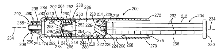
[0059] FIGS. 13-15 illustrate another embodiment of a syringe 200. FIG. 13 is
a cross-sectional
view of the syringe 200, illustrating an alternative embodiment of an
intermediate flow control plunger or
flow through valve plunger 202. In the illustrated embodiment of FIG. 13, the
syringe 200 may include a
primary plunger 204 and an elongated fluid container or syringe barrel 206
having an external fluid
coupling such as a luer fitting 208. The luer fitting 208 may be coupled to a
variety of fluid exchange or
delivery systems, which may include tubing, valves, gravity fed containers,
power injectors, electronic
controls, injection needles, and so forth. In addition, the embodiment of
FIGS. 13-15 may be coupled to
or generally associated with a radioisotope generator, a fluid dispensing
system, a power injector (e.g.,
motor, worm drive, radiation shield, etc.), a support structure, a rotatable
arm, a stand, an electronic
control unit, a computer, an imaging system, a diagnostic system, or a
combination thereof.
[0060] The primary plunger 204 includes a primary plunger head 210 coupled to
a pushrod 212.
For example, the primary plunger head 210 may be removably coupled to the
pushrod 212 via a variety
of fastening mechanisms, such as mating threads, snap fit mechanisms,
compression fit mechanisms,
or various tool free fasteners. In the illustrated embodiment, the primary
plunger head 210 may include
a generally cylindrical body 214 having a flat side 216 and an opposite curved
or conical side 218. In
addition, the primary plunger head 210 may include one or more outer seals,
such as a plurality of
sequential o-rings 220 and 222, disposed about the generally cylindrical body
214. The primary
plunger head 210 may include a removable fastening mechanism, such as an
internally threaded
member or female threads 224 extending inward from the flat side 216.
Similarly, the pushrod 212 may
include a removable fastening mechanism, such as an externally threaded member
or male threads
226, extending outwardly from a flat side 228. Thus, the primary plunger head
210 may be removably
coupled to the pushrod 212 by rotatingly driving the male threads 226 into the
female threads 224 until
the flat sides 216 and 228 may be generally flush with one another. In
addition, the pushrod 212 may
include an end member 230 disposed on an opposite end from the male threads
226. Similar to the
embodiment of FIGS. 1-12, the pushrod 212 may include a plurality of
lengthwise ribs 232, such as a
set of four lengthwise ribs, arranged symmetrically about a lengthwise or
central axis 234 of the primary
plunger 204. A plurality of measurement indicia 236 may be disposed along the
length of the pushrod
212 in a generally sequential offset arrangement.
12
CA 02607959 2007-11-06
WO 2006/124819 PCT/US2006/018806
[0061] As mentioned above, the syringe 200 of FIG. 13 may include one or more
floating valve
plungers or intermediate flow control plungers, such as the intermediate flow
control plunger 202. In
certain embodiments, the intermediate flow control plunger 202 may include a
generally central,
internal, or flow through check valve. In other words, the intermediate flow
control plunger 202 may be
configured to enable fluid to pass directly through rather than around the
intermediate flow control
plunger 202 in response to a pressure differential between opposite sides of
the intermediate flow
control plunger 202. In the illustrated embodiment, the intermediate flow
control plunger 202 may
include a fluid passage plunger insert 238 and a flexible plunger sleeve 240.
In certain embodiments,
the flexible plunger sleeve 240 may include a resilient, elastomeric, or
generally flexible material, while
the fluid passage plunger insert 238 may be generally rigid. In addition, the
fluid passage plunger 238
and the flexible plunger sleeve 240 may have generally circular or annular
geometries, which may be
disposed concentrically with respect to one another. Also, the intermediate
flow control plunger 202
may have a continuous outer seal, such as one or more o-rings, as discussed in
further detail below.
[0062] The illustrated fluid passage plunger insert 238 may include a
generally cylindrical body
portion 242 having an open end 244 and an opposing throat end 246. In
addition, the generally
cylindrical body portion 242 may include an annular groove 248 and a
protruding flange portion 250
disposed adjacent the open end 244. The throat end 246 may have a generally
tapered, inwardly
angled, or conical geometry, which includes one or more fluid passages. For
example, the throat end
246 may include axially offset passages 252, 254, which may be normally closed
or sealed by the
flexible plunger sleeve 240. In certain embodiments, the throat end 246 may
include fewer or greater
numbers of passages, such as 1, 3, 4, 5, 6, 7, 8, 9, 10, or more. These
passages, e.g., 252, 254,
enable fluid to flow directly through the interior of the intermediate flow
control plunger 202, rather than
around the periphery of the intermediate flow control plunger 202 at the seal
interface with the syringe
barrel 206. As illustrated, the axially offset passages 252, 254 may be
substantially covered and sealed
by a flexible mouth portion 256 of the flexible plunger sleeve 240. In other
words, the flexible mouth
portion 256 may be substantially or mostly closed across the throat end 246 of
the fluid passage
plunger insert 238 except for an opening therein (e.g., axial opening 258). As
illustrated, the axial
opening 258 may be disposed along the central axis 234, whereas the axially
offset passages 252, 254
may be disposed at a substantial distance or offset from the central axis 234.
[0063] The flexible plunger sleeve 240 includes a generally cylindrical body
260 having a plurality
of annular outer seals (e.g., o-ring portions 262, 264) and a generally
annular latch portion 266. In the
illustrated embodiment, the generally cylindrical body 260 of the flexible
plunger sleeve 240 may be
disposed concentrically about the generally cylindrical body portion 242 of
the fluid passage plunger
insert 238, such that the latch portion 266 may extend removably into the
annular groove 248. As
such, the fluid passage plunger insert 238 may be removably coupled or snap
fit with the flexible
plunger sleeve 240, such that the intermediate flow control plunger 202 may be
disassembled, cleaned,
and reused if desirable.
[0064] In certain embodiments, the fluid passage plunger insert 238 may be
molded, machined, or
generally manufactured with a variety of generally rigid materials, e.g.,
plastic. The flexible plunger
sleeve 240 may be molded or generally manufactured from a variety of flexible
or resilient materials,
13
CA 02607959 2007-11-06
WO 2006/124819 PCT/US2006/018806
such as rubber. As discussed in further detail below, the fluid passage
plunger insert 238 cooperates
with the flexible plunger sleeve 240 to at least substantially or entirely
separate fluids disposed on
opposite sides of the intermediate flow control plunger 202. Upon reaching or
passing a certain
pressure differential between opposite sides of the intermediate flow control
plunger 202, the flexible
plunger sleeve 240 may enable fluid flow directly through an interior of the
fluid passage plunger insert
238 rather than around the periphery of the intermediate flow control plunger
202.
[0065] As further illustrated in FIG. 13, the syringe barrel 206 includes an
interior surface 268
defining a generally cylindrical passageway and an exterior surface 270
exhibiting a generally
cylindrical geometry 270, which both extend lengthwise along the syringe
barrel 206 between a first end
272 and a second end 274 thereof. In certain applications, one or more of the
intermediate flow control
plungers 202 and the primary plunger 204 may be disposed lengthwise along the
interior surface 268
through an opening 276 at the first end 272 of the barrel 206. The plungers
202 and 204 may be offset
from one another and from the second end 274 of the barrel 206 to accommodate
two or more
substances or fluids. For example, a first medical fluid 278 may be disposed
between the intermediate
flow control plunger 202 and the second end 274 of the barrel 206. In
addition, a second medical fluid
280 may be disposed between the primary plunger head 210 and the second
intermediate flow control
plunger 202. In certain embodiments, the first medical fluid 278 may include a
radiopharmaceutical, a
contrast agent, a drug, or a combination thereof. By further example, the
second medical fluid 280 may
include a biocompatible flushing or cleaning substance, such as a heparin
solution, sterilized water, a
glucose solution, saline, or another suitable substance. The interspacing
between the one or more
secondary floating valve plungers or intermediate flow control plungers 202,
the primary plunger head
210, and the second end 274 of the barrel 206 may depend on the volume,
quantity, or dose of the first
medical fluid 278, the second medical fluid 280, and so forth.
[0066] The syringe barrel 206 may include a flow control actuator 282
extending inwardly (e.g.,
toward the axis 234) from the interior surface 268 of the barrel 206 near the
second end 274 thereof.
As discussed in further detail below, the flow control actuator 282 may engage
the outer periphery of
the intermediate flow control plunger 202, such that the flexible mouth
portion 256 may be forced
forward away from the throat end 246 to enable injection or general flow of
the second medical fluid
234. In other words, the first medical fluid 278 disposed in a first chamber
284 may be forced outwardly
through the luer fitting 208 in response to forward movement of the
intermediate flow control plunger
202. Upon reaching the flow control actuator 282, the flexible plunger sleeve
240 of the intermediate
flow control plunger 202 opens in a forward direction to enable the second
medical fluid 280 disposed in
a second chamber 286 to flow directly through the interior of the intermediate
flow control plunger 202
in response to axial movement of the primary plunger 204. Thus, the flow
control actuator 282 may be
described or defined as a plunger check valve actuator, which triggers or
actuates the transition of the
check valve 240 from a generally closed position to an open position enabling
flow through the interior
of the intermediate flow control plunger 202.
[0067] In the illustrated embodiment, the luer fitting 208 may include a male
luer 288 and a luer
collar 290. For example, the luer collar 290 may be disposed concentrically
about the male luer 288,
such that these components 288, 290 define an interspace 292 having one or
more removable
14
CA 02607959 2007-11-06
WO 2006/124819 PCT/US2006/018806
fastening mechanisms. By further example, the male luer 288 may include a
compression fitting or
tapered or external surface 294, while the luer collar 290 may include
internal threads 296. In certain
embodiments, the luer fitting 208 may include a flow control mechanism (e.g.,
a manual or electronic
valve) to open and close the fluid flow relative to the syringe 200. The luer
fitting 208 may include a
generally central fluid flow passage 298 extending through the male luer 288
along the axis 234.
[0068] FIG. 14 is a partial cross-sectional view of the syringe 200 of FIG.
13, further illustrating a
first injection of the medical fluid 278 from the syringe 200 immediately
prior to an injection transition or
intermediate position between multiple/sequential injections of the medical
fluids 278, 280. The first
injection of the medical fluid 278 is represented by arrows 300. Specifically,
the illustrated syringe 200
can permit passage of the fist medical fluid 278 (e.g., a radiopharmaceutical
or contrast agent) followed
by the second medical fluid 280 (e.g., a biocompatible flush) through the
central fluid flow passage 298
of the luer fitting 208 via the intermediate flow control plunger 202. In the
illustrated embodiment, the
intermediate flow control plunger 202 may be abutted against the flow control
actuator 282 after
discharging the first medical fluid 278. The first medical fluid 278 may be
discharged from the first
chamber 284 between the intermediate flow control plunger 202 and the second
end 274 of the syringe
barrel 206 by depressing the primary plunger 204 lengthwise along the axis
234. As the primary
plunger 204 moves lengthwise along the syringe barrel 206, the flexible
plunger sleeve 240 remains
sealed against the fluid passage plunger insert 238 due to the pressure
differential between the first
and second chambers 284, 286. Upon reaching the flow control actuator 282, the
intermediate flow
control plunger 202 may become stationary to actuate the flexible plunger
sleeve 240.
[0069] In other words, the flexible plunger sleeve 240 may remain closed or
sealed with the fluid
passage plunger insert 238 as long as the intermediate flow control plunger
202 is capable of moving in
response to a pressure differential between the first and second cavities or
chambers 284, 286. As
such, the movement of the intermediate flow control plunger 202 maintains a
fluid pressure balance
between the first and second chambers 284, 286, such that the seal is
maintained by the flexible
plunger sleeve 240. When movement is no longer possible at the flow control
actuator 282, the force or
pressure of the second medical fluid 280 disposed in the second chamber 286
overcomes the flexible
plunger sleeve 240 to enable discharge of the second medical fluid 280. At
this stage, the primary
plunger 204 moves lengthwise along the syringe barrel 206 while the
intermediate flow control plunger
202 remains stationary.
[0070] FIG. 15 is a partial cross-sectional view of the syringe 200 of FIGS.
13-14, further
illustrating actuation of the intermediate flow control plunger 202 at the
flow control actuator 282. As
illustrated, the flexible mouth portion 256 of the flexible plunger sleeve 240
is disposed at an offset
away from the throat end 246 of the fluid passage plunger insert 238. In other
words, a gap 302, may
exist between the flexible mouth portion 256 and the throat end 246. In this
generally unrestricted
configuration, the second medical fluid 280 disposed between the primary
plunger head 210 and the
intermediate flow control plunger 202 may be forced through the passages 252,
254, the gap 302, and
out through the central fluid flow passage 298 as illustrated by arrows 304,
306, and 308, respectively.
In certain embodiments, as discussed above, the second medical fluid 280 may
include a biocompatible
flushing fluid, such as a heparin solution, sterilized water, a glucose
solution, saline, or another suitable
CA 02607959 2007-11-06
WO 2006/124819 PCT/US2006/018806
medical fluid. Accordingly, the second fluid injection or discharge may serve
to substantially flush out or
clean the various passages and interior portions of the syringe 200.
[0071] In certain embodiments, the syringes illustrated and described above
with reference to
FIGS. 1-15 may be filled or pre-filled with one or more medical fluids, such
as contrast agents,
radiopharmaceuticals, tagging agents, biocompatible flushes, or combinations
thereof. For example,
the disclosed syringes, e.g., 20, 110, 130, 170, and 200 may be filled or pre-
filled with a first medical
fluid in a first chamber and a second medical fluid in a second chamber. The
first medical fluid may
include a contrast agent for medical imaging, such as magnetic resonance
imaging (MRI), computed
tomography (CT), radiography (e.g., x-ray), or ultrasound. Alternatively, the
first medical fluid may
include a radioisotope or radiopharmaceutical for radiation-based treatment or
medical imaging, such
as positron emission tomography (PET) or single photon emission computed
tomography (SPECT). In
addition, the second medical fluid may include a biocompatible flush, such as
heparin solution,
sterilized water, glucose solution, saline, or another suitable substance. The
disclosed syringes may be
used to inject the first and second medical fluids one after another into a
subject or patient.
Alternatively, the disclosed multi-chamber, multi-stage, or sequential
injection syringes, e.g., 20, 110,
130, 170, and 200 may be filled or pre-filled with a single medical fluid,
such as a radiopharmaceutical
or a contrast agent.
[0072] In certain embodiments, the subject (e.g., patient) may be scanned or
generally imaged by
a suitable medical diagnostic and/or imaging system, such as listed above. For
example, after the
radiopharmaceutical enters the blood stream and focuses on a particular organ
or area of interest, the
diagnostic and/or imaging system may function to acquire imaging data, process
the data, and output
one or more images. Thus, the diagnostic and/or imaging system may include
detector/acquisition
hardware and software, data/image processing hardware and software, data/image
storage hardware
and software, a display, a printer, a keyboard, a mouse, a computer
workstation, a network, and other
associated equipment.
[0073] FIG. 16 is a flowchart illustrating an embodiment of a method of use or
syringe preparation
process 350 utilizing one or more of the multi-chamber, multi-stage, or
sequential injection syringes,
e.g., 20, 110, 130, 170, and 200, of FIGS. 1-15. As illustrated, the process
350 may include filling a first
chamber of a syringe with a first medical fluid (block 352). For example, the
first medical fluid may
include a radiopharmaceutical or a contrast agent. The process 350 may then
include separating the
first chamber from a second chamber of the syringe with an intermediate
plunger having a flow-through
check valve (block 354). For example, the intermediate plunger may include the
intermediate flow
control plunger 30 of FIGS. 1-12 or the intermediate flow control plunger 202
of FIGS. 13-15. The
process 350 also may include filling the second chamber of the syringe with a
second medical fluid
(block 356). For example, the second medical fluid may include a biocompatible
flush, such as heparin
solution, sterilized water, glucose solution, saline, or another suitable
substance. In addition, the
process 350 may include closing the syringe with a primary plunger disposed
about the second
chamber opposite from the intermediate plunger (block 358).
16
CA 02607959 2007-11-06
WO 2006/124819 PCT/US2006/018806
[0074] FIG. 17 is a flowchart illustrating an embodiment of a diagnostic
imaging process 360
utilizing one or more of the multi-chamber, multi-stage, or sequential
injection syringes, e.g., 20, 110,
130, 170, and 200, as illustrated in FIGS. 1-15. As illustrated, the process
360 may include detecting a
medical fluid administered to a subject (e.g., a patient) from a sequential
injection syringe having a flow-
through check valve (block 362). The detection may include a variety of
imaging modalities. The
medical fluid may enable detection, or enhance detection, or tag a particular
organ, or otherwise
improve the imaging detection of a particular area of interest in the patient.
For example, the syringe
filled in the process 350 of FIG. 16 may be used to inject a subject with a
radiopharmaceutical, a
contrast agent, or another substance. By further example, one of the multi-
chamber, multi-stage, or
sequential injection syringes, e.g., 20, 110, 130, 170, and 200, as
illustrated with reference to FIGS. 1-
15 may be used to inject a radiopharmaceutical or a contrast agent into a
subject. As discussed above,
a contrast agent may be used for medical imaging, such as magnetic resonance
imaging (MRI),
computed tomography (CT), radiography (e.g., x-ray), or ultrasound.
Alternatively, a radioisotope or
radiopharmaceutical may be used for radiation-based treatment or medical
imaging, such as positron
emission tomography (PET) or single photon emission computed tomography
(SPECT). At block 364,
the process 360 may include processing data associated with the medical fluid
in the subject. The
process 360 also may include outputting an image of the subject associated
with the medical fluid in the
subject (block 366). Again, the foregoing procedures and resulting image
directly benefit from the one
or more medical fluids (e.g., radiopharmaceutical or contrast agent)
administered with. the multi-
chamber, multi-stage, or sequential injection syringes, e.g., 20, 110, 130,
170, and 200, as illustrated
and described with reference to FIGS. 1-15
[0075] FIG. 18 is a flowchart illustrating an exemplary nuclear medicine
process utilizing one or
more of the multi-chamber, multi-stage, or sequential injection syringes,
e.g., 20, 110, 130, 170, and
200, as illustrated with reference to FIGS. 1-15. As illustrated, the process
410 begins by providing a
radioactive isotope for nuclear medicine at block 412. For example, block 412
may include eluting
technetium-99m from a radioisotope generator as discussed in further detail
below. At block 414, the
process 410 proceeds by providing a tagging agent (e.g., an epitope or other
appropriate biological
directing moiety) adapted to target the radioisotope for a specific portion,
e.g., an organ of a patient. At
block 416, the process 410 then proceeds by combining the radioactive isotope
with the tagging agent
to provide a radiopharmaceutical for nuclear medicine. In certain embodiments,
the radioactive isotope
may have natural tendencies to concentrate toward a particular organ or tissue
and, thus, the
radioactive isotope may be characterized as a radiopharmaceutical without
adding any supplemental
tagging agent. At block 418, the process 410 may then involve filling a
syringe with the
radiopharmaceutical and another medical fluid in sequential first and second
chambers, as discussed in
detail above. For example, block 418 may include the process 350 of FIG. 16,
and may include filling
one of the multi-chamber, multi-stage, or sequential injection syringes, e.g.,
20, 110, 130, 170, and 200,
as illustrated with reference to FIGS. 1-15. At block 420, the process 410
then may proceed by
injecting the radiopharmaceutical into a patient from the first chamber of the
syringe. At block 422, the
process 410 may continue by injecting the other medical fluid into the patient
from the second chamber
of the syringe. Again, the other fluid may include a biocompatible flush or
another selected medical
17
CA 02607959 2007-11-06
WO 2006/124819 PCT/US2006/018806
fluid. After a pre-selected time, the process 410 proceeds by
detecting/imaging the
radiopharmaceutical tagged to the patient's organ or tissue (block 424). For
example, block 424 may
include using a gamma camera or other radiographic imaging device to detect
the radiopharmaceutical
disposed on or in or bound to tissue of a brain, a heart, a liver, a tumor, a
cancerous tissue, or various
other organs or diseased tissue.
[0076] FIG. 19 is a block diagram of an exemplary system 426 for providing one
or more of the
multi-chamber, multi-stage, or sequential injection syringes, e.g., 20, 110,
130, 170, and 200, as
illustrated in FIGS. 1-15 with one or more medical fluids (e.g.,
radiopharmaceutical and biocompatible
flush) for use in a nuclear medicine application. As illustrated, the system
426 may include a
radioisotope elution system 428 having a radioisotope generator 430, an eluant
supply container 432,
and an eluate output container or dosing container 434. In certain
embodiments, the eluate output
container 434 may be evacuated (in vacuum), such that the pressure
differential between the eluant
supply container 432 and the eluate output container 434 facilitates
circulation of an eluant (e.g., saline)
through the radioisotope generator 430 and out through an eluate conduit into
the eluate output
container 434. As the eluant (e.g., a saline solution) circulates through the
radioisotope generator 430,
the circulating eluant generally washes out or elutes a radioisotope (e.g.,
Technetium-99m). For
example, one embodiment of the radioisotope generator 430 includes a radiation
shielded outer casing
(e.g., lead shell) that encloses a radioactive parent, such as molybdenum-99,
adsorbed to the surfaces
of beads of alumina or a resin exchange column. Inside the radioisotope
generator 430, the parent
molybdenum-99 transforms, with a half-life of about 67 hours, into metastable
technetium-99m. The
daughter radioisotope (e.g., technetium-99m) is generally held less tightly
than the parent radioisotope
(e.g., molybdenum-99) within the radioisotope generator 430. Accordingly, the
daughter radioisotope
can be extracted or washed out with a suitable eluant, such as an oxidant-free
physiologic saline
solution. The eluate output from the radioisotope generator 430 into the
eluate output container 434
generally includes the eluant and the washed out or eluted radioisotope from
within the radioisotope
generator 430. Upon receiving the desired amount of eluate within the eluate
output container 434, a
valve may be closed to stop the eluant circulation and output of eluate. As
discussed in further detail
below, the extracted daughter radioisotope can then, if desired, be combined
with a tagging agent to
facilitate diagnosis or treatment of a patient (e.g., in a nuclear medicine
facility).
[0077] As further illustrated in FIG. 19, the system 426 also includes a
radiopharmaceutical
production system 436, which functions to combine a radioisotope 438 (e.g.,
technetium-99m solution
acquired through use of the radioisotope elution system 428) with a tagging
agent 440. In some
embodiment, this radiopharmaceutical production system 436 may refer to or
include what are known in
the art as "kits" (e.g., TechnescanTM kit for preparation of a diagnostic
radiopharmaceutical). Again, the
tagging agent may include a variety of substances that are attracted to or
targeted for a particular
portion (e.g., organ, tissue, tumor, cancer, etc.) of the patient. As a
result, the radiopharmaceutical
production system 436 produces or may be utilized to produce a
radiopharmaceutical including the
radioisotope 438 and the tagging agent 440, as indicated by block 442. The
illustrated system 426 may
also include a radiopharmaceutical dispensing system 444, which facilitates
extraction of the
radiopharmaceutical into a syringe 446 having an intermediate plunger with a
flow-through check valve.
18
CA 02607959 2007-11-06
WO 2006/124819 PCT/US2006/018806
In the illustrated embodiment, the syringe may be one of the multi-chamber,
multi-stage, or sequential
injection syringes, e.g., 20, 110, 130, 170, and 200, as illustrated and
described above with reference to
FIGS. 1-15. Thus, the system 426 also may fill the syringe with an additional
medical fluid, such as a
biocompatible flush. For example, the multi-chamber, multi-stage, or
sequential injection syringes, e.g.,
20, 110, 130, 170, and 200, of FIGS. 1-15 may be filled with a
radiopharmaceutical and a biocompatible
flush in sequential chambers separated by the intermediate flow control
plunger, e.g., 30 or 202. In
certain embodiments, the various components and functions of the system 426
may be disposed within
a radiopharmacy, which prepares the syringe 446 of the radiopharmaceutical for
use in a nuclear
medicine application. For example, the syringe 446 may be prepared and
delivered to a medical facility
for use in diagnosis or treatment of a patient.
[0078] FIG. 20 is a block diagram of an exemplary nuclear medicine imaging
system 448 utilizing
the multi-chamber, multi-stage, or sequential injection syringe 446 of
radiopharmaceutical provided
using the system 426 of FIG. 19. As illustrated, the nuclear medicine
imagining system 448 includes a
radiation detector 450 having a scintillator 452 and a photo detector 454. In
response to radiation 456
emitted from a tagged organ within a patient 458, the scintillator 452 emits
light that is sensed and
converted to electronic signals by the photo detector 454. Although not
illustrated, the imaging system
448 also can include a collimator to collimate the radiation 456 directed
toward the radiation detector
450. The illustrated imaging system 448 also includes detector acquisition
circuitry 460 and image
processing circuitry 462. The detector acquisition circuitry 460 generally
controls the acquisition of
electronic signals from the radiation detector 450. The image processing
circuitry 462 may be
employed to process the electronic signals, execute examination protocols, and
so forth. The illustrated
imaging system 448 also includes a user interface 464 to facilitate user
interaction with the image
processing circuitry 462 and other components of the imaging system 448. As a
result, the imaging
system 448 produces an image 466 of the tagged organ within the patient 458.
Again, the foregoing
procedures and resulting image 466 directly benefit from the one or more
medical fluids (e.g.,
radiopharmaceutical) administered with the multi-chamber, multi-stage, or
sequential injection syringes,
e.g., 20, 110, 130, 170, and 200, as illustrated and described with reference
to FIGS. 1-15.
[0079] When introducing elements of various embodiments of the present
invention, the articles
"a", "an", "the", and "said" are intended to mean that there are one or more
of the elements. The terms
"comprising", "including", and "having" are intended to be inclusive and mean
that there may be
additional elements other than the listed elements. Moreover, the use of
"top", "bottom", "above",
"below" and variations of these terms is made for convenience, but does not
require any particular
orientation of the components.
[0080] While the invention may be susceptible to various modifications and
alternative forms,
specific embodiments have been shown by way of example in the figures and have
been described in
detail herein. However, it should be understood that the invention is not
intended to be limited to the
particular forms disclosed. Rather, the invention is to cover all
modifications, equivalents, and
alternatives failing within the spirit and scope of the invention as defined
by the following appended
claims.
19