Note: Descriptions are shown in the official language in which they were submitted.
CA 02608017 2007-11-09
WO 2006/124068
PCT/US2005/043144
Atty. Docket No.: 9469.19606-AB PCT
- 1 -
,
SYSTEMS FOR ELECTRICAL STIMULATION OF NERVES
IN ADIPOSE TISSUE REGIONS
Field of the Invention
This invention relates to systems and methods for
electrical stimulation on nerves in adipose tissue
regions to provide functional and/or therapeutic
outcomes.
Background of the Invention
I. Neuromodulation Stimulation
Neuromodulation stimulation (the electrical
excitation of nerves, often afferent nerves, to
indirectly affect the stability or performance of a
physiological system) can provide functional and/or
therapeutic outcomes. While existing systems and methods
can provide remarkable benefits to individuals requiring
neuromodulation stimulation, many limitations and issues
still remain. For example, existing systems can often
require the user to wear an external stimulator, which
may provide a positive functional outcome, but may also
negatively affect quality of life issues.
A variety of products and treatment methods are
available for neuromodulation stimulation. As an example,
neuromodulation stimulation has been used for the
treatment of sexual dysfunction, which affects both men
and women. A wide range of options exist for the
restoration of sexual function. Treatments include
CA 02608017 2007-11-09
WO 2006/124068
PCT/US2005/043144
Atty. Docket No.: 9469.19606-AB PCT
- 2 -
everything from medications, simple mechanical devices,
psychological counseling, external stimulators, and
surgically implanted devices.
Both external and implantable devices are available
for the purpose of neuromodulation stimulation for the
restoration of sexual function. The operation of these
devices typically includes the use of an electrode placed
either on the external surface of the skin or a
surgically implanted electrode. Although these modalities
have shown the ability to provide a neuromodulation
stimulation with positive effects, they have received
limited acceptance by patients because of their
limitations of portability, limitations of treatment
regimes, and limitations of ease of use and user control.
II. Sexual Dysfunction
One form of male sexual dysfunction is know as
Erectile Dysfunction (ED), and is often referred to as
"impotency." There are some common diseases such as
diabetes, Peyronie's disease, heart disease, and prostate
cancer that are associated with impotency or have
treatments that may cause impotency. And in some cases
the cause may be psychological.
Erectile Dysfunction is common problem affecting men
and is defined as the inability to achieve or maintain a
penile erection sufficient for sexual activity. It is
estimated that 35% to 50% of all men aged '40 to 70 have
some form of ED, nearly 46 million Americans have ED, and
over 150 million men have ED worldwide. It is also
estimated that sexual dysfunctions occur in 43 percent of
women in the United States. It would cost $3.5 billion
per year if only one fifth of Americans with ED were
treated with the first line of treatment (oral therapy
such as PDE-5 inhibitors), and the cost for the second
line of treatment (such as injection or transurethral
administration of alprostadil) is approximately twice as
CA 02608017 2007-11-09
WO 2006/124068
PCT/US2005/043144
Atty. Docket No.: 9469.19606-AB PCT
- 3 -
expensive. A cost-effective therapy is needed because the
number of men seeking treatment tripled between 1997 and
2000 and is expected to increase as awareness of
treatment options for ED becomes more widespread.
The severity of erectile dysfunction can range from
1) mild ED, in which a man is occasionally unable to
achieve and sustain an erection sufficient for
intercourse, to 2) frequent or moderate ED to 3) severe
or complete ED, in which a man is never able to produce
and sustain an erection sufficient for intercourse. The
prevalence of moderate to complete ED increases with age.
Approximately 20% of men aged 40 years have moderate to
severe ED and approximately 70% of men aged 70 years have
moderate to severe ED. Over 70% of men with ED report
that their quality of life is moderately to severely
reduced by ED, and over 70% of men with ED feel hurt by
the response of their partner to their ED and feel "to
some extent a failure" because of their ED. Thus, ED is
often associated with poor self-image, depression, and it
can affect interpersonal relationships and lead to
increased mental stress.
ED is often a result of a combination of
psychological and organic factors, but it is thought to
be purely psychological in origin in less than 30% of the
cases. Organic factors can include complications from
neurologic diseases (stroke, multiple sclerosis,
Alzheimer's disease, brain or spinal tumors), chronic
renal failure, prostate cancer, diabetes, trauma,
surgery, medications, and abnormal structure. However,
most cases of ED are associated with vascular diseases.
An erection cannot be sustained without sufficient blood
flow into and entrapment within the erectile bodies of
the penis, and vascular related ED can be due to a
malfunction of either the arterial or the venous system.
In a healthy individual, penile erection is
CA 02608017 2007-11-09
WO 2006/124068
PCT/US2005/043144
Atty. Docket No.: 9469.19606-AB PCT
- 4 -
generated by increased blood low into the penis via
arterial dilation and decreased blood flow from the penis
via venous occlusion. Arterial dilation is generated by
activation of the cavernous nerve (a parasympathetic
nerve), which causes relaxation of corporeal smooth
muscle of the cavernosal and trabecular spaces. Penile
erection begins with the filling and expansion of the
three erectile bodies: the corpus spongiosum and the two
corpora cavernosa. This expansion compresses the venules,
preventing blood from leaving the penis and furthering
the erection.
Persons with vasculogenic erectile dysfunction are
unable to achieve penile erection due to either
insufficient arterial blood flow or insufficient venous
occlusion or both. Normal reflex erection coordinates
dilation of penile blood vessels, auymenting vascular
filling, and venous occlusion, preventing leakage and
increasing penile stiffness.
Stimulation of a target nerve N, such as the dorsal
nerve of the penis (DNP) afferents activates spinal
circuitry that coordinates efferent activity in the
cavernous nerve (CN), increasing filling via dilation of
penile arteries, and efferent activity in the pudendal
nerve (PN), preventing leakage via occlusion of penile
veins, producing a sustained reflex erection (see Fig.
1).
Figs. 2 and 3 show a profile and cross-section of
the penis, illustrating the anatomical relationship of
the erectile tissue (corpora cavernosa and corpus
spongiosum) inside the penis. Figs. 4 and 5 show the
physiological changes in the size of the penile arteries,
erectile tissue, and veins during erection. Fig. 4 shows
the penile arteries constricted, the erectile tissue
collapsed, and the veins open prior to an erection.
Arterial dilation leads to increased inflow of blood,
CA 02608017 2007-11-09
WO 2006/124068
PCT/US2005/043144
Atty. Docket No.: 9469.19606-AB PCT
- 5 -
which fills and expands the erectile tissue as the veins
are compressed to decrease outflow of blood from the
erectile tissue, as shown in Fig. 5.
III. Methods of Treatment For ED
Methods of treatment for erectile dysfunction are
available but are either often discontinued due to loss
of efficacy or side effects or reserved as a final
recourse requiring irrevocable damage. Three lines of
treatment exist for ED. Oral therapy (PDE-5 inhibitors)
is usually the first line of treatment, and it can be
effective in up to 70% of men when it is first
administered, but half of the patients stop taking PDE-5
inhibitors because they lose their effectiveness within
one to three years. The second line of treatment is
usually a minimally invasive therapy such as a vacuum
device or direct administration of a vasoactive agent.
The second-line treatments are usually effective in 33%
to 70% of men, but they are also later discontinued by
over half of the patients, often due to side effects such
as pain or local damage at the site of administration.
For the 30% to 65% of men who fail or discontinue oral
therapy, the total cost for the second line of treatment
(vacuum device or alprostadil, administered via injection
or transurethrally) would be $1 to $6 billion. However,
side effects of pain and local damage are associated with
the second line of treatment, and at least half of the
men discontinue this form of therapy. If the men who
failed or discontinued both the first and second lines of
treatment chose to receive a penile prosthesis, the total
cost would be over $20 billion. Yet, implantation of a
penile prosthesis is reserved for the final method of
treatment because the implantation causes permanent
(irrevocable) damage to the erectile tissue resulting in
the loss of any future erection if the implant is
removed. Thus, an alternative approach is needed that can
CA 02608017 2007-11-09
WO 2006/124068
PCT/US2005/043144
Atty. Docket No.: 9469.19606-AB PCT
- 6 -
provide a multitude of advantages over the current
therapies.
IV. Neuromodulation Stimulation to Evoke Erection
Systemic side effects (headache, flushing,
dyspepsia, etc.) and permanent damage to the corpora
cavernosa may be avoided by electrically stimulating a
peripheral nerve to activate a reflex that coordinates
arterial dilation with venous occlusion, producing an
erection. In anesthetized, spinalized rats, electrical
stimulation of afferent pathways in the dorsal nerve of
the penis (DNP) can produce an increase in corpus
cavernous pressure (CCP). The increase in CCP is gated to
the onset and offset of stimulation and has been
sustained for up to fifteen minutes. Previous results in
the dog demonstrated that reflex erections are repeatable
for a period of three to five hours. Stimulation of the
DNP leads to transient increases in the EMG activity of
the ischiocavernosus (IC) and bulbospongiosus (BS)
muscles, which are responsible for venous occlusion.
Venous occlusion prevents leakage of blood from the penis
and explains why DNP stimulation can evoke supra-systolic
increases in penile pressure. These animal experiments
demonstrate that DNP stimulation can evoke a reflex
erection, but they do not determine if the reflex
erection is comparable to the erections evoked by the
present treatment methods.
An implantable stimulation system is needed that can
provide an erection quickly and is acceptable to men who
use or may need to use nitrates to treat cardiovascular
disease because over 35% of men with cardiovascular
disease develop ED. The loss of efficacy of oral therapy
is likely due to the long duration (four to eighteen
hours) of action, and the consistently elevated drug
concentrations can reduce the response to the drug via
tachyphylaxis or increased tolerance as seen with
CA 02608017 2013-07-16
66742-1226
- 7 -
nitroglycerin tolerance. No loss of efficacy is expected
with an implantable stimulation system that will only be
activated five minutes before and during erection, and it
will provide controlled release of neurotransmitter via
activation of a reflex in the central nervous system.
The implantable stimulation system may be activated
by the movement of a magnet over a magnetic reed switch
within the implantable pulse generator of the stimulation
system, or the press of a remote button, for example.
Unlike the second line of treatment, this approach will
not require a constrictive ring, needle insertion, or
urethral-suppository insertion, which can cause local
injury prior to each erection and lead to discontinuation
of treatment. In contrast to the penile implant, an
implantable stimulation system approach will not damage
the erectile tissue.
There remains a need for systems and methods that
can effectively restore sexual function, in a
straightforward manner, without requiring drug therapy
and complicated (and in some instanced irrevocable)
surgical procedures.
Summary of the Invention
One aspect of the invention provides systems and
methods for the treatment of sexual dysfunction by the
stimulation of the left and/or right branches of the
dorsal genital nerves using a stimulation electrode sized
and configured to be implanted in tissue in a region at
or near a pubic symphysis, and an implantable pulse
generator to convey electrical stimulation waveforms to
the stimulation electrode to stimulate the left branch
and/or the right branch of the dorsal genital nerves.
In some embodiments, the stimulation waveforms
conveyed to the stimulation electrode may affect afferent
stimulation of the left and/or right branches of the
dorsal genital nerves, the afferent stimulation
activating spinal circuitry that
CA 02608017 2013-07-16
66742-1226
=
-8 -
coordinates efferent activity in the cavernous nerve and
efferent activity in the pudendal nerve, producing a
sexual funption. The .electrical. stimulatibn waveform
includes at least a variable frequency component and/or a
variable duty cycle component and/or a variable amplitude
Component and/or a variable pause component to ward off
habituation. For example, the electrical stimulation
waveform may include 4 variable frequency. in the range of
about one Hz to about fifteen Hz,and a -.:variable
amplitude in the range of about 10.0:miOroampS to about 20
. .
milliamps, and.a variable duty cycle'in.:the.range of
about zero seconds to about ten seconds, and a variable
pause component in the range of about zero seconds to
about ten seconds.
An additional aspect of the invention provides
systems and methods for a stimulation electrode assembly
sized and configured for-placement in an adipose tissue
region to stimulate a nerve in the adipose tissue region.
The stimulation electrode assembly includes an elongated
lead sized and configured to be implanted within the
adipose tissue region, the lead ,including at least two
electrically conductive. portions to apply electrical
stimulation to nerve tissue in the adipose tissue region.
Each electrically conductive portion may comprise a
conductive surface area in the range of about 10 mm2 to
about 20 mm2. The at least two electrically conductive
portions can be configured to function as two individual
stimulating electrodes in a monopolar configuration or as
one stimulating electrode in ,a bipolar configuration.
In some embodiments, the lead also may include at
least two expandable anchoring structures deployable from
the lead .to engage adipose tissue and resist dislodgement
and/or migration of the at least two electrically
conductive portions within the adipose tissue region.
Each expandable anchoring structure may include two
circumferentially spaced-apart,
CA 02608017 2013-07-16
66742-1226
- 9 -
radiating shovel-like blade shaped members. These two shovel-
like blade shaped members may be spaced 180 degrees apart and
the at least two expandable anchoring structures may be
spaced 90 degrees apart.
According to a further aspect of the invention, there
is provided use of bilateral electrical stimulation of a left
branch and a right branch of a dorsal genital nerve of a
patient via at least one stimulation electrode located at a
target site at a pubic symphasis of the patient to treat sexual
dysfunction of the patient.
According to still a further aspect of the invention,
there is provided a device for treating sexual dysfunction
comprising: at least one stimulation electrode sized and
configured to be implanted in a tissue region at a target site
at a pubic symphasis of a patient, the at least one stimulation
electrode configured to provide bilateral electrical
stimulation of a left branch and a right branch of a dorsal
genital nerve of the patient.
CA 02608017 2013-07-16
66742-1226
- 9a -
Other features and advantages of the inventions are
set forth in the following specification and attached
drawings.
Brief Description of the Drawings =
Fig. 1 is a schematic view of the stimulation of a
target afferent nerve and the spinal circuitry activated
to coordinate efferent nerve activity for sexual
restoration.
Fig. 2 is a lateral cross-sectional view of a penis,
showing the relationship of the erectile. tissue inside
the penis.
Fig. 3 is an end section view of the penis taken
generally along line 3-3 of Fig. 2.
Fig. 4 is a side sectional view of penile tissue
prior to an erection.
Fig. 5 is a side sectional view of penile tissue as
= shown in Fig. 4, showing the changes in the penile tissue
causing an erection.
Fig. 6 is a view of a stimulation assembly that
provides electrical stimulation to central nervous system.
tissue, muscles and/or nerves inside the body using a
general purpose implantable pulse generator.
Figs. TA and 7B are front and side views of the,
general purpose implantable pulse generator shown in Fig.
6, which is powered by a primary battery. '
Figs. BA and 8B are anterior anatomic views of the
system shown in Fig. 6 after implantation in an adipose
tissue region at or near near the pubic symphysis.
Fig. 8C is an anterior anatomic view of an
alternative configuration of the system shown in Fig. 8B,
showing more than one lead and electrode implanted in the.
=
CA 02608017 2007-11-09
W02006/124068
PCT/US2005/043144
Atty. Docket No.: 9469.19606-AB PCT
- 10 -
targeted tissue region.
Fig. 9 is an anterior anatomic view of the pelvic
girdle in a human.
Fig. 10 is a lateral section view of the pelvic
girdle region shown in Fig. 9.
Fig. 11 is an inferior view of a female pelvic
girdle region.
Fig. 12 is an anatomic view showing the implantable
pulse generator shown in Figs. 7A and 7B in association
with an external programmer that relies upon wireless
telemetry, and showing the programmer's capability of
communicating with the implantable pulse generator up to
an arm's length away from the implantable pulse
generator.
Fig. 13 is a system view of an implantable pulse
generator system incorporating a clinician programmer
derivative and showing the system's capability of
communicating and transferring data over a network,
including a remote network.
Fig. 14 is a perspective graphical view of one
possible type of patient controller that may be used with
the implantable pulse generator shown in Figs. 7A and 7B.
Fig. 15 is a block diagram of a circuit that the
implantable pulse generator shown in Figs. 7A and 7B can
incorporate.
Fig. 16 is a circuit diagram showing a possible
circuit for the wireless telemetry feature used with the
implantable pulse generator shown in Figs. 7A and 7B.
Fig. 17 is a circuit diagram showing a possible
circuit for the stimulus output stage and output
multiplexing features used with the implantable pulse
generator shown in Figs. 7A and 7B.
Fig. 18 is a graphical view of a desirable biphasic
stimulus pulse output of the implantable pulse generator
for use with the system shown in Fig. 6.
CA 02608017 2007-11-09
WO 2006/124068
PCT/US2005/043144
Atty. Docket No.: 9469.19606-AB PCT
- 11 -
Fig.-19 is a circuit diagram showing a possible
circuit for the microcontroller used with the implantable
pulse generator shown in Figs. 7A and 7B.
Fig. 20 is a circuit diagram showing one possible
option for a power management sub-circuit where the sub-
circuit includes MOSFET isolation between the battery and
charger circuit (when used), the power management sub-
circuit being a part of the implantable pulse generator
circuit shown in Fig. 6.
Fig. 21 is a circuit diagram showing a second
possible option for a power management sub-circuit where
the sub-circuit does not include MOSFET isolation between
the battery and charger circuit (when used), the power
management sub-circuit being a part of the implantable
pulse generator circuit shown in Fig. 6.
Fig. 22 is a circuit diagram showing a possible
circuit for the VHH power supply feature used with the
implantable pulse generator shown in Figs. 7A and 7B.
Figs. 23 and 24 are anatomic section views of the
adipose tissue region with one lead and electrode
associated with the system shown in Fig. 6, after having
been implanted.
Figs. 25A and 25E are perspective views of the lead
and electrode associated with the system shown in Fig. 6.
Fig. 26 is a side interior view of a representative
embodiment of a lead of the type shown in Figs. 23 and
24.
Fig. 27 is an end section view of the lead taken
generally along line 27-27 in Fig. 26.
Fig. 28 is an elevation view, in section, of a lead
and electrode of the type shown in Figs. 23 and 24
residing within an introducer sheath for implantation in
a targeted tissue region, the anchoring members being
shown retracted within the sheath.
Fig. 29 is a perspective view of a molded cuff
CA 02608017 2007-11-09
WO 2006/124068 PCT/US2005/043144
Atty. Docket No.: 9469.19606-AB PCT
- 12 -
electrode prior to implantation.
Fig. 30 is a perspective view of an alternative
embodiment of the molded cuff electrode shown in Fig. 29,
showing the lead extending generally parallel from the
cuff electrode.
Figs. 31 and 32 are plan views showing both solid
and seymented embodiments for the electrically conductive
surface.
Fig. 33 is a perspective, diagrammatic view of the
molded cuff electrode shown in Fig. 29 implanted about a
nerve and coupled to a pulse generator to deliver a
neuromodular stimulation to achieve a desired therapeutic
result.
Fig. 34 is a side section view of the molded cuff
electrode taken generally along line 34-34 on Fig. 33.
Fig. 35 is a plan view of an alternative embodiment
of the conductive surfaces configuration.
Fig. 36 is a side section view of the alternative
embodiment shown in Fig. 35 positioned about a nerve N.
Fig. 37 is an applicator tool for placement of a
molded cuff electrode of the type shown in Fig. 29 about
a nerve, the applicator tool being shown before mounting
of the electrode with the electrode delivery mechanism in
an aft condition.
Fig. 38 is a side view of the applicator tool shown
in Fig. 37, with the electrode mounted and the electrode
delivery mechanism in an aft condition, ready to implant
the electrode about a nerve.
Fig. 39 is a side view of the applicator tool shown
in Fig. 37, with the electrode delivery mechanism =
translated to a forward condition to implant the
electrode about a nerve.
Fig. 40 is a plane view of a system of surgical
tools that can be use to implant the system shown in Fig.
6.
CA 02608017 2007-11-09
WO 2006/124068
PCT/US2005/043144
Atty. Docket No.: 9469.19606-AB PCT
- 13 -
Figs. 41 through 44 illustrate general steps of
implanting the system shown in Fig. 6 in either a single
surgical procedure or two surgical procedures.
The invention may be embodied in several forms
without departing from its spirit or essential
characteristics. The scope of the invention is defined in
the appended claims, rather than in the specific
description preceding them. All embodiments that fall
within the meaning and range of equivalency of the claims
are therefore intended to be embraced by the claims.
Description of the Preferred Embodiments
The various aspects of the invention will be
described in connection with the restoration of sexual
function (e.g., erectile restoration) by the unilateral
or bilateral stimulation of the left and/or right
branches of the dorsal genital nerves using a lead or
leads implanted in adipose or other tissue in the region
at or near the pubic symphysis, or electrode(s) implanted
on the left and/or right branches of the dorsal genital
nerves. That is because the features and advantages of
the invention are well suited for this purpose. Still, it
should be appreciated that the various aspects of the
invention can be applied in other forms and in other
locations in the body to achieve other objectives as
well. These objectives pertain to both male and female,
human and animal, and may include, but are not limited
to, erection, ejaculation, arousal, and lubrication.
I. System Overview
A. Neuromodulation Stimulation
Afferent stimulation produces a full penile erection
by activating sensory fibers with a stimulation pattern
that mimics the pattern of sensory signals sent to reflex
circuitry during coitus. The reflex circuitry then
coordinates the 1) increase in blood flow into the penis
via dilation of penile arteries with the 2) decrease in
CA 02608017 2007-11-09
WO 2006/124068
PCT/US2005/043144
Atty. Docket No.: 9469.19606-AB PCT
- 14 -
blood flow exiting the penis via occlusion of penile
veins (see Fig. 1).
The afferent pathway(s) may be activated by
stimulation of any genital nerve including the dorsal
penile nerve; the ilioinguinal nerve; the medial,
lateral, and posterior scrotal branches of the perineal
nerve; the cavernous nerve, the perineal branch of the
posterior femoral cutaneous nerve, the dorsal clitoral
nerve, the vaginal nerves, and the labial nerves, for
example. These pathways may also be activated by
stimulation of any spinal root which supplies any of
these genital nerves. Any combination of the genital
nerves and/or their spinal roots will be referred to as
the target nerve N.
An implant system 10 will be used to provide
electrical stimulation of a target nerve N (e.g., the
dorsal nerve of the penis) to provide sustainable
erections on-demand with a simple surgical procedure that
preserves the existing anatomy.
The electrical stimulation may be applied with any
type of electrical contact such as a lead 12 placed in,
on, around, or near any of the target nerve N named
above. Note that the electrode 16 may be in contact with
the target nerve N, or it may be some distance (on the
order of centimeters) away because it does not have to be
in contact with the target nerve N to activate it.
Stimulation may be applied through a lead, such as a
fine wire electrode, inserted via needle introducer in
proximity of a target nerve N. When proper placement is
confirmed, as indicated by patient sensation or visible
movement of related organs, such as the penis, scrotum,
or anal sphincter, (or clitoris for women), the needle
may be withdrawn, leaving the electrode in place.
Alternatively, stimulation may be applied through
any type of nerve cuff (spiral, helical, cylindrical,
CA 02608017 2007-11-09
W02006/124068
PCT/US2005/043144
Atty. Docket No.: 9469.19606-AB PCT
- 15 -
book, flat interface nerve electrode (FINE), slowly
closing FINE, etc.) that is surgically placed on or
around a target nerve N.
Stimulation may also be applied through a
penetrating electrode, such as an electrode array that is
comprised of any number 1) of needle-like electrodes
that are inserted into a target nerve N.
In all cases, the lead 12 may be routed
subcutaneously to an implantable pulse generator (IPG)
18. The IPG may be located some distance from the
electrode 16 or it may be integrated with the electrode,
eliminating the need to route the lead 12 subcutaneously.
Control of the stimulation parameters may be
provided by an external controller. The IPG external
controller (clinician programmer 36) may be a remote unit
that uses wireless communication (such as RF or magnetic
signals) to control the IPG 18. The implantable pulse
generator 18 may use regulated voltage (10 mV to 20 V),
regulated current (10 A to 50 mA), and/or passive charge
recovery to generate the stimulation waveform.
The pulse may by monophasic or biphasic. In the case
of the biphasic pulse, the pulse may be symmetrical or
asymmetrical. Its shape may be rectangular or exponential
or a combination of rectangular and exponential
waveforms. The pulse width of each phase may range
between 10 sec and 10 to the sixth power sec.
Pulses may be applied in continuous or intermittent
trains (i.e. the stimulus frequency changes as a function
of time). In the case of intermittent pulses, the on/off
duty cycle of pulses may be symmetrical or asymmetrical,
and the duty cycle may be regular and repeatable from one
intermittent burst to the next or the duty cycle of each
set of bursts may vary in a random (or pseudo random)
fashion. Varying the stimulus frequency and/or duty cycle
may assist in warding off habituation because of the
CA 02608017 2007-11-09
WO 2006/124068
PCT/US2005/043144
Atty. Docket No.: 9469.19606-AB PCT
- 16 -
stimulus modulation.
The stimulating frequency may range from 1 to 300
Hz, and the frequency of stimulation may be constant or
varying. In the case of applying stimulation with varying
frequencies, the frequencies may vary in a consistent and
repeatable pattern or in a random (or pseudo random)
fashion or a combination of repeatable and random
patterns.
B. The Implant System
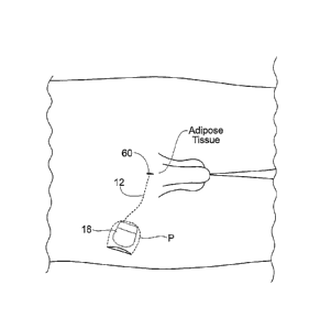
Fig. 6 shows an implant system 10 for the
restoration of sexual function in animals, including
humans.
The system 10 includes an implantable lead 12 having
a proximal and distal end coupled to an implantable pulse
generator or IPG 18. The lead 12 and the implantable
pulse generator 18 are shown implanted within a tissue
region T of a human or animal body.
The distal end of the lead 12 includes at least one
electrically conductive surface, which will in shorthand
be called an electrode 16. The electrode 16 is implanted
in electrical conductive contact with at least one
functional grouping of neural tissue, muscle, or at least
one nerve, or at least one muscle and nerve. The
implantable pulse generator 18 includes a connection
header 14 that desirably carries a plug-in receptacle
(connector) for the lead 12. In this way, the lead 12
electrically connects the electrode 16 to the implantable
pulse generator 18.
The implantable pulse generator 18 is sized and
configured to be implanted subcutaneously in tissue,
desirably in a subcutaneous pocket P, which can be remote
from the electrode 16, as Fig. 6 shows. Desirably, the
implantable pulse generator 18 is sized and configured to
be implanted using a minimally invasive surgical
procedure.
CA 02608017 2007-11-09
WO 2006/124068
PCT/US2005/043144
Atty. Docket No.: 9469.19606-AB PCT
- 17 -
The lead 12 and electrode 16 are sized and
configured to be implanted percutaneously in tissue, and
to be tolerated by an individual during extended use
without pain or discomfort. The comfort is both in terms
of the individual's sensory perception of the electrical
waveforms that the electrode applies, as well as the
individual's sensory perception of the physical presence
of the electrode and lead. In both categories, the lead
12 and electrode 16 are desirably "imperceptible."
In particular, one configuration of the lead 12 and
electrode 16 are sized and configured to reside with
stability in soft or adipose tissue in the lower anterior
pelvic region of the body (see Figs 8A and 8B). It has
been discovered that, when properly placed in this
region, one or more lead/ electrode(s) 16 are uniquely
able to deliver electrical stimulation current
simultaneously to both left and right branches of the
dorsal genital nerves, present near the clitoris in a
female and near the base of the penis of a male (see
Figs. 8A and 8B). Specific features of the lead 12 and
electrode 16 that make them well suited for this purpose,
as well as other purposes, will be described in greater
detail later.
As Figs. 7A and 7B show, the implantable pulse
generator 18 includes a circuit 20 that generates
electrical stimulation waveforms. An on-board, primary
battery 22 desirably provides the power. The implantable
pulse generator 18 also desirably includes an on-board,
programmable microcontroller 24, which carries embedded
code. The code expresses pre-programmed rules or
algorithms under which the desired electrical stimulation
waveforms are generated by the circuit 20. The
implantable pulse generator 18 may also include an
electrically conductive case 26, which can also serve as
the return electrode for the stimulus current introduced
CA 02608017 2007-11-09
W02006/124068
PCT/US2005/043144
Atty. Docket No.: 9469.19606-AB PCT
- 18 -
by the lead/electrode when operated in a monopolar
configuration.
As shown in Figs. 8A and 83, the implantation site
can comprise a tissue region on the posterior hip.
Alternatively, the implantation site can comprise a more
medial tissue region in the lower abdomen. There, the
pulse generator 18 can reside for extended use without
causing pain and/or discomfort and/or without effecting
body image.
The implant system 10 includes an external patient
controller 37 (see Figs. 8A and 14). The patient
controller 37 is sized and configured to be held or worn
by the individual to transcutaneously activate and
deactivate or modify the output of the pulse generator
18. The patient controller 37 may, e.g., be a simple
magnet that, when placed near the site where the pulse
generator 18 is implanted (see Fig. 14), toggles a
magnetic switch within the implantable pulse generator 18
between an on condition and an off condition, or advances
through a sequence of alternative stimulus modes pre-
programmed by the clinician into the implantable pulse
generator 18. Alternatively, the patient controller 37
may comprise more sophisticated circuitry that would
allow the individual to make these selections through an
RF field (magnetic and/or electric) that passes through
the skin and tissue within an arm's length distance (or
up to two meters) from the implanted pulse generator 18.
According to its programmed rules, when switched on,
the implantable pulse generator 18 generates prescribed
stimulation waveforms through the lead 12 and to the
electrode 16. These waveforms bilaterally stimulate the
left and right branches of the dorsal genital nerves in a
manner that achieves the desired physiologic response.
It has been discovered that bilateral stimulation of
the dorsal genital nerves achieved by placement of a
CA 02608017 2007-11-09
WO 2006/124068
PCT/US2005/043144
Atty. Docket No.: 9469.19606-AB PCT
- 19 -
single electrode 16 at a unique location in the body
(which will be described in greater detail later),
achieves the desired physiologic result. Alternatively,
more than one electrode may be placed to stimulate the
dorsal genital nerves (e.g., one or more electrodes to
stimulate the left branch and one or more electrodes to
stimulate the right branch, see Fig. 8C). Bilateral
stimulation may be achieved with a single electrode 16,
but due to anatomical variations in the patient or
20 and one on, in, or near the right branch of the dorsal
genital nerve.
Using the controller 26, the individual may turn on
or turn off the sexual restoration control waveforms at
will or adjust the waveforms to achieve the desired -
The system 10 desirably includes means for
selectively varying the frequency or range of frequencies
for a variable duration at which the stimulation
waveforms are applied by the one or more electrodes 18.
35 By modulating the frequency and /or duration of the
CA 02608017 2007-11-09
WO 2006/124068
PCT/US2005/043144
Atty. Docket No.: 9469.19606-AB PCT
- 20 -
stimulation waveform, the same system components and
placement of electrodes can serve to achieve markedly
different physiologic responses, and in addition, reduce
habituation.
The shape of the waveform can vary as well. It can,
e.g., be a typical square pulse, or possess a ramped
shape. The pulse, or the rising or falling edges of the
pulse, can present various linear, exponential,
hyperbolic, or quasi-trapezoidal shapes. The stimulation
waveform can be continuous, or it can be variable and
change cyclically or in step fashion in magnitude and
waveform over time.
In a non-limiting exemplary embodiment, the stimulus
waveforms may include a variable frequency for a variable
duration (e.g., a first stimulation at 5 Hz for 2
seconds, then 7 Hz for 3 seconds, then 6 Hz for 1 second,
and so on), intermittent stimulation (apply stimulation
in bursts separated by pauses in stimulation (e.g.,
stimulation for 3 seconds, rest for 2 seconds, repeat,
and so on). The stimulus waveforms may also include a
continuously or intermittently applied duty cycle of
pulses. This may be considered the same as changing the
frequency but it also refers to 1) the duration of bursts
of stimulation and 2) the duration of pauses between the
bursts. For example, a variable duty cycle for
intermittent pulses may include stimulation with 10
pulses, then off for 500 milliseconds, stimulation with
15 pulses, then off for 750 milliseconds, stimulation
with 5 pulses, then off for 2 seconds, and it could keep
going in this variable pattern.
The stimulus waveforms may also include stimulation
at different amplitudes. This may be beneficial because
increasing the amplitude may increase penile tumescence
to a certain degree, and then increasing the amplitude
further may be used to cause ejaculation. Thus, amplitude
CA 02608017 2007-11-09
WO 2006/124068
PCT/US2005/043144
Atty. Docket No.: 9469.19606-AB PCT
- 21 -
modulation may be used to control the response. Varying
the amplitude may also provide another form of anti-
habituation control, allowing a sexual function (e.g.,
erection) to remain more robust than if the target nerve
N was stimulated at a constant amplitude. Amplitude
modulation may also more realistically recreate the
varying level of fiber activation that occurs during
coitus.
The patient controller 37 and/or a clinician
programmer, for example, may include a manual-actuated
switch or control knob which an operator operates or
tunes to acquire a desired waveform frequency, given the
desired physiologic response.
C. The Anatomic Landmarks
As already described, certain components of the
implant system 10 are sized and configured to be
implanted in adipose tissue in the lower anterior pelvic
region, where it has been discovered that effective
bilateral stimulation of both the left and right branches
of the dorsal genital nerves can be achieved with one or
more electrodes. The main anatomic landmark guiding the
unique placement of these components is the pubic
symphysis.
As Fig. 9 shows, the hip bones are two large,
irregularly shaped bones, each of which develops from the
fusion of three bones, the ilium, ischium, and pubis. The
ilium is the superior, fan-shaped part of the hip bone.
The ala of the ilium represents the spread of the fan.
The iliac crest represents the rim of the fan. It has a
curve that follows the contour of the ala between the
anterior and posterior superior iliac spines.
As Figs. 9 and 10 show, the sacrum is formed by the
fusion of five originally separate sacral vertebrae. The
hip bones are joined at the pubic symphysis anteriorly
and to the sacrum posteriorly to form the pelvic girdle
CA 02608017 2007-11-09
WO 2006/124068
PCT/US2005/043144
Atty. Docket No.: 9469.19606-AB PCT
- 22 -
(see Fig. 9). The pelvic girdle is attached to the lower
limbs. Located within the pelvic girdle are the abdominal
viscera (e.g., the ileum and siymoid colon) and the
pelvic viscera (e.g., the urinary bladder and prostate
gland for males, and the urinary bladder and reproductive
organs such as the uterus and ovaries for females).
Within this bony frame (see Figs. 9 and 10), the
pudendal nerve is derived at the sacral plexus from the
anterior divisions of the ventral rami of S2 through S4
and carries afferent (sensory) and efferent (motor) nerve
components that innervate muscles and organs in the lower
abdomen. The pudendal nerve extends bilaterally, in
separate branches on left and right sides of the pelvic
girdle. Each branch accompanies the interior pudendal
artery and leaves the pelvis through the left and right
greater sciatic foramens between the piriformis and
coccygeus muscles. The branches hook around the ischial
spine and sacrospinous ligament, and enter the skin and
muscles of the perineum through the left and right lesser
sciatic foramen.
The Figures are largely based upon the anatomy of a
male, but the parts of the male perineum are homologues
of the female. As shown in Fig. 11, which is based on the
anatomy of a female, the bilateral left and right
branches extend anteriorly through the perineum, each
ending as the dorsal genital nerve of the penis or
clitoris. The genital nerves are the chief sensory nerve
of the external genitalia.
As Fig. 11 shows, in the female and male, adipose
tissue overlays the pubic symphysis. The bilateral
branches of the genital nerves innervate this tissue
region. In the female, this tissue region is known as the
mons pubis. In the male, the penis and scrotum extend
from this region. Further discussion regarding the
fixation of the lead 12 and electrode 16 in adipose
CA 02608017 2007-11-09
WO 2006/124068
PCT/US2005/043144
Atty. Docket No.: 9469.19606-AB PCT
- 23 -
tissue will be described later.
D. Conditions Required to Evoke Erection
Erection is a complex process involving control from
the autonomic and somatic nervous systems. There are two
peripheral neural pathways that control erection in cats
and dogs. The parasympathetic pathway (52-54) mediates
tactile, as well as psychically induced erection, while
the sympathetic pathway (T10-L2) mediates only
psychically induced erection. Although erection involves
many central and psychogenic factors, reflex erections
are mediated by a spinal mechanism, and do not require
participation of supraspinal structures.
The implant system 10 will focus on spinally-
mediated reflex erection, as this is most relevant to
restoration of sexual function. The afferents of the
erection reflex arises from the dorsal nerve of the penis
(DNP), while the efferent side includes both the
cavernous and pudendal nerves (see Fig. 1). The
cavernous nerve mediates engorgement of the penis as a
result of dilation of penile blood vessels (mediated by a
non-adrenergic non-cholinergic mechanism, putatively
nitric oxide), and venous occlusion may also play a role
in engorgement. The pudendal nerve carries the somatic
innervation of the bulbospongiosus which serves to
further increase cavernous pressure and penile stiffness,
and the ischiocavernosus which can also augment stiffness
of the penis.
Previous studies indicate that electrical
stimulation of the dorsal nerve of the penis can evoke
reflex penile erection before and after T8 spinal cord
transection in the rat. In the spinalized rat, DNP
stimulation produces a copulatory-like reflex, including
erectile and ejaculatory responses. DNP stimulation
evokes central reflexes with latencies of 50 milliseconds
to 150 milliseconds and is thought to mediate reflex
CA 02608017 2007-11-09
WO 2006/124068
PCT/US2005/043144
Atty. Docket No.: 9469.19606-1B PCT
- 24 -
erection. Stimulus frequencies of two Hz to ten Hz have
been successful in eliciting reflex erections before and
after spinalization, and spinalization increases the
combination of stimulus parameters that are successful in
evoking erection.
Low amplitude genital nerve stimulation is not
expected to cause pain because it has been observed that
low amplitude (5 + 3 mA), low frequency (10 Hz)
electrical stimulation of the dorsal genital (clitoral)
nerve created a sensation that was well tolerated by all
women (n = 17), often described as a thumping (24%),
buzzing (18%), or pulsing (12%) sensation, and the
amplitude could be increased to almost double (9 + 3 mA)
before it became uncomfortable.
The implant system 10 is sized and configured to
evoke a rigid erection and sustain an erection for about
30 minutes that is comparable in both 1) corpus cavernous
pressure (CCP) and 2) CCP/BP (blood pressure) to the
erection produced by intracavernous injection of
alprostadil. A rigid erection is defined by CCP BP and
a functional score of 4 or 5 (sufficient for sexual
intercourse or full erection) on the Schramek grading
system. The time to erection once the implant system 10
is turned on may be in the range of a few minutes (e.g.,
two to ten minutes). When the implant system is turned
off, the erection will subside comparable to a normal
healthy response.
E. Afferent and Efferent Stimulation
The system and methods described for afferent
stimulation can provide a more rigid and longer lasting
erection than methods that use efferent stimulation
because afferent stimulation activates a reflex that
coordinates the increase of filling via dilation of
penile arteries with the prevention of leakage via
occlusion of penile veins. Present stimulation methods do
CA 02608017 2007-11-09
WO 2006/124068
PCT/US2005/043144
Atty. Docket No.: 9469.19606-1B PCT
- 25 -
not stimulate both cavernous and pudendal nerves (or
nerve branches), nor do present stimulation methods use
reflexes to coordinate the individual processes involved
in erection. Specifically, afferent stimulation can 1)
provide a longer lasting erection because it activates a
reflex that controls the rate and amount of
neurotransmitter released from the cavernous nerve. On
the other hand, direct efferent stimulation of the
cavernous nerve can release excessive amounts of
neurotransmitter. The reflex activated by afferent
stimulation also 2) coordinates efferent activity in the
pudendal nerve to prevent leakage of blood from the penis
via occlusion of penile veins, whereas efferent
stimulation of the cavernous nerve does nothing to
prevent leakage of blood from the penis
Additionally, efferent stimulation risks generating
the perception of pain due to the current amplitude that
may be required. Afferent stimulation may avoid the
generation of pain because lower amplitudes of current
can be used to activate selectively the large sensory
fibers without activating the smaller C-fibers that
transmit signals to pain centers.
Nevertheless, a coordinated stimulation to both
afferent and efferent nerves, or efferent and efferent
nerves, including coordinated stimulation of both the
cavernous and pudendal nerves (or branches), may also be
used to produce the desired functional result.
II. Details of Implant System
A. The Implantable Pulse Generator
As previously described, Fig. 6 shows a system 10
for the functional restoration of sexual function. The
assembly includes an implantable lead 12 and electrode 16
coupled to an implantable pulse generator or IPG 18. The
lead 12 and the implantable pulse generator 18 are shown
implanted within a tissue region T of a human or animal
CA 02608017 2007-11-09
W02006/124068
PCT/US2005/043144
Atty. Docket No.: 9469.19606-AB PCT
- 26 -
body.
Desirably, the components of the implantable pulse
generator 18 are sized and configured so that they can
accommodate several different indications, without major
change or modification (see Fig. 7A). Examples of
components that desirably remain unchanged for different
indications include the case 26, the battery 22, the
microcontroller 24, much of the software (firmware) of
the embedded code, the power management circuitry 40, and
the stimulus power supply, both of which are part of the
circuitry 20. Thus, a new indication may require only
changes to the programming of the microcontroller 24.
Most desirably, the particular code is remotely embedded
in the microcontroller 24 after final assembly,
packaging, and sterilization of the implantable pulse
generator 18.
Certain components of the implantable pulse
generator 18 may be expected to change as the indication
changes; for example, due to differences in leads and
electrodes, the connection header 14 and associated
receptacle(s) for the lead may be configured differently
for different indications. Other aspects of the circuit
20 may also be modified to accommodate a different
indication; for example, the stimulator output stage(s),
sensor(s) and/or sensor interface circuitry.
In this way, the implantable pulse generator 18
accommodates implanting in diverse tissue regions and
also accommodates coupling to a lead 12 and an electrode
16 having diverse forms and configurations, again
depending upon the therapeutic or functional effects
desired. For this reason, the implantable pulse generator
can be considered to be general purpose or "universal."
1. Desirable Technical Features
The implantable pulse generator 18 can incorporate
various technical features to enhance its universality.
CA 02608017 2007-11-09
WO 2006/124068
PCT/US2005/043144
Atty. Docket No.: 9469.19606-AB PCT
- 27 -
a. Small, Composite Case
According to one desirable technical feature, the
implantable pulse generator 18 can be sized small enough
to be implanted (or replaced) with only local anesthesia.
As Figs. 7A and 7B show, the functional elements of the
implantable pulse generator 18 (e.g., circuit 20, the
microcontroller 24, the battery 22, and the connection
header 14) are integrated into a small, composite case
26. As can be seen in Fig. 2A and 2B, the implantable
pulse generator 18 may comprise a case 26 having a small
cross section, e.g., (5mm to 15mm thick) x (45mm to 60mm
wide) x (45mm to 60mm long). The overall weight of the
implantable pulse generator 18 may be approximately
twenty to thirty grams. These dimensions make possible
implantation of the case 26 with a small incision; i.e.,
suitable for minimally invasive implantation.
Additionally, a smaller or larger, but similarly shaped
IPG might be required for other applications, such as
with more stimulus channels (thus requiring a large
connection header) and/or a smaller or larger internal
battery.
The case 26 of the implantable pulse generator 18 is
desirably shaped with a smaller end 30 and a larger end
32. As Fig. 6 shows, this geometry allows the smaller end
30 of the case 26 to be placed into the skin pocket P
first, with the larger end 32 being pushed in last.
Desirably, the case 26 for the implantable pulse
generator 18 comprises a laser welded implant grade
titanium material. This construction offers high
reliability with a low manufacturing cost. The clam shell
construction has two stamped or successively drawn
titanium case halves that are laser welded around the
circuit assembly and battery 22 with feed-thrus.
Typically, a molded plastic spacing nest is used to hold
the battery 22, the circuit 20, and perhaps a power
CA 02608017 2007-11-09
WO 2006/124068
PCT/US2005/043144
Atty. Docket No.: 9469.19606-AB PCT
- 28 -
recovery (receive) coil (if a rechargeable battery is
used) together and secure them within the hermetically
sealed titanium case. An implantable pulse generator
having a rechargeable battery can be used of the type
described in copending United States Patent Application
Serial No. 11/150,418, filed 10 June 2005 and entitled
"Implantable Pulse Generator for Providing Functional
and/or Therapeutic Stimulation of Muscles and/or Nerves
and/or Central Nervous System Tissue," which is
incorporated herein by reference. The electronics may be
fabricated on a flexible or flex-rigid PC board using
very high density technique include adhesive flip-chip or
chip-on-board mounting of the larger semiconductor
devices. The tissue contact materials used in the
manufacture of the IPG may all have Master Files with FDA
demonstrating their biocompatibility.
The implantable pulse generator 18 shown in Figs. 7A
and 7B includes a clam-shell case 26 having a thickness
that is selected to provide adequate mechanical strength
The implantable pulse generator 18 may be implanted at a
target implant depth of not less than five millimeters
beneath the skin, and not more than fifteen millimeters
beneath the skin, although this implant depth may change
due to the particular application, or the implant depth
may change over time based on physical conditions of the
patient.
b. Primary Power Source
According to one desirable technical feature, the
implantable pulse generator 18 desirably possesses an
internal battery capacity sufficient to allow a service
life of greater than three years with the stimulus being
a high duty cycle, e.g., virtually continuous, low
frequency, low current stimulus pulses, or alternatively,
the stimulus being higher frequency and amplitude
stimulus pulses that are used only intermittently, e.g.,
CA 02608017 2007-11-09
WO 2006/124068
PCT/US2005/043144
Atty. Docket No.: 9469.19606-AB PCT
- 29 -
a very low duty cycle.
To achieve this feature, the primary battery 22 of
the implantable pulse generator 18 desirably comprises a
primary power source; most desirably an implant grade
Lithium Ion battery 22. Given the average quiescent
operating current (estimated at 8 A plus 35 A for a
wireless telemetry receiver pulsing on twice every
second) and a seventy percent efficiency of the stimulus
power supply, a 1.0 Amp-hr primary cell battery can
provide a service life of less than two years, which is
too short to be clinically or commercially viable for
this indication. Therefore, the implantable pulse
generator 18 desirably incorporates a primary battery,
e.g., a Lithium Ion battery. Given representative
desirable stimulation parameters (which will be described
later), a Lithium Ion battery with a capacity of at least
30mA-hr will operate for over three years. Lithium Ion
implant grade batteries are available from a domestic
supplier. A representative battery provides 35m-hr in a
package configuration that is of appropriate size and
shape to fit within the implantable pulse generator 18.
The implantable pulse generator 18 desirably
incorporates circuitry and/or programming to assure that
the implantable pulse generator 18 will suspend
stimulation, and perhaps fall-back to only very low rate
telemetry, and eventually suspends all operations when
the primary battery 22 has discharged the majority of its
capacity (i.e., only a safety margin charge remains).
Once in this dormant mode, the implantable pulse
generator may provide limited communications and is in
condition for replacement.
c. Wireless Telemetry
According to one desirable technical feature, the
system or assembly 10 includes an implantable pulse
generator 18, which desirably incorporates wireless
CA 02608017 2007-11-09
WO 2006/124068
PCT/US2005/043144
Atty. Docket No.: 9469.19606-AB PCT
- 30 -
telemetry (rather that an inductively coupled telemetry)
for a variety of functions to be performed within arm's
reach of the patient, the functions including receipt of
programming and clinical parameters and settings from the
clinician programmer 36, communicating usage history to
the clinician programmer, and providing user control of
the implantable pulse generator 18. Each implantable
pulse generator may also have a unique signature that
limits communication to only the dedicated controllers
(e.g., the matched patient controller, or a clinician
programmer configured for the implantable pulse generator
in question).
The implantable pulse generator 18 desirably
incorporates wireless telemetry as an element of the
implantable pulse generator circuit 20 shown in Fig. 15.
A circuit diagram showing a desired configuration for the
wireless telemetry feature is shown in Fig. 16. It is to
be appreciated that modifications to this circuit diagram
configuration which produce the same or similar functions
as described are within the scope of the invention.
As shown in Fig. 12, the assembly 10 desirably
includes a clinician programmer 36 that, through a
wireless telemetry 38, transfers commands, data, and
programs into the implantable pulse generator 18 and
retrieves data out of the implantable pulse generator 18.
In some configurations, the clinician programmer may
communicate with more than one implantable pulse
generator implanted in the same user.
The clinician programmer 36 may incorporate a custom
programmed general purpose digital device, e.g., a custom
program, industry standard handheld computing platform or
other personal digital assistant (PDA). The clinician
programmer 36 can include an on-board microcontroller
powered by a rechargeable battery. The rechargeable
battery of the clinician programmer 36 may be recharged
CA 02608017 2007-11-09
WO 2006/124068
PCT/US2005/043144
Atty. Docket No.: 9469.19606-AB PCT
- 31 -
by being docked on a charging base (not shown); or the
custom electronics of the clinician programmer may
receive power from the connected PDA. The microcontroller
carries embedded code which may include pre-programmed
rules or algorithms that allow a clinician to remotely
download program stimulus parameters and stimulus
sequences parameters into the implantable pulse generator
18. The microcontroller of the clinician programmer 36 is
also desirably able to interrogate the implantable pulse
generator and upload usage data from the implantable
pulse generator. Fig. 12 shows one possible application
where the clinician is using the programmer 36 to
interrogate the implantable pulse generator. Fig. 13
shows an alternative application where the clinician
programmer, or a clinician programmer derivative 33
intended for remote programming applications and having
the same or similar functionality as the clinician
programmer, is used to interrogate the implantable pulse
generator. As can be seen, the clinician programmer
derivative 33 is connected to a local computer, allowing
for remote interrogation via a local area network, wide
area network, or Internet connection, for example.
Using subsets of the clinician programmer software,
features of the clinician programmer 36 or clinician
programmer derivative 33 might include the ability of the
clinician or physician to remotely monitor and adjust
parameters using the Internet or other known or future
developed networking schemes. A clinician programmer
derivative 33 would desirably connect to the patient's
computer in their home through an industry standard
network such as the Universal Serial Bus (USE), where in
turn an applet downloaded from the clinician's server
would contain the necessary code to establish a reliable
transport level connection between the implantable pulse
generator 18 and the clinician's client software, using
CA 02608017 2007-11-09
WO 2006/124068
PCT/US2005/043144
Atty. Docket No.: 9469.19606-2B PCT
- 32 -
the clinician programmer derivative 33 as a bridge. Such
a connection may also be established through separately
installed software. The clinician or physician could then
view relevant diagnostic information, such as the health
of the unit or its current settings, and then modify the
stimulus settings in the IPG or direct the patient to
take the appropriate action. Such a feature would save
the clinician, the patient and the health care system
substantial time and money by reducing the number of
office visits during the life of the implant.
Other features of the clinician programmer, based on
an industry standard platform, might include the ability
to connect to the clinician's computer system in his or
hers office. Such features may take advantage of the
Conduit connection employed by Palm OS based devices.
Such a connection then would transfer relevant patient
data to the host computer or server for electronic
processing and archiving. With a feature as described
here, the clinician programmer then becomes an integral
link in an electronic chain that provides better patient
service by reducing the amount of paperwork that the
physician's office needs to process on each office visit.
It also improves the reliability of the service since it
reduces the chance of mis-entered or mis-placed
information, such as the record of the parameter setting
adjusted during the visit.
With the use of a patient controller 37 (see Fig.
14), the wireless link 38 allows a patient to control
certain parameters of the implantable pulse generator
within a predefined limited range. The parameters may
include the operating modes/states, increasing/decreasing
or optimizing stimulus patterns, or providing open or
closed loop feedback from an external sensor or control
source. The wireless telemetry 38 also desirably allows
the user to interrogate the implantable pulse generator
CA 02608017 2007-11-09
WO 2006/124068
PCT/US2005/043144
Atty. Docket No.: 9469.19606-1B PCT
- 33
18 as to the status of its internal battery 22. The full
ranges within these parameters may be controlled,
adjusted, and limited by a clinician, so the user may not
be allowed the full range of possible adjustments.
In one embodiment, the patient controller 37 is
sized and configured to couple to a key chain, as seen in
Fig 14. It is to be appreciated that the patient
controller 37 may take on any convenient shape, such as a
ring on a finger, or a watch on a wrist, or an attachment
to a belt, for example. The patient controller may also
use a magnetic switch to enable the user to turn the IPG
on/off.
The wireless telemetry may incorporate a suitable,
low power wireless telemetry transceiver (radio) chip set
that can operate in the MICS (Medical Implant
Communications Service) band (402MHz to 405MHz) or other
VHF/UHF low power, unlicensed bands. A wireless telemetry
link not only makes the task of communicating with the
implantable pulse generator 18 easier, but it also makes
the link suitable for use in motor control applications
where the user issues a command to the implantable pulse
generator to produce muscle contractions to achieve a
functional goal (e.g., to stimulate ankle flexion to aid
in the gait of an individual after a stroke) without
requiring a coil or other component taped or placed on
the skin over the implanted implantable pulse generator.
Appropriate use of power management techniques is
important to the effective use of wireless telemetry.
Desirably, the implantable pulse generator is exclusively
the communications slave, with all communications
initiated by the external controller (the communications
master). The receiver chip of the implantable pulse
generator is OFF more than 99% of the time and is pulsed
on periodically to search for a command from an external
controller, including but not limited to the clinician
CA 02608017 2007-11-09
WO 2006/124068
PCT/US2005/043144
Atty. Docket No.: 9469.19606-2B PCT
- 34 -
programmer 36 and the patient controller 37.
Communications protocols include appropriate check and
message acknowledgment handshaking to assure the
necessary accuracy and completeness of every message.
Some operations (such as reprogramming or changing
stimulus parameters) require rigorous message accuracy
testing and acknowledgement. Other operations, such as a
single user command value in a string of many consecutive
values, might require less rigorous checking and a more
loosely coupled acknowledgement.
The timing with which the implantable pulse
generator enables its transceiver to search for RF
telemetry from an external controller is precisely
controlled (using a time base established by a quartz
crystal) at a relatively low rate, e.g., the implantable
pulse generator may look for commands from the external
controller at a rate of less than one (1) Hz. This
equates to a monitoring interval of several seconds. It
is to be appreciated that the monitoring rate may vary
faster or slower depending on the application, (e.g.,
twice per second; i.e., every 500 milliseconds). This
allows the external controller to time when the
implantable pulse generator responds to a command and
then to synchronize its commands with when the
implantable pulse generator will be listening for
commands. This, in turn, allows commands issued within a
short time (seconds to minutes) of the last command to be
captured and acted upon without having to 'broadcast' an
idle or pause signal for 500 milliseconds before actually
issuing the command in order to know that the implantable
pulse generator will have enabled its receiver and
received the command. Similarly, the communications
sequence is configured to have the external controller
issue commands in synchronization with when the
implantable pulse generator will be listening for a
CA 02608017 2007-11-09
WO 2006/124068
PCT/US2005/043144
Atty. Docket No.: 9469.19606-AB PCT
- 35 -
command. Similarly, the command set implemented is
selected to minimize the number of messages necessary and
the length of each message consistent with the
appropriate level of error detection and message
integrity monitoring. It is to be appreciated that the
monitoring rate may vary faster or slower depending on
the application; and may vary over time within a given
application.
A suitable radio chip is used for the half duplex
wireless communications, e.g., the AMIS-52100 (AMI
Semiconductor; Pocatello, Idaho). This transceiver chip
is designed specifically for the MICS and its European
counter-part the ULP-MI (Ultra Low Power-Active Medical
Implant) band. This chip set is optimized by micro-power
operation with rapid start-up, and RF 'sniffing'
circuitry.
d. Stimulus Output Stage
According to one desirable technical feature, the
implantable pulse generator 18 desirably uses a single
stimulus output stage (generator) that is directed to one
or more output channels (electrode surfaces) by analog
switch(es) or analog multiplexer(s). Desirably, the
implantable pulse generator 18 will deliver at least one
channel of stimulation via a lead/electrode. For
applications requiring more stimulus channels, several
channels (perhaps up to four) can be generated by a
single output stage. In turn, two or more output stages
could be used, each with separate multiplexing to
multiple channels, to allow an implantable pulse
generator with eight or more stimulus channels. The
stimulation waveform output of the IPG desirably has an
asymmetrically biphasic waveform (net DC current less
than 101uA), and an RC recovery phase with programmable
interphase delay. The stimulus parameters (amplitude,
pulse duration, and frequency) are independently
CA 02608017 2007-11-09
WO 2006/124068
PCT/US2005/043144
Atty. Docket No.: 9469.19606-AB PCT
- 36 -
adjustable with amplitude output ranging from 0.5 mA to
20 mA, pulse duration ranging from 0 to 500 microseconds,
and frequency ranging from 1 (one) to 300 Hz. In one
embodiment, the applied stimulus frequency may be in the
range of about one Hz to about fifteen Hz. The stimulus
current (amplitude) and pulse duration being programmable
on a channel to channel basis and adjustable over time
based on a clinically programmed sequence or regime or
based on user (patient) commands received via the
wireless communications link.
A circuit diagram showing a desired configuration
for the stimulus output stage feature is shown in Fig.
17. It is to be appreciated that modifications to this
circuit diagram configuration which produce the same or
similar functions as described are within the scope of
the invention.
Desirably, the implantable pulse generator 18
includes a single stimulus generator (with its associated
DC current blocking output capacitor) which is
multiplexed to a number of output channels; or a small
number of such stimulus generators each being multiplexed
to a number of output channels. This circuit architecture
allows multiple output channels with very little
additional circuitry. A typical, biphasic stimulus pulse
is shown in Fig. 18. Note that the stimulus output stage
circuitry 46 may incorporate a mechanism to limit the
recovery phase current to a small value (perhaps 0.5mA).
Also note that the stimulus generator (and the associated
timing of control signals generated by the
microcontroller) may provide a delay (typically of the
order of 100 microseconds) between the cathodic phase and
the recovery phase to limit the recovery phase diminution
of the cathodic phase effective at eliciting a neural
excitation. The charge recovery phase for any electrode
(cathode) must be long enough to assure that all of the
CA 02608017 2007-11-09
WO 2006/124068
PCT/US2005/043144
Atty. Docket No.: 9469.19606-AB PCT
- 37 -
charge delivered in the cathodic phase has been returned
in the recovery phase; i.e., greater than or equal to
five time constants are allowed for the recovery phase.
This will allow the stimulus stage to be used for the
next electrode while assuring there is no net DC current
transfer to any electrode. Thus, the single stimulus
generator having this characteristic would be limited to
four channels (electrodes), each with a maximum frequency
of 30 Hz to 50 Hz. This operating frequency exceeds the
needs of many indications for which the implantable pulse
generator is well suited. For applications requiring more
channels (or higher composite operating frequencies), two
or more separate output stages might each be multiplexed
to multiple (e.g., four) electrodes.
e. The Lead Connection Header
According to one desirable technical feature, the
implantable pulse generator 18 desirably includes a lead
connection header 14 for connecting the lead(s) 12 that
will enable reliable and easy replacement of the
lead/electrode (see Figs. 7A and 7B), and includes a
small antenna 54 for use with the wireless telemetry
feature.
The implantable pulse generator desirably
incorporates a connection header (top header) 14 that is
easy to use, reliable, and robust enough to allow
multiple replacements of the implantable pulse generator
after many years (e.g., more than ten years) of use. The
surgical complexity of replacing an implantable pulse
generator is usually low compared to the surgical
complexity of correctly placing the implantable lead
12/electrode 16 in proximity to the target nerve/tissue
and routing the lead 12 to the implantable pulse
generator. Accordingly, the lead 12 and electrode 16
desirably has a service life of at least ten years with a
probable service life of fifteen years or more. Based on
CA 02608017 2007-11-09
W02006/124068
PCT/US2005/043144
Atty. Docket No.: 9469.19606-AB PCT
- 38 -
the clinical application, the implantable pulse generator
may not have this long a service life. The implantable
pulse generator service life is largely determined by the
power capacity of the Lithium Ion battery 22, and is
likely to be three to ten years, based on the usage of
the device. Desirably, the implantable pulse generator 18
has a service life of at least three years.
As described above, the implantable pulse generator
preferably will use a laser welded titanium case. As with
other active implantable medical devices using this
construction, the implantable lead(s) 12 connect to the
implantable pulse generator through a molded or cast
polymeric connection header 14 (top header). Metal-
ceramic or metal-glass feed-thrus maintain the hermetic
seal of the titanium capsule while providing electrical
contact to the electrical contacts of the lead
12/electrode 16.
The standard implantable connectors may be similar
in design and construction to the low-profile IS-1
connector system (per ISO 5841-3). The I5-1 connectors
have been in use since the late 1980s and have been shown
to be reliable and provide easy release and re-connection
over several implantable pulse generator replacements
during the service life of a single pacing lead. Full
compatibility with the IS-1 standard, and mating with
pacemaker leads, is not a requirement for the implantable
pulse generator.
The implantable pulse generator connection system
may include a modification of the IS-1 connector system,
which shrinks the axial length dimensions while keeping
the format and radial dimensions of the IS-1. For
application with more than two electrode conductors, the
top header 14 may incorporate one or more connection
receptacles each of which accommodate leads with
typically four conductors. When two or more leads are
CA 02608017 2007-11-09
WO 2006/124068 PCT/US2005/043144
=
Atty. Docket No.: 9469.19606-AB PCT
- 39 -
accommodated by the header, these leads may exit the
connection header in opposite directions (i.e., from
opposite sides of the header).
These connectors can be similar to the banded axial
connectors used by other multi-polar implantable pulse
generators or may follow the guidance of the draft IS-4
implantable connector standard. The design of the
implantable pulse generator housing and header 14
preferably includes provisions for adding the additional
feed-thrus and larger headers for such indications.
The inclusion of the UHF antenna 54 for the wireless
telemetry inside the connection header (top header) 14 is
necessary as the shielding offered by the titanium case
will severely limit (effectively eliminate) radio wave
propagation through the case. The antenna 54 connection
will be made through a feed-thru similar to that used for
the electrode connections. Alternatively, the wireless
telemetry signal may be coupled inside the implantable
pulse generator onto a stimulus output channel and
coupled to the antenna 54 with passive filtering/coupling
elements/methods in the connection header 14.
f. The Microcontroller
According to one desirable technical feature, the
implantable pulse generator 18 desirably uses a standard,
commercially available micro-power, flash programmable
microcontroller 24 or processor core in an application
specific integrated circuit (ASIC). This device (or
possibly more than one such device for a computationally
complex application with sensor input processing) and
other large semiconductor components may have custom
packaging such as chip-on-board, solder flip chip, or
adhesive flip chip to reduce circuit board real estate
needs.
A circuit diagram showing a desired configuration
for the microcontroller 24 is shown in Fig. 19. It is to
CA 02608017 2007-11-09
WO 2006/124068
PCT/US2005/043144
Atty. Docket No.: 9469.19606-AB PCT
- 40 -
be appreciated that modifications to this circuit diagram
configuration which produce the same or similar functions
as described are within the scope of the invention.
g. Power Management Circuitry
According to one desirable technical feature, the
implantable pulse generator 18 desirably includes
efficient power management circuitry as an element of the
implantable pulse generator circuitry 20 shown in Fig.
15. The power management circuitry is generally
responsible for the efficient distribution of power and
monitoring the battery 22. In addition, the operation of
the implantable pulse generator 18 can be described in
terms of having operating modes as relating to the
function of the power management circuitry. These modes
may include, but are not limited to IPG Active and IPG
Dormant. These modes will be described below in terms of
the principles of operation of the power management
circuitry using possible circuit diagrams shown in Figs.
and 21. Fig. 20 shows one possible power management
20 sub-circuit having MOSFET isolation between the battery
22 and a charger circuit (when used). Fig. 21 shows
another possible power management sub-circuit diagram
without having MOSFET isolation between the battery 22
and the charger circuit (when used). In the circuit
without the isolation MOSFET (see Fig. 21), the leakage
current of the disabled charge control integrated circuit
chip (U1) must be very low to prevent this leakage
current from discharging the battery 22 in all modes
(including the Dormant Mode). Except as noted, the
description of these modes applies to both circuits.
i. IPG Active Mode
The IPG Active mode occurs when the implantable
pulse generator 18 is operating normally. In this mode,
the implantable pulse generator may be generating
stimulus outputs or it may be waiting for the next
CA 02608017 2007-11-09
WO 2006/124068
PCT/US2005/043144
Atty. Docket No.: 9469.19606-AB PCT
- 41 -
request to generate stimulus in response to a timed
neuromodulation sequence or a telemetry command from an
external controller. In this mode, the implantable pulse
generator is active (microcontroller 24 is powered and
coordinating wireless communications and may be timing &
controlling the generation and delivery of stimulus
pulses).
i(a).Principles of Operation,
IPG Active Mode
In the IPG Active mode, as can be seen in Fig. 20,
the lack of DC current from VRAW means that Q5 is held
off. This, in turn, holds Q3 off and a portion of the
power management circuitry is isolated from the battery
22. In Fig. 21, the lack of DC current from VRAW means
that Ul is disabled. This, in turn, keeps the current
drain from the battery 22 to an acceptably low level,
typically less than 1 A.
IPG Dormant Mode
The IPG Dormant mode occurs when the implantable
pulse generator 18 is completely disabled (powered down).
In this mode, power is not being supplied to the
microcontroller 24 or other enabled circuitry. This is
the mode for the long-term storage of the implantable
pulse generator before or after implantation. The Dormant
mode may only be exited by placing a pacemaker magnet (or
comparable device) over the implantable pulse generator
18 for a predetermined amount of time, e.g., five
seconds.
ii(a).Principles of Operation,
IPG Dormant Mode
In the IPG Dormant mode, VBAT is not delivered to
the remainder of the implantable pulse generator
circuitry because Q4 is turned off. The Dormant mode is
stable because the lack of VBAT means that VCC is also
not present, and thus Q6 is not held on through R8 and
CA 02608017 2007-11-09
WO 2006/124068
PCT/US2005/043144
Atty. Docket No.: 9469.19606-AB PCT
- 42 -
R10. Thus the battery 22 is completely isolated from all
load circuitry (the VCC power supply and the VHH power
supply).
The Dormant mode is entered through the application
of a long magnet placement over Si (magnetic reed switch)
or through the reception of a command by the wireless
telemetry. In the case of the telemetry command, the
PortD4, which is normally configured as a microcontroller
input, is configured as a logic output with a logic low
(0) value. This, in turn, discharges C8 through R12 and
turns off Q6; which, in turn, turns off Q4 and forces the
implantable pulse generator into the Dormant mode. Note
that R12 is much smaller in value than R10, thus the
microcontroller 24 can force C8 to discharge even though
VCC is still present.
In Fig. 20, the lack of DC current from VRAW means
that Q5 is held off. This, in turn, holds Q3 off and a
portion of the power management circuitry is isolated
from the battery 22. Also, Q4 was turned off. In Fig. 21,
the lack of DC current from VRAW means that U1 is
disabled. This, in turn, keeps the current drain from the
battery 22 to an acceptably low level, typically less
than 1 A.
2. Representative Implantable Pulse
Generator Circuitry
Fig. 15 shows an embodiment of a block diagram
circuit 20 for the primary cell implantable pulse
generator 18 that takes into account the desirable
technical features discussed above. The circuit 20 can be
grouped into functional blocks, which generally
correspond to the association and interconnection of the
electronic components.
In Fig. 15, seven functional blocks are shown: (1)
The Microprocessor or Microcontroller 24; (2) the Power
Management Circuit 40; (3) the VCC Power Supply 42; (4)
CA 02608017 2007-11-09
WO 2006/124068
PCT/US2005/043144
Atty. Docket No.: 9469.19606-7B PCT
- 43 -
the VHH Power Supply 44; (5) the Stimulus Output Stage(s)
46; (6) the Output Multiplexer(s) 48; and (7) the
Wireless Telemetry Circuit 50.
For each of these blocks, the associated functions,
possible key components, and circuit description are now
described.
a. The Microcontroller
The Microcontroller 24 is responsible for the
following functions:
(1) The timing and sequencing of the stimulator
stage and the VHH power supply used by the stimulator
stage,
(2) The sequencing and timing of power management
functions,
(3) The monitoring of the battery voltage, the
stimulator voltages produced during the generation of
stimulus pulses, and the total circuit current
consumption, VHH, and VCC,
(4) The timing, control, and interpretation of
commands to and from the wireless telemetry circuit,
(5) The logging (recording) of patient usage data as
well as clinician programmed stimulus parameters and
configuration data, and
(6) The processing of commands (data) received from
the user (patient) via the wireless link to modify the
characteristics of the stimulus being delivered.
The use of a microcontroller incorporating flash
programmable memory allows the operating program of the
implantable pulse generator as well as the stimulus
parameters and settings to be stored in non-volatile
memory (data remains safely stored even if the battery 22
becomes fully discharged; or if the implantable pulse
generator is placed in the Dormant mode). Yet, the data
(operating program, stimulus parameters, usage history
log, etc.) can be erased and reprogrammed thousands of
CA 02608017 2007-11-09
WO 2006/124068
PCT/US2005/043144
Atty. Docket No.: 9469.19606-AB PCT
- 44 -
times during the life of the implantable pulse generator.
The software (firmware) of the implantable pulse
generator must be segmented to support the operation of
the wireless telemetry routines while the flash memory of
the microcontroller 24 is being erased and reprogrammed.
Similarly, the VCC power supply 42 must support the power
requirements of the microcontroller 24 during the flash
memory erase and program operations.
Although the microcontroller 24 may be a single
component, the firmware is developed as a number of
separate modules that deal with specific needs and
hardware peripherals. The functions and routines of these
software modules are executed sequentially; but the
execution of these modules are timed and coordinated so
as to effectively function simultaneously. The
microcontroller operations that are associated directly
with a given hardware functional block are described with
that block.
The Components of the Microcontroller Circuit may
include:
(1) A single chip microcontroller 24. This
component may be a member of the Texas Instruments
MSP430 family of flash programmable, micro-power, highly
integrated mixed signal microcontroller. Likely family
members to be used include the MSP430F1610, MSP430F1611,
MSP430F1612, MSP430F168, and the MSP430F169. Each of
these parts has numerous internal peripherals, and a
micropower internal organization that allows unused
peripherals to be configured by minimal power
dissipation, and an instruction set that supports bursts
of operation separated by intervals of sleep where the
microcontroller suspends most functions.
(2) A miniature, quartz crystal (X1) for
establishing precise timing of the microcontroller. This
may be a 32.768KHz quartz crystal.
CA 02608017 2007-11-09
WO 2006/124068
PCT/US2005/043144
Atty. Docket No.: 9469.19606-AB PCT
- 45 -
(3) Miscellaneous power decoupling and analog
signal filtering capacitors.
b. Power Management Circuit
The Power Management Circuit 40 (including
associated microcontroller actions) is responsible for
the following functions:
(1) monitor the battery voltage,
(2) suspend stimulation when the battery voltage
becomes very low, and/or suspend all operation (go into
the Dormant mode) when the battery voltage becomes
critically low,
(3) communicate (through the wireless telemetry
link 38) with the external equipment the charge status of
the battery 22,
(4) prevent (with single fault tolerance) the
delivery of excessive current from the battery 22,
(5) provide battery power to the rest of the
circuitry of the implantable pulse generator, i.e., VCC
and VHH power supplies,
(6) provide isolation of the Lithium Ion battery
22 from other circuitry while in the Dormant mode,
(7) provide a hard microprocessor reset and force
entry into the Dormant mode in the presence of a
pacemaker magnet (or comparable device), and
(8) provide the microcontroller 24 with analog
voltages with which to measure the magnitude of the
battery voltage and the appropriate battery current flow
(drain and charge).
The Components of the Power Management Circuit may
include:
(1) Low on resistance, low threshold P channel
MOSFETs with very low off state leakage current (Q2, Q3,
and Q4).
(2) Analog switches (or an analog multiplexer) U3.
(3) Logic translation N-channel MOSFETs (Q5 & Q6)
CA 02608017 2007-11-09
WO 2006/124068
PCT/US2005/043144
Atty. Docket No.: 9469.19606-AD PCT
- 46 -
with very low off state leakage current.
c. The VCC Power Supply
The VCC Power Supply 42 is generally responsible for
the following functions:
(1) Some of the time, the VCC power supply passes
the battery voltage to the circuitry powered by VCC, such
as the microcontroller 24, stimulator output stage 46,
wireless telemetry circuitry 50, etc.
(2) At other times, the VCC power supply
fractionally steps up the voltage to about 3.3V; (other
useable voltages include 3.0V, 2.7V, etc.) despite
changes in the Lithium Ion battery 22 voltage. This
higher voltage is required for some operations such as
programming or erasing the flash memory in the
microcontroller 24, (i.e., in circuit programming).
The voltage converter / switch part at the center of
the VCC power supply may be a charge pump DC to DC
converter. Typical choices for this part may include the
Maxim MAX1759, the Texas Instruments TPS60204, or the
Texas Instruments REG710, among others.
The characteristics of the VCC Power Supply might
include:
(1) high efficiency and low quiescent current, i.e.,
the current wasted by the power supply in its normal
operation. This value should be less than a few
microamperes; and
(2) drop-out voltage, i.e., the minimal difference
between the VEAT supplied to the VCC power supply and its
output voltage. This voltage may be less than about 100mV
even at the current loads presented by the transmitter of
the wireless telemetry circuitry 50.
(3) The VCC power supply 42 may allows in-circuit
reprogramming of the implantable pulse generator
firmware, or optionally, the implantable pulse generator
18 may not use a VCC power supply, which may not allow
CA 02608017 2007-11-09
WO 2006/124068
PCT/US2005/043144
Atty. Docket No.: 9469.19606-AB PCT
- 47 -
in-circuit reprogramming of the implantable pulse
generator firmware.
d. VHH Power Supply
A circuit diagram showing a desired configuration
for the VHH power supply 44 is shown in Fig. 22. It is to
be appreciated that modifications to this circuit diagram
configuration which produce the same or similar functions
as described are within the scope of the invention.
The VHH Power Supply 44 is generally responsible for
the following functions:
(1) Provide the Stimulus Output Stage 46 and the
Output Multiplexer 48 with a programmable DC voltage
between the battery voltage and a voltage high enough to
drive the required cathodic phase current through the
electrode circuit plus the voltage drops across the
stimulator stage, the output multiplexer stage, and the
output coupling capacitor. VHH is typically 12VDC or less
for neuromodulation applications; and 25V or less for
muscle stimulation applications.
The Components of the VHH Power Supply might
include:
(1) Micropower, inductor based (fly-back topology)
switch mode power supply (1310); e.g., Texas Instruments
TPS61045, Texas Instruments TPS61041, or Linear
Technology LT3464 with external voltage adjustment
components.
(2) L6 is the flyback energy storage inductor.
(3) C42 & C43 form the output capacitor.
(4) R27, R28, and R29 establish the operating
voltage range for VHH given. the internal DAC which is
programmed via the SETVHH logic command from the
microcontroller 24.
(5) Diode D9 serves no purpose in normal operation
and is added to offer protection from over-voltage in the
event of a VHH circuit failure.
CA 02608017 2007-11-09
WO 2006/124068
PCT/US2005/043144
Atty. Docket No.: 9469.19606-AB PCT
- 48 -
(6) The microcontroller 24 monitors VHH for
detection of a VHH power supply failure, system failures,
and optimizing VHH for the exhibited electrode circuit
impedance.
e. Stimulus Output Stage
The Stimulus Output Stage(s) 46 is responsible for
the following functions:
(1) Generate the identified biphasic stimulus
current with programmable (dynamically adjustable during
use) cathodic phase amplitude, pulse width, and
frequency. The recovery phase may incorporate a maximum
current limit; and there may be a delay time (most likely
a fixed delay) between the cathodic phase and the
recovery phase (see Fig. 18). Typical currents (cathodic
phase) for neuromodulation applications range between
about 100 microamps and about 20 milliamps. For
applications using nerve cuff electrodes or other
electrodes that are in very close proximity to the
excitable neural tissue, stimulus amplitudes of less than
one milliamp might be necessary because of this close
proximity. Electrode circuit impedances can vary with the
electrode and the application, but are likely to be less
than 2,000 ohms and greater than 100 ohms across a range
of electrode types.
The Components of the Stimulus Output Stage may
include:
(1) The cathodic phase current through the
electrode circuit is established by a high gain (HFE) NPN
transistor (Q7) with emitter degeneration. In this
configuration, the collector current of the transistor
(Q7) is defined by the base drive voltage and the value
of the emitter resistor (R24).
Two separate configurations are possible: In the
first configuration (as shown in Fig. 17), the base drive
voltage is provided by a DAC peripheral inside the
CA 02608017 2007-11-09
WO 2006/124068
PCT/US2005/043144
Atty. Docket No.: 9469.19606-18B PCT
- 49 -
microcontroller 24 and is switched on and off by a timer
peripheral inside the microcontroller. This switching
function is performed by an analog switch (U8). In this
configuration, the emitter resistor (R24) is fixed in
value and fixed to ground.
In a second alternative configuration, the base
drive voltage is a fixed voltage pulse (e.g., a logic
level pulse) and the emitter resistor is manipulated
under microcontroller control. Typical options may
include resistor(s) teLminated by microcontroller IO port
pins that are held or pulsed low, high, or floating; or
an external MOSFET that pulls one or more resistors from
the emitter to ground under program control. Note that
the pulse timing need only be applied to the base drive
logic; the timing of the emitter resistor manipulation is
not critical.
The transistor (Q7) desirably is suitable for
operation with VHH on the collector. The cathodic phase
current through the electrode circuit is established by
the voltage drop across the emitter resistor. Diode D7,
if used, provides a degree of temperature compensation to
this circuit.
(2) The microcontroller 24 (preferably including a
programmable counter/timer peripheral) generates stimulus
pulse timing to generate the cathodic and recovery phases
and the interphase delay. The microcontroller 24 also
monitors the cathode voltage to confirm the correct
operation of the output coupling capacitor, to detect
system failures, and to optimize VHH for the exhibited
electrode circuit impedance; i.e., to measure the
electrode circuit impedance. Additionally, the
microcontroller 24 can also monitor the pulsing voltage
on the emitter resistor; this allows the fine adjustment
of low stimulus currents (cathodic phase amplitude)
through changes to the DAC value.
CA 02608017 2007-11-09
WO 2006/124068
PCT/US2005/043144
Atty. Docket No.: 9469.19606-AB PCT
- 50 -
f. The Output Multiplexer
The Output Multiplexer 48 is responsible for the
following functions:
(1) Route the Anode and Cathode connections of the
Stimulus Output Stage 46 to the appropriate electrode
based on addressing data provided by the microcontroller
24.
(2) Allow recharge (recovery phase) current to
flow from the output coupling capacitor back through the
electrode circuit with a programmable delay between the
end of the cathodic phase and the beginning of the
recovery phase (the interphase delay).
The circuit shown in Fig. 17 may be configured to
provide monopolar stimulation (using the case 26 as the
return electrode) to Electrode 1, to Electrode 2, or to
both through time multiplexing. This circuit could also
be configured as a single bipolar output channel by
changing the hardwire connection between the circuit
board and the electrode; i.e., by routing the CASE
connection to Electrode 1 or Electrode 2. The use of four
or more channels per multiplexer stage (i.e., per output
coupling capacitor) is possible.
The Components of the Output Multiplexer might
include:
(1) An output coupling capacitor in series with
the electrode circuit. This capacitor is desirably
located such that there is no DC across the capacitor in
steady state. This capacitor is typically charged by the
current flow during the cathodic phase to a voltage range
of about 1/4th to 1/10th of the voltage across the
electrode circuit during the cathodic phase. Similarly,
this capacitor is desirably located in the circuit such
that the analog switches do not experience voltages
beyond their ground of power supply (VHH).
(2) The'analog switches (U7) must have a suitably
CA 02608017 2007-11-09
WO 2006/124068
PCT/US2005/043144
Atty. Docket No.: 9469.19606-AB PCT
- 51 -
high operating voltage, low ON resistance, and very low
quiescent current consumption while being driven from the
specified logic levels. Suitable analog switches include
the Vishay/Siliconix DG412HS, for example.
(3) Microcontroller 24 selects the electrode
connections to carry the stimulus current (and time the
interphase delay) via address lines.
(4) Other analog switches (U9) may be used to
sample the voltage of VHH, the CASE, and the selected
Electrode. The switched voltage, after the voltage
divider formed by R25 and R26, is monitored by the
microcontroller 24.
g. Wireless
Telemetry Circuit
The Wireless Telemetry circuit 50 is responsible for
the following functions:
(1) Provide reliable, bidirectional communications
(half duplex) with an external controller, programmer, or
an optional charger 34, for example, via a VHF-UHF RF
link (likely in the 402MHZ to 405MHz MICS band per FCC 47
CFR Part 95 and the Ultra Low Power - Active Medical
Implant (AMI) regulations of the European Union). This
circuit will look for RF commands at precisely timed
intervals (e.g., twice a second), and this function must
consume very little power. Much less frequently this
circuit will transmit to the external controller. This
function should also be as low power as possible; but
will represent a lower total energy demand than the
receiver in most of the anticipated applications. The RF
carrier is amplitude modulated (on-off keyed) with the
digital data. Serial data is generated by the
microcontroller 24 already formatted and timed. The
wireless telemetry circuit 50 converts the serial data
stream into a pulsing carrier signal during the transit
process; and it converts a varying RF signal strength
into a serial data stream during the receive process.
CA 02608017 2007-11-09
WO 2006/124068
PCT/US2005/043144
Atty. Docket No.: 9469.19606-AB PCT
- 52 -
The Components of the Wireless Telemetry Circuit
might include:
(1) a crystal controlled, micropower transceiver
chip such as the AMI Semiconductor AMIS-52100 (136). This
chip is responsible for generating the RF carrier during
transmissions and for amplifying, receiving, and
detecting (converting to a logic level) the received RF
signals. The transceiver chip must also be capable of
quickly starting and stopping operation to minimize power
consumption by keeping the chip disabled (and consuming
very little power) the majority of the time; and powering
up for only as long as required for the transmitting or
receiving purpose.
(2) The transceiver chip has separate transmit and
receive ports that must be switched to a single
antenna/feedthru. This function is performed by the
transmit/receive switch (U5) under microcontroller
control via the logic line XMIT. The microcontroller 24
controls the operation of the transceiver chip via an I2C
serial communications link. The serial data to and from
the transceiver chip may be handled by a UART or a SPI
peripheral of the microcontroller. The message
encoding/decoding and error detection may be perfoLmed by
a separate, dedicated microcontroller; else this
processing will be time shared with the other tasks of
the only microcontroller.
The various inductor and capacitor components (136)
surrounding the transceiver chip and the transmit/receive
switch (135) are impedance matching components and
harmonic filtering components, except as follows:
(1) X2, C33 and C34 are used to generate the
crystal controlled carrier, desirably tuned to the
carrier frequency divided by thirty-two,
(2) L4 and C27 form the tuned elements of a VCO
(voltage controlled oscillator) operating at twice the
CA 02608017 2007-11-09
WO 2006/124068
PCT/US2005/043144
Atty. Docket No.: 9469.19606-AB PCT
- 53 -
carrier frequency, and
(3) R20, C29,
and C30 are filter components of the
PLL (phase locked loop) filter.
B. Lead and Electrode
As previously described, the system 10 includes an
implantable pulse generator 18, a lead 12, and an
electrode 16. Two possible types of electrodes will be
described below, although any number of electrode types
may be used.
1. Implantation in Adipose Tissue
Neurostimulation leads and electrodes that may be
well suited for implantation in muscle tissue are not
well suited for implantation in soft adipose tissue in
the targeted location at or near the pubic symphysis.
This is because adipose tissue is unlike muscle tissue,
and also because the vascularization and innervation of
tissue at or near the pubic symphysis is unlike tissue in
a muscle mass. Muscular tissue is formed by tough bundles
of fibers with intermediate areolar tissue. The fibers
consist of a contractile substance enclosed in a tubular
sheath. The fibers lend bulk, density, and strength to
muscle tissues that are not found in soft adipose tissue.
Muscles are also not innervated with sensory nerves or
highly vascularized with blood vessels to the extent
found in the pubic region of the body.
Adipose tissue (see Fig. 23) consists of small
vesicles, called fat-cells, lodged in the meshes of
highly vascularized areolar tissue containing minute
veins, minute arteries, and capillary blood vessels. The
fat-cells vary in size, but are about the average
diameter of 1/500 of an inch. They are formed of an
exceedingly delicate protoplasmic membrane, filled with
fatty matter, which is liquid during life and turns solid
after death. They are round or spherical where they have
not been subject to pressure; otherwise they assume a
CA 02608017 2007-11-09
WO 2006/124068
PCT/US2005/043144
Atty. Docket No.: 9469.19606-AB PCT
- 54 -
more or less angular outline. The fat-cells are contained
in clusters in the areolae of fine connective tissue, and
are held together mainly by a network of capillary blood
vessels, which are distributed to them.
In one embodiment, the lead 12 and electrode 16 are
sized and configured to be inserted into and to rest in
soft adipose tissue (see Fig. 23), such as in the lower
abdomen for example, without causing pain or discomfort
or impact body image. Desirably, the lead 12 and
electrode 16 can be inserted using a small (e.g., smaller
than 16 gauge) introducer with minimal tissue trauma. The
lead 12 and electrode 16 are formed from a biocompatible
and electrochemically suitable material and possess no
sharp features that can irritate tissue during extended
use. Furthermore, the lead 12 and electrode 16 possess
mechanical characteristics including mechanical
compliance (flexibility) along their axis (axially), as
well as perpendicular to their axis (radially), and
unable to transmit torque, to flexibly respond to dynamic
stretching, bending, and crushing forces that can be
encountered within soft, mobile adipose tissue in this
body region without damage or breakage, and to
accommodate relative movement of the pulse generator 18
coupled to the lead 12 without imposing force or torque
to the electrode 16 which tends to dislodge the
electrode.
Furthermore, the lead 12 and electrode 16 desirably
include an anchoring means 70 for providing retention
strength to resist migration within or extrusion from
soft, mobile adipose tissue in this body region in
response to force conditions normally encountered during
periods of extended use (see Fig. 24). In addition, the
anchoring means 70 is desirably sized and configured to
permit the electrode 16 position to be adjusted easily
during insertion, allowing placement at the optimal
CA 02608017 2007-11-09
WO 2006/124068
PCT/US2005/043144
Atty. Docket No.: 9469.19E06-AB PCT
- 55 -
location where bilateral stimulation of the left and
right branches of the genital nerves occurs. The
anchoring means 70 functions to hold the electrode at the
implanted location despite the motion of the tissue and
small forces transmitted by the lead due to relative
motion of the connected pulse generator due to changes in
body posture or external forces applied to the abdomen.
However, the anchoring means 70 should allow reliable
release of the electrode 16 at higher force levels, to
permit withdrawal of the implanted electrode 16 by
purposeful pulling on the lead 12 at such higher force
levels, without breaking or leaving fragments, should
removal of the implanted electrode 16 be desired.
The lead 12 and electrode 16 is sized and configured
to be anchored solely in soft adipose tissue, with no
dependence on support or stability from muscle tissue.
The lead 12 and electrode 16 are particularly well suited
for placement in this soft adipose tissue because of the
unique shape, size, spacing, and orientation of the
anchoring means 70, which allows the lead 12 and
electrode 16 to be used for other indications in addition
to sexual restoration, such as in the field of urology
(e.g., stimulation of nerves in adipose tissue for the
treatment of incontinence).
a. The Lead
Figs. 26 and 27 show a representative embodiment of
a lead 12 and electrode 16 that provide the foregoing
features. The implantable lead 12 comprises a molded or
extruded component 72, which encapsulates a coiled
stranded wire element 74, and a connector 75 (shown in
Fig. 24). The wire element may be trifilar, as shown in
Fig. 26, and may be constructed of coiled MP35N nickel-
cobalt wire or wires that have been coated in
polyurethane. The molded or extruded lead 12 can have an
outside diameter as small as about one (1) mm. The lead
CA 02608017 2007-11-09
WO 2006/124068
PCT/US2005/043144
Atty. Docket No.: 9469.19606-AB PCT
- 56 -
12 may be approximately 10 cm to 40 cm in length. The
lead 12 provides electrical continuity between the
connector 75 and the electrode 16.
The coil's pitch can be constant or, as Fig. 26
shows, the coil's pitch can alternate from high to low
spacing to allow for flexibility in both compression and
tension. The tight pitch will allow for movement in
tension, while the open pitch will allow for movement in
compression.
A standard IS-1 or similar type connector 75 at the
proximal end provides electrical continuity and
mechanical attachment to the IPG. The lead 12
and
connector 75 all may include provisions for a guidewire
that passes through these components and the length of
the lead 12 to the conductive electrode 16 at the distal
end.
b. The Electrode
The electrode 16 may comprise one or more
electrically conductive surfaces. Two conductive surfaces
are show in Fig. 24. The two conductive surfaces can be
used either A) as two individual stimulating (cathodic)
electrodes in monopolar configuration using the casing 26
of the IPG 18 as the return (anodic) electrode or B) in
bipolar configuration with one electrode functioning as
the stimulating (cathodic) electrode and the other as the
return (anodic) electrode.
In general, bipolar stimulation is more specific
than monopolar stimulation - the area of stimulation is
much smaller - which is good if the electrode 16 is close
to the target nerve N. But if the electrode 16 is farther
from the target nerve N, then a monopolar configuration
could be used because with the IPG 18 acting as the
return electrode, activation of the nerve is less
sensitive to exact placement than with a bipolar
configuration.
CA 02608017 2007-11-09
WO 2006/124068
PCT/US2005/043144
Atty. Docket No.: 9469.19606-AB PCT
- 57 -
In use, a physician may first attempt to place the
electrode 16 close to the target nerve N so that it could
be used in a bipolar configuration, but if bipolar
stimulation failed to activate the nerve, then the
electrode 16 could be switched to a monopolar
configuration. Two separate conductive surfaces on the
electrode 16 provide an advantage because if one
conductive surface fails to activate the target nerve N
because it is too far from the nerve, then stimulation
with the second conductive surface could be tried, which
might be closer to the target nerve N. Without the second
conductive surface, a physician would have to reposition
the electrode to try to get closer to the target nerve N.
The electrode 16, or electrically conductive surface
or surfaces, can be formed from PtIr (platinum-iridium)
or, alternatively, 316L stainless steel, and possess a
conductive surface of approximately 10 mm2 - 20 mm2. This
surface area provides current densities up to 2mA/mm2
with per pulse charge densities less than 0.5pC/mm2.
These dimensions and materials deliver a charge safely
within the stimulation levels supplied by the IPG.
Each conductive surface has an axial length in the
range of about three to five millimeters in length. When
two or more conductive surfaces are used, either in the
monopolar or bipolar configurations as described, there
will be an axial spacing between the conductive surfaces
in the range of 1.5 to 2.5 millimenters.
c. The Anchoring Means
In the illustrated embodiment (see Figs. 24 and 25),
the lead is anchored by anchoring means 70 specifically
designed to secure the electrode 16 in the layer of
adipose tissue in electrical proximity to the target
nerve N, without the support of muscle tissue. The
anchoring means 70 takes the form of an array of shovel-
like blades or scallops 76 proximal to the proximal-most
CA 02608017 2007-11-09
WO 2006/124068
PCT/US2005/043144
Atty. Docket No.: 9469.19606-AB PCT
- 58 -
electrode 16 (although a blade 76 or blades could also be
proximal to the distal most electrode 16, or could also
be distal to the distal most electrode 16). The blades 76
are desirably present relatively large, generally planar
surfaces, and are placed in multiple rows axially along
the lead 12. The blades 76 may also be somewhat arcuate
as well, or a combination of arcuate and planar surfaces.
A row of blades 76 comprises two blades 76 spaced 180
degrees apart. The blades 76 may have an axial spacing
between rows of blades in the range of six to fourteen
millimeters, and each row may be spaced apart 90 degrees.
The blades 76 are normally biased toward a radially
outward condition into tissue. In this condition, the
large surface area and orientation of the blades 76 allow
the lead 12 to resist dislodgement or migration of the
electrode 16 out of the correct location in the
surrounding tissue. In the illustrated embodiment, the
blades 76 are biased toward a proximal-pointing
orientation, to better resist proximal migration of the
electrode 16 with lead tension. The blades 76 are
desirably made from a polymer material, e.g., high
durometer silicone, polyurethane, or polypropylene,
bonded to or molded with the lead 12.
The blades 76 can be deflected toward a distal
direction in response to exerting a pulling force on the
lead 12 at a threshold axial force level, which is
greater than expected day-to-day axial forces. The blades
76 are sized and configured to yield during proximal
passage through tissue in result to such forces, causing
minimal tissue trauma, and without breaking or leaving
fragments, despite the possible presence of some degree
of tissue in-growth. This feature permits the withdrawal
of the implanted electrode 16, if desired, by purposeful
pulling on the lead 12 at the higher axial force level.
Desirably, the anchoring means 70 is prevented from
CA 02608017 2007-11-09
WO 2006/124068
PCT/US2005/043144
Atty. Docket No.: 9469.19606-AB PCT
- 59 -
fully engaging body tissue until after the electrode 16
has been deployed. The electrode 16 is not deployed until
after it has been correctly located during the
implantation (installation) process.
More particularly, and as will be described in
greater detail later, the lead 12 and electrode 16 are
intended to be percutaneously introduced through a sleeve
154 shown in Fig. 40 (this is also shown in Figs. 41 and
42). As shown in Fig. 28, the blades 76 assume a
collapsed condition against the lead 12 body when within
the sleeve 154. In this condition, the blades 76 are
shielded from contact with tissue. Once the location is
found, the sleeve 154 can be withdrawn, holding the lead
12 and electrode 16 stationary. Free of the sleeve 154,
the blades 76 spring open to assume their radially
deployed condition in tissue, fixing the electrode 16 in
the desired location.
The position of the electrode 16 relative to the
anchoring means 70, and the use of the sleeve 154, allows
for both advancement and retraction of the electrode
delivery sleeve 154 during implantation while
simultaneously delivering test stimulation. The sleeve
154 can be drawn back relative to the lead 12 to deploy
the electrode 16 anchoring means 70, but only when the
physician determines that the desired electrode location
has been reached. The withdrawal of the sleeve 154 from
the lead 12 causes the anchoring means 70 to deploy
without changing the position of electrode 16 in the
desired location (or allowing only a small and
predictable, set motion of the electrode). Once the
sleeve 154 is removed, the flexible, silicone-coated or
polyurethane-coat lead 12 and electrode 16 are left
implanted in the tissue.
2. Molded Nerve Cuff
In an alternative embodiment, a lead 12 and a cuff
CA 02608017 2007-11-09
WO 2006/124068
PCT/US2005/043144
Atty. Docket No.: 9469.19606-2B PCT
- 60 -
electrode 16' may be used. As Fig. 29 shows, the cuff
electrode 16' includes at least one electrically
conductive surface 88. In the illustrated embodiment,
there are three individually controllable electrically
conductive surfaces 88, although more or less may be
used. The surface 88 may be solid, as shown in Fig. 31,
or the surface may be segmented into isolated conductive
seyments electrically coupled by a wire, as shown in Fig.
32. It is to be appreciated that additional alternative
configurations are possible as well.
In this arrangement, the lead 12 (see Fig. 33)
comprises a molded component 98, which encapsulates a
coiled trifilar stranded wire element 100. Each wire of
the element 100 is coupled to one of the electrically
conductive solid or segmented surfaces 88. These surfaces
may be manufactured using a thin film of metal deposited
on a liquid crystal polymer substrate, or from strips of
platinum, for example.
As Fig. 29 shows, the cuff electrode 16' comprises a
body 90 that may be molded from a low durometer elastomer
material 106 (e.g., silicone, such as a two part,
translucent, pourable silicone elastomer, e.g., Nusil
MED-4211). The electrically conductive surfaces 88 are
integrated with the body 90 during the molding process.
Additional alternative configurations of segmented
conductive surfaces and the molding process of the cuff
electrode 16' is described in co-pending United States
Patent Application Serial No. 11/196,995, filed 4 August
2005 and entitled "Devices, Systems, and Methods
Employing a Molded Nerve Cuff Electrode," which is
incorporated herein by reference.
The molded body 90 of the cuff electrode 16' is
shaped or formed during the molding process to normally
assume a curled or tubular spiral or rolled
configuration. As shown in Fig. 29, in its normal coiled
CA 02608017 2007-11-09
WO 2006/124068
PCT/US2005/043144
Atty. Docket No.: 9469.19606-AB PCT
- 61 -
condition, the body 90 extends in a spiral having a range
of shout 450 degrees to about 560 degrees from end to
end, and in one embodiment about 540 degrees from end to
end. The body 90 can be elastically uncoiled to increase
its inner diameter (as Figs. 33 and 34 show), e.g., to be
initially fitted about the periphery of the target nerve
N, and in response to post-operative changes in the
diameter of the target nerve N that might occur due to
swelling. The elasticity of the body 90 wraps the
electrically conductive surfaces snugly against the
periphery of the targeted nerve N. The elasticity of the
body 90 is selected to snugly wrap about the target nerve
N without causing damage or trauma. To this end, it is
believed desirable that the elastic memory of the cuff
electrode 16' exhibits a predictable and repeatable
pressure vs. diameter relationship that gradually
increases pressure with increase in diameter to allow the
electrode to fit snuggly about the periphery of a nerve,
but not too tightly to cause damage (i.e., exerts a
.20 maximum pressure about the target nerve N that does not
exceed about 20 mmHg).
As Fig. 29 shows, the electrode 16', being a molded
component, desirably includes a molded or over-molded
section forming a strain relief boot 110 at the junction
,between the lead 12 and the cuff body 90. The boot 110
strengthens the junction, to resist the effect of torque
forces that might be applied during implantation and use
along the lead 12. In addition, the strain relief boot
110 helps to prevent tension and/or motion from damaging
the lead to cuff interface for a longer flex life. Fig.
30 shows an alternative embodiment where the lead 12 and
strain relief boot 110 are generally parallel to the cuff
body 90. The strain relief boot 110 may take on any
desired shape (i.e., coiled, bent, cone, or zigzag) to
aid in its strain relief properties and to improve
CA 02608017 2007-11-09
WO 2006/124068
PCT/US2005/043144
Atty. Docket No.: 9469.19606-AB PCT
- 62 -
manufacturability. It is to be appreciated that the lead
to cuff interface may be at any desired angle and is not
limited to a parallel or perpendicular configuration.
As Fig. 34 shows, when wrapped about the target
nerve N, the electrically conductive surfaces 88 make and
sustain circumferential contact substantially about the
entire periphery of the target nerve N. In an alternative
embodiment shown in Figs. 35 and 36, the electrically
conductive surfaces 88 may be positioned so as to make
contact with the target nerve N along the axis of the
nerve, and around only a portion of the circumference of
the target nerve N. Fig. 35 shows an uncoiled cuff body
90 including three electrically conductive surfaces 88.
Fig. 36 shows the conductive surfaces 88 positioned along
a length (the axis) of the target nerve N.
In a representative embodiment, the body 90
possesses a minimum diameter (when in its noLmally coiled
condition) of as small as one (1) mm, which makes it well
suited for implantation about small nerves. The minimum
diameter of the body 90 can, of course, be molded to
possess larger minimum diameters, to provide a family of
nerve cuff electrodes 16' of different diameters that
accommodate the range of diameters of human and animal
nerves, from small to large.
The electrically conductive surfaces 88 are made,
e.g., from strips of platinum, either as one long strip,
or as segmented strips that are connected to each other
by at least one wire. In addition, these or alternative
configurations may be manufactured using a thin film of
metal deposited on a liquid crystal polymer substrate.
The electrically conductive surface 88 measures at least
one mm of length along the axis of the target nerve N and
at least one mm of width along the circumference of the
target nerve N. In one representative embodiment, the
strips 88 each measure about 10 mm x 2 mm x .0254 mm in
CA 02608017 2007-11-09
WO 2006/124068
PCT/US2005/043144
Atty. Docket No.: 9469.19606-AB PCT
- 63 -
length, width, and thickness, respectively. The geometry
allows the molded elastomeric body 90 to securely hold
the strips without migration, with the surfaces 88
exposed for contact with the nerve. In the illustrated
embodiment, the electrically conductive surfaces 88 are
carried in an exposed array circumferentially against and
along the axis of the target nerve N. This geometry is
well suited for applying neuromodulation stimulation, as
well as nerve conduction blocks, and has application for
use in other indications as well. Other geometries and
configurations can, of course, be used for other
indications.
a. Implanting the Nerve Cuff
Due to its mechanical and physical properties, the
molded cuff electrode 16' shown in Fig. 29 is, in use,
well suited for placement about a peripheral nerve to
deliver a neuromodulation stimulation. This is because
the electrode 16' (i) reliably establishes and maintains
circumferential contact about substantially the entire
nerve periphery, (ii) exhibits a predictable and
repeatable diameter vs. pressure curve, (iii) is adaptive
to post-operative swelling, and (iv) resists the effects
of translational and rotational forces to stay in place
post-operatively.
i. Implant Applicator Tool
As shown in Figs. 37 to 39, the implantation of the
electrode 16' can be facilitated by use of an applicator
tool 44. While tools of various configurations can be
used, the applicator tool 114 shown in Figs. 37 to 39
includes an applicator body 116 with a handle 118. As
Fig. 37 shows, the applicator body 116 comprises an open
ended, inverted trough for fitment over a portion of a
target nerve N. As will be described later, the
curvilinear form of the body 116 accommodates mounting of
the electrode 16' in an uncoiled condition.
CA 02608017 2007-11-09
WO 2006/124068
PCT/US2005/043144
Atty. Docket No.: 9469.19606-AB PCT
- 64 -
The applicator tool 114 also includes a slider 120
carried on the body 116. The slider 120 moves along the
axis of the body 116 between a forward position (Fig. 39)
and an aft position (Fig. 38). A scissors-type linkage
122 is coupled, to the handle 118 so an operator can
easily affect movement of the slider 120 fore and aft.
Opening the linkage 122 moves the slider 120 aft (see
Fig. 38); closing the linkage 122 moves the slider
forward (see Fig. 39).
The inverted trough shape of the applicator body 116
is sized and configured so that, when the slider 120 is
in is aft position, the electrode 16' can be uncoiled and
mounted on the body 116 forward of the slider 120, as
Fig. 38 shows. This is desirably accomplished immediately
before placing the applicator tool 114 in the targeted
position on the target nerve N, which is shown in Fig.
38.
Closing the linkage 122 (as Fig. 39 shows), moves
the slider 120 forward. The slider pushes against the
electrode 16' and ultimately ejects the electrode 16'
from the applicator body 116 onto the target nerve N (as
Fig. 39 shows). Free of the trough-shaped applicator body
116, the elastic memory of the molded electrode 16'
causes it to coil about the target nerve N, as Figs. 33
and 34 show. The applicator tool 114 can now be removed
from the target nerve N, leaving the electrode 16'
implanted about it.
The applicator tool 114 can be formed of a metal or
plastic material. Desirably, the tool 114 is molded from
snap together medical grade plastic parts (e.g.,
polystyrene), and is supplied as part of a sterile kit
with the electrode 16' as a single-use device.
The applicator tool 114 makes possible a
straightforward and reliable placement of the electrode
16' into humans and animals, e.g., installation in vivo
CA 02608017 2007-11-09
WO 2006/124068
PCT/US2005/043144
Atty. Docket No.: 9469.19606-AB PCT
- 65 -
desirably is accomplished in one minute or less.
III. IMPLANT TOOLS
The implant system 10 shown in Fig. 6 makes
desirable a system of physician surgical tools (shown in
Fig. 40) to facilitate implantation of the implant system
in the intended way, desirably on an outpatient basis.
The surgical tool system 150 shown in Fig. 40
includes a needle 152 (or trocar) and a companion
introducer sleeve 154. The sleeve 154 is electrically
10 insulated or
insulated except at its tip. The needle 152
is also electrically insulated, except at its tip. The
tool system 150 also includes a tunneling tool 156.
The tool system 150 also includes an external pulse
generator 158, which operates to generate stimulation
wave pulses of the same type as the implanted pulse
generator 18. The external pulse generator 158 includes a
connector cable 160 to couple the pulse generator 158 to
the needle 152. A patch electrode 162 is also included,
which is to be placed on the skin of the individual and
coupled to the external pulse generator 158, to serve as
a return path for the stimulation waveforms.
Using the surgical tool system 150, the implant
system 10 can be implanted in the manner shown in Figs.
8A and 8B.
In the above description, the surgical tool system
150 is used to implant the implant system 10 in a single
surgical procedure. Alternatively, and desirably, a two-
stage surgical procedure can be used.
The first stage comprises a screening phase that
performs test stimulation using a temporary external
pulse generator to evaluate if an individual is a
suitable candidate for extended placement of the
implantable pulse generator. The first stage can be
conducted, e.g., during a nominal two week period. If the
patient is a suitable candidate, the second stage can be
CA 02608017 2007-11-09
W02006/124068
PCT/US2005/043144
Atty. Docket No.: 9469.19606-AB PCT
- 66 -
scheduled, which is the implantation of the pulse
generator 18 itself, as described below.
IV. Implantation Methodology
Representative surgical techniques will now be
described to place the system 10 in a desired location.
Additional representative surgical techniques can be used
as described in co-pending United States Patent
Application Serial No. 11/149,654, filed 10 June 2005 and
entitled "Systems and Methods for Bilateral Stimulation
of Left and Right Branches of the Dorsal Genital Nerves
to Treat Dysfunctions Such as Urinary Incontinence,"
which is incorporated herein by reference. The electrode
16 and lead 12 are placed at the targeted tissue site
(e.g., in adipose tissue at or near the pubic symphysis),
and the IPG 18 is placed remote from the targeted tissue
site. It is this desired placement Of the lead 12 and
electrode 16 that makes possible the bilateral
stimulation of both left and right branches of the dorsal
genital nerves with a single lead 12 to provide sexual
restoration (e.g., erectile restoration).
Before implantation, it is recommended that an oral
broad spectrum antibiotic is given and continued for five
days. The lower abdomen from the pubic symphysis to
umbilicus and from the anterior iliac spines bilaterally
are prepped with Betadine (or Hibiclens Solutions for
cases of Betadine allergy).
As before generally described, implantation of the
implant system 10 shown in Fig. 6 can entail a single
surgical procedure or optionally a two-step surgical
procedure.
A. Single Surgical Procedure
The site for the needle puncture 60 is located
midline or near-midline, near the inferior border of the
pubic symphysis aiming toward the base of the penis (or
clitoris in females). Local anesthesia (e.g., 1%-
CA 02608017 2007-11-09
WO 2006/124068
PCT/US2005/043144
Atty. Docket No.: 9469.19606-AB PCT
- 67 -
Lidocaine (2-5 ccs) or equivalent) is injected prior to
making the anticipated needle 152 puncture site.
Once local anesthesia is established, as shown in
Fig. 41, the needle 152 is placed tip-first into the
sleeve 154 and the needle 152 and sleeve 154 are advanced
percutaneously into the anesthetized site 60 to a depth
necessary to reach the target site between the pubic
symphysis and the base of the penis (into the pelvis 4-6
cm rostral to the crus at the base of the penis in
proximity to dorsal genital nerve). As Fig. 42 shows, the
needle 152 is coupled to the external pulse generator 158
(via the cable 160), to apply stimulation waveforms
through the needle tip concurrent with positioning of the
needle 152. A patch electrode 162 placed on the skin of
the individual is also coupled to the external pulse
generator 158 to serve as a return path for the
stimulation waveforms.
The physician monitors patient-reported sensation or
visible movement of related organs, such as the penis,
scrotum, or anal sphincter, (or clitoris for women)', in
concert with applying stimulation waveforms through the
needle tip, penetrating and withdrawing the needle 152 as
necessary in a minimally invasive way, until a
subcutaneous location where optimal intended stimulation
results are realized (e.g., bilateral stimulation of both
left and right branches of the genital nerves).
When the desired response is achieved, the needle
152 is removed leaving the sleeve 154 in place. The lead
12 is then inserted into the sleeve 154. The lead 12 is
fed into the sleeve 154 using a guidewire 155 down the
center of the lead 12 (see Fig. 43). A visual marking on
the outside of the lead 12 confirms it is fully inserted
into the sleeve 154. The guidewire is then withdrawn
from the lead 12. Test stimulation is delivered via the
lead 12 to confirm proper location. The sleeve 154 is
CA 02608017 2007-11-09
W02006/124068
PCT/US2005/043144
Atty. Docket No.: 9469.19606-AB PCT
- 68 -
then removed, leaving the lead 12 anchored in place,
Confirmatory stimulation can again be applied to the lead
12.
Next, a subcutaneous pocket P is made and sized to
accept the implantable pulse generator 18. The pocket P
is formed remote from the electrode 16. The puncture site
60 where the lead 12 exits the skin is slightly enlarged
with a scalpel. The tunneler 156 is then inserted into
the IPG site P and subcutaneously passed through tissue
to the lead exit site 60. The lead 12 is inserted into
the tunneler 156 and the lead 12 is passed under the skin
to the IPG site P. where its connector is mated to the
IPG connector. The IPG 18 and the attached lead 12 are
then inserted into the subcutaneous pocket P (see Fig.
44) and the incisions at both the pocket P and the lead
site 60 are then sutured closed.
B. Two Stage Surgical Procedure
As before described, the first stage installs the
electrode 16 and lead 12 in the manner described above,
and connects the lead 12 to a temporary external pulse
generator 158. If the use of the external pulse generator
158 achieves the desired results after a predefined test
period (e.g., two weeks), a pulse generator 18 is
implanted in the second stage in the manner described
above.
When the procedure is completed, the stimulus
parameters for therapy can be programmed into the IPG 18
by the clinician using the clinician programmer 36. As
previously described, the clinician programmer 36 may be
a Palm-based device that uses wireless communication to
program the patient's stimulus parameters up to two
meters away from the IPG 18 (see Fig. 12). Stimulus
parameters (amplitude, pulse duration, frequency, duty
cycle, etc.) are programmed to elicit the erection and be
comfortable to the patient. The patient may turn the IPG
CA 02608017 2007-11-09
WO 2006/124068 PCT/US2005/043144
Atty. Docket No.: 9469.19606-AB PCT
-69-
18 On or Off using the patient controller, as previously
described.
The various tools and devices as just described can
be consolidated for use in a functional kit or kits. The
kits can take various forms. A single kit may include the
necessary components for carrying out a single stage
implant procedure as previously described. Alternatively,
more than one kit may be constructed for carrying out the
two stage implant procedure. Each kit also preferably
includes directions for using the contents of the kit to
carry out a desired procedure. The instructions for use
can also be available through an internet web page.
V. Representative Indications
Due to its technical features, the implant system 10
can be used to provide beneficial results in diverse
therapeutic and functional restorations indications.
For example, in the field of urology, possible
indications for use of the implant system 10 include the
treatment of (i) urinary and fecal incontinence; (ii)
micturition/retention; (iii) pelvic floor muscle
activity; and/or (iv) pelvic pain; (v)
defecation/constipation; and (vi) restoration of sexual
function.
Restoration of sexual function pertains to both male
and females. Male restoration may include both erection
and/or ejaculation actions, for example. Female
restoration may include both arousal (engorgement) and/or
lubrication, for example.
The implant system 10 can be used for veterinary
uses. The ability to control/activate sexual actions such
as erection and/or ejaculation actions may be used in
animal reproduction technologies, such as artificial
insemination. Artificial insemination is commonly used
for selective reproduction of bovines, swine, dogs, and
cats, as non-limiting examples.
CA 02608017 2007-11-09
WO 2006/124068
PCT/US2005/043144
Atty. Docket No.: 9469.19606-AB PCT
- 70 -
The implant system 10 can be used for deep brain
stimulation in the treatment of (i) Parkinson's disease;
(ii) multiple sclerosis; (iii) essential tremor; (iv)
depression; (v) eating disorders; (vi) epilepsy; and/or
(vii) minimally conscious state.
The implant system 10 can be used for pain
management by interfering with or blocking pain signals
from reaching the brain, in the treatment of, e.g., (i)
peripheral neuropathy; and/or (ii) cancer.
The implant system 10 can be used for vagal nerve
stimulation for control of epilepsy, depression, or other
mood/psychiatric disorders.
The implant system 10 can be used for the treatment
of obstructive sleep apnea.
The implant system 10 can be used for gastric
stimulation to prevent ref lux or to reduce appetite or
food consumption.
The implant system 10 can be used in functional
restorations indications such as the restoration of motor
control, to restore (i) impaired gait after stroke or
spinal cord injury (SCI); (ii) impaired hand and arm
function after stroke or SCI; (iii) respiratory
disorders; (iv) swallowing disorders; (v) sleep apnea;
and/or (vi) neurotherapeutics, allowing individuals with
neurological deficits, such as stroke survivors or those
with multiple sclerosis, to recover functionally.
The foregoing is considered as illustrative only of
the principles of the invention. Furthermore, since
numerous modifications and changes will readily occur to
those skilled in the art, it is not desired to limit the
invention to the exact construction and operation shown
and described. While the preferred embodiment has been
described, the details may be changed without departing
from the invention, which is defined by the claims.
Various features of the invention are set forth in
CA 02608017 2007-11-09
WO 2006/124068
PCT/US2005/043144
Atty. Docket No.: 9469.19606-7B PCT
- 71 -
the following claims.