Note: Descriptions are shown in the official language in which they were submitted.
CA 02608236 2007-11-09
WO 2006/124700 PCT/US2006/018592
P53 VACCINES FOR THE TREATMENT OF CANCERS
[0001] The present invention claims priority to U.S. Provisional Patent
Application
Serial No. 60/680,284, filed May 12, 2005, which is incorporated by reference
herein in its
entirety.
STATEMENT REGARDING FEDERALLY SPONSORED
RESEARCH OR DEVELOPMENT
[0002] The present invention utilized funds from grant number CA61242 from the
National Cancer Institute. The United States Govermnent may have certain
rights in the
invention.
FIELD OF THE INVENTION
[0003] The present invention relates generally to at least the fields of cell
biology,
immunology, molecular biology, and cancer therapy. More particularly, it
concerns a method of
eliciting or promoting an immune response, such as a cytotoxic T lymphocyte
response directed
against self gene antigens presented by hyperproliferative cells that are
resistant to at least one
hyperproliferative disease treatment.
BACKGROUND OF THE INVENTION
[0004] Normal tissue homeostasis is a highly regulated process of cell
proliferation
and cell death, and an imbalance of either cell proliferation or cell death
can develop into a
cancerous state (Solyanik et al., 1995; Stokke et al., 1997; Mumby and Walter,
1991; Natoli et
al., 1998; Magi-Galluzzi et al., 1998). The maintenance of cell proliferation
and cell death is at
least partially regulated by proto-oncogenes. A proto-oricogene can encode
proteins that induce
cellular proliferation (e.g., sis, erbB, src, ras and myc), proteins that
inhibit cellular proliferation
(e.g., Rb, p53, NF1 and WTl) or proteins that regulate programmed cell death
(e.g., bcl-2) (Ochi
et al., 1998; Johnson and Hamdy, 1998; Liebermann et al., 1998). However,
genetic
rearrangements or mutations to these proto-oncogenes results in the conversion
of a proto-
oncogene into a potent cancer causing oncogene. Often, a single point mutation
is enough to
transform a proto-oncogene into an oncogene. For example, a point mutation in
the p53 tumor
suppressor protein results in the complete loss of wild-type p53 function
(Vogelstein and
Kinzler, 1992; Fulchi et al., 1998) and acquisition of "dominant" tumor
promoting function.
CA 02608236 2007-11-09
WO 2006/124700 PCT/US2006/018592
[0005] Immunotherapy, a rapidly evolving area in cancer research, is one
option
for the treatment of certain types of cancers. For example, the immune system
identifies tumor
cells as being foreign and thus are targeted for destruction by the immune
system. Unfortunately,
the response typically is not sufficient to prevent most tumor growths.
However, recently there
has been a focus in the area of immunotherapy to develop methods that augment
or supplement
the natural defense mechanism of the immune system. Examples of
immunotherapies currently
under investigation or in use are immune adjuvants (e.g., Mycobacter=iurn
bovis, Plasrnodium
falciparuna, dinitrochlorobenzene and aromatic compounds) (U.S. Pat. No.
5,801,005; U.S. Pat.
No. 5,739,169; Hui and Hashimoto, 1998; Christodoulides et al., 1998);
cytokine therapy (e.g.,
interferons a, (3 and y; IL-1, GM-CSF and TNF) (Bukowski et al., 1998;
Davidson et al., 1998;
Hellstrand et al., 1998); gene therapy (e.g., TNF, IL-1, IL-2, p53) (Qin et
al., 1998; Austin-
Edward and Villaseca, 1998; U.S. Pat. No. 5,830,880 and U.S. Pat. No.
5,846,945); and
monoclonal antibodies (e.g., anti-ganglioside GM2, anti-HER-2, anti-p 185)
(Pietras et al., 1998;
Hanibuchi et al., 1998; U.S. Pat. No. 5,824,311).
[0006] As mentioned above, proto-oncogenes play an important role in cancer
biology. For example, Rb, p53, NF1 and WTl tumor suppressors are essential for
the
maintenance of the non-tumorogenic phenotype of cells (reviewed by Soddu and
Sacchi, 1998).
Approximately 50% of all cancers have been found to be associated with
mutations of the p53
gene, which result in the loss of p53 tumor suppressor properties (Levine et
al., 1991; Vogelstein
and Kinzler, 1992; Hartmann et al., 1996a; Hartmann et al., 1996b). Mutations
in the p53 gene
also result in the prolongation of the p53 half-life in cells and the
overexpression of p53 protein.
In normal cells, p53 is undetectable due to its high turnover rate. Thus, p53
overexpression in
cancerous cells results in multiple immunogenic p53 epitopes that can be used
in
immunotherapy. The high incidence of cancer related to mutations of the p53
gene has prompted
many research groups to investigate p53 as a route of cancer treatment via
gene replacement. The
proto-oncogenes sis, erbB, src, ras and inyc, encoding proteins that induce
cellular proliferation,
and the proto-oncogenes of the Bcl-2 family that regulate programmed cell
death also play
important roles in the non-tumorogenic phenotype of cells.
[0007] A few also have explored the use of p53 in immunotherapy. For example,
in
an in vitro assay, p53 mutant peptides capable of binding to HLA-A2.1 and
inducing primary
cytotoxic T lymphocyte (CTL) responses were identified (Houbiers et al.,
1993). In a study in
which synthetic p53 mutant and wild-type peptides were screened for
immunogenicity in mice, it
2
CA 02608236 2007-11-09
WO 2006/124700 PCT/US2006/018592
was observed that only mutant p53 epitopes were capable of eliciting a CTL
response (Bertholet
et al., 1997). In contrast, the immunization of BALB/c mice with bone marrow-
derived dendritic
cells (DC) in the presence of GM-CSF/IL-4 and prepulsed with the H-2Kd binding
wild-type p53
peptide (232-240) was observed to induce p53 anti-peptide CTL response (Ciemik
et al., 1996;
Gabrilovich et al., 1996; Yanuck et al., 1993; DeLeo, 1998; Mayordomo et al.,
1996). Further,
the intradermal and intramuscular injection of naked plasmid DNA encoding
human wild-type
p53 and the intravenous injection of human wild-type p53 presented by a
recombinant canarypox
vector have been successful in the destruction of tumors (Hurpin et al.,
1998).
[0008] Pre-clinical studies using mouse models (Ishida et al., 1999; Murakami
et
al., 2004; Espenschied et al., 2003; Blaszczyk-Thurin et al., 2002; Cicinnati
et al., 2005) and an
ex vivo human culture model (Nikitin et al., 2001) have demonstrated that the
induction of an
anti-p53 CTL cell response has selectively killed tumor cells and spare normal
cells.
Furthermore, anti-p53 T cells have been shown to be present in cancer patients
(Hoffmann et al.,
2005; Sirianni et al., 2004; van der Burg et al., 2003).
[0009] Another critical element of cancer vaccines is a selection of adequate
carrier
for TAA. This vehicle should help to activate the primary immune response and
if necessary to
overcome tolerance to self-proteins. Dendritic cells (DC) are most potent
antigen presenting cells
and are actively used in cancer immunotherapy (reviewed in Gabrilovich, 2002).
In recent years
it became increasingly clear that success of DC-based immunotherapy depends of
activation
status of these cells. Adenovirus provides one exemplary effective means to
activate DCs. It
induced up-regulation of MHC class II and co-stimulatory molecules on DC
surface, production
of IL-12, Thl, and pro-inflammatory cytokines (Nikitina et al., 2002; Tan et
al., 2005; Herrera et
al., 2002). Adenovirus also provides excellent tool for gene delivery into DCs
(reviewed in
Humrich and Jenne, 2003; Gamvrellis et al., 2004).
[0010] WO 00/54839 describes dendritic cells transduced with a wild-type self
gene for the treatment of hyperproliferative disease.
[0011] Despite the foregoing, there currently exist no methods of self gene-
based
immunotherapy capable of utilizing wild-type self genes to generate an
antitumor immune
response specific for a variety of therapy-resistant cells overexpressing
different mutant self
proteins. This would permit the treatment of any cancerous or pre-cancerous
cell associated with
increased or altered expression of the self gene. Further, it would eliminate
the need to identify
3
CA 02608236 2007-11-09
WO 2006/124700 PCT/US2006/018592
the site of self gene mutation in each patient and generate customized self
gene mutant peptides
for immunotherapy. Thus, the need exists for an immunotherapy that is capable
of attenuating or
enhancing the natural immune systems CTL response against hyperproliferative
cells with
increased or altered expression of mutant self gene antigens.
SUMMARY OF THE INVENTION
[0012] It is clear that new therapeutic approaches are needed to improve the
outcome of cancer, and vaccines may represent one of such approaches. Although
some clinical
trials performed in recent years demonstrated encouraging results, most of the
trials showed very
limited clinical response (Rosenberg et aL, 2004). The results of these trials
exposed major
challenges to successful cancer immunotherapy. One of the most important of
them is
identification of suitable tumor associated antigen (TAA). An ideal TAA would
not only be
expressed in a significant proportion of cancer patients, but survival of
tumor cells would depend
on the presence of molecules comprising TAA. This would prevent tumor cells
from escaping
immune recognition by losing these molecules. The tumor suppressor gene, p53,
has many
features of an ideal TAA and is employed as merely an exemplary embodiment in
the present
invention.
[0013] Generally, the present invention concerns compositions and methods
related
to cancer vaccines, particularly for the treatment of cancers, including
therapy-resistant cancers.
There exists a need for an immunotherapy that is capable of augmenting the
natural immune
system's CTL response against therapy-resistant hyperproliferative cells
expressing an altered
self gene antigen. The present invention also provides a method of eliciting a
cytotoxic T
lymphocyte response directed against p53 antigens presented by
hyperproliferative therapy-
resistant cells. In one embodiment of the invention, there is provided a
method for treating an
individual with a therapy-resistant hyperproliferative disease and/or
preventing an individual
from having a therapy-resistant hyperproliferative disease. In particular
aspects, an individual
having at least one cancer cell resistant to a cancer treatment is treated
with a dendritic cell
comprising a self gene product, and in additional embodiments the treatment
further comprises
an additional therapy. The additional therapy may be of any suitable kind of
cancer treatment,
although in particular aspects the additional therapy is chemotherapy. In
further specific
embodiments, the chemotherapy upregulates expression of p53 and/or a death
receptor, for.
example.
4
CA 02608236 2007-11-09
WO 2006/124700 PCT/US2006/018592
[0014] The treatment of a hyperproliferative disease in the present invention
may
comprise the steps of identifying an individual with a hyperproliferative
disease, characterized
by alteration or increased expression of a self gene product in at least some
of the
hyperproliferative cells in the individual. In alternative embodiments,
however, an individual
.may have previously been identified with a hyperproliferative disease
characterized by alteration
or increased expression of a self gene product in at least some of the
hyperproliferative cells in
the individual. Following identification of a subject with a
hyperproliferative disease, an
expression construct comprising a self gene under the control of a promoter
operable in
eukaryotic dendritic cells is administered to the subject. In particular
aspects, the self gene
product is expressed by dendritic cells and presented to immune effector
cells, thereby
stimulating an anti-self gene product response. In alternative embodiments,
the self gene product
that may be altered or have increased expression in the individual is not
identified directly or
indirectly, yet the expression construct comprising a self gene under the
control of a promoter
operable in eukaryotic dendritic cells is administered to the subject, such as
intradermally, for
example. The selection of the self gene product in the alternative embodiment
may comprise
known statistically favorable susceptibilities of self gene products as in a
population of
individuals. For example, a self gene product that is known to be mutated
frequently in
individuals that have cancer or that are susceptible thereto may be employed
in the invention.
Individuals having a high risk for developing a particular cancer include
those having particular
altered genes and/or gene expression, for example for p53, BRCA1, BRCA2, APC,
DPC4, NF-1,
NF-2, p16, p27, or RB; having a preneoplastic condition; personal history of
cancer; family
history of cancer; unprotected exposure to strong sunlight; tobacco use; and
so forth.
[0015] In one embodiment, the self-gene product comprises an oncogene, wherein
the oncogene may be selected from the group consisting of tumor suppressors,
tumor-associated
genes, growth factors, growth-factor receptors, signal transducers, honnones,
cell cycle
regulators, nuclear factors, transcription factors and apoptic factors. In
particular embodiments,
the tumor suppressor is selected from the group consisting of mda-7, Rb, p53,
p16, p19, p21,
p73, DCC, APC, NF-1, NF-2, PTEN, FHIT, C-CAM, E-cadherin, MEN-I, MEN-II, ZACl,
VHL, FCC, MCC , PMS1, PMS2, MLH-1, MSH-2, DPC4, BRCA1, BRCA2 and WT-1. In
preferred embodiments, the tumor suppressor is p53. In preferred embodiments,
the growth-
factor receptor is selected from the group consisting of FMS, ERBB/HER, ERBB-
2/NEU/HER-
2, ERBA, TGF-(3 receptor, PDGF receptor, MET, KIT and TRK. In preferred
embodiments, the
CA 02608236 2007-11-09
WO 2006/124700 PCT/US2006/018592
signal transducer is selected from the group consisting of SRC, AB1, RAS,
AKT/PKB, RSK-1,
RSK-2, RSK-3, RSK-B, PRAD, LCK and ATM. In preferred embodiments, the
transcription
factor or nuclear factor is selected from the group consisting of JUN, FOS,
MYC, BRCA1,
BRCA2, ERBA, ETS, EVII, MYB, HMGI-C, HMGI/LIM, SKI, VHL, WT1, CEBP-[i, NFKB,
IKB, GL1 and REL. In preferred embodiments, the growth factor is selected from
the group
consisting of SIS, HST, INT-l/WT1 and INT-2. In preferred embodiments, the
apoptic factor is
selected from the group consisting of Bax, Bak, Bim, Bik, Bid, Bad, Bcl-2,
Harakiri and ICE
proteases. In preferred embodiments, the tumor-associated gene is selected
from the group
consisting of CEA, mucin, MAGE and GAGE.
[0016] The expression construct may be a viral vector, wherein the viral
vector is
an adenoviral vector, a retroviral vector, a vaccinia viral vector, an adeno-
associated viral vector,
a polyoma viral vector, an alphavirus vector, or a herpesviral vector. In
particular embodiments,
the viral vector is an adenoviral vector.
[0017] In certain embodiments, the adenoviral vector is replication-defective.
In
another embodiment, the replication defect is a deletion in the El region of
the virus. In certain
embodiments, the deletion maps to the E1B region of the virus. In other
embodiments, the
deletion encompasses the entire ElB region of the virus. In another
embodiment, the deletion
encompasses the entire El region of the virus.
[0018] . In one embodiment of the present invention, the promoter operable in
eukaryotic cells may be selected from the group consisting of CMV IE, dectin-
1, dectin-2,
human CD11c, F4/80, MHC class II, and other promoters, whether natural or
synthetic that
function in the target cells. In preferred embodiments, the promoter is CMV
IE. In another
embodiment the expression vector further comprises a polyadenylation signal.
[0019] It is contemplated, in one embodiment of the present invention, that
the
hyperproliferative disease is cancer, wherein the cancer may be selected from
the group
consisting of lung, head, neck, breast, pancreatic, prostate, renal, bone,
testicular, cervical,
gastrointestinal, lymphoma, brain, colon, skin and bladder. In other
embodiments, the
hyperproliferative disease is non-cancerous and may be selected from the group
consisting of
rheumatoid arthritis (RA), inflammatory bowel disease (IBD), osteoarthritis
(OA), pre-neoplastic
lesions in the lung, and psoriasis, for example.
6
CA 02608236 2007-11-09
WO 2006/124700 PCT/US2006/018592
[0020] In other embodiments, the subject treated for a hyperproliferative
disease is
a human. It is contemplated, in certain embodiments, administering to the
subject at least a first
cytokine selected from the group consisting GM-CSF, IL-4, C-KIT, Steel factor,
TGF-(3, TNF-a
and FLT3 ligand. In yet another embodiment, a second cytokine, different from
the first
cytokine, is administered to the subject. In another embodiment, the cytokine
is administered as a
polynucleotide encoded by the expression construct. In other embodiments, the
immune effector
cells are CTLs.
[0021] Also contemplated in the present invention is intradermal
administration of
the expression construct by a single injection or multiple injections. In one
embodiment, the
injections are performed local to a hyperproliferative or tumor site. In
another embodiment, the
injections are performed regional to a hyperproliferative or tumor site. In
still another
embodiment, the injections are performed distal to a hyperproliferative or
tumor site. It is further
contemplated that the injections are performed at the same time, at different
times or via
continuous infusion.
[0022] In particular aspects, the present invention comprises a method for
inducing
a p53-directed immune response in a subject having therapy-resistant cancer
comprising the
steps of obtaining dendritic cells from a subject, infecting the dendritic
cells with an adenoviral
vector comprising a p53 gene under the control of a promoter operable in
eukaryotic cells and
administering the adenovirus-infected dendritic cells to the subject, whereby
p53 expressed in
the dendritic cells is presented to immune effector cells, thereby stimulating
an anti-p53
response.
[0023] Therapy to which the subject may have resistant cancer may be of any
kind,
although in particular embodiments the therapy may)comprise chemotherapy,
radiation, or both.
In some embodiments of the present invention, there is a method of conferring
or restoring
chemosensitivity to one or more drug and/or radiation-resistant
hyperproliferative cells in a
subject, wherein the hyperproliferative disease is characterized by alteration
or increased
expression of a self gene product, comprising providing to the subject a
dendritic cell expressing
the self gene product. In specific embodiments, the method further comprises
administering to
the subject a further drug- or radiation therapy. Providing the dendritic cell
may comprise
administering a dendritic cell transformed with an expression construct
expressing the self gene
product or it may encompass administering an expression construct expressing
the self gene
7
CA 02608236 2007-11-09
WO 2006/124700 PCT/US2006/018592
product to a dendritic cell in the subject. In specific embodiments, the
hyperproliferative disease
comprises metastatic cancer, including therapy-resistant metastatic cancer.
[0024] Thus, in particular embodiments of the invention, there is a method of
providing to an individual with a therapy-resistant hyperproliferative disease
an immunogenic
composition comprising a dendritic cell having a self gene product, which is
preferably
expressed. In fu.rther embodiments, the individual is provided a cancer
therapy in addition to the
immunogenic composition, and in certain aspects the two therapies work in an
additive manner
or in a synergistic manner to treat the hyperproliferative disease, including
hyperproliferative
cells that are resistant to a cancer treatment. In additional embodiments, the
dendritic cell
expressing a self gene product is considered as comprising a vaccine.
[0025] In one embodiment of the present invention, there is a method of
conferring
or restoring sensitivity to one or more therapy-resistant hyperproliferative
cells in a subject,
wherein said hyperproliferative cells are characterized by alteration or
increased expression of a
self gene product, comprising providing to said subject a dendritic cell
expressing said self gene
product. In a specific embodiment, the therapy-resistant hyperproliferative
cells are further
defined as resistant to a drug, radiation, or both. In a further specific
embodiment, the therapy-
resistant hyperproliferative cells are further defined as resistant to an
interferon, interleukin,
antibody, inhibitor, mixture thereof, or combination thereof. In specific
embodiments, the
antibody is further defined as a monoclonal antibody, such as a monoclonal
antibody against
Her-2/neu, including Herceptin . In particular aspects, the monoclonal
antibody is further
defined as a monoclonal antibody against VEGF, such as Avastin; for example.
In additional
specific embodiments, the inhibitor is further defined as a VEGF inhibitor.
[0026] The drug to which the cell is resistant may be any one or more drugs,
although in particular aspects the drug comprises Taxol, topotecan, cisplatin,
carboplatin,
adriamycin, or taxotere, for example. The drug may be an alkylating agent,
such as busulfan,
cisplatin, or ifosfamide, for example. The drug may be an anthracycline, such
as doxorubicin or
epirubicin, for example. The drug may be an anti-metabolite, such a
fluorouracil or methotrexate,
for example. The drug may, be a topoisomerase inhibitor, such as bleomycin,
etoposide, or
gemcitabine, for example. The drug may be a microtubule inhibitor, such as
taxol, taxotere, or
vinblastine, for example. The drug may be a monoclonal antibody, such as
trastuzumab
(Herceptin ), bevacizumab (Avastin ), imatinib mesylate (Gleevec(V), gefitinib
(Iressa ),
8
CA 02608236 2007-11-09
WO 2006/124700 PCT/US2006/018592
erlotinib (Tarceva ), or cetuximab (Erbitux ), for example. The drug may be
cyclophosphamide. The drug may be an alkylating agent. The drug may be a
topoisomerase 1
inhibitor, such as irinotecan.
[0027] In some embodiments, methods of the present invention fiu-ther comprise
administering to a subject an additional therapy, such as one comprising a
drug, a metal,
radiation, surgery, gene therapy, immunotherapy, hormone therapy, or a
combination thereof. In
specific embodiments, the chemotherapy comprises a composition that
upregulates expression of
p53, Fas, a death receptor, or a combination thereof. In additional specific
embodiments, the
dendritic cell and the additional therapy are provided to the subject
concomitantly or in
succession. In particular, the dendritic cell may be provided to the subject
prior to the further
therapy, such as within about one to twelve months of providing the dendritic
cell to the subject.
In certain aspects, the dendritic cell and the additional therapy are provided
more than once, such
as in cycles. In other specific embodiments, the dendritic cell is provided to
the subject
subsequent to the additional therapy, such as within about one to two months
of providing the
further therapy to the subject.
[0028] In particular aspects of the invention, there is administration of a
dendritic
cell transformed with an expression construct, such as an adenoviral vector,
expressing said self
gene product, such as p53. The self gene product may be a tumor suppressor or
a proto-
oncogene product. It may also be a gene product that is upregulated in cancer
cells. In particular
aspects, the self gene product comprises survivin, Her2/neu, CEA, ras, TERT,
NY-ESO, PSA,
CEA, MART, MAGE1, MAGE 3, gplOO, BAGE, GAGE, TRP-1, TRP-2, mda-7, susl, or
PMSA.
[0029] In specific embodiments, the hyperproliferative cells in the invention
are
therapy-resistant cancer cells, such as metastatic cancer cells, for example.
In additional specific
embodiments, the cancer cells are small cell lung cancer cells. The
hyperproliferative cells may
be cells from lung cancer, breast cancer, colon cancer, melanoma, liver
cancer, brain cancer,
prostate cancer, kidney cancer, sarcoma, pancreatic cancer, lymphoma, or
leukemia, for example.
[0030] In particular, hyperproliferative cells that may be treated by methods
and
compositions of the invention include at least cells from the bladder, blood,
bone, bone marrow,
brain, breast, colon, esophagus, gastrointestine, gum, head, kidney, liver,
lung, nasopharynx,
neck, ovary, prostate, skin, stomach, testis, tongue, or uterus. In addition,
the cancer may
9
CA 02608236 2007-11-09
WO 2006/124700 PCT/US2006/018592
specifically be of the following nonlimiting histological types: neoplasm,
malignant; carcinoma;
carcinoma, undifferentiated; giant and spindle cell carcinoma; small cell
carcinoma; papillary
carcinoma; squamous cell carcinoma; lymphoepithelial carcinoma; basal cell
carcinoma;
pilomatrix carcinoma; transitional cell carcinoma; papillary transitional cell
carcinoma;
adenocarcinoma; gastrinoma, malignant; cholangiocarcinoma; hepatocellular
carcinoma;
combined hepatocellular carcinoma and cholangiocarcinoma; trabecular
adenocarcinoma;
adenoid cystic carcinoma; adenocarcinoma in adenomatous polyp; adenocarcinoma,
familial
polyposis coli; solid carcinoma; carcinoid tumor, malignant; branchiolo-
alveolar
adenocarcinoma; papillary adenocarcinoma; chromophobe carcinoma; acidophil
carcinoma;
oxyphilic adenocarcinoma; basophil carcinoma; clear cell adenocarcinoma;
granular cell
carcinoma; follicular adenocarcinoma; papillary and follicular adenocarcinoma;
nonencapsulating sclerosing carcinoma; adrenal cortical carcinoma; endometroid
carcinoma;
skin appendage carcinoma; apocrine adenocarcinoma; sebaceous adenocarcinoma;
ceruminous
adenocarcinoma; mucoepidermoid carcinoma; cystadenocarcinoma; papillary
cystadenocarcinoma; papillary serous cystadenocarcinoma; mucinous
cystadenocarcinoma;
mucinous adenocarcinoma; signet ring cell carcinoma; infiltrating duct
carcinoma; inedullary
carcinoma; lobular carcinoma; inflammatory carcinoma; paget's disease,
mammary; acinar cell
carcinoma; adenosquamous carcinoma; adenocarcinoma w/squamous metaplasia;
thymoma,
malignant; ovarian stromal tumor, malignant; thecoma, malignant; granulosa
cell tumor,
malignant; androblastoma, malignant; sertoli cell carcinoma; leydig cell
tumor, malignant; lipid
cell tumor, malignant; paraganglioma, malignant; extra-mammary paraganglioma,
malignant;
pheochromocytoma; glomangiosarcoma; malignant melanoma; amelanotic melanoma;
superficial spreading melanoma; malig melanoma in giant pigmented nevus;
epithelioid cell
melanoma; blue nevus, malignant; sarcoma; fibrosarcoma; fibrous histiocytoma,
malignant;
myxosarcoma; liposarcoma; leiomyosarcoma; rhabdomyosarcoma; embryonal
rhabdomyosarcoma; alveolar rhabdomyosarcoma; stromal sarcoma; mixed tumor,
malignant;
mullerian mixed tumor; nephroblastoma; hepatoblastoma; carcinosarcoma;
mesenchymoma,
malignant; brenner tumor, malignant; phyllodes tumor, malignant; synovial
sarcoma;
mesothelioma, malignant; dysgerminoma; embryonal carcinoma; teratoma,
malignant; struma
ovarii, malignant; choriocarcinoma; mesonephroma, malignant; hemangiosarcoma;
hemangioendothelioma, malignant; kaposi's sarcoma; hemangiopericytoma,
malignant;
lymphangiosarcoma; osteosarcoma; juxtacortical osteosarcoma; chondrosarcoma;
chondroblastoma, malignant; mesenchymal chondrosarcoma; giant cell tumor of
bone; ewing's
CA 02608236 2007-11-09
WO 2006/124700 PCT/US2006/018592
sarcoma; odontogenic tumor, malignant; ameloblastic odontosarcoma;
ameloblastoma,
malignant; ameloblastic fibrosarcoma; pinealoma,. malignant; chordoma; glioma,
malignant;
ependymoma; astrocytoma; protoplasmic astrocytoma; fibrillary astrocytoma;
astroblastoma;
glioblastoma; oligodendroglioma; oligodendroblastoma; primitive
neuroectodermal; cerebellar
sarcoma; ganglioneuroblastoma; neuroblastoma; retinoblastoma; olfactory
neurogenic tumor;
meningioma, malignant; neurofibrosarcoma; neurilemmoma, malignant; granular
cell tumor,
malignant; malignant lymphoma; hodgkin's disease; hodgkin's; paragranuloma;
malignant
lymphoma, small lymphocytic; malignant lymphoma, large cell, diffuse;
malignant lymphoma,
follicular; mycosis fungoides; other specified non-hodgkin's lymphomas;
malignant
histiocytosis; multiple myeloma; mast cell sarcoma; immunoproliferative small
intestinal
disease; leukemia; lymphoid leukemia; plasma cell leukemia;, erythroleukemia;
lymphosarcoma
cell leukemia; myeloid leukemia; basophilic leukemia; eosinophilic leukemia;
monocytic
leukemia; mast cell leukemia; megakaryoblastic leukemia; myeloid sarcoma; and
hairy cell
leukemia.
[0031] In specific aspects of the invention, patients with extensive stage
small cell
lung cancer were vaccinated with dendritic cells transduced with adenoviral
vector comprising
wild-type p53 gene. A p53-specific T-cell response to vaccination was observed
in many of the
patients. Antigen-specific immune response to vaccination correlated
positively with a moderate
increase in the titer of anti-adenovirus antibody and negatively with
accumulation of immature
myeloid cells. No association between antigen-specific response to vaccination
and the presence
and functional activity of DCs and T cells was found. Only one patient
demonstrated objective
clinical response to vaccination, whereas most of the patients had disease
progression. However,
these patients showed very high rate of objective clinical response to
chemotherapy that was
started immediately after vaccination. This clinical response closely
correlated with antigen-
specific immune response. In specific embodiments, the present invention
concerns a new
paradigm in cancer immunotherapy, wherein vaccination is particularly
effective not as a single
modality but in direct synergy with another cancer treatment, such as
chemotherapy.
[0032] In additional embodiments of the invention, there is treatment and/or
prevention of Li-Fraumeni syndrome, for example utilizing a dendritic cell
comprising the p53
self gene. As ras is up-regulated in pancreatic and colorectal cancers, for
example, in these and
other cancers one could target ras by employing dendritic cells comprising a
t=as polynucleotide
in these subjects.
11
CA 02608236 2007-11-09
WO 2006/124700 PCT/US2006/018592
[0033] Thus, in one embodiment of the present invention there is a method of
conferring or restoring sensitivity to one or more therapy-resistant
hyperproliferative cells in a
subject, wherein said hyperproliferative cells are characterized by alteration
or increased
expression of a self gene product, comprising providing to said subject a
dendritic cell
expressing said self gene product. In a specific embodiment, the therapy-
resistant
hyperproliferative cells are further defined as resistant to a drug,
radiation, or both.
[0034] In a specific embodiment, the therapy-resistant hyperproliferative
cells are
further defined as resistant to an interferon, interleukin, antibody,
inhibitor, mixture thereof, or
combination thereof. The antibody may be further defmed as a monoclonal
antibody, which may
be further defined as a monoclonal antibody against Her-2/neu, such as
trastuzumab
(Herceptin ). The monoclonal antibody may abe further defined as a monoclonal
antibody
against VEGF, which may be further defined as bevacizumab (Avastin ). The
inhibitor may be
further defined as a VEGF inhibitor. The drug may comprise Taxol, topotecan,
cisplatin,
carboplatin, adriamycin, cyclophosphamide, or taxotere, for example. The drug
may be an
alkylating agent, such as busulfan, cisplatin, or ifosfamide. The drug may be
an anthracycline,
such as doxorubicin or epirubicin. The drug may be an anti-metabolite, such as
fluorouracil or
methotrexate. The drug may abe a topoisomerase inhibitor, such as bleomycin,
etoposide, or
geincitabine. The drug may be a microtubule inhibitor, such as taxol or
vinblastine. The drug
may be a monoclonal antibody, such as trastuzumab, bevacizumab, imatinib
mesylate, gefitinib,
or erlotinib.
[0035] In certain aspects, methods of the invention further comprising
administering to the subject an additional therapy, such as a drug, a metal,
radiation, surgery,
gene therapy, immunotherapy, hormone therapy, or a combination thereof. In
specific
embodiments, the additional therapy comprises cheinotherapy, such as
comprising a composition
that upregulates expression of p53, Fas, a death receptor, or a combination
thereof. In another
specific embodiment, the dendritic cell and the additional therapy are
provided to the subject
concomitantly or in succession. In an additional specific embodiment, the
dendritic cell is
provided to the subject prior to the further therapy. The additional therapy
may be provided to
the subject within about one to twelve months of providing the dendritic cell
to the subject, and
the dendritic cell and the additional therapy may be provided more than once.
In specific
aspects, the dendritic cell and the additional therapy are provided in cycles.
The dendritic cell
may be provided to the subject subsequent to the additional therapy. The
dendritic cell may be
12
CA 02608236 2007-11-09
WO 2006/124700 PCT/US2006/018592
provided to the subject within about one to two months of providing the
further therapy to the
subject. The providing may comprise administering a dendritic cell transformed
with an
expression construct expressing said self gene product, for example, wherein
providing
comprises administering an expression construct expressing the self gene
product to a dendritic
cell in the subject.
[0036] In particular aspects of the invention, the expression construct
comprises an
adenoviral vector. The self gene product comprises p53, in certain aspects.
The self gene
product may comprise a tumor suppressor or a proto-oncogene product. The self
gene product
may be further defined as a gene product that is upregulated in cancer cells.
In specific aspects,
the self gene product comprises survivin, Her2/neu, CEA, ras, TERT, NY-ESO,
PSA, CEA,
MART, MAGE 1, MAGE 3, gp 100, BAGE, GAGE, TRP-1, TRP-2, or PMSA.
[0037] Hyperproliferative cells of the present invention are cancer cells, in
certain
embodiments, including metastatic cancer cells, in some embodiments. The
hyperproliferative
cells may be small cell lung cancer cells or they may be cells from lung
cancer, breast cancer,
colon cancer, melanoma, liver cancer, brain cancer, prostate cancer, kidney
cancer, sarcoma,
pancreatic cancer, lymphoma, or leukemia.
[0035] Methods of the invention may further comprise delivering to the subject
an
agent that enhances the activity of the dendritic cell expressing the self
gene product, such as, for
example, an antibody, including a monoclonal antibod, for example a CD40
antibody. The
dendritic cell expressing the self gene product and the agent may be comprised
in the same
composition or they may be comprised in separate compositions. The dendritic
cell expressing
the self gene product and the agent are delivered to the subject at the same
time, in certain
embodiments, although the dendritic cell expressing the self gene product may
be delivered to
the subject prior to delivery of the agent to the subject, in alternative
embodiments. In specific
aspects, the dendritic cell expressing the self gene product is delivered to
the subject subsequent
to delivery of the agent to the subject. The subject has previously been
treated with
chemotherapy, radiation, or both, in specific embodiments of the invention.
[0039] Methods of the invention may further comprise the step of assaying a
sample from the subject for the hyperproliferative cells, and the sample may
comprise a biopsy,
blood, urine, cheek scrapings, saliva, cerebrospinal fluid, feces, nipple
aspirate, or a combination
thereof. The assaying of the sample may include assaying for a therapy-
resistance marker, such
13
CA 02608236 2007-11-09
WO 2006/124700 PCT/US2006/018592
as a mutation in one or more polynucleotides in one or more of the
hyperproliferative cells or an
upregulation or downregulation of expression of one or more polynucleotides,
compared to
normal non-cancerous cells of the same tissue, for example.
[0040] In a further embodiment of the invention, there is a method of treating
one
or more hyperproliferative cells in a subject, wherein said one or more
hyperproliferative cells
are resistant to a clinically-recognized therapy for the hyperproliferative
cells or wherein said
one or more hyperproliferative cells will become resistant upon exposure to
the clinically-
recognized therapy for the hyperproliferative cells, and wherein the
hyperproliferative cells are
characterized by alteration or increased expression of a self gene product,
comprising providing
to said subject a dendritic cell expressing said self gene product. In certain
aspects, the
hyperproliferative cells that will become resistant upon exposure to the
clinically-recognized
therapy comprise a polynucleotide having one or more mutations associated with
the resistance.
The method may further comprise delivering to the subject an agent that
enhances the activity of
the dendritic cell expressing the self gene product.
[0041] Moreover, the present invention can be used to prevent therapy-
resistant
cancer. The development of therapy-resistant cancer from cancer that is
sensitive to therapy may
be halted, disrupted, or delayed by methods of the invention. Thus, in one
embodiment there is a
method of treating or preventing the development of therapy-resistant
hyperproliferative cells,
wherein said hyperproliferative cells are characterized by alteration or
increased expression of a
self gene product, comprising providing to said subject a dendritic cell
expressing said self gene
product.
[0042] The foregoing has outlined rather broadly the features and technical
advantages of the present invention in order that the detailed description of
the invention that
follows may be better understood. Additional features and advantages of the
invention will be
described hereinafter which form the subject of the claims of the invention.
It should be
appreciated by those skilled in the art that the conception and specific
embodiment disclosed
may be readily utilized as a basis for modifying or designing other structures
for carrying out the
same purposes of the present invention. It should also be realized by those
skilled in the art that
such equivalent constructions do not depart from the spirit and scope of the
invention as set forth
in the appended claims. The novel features which are believed to be
characteristic of the
invention, both as to its organization and method of operation, together with
further objects and
14
CA 02608236 2007-11-09
WO 2006/124700 PCT/US2006/018592
advantages will be better understood from the following description when
considered in
connection with the accompanying figures. It is to be expressly understood,
however, that each
of the figures is provided for the purpose of illustration and description
only and is not intended
as a definition of the limits of the present invention.
BRIEF DESCRIPTION OF THE DRAWINGS
[0043] For a more complete understanding of the present invention, reference
is
now made to the following descriptions taken in conjunction with the
accompanying drawing, in
which:
[0044] FIG. 1 provides an exemplary conventional SCLC treatment schema.
[0045] FIG. 2 provides an exemplary Advexin -DC vaccine phase I/II trial in
patients with extensive SCLC.
[0046] FIG. 3 demonstrates exemplary Advexin vaccine schema first line
responses.
[0047] FIG. 4 shows Advexin vaccine schema second line treatment.
[0048] FIG. 5 shows Advexin /DC vaccine survival data in all patients for
response to second line treatment.
[0049] FIG. 6 provides a chart of response to second line chemotherapy.
[0050] FIG. 7 shows drug activity in resistant SCLC compared to that of the
present invention.
[0051] FIG. 8 shows Advexin !DC vaccine survival data in evaluable patients
receiving second line vaccine/CTX.
[0052] FIG. 9 provides an exemplary Advexin -DC vaccine phase II trial in
patients with extensive SCLC.
[0053] FIGS. 10A-lOB show characteristics of DCs generated from mononuclear
cells. DCs were prepared from frozen sample of mononuclear cells and infected
with Adv-p53
as described in the text. On day 7, cells were collected and labeled with
cocktail of FITC-
CA 02608236 2007-11-09
WO 2006/124700 PCT/US2006/018592
conjugated lineage specific antibodies and PerCP conjugated HLA-DR antibody
(FIG. IOA -
right panel) or isotype control IgG (FIG. 10A - left panel). Surface staining
cells were fixed,
permeabilized, and stained with isotype control (FIG. l OB - right top panel)
or anti-p53 antibody
(FIG. 10B - right bottom panel). To illustrate specificity of the staining non-
infected cells were
stained with isotype control IgG (FIG. l OB - left top panel) or anti-p53
antibody (FIG. l OB - left
bottom panel). Lin-HLA-DR+ cells were gated and staining with p53 was analyzed
within this
population of DCs.
[0054] FIGS. 11A-11C show an example of p53-specific mmune response to
immunization. In FIG. 1 1A, two HLA-A2 negative patients were vaccinated with
DC-Adv-p53
(3 vaccines with 2-week interval). Blood was collected before immunization, 3
weeks after last
vaccine and 2 months later. Cells were stimulated with ALVAC-p53 as described
in Material and
Methods. ALVAC with "empty" vector was used as control (ALVAC-cont). The
number of IFN-
y produced cells was evaluated in quadruplicates in ELISPOT and calculated per
2x10⁵
mononuclear cells. Average SD are shown. *- p<0.05 between cells incubated
with ALVAC-
p53 and ALVAC-cont. # - p<0.05 between pre- and post-vaccine samples. FIG. 11B
shows that
a HLA-A2 positive patient with extensive stage SCLC was vaccinated with DC-Adv-
p53. Blood
was collected before immunization and at different points after immunization.
The number of
IFN- y produced cells per 2x10⁵ mononuclear cells was evaluated in
quadruplicates in ELISPOT.
Cells were stimulated with HLA-A2 matched p53-derived peptide (LLGRNSFEV; SEQ
ID
NO:3), PSA-derived irrelevant peptide (FLTPKKLQCV; SEQ ID NO:2) or left in
medium alone
(control) Average+SD are shown. *- p<0.05 between cells incubated with p53 and
PSA peptide,
#- p<0.05 between pre- and post-vaccine samples. FIG. 11 C shows that samples
of peripheral
blood from HLA-A2 positive patient were collected before and after
immunization. Mononuclear
cells were stained with APC conjugated anti-CD8 antibody and PE conjugated p53
tetramer. All
CD8+ were gated and the proportion of tetramer positive cells within the
population of CD8+
cells was evaluated.
[0055] FIGS. 12A-12D show p53-specific response to vaccination. The results of
IFN- y ELISPOT assay from all tested patients are presented. The background
level of non-
specific IFN- 7 production (ALVAC-control or irrelevant peptide) was
subtracted. The number
of spots per 2x10⁵ cells are shown. All measurements were done in
quadruplicate. Only average
for each sample is shown. Not all HLA-A2 positive patients were tested both
with ALVAC-p53
and p53-derived peptide.
16
CA 02608236 2007-11-09
WO 2006/124700 PCT/US2006/018592
[0056] FIGS. 13A-13E show association between p53-specific cellular response,
anti-adenoviral humoral response and T-cell function prior vaccination. In
FIG. 13A, the titer of
anti-adenovirus IgG was calculated using serial dilution assay. Patients were
split in three
groups: patient with no increase in antibody titer after vaccination (11
patients), patients with
moderate increase in antibody titer (from 2 to 8 fold - 10 patients) and
patients with high
increase in the titer after vaccination (>8 fold - 12 patients). The
proportion of patients who
demonstrated cellular p53-specific immune response was calculated in each
group. P value (two-
tailed) was calculated using Mann Whitney test. In FIG. 13B, functional
activity of T cells prior
vaccination. Samples were collected prior vaccination. MNC were stimulated in
triplicates with
0.1 g TT (TT-response) or 5 g/ml PHA (PHA response). Stimulation index was
calculated as
the ratio between cell proliferation in the presence of stimuli and the medium
alone. Horizontal
bar represent minimal values in control group (n=6). Individual results are
shown. In FIG. 13C,
patients were split into two groups: with normal level of T-cell response to
stimulus and
decreased level of the response (below minimal control values). Proportion of
patients with
positive p53-specific response was calculated within each group. No
statistical differences were
found between the groups (For both stimuli p values in Fisher's Exact Test
were more than 0.4).
In FIG. 13D, MNC collected prior and 2-3 weeks after vaccination were stained
with PerCP-
conjugated anti-CD3 antibody, PE-conjugated anti-CD4 antibody and FITC-
conjugated anti-
CD25 antibody and analyzed by flow cytometry. The proportion of CD25high cells
within the
population of CD3+CD4+ T cells was calculated. Horizontal bar represent mean
of the values in
the groups (p values was > 0.2 in Munn Whitney test). In FIG. 13E, patients
were split into two
groups: with control and increased levels of CD4+CD25+ T cells (above maximal
control values).
Proportion of patients with positive p53-specific response was calculated
within each group. No
statistical differences were found between the groups (p values in Fisher's
Exact Test were more
than 0.3).
[0057] FIGS. 14A-14K show association between p53-specific response to
vaccination and DC phenotype and function. MNC were isolated from control
donors and SCLC
patients prior vaccination. Cells were stained with cocktail of antibodies and
analyzed using
multicolor flow cytometry as described in Methods. The proportion of DC (Lin-
HLA-DR+)
(FIG. 14A), mature DCs (Lin-CD83+) (FIG. 14B), and ImC (Lin-HLA-DR-CD33+)
(FIG. 14G)
were evaluated. Two-tailed p values were calculated using Munn Whitney test.
In FIG. 14D,
mean fluorescence intensity (MFI) of HLA-DR in Lin- cells. In FIG. 14E, N1NC
were used as
17
CA 02608236 2007-11-09
WO 2006/124700 PCT/US2006/018592
stimulators of allogeneic control T cells as described herein. Results of 1:1
ratio (MNC : T cells)
are shown. Each experiment was performed in triplicates. Two-tailed p values
were calculated
using Munn Whitney test. FIGS. 14C and 14F show percentage of patients with
positive p53-
specific response to vaccination (p53-responders) and negative response (p53
non-responders)
was calculated within the groups of patients with control and decreased level
of DC phenotype
prior vaccination. Differences between groups were not statistically
significant (two-tailed p
value in Fisher's exact test was more 0.3). FIG. 14H shows percentage of p53-
responders and
non-responders was calculated within the groups of patients with control and
elevated levels of
ImC prior to vaccine administration. Two-tailed p value in Fisher's exact test
is shown. FIG. 141
shows where mononuclear cells collected prior to and 2 to 3 weeks after
vaccination were
stained with a phycoerythrin-conjugated anti-CD3 antibody, an antigen-
presenting cell-
conjugated anti-CD4 antibody and a FITC-conjugated anti-CD25 antibody, and
analyzed by flow
cytometry. The proportion of CD25h'gi' cells within the population of
CD3+CD4+T cells was
calculated. Bar, mean of the values in the groups (P>0.2 in Mann-Whitney
test). In FIG. 14J,
patients were divided into two groups: with control and increased levels of
CD4+CD25+T cells
(above maximal control values). The proportion of patients with positive p53-
specific responses
was calculted within each group. No statistical differences were found between
the groups
(P>0.3 in Fisher's exact test). In FIG. 14K, mononuclear cells were isolated
from control donors
and patients with SCLC prior to vaccination. Cells were stained with a
cocktail of antibodies
and analyzed using multicolor flow cytometry. The proportion of immature
myeloid cells (Lin
HLA-DR'CD33-') was evaluated.
[00581 FIGS. 15A-15D show clinical response to vaccination. FIG. 15A shows
survival of platinum resistant patients. Survival from the time of the first
vaccine administration
of the 13 platinum resistant patients who received chemotherapy after the
vaccines. Median
survival is 9.3 months. FIG. 15B shows survival of all patients. Survival of
all 23 patients
treated with the vaccine from the time of the first vaccine administered. The
median survival is
months. FIG. 15C shows relationship between p53 specific immune response to
vaccination
and clinical response to chemotherapy. Eighteen patients who progressed after
vaccination and
were treated with second-line chemotherapy were split into two groups
according to their
immunological response to the vaccine. PD-progressive disease, SD- stable
disease, PR- partial
response, CR- complete response (all according to RESIST criteria). P was
calculated using
Wilcoxon sum rank test. FIG. 15D shows survival according to immune response.
Survival from
18
CA 02608236 2007-11-09
WO 2006/124700 PCT/US2006/018592
the first vaccine administration of the 22 patients who were evaluable for an
anti-p53 immune
response. The solid line represents patients who had a positive immune
response (median
survival, 12.1 months), and the dashed line represents those patients who did
not (median
survival, 7.9 months). The difference between the two survival curves has a p-
value of 0.075.
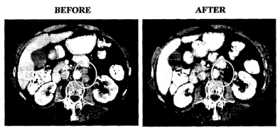
[0059] FIG. 16 shows a clinical response to vaccine. A patient with
progressive
disease in retroperitoneal lymph nodes (new, and positive on PET scan) 2
months after
cisplatin/etoposide was treated with 3 vaccines at the time of progression. A
PR was observed six
weeks after the first vaccine administration. On the left, an abdominal CT
scan performed 1
week prior to the first vaccine demonstrates 2 enlarged retroperitoneal lymph
nodes (circled,
each 2 cm in diameter). Two weeks after the third vaccine, the CT scan on the
right was obtained
demonstrating a greater than 60% reduction in the size of both lesions.
[0060] FIGS. 17A-17B concern association between immunologic and clinical
response to vaccination. In FIG. 17A, there are the results of IFN-y ELISPOT
assay from
patients who developed p53 immune response to vaccination. The background
level of
nonspecific IFN-7 production (irrelevant peptide) was subtracted. The number
of spots per
1x10⁵ cells are shown. All measure ments were done in quadruplicate. The mean
for each
sample is shown. In FIG. 17B, there are lymphocyte counts (x109/L) in patients
who were
treated with second-line chemotherapy. Columns, mean; bars, SD.
DETAILED DESCRIPTION OF THE INVENTION
[0061] The present invention is related in subject matter to U.S. Patent
Application
Publication No. 20030045499, which is incorporated by reference herein in its
entirety.
1. Definitions
[0062] As used herein the specification, "a" or "an" may mean one or more. As
used herein in the claim(s), when used in conjunction with the word
"comprising", the words "a"
or "an" may mean one or more than one. As used herein "another" may mean at
least a second
or more. Some embodiments of the invention may consist of or consist
essentially of one or
more elements, method steps, and/or methods of the invention. It is
contemplated that any
method or composition described herein can be implemented with respect to any
other method or
composition described herein.
19
CA 02608236 2007-11-09
WO 2006/124700 PCT/US2006/018592
[0063] The term "conferring or restoring chemosensitivity" as used herein
refers to
rendering a cancer cell responsive to cancer treatment wherein the cancer cell
is presently not
responsive to a cancer treatment, is predicted to be nonresponsive to a cancer
treatment, or is
susceptible to being nonresponsive to a cancer treatment, for example. More
specifically, the
proliferation of a cancer cell that is not affected by a particular cancer
treatment becomes
affected by a cancer treatment. The cancer cell may have come from a cancer,
such as in a
tumor, for example, that had been previously sensitive to a cancer treatment,
or the cancer cell
may have come from a cancer, such as in at.unor, for example, that was never
sensitive to a
cancer treatment. The cancer cell may be susceptible to becoming resistant to
one or more
cancer treatments; and the method of the invention prevents the cell from
becoming resistant to
one or more cancer treatments. In certain embodiments, the cancer cell is
susceptible to
becoming resistant to treatment because it comprises a mutation in one or more
polynucleotides
associated with resistance and/or it comprises upregulation or downregulation
of one or more
polynucleotides, wherein the upregulation or downregulation is associated with
resistance.
[0064] The term "first line therapy" as used herein refers to a first
treatment a
person receives after being diagnosed with cancer.
[0065] The term "immunogenic composition" as used herein refers to a
composition that elicits an immune response in the body of an individual. In
specific
embodiments, the immunogenic composition comprises a vaccine, which may be
defined as an
immunogenic composition that provides immunity upon subsequent challenge.
[0066]* The terms "resistant" or "therapy-resistant" as used herein refers to
cancer
comprising one or more cancer cells that are not able to be treated by one or
more cancer
treatments. For example, the cancer cell or cancer cells may still be able to
proliferate following
subjecting the cell to the treatment. In a specific embodiment, the cancer
treatment that one or
more cells are resistant to is chemotherapy. In other aspects, the resistance
may be to one or
more cancer therapies. In further specific embodiments, the resistant cells
develop resistance to
the therapy, whereas in alternative embodiments the resistant cells were
always resistant to the
therapy or comprised a biological or physiological phenotype or genotype
rendering it unable to
be sensitive to one or more cancer treatments.
[0067] In some embodiments an individual is treatable with the methods of the
invention wherein the individual has previously been treated with a cancer
treatment, such as
CA 02608236 2007-11-09
WO 2006/124700 PCT/US2006/018592
chemotherapy, radiation, or both for example, although in other embodiments
the individual has
not been previously treated with a cancer treatment. In aspects wherein the
individual has not
been previously treated with a cancer treatment, the individual may comprise
one or more cancer
cells that will become resistant upon exposure to the cancer. treatment. The
manifestation of this
resistance may occur immediately or soon after initiation of the cancer
treatment to which the
cells will' become resistant, or the resistance may not manifest until months
or years following
initation of the treatment. The one or more cancer cells that are resistant or
will become resistant
to the therapy may or may not be metastatic.
[0068] The therapy to which the individual has one or more resistant cells is
in the
context of treatment routinely given for a particular cancer. That is, the
therapy to which the
individual is resistant may be qualified in terms of a traditional cancer
treatment for that
particular cancer, and in certain aspects the invention may relate to
resistance to a clinically-
recognized therapy for a particular cancer. For example, skilled artisans
recognize that for breast
cancer, traditional, clinically-recognized therapy includes at least Herceptin
; aromatase
inhibitors (Arimidex [chemical name: anastrozole], Aromasin [chemical name:
exemestane],
and Femara(M [chemical name: letrozole]); tamoxifen, raloxifene, toremifene,
or Faslodex
(chemical name: fulvestrant). Exemplary clinically-recognized therapy for lung
cancer includes
at least cisplatin, etoposide, carboplatin, paclitaxel, docetaxel, vinorelbine
tartrate, doxorubicin,
vincristine sulfate, ifosfamide, and/or gemcitabine hydrochloride. Exemplary
clinically-
recognized therapy for prostate cancer includes at least docetaxel;
luteinizing hormone-releasing
hormone agonists, such as leuprolide, goserelin, and buserelin; antiandrogens,
such as flutamide
and bicalutamide; ketoconazole; and/or aminoglutethimide. One of skill in the
art recognizes
other conventional, clinically recognized treatments for other cancer types.
[0069] The term "second line therapy" as used herein refers to a therapy
additional
and subsequent to a first line therapy and in particular aspects is non-
identical to the first line
therapy. In cases where a human tumor responds (i.e., complete or partial
response) to a first
line therapy, the tumor is termed "sensitive" and, if the tumor recurs, second
line treatment may
involve re-administration of the same first line active therapy. However,
SCLC, for example, is
an especially aggressive cancer and has a very high frequency of tumor
recurrence. In cases
where tumors are treated with first line chemotherapy and the tumor either
fails to respond (i.e.,
does not regress) or continues to grow, these tumors are considered
"resistant" if tumor growth
occurs within 90 days of completion of a chemotherapy regimen. As described
above, for
21
CA 02608236 2007-11-09
WO 2006/124700 PCT/US2006/018592
resistant tumors, a different chemotherapy is used for subsequent treatment,
in specific
embodiments.
[00701 The term "sensitive" as used herein refers to cancer comprising one or
more
cancer cells that is able to be treated with a particular cancer treatment.
For example, the cell or
cells are not able to proliferate following subjecting the cell to the
treatment. In specific
embodiments, a cell that is sensitive to a particular cancer treatment is
killed by the treatment.
II. The Present Invention
[0071] The present invention contemplates the treatment of therapy-resistant
hyperproliferative disease. In particular aspects, the treatment is by
conferring or restoring
chemosensitivity to an individual with cancer, wherein one or more of the
cancer cells is resistant
to therapy, by administering a self gene product expression construct in
dendritic cells, which
subsequently present the processed self gene product antigen to immune
effector cells. In
specific embodiments, the self gene product expression construct comprises a
p53 expression
construct. The immune effector cells then mount an anti-self gene product
response, such as an
anti-p53 response, resulting in the destruction or lysis of hyperproliferative
cells presenting
mutant self gene product antigen, including therapy-resistant
hyperproliferative cells, such as
exemplary mutant p53 antigen. In particular embodiments, dendritic cells are
obtained from a
patient in which expression of the self gene product, such as p53, is
upregulated in
hyperproliferative cells. The dendritic cells obtained are infected with an
adenoviral vector
comprising a p53 gene and the p53 adenovirus-infected dendritic cells are
administered to the
individual. It is contemplated that infected dendritic cells will present self
gene antigens to
immune effector cells, stimulate an anti-self gene response in the patient,
and result in the
destruction or lysis of hyperproliferative cells presenting mutant self gene
antigen, including at
least some that are resistant to cancer therapy. In specific embodiments, the
hyperproliferative
disease and/or its resistance to a cancer therapy is characterized by
alteration or iricreased
expression of a self gene product.
[0072] In further embodiments, the present invention encompasses sensitizing
one
or more cells of a hyperproliferative disease, and in particular embodiments,
the disease and
diseased cells thereof are resistant to a drug, radiation, or both, for
example. The disease may be
generally characterized by an alteration and/or increased expression of a self
gene product and/or
the resistance of the disease to one or more particular therapies may be
generally characterized
22
CA 02608236 2007-11-09
WO 2006/124700 PCT/US2006/018592
by an alteration and/or increased expression of a self gene product. In
particular embodiments,
the subject with the disease is provided a dendritic cell expressing the self
gene product in
addition to administering to the subject a further treatment for the
hyperproliferative disease,
such as a drug or radiation therapy, for example.
[0073] In additional embodiments, there is a method of conferring or restoring
chemosensitivity to one or more chemotherapy-resistant cancer cells in an
individual, comprising
delivering to the individual a therapeutically effective amount of a dendritic
cell expressing a self
gene product and an additional treatment for the cancer. In a certain aspect
of the invention, the
composition comprises p53 in an adenoviral vector housed in a dendritic cell.
[0074] The dendritic cell expressing a self gene product may be considered an
immunogenic composition, and in particular embodiments, the invention
comprises methods of
providing the dendritic cell expressing a self gene product and of providing
another cancer
therapy nonidentical to the dendritic cell expressing the self gene product,
although dendritic
cells expressing other self gene products may be employed. The therapy that is
not the dendritic
cell expressing a self gene product may comprise any type of cancer therapy,
including, for
example, chemotherapy, radiation, gene therapy, surgery, immunotherapy,
hormone therapy, and
the like. The two separate therapies may be administered to an individual in
any suitabable
regimen, although in specific embodiments the immunogenic composition is
delivered
subsequent to the other therapy. Part or all of the dendritic cell therapy and
second therapy may
be repeated, such as by cycling of the therapies.
[0075] Thus, in particular embodiments of the invention, there is a method of
providing to an individual with a therapy-resistant hyperproliferative disease
an immunogenic
composition comprising a dendritic cell having a self gene product. In further
embodiments, the
individual is provided a cancer therapy in addition to the immunogenic
composition, and in
certain aspects the two therapies work in an additive manner or in a
synergistic manner to treat
the hyperproliferative disease, including hyperproliferative cells that are
resistant to a cancer
treatment. In additional embodiments, the dendritic cell expressing a self
gene product is
considered a vaccine.
III. Advexin -Dendritic Cell (DC)
[0076] Although any suitable composition comprising a dendritic cell
expressing a
self gene product may be employed in the invention, in specific aspects of the
invention an
23
CA 02608236 2007-11-09
WO 2006/124700 PCT/US2006/018592
Advexin -DC composition is utilized. As used herein, an Advexin -DC
composition
comprises wild-type p53 on a vector, wherein the vector is comprised in a
dendritic cell. In
particular aspects of the invention, the vector may be any suitable vector
such that it permits
expression of p53 within the dendritic cell. Exemplary embodiments of vectors
include
adenoviral vectors, viral vectors, adeno-associated viral vectors, retroviral
vectors, such as
lentiviral vectors, herpes viral vectors, or vaccinia viral vectors.
[0077] Although wild-type p53 is easily obtained by one of skill in the art,
an
exemplary wild-type sequence is provided in SEQ ID NO: 1 (National Center for
Biotechnology
Information GenBank Accession No. M14695). Other p53 sequences are available
in the
National Center for Biotechnology's GenBank database.
[0078] In other embodiments, a composition is employed pursuant to those =
described in U.S. Patent No. 6,726,907, which is incorporated by reference
herein in its entirety,
which includes a purified adenoviral vector composition comprising p53, for
example.
IV. Enhancement of Methods and Compositions
[0079] In some embodiments of the invention, a dendritic cell expressing a
self
gene product fiuther comprises one or more moieties to enhance the activity of
the dendritic cell
composition. The moiety may be added to the dendritic cell before or after the
dendritic cell was
manipulated to comprise the self gene product. In particular aspects of the
invention, a dendritic
cell was subjected to a composition to enhance its activity.
[0080] Any composition that enhances the activity of a dendritic cell
expressing a
self gene product may be employed in the invention, although in particular
aspects the moiety
comprises an antibody, and in specific embodiments the antibody is an
monoclonal antibody,
although optionally the antibody is a polyclonal antibody. In particular
embodiments, the
dendritic cell is subjected to anti-CD40 antibody (Nikitina et al., 2002).
Alternate methods for
promoting differentiation and activation of DC include treatment with pathogen
receptors and
inflammatory signals (see, for example Munz C, Steinman RM, Fujii S. Dendritic
cell
maturation by innate lymphocytes: coordinated stimulation of innate and
adaptive immunity. J
Exp Med. 2005 Jul 18;202(2):203-7).
[0081] The delivery method for any composition that enhances the activity of a
dendritic cell expressing a self gene product may be of any suitable kind. In
some embodiments,
24
CA 02608236 2007-11-09
WO 2006/124700 PCT/US2006/018592
for example, the enhancing composition is provided as a polynucleotide, a
polypeptide, a
peptide, a small molecule, and so forth, and the delivery method is
appropriately suited. For
example, a small molecule, polypeptide, and/or protein that enhances the
activity of a dendritic
cell expressing a self gene product may be delivered in a liposome to an
individual in need
thereof. Alternatively, a polynucleotide encoding the enhancing composition
may be utilized. In
certain aspects, a polynucleotide encoding the enhancing composition is the
same or different as
the polynucleotide that encodes the self gene product. In those embodiments
wherein the same
polynucleotide comprising a sequence that encodes a self gene product also
comprises a
sequence that encodes the enhancing composition, the two sequences may encode
a fusion gene
product or may encode two separate gene products. In further embodiments, the
sequence that
encodes a self gene product and the sequence that encodes the enhancing
composition are
regulated by different regulatory regions, although in alternative embodiments
they are regulated
by the same regulatory region. Any regulatory region, which in specific
embodiments may be
referred to as a promoter, may be a tissue-specific regulatory region, an
inducible regulatory
region, or a constitutive regulatory region, for example.
V. Subjects for Treatment with Methods of the Invention
[0082] Any individual may be treated with methods and compositions of the
invention. In certain aspects of the invention, the methods and compositions
concern cancer
vaccines. In particular embodiments, an individual is administered a vaccine
of the invention.
An individual suited for the methods and compositions of the invention may
have one or more
risk factors for developing one or more types of cancer. A risk factor may be
defined as
anything that increases the chance of developing cancer, and in this case may
be anything that
increases the chance of developing therapy-resistant cancer. The risk of
developing therapy-
resistant cancer may manifest before, during, or after administration of the
therapy to which
resistance has occurred.
[0083] The following risk factors may apply in general to developing cancer or
specifically to developing therapy-resistant cancer, and thus, in specific
embodiments the
individual has one or more risk factors for developing cancer, including
therapy-resistant cancer.
Although different cancers have different risk factors, some risk factors
apply to more than one
type of cancer, such as having a preneoplastic condition, a personal history
of cancer, a family
history of cancer, and/or having altered genes and/or gene expression, for
example for p53.
Some risk factors are specific to one or more types of cancer, such as having
particular altered
CA 02608236 2007-11-09
WO 2006/124700 PCT/US2006/018592
genes and/or gene expression, for example BRCA1 or BRCA2 for breast cancer;
unprotected
exposure to strong sunlight for skin cancer; tobacco use for cancers of the
lungs, larynx, mouth,
throat, esophagus, kidneys, bladder, colon, and several other organs; and so
forth.
[0084] Risk factors for individuals developing therapy-resistant cancer may be
of
any kind, although in specific embodiments they comprise one or more mutations
and/or
expression alterations identified with a particular polynucleotide. Examples
include EGFR
mutation and resistance of non-small-cell lung cancer to gefitinib (Kobayashi
et al., 2005);
melanocyte master regulator MITF (microphthalmia-associated transcription
factor) and
resistance to skin cancer (Garraway et al., 2005); ZNRD1 expression changes in
gastric cancer
cells (Zhang et al., 2003), for example. The classic mechanism for conferring
resistance to
chemotherapies is via up-regulation of the P-glycoprotein family of genes,
responsible for
conferring the mdr (multi-drug resistance) phenotype (Clarke R, Leonessa F,
Trock B.
Multidrug resistance/P-glycoprotein and breast cancer: review and meta-
analysis. Semin Oncol.
2005 Dec;32(6 Suppl 7):S9-15.). Mutations associated with resistance to breast
cancer include
estrogen receptor mutations in tamoxifen-resistant breast cancer (Karnik et
al., 1994); a mutation
in 482 (R482) in human BreastCancer Resistance Protein (BCRP) associated with
doxorubicin
resistance (Allen et al., 2002);
-[0085] An individual with one or more risk factors for developing therapy-
resistant
cancer may be administered the methods and compositions of the present
invention at any time,
including before developing therapy-resistant cancer, after developing therapy-
resistant cancer,
or both.
VI. Hyperproliferative Disease
[0086] Cancer has become one of the leading causes of death in the Western
world,
second only behind heart disease. Current estimates project that one person in
three in the U.S.
will develop cancer, and that one person in five will die from cancer. Cancers
can be viewed
from an immunologic perspective as altered self cells that have lost the
normal growth-regulating
mechanisms.
[0087] Oncogenes are polynucleotides that have the potential to cause a normal
cell
to become cancerous. There are currently three major categories of oncogenes
reflecting their
different activities. One category of oncogenes encode proteins that induce
cellular proliferation.
A second category of oncogenes, called tumor-suppressors genes or anti-
oncogenes, function to
26
CA 02608236 2007-11-09
WO 2006/124700 PCT/US2006/018592
inhibit excessive cellular proliferation. The third category of oncogenes
either block or induce
apoptosis by encoding proteins that regulate programmed cell death.
[0088] In one embodiment of the , present invention, the treatment of
hyperproliferative disease involves the administration of a self gene
expression construct to
dendritic cells, and in specific embodiments, the administration is
intradermally. It is
contemplated that the dendritic cells present the processed self gene wild-
type antigens to
immune effector cells, which mount an anti-self gene response, resulting in
the destruction or
lysis of hyperproliferative cells presenting mutant self antigen. The three
major categories of
oncogenes are discussed below and listed in Table 1.
[0089] In particular embodiments, the present invention may be employed in the
treatment of any type of cancer, including, for example, lung, breast,
prostate, colon, pancreatic,
brain, skin, thyroid, liver, kidney, spleen, esophageal, ovarian, cervical,
uterine, testicular, bone,
pituitary gland, stomach, blood, bone marrow, and lymphatic system.
[0090] In specific embodiments, the present invention is utilized for the
treatment
of small cell lung cancer. Small cell lung cancer (SCLC) constitutes 15-20% of
the
approximately 170,000 new cases of lung cancer seen annually in the US. SCLC
is the most
aggressive form of lung cancer, with 5 year survival rates of <10%. Diagnosis
of extensive stage
disease (ES) comprises approximately two-thirds of new SCLC cases, and results
in survival of
only 2-4 months if untreated, and survival increases to 6-7 months with
aggressive chemotherapy
regimens. Both limited stage and extensive stage disease are very responsive
to first line
chemotherapy with response rates of greater than 50% routinely observed.
However, these
responses almost invariably are short-lived and disease recurrence in ES
patients occurs
frequently. After relapse or failure to respond to chemotherapy, patients
generally succumb to
disease within a few months (Schiller, 2001). Treatment of patients with
relapsed SCLC is
especially challenging: if patients are platinum-resistant (i.e., disease
progression occurs within 3
months of completion of a platinum regimen), median survival ranges from 3.7
to 4.7 months.
For platinum-sensitive patients, median survival ranges from 5.8-6.9 months
(Eckardt, 2005).
A. Inducers of Cellular Proliferation
[0091] The proteins that induce cellular proliferation further fall into
various
categories dependent on function. The commonality of all of these proteins is
their ability to
27
CA 02608236 2007-11-09
WO 2006/124700 PCT/US2006/018592
regulate cellular proliferation. For example, a form of PDGF, the sis oncogene
is a secreted
growth factor. Oncogenes rarely arise from genes encoding growth factors, and
at the present, sis
is the only known naturally occurring oncogenic growth factor.
[0092] The proteins fms, erbA, erbB and neu are growth factor receptors.
Mutations to these receptors result in loss of regulatable function. For
example, a point mutation
affecting the transmembrane domain of the nue receptor protein results in the
nue oncogene. The
erbA oncogene is derived from the intracellular receptor for thyroid hormone.
The modified
oncogenic erbA receptor is believed to compete with the endogenous thyroid
hormone receptor,
causing uncontrolled growth.
[0093] The largest class of oncogenes are the signal transducing proteins
(e.g., src,
abl and ras) are signal transducers. The protein src, is a cytoplasmic protein-
tyrosine kinase, and
its transformation from proto-oncogene to oncogene in some cases, results via
mutations at
tyrosine residue 527. In contrast, transformation of GTPase protein ras from
proto-oncogene to
oncogene, in one example, results from a valine to glycine mutation at amino
acid 12 in the
sequence, reducing ras GTPase activity.
[0094] The proteins jun, fos and myc are proteins that directly exert their
effects on
nuclear functions as transcription factors. Table 1 lists a variety of the
oncogenes described in
this section and many of those not described.
B. Inhibitors of Cellular Proliferation
[0095] The tumor suppressor oncogenes function to inhibit excessive cellular
proliferation. The inactivation of these genes results destroys their
inhibitory activity, resulting in
unregulated proliferation. The tumor suppressors p53, p16 and C-CAM are
described below.
[0096] High levels of mutant p53 have been found in many cells transformed by
chemical carcinogenesis, ultraviolet radiation, and several viruses. The p53
gene is a frequent
target of mutational inactivation in a wide variety of human tumors and is
already documented to
be the most frequently-mutated gene in common human cancers. It is mutated in
over 50% of
human NSCLC (Hollstein et al., 1991) and in a wide spectrum of other tumors. A
variety of
cancers have been associated with mutations of the p53 gene, which result in
the loss of p53
tumor suppressor properties. Mutations in the p53 gene further account for
approximately 50%
of all cancers that develop (Vogelstein and Kinzler, 1992; Levine et al.,
1991), with the majority
28
CA 02608236 2007-11-09
WO 2006/124700 PCT/US2006/018592
of these mutations being single-base missense mutations (Kovach et al., 1996).
It has been
observed that mutations resulting in a loss of p53 function also result in
high nuclear and
cytoplasmic concentrations (i.e., overexpression) of mutant p53 protein
(Oldstone et al., 1992;
Finlay et al., 1988). In contrast, functional wild-type p53 protein is
expressed at very low levels
in cells.
[0097] The high cellular concentrations of p53 mutant protein has recently
received
much attention as an avenue for cancer immunotherapy. The general concept is
to elicit an
immune response against tumor cells presenting mutant p53 peptides bound to
MHC molecules
on the cell surface. The generation of an anti-tumor response using mutant p53
peptides as
antigens has been demonstrated in several studies (McCarty et al., 1998;
Gabrilovich et al.,
1996; Mayordomo et al., 1996; Zitvogel et al., 1996) However, this approach to
cancer
immunotherapy has several limitations. For example, p53 mutations can occur at
many different
sites in the protein, making it necessary to identify the site of the mutation
in each patient before
creating a specific mutant peptide for p53 cancer therapy. Further, not all
mutations are
contained in regions of the protein known to bind to MHC molecules, and
therefore would not
elicit an anti-tumor response (DeLeo, 1998).
[0098] The limitations described above have stimulated the search for
antigenic
epitopes in wild-type p53 sequences common to the vast majority of tumor
derived p53 proteins.
Wild-type p53 peptide-specific cytotoxic T lymphocytes have been produced from
human and
murine responding lymphocytes, some of which recognized p53-overexpressing
tumors in vitro
and in vivo (Theobald, et al., 1995; Ropke et al., 1996; Nijman et al., 1994;
U.S. Pat. No.
5,747,469, specifically incorporated herein by reference in its entirety).
However, since the
presentation of antigens is MHC class I restricted, only certain peptides can
successfully be
administered in certain patients, due to the polymorphic nature of the MHC
class I peptide
binding site. Further, it is not practical to identify all possible p53
peptides binding to a particular
individuals repertoire of MHC molecules. Additionally, a peptide vaccine that
does bind to a
patient's class I MHC may not be sufficiently presented by MHC class II, the
molecules crucial
in the induction of CD4+.T cell immune responses.
[0099] Researchers have to attempted to identify multiple p53 epitopes, which
should permit more effective immune responses against tumor cells expressing
multiple p53
genes with mutations at different sites. This could be accomplished by
immunizing cells with
29
CA 02608236 2007-11-09
WO 2006/124700 PCT/US2006/018592
intact wild-type p53 to take advantage of the overexpression of the whole p53
polypeptide in
most human tumors. The dendritic cell (DC) is the cell type best suited for
vaccine antigen
delivery (described further herein), as they are the most potent antigen
presenting cells, effective
in the stimulation of both primary and secondary immune responses (Steinman,
1991; Celluzzi
and Falo, 1997). It is contemplated in the present invention that the
transduction.of dendritic
cells with wild-type p53 protein, using a viral expression construct, will
elicit a potent antitumor
immune response specific for a variety of cells expressing different mutant
p53 proteins. Further,
since most mutations of p53 are single-base missense mutations, the approach
of the present
invention overcomes the limitations of identifying the site of the p53
mutation and subsequent
preparation of a customized mutant peptide for immunotherapy. Thus, the method
of the present
invention provides the basis for a simple and novel approach to immunotherapy
based cancer
treatment.
[0100] Wild-type p53 is recognized as an important growth regulator in many
cell
types. Missense mutations are common for the p53 gene and are essential for
the transforming
ability of the oncogene. A single genetic change prompted by point mutations
can create
carcinogenic p53. Unlike other oncogenes, however, p53 point mutations are
known to occur in
at least 30 distinct codons, often creating dominant alleles that produce
shifts in cell phenotype
without a reduction to homozygosity. Additionally, many of these dominant
negative alleles
appear to be tolerated in the organism and passed on in the germ line. Various
mutant alleles
appear to range from minimally dysfunctional to strongly penetrant, dominant
negative alleles
(Weinberg, 1991).
[0101] Another inhibitor of cellular proliferation is p16. The major
transitions of
the eukaryotic cell cycle are triggered by cyclin-dependent kinases, or CDK's.
One CDK, cyclin-
dependent kinase 4 (CDK4), regulates progression through the G1. The activity
of this enzyme
may be to phosphorylate Rb at late Gl. The activity of CDK4 is controlled by
an activating
subunit, D-type cyclin, and by an inhibitory subunit, the p16INK4 has been
biochemically
characterized as a protein that specifically binds to and inhibits CDK4, and
thus may regulate Rb
phosphorylation (Serrano et al., 1993; Serrano et al., 1995). Since the
p16rNK4 protein is a CDK4
inhibitor (Serrano, 1993), deletion of this gene may increase the activity of
CDK4, resulting in
hyperphosphorylation of the Rb protein. p16 also is known to regulate the
function of CDK6.
CA 02608236 2007-11-09
WO 2006/124700 PCT/US2006/018592
[0102] p161NK4 belongs to a newly described class of CDK-inhibitory proteins
that
also includes B 21 WAFI KIPI mrx4
p16 , p , and p27 . The p16 gene maps to 9p2l, a chromosome region
frequently deleted in many tumor types. Homozygous deletions and mutations of
the p 16INK4
gene are frequent in human tumor cell lines. This evidence suggests that the p
16INK4 gene is a
tumor suppressor gene. This interpretation has been challenged, however, by
the observation that
the frequency of the p 16INK4 gene alterations is much lower in primary
uncultured tumors than in
cultured cell lines (Caldas et al., 1994; Cheng et al., 1994; Hussussian et
al., 1994; Kamb et al.,
1994; Kamb et al., 1994; Mori et al., 1994; Okamoto et al., 1994; Nobori et
al., 1995; Orlow et
al., 1994; Arap et al., 1995). Restoration of wild-type p161NK4 function by
transfection with a
plasmid expression vector reduced colony formation by some human cancer cell
lines (Okamoto,
1994; Arap, 1995).
[0103] C-CAM is expressed in virtually all epithelial cells (Odin and Obrink,
1987). C-CAM, with an apparent molecular weight of 105 kD, was originally
isolated from the
plasma membrane of the rat hepatocyte by its reaction with specific antibodies
that neutralize
cell aggregation (Obrink, 1991). Recent studies indicate that, structurally, C-
CAM belongs to the
immunoglobulin (Ig) superfamily and its sequence is highly homologous to
carcinoembryonic
antigen (CEA) (Lin and Guidotti, 1989). Using a baculovirus expression system,
Cheung et al.
(1993) demonstrated that the first Ig domain of C-CAM is critical for cell
adhesive activity.
[0104] Cell adhesion molecules, or CAM's are known to be involved in a complex
network of molecular interactions that regulate organ development and cell
differentiation
(Edelman, 1985). Recent data indicate that aberrant expression of CAM's maybe
involved in the
tumorigenesis of several neoplasms; for example, decreased expression of E-
cadherin, which =is
predominantly expressed in epithelial cells, is associated with the
progression of several kinds of
neoplasms (Edelman and Crossin, 1991; Frixen et al., 1991; Bussemakers et al.,
1992; Matsura
et al., 1992; Umbas et al., 1992). Also, Giancotti and Ruoslahti (1990)
demonstrated that
increasing expression of a5(31 integrin by gene transfer can reduce
tumorigenicity of Chinese
hamster ovary cells in vivo. C-CAM now has been shown to suppress tumors
growth in vitro and
in vivo.
[0105] Other tumor suppressors that may be employed according to the present
invention include RB, APC, DCC, NF-1, NF-2, WT-1, MEN-I, MEN-II, zacl, p73,
VHL,
MMAC1, FCC and MCC (see Table 1).
31
CA 02608236 2007-11-09
WO 2006/124700 PCT/US2006/018592
C. Regulators of Programmed Cell Death
[0106] Apoptosis, or programmed cell death, is an essential occurring process
for
normal embryonic development, maintaining homeostasis in adult tissues, and
suppressing
carcinogenesis (Kerr et al., 1972). The Bcl-2 family of proteins and ICE-like
proteases have been
demonstrated to be important regulators and effectors of apoptosis in other
systems. The Bcl-2
protein, discovered in association with follicular lymphoma, plays a prominent
role in controlling
apoptosis and enhancing cell survival in response to diverse apoptotic stimuli
(Bakhshi et al.,
1985; Cleary and Sklar, 1985; Cleary et al., 1986; Tsujimoto et al., 1985;
Tsujimoto and Croce,
1986). The evolutionarily conserved Bcl-2 protein now is recognized to be a
member of a family=
of related proteins which can be categorized as death agonists or death
antagonists.
[0107] Subsequent to its discovery, it was shown that Bcl-2 acts to suppress
cell
death triggered by a variety of stimuli. Also, it now is apparent that there
is a family of Bcl-2 cell
death regulatory proteins which share in common structural and sequence
homologies. These
different family members have been shown to either possess similar functions
to Bcl-2 (e.g.,
Bc1XL, Bclw, Mcl-l, Al, Bfl-1) or counteract Bcl-2 function and promote cell
death (e.g., Bax,
Bak, Bik, Bim, Bid, Bad, Harakiri).
Table 1: Oncogenes
Gene Source Human Disease Function
Growth Factors' FGF family member
HST/KS Transfection
INT-2 MMTV promoter FGF family member
Insertion
INTI/WNTI MMTV promoter Factor-like
Insertion
SIS Simian sarcoma PDGF B
virus
Receptor Tyrosine Kinasesl 2
ERBB/HER Avian Amplified, deleted EGF/TGF-a/
erythroblastosis squamous cell amphiregulin/
virus; ALV cancer; hetacellulin
promoter glioblastoma receptor
insertion;
amplified
human tumors
ERBB-2/NEU/HER Transfected from rat Amplified breast, Regulated by NDF/
-2 Glioblatoms ovarian, gastric heregulin and
cancers EGF-
related factors
32
CA 02608236 2007-11-09
WO 2006/124700 PCT/US2006/018592
Gene Source Human Disease Function
FMS SM feline sarcoma CSF-1 receptor
virus
KIT HZ feline sarcoma MGF/Steel receptor
virus hematopoieis
TRK Transfection from NGF (nerve growth
human colon factor) receptor
cancer
MET Transfection from Scatter factor/HGF
human receptor
osteosarcoma
RET Translocations and Sporadic thyroid Orphan receptor Tyr
point mutations cancer; kinase
familial medullary
thyroid cancer;
multiple endocrine
neoplasias 2A and
2B
ROS URII avian sarcoma Orphan receptor Tyr
Virus kinase
PDGF receptor Translocation Chronic TEL(ETS-like
myclomonocytic transcription
leukemia factor)/
PDGF receptor
gene
fusion
TGF-(3 receptor Colon carcinoma
mismatch mutation
target
NONRECEPTOR TYROSINE KINASES 1
ABI. Abelson Mul.V Chronic Interact with RB,
myelogenous RNA
leukemia polymerase, CRK,
translocation CBL
with BCR
FPS/FES Avian Fujinami
SV;GA
FeSV
LCK Mul.V (murine Src family; T cell
leukemia signaling; interacts
virus) promoter CD4/CD8 T cells
insertion
SRC Avian Rous Membrane-
sarcoma associated Tyr
Virus kinase with
signaling function;
activated by
receptor kinases
33
CA 02608236 2007-11-09
WO 2006/124700 PCT/US2006/018592
Gene Source Human Disease Function
YES Avian Y73 virus Src family;
signaling
SER/THR PROTEIN KINASES'
AKT AKT8 murine Regulated by
retrovirus PI(3)K;
regulate 70-kd S6
k
MOS Maloney murine SV GVBD; cystostatic
factor; MAP
kinase
kinase
PIM-1 Promoter insertion
Mouse
RAF/MIL 3611 murine SV; Signaling in RAS
MH2 pathway
avian SV
MISCELLANEOUS CELL SURFACE'
APC Tumor suppressor Colon cancer Interacts with
catenins
DCC Tumor suppressor Colon cancer CAM domains
E-cadherin Candidate tumor Breast cancer Extracellular
Suppressor homotypic
binding;
intracellular
interacts with
catenins
PTC/NBCCS Tumor suppressor Nevoid basal cell 12 transmembrane
and cancer domain; signals
Drosophilia syndrome (Gorline through Gli
homology syndrome) homogue
CI to antagonize
hedgehog pathway
TAN-1 Notch Translocation T-ALI. Signaling
homologue
MISCELLANEOUS SIGNALINGI 3
BCL-2 Translocation B-cell lymphoma Apoptosis
CBL Mu Cas NS-1 V Tyrosine-
phosphorylated
RING
finger interact Abl
CRK CT1010 ASV Adapted SH2/SH3
interact Abl
DPC4 Tumor suppressor Pancreatic cancer TGF-(3-related
signaling
pathway
34
CA 02608236 2007-11-09
WO 2006/124700 PCT/US2006/018592
Gene Source Human Disease Function
MAS Transfection and Possible angiotensin
Tumorigenicity receptor
NCK Adaptor SH2/SH3
GUANINE NUCLEOTIDE EXCHANGERS AND BINDING
PROTEINS3 4
BCR Translocated with Exchanger; protein
ABL kinase
in CML
DBL Transfection Exchanger
GSP
NF-1 Hereditary tumor Tumor suppressor RAS GAP
Suppressor neurofibromatosis
OST Transfection Exchanger
Harvey-Kirsten, N- HaRat SV; Ki Point mutations in Signal cascade
RAS RaSV; many
Balb-MoMuSV; human tumors
Transfection
VAV Transfection S 112/S 113;
exchanger
NUCLEAR PROTEINS AND TRANSCRIPTION FACTORS1 s-9
BRCA1 Heritable suppressor Mammary Localization
cancer/ovarian unsettled
cancer
BRCA2 Heritable suppressor Mammary cancer Function unknown
ERBA Avian thyroid hormone
erythroblastosis receptor
Virus (transcription)
ETS Avian E26 virus DNA binding
EVII MuLV promotor AML Transcription factor
Insertion
FOS FBI/FBR murine 1 transcription
osteosarcoma factor
viruses with c-JUN
GLI Amplified glioma Glioma Zinc finger; cubitus
interruptus
homologue
is in hedgehog
signaling pathway;
inhibitory link
PTC
and hedgehog
HMGG/LIM Translocation Lipoma Gene fusions high
t(3:12) mobility group
t(12:15) HMGI-C (XT-
hook)
and transcription
CA 02608236 2007-11-09
WO 2006/124700 PCT/US2006/018592
Gene Source Human Disease Function
factor
LIM or acidic
domain
JUN ASV-17 Transcription factor
AP-1 with FOS
MLL/VHRX + Translocation/fusion Acute myeloid Gene fusion of
ELI/MEN ELL with MLL leukemia DNA-
Trithorax-like gene binding and methyl
transferase MLL
with
ELI RNA pol II
elongation factor
MYB Avian DNA binding
myeloblastosis
Virus
MYC Avian MC29; Burkitt's lymphoma DNA binding with
Translocation B- MAX partner;
cell cyclin
Lymphomas; regulation; interact
promoter RB; regulate
Insertion avian apoptosis
leukosis
Virus
N-MYC Amplified Neuroblastoma
L-MYC Lung cancer
REL Avian NF-xB family
transcription factor
Retriculoendothelio
sis
Virus
SKI Avian SKV770 Transcription factor
Retrovirus
VHL Heritable suppressor Von Hippel-Landau Negative regulator
syndrome or
elongin;
transcriptional
elongation
complex
WT-1 Wilm's tumor Transcription factor
CELL CYCLE/DNA DAMAGE
RESPONSEIO-"
ATM Hereditary disorder Ataxia- Protein/lipid kinase
telangiectasia homology; DNA
damage response
upstream in P53
pathway
BCL-2 Translocation Follicular Apoptosis
36
CA 02608236 2007-11-09
WO 2006/124700 PCT/US2006/018592
Gene Source Human Disease Function
lymphoma
FACC Point mutation Fanconi's anemia
group
C (predisposition
leukemia
FHIT Fragile site 3p14.2 Lung carcinoma Histidine triad-
related
diadenosine
5',3f'rf-
l p4
P
tetraphosphate
asymmetric
hydrolase
hMLI/MutL HNPCC Mismatch repair;
MutL
homologue
hMSH2/MutS HNPCC Mismatch repair;
MutS
homologue
hPMS 1 HNPCC Mismatch repair;
MutL
homologue
hPMS2 HNPCC Mismatch repair;
MutL
homologue
INK4/MTS1 Adjacent INK-4B at Candidate MTSl p16 CDK inhibitor
9anti-estrogen suppressor and
receptor tyrosine MLM
kinase inhibitor; melanoma gene
CDK complexes
INK4B/MTS2 Candidate p15 CDK inhibitor
suppressor
MDM-2 Amplified Sarcoma Negative regulator
p53
p53 Association with Mutated >50% Transcription factor;
SV40 human checkpoint control;
T antigen tumors, including apoptosis
hereditary Li-
Fraumeni
syndrome
PRAD1/BCL1 Translocation with Parathyroid Cyclin D
Parathyroid adenoma;
hormone B-CLL
or IgG
RB Hereditary Retinoblastoma; Interact cyclin/cdk;
Retinoblastoma; osteosarcoma; regulate E2F
Association with breast transcription factor
many cancer; other
37
CA 02608236 2007-11-09
WO 2006/124700 PCT/US2006/018592
Gene Source Human Disease Function
DNA virus tumor sporadic
Antigens cancers
XPA xeroderma Excision repair;
pigmentosum; skin photo-
cancer product
predisposition recognition;
zinc finger
VII. Immunologic Responses Related to Self Gene Tumorogenicity
[0108] In one embodiment of the present irivention, hyperproliferative disease
in
which expression of a self gene is upregulated in therapy-resistant
hyperproliferative cells is
treated by administering a self gene expression construct capable of eliciting
an anti-self gene
response. The self gene p53 will be referred to herein as merely an exemplary
embodiment.
[0109] Following delivery of the p53 expression construct to a given antigen-
presenting cell, a cascade of immunologic events must ensue to stimulate the
desired anti-p53
response. Thus, a basic understanding of the immunologic responses related to
p53 expression
and more generally, self gene expression in hyperproliferative disease, is
necessary.
A. Cytotoxic T Lymphocytes
[0110] T lymphocytes arise from hematopoietic stem cells in the bone marrow,
and
migrate to the thymus gland to mature. T cells express a unique antigen
binding receptor on their
membrane (T-cell receptor), which can only recognize antigen in association
with major
histocompatibility complex (MHC) molecules on the surface of other cells.
There are at least two
populations of T cells, known as T helper cells and T cytotoxic cells. T
helper cells and T
cytotoxic cells are primarily distinguished by their display of the membrane
bound glycoproteins
CD4 and CD8, respectively. T helper cells secret various lymphokines, that are
crucial for the
activation of B cells, T cytotoxic cells, macrophages and other cells of the
immune system. In
contrast, a T cytotoxic cells that recognizes an antigen-MHC complex
proliferates and
differentiates into an effector cell called a cytotoxic T lymphocyte (CTL).
CTLs eliminate cells
of the body displaying antigen, such as virus infected cells and tumor cells,
by producing
substances that result in cell lysis.
[0111] An important aspect of the present invention is the stimulation of a
CTL
response directed against wild-type self gene antigen. It has been observed
that mutations of the
p53 gene result in the overexpression of the mutant p53 protein in tumor cells
(Harris, 1996),
38
CA 02608236 2007-11-09
WO 2006/124700 PCT/US2006/018592
while wild-type p53 is expressed at low levels in normal cells. It has further
been demonstrated
that wild-type and mutant p53 peptides can stimulate a CTL response against
tumor cells
expressing p53 antigenic peptides (DeLeo, 1998; Mayordomo et al., 1996). It is
contemplated in
the present invention that a similar anti-self gene CTL response will be
stimulated by
immunizing dendritic cells with intact wild-type self gene polypeptide, and
thus can be used as a
treatment for hyperproliferative disease.
B. Antigen-presenting Cells
[0112] Antigen-presenting cells, which include macrophages, B lymphocytes, and
dendritic cells, are distinguished by their expression of a particular MHC
molecule. APCs
internalize antigen and re-express a part of that antigen, together with the
MHC molecule on
their outer cell membrane.
[0113] In a preferred embodiment of the present invention, dendritic cells are
the
antigen-presenting cells of choice for self gene delivery and antigen
presentation. Dendritic cells
are the most potent antigen-presenting cells for the initiation of antigen-
specific T cell activation
(Arthur et al., 1997). They are also excellent candidates for short term
culture and a variety of
gene transfer methods (e.g., DNA/liposome complexes, electroporation, CaPO4
precipitation,
and recombinant adenovirus) (Arthur et al., 1997). Human and mouse dendritic
cells have been
successfully modified by adenoviral gene transfer (Sonderbye et al., 1998). In
this study, an
adenovirus (AdLacZ) was used to express intracellular beta-galactosidase (beta-
gal) antigen in
the dendritic cells, with approximately 40% of the cells transduced with
AdLacZ expressing high
levels of beta-gal. In addition, the subcutaneous immunization of mouse
dendritic cells with the
ovalbumin (OVA) peptide induced an OVA-specific CDB+CTL response (Celluzzi and
Falo,
1997).
C. Major Histocompatibilty Complex
[0114] The major histocompatibility complex (MHC) is a large genetic complex
with multiple loci. The MHC loci encode two major classes of MHC membrane
molecules,
referred to as class I and class II MHCs. T helper lymphocytes generally
recognize antigen
associated with MHC class II molecules, and T cytotoxic lymphocytes recognize
antigen
associated with MHC class I molecules. In humans the MHC is refereed to as the
HLA complex
and in mice the H-2 complex. An important aspect of the present invention is
the immunization
of dendritic cells with the intact wild-type self gene to take advantage of
the relative
39
CA 02608236 2007-11-09
WO 2006/124700 PCT/US2006/018592
overexpression of the whole self gene molecule in most human tumors. The
approach of p53
immunotherapy is contemplated in one embodiment, to overcome previous
immunotherapies that
immunized animals with mutant p53 peptides as antigens (Gabrilovich et al.,
1996; Mayordomo
et al., 1996; Zitgovel et al., 1996). Although the approaches above using
mutant p53 peptides
were effective at generating anti-tumor responses, they have several
limitations. For example,
p53 mutations and other self genes occur at many sites in the protein, making
it necessary to
identify the site of mutation in each patient before constructing a customized
mutant peptide for
therapy. Furthermore, not all mutations are contained in regions of the
protein known to bind to
MHC molecules. In another study using wild-type 53 peptides, CTLs were
generated from
human and murine responding lymphocytes, some of which recognized p53
overexpressing
tumors in vitro (Theobald et al., 1995; Ropke et al., 1996; Nijman et al.,
1994). However, since
presentation of antigens is MHC class I restricted, only certain oligopeptides
can be used in
certain patients, because of the highly polymorphic MHC class I peptide
binding site. It is
contemplated in the present invention that immunizing dendritic cells with
intact, wild-type self
gene protein, will generate a variety of self gene antigens for MHC class I
presentation and thus
effectively stimulate a cytolytic T lymphocyte response.
VIII. Assays for Self Gene Upregulation or Altered Expression
[0115] In one embodiment of the present invention, the identification of a
patient
with a therapy-resistant hyperproliferative disease in which self gene
expression is upregulated is
desired. In patients with a therapy-resistant hyperproliferative disease, a
sample of the
hyperproliferative tissue will be used to assay upregulation, for example. A
wide variety of
detection methods can be employed in the present invention to detect the self
gene status of at
least one therapy-resistant cell, in certain embodiments. There are numerous
antibodies to the
oncogenic proteins, for example, and hence any assay that utilizes antibodies
for detection, for
example, ELISAs, Western Blotting, immunoassay techniques, etc., are
contemplated as useful
in the present invention. Alternatively, assays that employ nucleotide probes
may be used to
identify the presence of self gene, for example, Southern blotting, Northern
blotting or PCRTM
techniques. All the above techniques are well known to one of skill in the art
and could be
utilized in the present invention without undue experimentation.
A. ELISAs, Immunoassay and Immunohistological Assay.
[0116] In a particular embodiment of the present invention, immunohistological
assays are used to detect self gene increased or altered expression in therapy-
resistant tumor
CA 02608236 2007-11-09
WO 2006/124700 PCT/US2006/018592
samples (e.g., tissue sections). Exemplary methods of immunohistochemistry
assays and
immunfluorescence assays have previously been described (U.S. Pat. No.
5,858,723;
W094/11514, specifically incorporated herein by reference in its entirety).
Further
immunoassays encoinpassed by the present invention include, but are not
limited to those
described in U.S. Pat. No. 4,367,110 (double monoclonal antibody sandwich
assay) and U.S. Pat.
No. 4,452,901 (western blot). Other assays include immunoprecipitation of
labeled ligands and
immunocytochemistry, both in vitro and in vivo. Immunoassays generally are
binding assays.
Certain preferred immunoassays are the various types of enzyme linked
immunosorbent assays
(ELISAs) and radioimmunoassays (RIA) known in the art.
[0117] In one exemplary ELISA, the anti-self gene antibodies are immobilized
on a
selected surface, such as a well in a polystyrene microtiter plate, dipstick
or column support.
Then, a test composition suspected of containing the desired antigen, such as
a clinical sample, is
added to the wells. After binding and washing to remove non-specifically bound
immune
complexes, the bound antigen may be detected. Detection is generally achieved
by the addition
of another antibody, specific for the desired antigen, that is linked to a
detectable label. This type
of ELISA is known as a"sandwich ELISA." Detection also may be achieved by the
addition of a
second antibody specific for the desired antigen, followed by the addition of
a third antibody that
has binding affinity for the second antibody, with the third antibody being
linked to a detectable
label.
B. Southern and Northern Blotting Techniques
[0118] Southern and Northern blotting are commonly used techniques in
molecular
biology and well within the grasp of one skilled in the art. Southern and
Northern blotting
samples are obtained from the hyperproliferative tissue. The DNA and RNA from
test cells is
recovered by gentle cell rupture in the presence of a cation chelator such as
EDTA. The proteins
and other cell milieu are removed by admixing with saturated phenol or
phenol/chloroform and
centrifugation of the emulsion. The DNA and RNA is in the upper aqueous phase,
it is
deproteinized and mixed with ethanol. This solution allows the DNA and RNA to
precipitate, the
DNA and RNA can then be recover using centrifugation. In the case of RNA
extraction, RNAse
inhibitors such as DEPC are needed to prevent RNA degradation.
[0119] Electrophoresis in agarose or polyacrylamide gels is the most usual way
to
separate DNA and RNA molecules. Southern blotting will confirm the identity of
the self gene
41
CA 02608236 2007-11-09
WO 2006/124700 PCT/US2006/018592
encoding DNA. This is achieved by transferring the DNA from the intact gel
onto nitrocellulose
paper. The nitrocellulose paper is then washed in buffer that has for example,
a radiolabelled
cDNA containing a sequence complementary to wild-type self gene DNA. The
probe, binds
specifically to the DNA that encodes a region of self gene and can be detected
using
autoradiography by contacting the probed nitrocellulose paper with
photographic film. Self gene
-encoding mRNA can be detected in a similar manner by a process known as
Northern blotting.
For a more detailed description of buffers gel preparation, electrophoresis
condition etc., the
skilled artisan is referred to Sambrook, 1989.
C. Polymerase Chain Reaction (PCRTM)
[0120] PCRTM is a powerful tool in modern analytical biology. Short
oligonucleotide sequences usually 15-35 bp in length are designed, homologous
to flanking
regions either side of the self gene sequences to be amplified. The primers
are added in excess to
the source DNA, in the presence of buffer, enzyme, and free nucleotides. The
source DNA is
denatured at 95 C and then cooled to 50-60 C to allow the primers to anneal.
The temperature is
adjusted to the optimal temperature for the polymerase for an extension phase.
This cycle is
repeated 25-40 times.
[0121] In particular the present invention uses PCRTM to detect the self gene
status
of cells. Mutations in the self gene are first detected with Single Strand
Conformation
Polymorphism (SSCP) which is based on the electrophoretic determination of
conformational
changes in single stranded DNA molecules induced by point mutations or other
forms of slight
nucleotide changes. To identify where the mutation is located at within the
self gene, each exon
is separately amplified by PCRTM using primers specific for the particular
exon. After
amplification, the. PCRTM product is denatured and separated out on a
polyacrylamide gel to
detect a shift in mobility due to a conformational change which resulted
because of a point
mutation or other small nucleotide change in the gene. Mutations result in a
change in the
physical conformation of the DNA as well as change in the electrical charge of
the molecule.
Thus during electrophoresis when an electrical charge is applied to the
molecule, DNA that is
slightly different in shape and charge as compared to wild-type will move at a
different rate and
thus occupy a different position in the gel. After determination of which DNA
fragment contains
the mutation, the specific nucleotide changes are detected by DNA sequencing
of the amplified
PCRTM product. Sequencing of linear DNA- breaks down the DNA molecule into its
individual
nucleotides in the order with which they are assembled in the intact molecule.
Separation of the
42
CA 02608236 2007-11-09
WO 2006/124700 PCT/US2006/018592
individual nucleotides by electrophoresis on a sequencing gel allows detection
of individual
nucleotide changes compared to wild-type and is used to determine homo- or
heterozygocity of a
mutation, which is easily distinguished by the appearance of a single or
double band in the
sequencing gel.
IX. Self Gene Delivery
[0122] Many types of cancer have been associated with mutations in oncogenes.
These mutations typically result in the overexpression of a mutant self gene
protein in tumor
cells. It has been further demonstrated that wild-type p53 peptide specific
cytotoxic T
lymphocytes were generated from human and murine responding lymphocytes and
recognized
p53 overexpressing tumors in vitro (Theobald et al., 1995; Ropke et al., 1996;
Nijman et al.,
1994). In other aspects, the resistance of a cancer to one or more therapies
is related to the
activity and/or expression of a self gene product, such as its overexpression.
The present
invention contemplates the in vivo treatment of hyperproliferative diseases by
eliciting an anti-
self gene immune response directed against cells presenting self gene antigen
on their surface. In
certain embodiments of the present invention, an expression construct
comprising a self gene
under the control of a promoter operable in eukaryotic cells is administered
and expressed in
dendritic cells in order to prime an immune response against p53, as an
example.
A. Viral Trarisformation
1. Adenoviral Infection
[0123] One method for delivery of the recombinant DNA involves the use of an
adenovirus expression vector. Although adenovirus vectors are known to have a
low capacity for
integration into genomic DNA, this feature is counterbalanced by the high
efficiency of gene
transfer afforded by these vectors. "Adenovirus expression vector" is meant to
include those
constructs containing adenovirus sequences sufficient to (a) support packaging
of the construct
and (b) to ultimately express a recombinant gene construct that has been
cloned therein.
[0124] The vector comprises a genetically engineered form of adenovirus.
Knowledge of the genetic organization or adenovirus, a 36 kb, linear, double-
stranded DNA
virus, allows substitution of large pieces of adenoviral DNA with foreign
sequences up to 7 kb
(Grunhaus and Horwitz, 1992). In contrast to retrovirus, the adenoviral
infection of host cells
does not result in chromosomal integration because adenoviral DNA can
replicate in an episomal
43
CA 02608236 2007-11-09
WO 2006/124700 PCT/US2006/018592
manner without potential genotoxicity. Also, adenoviruses are structurally
stable, and no genome
rearrangement has been detected after extensive amplification.
[0125] Adenovirus is particularly suitable for use as a gene transfer vector
because
of its mid-sized genome, ease of manipulation, high titer, wide target-cell
range and high
infectivity. Both ends of the viral genome contain 100-200 base pair inverted
repeats (ITRs),
which are cis elements necessary for viral DNA replication and packaging. The
early (E) and late
(L) regions of the genome contain different transcription units that are
divided by the onset of
viral DNA replication. The El region (ElA and E1B) encodes proteins
responsible for the
regulation of transcription of the viral genome and a few cellular genes. The
expression of the E2
region (E2A and E2B) results in the synthesis of the proteins for viral DNA
replication. These
proteins are involved in DNA replication, late gene expression and host cell
shut-off (Renan,
1990). The products of the late genes, including the majority of the viral
capsid proteins, are
expressed only after significant processing of a single primary transcript
issued by the major late
promoter (MLP). The MLP, (located at 16.8 m.u.) is particularly efficient
during the late phase
of infection, and all the mRNA's issued from this promoter possess a 5'-
tripartite leader (TPL)
sequence which makes them preferred mRNA's for translation.
[0126] In a current system, recombinant adenovirus is generated from
homologous
recombination between shuttle vector and provirus vector. Due to the possible
recombination
between two proviral vectors, wild-type adenovirus may be generated from this
process.
Therefore, it is critical to isolate a single clone of virus from an
individual plaque and examine
its genomic structure.
[0127] Generation and propagation of the current adenovirus vectors, which are
replication deficient, depend on a unique helper cell line, designated 293,
which was transformed
from human embryonic kidney cells by Ad5 DNA fragments and constitutively
expresses El
proteins (Graham et al., 1977). Since the E3 region is dispensable from the
adenovirus genome
(Jones and Shenk, 1978), the current adenovirus vectors, witli the help of 293
cells, carry foreign
DNA in either the El, the D3 or both regions (Graham and Prevec, 1991). In
nature, adenovirus
can package approximately 105% of the wild-type genome (Ghosh-Choudhury et
al., 1987),
providing capacity for about 2 extra kb of DNA. Combined with the
approximately 5.5 kb of
DNA that is replaceable in the El and E3 regions, the maximum capacity of the
current
44
CA 02608236 2007-11-09
WO 2006/124700 PCT/US2006/018592
adenovirus vector is under 7.5 kb, or about 15% of the total length of the
vector. More than 80%
of the adenovirus viral genome remains in the vector backbone.
[0128] . Helper cell lines may be derived from human cells such as human
embryonic kidney cells, muscle cells, hematopoietic cells or other human
embryonic
mesenchymal or epithelial cells. Alternatively, the helper cells may be
derived from the cells of
other mammalian species that are permissive for human adenovirus. Such cells
include, e.g.,
Vero cells or other monkey embryonic mesenchymal or epithelial cells. As
stated above, the
preferred helper cell line is 293.
[0129] Racher et al. (1995) have disclosed improved methods for culturing 293
cells and propagating adenovirus. In one format, natural cell aggregates are
grown by inoculating
individual cells into 1 liter siliconized spinner flasks (Techne, Cambridge,
UK) containing 100-
200 ml of medium. Following stirring at 40 rpm, the cell viability is
estimated with trypan blue.
In another format, Fibra-Cel microcarriers (Bibby Sterlin, Stone, UK) (5 g/1)
is employed as
follows. A cell inoculum, resuspended in 5 ml of medium, is added to the
carrier (50 ml) in a 250
ml Erlenmeyer flask and left stationary, with occasional agitation, for 1 to 4
h. The medium is
then replaced with 50 ml of fresh medium and shaking initiated. For virus
production, cells are
allowed to grow to about 80% confluence, after which time the medium is
replaced (to 25% of
the final volume) and adenovirus added at an MOI of 0.05. Cultures are left
stationary overnight,
following which the volume is increased to 100% and shaking commenced for
another 72 h.
[0130] The adenovirus vector may be replication defective, or at least
conditionally
defective, the nature of the adenovirus vector is not believed to be crucial
to the successful
practice of the invention. The adenovirus may be of any of the 42 different
known serotypes or
subgroups A-F. Adenovirus type 5 of subgroup C is the preferred starting
material in order to
obtain the conditional replication-defective adenovirus vector for use in the
present invention.
This is because Adenovirus type 5 is a human adenovirus about which a great
deal of
biochemical and genetic information is known, and it has historically been
used for most
constructions employing adenovirus as a vector.
[0131] As stated above, the typical vector according to the present invention
is
replication defective and will not have an adenovirus El region. Thus, it will
be most convenient
to introduce the transforming construct at the position from which the E1-
coding sequences have
been removed. However, the position of insertion of the construct within the
adenovirus
CA 02608236 2007-11-09
WO 2006/124700 PCT/US2006/018592
sequences is not critical to the invention. The polynucleotide encoding the
gene of interest may
also be inserted in lieu of the deleted E3 region in E3 replacement vectors as
described by
Karlsson et al. (1986) or in the E4 region where a helper cell line or helper
virus complements
the E4 defect.
[0132] Adenovirus growth and manipulation is known to those of skill in the
art,
and exhibits broad host range in vitro and in vivo. This group of viruses can
be obtained in high
titers, e.g., 109-1011 plaque-forming units per ml, and they are highly
infective. The life cycle of
adenovirus does not require integration into the host cell genome. The foreign
genes delivered by
adenovirus vectors are episomal and, therefore, have low genotoxicity to host
cells. No side
effects have been reported in studies of vaccination with wild-type adenovirus
(Couch et al.,
1963; Top et al., 1971), demonstrating their safety and therapeutic potential
as in vivo gene
transfer vectors.
[0133] Adenovirus vectors have been used in eukaryotic gene expression
(Levrero
et al., 1991; Gomez-Foix et al., 1992) and vaccine development (Grunhaus and
Horwitz, 1992;
Graham and Prevec, 1992). Animal studies have suggested that recombinant
adenovirus could be
used for gene therapy (Stratford-Perricaudet and Perricaudet, 1991; Stratford-
Perricaudet et al.,
1990; Rich et al., 1993). Studies in administering recombinant adenovirus to
different tissues
include trachea instillation (Rosenfeld et al., 1991; Rosenfeld et al., 1992),
muscle injection
(Ragot et al., 1993), peripheral intravenous injections (Herz and Gerard,
1993) and stereotactic
inoculation into the brain (Le Gal La Salle et al., 1993).
2. Retroviral Infection [0134] The retroviruses are a group of single-stranded
RNA viruses characterized
by an ability to convert their RNA to double-stranded DNA in infected cells by
a process of
reverse-transcription (Coffin, 1990). The resulting DNA then stably integrates
into cellular
chromosomes as a provirus and directs synthesis of viral proteins. The
integration results in the
retention of the viral gene sequences in the recipient cell and its
descendants. The retroviral
genome contains three genes, gag, pol, and env that code for capsid proteins,
polymerase
enzyme, and envelope components, respectively. A sequence found upstream from
the gag gene
contains a signal for packaging of the genome into virions. Two long terminal
repeat (LTR)
sequences are present at the 5' and 3' ends of the viral genome. These contain
strong promoter
46
CA 02608236 2007-11-09
WO 2006/124700 PCT/US2006/018592
and enhancer sequences and are also required for integration in the host cell
genome (Coffin,
1990).
[01351 In order to construct a retroviral vector, a nucleic acid encoding a
gene of
interest is inserted into the viral genome in the place of certain viral
sequences to produce a virus
that is replication-defective. In order to produce virions, a packaging cell
line containing the gag,
pol, and env genes but without the LTR and packaging components is constructed
(Mann et al.,
1983). When a recombinant plasmid containing a cDNA, together with the
retroviral LTR and
packaging sequences is introduced into this cell line (by calcium phosphate
precipitation for
example), the packaging sequence allows the RNA transcript of the recombinant
plasmid to be
packaged into viral particles, which are then secreted into the culture media
(Nicolas and
Rubenstein, 1988; Temin, 1986; Mann et al., 1983). The media containing the
recombinant
retroviruses is then collected, optionally concentrated, and used for gene
transfer. Retroviral
vectors are able to infect a broad variety of cell types. However, integration
and stable
expression require the division of host cells (Paskind et al., 1975).
[0136] Concern with the use of defective retrovirus vectors is the potential
appearance of wild-type replication-competent virus in the packaging cells.
This can result from
recombination events in which the intact sequence from the recombinant virus
inserts upstream
from the gag, pol, env sequence integrated in the host cell genome. However,
packaging cell
lines are available that should greatly decrease the likelihood of
recombination (Markowitz et al.,
1988; Hersdorffer et al., 1990).
3. AAV Infection
[0137] Adeno-associated virus (AAV) is an attractive vector system for use in
the
present invention as it has a high frequency of integration and it can infect
nondividing cells,
thus making it useful for delivery of genes into mammalian cells in tissue
culture (Muzyczka,
1992). AAV has a broad host range for infectivity (Tratschin, et al., 1984;
Laughlin, et al., 1986;
Lebkowski, et al., 1988; McLaughlin, et al., 1988), which means it is
applicable for use with the
present invention. Details concerning the generation and use of rAAV vectors
are described in
U.S. Pat. No. 5,139,941 and U.S. Pat. No. 4,797,368, each incorporated herein
by reference.
[0138] Studies demonstrating the use of AAV in gene delivery include LaFace et
al. (1988); Zhou et al. (1 993); Flotte et al. (1993); and Walsh et al.
(1994). Recombinant AAV
47
CA 02608236 2007-11-09
WO 2006/124700 PCT/US2006/018592
vectors have been used successfully for in vitro and in vivo transduction of
marker genes (Kaplitt
et al., 1994; Lebkowski et al., 1988; Sarnulski et al., 1989; Shelling and
Smith, 1994; Yoder et
al., 1994; Zhou et al., 1994; Hermonat and Muzyczka, 1984; Tratschin et al.,
1985; McLaughlin
et al., 1988) and genes involved in human diseases (Flotte et al., 1992; Luo
et al., 1994; Ohi et
al., 1990; Walsh et al., 1994; Wei et al., 1994). Recently, an AAV vector has
been approved for
phase I human trials for the treatment of cystic fibrosis.
[0139] AAV is a dependent parvovirus in that it requires coinfection with
another
virus (either adenovirus or a member of the herpes virus family) to undergo a
productive
infection in cultured cells (Muzyczka, 1992). In the absence of coinfection
with helper virus, the
wild-type AAV genome integrates through its ends into human chromosome 19
where it resides
in a latent state as a provirus (Kotin et al., 1990; Samulski et al., 1991).
rAAV, however, is not
restricted to chromosome 19 for integration unless the AAV Rep protein is also
expressed
(Shelling and Smith, 1994). When a cell carrying an AAV provirus is
superinfected with a helper
virus, the AAV genome is "rescued" from the chromosome or from a recombinant
plasmid, and a
normal productive infection is established (Samulski et al., 1989; McLaughlin
et al., 1988; Kotin
et al., 1990; Muzyczka, 1992).
[0140] Typically, recombinant AAV (rAAV) virus is made by cotransfecting a
plasmid containing the gene of interest flanked by the two AAV terminal
repeats (McLaughlin et
al., 1988; Samulski et al., 1989; each incorporated herein by reference) and
an expression
plasmid containing the wild-type AAV coding sequences without the terminal
repeats, for
example pIM45 (McCarty et al., 1991; incorporated herein by reference). The
cells are also
infected or transfected with adenovirus or plasmids carrying the adenovirus
genes required for
AAV helper fitnction. rAAV virus stocks made in such fashion are contaminated
with adenovirus
which must be physically separated from the rAAV particles (for example, by
cesium chloride
density centrifugation). Alternatively, adenovirus vectors containing the AAV
coding regions or
cell lines containing the AAV coding regions and some or all of the adenovirus
helper genes
could be used (Yang et al., 1994a; Clark et al., 1995). Cell lines carrying
the rAAV DNA as aa
integrated provirus can also be used (Flotte et al., 1995).
4. Other Viral Vectors
[0141] Other viral vectors may be employed as constructs in the present
invention.
Vectors derived from viruses such as vaccinia virus (Ridgeway, 1988; Baichwal
and Sugden,
48
CA 02608236 2007-11-09
WO 2006/124700 PCT/US2006/018592
1986; Coupar et al., 1988) and herpesviruses may be employed. They offer
several attractive
features for various mammalian cells (Friedmann, 1989; Ridgeway, 1988;
Baichwal and Sugden,
1986; Coupar et al., 1988; Horwich et al., 1990). Alternatively, Alphavirus
vectors and replicons
may be employed (Leitner et al., 2000; Caley et al., 1999).
[0142] A molecularly cloned strain of Venezuelan equine encephalitis (VEE)
virus
has been genetically refined as a replication competent vaccine vector for the
expression of
heterologous viral proteins (Davis et al., 1996). Studies have demonstrated
that VEE infection
stimulates potent CTL responses and has been sugested that VEE may be an
extremely useful
vector for immunizations (Caley et al., 1997). It is contemplated in the
present invention, that
VEE virus may be useful in targeting dendritic cells.
[0143] With the recent recognition of defective hepatitis B viruses, new
insight was
gained into the structure-function relationship of different viral sequences.
In vitro studies
showed that the virus could retain the ability for helper-dependent packaging
and reverse
transcription despite the deletion of up to 80% of its genome (Horwich et al.,
1990). This
suggested that large portions of the genome could be replaced with foreign
genetic material.
Chang et al. recently introduced the chloramphenicol acetyltransferase (CAT)
gene into duck
hepatitis B virus genome in the place of the polymerase, surface, and pre-
surface coding
sequences. It was cotransfected with wild-type virus into an avian hepatoma
cell line. Culture
media containing high titers of the recombinant virus were used to infect
primary duckling
hepatocytes. Stable CAT gene expression was detected for at least 24 days
after transfection
(Chang et al., 1991).
[0144] In still further embodiments of the present invention, the nucleic
acids to be
delivered are housed within an infective virus that has been engineered to
express a specific
binding ligand. The virus particle will thus bind specifically to the cognate
receptors of the target
cell and deliver the contents to the cell. A novel approach designed to allow
specific targeting of
retrovirus vectors was recently developed based on the chemical modification
of a retrovirus by
the chemical addition of lactose residues to the viral envelope. This
modification can permit the
specific infection of hepatocytes via sialoglycoprotein receptors.
[0145] Another approach to targeting of recombinant retroviruses was designed
in
which biotinylated antibodies against a retroviral envelope protein and
against a specific cell
receptor were used. The antibodies were coupled via the biotin components by
using streptavidin
49
CA 02608236 2007-11-09
WO 2006/124700 PCT/US2006/018592
(Roux et al., 1989). Using antibodies against major histocompatibility complex
class I and class
II antigens, they demonstrated the infection of a variety of human cells that
bore those surface
antigens with an ecotropic virus in vitro (Roux et al., 1989).
B. Non-viral Delivery
[0146] In addition to viral delivery of the self gene, the following are
additional
methods of recombinant gene delivery to a given host cell and are thus
considered in the present
invention.
1. Electroporation
[0147] In certain preferred embodiments of the present invention, the gene
construct is introduced into the dendritic cells via electroporation.
Electroporation involves the
exposure of a suspension of cells and DNA to a high-voltage electric
discharge.
[0148] Transfection of eukaryotic cells using electroporation has been quite
successful. Mouse pre-B lymphocytes have been transfected with human kappa-
immunoglobulin
genes (Potter et al., 1984), and rat hepatocytes have been transfected with
the chloramphenicol
acetyltransferase gene (Tur-Kaspa et al., 1986) in this manner.
[0149] It is contemplated that electroporation conditions for dendritic cells
from
different sources may be optimized. One may particularly wish to optimize such
parameters as
the voltage, the capacitance, the time and the electroporation media
composition. The execution
of other routine adjustments will be known to those of skill in the art.
2. Particle Bombardment
[0150] Another embodiment of the invention for transferring a naked DNA
construct into cells involves particle bombardment. This method depends on the
ability to
accelerate DNA-coated microprojectiles to a high velocity allowing them to
pierce cell
membranes and enter cells without killing them (Klein et al., 1987). The
microprojectiles used
have consisted of biologically inert substances such as tungsten, platinum or
gold beads.
[0151] It is contemplated that in some instances DNA precipitation onto met al
particles would not be necessary for DNA delivery to a recipient cell using
particle
bombardment. It is contemplated that particles may contain DNA rather than be
coated with
CA 02608236 2007-11-09
WO 2006/124700 PCT/US2006/018592
DNA. Hence it is proposed that DNA-coated particles may increase the level of
DNA delivery
via particle bombardment but are not, in and of themselves, necessary.
[01521 Several devices for accelerating small particles have been developed.
One
such device relies on a high voltage discharge to generate an electrical
current, which in turn
provides the motive force (Yang et al., 1990). Another method involves the use
of a Biolistic
Particle Delivery System, which can be used to propel particles coated with
DNA through a
screen, such as stainless steel or Nytex screen, onto a filter surface covered
with cells in
suspension. The screen disperses the particles so that they are not delivered
to the recipient cells
in large aggregates. It is believed that a screen intervening between the
projectile apparatus and
the cells to be bombarded reduces the size of projectile aggregates and may
contribute to a higher
frequency of transformation by reducing the damage inflicted on the recipient
cells by projectiles
that are too large.
[0153] For the bombardnient, cells in suspension are preferably concentrated
on
filters, or alternatively on solid culture medium. The cells to be bombarded
are positioned at an
appropriate distance below the macroprojectile stopping plate. If desired, one
or more screens are
also positioned between the acceleration device and the cells to be bombarded.
[0154] In bombardment transformation, one may optimize the prebombardment
culturing conditions and the bombardment parameters to yield the maximum
numbers of stable
transformants. Both the physical and biological parameters for bombardment are
important in
this technology. Physical factors are those that involve manipulating the
DNA/microprojectile
precipitate or those that affect the flight and velocity or either the macro-
or microprojectiles.
Biological factors include all steps involved in manipulation of cells before
and immediately
after bombardment, the osmotic adjustment of target cells to help alleviate
the trauma associated
with bombardment, and also the nature of the transforming DNA, such as
linearized DNA or
intact supercoiled plasmids. It is believed that pre-bombardment manipulations
are especially
important for successful transformation of primordial germ cells.
[0155] Accordingly, it is contemplated that one may wish to adjust various of
the
bombardment parameters in small scale studies to fully optimize the
conditions. One may
particularly wish to adjust physical parameters such as gap distance, flight
distance, tissue
distance and helium pressure. One may also optimize the trauma reduction
factors by modifying
conditions which influence the physiological state of the recipient cells and
which may therefore
51
CA 02608236 2007-11-09
WO 2006/124700 PCT/US2006/018592
influence transformation and integration efficiencies. For example, the
osmotic state, tissue
hydration and the subculture stage or cell cycle of the recipient cells may be
adjusted for
optimum transformation. The execution of other routine adjustments will be
known to those of
skill in the art.
3. Calcium Phosphate Co-precipitation or DEAE-dextran
Treatment
[0156] In other embodiments of the present invention, the transgenic construct
is
introduced to the cells using calcium phosphate co-precipitation. Mouse
primordial germ cells
have been transfected with the SV40 large T antigen, with excellent results
(Watanabe et al.,
1997). Human KB cells have been transfected with adenovirus 5 DNA (Graham and
Van Der
Eb, 1973) using this technique. Also in this manner, mouse L(A9), mouse C127,
CHO, CV-1,
BHK, NIH3T3 and HeLa cells were transfected with a neomycin marker gene (Chen
and
Okayama, 1987), and rat hepatocytes were transfected with a variety of marker
genes (Rippe et
al., 1990).
[0157] In another embodiment, the expression construct is delivered into the
cell
using DEAE-dextran followed by polyethylene glycol. In this manner, reporter
plasmids were
introduced into mouse myeloma and erythroleukemia cells (Gopal, 1985).
4. Direct Microinjection or Sonication Loading
[0158] Further embodiments of the present invention include the introduction
of
the gene construct by direct microinjection or sonication loading. Direct
microinjection has been
used to introduce nucleic acid constructs into Xenopus oocytes (Harland and
Weintraub, 1985),
and LTK fibroblasts have been transfected with the thymidine kinase gene by
sonication loading
(Fechheimer et al., 1987).
5. Liposome Mediated Transformation
[0159] In a further embodiment of the invention, the gene construct may be
entrapped in a liposome. Liposomes are vesicular structures characterized by a
phospholipid
bilayer membrane and an inner aqueous medium. Multilamellar liposomes have
multiple lipid
layers separated by aqueous medium. They form spontaneously when phospholipids
are
suspended in an excess of aqueous solution. The lipid components undergo self-
rearrangement
before the formation of closed structures and entrap water and dissolved
solutes between the
52
CA 02608236 2007-11-09
WO 2006/124700 PCT/US2006/018592
lipid bilayers (Ghosh and Bachhawat, 1991). Also contemplated is a gene
construct complexed
with Lipofectamine (Gibco BRL) or DOTAP-Cholesterol formulations.
[0160] Liposome-mediated nucleic acid delivery and expression of foreign DNA
in
vitro,has been very successful (Nicolau and Sene, 1982; Fraley et al., 1979;
Nicolau et al., 1987).
Wong et al. (1980) demonstrated the feasibility of liposome-mediated delivery
and expression of
foreign DNA in cultured chick embryo, HeLa and hepatoma cells.
[0161] In certain embodiments of the invention, the liposome may be complexed
with a hemagglutinating virus (HVJ). This has been shown to facilitate fusion
with the cell
membrane and promote cell entry of liposome-encapsulated DNA (Kaneda et al.,
1989). In other
embodiments, the liposome may be complexed or employed in conjunction with
nuclear non-
histone chromosomal proteins (HMG-1) (Kato et al., 1991). In yet further
embodiments, the
liposome may be complexed or employed in conjunction with both HVJ and HMG-1.
C. Vectors and Regulatory Signals
[0162] Vectors of the present invention are designed, primarily, to transform
dendritic cells with the self gene under the control of regulated eukaryotic
promoters (i.e.,
inducible, repressable, tissue specific). Also, the vectors usually will
contain a selectable marker
if, for no other reason, to facilitate their production in vitro. However,
selectable markers may
play an important role in producing recombinant cells and thus a discussion of
promoters is
useful here. Table 2 and Table 3 below, list inducible promoter elements and
enhancer elements,
respectively.
53
CA 02608236 2007-11-09
WO 2006/124700 PCT/US2006/018592
Table 2 - Inducible Elements
Element Inducer References
MT IT Phorbol Ester (TPA) Palmiter et al., 1982; Haslinger and
Heavy metals Karin, 1985; Searle et al., 1985;
Stuart et al., 1985; Imagawa et al.,
1987; Karin et al., 1987; Angel et
al., 1987b; MeNeall et al., 1989
MMTV (mouse Glucocorticoids Huang et al., 1981; Lee et al., 1981;
mammary tumor virus) Majors and Varmus, 1983;
Yamamoto et al., 1983; Lee et al.,
1984; Ponta et al., 1985; Si. e., i et
al., 1986
13-Interferon poly(rI)X Tavernier et al., 1983
poly(rc)
Adenovirus 5 E2 Ela Imperiale and Nevins, 1984
Collagenase Phorbol Ester (TPA) Angel et al., 1987a
Stromelysin Phorbol Ester (TPA) Angel et al., 1987b
SV40 Phorbol Ester (TFA) Angel et al., 1987b
Murine MX Gene Interferon, Newcastle Hug et al., 1988
Disease Virus
GRP78 Gene A23187 Resendez et al., 1988
a-2-Macroglobulin IL-6 Kunz et al., 1989
Vimentin Serum Rittling et al., 1989
MHC Class I Gene H-2xb Interferon Blanar et al., 1989
HSP70 Ela, SV40 Large T Taylor et al., 1989; Taylor and
Antigen Kingston, 1990a,b
Proliferin Phorbol Ester-TPA Mordacq and Linzer, 1989
Tumor Necrosis Factor PMA Hensel et al., 1989
Thyroid Stimulating Thyroid Hormone Chatterjee et al., 1989
Hormone a Gene
54
CA 02608236 2007-11-09
WO 2006/124700 PCT/US2006/018592
Table 3 - Other Promoter/Enhancer Elements
Promoter/Enhancer References
Immunoglobulin Heavy Chain Banerji et al., 1983; Gillies et al., 1983;
Grosschedl and
Baltimore, 1985; Atchinson and Perry, 1986, 1987; Imler et
al., 1987; Neuberger et al., 1988; Kiledjian et al., 1988;
Immunoglobulin Light Chain Queen and Baltimore, 1983; Picard and Schaffner,
1985
T-Cell Receptor Luria et al., 1987, Winoto and Baltimore, 1989; Redondo et
al., 1990
HLA DQ a and DQ (3 Sullivan and Peterlin, 1987
(3-Interferon Goodbourn et al., 1986; Fujita et al., 1987; Goodbourn and
Maniatis, 1985
Interleukin-2 Greene et al., 1989
Interleukin-2 Receptor Greene et al., 1989; Lin et al., 1990
MHC Class 115 Koch et al., 1989
MHC Class II HLA-DRa Sherman et al., 1989
(3-Actin ~ Kawainoto et al., 1988; Ng et al., 1989
Muscle Creatine Kinase Jaynes et al., 1988; Horlick and Benfield, 1989;
Johnson et al.,
1989a
Prealbumin (Transthyretin) Costa et al., 1988
Elastase I Omitz et al., 1987
Metallothionein Karin et al., 1987; Culotta and Hamer, 1989
Collagenase Pinkert et al., 1987; Angel et al., 1987
Albumin Gene Pinkert et al., 1987, Tronche et al., 1989, 1990 .
a-Fetoprotein Godbout et al., 1988; Campere and Tilghman, 1989
y-Globin Bodine and Ley, 1987; Perez-Stable and Constantini, 1990
(3-Globin Trudel and Constantini, 1987
c-fos Cohen et al., 1987
c-HA-ras Triesman, 1985; Deschamps et al., 1985
Insulin Edlund et al., 1985
Neural Cell Adhesion Molecule Hirsch et al., 1990
(NCAM)
al_Antitrypain Latimer et al., 1990
H2B (TH2B) Histone Hwang et al., 1990
Mouse or Type I Collagen Rippe et al., 1989
CA 02608236 2007-11-09
WO 2006/124700 PCT/US2006/018592
Promoter/Enhancer References
Glucose-Regulated Proteins (GRP94 Chang et al., 1989
and GRP78)
Rat Growth Hormone Larsen et al., 1986
Human Serum Amyloid A (SAA) Edbrooke et aL, 1989
Troponin I (TN I) Yutzey et al., 1989
Platelet-Derived Growth Factor Pech et al., 1989
Duchenne Muscular Dystrophy Klamut et al., 1990
SV40 Banerji et al., 1981; Moreau et al., 1981; Sleigh and Lockett,
1985; Firak and Subramanian, 1986; Herr and Clarke, 1986;
Imbra and Karin, 1986; Kadesch and Berg, 1986; Wang and
Calame, 1986; Ondek et al., 1987; Kuhl et al., 1987 Schaffner
et al., 1988
Polyoma Swartzendruber and Lehman, 1975; Vasseur et al., 1980;
Katinka et al., 1980, 1981; Tyndell et al., 1981; Dandolo et al.,
1983; Hen et al., 1986; Si. e., i et al., 1988; Campbell and
Villarreal, 1988
Retroviruses Kriegler and Botchan, 1983; Kriegler et al., 1984a,b; Bosze et
al., 1986; Miksicek et al., 1986; Celander and Haseltine, 1987;
Thiesen et al., 1988; Celander et al., 1988; Chol et al., 1996;
Reisman and Rotter, 1989
Papilloma Virus Campo et al., 1983; Lusky et al., 1983; Spandidos and Wilkie,
1983; Spalholz et al., 1985; Lusky and Botchan, 1986; Cripe et
al., 1987; Gloss et al., 1987; Hirochika et al., 1987, Stephens
and Hentschel, 1987
Hepatitis B Virus Bulla and Siddiqui, 1988; Jameel and Siddiqui, 1986; Shaul
and Ben-Levy, 1987; Spandau and Lee, 1988
Human Immunodeficiency Virus Muesing et al., 1987; Hauber and Cullan, 1988;
Jakobovits et
al., 1988; Feng and Holland, 1988; Takebe et al., 1988;
Berkhout et al., 1989; Laspia et al., 1989; Sharp and
Marciniak, 1989; Braddock et al., 1989
Cytomegalovirus Weber et al., 1984; Boshart et al., 1985; Foecking and
Hofstetter, 1986
Gibbon Ape Leukemia Virus Holbrook et al., 1987; Quinn et al., 1989
[0163] Preferred for use in the present invention is the cytomegalovirus (CMV)
promoter. This promoter is commercially available from Invitrogen in the
vector pcDNAIII,
which is preferred for use in the present invention. Also contemplated as
useful in the present
invention are the dectin-1 and dectin-2 promoters. Below are a list of
additional viral promoters,
cellular promoters/enhancers and inducible promoters/enhancers that could be
used in
combination with the present invention. Additionally any promoter/enhancer
combination (as per
56
CA 02608236 2007-11-09
WO 2006/124700 PCT/US2006/018592
the Eukaryotic Promoter Data Base EPDB) could also be used to drive expression
of structural
genes encoding oligosaccharide processing enzymes, protein folding accessory
proteins,
selectable marker proteins or a heterologous protein of interest.
[0164] The use of internal ribosome binding sites (IRES) elements are used to
create multigene, or polycistronic, messages. IRES elements are able to bypass
the ribosome
scanning model of 5'-methylated cap-dependent translation and begin
translation at internal sites
(Pelletier and Sonenberg, 1988). IRES elements from two members of the
picomavirus family
(polio and encephalomyocarditis) have been described (Pelletier and Sonenberg,
1988), as well
an IRES from a mammalian message (Macejak and Sarnow, 1991). IRES elements can
be linked
to heterologous open reading frames. Multiple open reading frames can be
transcribed together,
each separated by an IRES, creating polycistronic messages. By virtue of the
IRES element, each
open reading frame is accessible to ribosomes for efficient translation.
Multiple genes can be
efficiently expressed using a single promoter/enhancer to transcribe a single
message. Another
signal that may prove useful is a polyadenylation signal (hGH, BGH, SV40).
[0165] As discussed above, in certain embodiments of the invention, a cell may
be
identified and selected in vitro or in vivo by including a marker in the
expression construct. Such
markers confer an identifiable change to the cell permitting easy
identification of cells containing
the expression construct. Usually, the inclusion of a drug selection marker
aids in cloning and in
the selection of transformants, for example, genes that confer resistance to
neomycin, puromycin,
hygromycin, DHFR, GPT, zeocin, tetracycline and histidinol are useful
selectable markers.
Alternatively, enzymes such as herpes simplex virus thymidine kinase (tk) or
chloramphenicol
acetyltransferase (CAT) may be employed.
[0166] The promoters and enhancers that control the transcription of protein
encoding genes in eukaryotic cells are composed of 'multiple genetic elements.
The cellular
machinery is able to gather and integrate the regulatory information conveyed
by each element,
allowing different genes to evolve distinct, often complex patterns of
transcriptional regulation.
[0167] The term promoter will be used here to refer to a group of
transcriptional
control modules that are clustered around the initiation site for RNA
polymerase II. Much of the
thinking about how promoters are organized derives from analyses of several
viral promoters,
including those for the HSV thymidine kinase (tk) and SV40 early transcription
units. These
studies, augmented by more recent work, have shown that promoters are composed
of discrete
57
CA 02608236 2007-11-09
WO 2006/124700 PCT/US2006/018592
functional modules, each consisting of approximately 7-20 bp of DNA, and
containing one or
more recognition sites for transcriptional activator proteins.
[0168] At least one module in each promoter functions to position the start
site for
RNA synthesis. The best known example of this is the TATA box, but in some
promoters
lacking a TATA box, such as the promoter for the mammalian terminal
deoxynucleotidyl
transferase gene and the promoter for the SV 40 late genes, a discrete element
overlying the start
site itself helps to fix the place of initiation.
[0169] Additional promoter elements regulate the frequency of transcriptional
initiation. Typically, these are located in the region 30-110 bp upstream of
the start site, although
a number of promoters have recently been shown to contain functional elements
downstream of
the start site as well. The spacing between elements is flexible, so that
promoter function is
preserved when elements are inverted or moved relative to one another. In the
tk promoter, the
spacing between elements can be increased to 50 bp apart before activity
begins to decline.
Depending on the promoter, it appears that individual elements can function
either co-
operatively or independently to activate transcription.
[0170] Enhancers were originally detected as genetic elements that increased
transcription from a promoter located at a distant position on the same
molecule of DNA. This
ability to act over a large distance had little precedent in classic studies
of prokaryotic
transcriptional regulation. Subsequent work showed that regions of DNA with
enhancer activity
are organized much like promoters. That is, they are composed of many
individual elements,
each of which binds to one or more transcriptional proteins.
[0171] The basic distinction between enhancers and promoters is operational.
An
enhancer region as a whole must be able to stimulate transcription at a
distance; this need not be
true of a promoter region or its component elements On the other hand, a
promoter must have
one or more elements that direct initiation of RNA synthesis at a particular
site and in a
particular orientation, whereas enhancers lack these specificities. Aside from
this operational
distinction, enhancers and promoters are very similar entities.
[0172] Promoters and enhancers have the same general function of activating
transcription in the cell. They are often overlapping and contiguous, often
seeming to have a very
similar modular organization. Taken together, these considerations suggest
that enhancers and
58
CA 02608236 2007-11-09
WO 2006/124700 PCT/US2006/018592
promoters are homologous entities and that the transcriptional activator
proteins bound to these
sequences may interact with the cellular transcriptional machinery in
fundamentally the same
way.
[0173] In any event, it will be understood that promoters are DNA elements
which
when positioned functionally upstream of a gene leads to the expression of
that gene. Most
transgene constructs of the present invention are functionally positioned
downstream of a
promoter element.
X. Pharmaceutical Compositions and Routes of Self Gene Delivery
[0174] In a preferred embodiment of the present invention, a method of
tieating a
subject with a hyperproliferative disease in which self gene expression is
increased or altered is
contemplated, and in particular aspects one or more cells of the subject are
resistant to one or
more therapies of the hyperproliferative disease. Hyperproliferative diseases
or resistance of a
therapy thereto that are most likely to be treated in the present invention
are those that result
from mutations in the self gene and the overexpression of self gene protein in
the resistant
hyperproliferative cells. Examples of hyperproliferative diseases contemplated
for treatment are
lung cancer, head and neck cancer, breast cancer, pancreatic cancer, prostate
cancer, renal
cancer, bone cancer, testicular cancer, cervical cancer, gastrointestinal
cancer, lymphomas, pre-
neoplastic lesions in the lung, colon, breast and bladder and any other
hyperproliferative diseases
that involve mutations and upregulation of self gene expression, for example.
An important
aspect of this embodiment is the delivery of a self gene adenoviral vector to
dendritic cells, for
processing and presentation of self gene antigenic peptides to immune effector
cells, thereby
stimulating an anti-self gene response. In one embodiment, a self gene
adenovirus concentration
range of 100-300 PFU/cell transduces greater than 50% of the dendritic cells.
The preferred
mode of delivering the self gene construct in the present invention is by
adenoviral vector, in a
certain aspect of the invention.
[0175] In a preferred embodiment of the present invention, a method of
treating a
subject with a therapy-resistant hyperproliferative disease in which p53
expression is upregulated
is contemplated. Hyperproliferative diseases and therapy resistances thereof
that are most likely
to be treated in the present invention are those that result from mutations in
the p53 gene and the
overexpression of p53 protein in the hyperproliferative cells. Examples of
hyperproliferative
diseases contemplated for treatment are lung cancer, head and neck cancer,
breast cancer,
59
CA 02608236 2007-11-09
WO 2006/124700 PCT/US2006/018592
pancreatic cancer, prostate cancer, renal cancer, bone cancer, testicular
cancer, cervical cancer,
gastrointestinal cancer, lymphomas, pre-neoplastic lesions in the lung, colon,
rectal, breast and -
bladder and any other hyperproliferative diseases that involve mutations and
upregulation of p53
expression. An important aspect of this embodiment is the delivery of a p53
adenoviral vector to
dendritic cells, for processing and presentation of p53 antigenic peptides to
immune effector
cells, thereby stimulating an anti-p53 response. In one embodiment, a p53
adenovirus
concentration range of 100-300 PFU/cell transduces greater than 50% of the
dendritic cells. The
preferred mode of delivering the p53 adenoviral vector construct in the
present invention is by
intradermal injection of dendritic cells, although other modes are
contemplated. In certain
embodiments, the injection site is pretreated with chemokines or cytokines to
elicit dendritic cell
migration and maturation to the site of intradermal injection. In further
embodiments,
administration of the self gene adenoviral vector to dendritic cells comprises
multiple
intradermal injections. For example, the treatment of certain cancer types may
require at least 3
or more immunizations, every 2-4 weeks. Dendritic cell intradermal injection
may further be
performed local, regional, or distal to the site of tumor growth, as well as
subcutaneous,
intraperitoneal or injection into or near a draining lymph node, for example.
Identifying,
isolating, and obtaining dendritic cells are described herein.
[0176] In certain embodiments, the present invention also concerns
formulations of
one or more self gene adenovirus compositions for administration to a mammal,
that transduces
dendritic cells of the mammal. For the treatment of therapy-resistant
hyperproliferative disease in
humans, it is contemplated that the adenovirus vector is replication-
defective, comprising a self
gene under the control of a promoter operable in eukaryotic cells (e.g., CMV
IE, dectin-1, dectin-
2). It will also be understood that, if desired, the self gene compositions
disclosed herein may be
administered in combination with other agents as well, such as, e.g., various
pharmaceutically-
active agents. As long as the composition comprises at least one self gene
expression construct,
there is virtually no limit to other components which may also be included,
given that the
additional agents do not cause a significant adverse effect upon contact with
the dendritic cells.
[0177] Adjuvants are substances that non-specifically enhance or potentiate
the
immune response (e.g., CTLs) to an antigen, and would thus be considered
useful in
formulations of the present invention. For example, cholera toxin acts locally
as a mucosal
adjuvant for the induction of peptide-specific CTLs following intranasal
immunization of
dendritic cells with CTL epitope peptides (Porgador et al., 1997; Porgador et
al., 1998). Several
CA 02608236 2007-11-09
WO 2006/124700 PCT/US2006/018592
immunological adjuvants (e.g., MF59) specific for dendritic cells and their
preparation have been
described previously (Dupis et al., 1998; Allison, 1997; Allison, 1998). The
use of such
adjuvants in the present invention are considered. In another embodiment of
the present
invention, cytokines are used in combination with the delivery of the p53
expression construct.
Cytokines are secreted, low-molecular weight proteins that regulate the
intensity and duration of
the immune response by exerting a variety of effects on lymphocytes and other
immune cells.
Several cytokines have been directly linked to influencing dendritic cell
migration to lymphoid
tissues (e.g., TNF-a), accelerating the maturation of dendritic cells into
efficient antigen-
presenting cells for T-lymphocytes (e.g., GM-CSF, IL-1 and IL-4) (Dupis et
al., 1998; Allison,
1997; Allison, 1998; U.S. Pat. No. 5,849,589, specifically incorporated herein
by reference in its
entirety) and acting as immunoadjuvants (e.g., IL-12) (Gabrilovich et al.,
1996). The use of these
and other cytokines (e.g., FLT-3 ligand, CD 40) are considered in the present
invention.
[0178] The formulation of pharmaceutically-acceptable excipients and carrier
solutions are well-known to those of skill in the art, as is the development
of suitable dosing and
treatment regimens for using the particular compositions described herein in a
variety of
treatment regimens, including, e.g., intradermal, parenteral, intravenous,
intramuscular,
intranasal, intratumoral, intrathecal, and/or oral administration and
formulation.
A. Injectable Compositions and Delivery
[0179] The preferred method of the self gene adenovirus expression construct
delivery to dendritic cells in the present invention is via intradermal
injection. However, the
pharmaceutical compositions disclosed herein may alternatively be administered
parenterally,
intravenously, intramuscularly, or even intraperitoneally as described in U.S.
Pat. No. 5,543,158;
U.S. Pat. No. 5,641,515 and U.S. Pat. No. 5,399,363 (each specifically
incorporated herein by
reference in its entirety). Injection of self gene constructs and transduced
dendritic cells may be
delivered by syringe or any other method used for injection of a solution, as
long as the
expression construct or transduced cells can pass through the particular gauge
of needle required
for injection. A novel needleless injection system has recently been described
(U.S. Pat. No.
5,846,233) having a nozzle defining an ampule chamber for holding the solution
and an energy
device for pushing the solution out of the nozzle to the site of delivery. A
syringe system has also
been described for use in gene therapy that permits multiple injections of
predetermined
quantities of a solution precisely at any depth (U.S. Pat. No. 5,846,225).
61
CA 02608236 2007-11-09
WO 2006/124700 PCT/US2006/018592
[0180] Solutions of the active compounds as free base or pharmacologically
acceptable salts may be prepared in water suitably mixed with a surfactant,
such as
hydroxypropylcellulose. Dispersions may also be prepared in glycerol, liquid
polyethylene
glycols, and mixtures thereof and in oils, for example. Under ordinary
conditions of storage and
use, these preparations contain a preservative to prevent the growth of
microorganisms. The
pharmaceutical forms suitable for injectable use include sterile aqueous
solutions or dispersions
and sterile powders for the extemporaneous preparation of sterile injectable
solutions or
dispersions (U.S. Pat. No. 5,466,468, specifically incorporated herein by
reference in its
entirety). In all cases the forin must be sterile and must be fluid to the
extent that easy
syringability exists. It must be stable under the conditions of manufacture
and storage and must
be preserved against the contaminating action of microorganisms, such as
bacteria and fungi.
The carrier can be a solvent or dispersion medium containing, for example,
water, ethanol,
polyol (e.g., glycerol, propylene glycol, and liquid polyethylene glycol, and
the like), suitable
mixtures thereof, and/or vegetable oils. Proper fluidity may be maintained,
for example, by the
use of a coating, such as lecithin, by the maintenance of the required
particle size in the case of
dispersion and by the use of surfactants. The prevention of the action of
microorganisms can be
brought about by various antibacterial and antifungal agents, for example,
parabens,
chlorobutanol, phenol, sorbic acid, thimerosal, and the like. In many cases,
it will be preferable
to include isotonic agents, for example, sugars or sodium chloride. Prolonged
absorption of the
injectable compositions can be brought about by the use in the compositions of
agents delaying
absorption, for example, aluminum monostearate and gelatin.
[0181] For parenteral administration in an aqueous solution, for example, the
solution should be suitably buffered if necessaiy and the liquid diluent first
rendered isotonic
with sufficient saline or glucose. These particular aqueous solutions are
especially suitable for
intravenous, intramuscular, subcutaneous and intraperitoneal administration.
In this connection,
sterile aqueous media which can be employed will be known to those of skill in
the art in light of
the present disclosure. For example, one dosage may be dissolved in 1 ml of
isotonic NaC1
solution and either added to 1000 ml of hypodermoclysis fluid or injected at
the proposed site of
infusion, (see for example, "Remington's Pharmaceutical Sciences" 15th
Edition, pages 1035-
1038 and 1570-1580). Some variation in dosage will necessarily occur depending
on the
condition of the subject being treated. The person responsible for
administration will, in any
event, determine the appropriate dose for the individual subject. Moreover,
for human
62
CA 02608236 2007-11-09
WO 2006/124700 PCT/US2006/018592
administration, preparations should meet sterility, pyrogenicity, general
safety, and purity
standards as required by FDA Office of Biologics standards.
[0182] Sterile injectable solutions are prepared by incorporating the active
compounds in the required amount in the appropriate solvent with various of
the other
ingredients enumerated above, as required, followed by filtered sterilization.
Generally,
dispersions are prepared by incorporating the various sterilized active
ingredients into a sterile
vehicle which contains the basic dispersion medium and the required other
ingredients from
those enumerated above. In the case of sterile powders for the preparation of
sterile injectable
solutions, the preferred methods of preparation are vacuum-drying and freeze-
drying techniques
which yield a powder of the active ingredient plus any additional desired
ingredient from a
previously sterile-filtered solution thereof.
[0183] The compositions disclosed herein may be formulated in a neutral or
salt
form. Pharmaceutically-acceptable salts, include the acid addition salts
(formed with the free
amino groups of the protein) and which are formed with inorganic acids such
as, for example,
hydrochloric or phosphoric acids, or such organic acids as acetic, oxalic,
tartaric, mandelic, and
the like. Salts formed with the free carboxyl groups can also be derived from
inorganic bases
such as, for example, sodium, potassium, ammonium, calcium, or ferric
hydroxides, and such
organic bases as isopropylamine, trimethylaniine, histidine, procaine and the
like. Upon
formulation, solutions will be administered in a manner compatible with the
dosage formulation
and in such amount as is therapeutically effective. The formulations are
easily administered in a
variety of dosage forms such as injectable solutions, drug release capsules
and the like.
[0184] As used herein, "carrier" includes any and all solvents, dispersion
media,
vehicles, coatings, diluents, antibacterial and antifungal agents, isotonic
and absorption delaying
agents, buffers, carrier solutions, suspensions, colloids, and the like. The
use of such media and
agents for pharmaceutical active substances is well known in the art. Except
insofar as any
conventional media or agent is incompatible with the active ingredient, its
use in the therapeutic
compositions is contemplated. Supplementary active ingredients can also be
incorporated into
the compositions:
[0185] The phrase "pharmaceutically-acceptable" refers to molecular entities
and
compositions that do not produce an allergic or similar untoward reaction when
administered to a
human. The preparation of an aqueous composition that contains a protein as an
active ingredient
63
CA 02608236 2007-11-09
WO 2006/124700 PCT/US2006/018592
is well understood in the art. Typically, such compositions are prepared as
injectables, either as
liquid solutions or suspensions; solid forms suitable for solution in, or
suspension in, liquid prior
to injection can also be prepared. The preparation can also be emulsified.
B. Oral Compositions and Delivery
[0186] The pharmaceutical compositions disclosed herein may be delivered via
oral administration to an animal, and as such, these compositions may be
formulated with an
inert diluent or with an assimilable edible carrier, or they may be enclosed
in hard- or soft-shell
gelatin capsule, or they may be compressed into tablets, or they may be
incorporated directly
with the food of the diet.
[0187] The active compounds may even be incorporated with excipients and used
in the form of ingestible tablets, buccal tables, troches, capsules, elixirs,
suspensions, syrups,
wafers, and the like (Mathiowitz et al., 1997; Hwang et al., 1998; U.S. Pat.
No. 5,641,515; U.S.
Pat. No. 5,580,579 and U.S. Pat. No. 5,792,451, each specifically incorporated
herein by
reference in its entirety). The tablets, troches, pills, capsules and the like
may also contain the
following: a binder, as gum tragacanth, acacia, cornstarch, or gelatin;
excipients, such as
dicalcium phosphate; a disintegrating agent, such as corn starch, potato
starch, alginic acid and
the like; a lubricant, such as magnesium stearate; and a sweetening agent,
such as sucrose,
lactose or saccharin may be added or a flavoring agent, such as peppermint,
oil of wintergreen,
or cherry flavoring. When the dosage unit form is a capsule, it may contain,
in addition to
materials of the above type, a liquid carrier. Various other materials may be
present as coatings
or to otherwise modify the physical form of the dosage unit. For instance,
tablets, pills, or
capsules may be coated with shellac, sugar or both. A syrup of elixir may
contain the active
compounds sucrose as a sweetening agent methyl and propylparabens as
preservatives, a dye and
flavoring, such as cherry or orange flavor. Of course, any material used in
preparing any dosage
unit form should be pharmaceutically pure and substantially non-toxic in the
amounts employed.
In addition, the active compounds may be incorporated into sustained-release
preparation and
formulations.
[0188] Typically, these formulations may contain at least about 0.1% of the
active
compound or more, although the percentage of the active ingredient(s) may, of
course, be varied
and may conveniently be between about 1 or 2% and about 60% or 70% or more of
the weight or
volume of the total formulation. Naturally, the amount of active compound(s)
in each
64
CA 02608236 2007-11-09
WO 2006/124700 PCT/US2006/018592
therapeutically useful composition may be prepared is such a way that a
suitable dosage will be
obtained in any given unit dose of the compound. Factors such as solubility,
bioavailability,
biological half-life, route of administration, product shelf life, as well as
other pharmacological
considerations will be contemplated by one skilled in the art of preparing
such pharmaceutical
formulations, and as such, a variety of dosages and treatment regimens may be
desirable.
[0189] For oral administration the compositions of the present invention may
alternatively be incorporated with one or more excipients in the form of a
mouthwash, dentifrice,
buccal tablet, oral spray, or sublingual orally-administered formulation. For
example, a
mouthwash may be prepared incorporating the active ingredient in the required
amount in an
appropriate solvent, such as a sodium-borate solution (Dobell's Solution).
Alternatively, the
active ingredient may be incorporated into an oral solution such as those
containing sodium
borate, glycerin and potassium bicarbonate, or dispersed in a dentifrice,
including: gels, pastes,
powders and slurries, or added in a therapeutically effective amount to a
paste dentifrice that may
include water, binders, abrasives, flavoring agents, foaming agents, and
humectants, or
alternatively fashioned into a tablet or solution form that may be placed
under the tongue or
otherwise dissolved in the mouth.
C. Additional Modes of Delivery
[01901 In addition to the methods of delivery described above, the following
techniques are also contemplated as alternative methods of self gene delivery.
Sonophoresis (i.e.,
ultrasound)has been used and described in U.S. Pat. No. 5,656,016
(specifically incorporated
herein by reference in its entirety) as a device for enhancing the rate and
efficacy of drug
permeation into and through the circulatory system. Other drug delivery
alternatives
contemplated are intraosseous injection (U.S. Pat. No. 5,779,708), microchip
devices (U.S. Pat.
No. 5,797,898), ophthalmic formulations (Bourlais et al., 1998), transdermal
matrices (U.S. Pat.
No. 5,770,219 and U.S. Pat. No. 5,783,208), rectal delivery (U.S. Pat. No.
5,811,128) and
feedback controlled delivery (U.S. Pat. No. 5,697,899), each specifically
incorporated herein by
reference in its entirety.
XI. Monitoring Immune Response
[0191] In one embodiment of the present invention, self gene adenovirus
vectors
are intradermally administered to dendritic cells. Subsequently, the dendritic
cells express and
present self gene antigens to immune effector cells, thereby stimulating an
anti-self gene
CA 02608236 2007-11-09
WO 2006/124700 PCT/US2006/018592
response. In another embodiment, the immune effector cells are cytotoxic T
lymphocytes
(CTLs). Thus, an important aspect of the invention is the ability to monitor
immune responses,
specifically CTLs.
A. CTL Assay
[0192] Cytotoxic T lymphocyte activity can be assessed in freshly isolated
peripheral blood mononuclear cells (PBMC), in phytohaemaglutinin-stimulat- ed
IL-2 expanded
cell lines established from PBMC (Bernard et al., 1998) or by T cells isolated
from previously
immunized subjects and restimulated for 6 days with DC infected with
Adenovirus self gene
using standard 6 h 51Cr release microtoxicity assays. Colonic T-cells have
been tested for their
ability to mediate both perforin and Fas ligand-dependent killing in
redirected cytotoxicity assays
(Simpson et al., 1998). The colon cytotoxic T lymphocytes displayed both Fas-
and perforin-
dependent killing. Recently, an in vitro dehydrogenase release assay has been
developed that
takes advantage of a new fluorescent amplification system (Page et al., 1998).
This approach is
sensitive, rapid, reproducible and may be used advantageously for mixed
lymphocyte reaction
(MLR). It may easily be further automated for large scale cytotoxicity testing
using cell
membrane integrity, and is thus considered in the present invention. In
another fluorometric
assay developed for detecting cell-mediated cytotoxicity, the fluorophore used
is the non-toxic
molecule alamar blue (Nociari et al., 1998). The alamarBlue is fluorescently
quenched (i.e., low
quantum yield) until mitochondrial reduction occurs, which then results in a
dramatic increase in
the alamarBlue fluorescence intensity (i.e., increase in the quantum yield).
This assay is reported
to be extremely sensitive, specific and requires a significantly lower number
of effector cells
than the standard 51Cr release assay.
B. Anti-CTL Antibodies
[0193] It is also contemplated in the present invention, that antibodies
directed
against specific CTL epitopes may be used to assay CTL immune responses. The
culturing and
activation of mononuclear leukocytes with a standard stimulus known to
activate such cells has
been described in U.S. Pat. No. 5,843,689 (specifically incorporated herein by
reference in its
entirety). After culturing, aliquots of the cells are incubated with
fluorophore-conjugated
monoclonal antibodies to antigenic determinants of a particular mononuclear
subclass (e.g.,
CTLs). The incubated aliquots are analyzed on a flow cytofluorometer. It is
contemplated that
66
CA 02608236 2007-11-09
WO 2006/124700 PCT/US2006/018592
the use of CTL specific monoclonal antibodies and fluorophore-conjugated
monoclonal
antibodies (e.g., CD8+, FasL, CD4+) will be of particular use as assays in the
present invention.
XII. Ex vivo Preparation of Dendritic Cells
[0194] In one embodiment of the present invention, a method for a self gene-
directed (such as p53-directed, for example) immune response in a subject is
induced by at least
one of the following: 1) obtaining dendritic cells from the subject, 2)
infecting dendritic cells
with an adenoviral vector comprising a p53 gene under the control of a
promoter operable in
eukaryotic cells; and 3) the p53 adenovirus-infected dendritic cells are
administered to the
subject. It is contemplated that infected dendritic cells will present p53
antigens to immune
effector cells and therefore stimulate an anti-p53 response in the subject.
Thus, an important
aspect of the present invention is to obtain dendritic cells from the subject
or induce precursor
cells (e.g., monocytes) to differentiate into dendritic cells -for infection
with p53 adenoviral
vectors for use in treatment of hyperproliferative disease.
[0195] It has been observed experimentally that patients with advanced stages
of
certain types of cancer have reduced function of dendritic cells (i.e.,
defective antigen
presentation), but that these patients could give rise to functional dendritic
cells through the in
vitro growth and stimulation of stem cells (Gabrilovich et al., 1997). The
stem cells were
obtained from the cancer patients, stimulated to differentiate into dendritic
cells by the addition
of granulocyte/macrophage colony-stimulating factor and IL4, and observed to
elicit much
higher levels of CTL responses than mature dendritic cells obtained from the
cancer patients
(Gabrilovich et al., 1997). Thus, it is contemplated in the present invention
that stem cell
precursor stimulated dendritic cell differentiation is used as a method for ex
vivo treatment of
hyperproliferative disease.
[0196] A method of culturing and inducing the differentiation of monocytes
into
dendritic cells has been described in U.S. Pat. No. 5,849,589 (specifically
incorporated herein by
reference in its entirety). The method of monocyte differentiation into
dendritic cells consists of
a culture medium stimulated with GM-CSF, IL-4 and TNFa. An alternate method of
isolating
dendritic cells has been described by Cohen et al. (U.S. Pat. No. 5,643,786,
specifically
incorporated herein by reference in its entirety). This method involves
elutriating peripheral
blood samples in at least four flow rates from an elutriation rotor. Calcium
ionophore is used to
stimulate monocytes isolated during the process into dendritic cells and
treatment for diseases
67
CA 02608236 2007-11-09
WO 2006/124700 PCT/US2006/018592
involving re-introduction of the activated dendritic cells are also disclosed.
It is also possible to
prepare immortalized precursor cells that is considered useful in the present
invention (U.S. Pat.
No. 5,830,682; U.S. Pat. No. 5,811,297, each specifically incorporated herein
by reference in its
entirety). In another example, an immature dendritic cell line derived from
p53 growth
suppressor gene deficient animals are prepared (U.S. Pat. No. 5,648,219,
specifically
incorporated herein by reference in its entirety). The immature dendritic cell
line may be induced
to become an activated, immortalized dendritic cell line that will stimulate T-
cell proliferation
and is thus contemplated for use in the present invention. Methods and
compositions for use of
human dendritic cells to activate T-cells for immunotherapeutic responses
against primary and
metastatic prostate cancer have also been described (U.S. Pat. No. 5,788,963,
specifically
incorporated herein by reference in its entirety). After the exposure of the
dendritic cells to
prostate cancer antigen in vitro, the dendritic cells are administered to a
prostate cancer patient to
activate T-cell responses in vivo. An important embodiment of the invention
described above
(U.S. Pat. No. 5,788,963) is a method to extend the life span of the human
dendritic cells by
cryopreservation. This method may be of important utility in the present
invention for long term
storage of p53 adenoviral-infected dendritic cells.
XIII. Pharmaceuticals and Methods of Treating Cancer
[0197] In a particular aspect, the present invention provides methods for the
treatment of various therapy-resistant hyperproliferative diseases. Treatment
methods will
involve treating an individual with an effective amount of dendritic cells
comprising a self gene
of interest. An effective amount is described, generally, as that amount
sufficient to detectably
and repeatedly to ameliorate, reduce, minimize or limit the extent of the
disease or its symptoms,
including its resistance to one or more therapies. More rigorous definitions
may apply, including
elimination, eradication or cure of a therapy-resistant disease.
[0198] To kill cells, inhibit cell growth, inhibit metastasis, decrease tumor
or tissue
size and otherwise reverse or reduce the malignant phenotype of therapy-
resistant tumor cells,
using the methods and compositions of the present invention, one would
generally contact a
dendritic cell with the therapeutic expression construct. This may be combined
with
compositions comprising other agents effective in the treatment of therapy-
resistant
hyperproliferative cells. These compositions would be provided in a combined
amount effective
to kill or inhibit proliferation of the cell. This process may involve
contacting the cells with the
expression construct and the agent(s) or factor(s) at the same time. This may
be achieved by
68
CA 02608236 2007-11-09
WO 2006/124700 PCT/US2006/018592
contacting the cell with a single composition or pharmacological formulation
that includes both
agents, or by contacting the cell with two distinct compositions or
formulations, at the same time,
wherein one composition includes the expression construct and the other
includes the second
agent. Although in particular embodiments the exemplary p53 construct is
administered within a
dendritic cell, the additional therapy may or may not be administered in a
dendritic cell or in the
dendritic cell housing the exemplary p53 construct.
[0199] Alternatively, the dendritic cell therapy may precede or follow the
other
agent treatment by intervals ranging from minutes to weeks. In embodiments
where the other
agent and expression construct are applied separately to the cell, one would
generally ensure that
a significant period of time did not expire between the time of each delivery,
such that the agent
and expression construct would still be able to exert an advantageously
combined effect on the
cell. In such instances, it is contemplated that one rriay contact the cell or
individual with both
modalities within about 12-24 h of each other and, more preferably, within
about 6-12 h of each
other. In some situations, it may be desirable to extend the time period for
treatment
significantly, however, where several d (2, 3, 4, 5, 6 or 7) to several wk (1,
2, 3, 4, 5, 6, 7 or 8)
lapse between the respective administrations.
[0200] Various combinations may be employed, such as the exemplary case
wherein the dendritic cell comprising the self gene product is "A" and the
other therapy is "B":
[0201] A/B/A B/A/B B/B/A A/A/B A/B/B B/A/A A/B/B/B B/A/B/B B/B/B/A
B/B/A/B A/A/B/B A/B/A/B A/B/B/A B/B/A/A B/A/B/A B/A/A/B A/A/A/B B/A/A/A
A/B/A/A A/A/B/A
[0202] Administration of the therapeutic expression constructs of the present
invention to a patient will follow general protocols for the administration of
chemotherapeutics,
taking into account the toxicity, if any, of the vector. It is expected that
the treatment cycles
would be repeated as necessary. It also is contemplated that various standard
therapies, as well as
surgical intervention, may be applied in combination with the described
dendritic cell therapy.
[0203] Aqueous compositions of the present invention comprise an effective
amount of the compound, dissolved or dispersed in a pharmaceutically
acceptable carrier or
aqueous medium. Such compositions can also be referred to as inocula. The
phrases
"pharmaceutically or pharmacologically acceptable" refer to molecular entities
and compositions
69
CA 02608236 2007-11-09
WO 2006/124700 PCT/US2006/018592
that do not produce an adverse, allergic or other untoward reaction when
administered to an
animal, or a human, as appropriate. As used herein, "pharmaceutically
acceptable carrier"
includes any and all solvents, dispersion media, coatings, antibacterial and
antifungal agents,
isotonic and absorption delaying agents and the like. The use of such media
and agents for
pharmaceutical active substances is well known in the art. Except insofar as
any conventional
media or agent is incompatible with the active ingredient, its use in the
therapeutic compositions
is contemplated. Supplementary active ingredients also can be incorporated
into the
compositions.
[0204] The treatments may include various "unit doses." Unit dose is defined
as
containing a predetermined-quantity of the therapeutic composition calculated
to produce the
desired responses in association with its administration, i.e., the
appropriate route and treatment
regimen. The quantity to be administered, and the particular route and
formulation, are within the
skill of those in the clinical arts. Also of import is the subject to be
treated, in particular, the state
of the subject and the protection desired. A unit dose need not be
administered as a single
injection but may comprise continuous infusion over a set period of time. Unit
dose of the
present invention may conveniently may be described in terms of plaque forming
units (pfu) of
the viral construct. Unit doses range from 103, 104, 105, 106, 101, 108, 109,
1010, 1011, 1012, 1013
pfu and higher.
[0205] Preferably, patients will have adequate bone marrow function (defined
as a
peripheral absolute granulocyte count of >2,000/mm3 and a platelet count of
100,000/mm3),
adequate liver function (bilirubin <1.5 mg/dl) and adequate renal function
(creatinine <1.5
mg/dl).
A. Gene Therapy
[0206] One of the preferred embodiments of the present invention involves "the
use
of viral vectors to deliver therapeutic genes to dendritic cells for the
treatment of cancer, and this
embodiment may concern the dendritic cell/self gene product, the other
therapy, or both
therapies. Resistant cancer cells to be treated include cancers of the lung,
brain, prostate, kidney,
liver, ovary, breast, skin, stomach, esophagus, head and neck, testicles,
colon, cervix, lymphatic
system and blood. Of particular interest are non-small cell lung carcinomas
including squamous
cell carcinomas, adenocarcinomas and large cell undifferentiated carcinomas,
tumor suppressors,
antisense oncogenes, and inhibitors of apoptosis.
CA 02608236 2007-11-09
WO 2006/124700 PCT/US2006/018592
[0207] According to the present invention, one may treat the resistant cancer
by
directly injection a tumor with the viral vector. Alternatively, the resistant
tumor may be infused
or perfused with the vector using any suitable delivery vehicle. Local or
regional administration,
with respect to the tumor, also is contemplated. Finally, systemic
administration may be
performed. Continuous administration also may be applied where appropriate,
for example,
where a tumor is excised and the tumor bed is treated to eliminate residual,
microscopic disease.
Delivery via syringe or catherization is preferred. Such continuous perfusion
may take place for
a period from about 1-2 hours, to about 2-6 hours, to about 6-12 hours, to
about 12-24 hours, to
about 1-2 days, to about 1-2 wk or longer following the initiation of
treatment. Generally, the
dose of the therapeutic composition via continuous perfusion will be
equivalent to that given by
a single or multiple injections, adjusted over a period of time during which
the perfusion occurs.
[0208] For tumors of >4 cm, the volume to be administered will be about 4-10
ml
(preferably 10 ml), while for tumors of <4 cm, a volume of about 1-3 ml will
be used (preferably
3 ml). Multiple injections delivered as single dose comprise about 0.1 to
about 0.5 ml volumes.
The viral particles may advantageously be contacted by administering multiple
injections to the
tumor, spaced at approximately 1 cm intervals.
[0209] In certain embodiments, the tumor being treated may not, at least
initially,
be resectable. Treatments with therapeutic viral constructs may increase the
resectability of the
tumor due to shrinkage at the margins or by elimination of certain
particularly invasive portions.
Following treatments, resection may be possible. Additional viral treatments
subsequent to
resection will serve to eliminate microscopic residual disease at the tumor
site.
[0210] A typical course of treatment, for a primary tumor or a post-excision
tumor
bed, will involve multiple doses. Typical primary tumor treatment involves a 6
dose application
over a two-week period. The two-week regimen may be repeated one, two, three,
four, five, six
or more times. During a course of treatment, the need to complete the planned
dosings may be
re-evaluated.
B. Chemotherapy
[0211] Cancer therapies also include a variety of combination therapies with
both
chemical and radiation based treatments. Combination chemotherapies include,
for example,
cisplatin (CDDP), carboplatin, procarbazine, mechlorethamine,
cyclophosphamide,
71
CA 02608236 2007-11-09
WO 2006/124700 PCT/US2006/018592
camptothecin, ifosfamide, melphalan, chlorambucil, bisulfan, nitrosurea,
dactinomycin,
daunorubicin, doxorubicin, bleomycin, plicomycin, mitomycin, etoposide (VP
16), tamoxifen,
taxol, transplatinum, 5-fluorouracil, vincristin, vinblastin and methotrexate
or any analog or
derivative variant thereof.
[0212] In specific embodiments, chemotherapy is employed that upregulates
expression of p53.
C. Radiotherapy
[02131 Other factors that cause DNA damage and have been used extensively
include what are commonly known as y-rays, X-rays, and/or the directed
delivery of
radioisotopes to tumor cells. Other forms of DNA damaging factors are also
contemplated such
as microwaves and UV-irradiation. It is most likely that all of these factors
effect a broad range
of damage on DNA, on the precursors of DNA, on the replication and repair of
DNA, and on the
assembly and maintenance of chromosomes. Dosage ranges for X-rays range from
daily doses of
50 to 200 roentgens for prolonged periods of time (3 to 4 wk), to single doses
of 2000 to 6000
roentgens. Dosage ranges for radioisotopes vary widely, and depend on the half-
life of the
isotope, the strength and type of radiation emitted, and the uptake by the
neoplastic cells.
[0214] The terms "contacted" and "exposed," when applied to a cell, are used
herein to describe the process by which a therapeutic construct and a
chemotherapeutic or
radiotherapeutic agent are delivered to a target cell or are placed in direct
juxtaposition with the
target cell. To achieve cell killing or stasis, both agents are delivered to a
cell in a combined
amount effective to kill the cell or prevent it from dividing.
XIV. Chemotherapy
(0215] In some embodiments of the invention, chemotherapy relates to the
invention. For example, a subject may be or a subject may become resistant to
one or more
particular chemotherapies, and/or a chemotherapy may be employed in
conjunction with a
method of the present invention. The tenn "chemotherapy" refers to the use of
drugs to treat
cancer. A "chemotherapeutic agent" is used to connote a compound or
composition that is
administered in the treatment of cancer. These agents or drugs are categorized
by their mode of
activity within a cell, for example, whether and at what stage they affect the
cell cycle.
Alternatively, an agent may be characterized based on its ability to directly
cross-link DNA, to
72
CA 02608236 2007-11-09
WO 2006/124700 PCT/US2006/018592
intercalate into DNA, or to induce chromosomal and mitotic aberrations by
affecting nucleic acid
synthesis. Most chemotherapeutic agents fall into the following categories:
alkylating agents,
antimetabolites, antitumor antibiotics, mitotic inhibitors, and nitrosoureas.
[0216] Examples of chemotherapeutic agents include alkylating agents such as
thiotepa and cyclosphosphamide; alkyl sulfonates such as busulfan, improsulfan
and piposulfan;
aziridines such as benzodopa, carboquone, meturedopa, and uredopa;
ethylenimines and
methylamelamines including altretamine, triethylenemelamine,
trietylenephosphoramide,
triethiylenethiophosphoramide and trimethylolomelamine; acetogenins
(especially bullatacin and
bullatacinone); a camptothecin (including the synthetic analogue topotecan);
bryostatin;
callystatin; CC-1065 (including its adozelesin, carzelesin and bizelesin
synthetic analogues);
cryptophycins (particularly cryptophycin 1 and cryptophycin 8); dolastatin;
duocarmycin
(including the synthetic analogues, KW-2189 and CB1-TM1); eleutherobin;
pancratistatin; a
sarcodictyin; spongistatin; nitrogen mustards such as chlorambucil,
chlornaphazine,
cholophosphamide, estramustine, ifosfamide, mechlorethamine, mechlorethamine
oxide
hydrochloride, melphalan, novembichin, phenesterine, prednimustine,
trofosfamide, uracil
mustard; nitrosureas such as carmustine, chlorozotocin, fotemustine,
lomustine, nimustine, and
ranimnustine; antibiotics such as the enediyne antibiotics (e.g.,
calicheamicin, especially
calicheamicin gammall and calicheamicin omegaIl; dynemicin, including
dynemicin A;
bisphosphonates, such as clodronate; an esperamicin; as well as
neocarzinostatin chromophore
and related chromoprotein enediyne antiobiotic chromophores, aclacinomysins,
actinomycin,
authramycin, azaserine, bleomycins, cactinomycin, carabicin, carminomycin,
carzinophilin,
chromomycinis, dactinomycin, daunorubicin, detorubicin, 6-diazo-5-oxo-L-
norleucine,
doxorubicin (including morpholino-doxorubicin, cyanomorpholino-doxorubicin, 2-
pyrrolino-
doxorubicin and deoxydoxorubicin), epirubicin, esorubicin, idarubicin,
marcellomycin,
mitomycins such as mitomycin C, mycophenolic acid, nogalarnycin, olivomycins,
peplomycin,
potfiromycin, puromycin, quelamycin, rodorubicin, streptonigrin, streptozocin,
tubercidin,
ubenimex, zinostatin, zorubicin; anti-metabolites such as methotrexate and 5-
fluorouracil (5-
FU); folic acid analogues such as denopterin, methotrexate, pteropterin,
trimetrexate; purine
analogs such as fludarabine, 6-mercaptopurine, thiamiprine, thioguanine;
pyrimidine analogs
such as ancitabine, azacitidine, 6-azauridine, carmofur, cytarabine,
dideoxyuridine, doxifluridine,
enocitabine, floxuridine; androgens such as calusterone, dromostanolone
propionate,
epitiostanol, mepitiostane, testolactone; anti-adrenals such as
aminoglutethimide, mitotane,
73
CA 02608236 2007-11-09
WO 2006/124700 PCT/US2006/018592
trilostane; folic acid replenisher such as frolinic acid; aceglatone;
aldophosphamide glycoside;
aminolevulinic acid; eniluracil; amsacrine; bestrabucil; bisantrene;
edatraxate; defofamine;
demecolcine; diaziquone; elformithine; elliptinium acetate; an epothilone;
etoglucid; gallium
nitrate; hydroxyurea; lentinan; lonidainine; maytansinoids such as maytansine
and ansamitocins;
mitoguazone; mitoxantrone; mopidanmol; nitraerine; pentostatin; phenamet;
pirarubicin;
losoxantrone; podophyllinic acid; 2-ethylhydrazide; procarbazine; PSK
polysaccharide
complex); razoxane; rhizoxin; sizofiran; spirogermanium; tenuazonic acid;
triaziquone; 2,2',2"-
trichlorotriethylamine; trichothecenes (especially T-2 toxin, verracurin A,
roridin A and
anguidine); urethan; vindesine; dacarbazine; mannomustine; mitobronitol;
mitolactol;
pipobroman; gacytosine; arabinoside ("Ara-C"); cyclophosphamide; thiotepa;
taxoids, e.g.,
paclitaxel and doxetaxel; chlorambucil; gemcitabine; 6-thioguanine;
mercaptopurine;
methotrexate; platinum coordination complexes such as cisplatin, oxaliplatin
and carboplatin;
vinblastine; platinum; etoposide (VP-16); ifosfaniide; mitoxantrone;
vincristine; vinorelbine;
novantrone; teniposide; edatrexate; daunomycin; aminopterin; xeloda;
ibandronate; irinotecan
(e.g., CPT-11); topoisomerase inhibitor RFS 2000; difluorometlhylornithine
(DMFO); retinoids
such as retinoic acid; capecitabine; and pharmaceutically acceptable salts,
acids or derivatives of
any of the above.
[0217] Also included in this definition are anti-hormonal agents that act to
regulate
or inhibit hormone action on tumors such as anti-estrogens and selective
estrogen receptor
modulators (SERMs), including, for example, tamoxifen, raloxifene,
droloxifene, 4-
hydroxytamoxifen, trioxifene, keoxifene, LY117018, onapristone, and
toremifene; aromatase
inhibitors that inhibit the enzyme aromatase, which regulates estrogen
production in the adrenal
glands, such as, for example, 4(5)-imidazoles, aminoglutethimide, megestrol
acetate,
exemestane, formestanie, fadrozole, vorozole, letrozole, and anastrozole; and
anti-androgens
such as flutamide, nilutamide, bicalutamide, leuprolide, and goserelin; as
well as troxacitabine (a
1,3-dioxolane nucleoside cytosine analog); antisense oligonucleotides,
particularly those which
inhibit expression of genes in signaling pathways implicated in abherant cell
proliferation, such
as, for example, PKC-alpha, Ralf and H-Ras; ribozymes such as a VEGF
expression inhibitor
and a HER2 expression inhibitor; vaccines such as gene therapy vaccines and
pharmaceutically
acceptable salts, acids or derivatives of any of the above.
74
CA 02608236 2007-11-09
WO 2006/124700 PCT/US2006/018592
[0218] List of FDA-approved oncology drugs with approved indications and date
of approval, which may be obtained on the world wide web address of the U.S.
Food and Drug
Administration:
May
Aldesieukin Proleukin Chiron Corp 05
1992
Accel. Approv. (clinical benefit not established) Campath is May
indicated for the treatment of B-cell chronic lymphocytic Millennium and
Alemtuzumab Campath 07
leukemia (B-CLL) in patients who have been treated with ILEX Partners, LP 2001
alkylating agents and who have failed fludarabine therapy.
Topical treatment of cutaneous lesions in patients with AIDS- Ligand Feb
alitretlnoin Panretin related Kaposi's sarcoma. Pharmaceuticals 02
1999
Patients with leukemia, lymphoma and solid tumor malignancies May
allopurinol Z io rim who are receiving cancer therapy which causes elevations
of GlaxoSmithKline 17
serum and urinary uric acid levels and who cannot tolerate oral 1996
therapy.
Single agent palliative treatment of patients with persistent or Dec
altretamine Hexalen recurrent ovarian cancer following first-line therapy with
a US Bioscience 26
cisplatin and/or alkylating agent based combination. 1990
To reduce the cumulative renal toxicity associated with repeated Dec
amifostine Ethvol administration of cisplatin in patients with advanced
ovarian US Bioscience 08
cancer 1995
Accel. Approv. (clinical benefit not established) Reduction of Mar
amifostine Et~ol platinum toxicity in non-small cell lung cancer US Bioscience
15
1996
To reduce post-radiation xerostomia for head and neck cancer Jun
amifostine Ethvol where the radiation port includes a substantial portion of
the US Bioscience 24
parotid glands. 1999
Accel. Approv. (clinical benefit not established) for the adjuvant Sep
anastrozole 1Arimidex treatment of postmenopausal women with hormone receptor
AstraZeneca 05
positive early breast cancer 2002
Treatment of advanced breast cancer in postmenopausal AstraZeneca Dec
anastrozole Arimidex women with disease progression following tamoxifen
therapy. Pharmaceuticals 27
1995
For first-line treatment of postmenopausal women with hormone Sep
anastrozole Arimidex receptor positive or hormone receptor unknown locally
AstraZeneca 01
ladvanced or metastatic breast cancer. Pharmaceuticals 2000
Second line treatment of relapsed or refractory APL following Sep
arsenic trioxide Trisenox ATRA plus an anthracycline. Cell Therapeutic 25
2000
ELSPAR is indicated in the therapy of patients with acute Aug
Asparaginase Elsaar lymphocytic leukemia. This agent is useful primarily in
Merck & Co. Inc 01
combination with other chemotherapeutic agents in the 2002
induction of remissions of the disease in pediatric patients.
Organon Teknika Aug
BCG Live TICE BCG Corp 21
1998
For the treatment by oral capsule of cutaneous manifestations Dec
bexarotene capsules Targretin of cutaneous T-cell lymphoma in patients who are
refractory to ~ 29
at least one prior systemic therapy. Pharmaceuticals 1999
For the topical treatment of cutaneous manifestations of Jun
[bexarotene pel Targretin cutaneous T-cell lymphoma in patients who are
refractory to at Li 28
least one prior systemic therapy. Pharmaandceuticals 2000
Bristol-Myers Jul
bleomycin Blenoxane Sguibb 31
1973
Sclerosing agent for the treatment of malignant pleural effusion Bristol-Myers
Feb
bleomycin Blenoxane 20
(MPE) and prevention of recurrent pleural effusions. Syuibb 1996
Use in combination with cyclophoshamide as conditioning Feb
[busulfan intravenous Busulfex regimen prior to allogeneic hematopoietic
progenitor cell Orphan Medical, Inc 04
transplantation for chronic myelogenous leukemia. 1999
Jun
busulfan oral Mvleran Chronic Myelogenous Leukemia- palliative therapy
GlaxoSmithKiine 26
1954
CA 02608236 2007-11-09
WO 2006/124700 PCT/US2006/018592
Pharmacia & Feb
calusterone Methosarb 20
Upiohn Company 1973
Accel. Approv. (clinical benefit subsequently established)
Treatment of metastatic breast cancer resistant to both
paclitaxel and an anthracycline containing chemotherapy Apr
capecitabine Xeloda regimen or resistant to paclitaxel and for whom further
Roche 30
anthracycline therapy may be contraindicated, e.g., patients 1998
who have received cumulative doses of 400 mg/m2 of
doxorubicin or doxorubicin equivalents
Initial therapy of patients with metastatic colorectal carcinoma
when treatment with fluoropyrimidine therapy alone is preferred. Apr
capecitabine Xeloda Combination chemotherapy has shown a survival benefit
Roche 30
compared to 5-FU/LV alone. A survival benefit over 5_FU/LV 2001
has not been demonstrated with Xeloda monotherapy.
Treatment in combination with docetaxel of patients with Sep
capecitabine Xeloda Imetastatic breast cancer after failure of prior
anthracycline lEoche 07
containing chemotherapy 2001
Palliative treatment of patients with ovarian carcinoma recurrent Bristol-
Myers Mar
carboplatin Paraplatin after prior chemotherapy, including patients who have
been 03
previously treated with cisplatin. Sgibb 1989
Initial chemotherapy of advanced ovarian carcinoma in Bristol-Myers Jul
carboplatin Paraplatin combination with other approved chemotherapeutic
agents. Squibb 05
1991
Bristol-Myers Mar
carmustine BCNU, BICNU Squibb 07
1977
carmustine with Guilford Sep
For use in addition to surgery to prolong survival in patients with
Polifeprosan 20 Gliadel Wafer Pharmaceuticals 23
Implant recurrent glioblastoma multiforme who qualify for surgery. ilne. 1996
Accel. Approv. (clinical benefit not established) Reduction of Dec
celecoxib Celebrex polyp number in patients with the rare genetic disorder of
Searle 123
familial adenomatous polyposis. 11999
chiorambucil Leukeran Chronic Lymphocytic Leukemia- palliative therapy
GlaxoSmithKiine
Mar
chlorambucil Leukeran ]GlaxoSmithkline 18
1957
Metastatic testicular-in established combination therapy with
other approved chemotherapeutic agents in patients with Dec
cisplatin Platinol metastatic testicular tumors whoc have already received
Bristol-Myers 19
appropriate surgical and/or radiotherapeutic procedures. An Squibb 1978
established combination therapy consists of Platinol, Blenoxane
and Velbam.
Metastatic ovarian tumors - in established combination therapy
with other approved chemotherapeutic agents: Ovarian-in
established combination therapy with other approved
chemotherapeutic agents in patients with metastatic ovarian Dec
tumors who have already received appropriate surgical and/or Bristol-Myers
cisplatin Platinol radiotherapeutic procedures. An established combination
Sguibb 19
consists of Platinol and Adriamycin. Platinol, as a single agent, 1978
is indicated as secondary therapy in patients with metastatic
ovarian tumors refractory to standard chemotherapy who have
not previously received Platinol therapy.
as a single agent for patients with transitional cell bladder Apr
Bristol-Myers
cisplatin Platinol cancer which is no longer amenable to local treatments such
as 22
surgery and/or radiotherapy. Squibb 1993
R.W.Johnson Feb
cladribine Leustatin, 2-CdA ITreatment of active hairy cell leukemia.
Pharmaceutical 26
Research Institute 1993
Bristol-Myers Nov
cyclophosphamide Cytoxan, Neosar Squibb 16
1959
Bristol-Myers 1Nov
cyclophosphamide [Cvtoxan Iniection Sguibb 16
1959
Bristol-Myers Apr
cyclophosphamide Cytoxan Iniection S uibb 29
1987
Bristol-Myers Apr
cyclophosphamide Cytoxan Tablet Squibb 29
1987
cvtarabine Cvtosar-U Pharmacia & Jun
76
CA 02608236 2007-11-09
WO 2006/124700 PCTIUS2006/018592
Upiohn CompanV 17
1969
Accel. Approv. (clinical benefit not established) Intrathecal ISkye
Acytarabine liposomal DepOCyt therapy of lymphomatous meningitis
Phannaceuticals 01
1999
May
dacarbazine 1DTIC-Dome Baver 27
1975
dactinomycin, Feb
actinomycin D lCosmeQen Merck 04
1964
dactinomycin, Dec
actinomycin D Cosmegan Merck 10
1964
Sep
Darbepoetin alfa IAranesP Treatment of anemia associated with chronic renal
failure. Amnen, Inc 17
2001
Aranesp is indicated for the treatment of anemia in patients with Jul
{DarbePoetin alfa IAranesP non- myeloid malignancies where anemia is due to
the effect of AmQen. Inc 19
concomitantly administered chemotherapy. 2002
First line cytotoxic therapy for advanced, HIV related Kaposi's Apr
daunorubicin liposomal DanuoXome sarcoma. Nexstar, Inc. 08
1996
daunorubicin, Leukemia/myelogenous/monocytiGerythroid of adults/remission Jan
daunomycin Daunorubicin induction in acute lymphocytic leukemia of children
and adults. Bedford Labs 30
1998
daunorubicin, In combination with approved anticancer drugs for induction of
Mar
daunomycin Cerubidine remission in adult ALL. Wyeth Ayerst 11
1987
Accel. Approv. (clinical benefit not established) treatment of Feb
Denileukin diftitox Ontak patients with persistent or recurrent cutaneous T-
cell lymphoma Seragen, Inc 05
whose malignant cells express the CD25 component of the IL-2 1999
receptor
' Accel. Approv. (clinical benefit subsequently established) May
Pharmacia &
dexrazoxane Zinecard Prevention of cardiomyopathy associated with doxorubicin
Upiohn Company 26
administration 1995
reducing the incidence and severity of cardiomyopathy
associated with doxorubicin administration in women with Oct
metastatic breast cancer who have received a cumulative Pharmacia &
dexrazoxane Zinecard 31
doxorubicin dose of 300 mg/m2 and who will continue to receive Upiohn Company
2002
doxorubicin therapy to maintain tumor control. It is not
recommended for use with the initiation of doxorubicin therapy.
Accel. Approv. (clinical benefit subsequently established)
Treatment of patients with locally advanced or metastatic breast May
Aventis
docetaxel Taxotere cancer who have progressed during anthracycline-based
Pharmaceutical 14
therapy or have relapsed during anthracycline-based adjuvant 1996
therapy.
For the treatment of locally advanced or metastatic breast Jun
cancer which has progressed during anthracycline-based Aventis
docetaxel Taxotere treatment or relapsed during anthracycline-based adjuvant
Pharmaceutical 22
1998
therapy.
For locally advanced or metastatic non-small cell lung cancer Aventis Dec
docetaxel Taxotere after failure of prior platinum-based chemotherapy.
Pharmaceutical 23
1999
Aventis Nov
docetaxel Taxotere 27
Pharmaceutical 2002
in combination with cisplatin for the treatment of patients with Nov
unresectable, locally advanced or metastatic non-small cell lung Aventis
docetaxel Taxotere cancer who have not previously received chemotherapy for
this Pharmaceutical 27
2002
condition.
Pharmacia & Aug
doxorubicin Adriamycin, Rubex Upiohn Company 07
1974
Adriamycin PFS Dec
Pharmacia &
doxorubicin IJectionintravenous Antibiotic, antitumor agent. Upiohn Companv 23
iniection 1987
Accel. Approv. (clinical benefit not established) Treatment of
AIDS-related Kaposi's sarcoma in patients with disease that has Seguus Nov
doxorubicin liposomal Doxil Pharmaceuticals, 17
progressed on prior combination chemotherapy or in patients Inc. 1995
who are intolerant to such therapy.
77
CA 02608236 2007-11-09
WO 2006/124700 PCT/US2006/018592
Accel. Approv. (clinical benefit not established) Treatment of Seauus IJun
doxorubicin liposomal Doxii metastatic carcinoma of the ovary in patient with
disease that is Pharmaceuticals, 128
refractory to both paclitaxel and platinum based regimens Inc. 1999
DROMOSTANOLONE 1Oct
PROPIONATE DROMOSTANOLONE Eli Lill 26
1961
DROMOSTANOLONE MASTERONE Oct
PROPIONATE INJECTION SYNTEX 08
1964
lDiluent for the intrathecal administration of methotrexate sodium Sep
IEhhiotts B Solution Elliott's B Solution and cytarabine for the prevention or
treatment of meningeal jOrphan Medical, Inc 27
leukemia or lymphocytic lymphoma. 1996
A component of adjuvant therapy in patients with evidence of Sep
epirubicin Ellence axillary node tumor involvement following resection of
primary Pharmacia &
Upiohn Company 15
breast cancer. 1999
EPOGENB is indicated for the reatment of anemia related to
therapy with zidovudine in HIV- infected patients. EPOGENB is
indicated to elevate or maintain the red blood cell level (as
manifested by the hematocrit or hemoglobin determinations) Jul
Epoetin alfa epogen and to decrease the need for transfusions in these
patients. Amgen, Inc 26
EPOGEND is not indicated for the treatment of anemia in HIV- 1999
infected patients due to other factors such as iron or folate
deficiencies, hemolysis or gastrointestinal bleeding, which
should be managed appropriately.
EPOGENB is indicated for the treatment of anemic patients Jul
(hemoglobin > 10 to _< 13 g/dL) scheduled to undergo elective,
Epoetin alfa e o en noncardiac, nonvascular surgery to reduce the need for
Ampen, Inc 26
allogeneic blood transfusions. 1999
EPOGENB is indicated for the treatment of anemia in patients
with non-myeloid malignancies where anemia is due to the
effect of concomitantly administered chemotherapy. EPOGEND
is indicated to decrease the need for transfusions in patients Jul
Epoetin alfa e o en who will be receiving concomitant chemotherapy for
a'minimum Amgen, Inc 26
of 2 months. EPOGENB is not indicated for the treatment of 1999
anemia in cancer patients due to other factors such as iron or
folate deficiencies, hemolysis or gastrointestinal bleeding, which
should be managed appropriately.
EPOGEN is indicated for the treatment of anemia associated Jul
Epoetin alfa egogen with CRF, including patients on dialysis (ESRD) and
patients Amgen, Inch 26
not on dialysis. 1999
Pharmacia & Dec
estramustine Emcvt palliation of prostate cancer Upiohn Company 24
1981
Management of refractory testicular tumors, in combination with Bristol-Myers
May
etoposide phosphate Etogophos other approved chemotherapeutic agents. Squibb
17
1996
Management of small cell lung cancer, first-line, in combination Bristol-Myers
May
etoposide phosphate Etopophos with other approved chemotherapeutic agents.
Squibb 17
1996
Management of refractory testicular tumors and small cell lung Bristol-Mvers
Feb
etoposide phosphate Etopophos cancer. Squibb 27
1998
Refractory testicular tumors-in combination therapy with other Nov
etoposide, VP-16 Ve esid approved chemotherapeutic agents in patients with
refractory Bristol-Myers 10
testicular tumors who have already received appropriate Sguibb 1983
surgical, chemotherapeutic and radiotherapeutic therapy.
In combination with other approved chemotherapeutic agents as Bristol-Myers
Dec
etoposide, VP-16 VePesid first line treatment in patients with small cell lung
cancer. Squibb 30
1986
In combination with other approved chemotherapeutic agents as Bristol-Myers
Dec
etoposide, VP-16 [VePesid first line treatment in patients with small cell
lung cancer. Squibb 30
1986
Treatment of advance breast cancer in postmenopausal women Pharmacia & Oct
exemestane Aromasin whose disease has progressed following tamoxifen therapy.
Uplohn Company 21
1999
Feb
Filgrastim Neupogen Amgen, Inc 20
1991
NEUPOGEN is indicated to reduce the duration of neutropenia Apr
and neutropenia-related clinical sequelae, eg, febrile
Filgrastim Neupogen neutropenia, in patients With nonmyeloid malignancies
Amgen, Inc 02
undergoing myeloablative chemotherapy followed by marrow 1998
78
CA 02608236 2007-11-09
WO 2006/124700 PCT/US2006/018592
transplantation.
NEUPOGEN is indicated to decrease the incidence of infection,
as manifested by febrile neutropenia, in patients with Apr
Filarastim Neupogen nonmyeloid malignancies receiving myelosuppressive Amgen,
Inc 02
anticancer drugs associated with a significant incidence of 1998
severe neutropenia with fever.
INEUPOGEN is indicated for reducing the time to neutrophil Apr
Filgrastim Neuaoaen recovery and the duration of fever, following induction or
Amgen, Inc 02
consolidation hemotherapy treatment of adults with AML. 1998
floxuridine Dec
FUDR Roche 18
(intraarterial) 1970
Palliative treatment of patients with 8-cell lymphocytic leukemia
fludarabine Fludara (CLL) who have not responded or have progressed during
Berlex Laboratories A8r
treatment with at least one standard alkylating agent containing Inc. 1991
regimen,
Apr
fluorouracil. 5-FU Adrucil prolong survival in combination with leucovorin ICN
Puerto Rico 125
1962
the treatment of hormone receptor-positive metastatic breast Apr
fulvestrant [slodex cancer in postmenopausal women with disease progression
IPR 25
following antiestrogen therapy 2002
Treatment of patients with locally advanced (nonresectable May
e~emcitabine Gemzar stage 11 or Ili) or metastatic (stage IV) adenocarcinoma
of the Eli Lillv 15
pancreas. Indicated for first-line treatment and for patients 1996
previously treated with a 5-fluorouracil-containing regimen.
For use in combination with cisplatin for the first-line treatment Aug
gemcitabine Gemzar of patients with inoperable, locally advanced (Stage IIIA
or IIIB) Eli Lill 25
or metastatic (Stage IV) non-small cell lung cancer, 1998
Accel. Approv. (clinical benefit not established) Treatment of May
aemtuzumab CD33 positive acute myeloid leukemia in patients in first relapse
azooamicin M I~~ who are 60 years of age or older and who are not considered
Wyeth Ayerst 17
candidates for cytotoxic chemotherapy. 2000
Palliative treatment of advanced breast cancer in pre- and AstraZeneca Dec
goserelin acetate Zoladex Imnlant perimenopausal women. Pharmaceuticals 18
1995
goserelin acetate Zoladex AstraZeneca Dec
Pharmaceuticals 18
1995
hydroxyurea Hydrea Bristol-Myers Dea
Souibb 07
1967
{hvdroxvurea Hydrea Bristol-Myers Feb
Decrease need for transfusions in sickle cell anemia gauibb 25
1998
Accel. Approv. (clinical benefit not established) treatment of
[britumomab Tiuxetan Zevalin patients with relapsed or refractory low-grade,
follicular, or IDEC Feb
transformed B-cell non-Hodgkin's Iym homa, including 19
p g patients Corp 2002
with Rituximab refractory follicular non-Hodgkin's lymphoma.
For use in combination with other approved antileukemic drugs Sep
idarubicin [ldamvcin for the treatment of acute myeloid leukemia (AML) in
adults. Adria Laboratories 27
1990
In combination with other approved antileukemic drugs for the Pharmacia & Feb
Ildubicin Idamvcin treatment of acute non-lymphocytic leukemia in adults.
Upiohn Comaany 17
1997
ifosfamide IFEX Third line chemotherapy of germ cell testicular cancer when
1Dec
Bristol-Myers used in combination with certain other approved antineoplastic
30
agents. Squibb 1988
Accei. Approv. (clinical benefit not established) Initial therapy of ay
imatinib mesylate IGleevec chronic myelogenous leukemia Novartis p
2001
Accel. Approv. (clinical benefit not established) metastatic or Feb
imatinib mesvlate Gleevec unresectable malignant gastrointestinal stromal
tumors Novartis 01
12002
Accel. Approv. (clinical benefit not established) Initial treatment Dec
imatinib mesvlate Gleevec of newly diagnosed Ph+ chronic myelogenous leukemia
(CML). Novartis 20
2002
Nov
Interteron alfa-2a Roferon-A Hoffmann-La Roche 01
Inc 1996
interferon alfa-2b Intron A Interferon alfa-2b, recombinant for injection is
indicated as Scherinp Corp Nov
79
CA 02608236 2007-11-09
WO 2006/124700 PCT/US2006/018592
adjuvant to surgical treatment in patients 18 years of age or 06
older with malignant melanoma who are free of disease but at 1997
high risk for systemic recurrence within 56 days of surgery.
Interferon alfa-2b, recombinant for Injection is indicated for the Nov
initial treatment of clinically aggressive follicular Non-Hodgkin's
Interferon alfa-2b Intron A Schering Corp 06
Lymphoma in conjunction with anthracycline-containing 1997
combination chemotherapy in patients 18 years of age or older.
Interferon alfa-2b, recombinant for Injection is indicated for 1Nov
intralesional treatment of selected patients 18 years of age or
Interferon alfa-2b Intron A older with condylomata acuminata involving
external surfaces of Schering Corp 06
the genital and perianal areas. 1997
Interferon alfa-2b, recombinant for Injection is indicated for the
treatment of chronic hepatitis C in patients 18 years of age or Nov
Interferon alfa-2b Intron A older with compensated liver disease who have a
history of Schering Corp 06
blood or blood-product exposure and/or are HCV antibody 1997
positive.
Interferon alfa-2b, recombinant for Injection is indicated for the Nov
Interferon alfa-2b Intron A treatment of chronic hepatitis B in patients 18
years of age or Schering Corp 106
oider with compensated liver disease and HBV replication. 1997
Interferon alfa-2b, recombinant for Injection is indicated for the Nov
Interferon alfa-2b Intron A treatment of patients 18 years of age or older
with hairy cell Schering Corp 06
leukemia. 1997
Interferon alfa-2b, recombinant for Injection is indicated for the
treatment of selected patients 18 years of age or older with
AIDS-Related Kaposi's Sarcoma. The likelihood of response to Nov
Interferon alfa-2b Intron A INTRON A therapy is greater in patients who are
without Schering Corp 06
systemic symptoms, who have limited lymphadenopathy and 1997
who have a relatively intact immune system as indicated by total
CD4 count.
Jun
Interferon alfa-2b lintron A Schering Corp 21
2002
Jun
Interferon alfa-2b lIntron A Schering Corp 21
2002
Jun
Interferon alfa-2b lIntron A Intron A Schering Corp 21
2002
Accel. Approv. (clinical benefit subsequently established) Jun
Treatment of patients with metastatic carcinoma of the colon or Pharmacia &
irinotecan Camptosar rectum whose disease has recurred or progressed following
5- Upiohn Company 14
1996
FU-based therapy.
Follow up of treatment of metastatic carcinoma of the colon or Oct
Pharmacia &
irinotecan Camptosar rectum whose disease has recurred or progressed following
5- ~iohn Company 22
FU-based d therapy. 1998
For first line treatment n combination with 5-FU/leucovorin of iPharmacia &
Apr
irinotecan [CamPtosar metastatic carcinoma of the colon or rectum. Upiohn
Company 20
2000
Treatment of advanced breast cancer in postmenopausal Jul
letrozole Femara women. Novartis 25
1997
First-line treatment of postmenopausal women with hormone Jan
letrozole Femara receptor positive or hormone receptor unknown locally
Novartis 110
advanced or metastatic breast cancer. 12001
Jan
letrozole Femara Novartis 17
2003
Welicovorin, Leucovorin calcium is indicated fro use in combination with 5-
Immunex Jun
leucovorin fluorouracil to prolong survival in the palliative treatment of 20
Leucovorin patients with advanced colorectal cancer. Corporation 1952
Immunex Jan
leucovorin Leucovorin Corporation 30
1987
Immunex Jan
leucovorin Leucovorin Corporation 30
11987
Immunex Aug
leucovorin Leucovorin Corporation 31
1988
In combination with fluorouracil to prolong survival in the Lederle Dec
[leucovorin Leucovorin palliative treatment of patients with advanced
colorectal cancer. Laboratories 12
1991
CA 02608236 2007-11-09
WO 2006/124700 PCT/US2006/018592
Adjuvant treatment in combination with 5-fluorouracil after Janssen Research
Jun
levamisole Ergamisol surgical resection in patients with Dukes' Stage C colon
cancer. Foundation 18
1990
Bristol-Myers Aug
lomustine, CCNU CeeBU S uibb 04
1976
meclorethamine. Mar
nitrogen mustard Mustargen Merck 15
1949
Bristol-Myers Aug
megestrol acetate Megace Souibb 18
1971
Jan
melphalan, L-PAM lAlkeran GlaxoSmithKline 17
1964
Systemic administration for palliative treatment of patients with Nov
melphalan, L-PAM Alkeran multiple myeloma for whom oral therapy is not
appropriate. GlaxoSmithKline 18
1992
Sep
mercaptopurine, 6-MP Purinethol GlaxoSmithKline 11
1953
Dec
mesna Mesnex Prevention of ifosfamide-induced hemorrhagic cystitis Asta Medica
30
1988
Lederle Dec
methotrexate Methotrexate Laboratories 07
1953
Lederle Aug
methotrexate Methotrexate Laboratories 10
1959
Lederle Nov
methotrexate Methotrexate Laboratories 01
1971
Lederle Nov
methotrexate Methotrexate Laboratories 01
1971
Lederle Apr
methotrexate Methotrexate osteosarcoma Laboratories 07
1988
Lederle Oct
methotrexate Methotrexate Laboratories 31
1988
For the use of UVADEX with the UVAR Photopheresis System Feb
in the palliative treatment of the skin manifestations of
methoxsalen Uvadex cutaneous T-cell lymphoma (CTCL) that is unresponsive to
Therakos 25
other forms of treatment. 1999
Bristol-Myers May
mitomycin C Mutamycin Sauibb 128
1974
therapy of disseminated adenocarcinoma of the stomach or Nov
pancreas in proven combinations with other approved
mitomycin C Mitozytrex chemotherapeutic agents and as palliative treatment
when other Superaen 14
2002
modalities have failed.
Bristol-Myers Jul
mitotane Lysodren Sguibb 08
1970
For use in combination with corticosteroids as initial Nov
Immunex
mitoxantrone 1N0vantr0ne chemotherapy for the treatment of patients with pain
related to 13
advanced hormone-refractory prostate cancer. Corporation 1996
For use with other approved drugs in the initial therapy for acute Lederle Dec
mitoxantrone Novantrone nonlymphocytic leukemia (ANLL) in adults. Laboratories
23
1987
nandrolone Oct
phenpropionate Duraboiin-50 Organon 30
1959
Boehringer
Inaelheim Pharma Aug
Nofetumomab Verluma KG (formerly Dr. 20
Karl Thomae 1996
GmbH
F2Eelvekin Neumeaa Genetics Institute, Nov
81
CA 02608236 2007-11-09
WO 2006/124700 PCT/US2006/018592
Inc 25
1997
[Genetics Institute, Sep
Oprelvekin Neumeaa inc 18
2002
Neumega is indicated for the prevention of severe
thrombocytopenia and the reduction of the need for platelet Sep
Genetics Institute,
Oprelvekin Neumega transfusions following myelosuppressive chemotherapy in
adult Inc 18
patients with nonmyeloid malignancies who are at high risk of 2002
severethrombocytopenia.
Accel. Approv. (clinical benefit not established) in combination
with infusional 5-FU/LV, is indicated for the treatment of patients Aug
oxaliplatin Eloxatin with metastatic carcinoma of the colon or rectum whose
disease Sanofi Synthelabo 09
has recurred or progressed during or within 6 months of 2002
completion of first line therapy with the combination of bolus 5-
FU/LV and irinotecan.
treatment of advanced AIDS-related Kaposi's sarcoma after Baker Norton Dec
aclitaxel [Paxene failure of first line or subsequent systemic chemotherapy
Pharmaceuticals. 24
Inc 1997
Treatment of patients with metastatic carcinoma of the ovary Bristoi-Myers
1Dec
paclitaxel Taxol 129
after failure of first-line or subsequent chemotherapy. jSguibb 1992
Treatment of breast cancer after failure of combination Apr
chemotherapy for metastatic disease or relapse within 6 months Bristol-Myers
paclitaxel Taxol 13
of adjuvant chemotherapy. Prior therapy should have included Squibb 1994
an anthracycline unless clinically contraindicated.
New dosing regimen for patients who have failed initial or Jun
Bristol-Myers
paclitaxel Taxol subsequent chemotherapy for metastatic carcinoma of the 22
ovary Squibb 1994
Bristol-Mvers Aug
paclitaxel Fa,,l second line therapy for AIDS related Kaposi's sarcoma. 04
Squibb 1997
For first-line therapy for the treatment of advanced carcinoma of Bristol-
Mvers Apr
paclitaxel Taxol the ovary in combination with cisplatin. Souibb 09
1998
for use in combination with cisplatin, for the first-line treatment Jun
1Bristol Myers
paclitaxel Taoi of non-small cell lung cancer in patients who are not
candidates 30
for potentially curative surgery and/or radiation therapy. Squibb 1998
For the adjuvant treatment of node-positive breast cancer. Oct
Bristol-Myers paciitaxel Taxol administered sequentially to standard
doxorubicin-containing 25
combination therapy. Squibb 1999
Bristol-Mvers Jun
paclitaxel Taxol First line ovarian cancer with 3 hour infusion. S uibb 20
2000
Treatment of osteolytic bone metastases of breast cancer in Sep
pamidronate Aredia conjunction with standard antineoplastic therapy. Novartis
22
1998
Ada4en (Peaademase Enzyme replacement therapy for patients with severe
combined Mar
pegademase Bovine immunodeficiency asa result of adenosine deaminase Enzon 21
deficiency. 1990
Feb
[PeasParase Oncaspar Enzon,Inc 01
1994
Neulasta is indicated to decrease the incidence of infection, as
manifested by febrile neutropenia, in patients with non-myeloid Jan
Pepfilgrastim Neulasta malignancies receiving myelosuppressive anti-cancer
drugs Amgen. Inc 31
associated with a clinically significant incidence of febrile 2002
neutropenia.
Single agent treatment for adult patients with alpha interferon Parke-Davis
Oct
pentostatin refractory hairy cell leukemia. Pharmaceutical Co. 11
1991
Single-agent treatment for untreated hairy cell leukemia patients Sep
with active disease as defined by clinically significant anemia, Parke-Davis
pentostatin N_i~ent neutropenia, thrombocytopenia, or disease-related
symptoms. Pharmaceutical Co. 29
(Supplement for front -line therapy.) 1993
Jul
pipobroman Vercyte Abbott Labs 01
1966
Iplicamvcin. May
mithramycin Mithracin Pfizer Labs 05
1970
82
CA 02608236 2007-11-09
WO 2006/124700 PCTIUS2006/018592
For use in photodynamic therapy (PDT) for palliation of patients QLT Dec
porfimer sodium Photofrin with completely obstructing esophageal cancer,. or
patients with phototherapeutics 27
partially obstructing esophageal cancer who cannot be Inc. 1995
satisfactorily treated with ND-YAG laser therapy.
For use in photodynamic therapy for treatment of microinvasive QLT Jan
porfimer sodium Photofrin endobronchial nonsmall cell lung cancer in patients
for whom Phototherageutics 09
surgery and radiotherapy are not indicated. Inc. 1998
For use in photodynamic therapy (PDT) for reduction of QLT Dec
obstruction and palliation of symptoms in patients with
porfimer sodium Photofrin completely or partially obstructing endobroncial
nonsmall cell Phototherapeutics 22
lung cancer (NSCLC). Inc. 1998
1Jul
procarbazine Matulane Sigma Tau Pharms 22
1969
1Dec
guinacrine Atabrine Abbott Labs 07
1964
ELITEK is indicated for the initial management of plasma uric
acid levels in pediatric patients with leukemia, lymphoma, and Jul
Sanofi-Synthelabo
Rasburicase Elitek solid tumor malignancies who are receiving anti-cancer
therapy Inc ~ 12
expected to result in tumor lysis and subsequent elevation of 2002
plasma uric acid.
1Nov
Rituximab Rituxan 1 Genentech, Inc 26
1997
Nov
Sargramostim Prokine Immunex Corp 07
11996
Pharmacia & May
streptozocin Zanosar [ntineopiastic agent. Upiohn Company 07
1982
For the prevention of the recurrence of malignant pleural Dec
talc Sclerosol effusion in symptomatic patients. Brvan 24
1997
AstraZeneca Dec
tamoxifen Nolvadex Pharmaceuticals 30
1977
As a single agent to delay breast cancer recurrence following Dec
AstraZeneca
tamoxifen Nolvadex total mastectomy and axillary dissection in postmenopausal
Pharmaceuticals 03
women with breast cancer (T1-3, N1, MO) 11986
For use in premenopausal women with metastatic breast cancer AstraZeneca Mar
tamoxifen Nolvadex 16
as an alternative to oophorectomy or ovarian irradiation Pharmaceuticals 1989
For use in women with axillary node-negative breast cancer AstraZeneca Jun
tamoxifen Nolvadex 21
adjuvant therapy. Pharmaceuticals
1990
AstraZeneca Apr
tamoxifen 1Nolvadex Metastatic breast cancer in men. Pharmaceuticals 01
1993
Equal bioavailability of a 20 mg Nolvadex tablet taken once a AstraZeneca Mar
tamoxifen Nolvadex day to a 10 mg Nolvadex tablet taken twice a day.
Pharmaceuticals 21
1994
to reduce the incidence of breast cancer in women at high risk AstraZeneca Oct
tamoxifen Nolvadex for breast cancer Pharmaceuticals 29
1998
In women with DCIS, following breast surgery and radiation, Jun
AstraZeneca
tamoxifen Nolvadex Nolvadex is indicated to reduce the risk of invasive breast
Pharmaceuticals 29
cancer. 2000
Accel. Approv. (clinical benefit not established) Treatment of Aug
adult patients with refractory anaplastic astrocytoma, i.e.,
temozolomide Temodar Schering 11
patients at first relapse with disease progression on a 1999
nitrosourea and procarbazine containing regimen
In combination with other approved anticancer agents for Jul
{teniPoside. VM-26 [Vumon induction therapy py in patients with refractory
childhood acute S~C uibb 14
lymphoblastic leukemia (all). 1992
Bristol-Myers Jun
testolactone Teslac Squibb 03
1969
Bristol-Myers May
testolactone Teslac Sguibb 27
1970
83
CA 02608236 2007-11-09
WO 2006/124700 PCT/US2006/018592
Jan
thioquanine, 6-TG Thioguanine GlaxoSmithKline 18
1966
Immunex Mar
thiotepa Thioplex Corporation 09
1959
Immunex Dec
thiotepa Thioplex Corporation 22
1994
Lederle Aug
thiotepa Thioplex Laboratories 15
1990
Treatment of patients with metastatic carcinoma of the ovary May
topotecan Hycamtin
after failure of initial or subsequent chemotherapy. GlaxoSmithKline 28
1996
Treatment of small cell lung cancer sensitive disease after
failure of first-line chemotherapy. In clinical studies submitted to Nov
topotecan Hycamtin support approval, sensitive disease was defined as disease
GlaxoSmithKiine 30
responding to chemotherapy but subsequently progressing at 1998
least 60 days (in the phase 3 study) or at least 90 days (in the
phase 2 studies) after chemotherapy
Treatment of advanced breast cancer in postmenopausal May
toremifene Fareston women. Orion Corp. 29
1997
Accel. Approv. (clinical benefit not established) Treatment of Jun
Tositumomab Bexxar patients with CD20 positive, follicular, non-Hodgkin's
lymphoma, Corixa Corporation 27
with and without transformation, whose disease is refractory to 2003
Rituximab and has relapsed following chemotherapy
HERCEPTIN as a single agent is indicated for the treatment of Sep
patients with metastatic breast cancer whose tumors
Trastuzumab IHercePtin Genentech.lnc 25
overexpress the HER2 protein and who have received one or 1998
more chemotherapy regimens for their metastatic disease.
Herceptin in combination with paclitaxel is indicated for Feb
treatment of patients with metastatic breast cancer whose
Trastuzumab Herceptin tumors overexpress the HER-2 protein and had not
received Genentech, Inc 09
2000
chemotherapy for their metastatic disease
Dec
Trastuzumab Herceptin Genentech, Inc 11
2001
Aug
Trastuzumab Herceptin iGenentech, Inc 128
2002
Aug
Trastuzumab Herceptin 1 1Genentech, Inc 28
2002
Induction of remission in patients with acute promyelocytic Nov
tretinoin, ATRA Vesanoid leukemia (APL) who are refractory to or unable to
tolerate Roche 22
anthracycline based cytotoxic chemotherapeutic regimens. 1995
Uracil Mustard Sep
Uracil Mustard Roberts Labs 13
Capsules 1962
For intravesical therapy of BCG-refractory carcinoma in situ Sep
(CIS) of the urinary bladder in patients for whom immediate
valrubicin Valstar Anthra --> Medeva 25
cystectomy would be associated with unacceptable morbidity or 1998
mortality.
Nov
vinblastine lvelban Eli Lilly 05
1965
Jul
vincristine Oncovin Eli Lilly 10
1963
Jul
vincristine Oncovin Eli Lilly 10
1963
Jul
Frstine Oncovin Eli Lill 10
1963
Jul
Fncratmne Oncovin Eli Lilly 10
11963
Ivincristine Oncovin lEri Lilly Jul
84
CA 02608236 2007-11-09
WO 2006/124700 PCTIUS2006/018592
1963
Jul
vincristine Oncovin Eli Lilly 10
1963
Jul
vincristine Oncovin Eli Lilly 10
1963
Single agent or in combination with cisplatin for the first-line Dec
vinorelbine Navelbine treatment of ambulatory patients with unresectable,
advanced GlaxoSmithKline 23
non-small cell lung cancer (NSCLC). 1994
Navelbine is indicated as a single agent or in combination with
cisplatin for the first-line treatment of ambulatory patients with Nov
vinorelbine Navelbine unreseactable, advanced non-small cell lung cancer
(NSCLC). GlaxoSmithKline 05
In patients with Stage IV NSCLC, Navelbine is indicated as a 2002
single agent or in combination with cisplatin. In Stage III
NSCLC, Navelbine is indicated in combination with cisplatin.
the treatment of patients with multiple myeloma and patients
with documented bone metastases from solid tumors, in Feb
zoledronate Zometa conjunction with standard antineoplastic therapy. Prostate
Novartis 22
cancer should have progressed after treatment with at least one 2002
hormonal therapy
XV. Vaccine and other Pharmaceutical Compositions and Administration
A. Vaccines
[0219) The present invention includes methods for preventing or ameliorating
therapy-resistant hyperproliferative disease. As such, the invention
contemplates vaccines for
use in both active and passive immunization embodiments. Immunogenic
compositions,
proposed to be suitable for use as a vaccine, may be prepared most readily
directly from the
dendritic cell comprising the self gene product or polynucleotide encoding
same and are
prepared for ready formulation into a desired vehicle.
[0220] Preparation of the vaccine may involve introducing nucleic acids
encoding
the self gene into the dendritic cell to be used as a vaccine. Vaccines
comprising the dendritic
cells are prepared as injectables either as liquid solutions or suspensions:
solid forms suitable for
solution in or suspension in liquid prior to injection may also be prepared.
The preparation may
also be emulsified. The active immunogenic ingredient may be mixed with
excipients that are
pharmaceutically acceptable and compatible with the active ingredient.
Suitable excipients are,
for example, water, saline, dextrose, glycerol, ethanol, or the like and
combinations thereof. In
addition, if desired, the vaccine may contain amounts of auxiliary substances
such as wetting or
emulsifying agents, pH buffering agents, or adjuvants that enhance the
effectiveness of the
vaccines. In specific embodiments, vaccines are formulated with a combination
of substances, as
described in U.S. Patent Nos. 6,793,923 and 6,733,754, which are incorporated
herein by
reference in their entirety.
CA 02608236 2007-11-09
WO 2006/124700 PCT/US2006/018592
[0221] Vaccines may be conventionally administered parenterally, by injection,
for
example, either subcutaneously or intramuscularly. Additional formulations
that are suitable for
other modes of administration include suppositories and, in some cases, oral
formulations. For
suppositories, traditional binders and carriers may include, for example,
polyalkalene glycols or
triglycerides: such suppositories may be formed from mixtures containing the
active ingredient
in the range of about 0.5% to about 10%, preferably about 1% to about 2%. Oral
formulations
include such normally employed excipients as, for example, pharmaceutical
grades of mannitol,
lactose, starch, magnesium stearate, sodium saccharine, cellulose, magnesium
carbonate and the
like. These compositions take the form of solutions, suspensions, tablets,
pills, capsules, films,
mouthwashes, sustained release formulations or powders and contain about 10%
to about 95% of
active ingredient, preferably about 25% to about 70%.
[0222] Typically, vaccines are administered in a manner compatible with the
dosage formulation, and in such amount as will be therapeutically effective
and immunogenic.
The quantity to be administered depends on the subject to be treated,
including the capacity of
the individual's immune system to react to the composition, and the degree of
protection desired.
Precise amounts of active ingredient required to be administered depend on the
judgment of the
practitioner. However, suitable dosage ranges are of the order of several
hundred micrograms
active ingredient per vaccination. Suitable regimes for initial administration
and booster shots
are also variable, but are typified by an initial administration followed by
subsequent
inoculations or other administrations.
[0223] The manner of application may be varied widely. Any of the conventional
methods for administration of a vaccine are applicable. These are believed to
include oral
application on a solid physiologically acceptable base or in a physiologically
acceptable
dispersion, paxenterally, by injection and the like. The dosage of the vaccine
will depend on the
route of administration and will vary according to the size and health of the
subject.
[0224] In many instances, it will be desirable to have multiple
administrations of
the vaccine, usually not exceeding six vaccinations, more usually not
exceeding four
vaccinations, and preferably one or more, usually at least about three
vaccinations. The
vaccinations may be at two to twelve week intervals, more usually from three
to five week
intervals. Periodic boosters at intervals of 1-5 years, usually three years,
will be desirable to
maintain protective levels of the antibodies. The course of the immunization
may be followed by
86
CA 02608236 2007-11-09
WO 2006/124700 PCT/US2006/018592
assays for antibodies against the antigens, as described supra, U.S. Patent
Nos. 3,791,932;
4,174,384; and 3,949,064, are illustrative of these types of assays.
B. Carriers
[0225] A given composition may vary in its immunogenicity. It is often
necessary
therefore to boost the host immune system. Exemplary and preferred carriers
are keyhole limpet
hemocyanin (KLH) and bovine serum albumin (BSA). Other albumins such as
ovalbumin,
mouse serum albumin, or rabbit serum albumin can also be used as carriers.
Means for
conjugating a polypeptide to a carrier protein are well known in the art and
include
glutaraldehyde, m-maleimidobencoyl-N-hydroxysuccinimide ester, carbodiimyde,
and bis-
biazotized benzidine.
C. Adjuvants
[02261 The immunogenicity of compositions can be enhanced by the use of non-
specific stimulators of the immune response, known as adjuvants. Suitable
adjuvants include all
acceptable immunostimulatory compounds, such as cytokines, toxins, or
synthetic compositions.
Adjuvants can facilitate one or more of the following: 1) trap the antigen in
the body to cause a
slow release; 2) attract cells involved in the immune response to the site of
administration; 3)
induce proliferation or activation of immune system cells; or 4) improve the
spread of the
antigen throughout the subject's body.
[0227] Adjuvants include, but are not limited to, oil-in-water emulsions,
water-in-
oil emulsions, mineral salts, polynucleotides, and natural substances.
Specific adjuvants that
may be used include IL-l, IL-2, IL-4, IL-7, IL-12, 7-interferon, GMCSP, BCG,
aluminum
hydroxide or other aluminum compound, MDP compounds, such as thur-MDP and nor-
MDP,
CGP (MTP-PE), lipid A, and monophosphoryl lipid A(MPL). RIBI, which contains
three
components extracted from bacteria, MPL, trehalose dimycolate (TDM), and cell
wall skeleton
(CWS) in a 2% squalene/Tween 80 emulsion. MHC antigens may even be used.
Others
adjuvants or methods are exemplified in U.S. Patent Nos. 6,814,971; 5,084,269;
6,656,462, each
of which is incorporated herein by reference.
[02281 Various methods of achieving adjuvant affect for the vaccine includes
use
of agents such as aluminum hydroxide or phosphate (alum), commonly used as
about 0.05 to
about 0.1% solution in phosphate buffered saline, admixture with synthetic
polymers of sugars
87
CA 02608236 2007-11-09
WO 2006/124700 PCT/US2006/018592
(Carbopol ) used as an about 0.25% solution, aggregation of the protein in the
vaccine by heat
treatment with temperatures ranging between about 70 to about 101 C for a 30-
second to 2-
minute period, respectively. Aggregation by reactivating with pepsin-treated
(Fab) antibodies to
albumin; mixture with bacterial cells (e.g., C. parvum), endotoxins or
lipopolysaccharide
components of Gram-negative bacteria; emulsion in physiologically acceptable
oil vehicles (e.g.,
mannide mono-oleate (Aracel A)); or emulsion with a 20% solution of a
perfluorocarbon
(Fluosol-DA ) used as a block substitute may also be employed to produce an
adjuvant effect.
[0229] Exemplary, often preferred adjuvants include complete Freund's adjuvant
(a
non-specific stimulator of the immune response containing killed Mycobacterium
tuberculosis),
incomplete Freund's adjuvants, and aluminum hydroxide.
[0230] In addition to adjuvants, it may be desirable to coadminister biologic
response modifiers (BRM) to enhance immune responses. BRMs have been shown to
upregulate
T cell immunity or downregulate suppresser cell activity. Such BRMs include,
but are not
limited to, Cimetidine (CIM; 1200 mg/d) (Smith/Kline, PA); or low-dose
Cyclophosphamide
(CYP; 300 mg/m2) (Johnson/ Mead, NJ) and cytokines such as g-interferon, IL-2,
or IL-12 or
genes encoding proteins involved in immune helper functions, such as B-7.
D. Lipid Components and Moieties
[0231] In certain embodiments, the present invention concerns compositions
comprising one or more lipids associated with a nucleic acid or a
polypeptide/peptide or a
dendritic cell comprising same. A lipid is a substance that is insoluble in
water and extractable
with an organic solvent. Compounds other than those specifically described
herein are
understood by one of skill in the art as lipids, and are encompassed by the
compositions and
methods of the present invention. A lipid component and a non-lipid may be
attached to one
another, either covalently or non-covalently.
[0232] A lipid may be a naturally occurring lipid or a synthetic lipid.
However, a
lipid is usually a biological substance. Biological lipids. are well known in
the art, and include
for example, neutral fats, phospholipids, phosphoglycerides, steroids,
terpenes, lysolipids,
glycosphingolipids, glucolipids, sulphatides, lipids with ether and ester-
linked fatty acids and
polymerizable lipids, and combinations thereof.
88
CA 02608236 2007-11-09
WO 2006/124700 PCTIUS2006/018592
[0233] In certain embodiments, a composition may comprise about 1%, about 2%,
about 3%, about 4% about 5%, about 6%, about 7%, about 8%, about 9%, about
10%, about
11%, about 12%, about 13%, about 14%, about 15%, about 16%, about 17%, about
18%, about
19%, about 20%, about 21%, about 22%, about 23%, about 24%, about 25%, about
26%, about
27%, about 28%, about 29%, about 30%, about 31%, about 32%, about 33%, about
34%, about
35%, about 36%, about 37%, about 38%, about 39%, about 40%, about 41%, about
42%, about
43%, about 44%, about 45%, about 46%, about 47%, about 48%, about 49%, about
50%, about
51%, about 52%, about 53%, about 54%, about 55%, about 56%, about 57%, about
58%, about
59%, about 60%, about 61%, about 62%, about 63%, about 64%, about 65%, about
66%, about
67%, about 68%, about 69%, about 70%, about 71%, about 72%, about 73%, about
74%, about
75%, about 76%, about 77%, about 78%, about 79%, about 80%, about 81%, about
82%, about
83%, about 84%, about 85%, about 86%, about 87%, about 88%, about 89%, about
90%, about
91%, about 92%, about 93%, about 94%, about 95%, about 96%, about 97%, about
98%, about
99%, or any range therebetween, of a particular lipid, lipid type, or non-
lipid component such as
an adjuvant, antigen, peptide, polypeptide, sugar, nucleic acid or other
material disclosed herein
or as would be known to one of skill in the art. In a non-limiting example, a
composition may
comprise about 10% to about 20% neutral lipids, and about 33% to about 34% of
a cerebroside,
and about 1% cholesterol. In another non-limiting example, a liposome may
comprise about 4%
to about 12% terpenes, wherein about 1% of the micelle is specifically
lycopene, leaving about
3% to about 11% of the liposome as comprising other terpenes; and about 10% to
about 35%
phosphatidyl choline, and about 1% of a non-lipid component. Thus, it is
contemplated that
compositions of the present invention may comprise any of the lipids, lipid
types or other
components in any combination or percentage range.
E. In Vitro, Ex Vivo, or In Vivo Administration
[0234] As used herein, the term in vitro administration refers to
manipulations
performed on cells removed from or outside of an animal, including, but not
limited to cells in
culture. The term ex vivo administration refers to cells which have been
manipulated in vitro,
and are subsequently administered to a living animal. The term in vivo
administration includes
all manipulations performed within an animal.
[0235] In certain aspects of the present invention, the compositions may be
administered either in vitro, ex vivo, or in vivo. U.S. Patents 4,690,915 and
5,199,942, both
89
CA 02608236 2007-11-09
WO 2006/124700 PCT/US2006/018592
incorporated herein by reference, disclose methods for ex vivo manipulation of
blood
mononuclear cells and bone marrow cells for use in therapeutic applications.
XVI. Kits
[0236] Various kits may be provided as part of the present invention. A kit
may
comprise components to identify hyperproliferative disease and/or components
to treat
hyperproliferative disease, and in particular embodiments the
hyperproliferative disease
comprises one that is resistant to at least one cancer treatment. ; In
particular embodiments, the kit
comprises an apparatus and/or reagent(s) for collection of one or more
dendritic cells from an
individual in need of a therapy. The kit may also comprise an apparatus and/or
reagent(s) for
delivery of an expression construct to a dendritic cell. In further
embodiments, the kit comprises
an apparatus and/or reagent(s) for a therapy in addition to a dendritic cell
comprising a self gene
product expression construct, such as a chemotherapy, one or more tools for
surgery, a reagent or
apparatus for radiation, and so forth.
[0237] When the components of the kit are provided in one or more liquid
solutions, the liquid solution preferably is an aqueous solution, with a
sterile aqueous solution
being particularly preferred. The components of the kit may also be provided
in dried or
lyophilized forms. When reagents or components are provided as a dried form,
reconstitution
generally is by the addition of a suitable solvent. It is envisioned that the
solvent also may be
provided in another container means. The kits of the invention may also
include an instruction
sheet outlining suggested therapies relevant to the present invention.
[0238] The kits of the present invention also will typically include a means
for
containing the vials in close confinement for commercial sale such as, e.g.,
injection or blow-
molded plastic containers into which the desired vials are retained.
Irrespective of the number or
type of containers, the kits of the invention also may comprise, or be
packaged with, an
instrument for assisting with sample collection, evaluation, therapy
administration, and so forth.
Such an instrument may be an inhalant, syringe, pipette, forceps, measured
spoon, eye dropper or
any such medically approved delivery vehicle, for example.
XVII. Prevention Embodiments
[0239] In certain aspects of the invention, the methods and compositions of
the
invention relate to the prevention of developing therapy-resistant
hyperproliferative disease. The
prevention of developing therapy-resistant hyperproliferative disease may
occur before the
CA 02608236 2007-11-09
WO 2006/124700 PCT/US2006/018592
subject has been diagnosed with cancer, after the subject has been diagnosed
with cancer but
before the subject has received cancer treatment, or after a subject has
received cancer treatment
but before resistance to the therapy has developed, for example.
[0240] "Preventioii" and "preventing" are used according to their ordinary and
plain meaning to mean "acting before" or such an act. In the context of a
particular disease or
health-related condition, those terms refer to administration or application
of an agent, drug, or
remedy to a subject or performance of a procedure or modality on a subject for
the purpose of
blocking the onset of a disease or health-related condition. In certain
embodiments of the present
invention, the methods involving delivery a dendritic cell expressing a self
gene product to
prevent a disease or health-related condition in a subject. An amount of a
pharmaceutical
composition that is suitable to prevent a disease or condition is an amount
that is known or
suspected of blocking the onset of the disease or health-related condition.
The invention
contemplates that a dendritic cell expressing a self gene product may be
provided to a subject to
prevent the onset of therapy-resistant cancer or prevent an increase in the
number of cancer cells
that are resistant to the therapy.
XVIII. Examples
[0241] The following examples are included to demonstrate preferred
embodiments of the invention. It should be appreciated by those of skill in
the art that the
techniques disclosed in the examples which follow represent techniques
discovered by the
inventors to function well in the practice of the invention, and thus can be
consideredito
constitute preferred modes for its practice. However, those of skill in the
art should, in light of
the present disclosure, appreciate that many changes can be made in the
specific embodiments
which are disclosed and still obtain a like or similar result without
departing from the concept,
spirit and scope of the invention. More specifically, it will be apparent that
certain agents which
are both chemically and physiologically related may be substituted for the
agents described
herein while the same or similar results would be achieved. All such similar
substitutes and
modifications apparent to those skilled in the art are deemed to be within the
spirit, scope and
concept of the invention as defined by the appended claims.
91
CA 02608236 2007-11-09
WO 2006/124700 PCTIUS2006/018592
EXAMPLE 1
EXEMPLARY ADVEXIN -DENDRITIC CELL STUDIES
[0242] In specific embodiments, individuals with cancer are treated with
methods
and compositions of the present invention, such as, for example, dendritic
cells comprising
Advexin . The cancer is resistant to at least one cancer treatment. The
following description
concerns exemplary embodiments only and may be applied to cancers other than
small cell lung
cancer (SCLC).
[0243] Patients with extensive stage disease (metastatic disease that has
spread
beyond the supraclavicular areas) may be de-bulked with chemotherapy (or
surgery, or radiation,
in specific embodiments), and then vaccinated with autologous dendritic cells
transduced with
Advexin . Vaccines #1-3 are given every 2 weeks; patients are then staged (2
weeks after
vaccine #3), and if no evidence of disease progression, are given an
additional 3 vaccinations on
a monthly schedule, for example.
[0244] Twenty-four patients have completed or are currently undergoing
treatment.
Most patients progressed during the first course of vaccinations (3 patients
completed 6
vaccinations). Thus the overall response rate to vaccine therapy was 0%.
[0245] Thirteen patients received second line chemotherapy and are evaluable
for
response (n=9 taxol; n=2 CDDP/ CPT11; n=1 topotecan; n=1 carbo/VP16). Response
to second
line CTX was impressive with the following: 0 that were CR; 7 that were PR; 2
that were SD;
and 4 that were PD, to give an overall response rate of 54%.
[0246] For these 13 evaluable patients, median survival from time of first
vaccine
was 9 months (Note - time from diagnosis to vaccine #1 is approx. 6 months,
i.e., 15 month
survival). For the 24 evaluable patients, survival from vaccine #1 = 8 months
(Note - time from
diagnosis to vaccine #1 is approx. 6 months, i.e., 14 mo survival) A predicted
survival time for
patients with extensive disease is 9 months (Kurup and Hanna, 2004 (median
survival is 8-10
months); Neubauer et al., 2004 (median survival is 7.2 months); Thomas et al.,
2001 (median
survival is 38 weeks)).
[0247] Thus it appears as though the vaccine is well-tolerated and provides a
significant survival advantage to patients with extensive stage disease.
92
CA 02608236 2007-11-09
WO 2006/124700 PCT/US2006/018592
[0248] For the vaccine study, a total number enrolled was 47 (up to July
2004); 22
patients were enrolled and consented but did not receive any study drug (wrong
haplotype;
rapidly expired etc.). Of the remaining subjects, 9/24 subjects received
vaccines and have died
(survival from vaccine #1 ranges 2-12 mo); the average was 6.5 months from
vaccine #1.
[0249] For 5 evaluable patients, time from enrollment to death = 16; 13; 10;
7; 3
months, with an average being 10 months. The response to primary CTX (n=24)
was as follows:
that were CR; 12 that were PR; 1 that was SD; and 6 that were PD. Primary CTX
was 75%
patients carbo/VP16; and 25% CDDP/ CPT11 (50% patients treated at Moffitt; 50%
in
community). Twenty four patients received vaccine: n=3 patients received 6
vaccines; 21
received 1-3 vaccines. Then, 23/24 patients progressed during or at the end of
course 1 (vaccine
1-3). 1 patient was SD after 3 vaccinations and PD after 6 vaccinations. The
TTP after vaccine
#6 was 1 month (n=3), and the ORR to vaccine =0%. In platinum-refractory
patients, RR to
second CTX was 10%, wherein 13 patients received second line CTX; and ORR. to
second line
CTX was 54%.
EXAMPLE 2
EXEMPLARY SMALL CELL LUNG CANCER EMBODIMENT OF THE INVENTION
[0250] Although the methods and compositions of the invention are applicable
to a
variety of cancer types, in a specific and exemplary embodiment of the
invention they are
employed for treatment of small cell lung cancer (SCLC), which accounts for
20% of all lung
cancers. SCLC is the most aggressive of any pulmonary tumor, with a 5-year
survival rate of
<5%. Without treatment, the median survival from diagnosis is 2-4 months.
[0251] Compared with other cell types of lung cancer, small cell carcinoma has
a
greater tendency to be widely disseminated by the time of diagnosis, but is
much more
responsive to chemotherapy and irradiation. However, the responses to therapy
are generally
short-lived, and disease recurrence is frequent. Platinum and etoposide
combination
chemotherapy remains the standard of care in SCLC, although epirubicin/
cisplatin shows similar
activity with slightly reduced toxicity. Triplet therapy, dose
intensification, and maintenance
therapy have not demonstrated meaningful survival improvements given the
increased associated
toxicity. In specific embodiments, treatment of limited stage disease results
in median survival
93
CA 02608236 2007-11-09
WO 2006/124700 PCT/US2006/018592
of 16-24 months, for example with one of more of the following: (Etoposide
(VP16)/cisplatin
(CDDP)/ radiotherapy (XRT)/ prophylactic cranial irradiation (PCI).
[0252] The disease is divided into two classes: 33% of patients present with
limited
stage disease, where disease is confined to the hemithorax of origin, the
mediastinum, or
supraclavicular lymph nodes. In limited-stage disease, median survival is 16
to 24 months. The
majority (67%) of patients present with extensive stage disease, which is
classified as tumors that
have spread beyond the supraclavicular areas: these patients have a worse
prognosis than patients
with limited-stage disease. Median survival ranges from 7-9 months, however,
long-term
disease-free survival is rare. Both disease classes exhibit frequent
recurrence after therapy.
Recent studies have further classified patients with extensive disease with
regards to duration of
response to primary therapy: those that do not progress within. 8-12 weeks of
chemotherapy are
considered chemo-sensitive and may be re-treated with the same class of CTX.
Those who recur
or progress in less than 8-12 weeks are considered CTX-resistant/ refractory
and require
treatment with a different class of CTX.
[02531 Aggressive treatment of extensive stage disease results in median
survival
of 7-9 months, but there is a very poor prognosis for recurrent disease in
patients who have
progressed during chemotherapy (CTX), with a median survival of 2-3 months.
[0254) This example applies the methods and compositions of the present to
SCLC
as an example only. In particular, a review of SCLC Vaccine Phase I/II Trial
in patients with
extensive stage small cell lung cancer is described. A novel patient-specific
treatment
comprising Advexin -treated dendritic cells is evaluated in a Phase I/II
clinical trial for
extensive stage SCLC. In specific embodiments of the invention, the treatment
is well-tolerated.
A subset of patients have received second line chemotherapy with a median
survival exceeding 9
months after vaccination (Advexin -DC vaccination is initiated 6-8 months
after primary
chemotherapy), indicating a survival advantage of >50% compared to historical
controls.
[0255] In particular aspects, twenty-four patients with extensive stage
disease have
completed or are currently undergoing treatment (eight patients received their
first vaccine since
June 2004). Five patients demonstrated disease stabilization during the first
cycle of vaccination
(3 vaccine injections) and three of these have received 6 vaccinations. Two
additional patients
are in process to receive the second cycle of vaccine.
94
CA 02608236 2007-11-09
WO 2006/124700 PCT/US2006/018592
[0256] Of these 24 evaluable patients, nine have died, thus median survival
has not
yet been attained, although the projected median survival from vaccine #1 is
8+ months (the time
from diagnosis to vaccine #1 is 6-8 months). Historically, the predicted
survival time for
patients with extensive SCLC is 7-9 months. Thus these 24 patients appear to
have significantly
improved survival (>14 months) compared to standard chemotherapy.
[0257] A subset of thirteen patients received second line chemotherapy and
were
evaluable for response (n=9 taxol; n=2 CDDP/ CPT11; n=1 topotecan; n=1
carbo/VP16).
Response to second line CTX was impressive with 0 that were CR; 7 that were
PR; 2 that were
SD; and 4 that were PD, to give an overall response rate of 54%. For these 13
evaluable patients,
median survival from time of first vaccine was 8+ months, wherein the time
from diagnosis to
vaccine # 1 is 6-8 months, i.e., 14+ month survival.
[0258] In specific embodiments of the invention, the vaccine protocol is
utilized in
earlier disease stage patients, i.e., those with limited stage SCLC.
[0259] Historically, vaccine therapies for cancer have failed to demonstrate
significant efficacy that translated into survival advantage. This is believed
to be due to the local
immunosupression afforded by bulky disease, and thus more recent studies have
focused on use
of adjuvant vaccination after debulking (surgery to remove a large portion of
tumor, which is
usually done in preparation of further treatment such as chemotherapy and/or
radiotherapy). In
this study, 5/5 patients who responded with CR to primary chemotherapy are
still alive, although
prolonged survival has not been observed. Of the 12 patients with PR to
primary chemotherapy,
have died. Fifty percent (n=3) of patients with PD after first line
chemotherapy are still alive.
Thus, in particular embodiments of the invention, patients who respond well to
primary
chemotherapy demonstrate potentiation of second line chemotherapy by
vaccination.
[0260] The recent Provenge Phase III trial showed a statistically significant
survival benefit in patients with asymptomatic, metastatic, androgen-
independent prostate
cancer. Note that inclusion criteria limited patients who were asymptomatic
and had Gleason
scores of <7: it is likely that these patients had low-volume disease upon
vaccination. The recent
Biomira trial in stage IIIb NSCLC also enrolled patients who exhibited stable
disease or who had
responded to first line chemotherapy or radiotherapy. Study results did not
support statistically
improved survival in a larger group of patients with stage IIIB and stage IV
disease, further
suggesting that minimal residual disease is a good target.
CA 02608236 2007-11-09
WO 2006/124700 PCT/US2006/018592
[0261] Thus the current study indicates that the vaccine is well tolerated and
provides a significant survival advantage to patients with extensive stage
disease. In particular
aspects of the invention, patient charts are reviewed, survival analyses are
updated, and p53
immunity with survival benefit is correlated. In additional embodiments, a
controlled multi-
center Phase II/III study is employed to evaluate and optimize the benefit of
Advexin -DC
vaccination in patients with extensive disease who have responded to first
line chemotherapy.
[0262] FIG. 1 provides an exemplary conventional SCLC treatment schema. In
contrast, FIG. 2 provides an exemplary Advexin -DC vaccine phase I/II trial in
patients with
extensive SCLC. FIG. 3 demonstrates exemplary Advexin vaccine schema first
line responses,
whereas FIG. 4 shows Advexin vaccine schema second line treatment. FIG. 5
shows
Advexin /DC vaccine survival data in all patients for response to second line
treatment.
[0263] FIG. 6 provides a chart of response to second line chemotherapy. In
particular, 13 patients were evaluable after second line CTX. TTP averaged
1.75 months after
first line CTX, but a TTP< 3 months defined the majority of these patients as
"drug-resistant."
For responses to second line CTX, 7 were PR; 2 that were SD; 4 that were PD;
the overall
response rate (ORR) is 54%; median survival is 9+ months from vaccine 1.
Historically, median
survival in drug-resistant patients is 3-6 months. For example, Taxol showed
median survival of
100 days with life-threatening toxicity in 20% of patients (Smit, 1998).
[0264] FIG. 7 shows drug activity in resistant SCLC compared to that of the
present invention.
[0265] FIG. 8 shows Advexin /DC vaccine survival data in evaluable patients
receiving second line vaccine/CTX. Median survival from Advexin vaccine is
greater than 9
months, which is considerably longer than a median survival of 6.1 months in
resistant cancer as
demonstrated in Ardizzoni et al. (2003).
[0266] FIG. 9 provides an exemplary Advexin -DC vaccine phase II trial in
patients with extensive SCLC.
[0267] In conclusion, Advexin therapy was well-tolerated and sensitized to
second line CTX in patients with recurrent disease. In particular, Advexin
vaccine therapy
provided substantial survival advantage in patients with extensive and
recurrent SCLC.
96
CA 02608236 2007-11-09
WO 2006/124700 PCT/US2006/018592
EXAMPLE 3
EXEMPLARY CLINICAL TRIAL OUTLINE
[0268] Between January 2003 and June 2005, 29 fully-evaluable patients for
both
immune response and clinical response were treated with the vaccine. All
patients had ES SCLC
at the time of vaccination (17 patients with newly diagnosed ES disease and 12
with relapsed
disease; Table 4). The median age was 63 years (range 39-76). Twenty patients
were vaccinated
after only one prior chemotherapy regimen (six patients after two regimens,
and three patietns
after three regimens). All patients had received prior platinum therapy.
Patient characteristics
are listed in Table 4.
Table 4: Patient Characteristics
No. %
Total 29 100
Gender M 13 45
F 16 55.
Age Median 63
Range 39-76
Performance Status ECOG 0-1 28 98
(PS) ECOG 2 1 2
Clinical Stage Extensive 17 59
Relapsed 12 41
No. Chemo Regimens 1 20 69
Before Vaccine 2 6 21
>3 3 10
No. Leukophereses 1 18 62
_2 11 38
<3 1 3
No. Vaccines 3 20 69
>3 8 28
[0269] DC were generated from peripheral blood mononuclear precursors and then
infected with an adenoviral construct containing wild-type p53 (ADVEXIN ) as
described in
Methods. A typical example of the cell phenotype after Ad-p53 treatment is
presented in FIG.
10A. The number of p53-positive DC was evaluated using flow cytometry (FIG.
lOB). Patients
were scheduled to receive 3 doses of vaccine with 2-week interval injected
intradermally. If
patients demonstrated stable disease, they were given 3 more doses of the
vaccine, once per
month. The total number of administered vaccines ranged from 2 to 6 (median =
3) with a total
of 82 vaccines administered throughout the study. The Phase I component of the
trial had an
initial goal to escalate vaccine dose from 5x10⁶ to 5x10⁷ p53+ DC. However,
generation of
97
CA 02608236 2007-11-09
WO 2006/124700 PCT/US2006/018592
greater than 5x10⁶ p53+DC per dose was difficult to achieve (>107 p53+ DC were
generated in
less than 10% of all cases). Therefore, to maintain consistency throughout the
trial, the present
inventors decided not to escalate the single dose of p53+DC to greater than
5x10⁶ cells. On
average, 7.7x10⁷ DC and 8.6x10⁶ p53+DC were generated per dose (Table 5).
Table 5. The number of DCs generated for vaccines
Total number of DCs The number of p53+ The number of p53+
generated per DCs generated per DCs injected per
vaccine vaccine vaccine
Median 2.42x10 4.66x10 4.78x10
Average 7.69x10 06 3.84x10
Maximum 1.59x10 2.7x10 5x10
Minimum 1.47x 10 2.4x 10 2.4x 10
[0270] The number of injected p53+DC was limited to 5x10 even if more cells
were generated. On average each patient received 3.8x10⁶ p53+DC per
vaccination. In 5 cases,
patients received less than 106 p53+DC because of difficulties in vaccine
production.
EXAMPLE 4
ANTIGEN-SPECIFIC CELLULAR IMMUNE RESPONSE TO THE VACCINE
[0271] ' To evaluate immunological response, samples of peripheral blood from
patients were collected before immunization, 2-3 weeks after completion of 3
rounds of
immunization and 2 months later. p53 specific immune response was evaluated in
IFN-y
ELISPOT using canarypox virus (ALVAC) containing wild-type p53 or control
virus. Use of
ALVAC containing the full-length p53 gene allowed for evaluation of p53
specific response
regardless of patients' HLA type. Development of an immune response to p53 was
considered
significant if it was at least 2 SD higher than the response to control ALVAC.
Response to
vaccination was considered significant if p53-specific response after
immunization was more
than 2 SD higher than p53-specific response before immunization and at least 2
SD higher than
response to control ALVAC.
[0272] Representative ELISPOT data are shown for exemplary patients 1 and 2
(FIG. 11A). Although baseline immune reactivity against control ALVAC and p53
were lower in
patient 2 than patient 1, in both cases significant immune reactivity was
generated against p53
three weeks post-vaccination (p=0.045; FIG. 1 1A). The magnitude of the
response had
decreased by 2 months post-vaccine, however signals were still significantly
above pre-vaccine
levels. Immune response was fiuther evaluated in a subset of 12 HLA-A2
positive patients,
98
CA 02608236 2007-11-09
WO 2006/124700 PCT/US2006/018592
using HLA-A2 matched p53-derived or control peptides. Illustrative results are
shown in FIG.
11B. In this patient, pre-vaccine immune reactivity was identical for control,
PSA and p53
peptides. One month after vaccination, significant p53-specific reactivity was
evident. The
immune response was maintained during the subsequent three monthly vaccine
treatments and
appeared to return to baseline levels three months after the last vaccine
(FIG. 11B). No induction
of immune reactivity was observed against the control PSA peptide indicating
the specificity of
the response. Response to the p53 peptide allows for more precise evaluation
of CD8+ T-cell
specific responses. In these patients the presence of antigen-specific CD8+ T
cells was also
evaluated and confirmed using tetramer staining (FIG. 11 C).
[0273] Significant p53 specific response to vaccination was found in 9 out of
19
patients (47.3%) using ALVAC-p53 and in 7 out of 12 patients (58.3%) using p53-
derived
peptide (FIG. 12A and 12B). FIGS. 12C and 12D show modest but significant p53-
specific T
cell responses to vaccination in 13 out of 25 patients (52%) using ALVAC-p53,
and in 7 out of
12 patients (58.3%) using the p53-derived peptide. Three patients who had
significant response
to vaccination measured using p53-derived peptide had not been tested with
ALVAC-p53 due to
technical reasons. Overall, 12 out of 22 tested patients (54.5%; termed p53
responders) had
statistically significant p53-specific response to immunization. When both
assays were tested
using the same patient cells, the response rate to ALVAC-p53 compared to p53-
derived peptide
was not significantly different (p>0.1), however a lower response was seen
using tetramer
staining. Only 3 out of 11 tested patients (27.2%) demonstrated significant
increase in tetramer
staining (data not shown). Pre-vaccination level of p53-specific immunity was
similar in patients
who immunologically responded to the vaccine and those who did not (FIG. 12A
and 12B). The
level of p53-specific immune response decreased dramatically 2 months after
completion of
vaccination. This coincided with second line chemotherapy, which started in
most patients 3-4
weeks after the end of vaccination.
EXAMPLE 5
ASSOCIATION BETWEEN CELLULAR AND HUMORAL IMMUNE RESPONSE TO
THE VACCINE
[0274] Detectable pre-immunization level of anti-p53 antibody was observed in
10
out of 22 tested patients and only 3 patients demonstrated significant
increase in the level of anti-
p53 antibody after immunization. Interestingly, all those patients had
detectable pre-vaccine
99
CA 02608236 2007-11-09
WO 2006/124700 PCT/US2006/018592
level of the antibody. Four out of 10 patients (40%) with detectable pre-
immunization level of
anti-p53 antibody had positive p53-specific cellular response to vaccination,
which was not
statistically different from the response rate in patients with no pre-
existing level of anti-p53
antibody.
[0275] Anti-adenovirus antibody may play a critical role in limiting the
effect of
adenovirus based cancer vaccines. The present inventors measured anti-Adv IgG
and IgM
antibodies by ELISA using serial dilution of patients' sera. Most patients had
detectable pre-
immunization level of anti-Adv IgG antibody. After three rounds of
immunization with Adv-p53
DC, the titer of anti-Adv antibody increased in 12 of 23 patients (52.1%).
Moderate increase (>2
and <8 fold) was observed in 10 patients and. substantial (>8-fold) increase
in 2 patients. Both
patients with substantial increase of anti-adenovirus response did not develop
p53-specific
cellular response to immunization. p53-specific cellular immune response to
vaccination was
observed in 9 out of 10 patients (90%) with moderately increased titer of anti-
Adv antibody, and
in only 4 out of 11 (36.3%) patients with no detectable increase in antibody
titer (two-tailed p-
value in Fisher's Exact Test = 0.011) (FIG. 13A). Thus, moderately increased
production of anti-
Adv antibody in response to immunization not only did not prevent the
development of a cellular
p53-specific immune response to vaccination, but was associated with positive
response. Anti-
adenovirus antibody detected in those patients had neutralizing activity with
median neutralizing
titer of 1600 (data not shown).
EXAMPLE 6
ASSOCIATION OF IMMUNE RESPONSE TO VACCINATION WITH PRE-
VACCINATION LEVEL OF T-CELL, DC ACTIVITY, AND THE PRESENCE OF
IMMATURE MYELOID CELLS
[0276] Generation of p53-specific cellular response may depend not only on the
quality of antigen stimulation but also on the functional activity of T-cells
and host antigen
presenting cells, specifically DCs. The present inventors characterized the
pre-existing condition
of host immune system influenced the outcome of vaccination. To address this
question, MNC
isolated from patients prior to vaccination were stimulated with either
tetanus toxoid (TT) or
PHA and cell proliferation was measured by uptake of 3H-thymidine. The normal
level of
response was established using MNC from healthy volunteers and donors.
Stimulation index (SI)
was used to assess T cell proliferation in response to stimuli. It was
calculated as the ratio
100
CA 02608236 2007-11-09
WO 2006/124700 PCT/US2006/018592
between cell proliferation in the presence of 0.1 g TT or 5 g/ml PHA and the
medium alone.
In healthy donors SI for TT stimulation ranged from 15 to 80 with median of
30.4, whereas SI
for PHA stimulation ranged from 25 to 90 with median 53.5. Nine out of 20
(45%) tested
patients had TT response below the lower limit of inormal samples and 9 out of
19 (47.3%) tested
patients had PHA response below control range (FIG. 13B). However, patients
with decreased T-
cell response to TT or PHA developed p53-specific immunity to vaccination at
the same rate as
the patients with normal levels of T-cell response (FIG. 13C).
[0277] Recent studies have suggested that natural CD4+CD25+ regulatory T cells
(Treg) might play important role in down-regulation of antitumor immune
response (reviewed in
Chattopadhyay et al., 2005). For initial evaluation of Treg population we
calculated the presence
CD251"gh cells within the total population of CD3+CD4+ T cells. No differences
in the
proportion of these cells were found between group of healthy donors and SCLC
patients prior
vaccination or immediately after completion of vaccination (FIG. 13D). The
present inventors
characterized p53 specific response to vaccination in the group of patients
with elevated levels of
CD4+CD25+ T cells. However, no statistically significant link between the
presence of
CD3}CD4+CD25+ cells in patients' blood before or after vaccination and p53
specific T-cell
response to vaccination was observed (FIG. 13E).
[0278] Recent studies have suggested that CD4+CD25+ regulatory T cells (Treg)
might play an important role in the down-regulation of antitumor immune
responses (reviewed in
Chattophadhyay et al., 2005). As an initial evaluation of the Treg population,
CD25h'gh cells
were enumeratd within the total population of CD3+CD4+ T cells. No differences
in the
proportion of these cells were found between the groups of healthy subjects
and patients with
SCLC prior to vaccination or 2 to 3 weeks after completion of vaccination
(FIG. 141).
Furthermore, no statistically significant link was found between the presence
of these cells in the
patients' blood before or after vaccination, and p53-specific T cell responses
to vacination (FIG.
14J).
[0279] The present inventors characterized association between the presence
and
functional activity of DCs prior vaccination and antigen-specific response to
vaccination. No
statistically significant decrease in the proportion of DCs (Liri HLA"DR") and
their mature
CD83+ subset was found in SCLC patients (FIG. 14A and 14B). A substantial
number of
patients had decreased level of DCs. The present inventors compared the level
of p53 specific
101
CA 02608236 2007-11-09
WO 2006/124700 PCT/US2006/018592
immune response to vaccination in patients who had decreased proportion of DCs
(below
minimum values in control group) with those who had normal level of DCs. No
differences were
found (FIG. 14C). Expression of HLA-DR on DCs from patients with SCLC was
significantly
lower than in healthy donors. Mean fluorescence intensity was decreased from
50.5 1.9 in
control group to 35.5 4.3 in SCLC prior vaccination (p=0.03) (FIG. 14D).
Even more
substantial decrease was seen in allogeneic mixed leukocyte reaction, the
function specifically
attributed to DCs. Proliferation of responders, allogeneic T cells, was
decreased from 13279 ~:
140.7 CPM after stimulation by MNC from control group to 1740 =1: 767.4 CPM
after stimulation
with MNC from SCLC patients (p<0.001) (FIG. 14E). However, immunological
response to
vaccination (p53 specific response) was the same in groups of patients with
control and
decreased expression of HLA-DR (FIG. 14F). For allogeneic MLR such analysis
was not
possible since all patients had reduced level of this test.
[0280] Next, the present inventors evaluated the level of immature myeloid
cells
(ImC) implicated into immunosuppressive activity in cancer (Kusmartsev and
Gabrilovich, 2002;
Gabrilovich, 2004). Patients with SCLC had elevated level of Liri HLA-DR+CD33+
ImC prior
vaccination (0.47 0.13% vs. 0.13 0.03% in control, p=0.03). After
vaccination their presence
increased even further to 0.70 0.13 (p=0.002) (FIG. 14G). In patients with
SCLC that had Liri
HLA-DR7CD33+ immature myeloid cells prior to vaccination, after vaccination,
their presence
increased even further (P=0.002) (FIG. 14K). All patients with normal level of
ImC prior
vaccination had developed p53 specific immune response to vaccination (100%),
compared with
only 25.0% of patients with elevated level of ImC (two-tail p=0.06 in Fisher's
exact test) (FIG.
14H). Two patients had normal level of ImC after vaccination. Both these
patients had p53
specific response comparing with 46.1% of patients with elevated level of ImC
after vaccination
demonstrated p53 specific immune response. Because of small sample size in a
group with
normal level of ImC statistical analysis of post-vaccination data was not
possible. Thus, it
appears that p53 specific immune response to vaccination was associated with
increased
presence of ImC prior vaccination. Because of small sample size statistical
analysis of post-
vaccination data was not possible.
102
CA 02608236 2007-11-09
WO 2006/124700 PCT/US2006/018592
EXAMPLE 7
CLINICAL RESPONSE TO VACCINATION AND ITS ASSOCIATION WITH
ANTIGEN-SPECIFIC IMMUNE RESPONSE
[0281] Toxicities associated with the administration of the vaccine were
infrequent
and mostly mild. Only 2 patients experienced grade 2 toxicities (1 fatigue, 1
arthralgia) with
vaccine administration, and vaccinations were never withheld due to the
presence of any toxicity.
The most frequently noted toxicities were: grade I arthralgialmyalgia (9
pts.), fatigue and
erythema at the site of vaccination (5 pts. each) and pain at the site of
vaccination (4 pts.). The
occurrence of toxicities was independent of the number of vaccines previously
received.
[0282] Twenty-three treated patients to date are evaluable for immune and
clinical
responses. One patient was removed from the immune response analysis due to
the loss of a
blood specimen. One patient achieved a PR after vaccination but has not been
included in the
clinical response analysis because she has not yet completed the immune
response analysis.
None of 23 fully evaluable patients who had measurable lesions had tumor
regressions in
response to the vaccines, but five had stable disease. All but 2 of the 23
patients eventually
developed progressive disease. Eighteen of the 21 patients with progressive
disease were treated
with additional chemotherapy (3 patients declined). Of these, 13 were platinum
resistant
(refractory) (progressed within 90 days of receiving a platinum containing
regimen). Thirteen
patients received Paclitaxel (Taxol), two patients - carboplatin/CPT- 11, two
patients
CDDP/CPT-11, and one patient carboplatin/VP-16. Historic objective response
rate to second-
line chemotherapy in patients with platinum resistant extensive stage SCLC is
2%-5% and for
studies where >50% patients had refractory disease (as in our patient
population), 6%-16%
(Davies et al., 2004). However, the present inventors found objective clinical
response (PR+CR)
in 66.7% of all 18 patients treated with second-line chemotherapy (Table 6).
Of the 13 platinum
resistant patients treated with the vaccine, who received various
chemotherapeutic regimens
when they progressed after receiving the vaccine, response rate was 61.5%
(Table 6). The
niedian survival of these platinum resistant patients (n=13) from the time of
the first vaccine
administration was 9.3 months with a lower 95% confidence interval of 7.1
months (FIG. 15A).
The overall survival of all 23 evaluable patients was 10 months from the time
of the first vaccine
administration, with a lower 95% confidence interval of 7.1 months (FIG. 15B).
Table 6. Response to second-line chemotherapy in vaccinated patients
103
CA 02608236 2007-11-09
WO 2006/124700 PCT/US2006/018592
All patients who received chemo after Platinum resistant patients who received
vaccine: n=21 chemo after vaccine: n=13
Response # % Res onse # %
CR 3 14.3% CR 1 8%
PR 10 47.6 lo PR 7 54%
SD 4 19.05% SD 3 23%
PD 4 19.05% PD 2 15%
CR+PR 13 61.9% CR+PR 8 61.5%
[0283] The present inventors evaluated the connection between immunological
response to immunization and clinical response to second-line chemotherapy.
Eight out of 9
patients (88.9%) with positive immunological response to immunization had CR
or PR to
second-line chemotherapy compared with 3 out of 9 patients (33.3%) with no
detectable
immunological response (two-tailed p=0.0497 in Fisher's exact test) (FIG.
15C). Patients with
positive immunological response to vaccination had improved overall survival
(median 12.1
months) than patients who did not respond immunologically to vaccination
(median survival, 7.9
months)(FIG. 15D). However, the difference between the two survival curves did
not reach
statistical significance (p = 0.075).
[0284] The administration of second line chemotherapy started in most patients
3
to 4 weeks after the end of the vaccination. To follow-up the status of the
specific immune
resopnse in these patietns, we evaluated p53-specific immune response 2 months
after th elast
vaccination. In most patients, there was a significant decrease in the p53-
specific immune
responses (FIGS. 12A, 17A). This decrease was not associated with significant
chemotherapy-
induced lymphopenia (FIG. 17B).
EXAMPLE 8
CLINICAL RESPONSE IN ONE PATIENT AFTER 3 VACCINES
[0285] One patient was not included in the clinical analysis above because she
has
not yet completed the immune response analysis. This patient achieved a PR
after the third
vaccine administration. This patient received 4 cycles of cisplatin and
etoposide concurrent with
thoracic radiation therapy immediately after the initial diagnosis. She
subsequently progressed 2
months after her last dose of cisplatin, with the appearance of several PET
positive enlarged
retroperitoneal lymph nodes. She received 3 vaccines at that time, and was
restaged 2 weeks
later. Overall RECIST measurements revealed a 60% decrease in the size of all
of her
measurable lesions (FIG. 16).
104
CA 02608236 2007-11-09
WO 2006/124700 PCT/US2006/018592
EXAMPLE 9
SIGNIFICANCE OF THE PRESENT INVENTION
[0286) The present invention regards an immune response generated after
intradermal administration of the exemplary Ad-p53 treated dendritic cells
(DC). Since both
Adv associated antigens and p53-derived epitopes are presented on the same DC,
evaluation of
anti-Adv immunity can serve as a correlate for functional activity of DC, in
specific
embodiments. However, very high levels of anti-Adv response may be detrimental
due to
immunologic competition between presenting epitopes. In three patients who
failed second line
chemotherapy, two did not have detectable increase in anti-Adv antibody
response and one had
very high (>8-fold) increase, whereas 6 out of 8 patients who responded to
chemotherapy had
moderately increased level of anti-Adv antibody and 2 did not have that
increase. However, the
data are consistent with the concept of shared antigen presentation by DC, and
thus anti-Adv
immunity may serve as a surrogate marker for induction of np53 immunity.
[0287) To optimize p53-specific response of immune system, the present
inventors
used DCs loaded with adenovirus containing wild-type p53 gene. Adenovirus is
not only
excellent tool for gene delivery into DCs (rev. in Humrich and Jenne, 2003;
Gamvrellis et al.,
2004) but also induced activation of these cells that manifests in up-
regulation of MHC class II
and co-stimulatory molecules on DC surface, production of IL-12, Thl, and pro-
inflammatory
cytokines as well as functional potency (Nikitina et al., 2002; Tan et al.,
2005; Herrera et al.,
2002; Miller et al., 2002; Korst et al., 2002; Miller et al., 2003). Thus,
adenovirus provides a
unique opportunity to combine Ag delivery and DC activation and may provide
additional
benefits for DC based cancer immunotherapy.
[0288] In specific aspects, Adv provide high-level transduction efficacy for
mally
cell types, regardless of the mitotic status of the cell (Becker et al.,
1994). Replication defective
Adv with deletions in the El region have been directly injected into people in
many clinical trials
(reviewed in Roth and Cristiano, 1997). Successful transduction of APC, and DC
in particular,
with model Ags has been reported (Broassart et al., 1997; Dietz and vuk-
Pavlovic, 1998).
Transduced DCs were able to effectively present the recombinant protein Ags.
The results of
pre-clinical studies using tumor bearing murine models and initial results
evaluating human p53-
specific CTL precursors have demonstrated that Ad-p53 transduced DC were able
to induce
potent antitumor responses (Nikitina et al., 2002). This response recognized
epitopes associated
105
CA 02608236 2007-11-09
WO 2006/124700 PCT/US2006/018592
with different mutant p53 and led to tumor protection from p53-associated
tumors as well as to a
significant decrease in the growth of established tumors (Nikitina et al.,
2002; Ishida et al.,
1999). Thus, p53 has many characteristics of "ideal" TAA and is a very
attractive candidate for
use in cancer immunotherapy.
[0289] DC play a crucial role in an antitumor immune response. Tumor
protection
as well as limited therapeutic effects were induced when DC were used for
induction of immune
responses (reviewed in Gabrilovich, 2002). Thus, DC are ideal candidates as
vehicles to deliver
specific Ags for induction of immunity. A number of clinical trials have
utilized DC based
vaccines in various types of cancers. These studies show that Ag-loaded DC
immunizations are
safe and promising in the treatment of cancer. They include trials in Non-
Hodgkin's lymphoma,
B-cell lymphoma, multiple myeloma, prostate cancer, malignant melanoma,
colorectal cancer,
etc. Currently it appears that one of the critical factors in DC based
immunization is the
activation status of DC. Immature DC are not able to stimulate potent immune
responses.
Moreover, they may induce inhibition of Ag-specific T-cells (Dhodapkar et al.,
2001.
Adenovirus provides a unique opportunity to combine Ag delivery and DC
activation. DC
transduced with Adv clearly become more mature using the phenotypic criterion
of up-regulation
of CD83 and down-regulation of CD14. Transduced DC also decrease production of
IL-10, and a
subset of transduced DC produce increased levels of IL-12 p70. This level of
maturation is
superior to that achieved by treatment of these cells with tumor necrosis
factor-alpha or
interferon-alpha but less pronounced than with CD40L trimer or a combination
of CD40L plus
interferon-gamma (Schumacher et al., 2004). Maturation by Adv transduction
alone leads to
efficient stimulation of Ag-specific T cells from both healthy donors and
patients with advanced
cancer using two defined human tumor-associated Ags, MART-1 and AFP
(Schumacher et al.,
2004). The ability of Adv to induce DC maturation/activation has been well
established
(Nikitina et al., 2002; Miller et al., 2003; Miller et al., 2002; Korst et
al., 2002). These data
indicate that the adenoviral construct can provide additional benefits for DC
based cancer
immunotherapy.
[0290] Selection of patients with ES SCLC allowed the present inventors not
only
administered vaccine to patients with relatively low tumor volume (after
initial chemotherapy)
but also provide an opportunity to evaluate clinical response to vaccination.
The present
invention evaluated the immune response generated after intradermal
administration of Ad-p53
treated DC. IFN-y ELISPOT currently is one of the most sensitive measure of
immune response
106
CA 02608236 2007-11-09
WO 2006/124700 PCT/US2006/018592
to vaccination. In the invention, the inventors used two different variants of
this test: one using
ALVAC p53 and the other one using HLA-A2 matched peptide. ALVAC allowed for
evaluation
of p53 specific response regardless of HLA-type of the patients. However, it
did not allow for
discrimination between CD4+ and CD8+ T cells response. Peptides allowed for
the analysis of
the specific CD8+ T cell response but could be used only in HLA-A2 positive
patients. In the
hands of the inventors, no differences in the frequency of the responses were
seen between
ALVAC and the peptide approach. Overall, 54.5% of all patients demonstrated a
p53-specific
response to vaccination. This rate is consistent with a previously reported
immunological
response rate in patients treated with other DC-based vaccines. The present
inventors
characterized the factors limiting p53-specific response to vaccination. Using
samples of blood
collected prior to vaccination, there was comparison of pre-vaccination level
of T cell and DC
function with antigen-specific response to vaccine. Although presence of DCs
in peripheral
blood was decreased only in a fraction of patients, many patients had
decreased T-cell and DC
function. These data are consistent with previously reported observations
(reviewed in
Gabrilovich, 2004; Gabrilovich and Pisarev, 2003). However, no association
between these
parameters and p53-specific response to vaccination have been found. CD4+CD25+
Treg have
been implicated in cancer association immune defects (Zou et al., 2005).
Increase in the
population of these cells is not always indicative of up-regulation of T,eg,
since a substantial
proportion of CD4+CD25+ T cells are represented by activated T cells.
Currently, several
markers could be used for more precise identification of TLeg population;
however, functional
tests remains the only reliable method to determine the nature of these cells
(Chattopadhyay et
al., 2005; Zou et al., 2005). The present inventors could not detect a
substantial increase in the
presence of a CD4+CD25h'gh population of T cells, which made further analysis
unnecessary. The
data does not necessarily indicate lack of involvement of Treg in SCLC.
Patients were treated
with platinum-based chemotherapy just six weeks before the analysis. In
specific embodimeiits,
chemotherapy could eliminate some of these cells, as it was previously
reported for
cyclophosphamide (Ghiringhelli et al., 2004). Patients with ES SCLC had
increased level of
ImC, the cells implicated in tumor-associated immune suppression (Kusmartsev
and Gabrilovich,
2002; Gabrilovich, 2004; Bronte et al., 2001). Importantly, 80% of patients
with normal pre-
vaccine level of ImC had a p53-specific response to vaccination, compared with
only 28.6% of
patients with elevated pre-vaccine level of ImC. Although those differences
did not reach
statistical significance (p=0.07), they demonstrate a strong trend and suggest
that an increase in
ImC may negatively affect an antigen-specific response to vaccine. After
vaccination the
107
CA 02608236 2007-11-09
WO 2006/124700 PCTIUS2006/018592
presence of ImC increased even further, with only two patients having normal
level of these cells
(both had positive response to vaccination). An increase in ImC may be caused
by the fact that
most of the patients had progressive disease by the time of evaluation. In
specific embodiments,
removal of ImC is beneficial in enhancing the effect of cancer vaccines.
[0291] Induction of anti-adenovirus antibody response is considered as one of
the
major limiting factor for use of this vector in gene therapy. In these
studies, 12 out of 23 patients
showed increased titer of anti-adenovirus antibody. Interestingly, a p53-
specific response to
vaccination was found primarily in patients with moderate increase in the
titer (90%). Only 1/3
of patients without increase in the titer had developed a positive p53-
specific response to
vaccination (p=0.011). Since both adenovirus and p53-derived antigens are
presented on the
same DC, evaluation of anti-Adv immunity may serve as a correlate for
functional activity of
DC. However, very high levels of anti-Adv response may be detrimental due to
immunologic
competition between presenting epitopes. Patients who developed very strong
anti-adenovirus
response failed to generate p53-specific response to vaccination. These data
are consistent with
the results obtained in animal models that demonstrated that limited anti-
adenovirus response
generated after immunization of mice with DCs transduced with different
adenoviral constructs
did not affect antigen-specific CTL activity (Nikitina et al., 2002; Brossart
et al., 1997).
[0292] Despite induction of an antigen-specific immune response in more than
half
of the patients, objective clinical response was observed in only one patient
(4.2%). Importantly,
it was similar to that described in previous clinical trials (Rosenberg et
al., 2004). However,
after treatment of patients with second-line chemotherapy, most of the
vaccinated patient had
objective clinical response (CR or PR) to the treatment. Importantly, clinical
response to
vaccination correlated with immunological response. Less than 40% of patients
who did not have
a p53-specific response to vaccination responded clinically to second-line
chemotherapy,
whereas almost 90% of p53 responders had objective clinical response to
vaccination. Induction
of p53 cellular immunity correlated with improved survival in this group of
incurable patients.
These data indicate that vaccination synergizes with chemotherapy in patients
with SCLC.
Chemotherapy eventually blunted antigen-specific T-cell response, since it was
practically
undetectable 6-8 weeks after start of the chemotherapy. In particular aspects,
a synergistic effect
of immunotherapy and chemotherapy is taking place during the first couple of
weeks after start
of the treatment. In specific embodiments, one or more of the following
mechanisms of the
observed effect may be employed: chemotherapy may down-regulate the effect of
tumor-
108
CA 02608236 2007-11-09
WO 2006/124700 PCT/US2006/018592
produced immunosuppressive factors that prevent CTLs to kill tumor cells;
chemotherapy can
up-regulate p53 in tumor cells, making them more susceptible to recognition by
CTLs;
chemotherapy may activate CTLs by up-regulating the level of expression of
perforin or
granzymes; and a pro-apoptotic effect of granzymes and chemotherapy may be
synergized on
molecular level.
[0293] It has been recently suggested that combination of cancer immunotherapy
and immunotherapy may provide potential significant benefit (Lake and
Robinson, 2005). The
present invention provides the first direct clinical demonstration in support
of this new emerging
paradigm in the practical application of cancer immunotherapy. A cancer
vaccine is enhanced in
combination with other methods of treatment, specifically chemotherapy.
[0294] In summary, the data presented herein indicate that active immunization
with the exemplary Ad-p53 treated DC in patients with ES or relapsed SCLC is
safe and results
in induction of p53-specific immune activation in >50% of patients. The
present inventors
provide a comprehensive analysis of p53 and Adv immune induction at both the
humoral and
cellular level. Induction of p53 cellular immunity correlated with improved
survival in this
group of incurable patients.
EXAMPLE 10
EXEMPLARY MATERIALS AND METHODS
[0295] Exemplary materials and methods suitable in the invention are described
herein, although one of skill in the art would recognizes that these are non-
limiting in nature.
[0296] Immune Activation Assays. Briefly, mononuclear cells were infected with
ALVAC-p53 or ALVAC-control and the number of IFN-7 producing cells was
evaluated using
automatic ELISPOT reader (CTL) as described previously (Pisarev et al., 2003).
as described
earlier (Nikitina et al., 2001). All ELISPOT experiments were performed in
quadruplicates. The
levels of anti-p53 and anti-Adv antibodies were evaluated in ELISA using at
least 4 serial
dilutions. Internal controls provided by manufacturers were used to establish
a "cut-off 'level.
[0297] Patient Eligibility. Before enrolling patients, the protocol was
reviewed
and approved by the FDA (BB-IND 9792), the NIH Office of Biotechnology
Activities'
Recombinant DNA Advisory Committee (OBA#0205-538), the University of South
Florida
109
CA 02608236 2007-11-09
WO 2006/124700 PCT/US2006/018592
Institutional Review Board, and the USF Institutional Biosafety Committee.
Patients aged 18 or
older with a histologic diagnosis of extensive stage SCLC were eligible to
participate. ECOG
performance status of 0-2, and adequate organ function (WBC > 3,000/mm3 and
ANC
>1500/mm3, platelets > 100,000/mm3, hematocrit > 25%, bilirubin < 2.0 mg/dl,
and creatinine <
2.0 mg/dl) were required. Patients with a pre-existing autoimmune disorder, an
immunodeficiency condition, a serious ongoing infection, or uncontrolled brain
metastases were
not eligible.
[0298] Treatment Plan. All patients were treated with conventional cytotoxic
chemotherapy prior to receiving the investigational vaccine. Patients who had
progressive
disease after chemotherapy were eligible if they otherwise met all other
inclusion criteria. At
least 6 weeks after the last dose of chemotherapy, the patients underwent
leukapheresis.
Vaccines were produced and administered by intra-dermal injection at 4
separate sites that drain
to bilateral axillary and inguinal lymph node basins. This was repeated on 3
separate occasions,
every 2 weeks. Two weeks after the third set of vaccines, the patients were re-
staged. Those
patients who did not exhibit progressive disease at this point underwent a
second leukapheresis
procedure, and received 3 additional sets of vaccines, this time every 4
weeks. Patients who
developed progressive disease after the third or sixth vaccine were offered
additional cytotoxic
chemotherapy.
[0299] Vaccine Production. Mononuclear cells for DC production were obtained
after leukapheresis and kept stored in liquid nitrogen. After thawing cells
were placed in X-
VIVO-15 medium (Biowhittaker, Walkersville, MD) in tissue culture flasks at a
concentration of
1.3-1.7x10⁶ cells per cm2 of available culturing surface. After 2-hr culture
non-adherent cells
were removed and the flasks were recharged with X-VIVO-15 medium supplemented
with 5
ng/ml GM-CSF (Immunex), 5 ng/ml IL-4 (R&D Systems, Minneapolis, MN), and 2%
human
serum albumin. The flasks were incubated for 48 hours, at which time
additional cytokine
supplemented medium were added to the flasks. The flasks were then incubated
for additional 72
hours. At the conclusion of incubation, the non-adherent and loosely adherent
cells will be
collected and used for 2-hr infection with Ad-p53 at a viral particle to cell
ratio of 15,000:1. The
optimal dose of adenovirus that would produce the highest level of human p53
expression with
the least anlount of toxicity to the dendritic cells was determined. At the
conclusion of the two-
hour incubation, X-VIVO medium was added to a final cell concentration of 106
cells/mL, and
cells were incubated in flasks for an additional 46 hours, at which time the
cells were harvested
110
CA 02608236 2007-11-09
WO 2006/124700 PCT/US2006/018592
and analyzed. Vaccine release criteria include: (a) negative Gram's staining;
(b) negative
mycoplasma test by PCR analysis; (c) maximum endotoxin concentration of
5EU/mL; and (d) a
mature DC phenotype with evidence of intracellular p53 expression by flow
cytometry analysis.
To determine the latter, cells were treated with "fix and perm" reagent
(Caltag). Staining with
p53 specific antibody followed by a PE labeled anti-murine antibody. After
washing of excess
antibody, surface staining for linage markers (CD3, CD14, CD19, CD20, CD56)
with FITC
tagged monoclonal antibodies, and for HLA-DR with PE tagged monoclonal
antibodies was
performed. The final product was analyzed on flow cytometry.
[0300] Vaccine administration. On the scheduled days for vaccine
administration, the appropriately tested DCs were suspended in 1 ml of sterile
PlasmaLyteA
medium. One quarter mL of the cell suspension was injected intradermally into
four separate
sites to include proximal upper and lower extremities. The patients were
monitored for acute
toxicity for at least 1 hour after the injections.
[0301] Briefly, mononuclear cells were infected with ALVAC-p53 or ALVAC-
control and the number of IFN-y producing cells was evaluated using automatic
ELISPOT reader
(CTL) as described previously (Pisarev et al., 2003) as described earlier
(Nikitina et al., 2001).
All ELISPOT experiments were performed in quadruplicates.
[0302] The levels of anti-p53 and anti-Adv antibodies were evaluated in ELISA
using at least 4 serial dilutions. Internal controls provided by manufacturers
were used to
establish a "cut-off' level.
[0303] Patient Assessment. Patients were monitored for toxicity, particularly
for
evidence of autoimmunity. CBC's to monitor for hematologic toxicity, serum
creatinine to
monitor for renal toxicity, LFT's to monitor for hepatic toxicity, an d a
standard clinical toxicity
assessment will be performed every other week throughout the period of
immunization. In
addition, a medical history and physical examination will be performed on a
monthly basis.
[0304] Immune Response Evaluation. Analysis of IFN- y producing cells in
ELISPOT assay. Peripheral blood mononuclear cells were collected from patients
prior
vaccination, 2-3 weeks after completion of 3 vaccination and 2 months later.
Samples were kept
in aliquots in liquid nitrogen. Samples from one patients were thawed and
analyzed
simultaneously. ALVAC-p53 a recombinant canarypoxvirus containing full-length
wild-type p53
111
CA 02608236 2007-11-09
WO 2006/124700 PCT/US2006/018592
was obtained from Aventis Pasteur (Toronto, Canada). ALVAC-control contains
empty vector.
Mononuclear cells were infected with ALVAC-p53 or ALVAC-control for 2 hr in
serum free
medium at multiplicity of infection (MOI) 4 plaque forming units (PFU) per
cell. After infection
cells were seeded in quadruplicates in complete culture medium supplemented
with IL-2 (2x10⁵
cells pre well) in 96-well plates pre-coated with anti-IFN-y antibody and
incubated for 36 hr. The
number of IFN-y producing cells was evaluated using automatic ELISPOT reader
(CTL) as
described previously (Nikitina et al., 2001; Pisarev et al., 2003).
[0305] In HLA-A2 positive patients MNC in parallel were incubated for 36 hr
with
g/ml of either p53-derived HLA-A2 binding peptide LLGRNSFEV or control PSA-
derived
peptide - FLTPKKLQCV. The number of IFN-y producing cells was evaluated in
ELISPOT
assay as described previously (Pisarev et al., 2003).
[03061 Tetramer staining. Tetramer HLA-A-0201/ LLGRNSFEV was made in
NIAID MHC tetramer core facility at Yerkes Regional Primate Research Center.
MNC cells
were stained for 60 min at 4oC with APC-conjugated anti-CD8 antibody and PE-
conjugated
tetramer (1:100 dilution). The proporlion of tetramer positive cells within
the population CD8+ T
cells was calculated.
[0307] Evaluation of humoral immune response. The levels of anti-p53 and anti-
Adv antibodies (IgG and IgM) were evaluated in ELISA using at least 4 serial
dilutions. Internal
controls provided by manufacturers were used to establish a"cut-off' level.
Samples were
always assayed in duplicate. The absorbance was read on a spectrophotometer at
a wavelength of
450 nm against a reference filter of 620 nm in order to compensate for
differences in the material
of the microtitre plate. The p53-Autoantibody Elisa PLUS kit (Oncogene
Research) was used to
measure circulating antibodies to p53 in human serum samples. Adenovirus
IgG/IgM ELISA kits
were purchased from IBL Immuno-Biological Laboratories (Hamburg, Gennany).
[0308] Statistical Analysis. All patients who received at least one vaccine
were
evaluable for toxicity from the time of their first treatment with the Ad-p53
DC vaccine. Three
patients who received at least one vaccine had early progression and were
removed from the
study prior to receiving the minimum treatment with 3 vaccines, and were
considered in the final
analysis. Survival estimates were determined using the method of Kaplan and
Meier with
112
CA 02608236 2007-11-09
WO 2006/124700 PCT/US2006/018592
variances calculated using Greenwood's formula. The log rank test was used to
determine the
significance of a difference between two survival curves.
REFERENCES
[0309] The following references, to the extent that they provide exemplary
procedural or other details supplementary to those set forth herein, are
specifically incorporated
herein by reference.
PATENTS AND PATENT APPLICATIONS
[0310] U.S. Pat. No. 4,430,434, issued Feb. 7, 1984
[0311] U.S. Pat. No. 4,452,901, issued Jun. 5, 1984.
[0312] U.S. Pat. No. 4,727,028, issued Feb. 23, 1988
[0313] U.S. Pat. No. 4,960,704, issued Oct. 2, 1990.
[0314] U.S. Pat. No. 5,399,363, issued Mar. 21, 1995.
[0315] U.S. Pat. No. 5,466,468, issued Nov. 14, 1995.
[0316] U.S. Pat. No. 5,543,158, issued Aug. 6, 1996.
[0317] U.S. Pat. No. 5,580,579, issued Dec. 3, 1996.
[03181 U.S. Pat. No. 5,602,306, issued Feb. 11, 1997
[0319] U.S. Pat. No. 5,633,016, issued May 27, 1997.
[0320] U.S. Pat. No. 5,639,940, issued Jun. 17, 1997.
[0321] U.S. Pat. No. 5,641,515, issued Jun. 24, 1997.
[0322] U.S. Pat. No. 5,643,786, issued Jul. 1, 1997.
[0323] U.S. Pat. No. 5,648,219, issued Jul. 15, 1997.
[0324] U.S. Pat. No. 5,656,016, issued Aug. 12, 1997.
113
CA 02608236 2007-11-09
WO 2006/124700 PCT/US2006/018592
[0325] U.S. Pat. No. 5,658,583, issued Aug. 19, 1997.
[0326] U.S. Pat. No. 5,697,899, issued Dec.16, 1997.
[0327] U.S. Pat. No. 5,707,644, issued Jan. 13, 1998.
[0328] U.S. Pat. No. 5,718,914, issued Feb. 17, 1998.
[0329] U.S. Pat. No. 5,720,936, issued Feb. 24, 1998.
[0330] U.S. Pat. No. 5,725,871, issued Mar. 10, 1998.
[0331] U.S. Pat. No. 5,739,169, issued Apr. 14, 1998.
[0332] U.S. Pat. No. 5,747,469, issued May 5, 1998.
[0333] U.S. Pat. No. 5,756,353, issued May 26, 1998.
[0334] U.S. Pat. No. 5,770,219, issued Jun. 23, 1998.
[0335] U.S. Pat. No. 5,780,045, issued Jul. 14, 1998.
[0336] U.S. Pat. No. 5,783,208, issued Jul. 21, 1998.
[0337] U.S. Pat. No. 5,788,963, issued Aug. 4, 1998.
[0338] U.S. Pat. No. 5,789,655, issued Aug. 4, 1998.
[0339] U.S. Pat. No. 5,792,451, issued Aug. 11, 1998.
[0340] U.S. Pat. No. 5,797,898, issued Aug. 25, 1998.
[0341] U.S. Pat. No. 5,798,339, issued Aug. 25, 1998.
[0342] U.S. Pat. No. 5,801,005, issued Sep. 1, 1998.
[0343] U.S. Pat. No. 5,804,212, issued Sep. 8, 1998.
[03441 U.S. Pat. No. 5,811,128, issued Sep. 22, 1998.
[0345] U.S. Pat. No. 5,811,297, issued Sep. 22, 1998.
114
CA 02608236 2007-11-09
WO 2006/124700 PCT/US2006/018592
[0346] U.S. Pat. No. 5,814,599, issued Sep. 29, 1998.
[0347] U.S. Pat. No. 5,824,311, issued Oct. 20, 1998.
[0348] U.S. Pat. No. 5,824,346, issued Oct. 20, 1998.
[0349] U.S. Pat. No. 5,827,530, issued Oct. 27, 1998.
[0350] U.S. Pat. No. 5,830,682, issued Nov. 3, 1998.
[0351] U.S. Pat. No. 5,830,880, issued Nov. 3, 1998.
[0352] U.S. Pat. No. 5,836,935, issued Nov. 17, 1998.
[0353] U.S. Pat. No. 5,843,689, issued Dec. 1, 1998.
[0354] U.S. Pat. No. 5,846,225, issued Dec. 8, 1998.
[0355] U.S. Pat. No. 5,846,233, issued Dec. 8, 1998.
[0356] U.S. Pat. No. 5,846,928, issued Dec. 8, 1998.
[0357] U.S. Pat. No. 5,849,589, issued Dec. 15, 1998.
[0358] EPO 0273085
[0359] WPO 94/11514
PUBLICATIONS
[0360] Allen et al., "A Mutation Hot Spot in the Bcrpl (Abcg2) Multidrug
Transporter in Mouse Cell Lines Selected for Doxorubicin Resistancel," Cancer
Research 62,
2294-2299, April 15, 2002.
[0361] Allison, "Immunological adjuvants and their modes of action," Arch.
Immunol. Ther. Exp. (Warsz), 45(203):141-147, 1997.
[0362] Allison, "The mode of action of immunological adjuvants," Dev. Biol.
Stand., 92:3-11, 1998.
[0363] Alt, Kellems, Bertino and Schimke, J. Biol. Chem., 253:1357, 1978.
115
CA 02608236 2007-11-09
WO 2006/124700 PCT/US2006/018592
[03641 Ardizzoni, A. et al. Topotecan, a new active drug in the second-line
treatment of small-cell lung cancer: a phase II study in patients with
refractory and sensitive
disease. The European Organization for Research and Treatment of Cancer Early
Clinical
Studies Group and New Drug Development Office, and the Lung Cancer Cooperative
Group. J
Clin Oncol. 15, 2090-6 (1997).
[0365] Arthur et al., "A comparison of gene transfer methods in human
dendritic
cells," Cancer Gene Ther., 4(1):17-25, 1997.
[0366] Austin-Ward, Villaseca, "Gene therapy and its applications," Rev. Med.
Chil., 126(7):838-45, 1998.
[0367] Baichwal et al., "Vectors for gene transfer derived from animal DNA
viruses: Transient and stable expression of transferred genes," In: Gene
transfer, Kucherlapati R,
ed., New York: Plenum Press, pp. 117-148, 1986.
[0368] Becker, T.C. et al. Use of Recombinant Adenovirus for Metabolic
Engineering of Mammalian Cells. Meth. Cell Biol. 43, 161-176. (1994).
[0369] Bernard et al., "HIV-specific cytotoxic T-lymphyocyte activity in
immunologically normal HIV-infected persons," AIDS, 12(16):2125-2139, 1998.
[0370] Bertholet et al., "Cytotoxic T lymphocyte responses to wild-type and
mutant mouse p53 peptides," Eur. J. Immunol., 27(3):798-801, 1997.
[0371] Blaszczyk-Thurin, M., Ertl, I.O. & Ertl, H.C. An experimental vaccine
expressing wild-type p53 induces protective immunity against glioblastoma
cells with high
levels of endogenous p53. Scand J Immunol. 56, 361-75 (2002).
[0372] Bodner, S.M. et al. Expression of mutant p53 proteins in lung cancer
correlates with the class of p53 gene mutation. Oncogene. 7, 743-9. (1992).
[0373] Bourlais et al., "Opthalmic drug delivery systems-recent advances."
Prog.
Retin. Eye Res., 17:33-58, 1998.
[0374] Brizel, "Future directions in toxicity prevention," Semin. Radiat.
Oncol.,
8(4 Suppl. 1):17-20, 1998.
116
CA 02608236 2007-11-09
WO 2006/124700 PCT/US2006/018592
[0375] Brossart, P., Goldrath, A.W., Butz, E.A., Martin, S. & Bevan, M.J.
Virus-
mediated delivery of antigenic epitopes into dendritic cells as a means to
induce CTL. J.
Immunol. 158, 3270-3276 (1997).
[0376] Bukowski, Rayman, Uzzo, Bloom, Sandstrom, Peereboom, Olencki, Budd,
McLain, Elson, Novick, Finke, "Signal transduiction abnormalities in T
lymphocytes from
patients with advanced renal carcinoma: clinical relevance and effects of
cytokine therapy," Clin.
Cancer Res., 4(10):2337-47, 1998.
[0377] Caldovic and Hackett Jr., "Development of position-independent
expression
vectors and their transfer into transgenic fish," Mol. Mar. Biol. Biotechnol.,
4(1):51-61, 1995.
[0378] Caley, Betts, Davis, Swanstrom, Frelinger, Johnston, "Venezuelan equine
encephalitis virus vectors expressing HIV-1 proteins: vector design strategies
for improved
vaccine efficacy," Vaccine 17(23-24):3124-35, 1999.
[0379] Carver, Dalrymple, Wright, Cottom, Reeves, Gibson, Keenan, Barrass,
Scott, Colman, et al., "Transgenic livestock as bioreactors: stable expression
of human al.pha-1-
antitrypsin by a flock of sheep," Biotechnology N.Y., 1(11): 1263-1270, 1993.
[0380) Celluzzi and Falo, "Epidermal dendritic cells induce potent antigen-
specific
CTL-mediated immunity,"J. Invest. Dermatol., 108(5):716-720,1997.
[0381] Chalfie et al., Science, 263:802-805,1994.
[0382] Chang et al., "Foreign gene delivery and expression in hepatocytes
using a
hepatitis B virus vector," Hepatology, 14:134A, 1991.
[03831 Chattopadhyay, et al., "Regulatory T cells and tumor immunity, " Cancer
Immunol Immunother, 54:1153-61, 2005.
[0384] Chen and Okayama, "High-efficiency transfection of mammalian cells by
plasmid DNA," Mol. Cell Biol., 7:2745-2752, 1987.
[0385] Chikamatsu, K. et al. Generation of anti-p53 cytotoxic T lymphocytes
from
human peripheral blood using auologous dendritic cells. Clin. Cancer Res. 5,
1281-1288 (1999).
117
CA 02608236 2007-11-09
WO 2006/124700 PCT/US2006/018592
[0386] Christodoulides, Brooks, Rattue, Heckels, "Immunization with
recombinant
class 1 outer-membrance protein from Neisseria meningitidis: influence of
liposomes and
adjuvants on antibody avidity, recognition of native protein and the induction
of a bactericidal
immune response against meningococci," Microbiology, 144(Pt 11):3027-37, 1998.
[0387] Cicinnati, V.R. et al. Impact of p53-based immunization on primary
chemically-induced tumors. Int J Cancer. 113, 961-70 (2005).
[0388] Ciernik, Berzofsky, Carbone, "Induction of cytotoxic T lymphocytes and
antitumor immunity with DNA vaccines expressing single T cell epitopes," J.
Immunol.,
156(7):2369-75, 1996.
[0389] Clark, Voulgaropoulou, Fraley, Johnson, "Cell lines for the production
of
recombinant adeno-associated virus," Human Gene Therapy, 6:1329-1341, 1995.
[0390] Clarke R, Leonessa F, Trock B. Multidrug resistance/P-glycoprotein and
breast cancer: review and meta-analysis. Semin Oncol. Dec;32(6 Supp17):S9-15,
2005.
[0391] Coffin, "Retroviridae and their replication," In: Virology, Fields et
al.
(eds.), New York: Raven Press, pp. 1437-1500, 1990.
[0392] Colberre-Garapin et al., J. Mol. Biol., 150:1, 1981.
[0393] Cotten, Wagner, Zatloukal, Phillips, Curiel, "High efficiency receptor-
mediated delivery of small and large (48 kilobase) gene constructs using the
endosome
disruption activity of defective or inactivated adenovirus particles," Proc.
Natl. Acad. Sci. USA,
89:6094-6098, 1992.
[0394] Couch et aL, "Immunization with types 4 and 7 adenovirus by selective
infection of the intestinal tract," Am. Rev. Resp. Dis., 88:394-403, 1963.
[0395] Coupar et al., "A general method for the construction of recombinant
vaccinia virus expressing multiple foreign genes," Gene, 68:1-10, 1988.
[0396] Cozzi, Tucker, Langford, Pino-Chavez, Wright, O'Connell, Young,
Lancaster, McLanghlin, Hunt, Bordin, White, "Characterization of pigs
transgenic for human
decay-accelerating factor," Transplantation, 64(10):1383-1392, 1997.
118
CA 02608236 2007-11-09
WO 2006/124700 PCT/US2006/018592
[0397] Curiel, "Gene transfer mediated by adenovirus-polylysine DNA
complexes," In: Viruses in Human Gene Therapy, J.-M. H. Vos (Ed.), Carolina
Academic Press,
Durham, N.C., pp. 179-212, 1994.
[0398] Curran, "Radiation-induced toxicities: the role of radioprotectants,"
Semin.
Radial. Oncol., 8(4 Suppl. 1):2-4, 1998.
[0399] D'Amico, D. et al. High frequency of somatically acquired p53 mutations
in
small-cell lung cancer cell lines and tumors. Oncogene. 7, 339-46. (1992).
[0400] Davidson, Musk, Wood, Morey, Ilton, Yu, Drury, Shilkin, Robinson,
"Intralesional cytokine therapy in cancer: a pilot study of GM-CSF infusion in
mesothelioma," J.
Immunother., 21(5):3 89-98, 1998.
[0401] Davies, A.M., Evans, W.K., Mackay, J.A. & Shepherd, F.A. Treatment of
recurrent small cell lung cancer. Hematol Oncol Clin North Am. 18, 387-416
(2004).
[0402] Davies, A.M., Evans, W.K., Mackay, J.A. & Shepherd, F.A. Treatment of
recurrent small cell lung cancer. Hematol Oncol Clin North Am. 18, 387-416
(2004).
[0403] De Leo, A.B. p53-based immunotherapy of cancer. Approaches ro reversing
unresponsiveness to T lymphocytes and preventing tumor escape. Adv
Otorhinolaryngol. 62,
134-50 (2005).
[0404] DeLeo, "p53-based immunotherapy of cancer," Crit. Rev. Immunol., 18(1-
2):29-35, 1998.
[0405] Dhodapkar, M.V., Steinman, R.M., Krasovsky, J., Munz, C. & Bhardwaj,
N. Antigen-specific Inhibition of Effector T Cell Function in Humans after
Injection of Immature
Dendritic Cells. J. Exp. Med. 193, 233-238 (2001).
[0406] Dietz, A., B., & Vuk-Pavlovic, S. High efficiency adenovirus-mediated
gene transfer to human dendritic cells. Blood. 91, 392-398 (1998).
[0407] Dupuis et al., "Dendritic cells internalize vaccine adjuvant after
intramuscular injection," Cell Immunol., 186(1):18-27, 1998.
119
CA 02608236 2007-11-09
WO 2006/124700 PCT/US2006/018592
[0408] Ebert, Selgrath, DiTulio, Denman, Smith, Memon, Schindler, Monastersky,
Vitale, Gordon, "Transgenic production of a variant of human tissue-type
plasminogen activator
in goat milk: generation of transgenic goats and analysis of expression,"
Biotechnology N.Y.,
9(9):83 5-838, 1991.
[0409] el-Kareh, Secomb, "Theoretical models for drug delivery in solid
tumors,"
Crit. Rev. Biomed. Eng., 25(6):503-71, 1997.
[0410] Erlandsson, "Molecular genetics of renal cell carcinoma," Cancer Genet.
Cytogenet, 104(1):1-18, 1998.
[0411] Erlandsson, "Molecular genetics of renal cell carcinoma," Cancer Genet.
Cytogenet., 104(1):1-18, 1998.
[0412] Espenschied, J. et al. CTLA-4 blockade enhances the therapeutic effect
of
an attenuated poxvirus vaccine targeting p53 in an established murine tumor
model. J Immunol.
170, 3401-7 (2003).
[0413] Eura, M. et al. A wild-type sequence p53 peptide presented by HLA-A24
induces cytotoxic T lymphocytes that recognize squamous cell carpinomas of the
head and neck.
Clin. Cancer Res. 6, 979-986 (2000).
[0414] Ferkol et al., FASEB J., 7:1081-1091, 1993.
[0415] Finlay et al., "Activating niutations for transformation by p53 produce
a
gene product that forms an hsc70-p53 complex with an altered half-life," Mol.
Cell. Biol., 8:531-
539, 1988.
[0416] Flotte, Afione, Conrad, McGrath, Solow, Oka, Zeitlin, Guggino, and
Carter,
"Stable in vivo expression of the cystic fibrosis transmembrane conductance
regulator with an
adeno-associated virus vector," Proc. Natl. Acad. Sci. USA, 90:10613-10617,
1993.
[0417] Flotte, Barraza-Ortiz, Solow, Afione, Carter, and Guggino, "An improved
system for packaging recombinant adeno-associated virus vectors capable of in
vivo
transduction," Gene Therapy, 2:29-37, 1995.
120
CA 02608236 2007-11-09
WO 2006/124700 PCT/US2006/018592
[0418] Flotte, Solow, Owens, Afione, Zeitlin, and Carter, "Gene expression
from
adeno associated virus vector in airway epithelial cells," Am. J. Respir. Cell
Mol. Biol., 7:349-
356, 1992.
[0419] Fraley et al., "Entrapment of a bacterial plasmid in phospholipid
vesicles:
Potential for gene transfer," Proc. Natl. Acad. Sci. USA, 76:3348-3352, 1979.
[0420] Franz, Mueller, Haartong, Frey, Katus, "Transgenic animal models: new
avenues in cardiovascular physiology," J. Mol. Med., 75(2):115-119, 1997. j
[0421] Friedmann, "Progress toward human gene therapy," Science, 244:1275-
1281, 1989.
[0422] Fulci, Ishii, Van Meir, "p53 and brain tumors: from gene mutations to
gene
therapy," Brain Pathol., 8(4):599-613, 1998.
[0423] Gabrilovich et al., "Decreased antigen presentation by dendritic cells
in
patients with breast cancer," Clin. Cancer Res., 3(3):483-490, 1997.
[04241 Gabrilovich et al., "Dendritic cells in antitumor immune responses. II.
Dendritic cells grown from bone marrow precursors, but not mature DC from
tumor-bearing
mice, are effective antigen carriers in the therapy of established tumors,"
Cell Immunol,
170(1):111-119, 1996.
[0425] Gabrilovich et al., "IL-12 and mutant P53 peptide-pulsed dendritic
cells for
the specific immunotherapy of cancer," J. Immunother. Emphasis Tumor Immunol.,
19(6):414-
418, 1996.
[0426] Gabrilovich, et al., "Dendritic cells in antitumor immune responses. I.
Defective antigen presentation in tumor-bearing hosts," Cell Immunol.,
170(1):101-10, 1996.
[04271 Gabrilovich, D.I. Dendritic cell vaccines for cancer treatment. Curr
Opin
Mol Ther. 4, 452-458 (2002).
[0428] Garroway et al., "Integrative genomic analyses identify MITF as a
lineage
survival oncogene amplified in malignant melanoma," Nature 436(7047):117-22,
2005.
121
CA 02608236 2007-11-09
WO 2006/124700 PCT/US2006/018592
[0429] Gertig and Hunter, "Genes and environment in the etiology of colorectal
cancer," Semin. Cancer Biol., 8(4):285-298, 1997.
[0430] Ghosh and Bachhawat, "Targeting of liposomes to hepatocytes," In. Liver
Diseases, Targeted Diagnosis and Therapy Using Specific Receptors and
Ligands," Wu G. and
C. Wu ed. New York: Marcel Dekker, pp. 87-104, 1991.
[0431] Ghosh-Choudhury et al., "Protein IX, a minor component of the human
adenovirus capsid, is essential for the packaging of full-length genomes,"
EMBO J, 6:1733-1739,
1987.
[0432] Gomez-Foix et al., "Adenovirus-mediated transfer of the muscle glycogen
phosphorylase gene into hepatocytes confers altered regulation of glycogen,"
J. Biol. Chem.,
267:25129-25134, 1992.
[0433] Gopal., "Gene transfer method for transient gene expression, stable
transfection, and cotransfection of suspension cell cultures," Mol. Cell
Biol., 5:1188-1190, 1985.
[0434] Graham and Prevec, "Adenovirus-based expression vectors and
recombinant vaccines," Biotechnology, 20:363-390, 1992.
[0435] Graham and Prevec, "Manipulation of adenovirus vector," In: Methods in
Molecular Biology: Gene Transfer and Expression Protocol, E. J. .Murray (ed.),
Clifton, N.J.:
Humana Press, 7:109-128, 1991.
[0436] Graham and van der Eb, "A new technique for the assay of infectivity of
human adenovirus 5 DNA", Virology, 52:456-467, 1973.
[0437] Graham et al., "Characteristics of a human cell line transformed by DNA
from human adenovirus type 5", J. Gen. Virol., 36:59-72, 1977.
[0438] Graham et aL, "Characteristics of a human cell line transformed by DNA
from human adenovirus type 5", J. Gen. Virol., 36:59-72, 1977.
[0439] Grunhaus and Horwitz, "Adenovirus as cloning vector," Seminar in
Virology, 3:237-252, 1992.
122
CA 02608236 2007-11-09
WO 2006/124700 PCT/US2006/018592
[0440] Hanibuchi, Yano, Nishioka, Yanagawa, Kawano, Sone, "Therapeutic
efficacy of mouse-human chimeric antiganglioside GM2 monoclonal antibody
against multiple
organ micrometastases of human lung cancer in NK cell-depleted SCID mice,"
Int. J. Cancer,
78(4):480-5, 1998.
[04411 Harland and Weintraub, "Translation of mammalian mRNA injected into
Xenopus oocytes is specifically inhibited by antisense RNA," J. Cell Biol.,
101:1094-1099,
1985.
[0442] Hartmann et al., "High frequency of p53 gene mutations in primary
breast
cancers in Japanese women, a low-incidence population," Br. J. Cancer,
73(8):896-901, 1996.
[0443] Hartmann et al., "Overexpression and mutations of p53 in metastatic
malignant melanomas," Int. J. Cancer, 67(3):313-317, 1996.
[0444] Haskell and Bowen, "Efficient production of transgenic cattle by
retroviral
infection of early embryos," Mol. Reprod. Dev., 40(3):386-390, 1995.
[0445] Hellstrand, Hermodsson, Naredi, Mellqvist, Brune, "Histamine and
cytokine therapy," Acta. Oncol., 37(4):347-53, 1998.
[0446] Hennonat and Muzyczka, "Use of adeno-associated virus as a mammalian
DNA cloning vector; transduction of neomycin resistance into mammalian tissue
culture cells,"
Proc. Natl. Acad. Sci. USA, 81:6466-6470, 1984.
[0447] Hersdorffer et al., "Efficient gene transfer in live mice using a
unique
retroviral packaging line," DNA Cell Biol., 9:713-723, 1990.
[0448] Herz and Gerard, "Adenovirus-mediated transfer of low density
lipoprotein
receptor gene acutely accelerates cholesterol clearance in normal mice," Proc.
Natl. Acad Sci.
USA 90:2812-2816, 1993.
[0449] Ho, Lau, Leung, Johnson, "Internal radiation therapy for patients with
primary or metastatic hepatic cancer: a review," Cancer, 83(9):1894-907, 1998.
123
CA 02608236 2007-11-09
WO 2006/124700 PCT/US2006/018592
[0450] Hoffinann, T.K., Bier, H., Donnenberg, A.D., Whiteside, T.L. & De Leo,
A.B. p53 as an immunotherapeutic target in head and neck cancer. Adv
Otorhinolaryngol. 62,
151-60 (2005).
[0451] Horwich et al. "Synthesis of hepadenovirus particles that contain
replication-defective duck hepatitis B virus genomes in cultured HuH7 cells,"
J. Virol., 64:642-
650, 1990.
[0452] Houbiers et al., "In vitro induction of human cytotoxic T lymphocyte
responses against peptides of mutant and wild-type p53," Eur. J. Immunol.,
23(9):2072-2077,
1993.
[0453] Hui, Hashimoto, "Pathways for potentiation of immunogenicity during
adjuvant-assisted immunizations with Plasmodium falciparum major merozoite
surface protein
1," Infect. Immun., 66(11):5329-36, 1998.
[0454] Hurpin et al., "The mode of presentation and route of administration
are
critical for the induction of immune responses to p53 and antitumor immunity,"
Vaccine, 16(2-
3):208-215, 1998.
[0455] Ishida, T. et al. Dendritic cells transduced with wild type p53 gene
elicit
potent antitumor immune responses. Clinic. Exper. Immunol. 117, 244-251
(1999).
[0456] Ishida, T. et al. Dendritic cells transduced with wild-type p53 gene
elicit
potent anti-tumour immune responses. Clin Exp Immunol. 117, 244-51. (1999).
[0457] Johnson and Hamdy, "Apoptosis regulating genes in prostate cancer
(review)," Oncol. Rep., 5(3):553-557, 1998.
[0458] Johnson, Hamdy, "Apoptosis regulating genes in prostate cancer
(review),"
Oncol. Rep., 5(3):553-7, 1998.
[0459] Jones and Shenk, "Isolation of deletion and substitution mutants of
adenovirus type 5," Cell, 13:181-188, 1978.
[0460] Kaneda et al., "Increased expression of DNA cointroduced with nuclear
protein in adult rat liver," Science, 243:375-378, 1989.
124
CA 02608236 2007-11-09
WO 2006/124700 PCT/US2006/018592
[0461] Kang, Cho, Kole, "Up-regulation of luciferase gene expression with
antisense oligonucleotides: implications and applications in functional assay
development,"
Biochemistry, 37(18):6235-6239, 1998.
[0462] Kaptitt, Leone, Samulski, Siao, Pfaff, O'Malley, During, "Long-term
gene
expression and phenotypic correction suing adeno-associated virus vectors in
the mammalian
brain," Nature Genetics, 8:148-154, 1994.
[0463] Karlsson et al., EMBO J., 5:2377-2385, 1986.
[0464] Karnik, et al. Estrogen receptor mutations in tamoxifen-resistant
breast
cancer, Cancer Research, Vo154, Issue 2 349-353, 1994.
[0465] Kato et al., "Expression of hepatitis beta. virus surface antigen in
adult rat
liver," J. Biol. Chem., 266:3361-3364, 1991.
[0466] Kaufman, "Selection and Coamplification of Heterologous Genes in
Mammalian Cells," Methods in Enzymology, 185:537-566, 1990.
[0467] Keilholz, U. et al. Immunologic Monitoring of Cancer Vaccine Therapy:
Results of a Workshop Sponsored by the Society for Biologic Therapy. J.
Immunoth. 25, 97-139
(2002).
[0468] Kelleher and Vos, "Long-term episomal gene delivery in human lymphoid
cells using human and avian adenoviral-assisted transfection," Biotechniques,
17(6):1110-1117,
1994.
[0469] Klein et al., "High-velocity microprojectiles for delivering nucleic
acids
into living cells," Nature, 327:70-73, 1987.
[0470] Kobayashi et al., "EGFR Mutation and Resistance of Non-Small-Cell Lung
Cancer to Gefitinib," New Engi J Med, Volume 352:786-792, 2005.
[0471] Kolmel, "Cytology of neoplastic meningosis," J. Neurooncol., 38(2-
3):121-
125, 1998.
125
CA 02608236 2007-11-09
WO 2006/124700 PCT/US2006/018592
[0472] Korst, R.J., Mahtabifard, A., Yamada, R. & Crystal, R.G. Effect of
adenovirus gene transfer vectors on the immunologic functions of mouse
dendritic cells. Mol
Ther. 5, 307-15 (2002).
[0473] Kosmas, C., Tsavaris, N.B., Malamos, N.A., Vadiaka, M. & Koufos, C.
Phase II study of paclitaxel, ifosfamide, and cisplatin as second-line
treatment in relapsed small-
cell lung cancer. J Clin Oncol. 19, 119-26 (2001).
[0474] Kotin, Siniscalco, Samulski, Zhu, Hunter, McLaughlin, Muzyczka, Bems,
"Site-specific integration by adeno-associated virus," Proc. Natl. Acad. Sci.
USA, 87:2211-2215,
1990.
[0475] Kovach, Hartmann, Blaszyk, Cunningham, Schaid, and Sommer, "Mutation
detection by highly sensitive methods indicates that p53 gene mutations in
breast cancer can
have important prognostic value," Proc. Natl. Acad. Sci. USA, 93:1093-1096,
1996.
[0476] Kuball, J. et al. Generating p53-specific cytotoxic T lymphocytes by
recombinant adenoviral vector-based vaccination in mice, but not man. Gene
Ther. 9, 833-43
(2002).
[0477] Kurup, Hanna, "Treatment of small cell lung cancer," Crit Rev Oncol
Hematol. 52(2):117-26, 2004.
[0478] LaFace, Hermonat, Wakeland, Peck, "Gene transfer into hematopoietic
progenitor cells mediated by an adeno-associated virus vector," Virology,
162:483-486, 1988.
[0479] Laughlin, Cardellichio, Coon, "Latent Infection of KB Cells with Adeno-
Associated Virus Type 2," J. Virol., 60:515-524, 1986.
[0480] Le Gal La Salle et al., "An adenovirus vector for gene transfer into
neurons
and glia in the brain," Science, 259:988-990, 1993.
[0481] Lebkowski, McNally, Okarma, and Lerch, "Adeno-associated virus: a
vector system for efficient introduction and integration of DNA into a variety
of mammalian cell
types," Mol. Cell. Biol., *:3988-3996, 1988.
126
CA 02608236 2007-11-09
WO 2006/124700 PCT/US2006/018592
[0482] Leitner, Ying, Driver, Dubensky, Restifo, "Enhancement of tumor-
specific
immune response with plasmid DNA replicon vectors," Cancer Res 60(1):51-5,
2000.
[0483] Levine, A. J., Momand, J., and Finlay, C. A. "The p53 tumor suppresor
gene," Nature, 351:453-456, 1991.
[0484] Levrero et al., "Defective and nondefective adenovirus vectors for
expressing foreign genes in vitro and in vivo," Gene, 101:195-202, 1991.
[0485] Liebermann, Gregory, Hoffman, "AP-1 (Fos/Jun) transcription factors in
hematopoietic differentiation and apoptosis," Int. J. Oncol., 12(3):685-700,
1998.
[0486] Luo, Zhou, Cooper, Munshi, Boswell, Broxmeyer, Srivastava, "Adeno-
associated virus 2 mediated transfer and functional expression of a gene
encoding the human
granulocyte-macrophage colony-stimulating factor," Blood, 82 (Supp.): 1,303A,
1994.
[0487] Magi-Galluzzi, Murphy, Cangi, Loda, "Proliferation apoptosis and cell
cycle regulation in prostatic carcinogenesis," Anal. Quant. Cytol. Histol.,
20(5):343-350, 1998.
[0488] Mangray and King, "Molecular pathobiology of pancreatic
adenocarcinoma," Front Biosci., 3:D1148-1160, 1998.
[0489] Mann et al., "Construction of a retrovirus packaging mutant and its use
to
produce helper-free defective retrovirus," Cell, 33:153-159, 1983.
[0490] Markowitz et al., "A safe packaging line for gene transfer: Separating
viral
genes on two different plasmids," J. Virol., 62:1120-1124, 1988.
[0491] Masters, G.A. et al. Phase II trial of gemcitabine in refractory or
relapsed
small-cell lung cancer: Eastern Cooperative Oncology Group Trial 1597. J Clin
Oncol. 21, 1550-
(2003).
[0492] Mathiowitz et al., "Biologically erodable microspheres as potential
oral
drug delivery systems." Nature, 386:410-414, 1998.
[0493] Mayer, Future developments in the selectivity of anticancer agents:
drug
delivery and molecular target strategies," Cancer Metastasis Rev., 17(2):211-
8, 1998.
127
CA 02608236 2007-11-09
WO 2006/124700 PCT/US2006/018592
[0494] Mayordomo et al., "Therapy of murine tumors with p53 wild-type and
mutant sequence peptide-based vaccines," J. Exp. Med., 183(4):1357-1365, 1996.
[0495] McCarty, Christensen, Muzyczka, "Sequences Required for Coordinate
Induction of Adeno-Associated Virus p19 and p40 Promoters by Rep Protein," J.
Virol.,
65:2936-2945, 1991.
[0496] McLaughlin, Collis, Hermonat, Muzyczka, "Adeno-Associated Virus
General Transduction Vectors: Analysis of Proviral Structures," J. Virol.,
62:1963-1973, 1988.
[0497] Menard, S., Pupa, S.M., Campiglio, M. & Tagliabue, E. Biologic and
therapeutic role of HER2 in cancer. Oncogene. 22, 6570-8 (2003). -
[0498] Miller, G., Lahrs, S., Pillarisetty, V.G., Shah, A.B. & DeMatteo, R.P.
Adenovirus infection enhances dendritic cell immunostimulatory properties and
induces natural
killer and T-cell-mediated tumor protection. Cancer Res. 62, 5260-6 (2002).
[0499] Miller, G., Lahrs, S., Shah, A.B. & DeMatteo, R.P. Optimization of
dendritic cell maturation and gene transfer by recombinant adenovirus. Cancer
Immunol
Immunother. 52, 347-58 (2003).
[0500] Mougin, Bernard, Lab, "Biology of papillomavirus II infections. Their
role
in the carcinogenesis of the cervix," Ann. Biol. Clin. (Paris), 56(l):21-8,
1998.
[0501] Mumby and Walter, "Protein phosphatases and DNA tumor viruses:
transformation through the back door?" Cell Regul., 2(8):589-598, 1991.
[0502] Munz C, Steinman RM, Fujii S. Dendritic cell maturation by innate
lymphocytes: coordinated stimulation of innate and adaptive immunity. J Exp
Med. Jul
18;202(2):203-7, 2005.
[0503] Murakami, T. et al. Antitumor effect of intratumoral administration of
bone
marrow-derived dendritic cells transduced with wild-type p53 gene. Clin Cancer
Res. 10, 3871-
80 (2004).
[0504] Muzyczka, "Use of Adeno-Associated Virus as a General Transduction
Vector for Mammalian Cells," Curr. Top. Microbiol. Immunol., 158:97-129, 1992.
128
CA 02608236 2007-11-09
WO 2006/124700 PCT/US2006/018592
[0505] Natoli, Costanzo, Guido, Moretti, Levrero, Apoptotic, non-apoptotic,
and
anti-apoptotic pathways of tumor necrosis factor signalling," Biochem.
Pharmacol., 56(8):915-
920, 1998.
[0506] Nicolas and Rubinstein, "Retroviral vectors," In: Vectors: A survey of
molecular cloning vectors and their uses, Rodriguez and Denhardt (eds.),
Stoneham:
Butterworth, pp. 494-513, 1988.
[05071 Nicolau and Sene, "Liposome-mediated DNA transfer in eukaryotic cells,"
Biochem. Biophys. Acta, 721:185-190, 1982.
[0508] Nijman et al., "p53, a potential target for tumor-directed T cells,"
Immunol.
Letters, 40:171-178, 1994.
[0509] Nikitina, E.Y. et al. An effective immunization and cancer treatment
with
activated dendritic cells transduced with full-length wild-type p53. Gene
Therapy. 9, 345-352
(2002).
[0510] Nikitina, E.Y. et al. Dendritic cells transduced with full-length wild-
type
p53 generate antitumor cytotoxic T lymphocytes from peripheral blood of cancer
patients. Clin
Cancer Res. 7, 127-35. (2001).
[0511] Nikitina, E.Y. et al. Dendritic Cells Transduced with Full-Length Wild-
Type p53 Generate Antitumor Cytotoxic T Lymphocytes from Peripheral Blood of
Cancer
Patients. Clin Cancer Res. 7, 127-135 (2001).
[0512] Nociari et al., "A novel one-step, highly sensitive fluorometric assay
to
evaluate cell-mediated cytotoxicity," J. Immunol. Methods, 213(2): 157-167,
1998.
[0513] Noguchi, Y., Chen, Y. & Old, L. A mouse mutant p53 product recognized
by CD4+ and CD8+ T cells. Proc. Natl. Acad. Sci. USA. 91, 3171-3175 (1994).
[0514] Ochi, Hasuoka, Mizushima, Matsumura, Harada, "A case of small
pancreatic cancer diagnosed by serial follow-up studies promptly by a positive
K-ras point
mutation in pure pancreatic juice," Am. J. Gastroenterol., 93(8):1366-1368,
1998.
129
CA 02608236 2007-11-09
WO 2006/124700 PCT/US2006/018592
[0515] Ohara, "Radiotherapy: a significant treatment option in management of
prostatic cancer," Gan. To. Kagaku. Ryoho., 25(6):823-8, 1998.
[0516] Ohi, Dixit, Tillery, and Plonk, "Construction and replication of an
adeno-
associated virus expression vector that contains human .lambda.-globin cDNA,"
Gene, 89L:279-
282, 1990.
[0517] Oldstone et al., "A common antiviral cytotoxic T-lymphocyte epitope for
diverse major histocompatibility complex haplotypes: implications for
vaccination," Proc. Natl.
Acad. Sci. USA, 89:2752-2755, 1992.
[0518] Ono, Hirose, Miyazaki, Yamamoto, Matsumoto,"Transgenic medaka fish
bearing the mouse tyrosinase gene: expression and transmission of the
transgene following
electroporation of the orange-colored variant," Pigment Cell Res., 10(3): 168-
175, 1997.
[0519] Page et al., "A non isotopic method for the meausrement of cell
membrane
integrity," Anticancer Res., 18(4A):2313-2316, 1998.
[0520] Parajuli, P. et al. Immunization with wild-type p53 gene sequences
coadministered with F1t3 ligand induces an antigen-specific type I T-cell
response. Cancer Res.
61, 8227-34 (2001).
[0521] Paskind et al., "Dependence of moloney murine leukemia virus production
on cell growth," Virology, 67:242-248, 1975.
[0522] Pelletier and Sonenberg, Nature, 334:320-325, 1988.
[0523] Perales et al., Proc. Natl. Acad. Sci. USA, 91:4086-4090, 1994.
[0524] Philpott, N.J., Nociari, M., Elkon, K.B. & Falck-Pedersen, E.
Adenovirus-
induced maturation of dendritic cells through a P13 kinase-mediated TNF-alpha
induction
pathway. Proc Natl Acad Sci U S A. 101, 6200-5 (2004).
[0525] Pietras, Pegram, Finn, Maneval, Slamon, "Remission of human breast
cancer xenografts on therapy with humanized monoclonal antibody to HER-2
receptor and
DNA-reactive drugs," Oncogene, 17(17):2235-49, 1998.
130
CA 02608236 2007-11-09
WO 2006/124700 PCT/US2006/018592
[0526] Pinto-Sietsma and Paul, "Transgenic rats as models for hypertension,"
J.
Hum. Hypertens., 11(9):577-581, 1997.
[0527] Pisarev, V. et al. Full-Length Dominant-Negative Survivin for Cancer
Immunotherapy. Clin Cancer Res. 15, 6523-6533 (2003).
[0528] Porgador et al., "Intranasal immunization with cytotoxic T-lymphocyte
epitope peptide and mucosal adjuvant cholera toxin: selective augmentation of
peptide-
presenting dendritic cells in nasal mucosa-associated lymphoid tissue,"
Infect. Immun.,
66(12):5876-5881, 1998.
[0529] Potter et al., "Enhancer-dependent expression of human k immunoglobulin
genes introduced into mouse pre-B lymphocytes by electroporation," Proc. Nat'l
Acad. Sci. USA,
81:7161-7165, 1984.
[0530] Qin, Tao, Dergay, Moy, Fawell, Davis, Wilson, Barsoum, "Interferon-beta
gene therapy inhibits tumor formation and causes regression of established
tumors in immune-
deficient mice," Proc. Natl. Acad. Sci. USA, 95(24):1411-6, 1998.
[0531] Racher et al., Biotechnology Techniques, 9:169-174, 1995.
[0532] Ragot et al., "Efficient adenovirus-mediated transfer of a human
minidystrophin gene to skelet al muscle of mdx mice," Nature, 361:647-650,
1993.
[0533] Renan, "Cancer genes: current status, future prospects, and applicants
in
radiotherapy/oncology," Radiother. Oncol., 19:197-218, 1990.
[0534] Rich et al., "Development and analysis of recombinant adenoviruses for
gene therapy of cystic fibrosis," Hum. Gene Ther., 4:461-476, 1993.
[0535] Ridgeway, "Mammalian expression vectors," In: Rodriguez R L, Denhardt
D T, ed. Vectors: A survey of molecular cloning vectors and their uses.
Stoneham: Butterworth,
pp. 467-492, 1988.
[0536] Rippe et al., "DNA-mediated gene transfer into adult rat hepatocytes in
primary culture," Mol. Cell Biol., 10:689-695, 1990.
131
CA 02608236 2007-11-09
WO 2006/124700 PCT/US2006/018592
[0537] Ropke et al., "Spontaneous human squamous cell carcinomas are killed by
a
human cytotoxic T lymphocyte clone recognizing a wild-type p53-derived
peptide," Proc. Natl.
Acad. Sci. USA, 93:14704-14707, 1996.
[0538] Rosenberg, S.A., Yang, J.C. & Restifo, N.P. Cancer immunotherapy:
moving beyond current vaccines. Nat Med. 10, 909-15 (2004).
[0539] Rosenfeld et al., "Adenovirus-mediated transfer of a recombinant al-
antitrypsin gene to the lung epithelium in vivo," Science, 252:431-434, 1991.
[0540] Rosenfeld et al., "In vivo transfer of the human cystic fibrosis
transmembrane conductance regulator gene to the airway epitheliurm," Cell,
68:143-155, 1992.
[0541] Roth, J.A. & Cristiano, R.J. Gene therapy for cancer: what have we done
and where are we going? J. Natl. Cancer Inst. 89, 21-39 (1997).
[0542] Roux et al., "A versatile and potentially general approach to the
targeting of
specific cell types by retroviruses: Application to the infection of human
cells by means of major
histocompatibility complex class I and class II antigens by mouse ecotropic
murine leukemia
virus-derived viruses," Proc. Natl. Acad. Sci. USA, 86:9079-9083, 1989.
[0543] Samulski, Chang, Shenk, "Helper-free stocks of recombinant adeno-
associated viruses: Normal iritegration does not require viral gene
expression," J. Virol.,
63:3822-3828, 1989.
[0544] Samulski, Zhu, Xiao, Brook, Housman, Epstein, Hunter, "Targeted
integration of adeno-associated virus (AAV) into human chromosome 19," EMBO
J., 10:3941-
3950, 1991.
[0545] Sandler, A.B. Irinotecan in small-cell lung cancer: the US experience.
Oncology (Huntingt). 15, 11-2 (2001).
[0546] Santerre, et al., Gene, 30:147, 1984
[0547] Saurwein-Teissl et al., "Whole virus influenza vaccine activates
dendritic
cells (DC) and stimulates cytokine production by peripheral blood mononuclear
cells (PBMC)
while subunit vaccines support T cell proliferation," Clin. Exp. Immunol.,
114(2):271-276, 1998.
132
CA 02608236 2007-11-09
WO 2006/124700 PCT/US2006/018592
[0548] Schumacher, L. et al. Human dendritic cell maturation by adenovirus
transduction enhances tumor antigen-specific T-cell responses. J Immunother.
27, 191-200
(2004).
[0549] Shelling and Smith, "Targeted integration of transfected and infected
adeno-
associated virus vectors containing the neomycin resistance gene," Gene
Therapy, 1:165-169,
1994.
[0550] Simpson et al., "Consequences of Fas-ligand and perforin expression by
colon T cells in a mouse model of inflammatory bowel disease,"
Gastroenterology, 115(4):849-
855, 1998.
[0551] Sirianni, N. et al. Effect of human papillomavirus-16 infection on CD8+
T-
cell recognition of a wild-type sequence p53264-272 peptide in patients with
squamous cell
carcinoma of the head and neck. Clin Cancer Res. 10, 6929-37 (2004).
[0552] Soddu and Sacchi, "p53: prospects for cancer gene therapy," Cytokines
Cell
Mol. Ther., 4(3):177-185, 1998.
[0553] Solyanik, Berezetskaya, Bulkiewicz, Kulik, "Different growth patterns
of a
cancer cell population as a funetion of its starting growth characteristics:
analysis by
mathematical modelling," Cell Prolif., 28(5):263-278, 1995.
[0554] Sonderbye el al., "In vivo and in vitro modulation of immune
stimulatory
capacity of primary dendritic cells by adenovirus-mediated gene transduction,"
Exp. Clin.
Immunogenet., 15(2):100-111, 1998.
[0555] Steinman, "The dendritic cell system and its role in immunogenecity,"
Annu. Rev. Immunol., 9:271-296, 1991.
[0556] Stokke, Smedshammer, Jonassen, Blomhoff, Skarstad, Steen, "Uncoupling
of the order of the S and M phases: effects of staurosporine on human cell
cycle kinases," Cell
Prolif., 30(5):197-218, 1997.
[0557] Stratford-Perricaudet and Perricaudet, "Gene transfer into animals: the
promise of adenovirus," In: Human Gene Transfer, Eds, O. Cohen-Haguenauer and
M. Boiron,
Editions John Libbey Eurotext, France, p. 51-61, 1991.
133
CA 02608236 2007-11-09
WO 2006/124700 PCT/US2006/018592
[05581 Stratford-Perricaudet et al., "Evaluation of the transfer and
expression in
mice of an enzyme-encoding gene using a human adenovirus vector," Hum. Gene
Ther., 1:241-
256, 1990.
[05591 Svane, I.M. et al. Vaccination with p53-peptide-pulsed dendritic cells,
of
. patients with advanced breast cancer: report from a phase I study. Cancer
Immunol Immunother.
53, 633-41 (2004).
[0560] Temin, "Retrovirus vectors for gene transfer: Efficient integration
into and
expression of exogenous DNA in vertebrate cell genome," In: Gene Transfer,
Kucherlapati (ed.),
New York, Plenum Press, pp. 149-188, 1986.
[0561] Theobald et al., "Targeting p53 as a general tumor antigen," Proc.
Natl.
Acad. Sci. USA, 92:11993011997, 1995.
1 [05621 Theobald, M. et al. The sequence alteration associated with a
mutational
hotspot in p53 protects cells from lysis by cytotoxic T lymphocytes specific
for a flanking
peptide epitope. J Exp Med. 188, 1017-28 (1998).
[05631 Thomas P, et al. "Phase JI trial of paclitaxel and carboplatin in
metastatic
small-cell lung cancer: a Groupe Francais de Pneumo-Cancerologie study." J
Clin Oncol.,
19(5):1320-5, 2001.
[0564] Thomson, S.A. et al. Minimal epitopes expressed in a recombinant
polyepitope protein are processed and presented to CD8+ cytotoxic T cells:
implications for
vaccine design. Proc. Natl. Acad. Sci. USA. 92, 5845-5849 (1995).
[0565] Top et al., "Immunization with live types 7 and 4 adenovirus vaccines.
II.
Antibody response and protective effect against acute respiratory disease due
to adenovirus type
7," J. Infect. Dis., 124:155-160, 1971.
[0566] Tratschin, Miller, Smith, Carter, "Adeno-associated virus vector for
high-
frequency integration, expression and rescue of genes in mammalian cells,"
Mol. Cell. Biol.,
5:32581-3260, 1985.
134
CA 02608236 2007-11-09
WO 2006/124700 PCT/US2006/018592
[0567] Tratschin, West, Sandbank, Carter, "A human parvovirus, adeno-
associated
virus, as a eucaryotic vector: transient expression and encapsidation of the
procaryotic gene for
chloramphenicol acetyltransferase," Mol. Cell. Biol., 4:2072-2081, 1984.
[0568] Tur-Kaspa et al., "Use of electroporation to introduce biologically
active
foreign genes into primary rat hepatocytes," Mol. Cell Biol., 6:716-718, 1986.
[0569] Van Cott, Lubon, Russell, Butler, Gwazdauskas,-Knight, Drohan,
Velander,
"Phenotypic and genotypic stability of multiple lines of transgenic pigs
expressing recombinant
human protein C," Transgenic Res., 6(3):203-212, 1997.
[0570] van der Burg, S.H. et al. Induction of p53-specific immune responses in
colorectal cancer patients receiving a recombinant ALVAC-p53 candidate
vaccine. Clin Cancer
Res. 8, 1019-27 (2002).
[0571] van der Burg, S.H. et al. Magnitude and polarization of P53-specific T-
helper immunity in connection to leukocyte infiltration of colorectal tumors.
Int J Cancer. 107,
425-33 (2003).
[0572] Vierboom, M.P.M. et al. Tumor eradication by wild-type p53-specific
cytotoxic T lymphocytes. J. Exper. Med. 186, 695-704 (1997).
[0573] Vierboom, M.P.M., Gabrilovich, D.I., Offringa, R., Kast, W.M. & Melief,
C.J.M. p53: a target for T-cell mediated immunotherapy. in Peptide-based
Cancer Vaccines (ed.
Kast, W.M.) 40-55 (Landes Bioscience, 2000).
[0574] Vogelstein and Kinzler, "p53 function and dysfunction," Cell, 70(4):523-
526, 1992.
[0575] Wagner et al., Science, 260:1510-1513, 1990.
[0576] Wahlstrom et al., Mol. Endrocrinol., 6:1013-1022, 1992.
[0577] Walsh, Nienhuis, Samulski, Brown, Miller, Young, Liu, "Phenotypic
correction of Fanconi anemia in human hematopoietic cells with a recombinant
adeno-associated
virus vector," Proc. Natl. Acad. Sci. USA, 89:7257-7261, 1994; J. Clin.
Invest., 94:1440-1448,
1994.
135
CA 02608236 2007-11-09
WO 2006/124700 PCT/US2006/018592
[0578] Wan, Y., Bramson, J., Carter, R., Graham, F. & Gauldie, J. Dendritic
cells
transduced with an adenoviral vector encoding a model tumor-associated antigen
for tumor
vaccination. Human Gene Therapy. 8, 1355-1363 (1997).
[0579] Watanabe, M., Shirayoshi, Y., Koshimizu, U., Hashimoto, S., Yonehara,
S.,
Eguchi, Y., Tsujimoto, Y. and Nakatsuji, N., "Gene transfection of mouse
primordial germ cells
in vitro and analysis of their survival and growth control," Exp. Cell Res.
230:76-83, 1997.
[0580] Wei, Wei, Samulski, Barranger, "Expression of the human
glucocerebrosidase and arylsulfatase A genes in murine and patient primary
fibroblasts
transduced by an adeno-associated virus vector," Gene Therapy, 1:261-268,
1994.
[0581] Wheeler, C.J., Das, A., Liu, G., Yu, J.S. & Black, K.L. Clinical
responsiveness of glioblastoma multiforme to chemotherapy after vaccination.
Clin Cancer Res.
10, 5316-26 (2004).
[0582] Wong et al., "Appearance of .beta.-lactamase activity in animal cells
upon
liposome mediated gene transfer," Gene, 10:87-94, 1980.
[0583] Wu and Wu, "Evidence for targeted gene delivery to HepG2 hepatoma cells
in vitro," Biochemistry, 27:887-892, 1988.
[0584] Wu and Wu, "Receptor-mediated in vitro gene transfections by a soluble
DNA carrier system," J. Biol. Chem., 262:4429-4432, 1987.
[0585] Wu and Wu, Adv. Drug Delivery Rev., 12:159-167, 1993.
[0586] Yang et al., "hz vivo and in vitro gene transfer to mammalian somatic
cells
by particle bombardment," Proc. Nat'l Acad. Sci. USA, 87:9568-9572, 1990.
[0587] Yang, Chen, Trempe, "Characterization of cell lines that inducibly
express
the adeno-associated virus Rep proteins," J. Virol., 68:4847-4856, 1994.
[0588] Yanuck, Carbone, Pendleton, Tsukui, Winter, Minna, Berzofsky, "A mutant
p53 tumor suppressor protein is a target for peptide-induced CD8+cytotoxlc T-
cells, Cancer
Res., 53(14):3257-61, 1993.
136
CA 02608236 2007-11-09
WO 2006/124700 PCT/US2006/018592
[0589] Yanuck, M. et al. Mutant p53 tumor suppressor protein is a target for
peptide-induced CD8+ cytotoxic T-cells. Cancer research. 53, 3257-3261 (1993).
[0590] Yeom, Fuhrmann, Ovitt, Brehm, Ohbo, Gross, Huibner, Scholer, "Germline
regulatory element of Oct-4 specific for totipotent cycle of embryonal cells,"
Development,
122:881-894, 1996.
[05911 Yoder, Kang, Zhou, Luo, Srivastava, "In vivo gene transfer in murine
hematopoietic reconstituting stem cells mediated by the adeno-associated virus
2-based vectors,"
Blood, 82 (Supp.):1:347A, 1994.
[05921 Zhou, Broxmyer, Cooper, Harrington, Srivastava, "Adeno-associated virus
2 mediated gene transfer in murine hematopoietic cells, Exp. Hematol. (NY),
21:928-933, 1993.
[0593] Zhou, Cooper, Kang, Ruggieri, Heimfeld, Srivastava, Broxmeyer, "Adeno-
associated virus 2-mediated high efficiency gene transfer into immature and
mature subsets of
hematopoietic progenitor cells in human umbilical cord blood," J. Exp. Med.,
179:1867-1875,
1994.
[0594] Zwaveling, S. et al. Antitumor efficacy of wild-type p53-specific
CD4(+) T-
helper cells. Cancer Res. 62, 6187-93 (2002).
[0595] Although the present invention and its advantages have been described
in
detail, it should be understood that various changes, substitutions and
alterations can be made
herein without departing from the spirit and scope of the invention as defined
by the appended
claims. Moreover, the scope of the present application is not intended to be
limited to the
particular embodiments of the process, machine, manufacture, composition of
matter, means,
methods and steps described in the specification. As one of ordinary skill in
the art will readily
appreciate from the disclosure of the present invention, processes, machines,
manufacture,
compositions of matter, means, methods, or steps, presently existing or later
to be developed that
perform substantially the same function or achieve substantially the same
result as the
corresponding embodiments described herein may be utilized according to the
present invention.
Accordingly, the appended claims are intended to include within their scope
such processes,
machines, manufacture, compositions of matter, means, methods, or steps.
137
CA 02608236 2007-11-09
WO 2006/124700 PCT/US2006/018592
SEQUENCE LISTING
<110> ANTONIA, SCOTT
GABRILOVICH, DMITRY I.
CHADA, SUNZL
MENANDER, KERSTIN B.
<120> P53 VACCINES FOR THE TREATMENT OF CANCERS
<130> INGN:131W0
<140> UNKNOWN
<141> 2006-05-12
<150> 60/680,284
<151> 2005-05-12
<160> 3
<170> PatentIn Ver. 2.1
<210> 1
<211> 1303
<212> DNA
<213> Homo sapiens
<400> 1
gtccaggagc aggtagctgc tgggctccgg ggacactttg cgttcgggct gggagcgtgc 60
tttccacgac ggtgacacgc ttccctggat tggcagccag actgccttcc gggtcactgc 120
catggaggag ccgcagtcag atcctagcgt cgagccccct ctgagtcagg aaacattttc 180
agacctatgg aaactacttc ctgaaaacaa cgttctgtcc cccttgccgt cccaagcaat 240
ggatgatttg atgctgtccc cggacgatat tgaacaatgg ttcactgaag acccaggtcc 300
agatgaagct cccagaatgc cagaggctgc tccccccgtg gcccctgcac cagcgactcc 360
tacaCCggCg gccCctgCac cagccccctc ctggcccctg tcatcttctg tcccttccca 420
gaaaacctac cagggcagct acggtttccg tctgggcttc ttgcattctg ggacagccaa 480
gtctgtgact tgcacgtact cccctgccct caacaagatg ttttgccaac tggccaagac 540
ctgccctgtg cagctgtggg ttgattccac acccccgccc ggcacccgcg tccgcgccat 600
ggccatctac aagcagtcac agcacatgac ggaggttgtg aggcgctgcc cccaccatga 660
gcgctgctca gatagcgatg gtctggcccc tcctcagcat cttatccgag tggaaggaaa 720
tttgcgtgtg gagtatttgg atgacagaaa cacttttcga catagtgtgg tggtgcccta 780
tgagccgcct gaggttggct ctgactgtac caccatccac tacaactaca tgtgtaacag 840
ttcctgcatg ggcggcatga accggaggcc catcctcacc atcatcacac tggaagactc 900
cagtggtaat ctactgggac ggaacagctt tgaggtgcgt gtttgtgcct gtcctgggag 960
agaccggcgc acagaggaag agaatctccg caagaaaggg gagcctcacc acgagctgcc 1020
cccagggagc actaagcgag cactgcccaa caacaccagc tcctctcccc agccaaagaa 1080
gaaaccactg gatggagaat atttcaccct tcagatccgt gggcgtgagc gcttcgagat 1140
gttccgagag ctgaatgagg ccttggaact caaggatgcc caggctggga aggagccagg 1200
ggggagcagg gctcactcca gccacctgaa gtccaaaaag ggtcagtcta cctcccgcca 1260
taaaaaactc atgttcaaga cagaagggcc tgactcagac tga 1303
<210> 2
<211> 10
<212> PRT
<213> Artificial Sequence
<220>
<223> Description of Artificial Sequence: Synthetic
Peptide
<400> 2
Phe Leu Thr Pro Lys Lys Leu Gln Cys Val
1/2
CA 02608236 2007-11-09
WO 2006/124700 PCT/US2006/018592
1 5 10
<210> 3
<211> 9
<212> PRT
<213> Artificial Sequence
<220> ,
<223> Description of Artificial Sequence: Synthetic
Peptide
<400> 3
Leu Leu Gly Arg Asn Ser Phe Glu Val
1 5
2/2