Note: Descriptions are shown in the official language in which they were submitted.
CA 02608286 2007-11-13
Attorney Docket No. 10139/03702
USPTO Customer No. 30636
OSTEOSYNTHESIS DEVICE
Inventor(s): Kaj KLAUE
The invention refers to a device according to the general concept of patent
claim 1, and
to a form of embodiment of the device and a drilling kit according to claim
18.
Such devices can be used for various medical indications, in particular for:
A) The fixation of joint fragments, meaning fragments exhibiting both bone and
cartilage
elements; and
B) The temporary splinting of toes, in particular for treating hammer toes or
other toe
misalignments.
Joint fragments occur for instance in the following cases:
al) in accidents, for instance in combination with ligament injuries and
eventual
dislocations;
a2) in chronic joint instabilities;
a3) in growth disturbances of adolescents (so-called osteochondrosis or
osteochondritis).
In the majority of these cases, the knee (geniculum), the ankle joint (talus),
the hip joint
(talus), and the thigh joint (femur).
Joint fragments typically measure between 2 and 30 mm and must, for an
impeccable
functioning of the joint, be anatomically fixated with precision. It is in
this connection
essential that the joint not be immobilized for ligament nurturing reasons. A
post-
operative treatment with a continuous, passive motion therapy (CPM =
continuous
CA 02608286 2007-11-13
2
passive motion) is recommended. Moreover, the joint must be kept stably
connected to
its base bone in a free moving manner.
The operations for the indications listed under A) above are known as
"ostechondrosyntheses". In these situations, the so-called inter-fragmentary
shearing
motions are particularly feared. In order to prevent these, fixations are
carried out with
trans-fragmentary pins inserted from the side of the joint into the epiphyseal
bone. The
preparation of such pins from a re-absorbable material is also known.
The joint fragments are mostly so small that only a single pin can be placed
inside them.
Several pins would also endanger the strength and perfusion of the bone
element. The
joint fragments are also often positioned in such a manner as to be accessed
in an
orthogonal and joint-side direction only with difficulty.
The state of the art for the indications listed above under B) is the
Kirschner wire-
fixation of the toe joints during the healing time (soft tissue and/or bone
healing), where
the wire projects from the toe tip. The disadvantage of this already known Art
lies in the
fact that the patient is barely able to work, because he has to wear a so-
called "bumper"
(for instance a hard rail).
The most common operation of this kind is the arthrodesis of the proximal
interphalangeal joint, meaning the growing together of the bones, where
unfortunately
only a joint resection (the so-called Hohmann operation) is carried out. Also
recommended is a functional operation whereby ligaments of the terminal
phalanx are
transferred to the base phalanx (the so-called Girdlestone and Taylor
operation, 1947).
Both operations require a 6-8 week mechanical immobilization.
The W02004/089255 describes a tubular device for the temporary splinting of
toes,
which is implanted by a guiding wire. However, this known device possesses
several
disadvantages, as follows:
- The round cross section of the tube causes a situation wherein individual
bones
can turn around the tube, meaning that a anti-torsional protection is lacking;
CA 02608286 2007-11-13
3
- An expensive operating technique (the inserted guiding wire may bend and
take
a wrong path; the tube may jam on the guiding wire; the tubular implant and
the
guiding wire are weakened in themselves (small wire size and central
channeling
in the tube; the application from a distal point, meaning originating from the
tow
body, sacrifices the distal interphalangeal joint).
The invention intends to offer remedies for the problems mentioned above. The
object
of the invention is to create a device making it possible to achieve the
following targets:
For the indications listed under A):
Al) A torsion-proof and slide-proof fixation of the joint fragment by using a
single pin;
A2) the use of an adequate drilling material (an alternately rotating,
flexible drill) and the
initial drilling through a curved drill bushing;
For the indications listed under B):
131) To guarantee a rotational stability of the implant, so as to be able to
consolidate the
arthrodesis a correct and stable position and thus achieve a natural position
of the toe
nail and the toe body;
B2) to correct a hyperflection defect of the proximal interphalangeal joint;
B3) to guarantee a ground contact of the toe body.
The invention solves the task by using a device possessing the characteristics
of claim
1, as well as by using a kit encompassing a form of embodiment of the device
and a drill
possessing the characteristics of claim 18.
The advantages achieved by the invention are essentially to be seen in the
fact that
thanks to the device according to the invention:
a) The uninvolved joint (distal interphalangeal joint) can be spared;
b) The inter-fragmentary stability can be drastically improved, thus insuring
consolidation;
c) The stability is particularly preserved in rotation.
CA 02608286 2007-11-13
4
In a particular form of embodiment of the invention, the non-circular cross
section of the
pin is realized over only part of its total length. This allows optimizing its
strength. The
non-circular cross section can be made in a polygonal, preferably triangular
shape.
Thank to the shape of this profile, the rotational protection can be
optimized.
In another form of embodiment of the invention, the central axis of the pin
may be
curved. The advantage of this configuration lies essentially in the fact that
the
anatomical axis of the toe is reconstructed in the sagittal axis.
The ground contact of the toe body is curved with a slight "bias". It has
proved
advantageous to intersect the tangents at both endpoints of the pin's central
axis at an
angle of 50 - 20 . The advantage of this embodiment is in the "nailing" of the
joint
fragment or tubular bone which is, in case of difficult accesses done from any
desired
side through a penetrating bushing.
In a further form of embodiment of the invention, the pin presents on its
mantle surface
at least three longitudinal edges or ridges. This achieves the advantage that
the profiled
edges of the pin are anchored in the ligament or bone tissue.
The longitudinal edges or longitudinal ridges may separate from each other by
concave
depressions.
The maximum outside diameter of the pin is advantageously 1.5 - 3.5 mm, and
the core
diameter 1.0 - 2.5 mm.
In another form of embodiment of the invention, the pin tapers in the
direction of the
centre of the curving radius of the central axis. This facilitates the
implantation of the
pin.
The pin is preferably formed whole. The pin may be rounded off on at least one
of its
extremities.
CA 02608286 2007-11-13
The bioresorbable material forming the pin is advantageously made essentially
brittle
and fractious. The bioresorbable material conveniently exhibits a breaking
elongation e
=(A x 100/L) < 10%. The advantage of such a material is in its better
resorbability.
5 In another form of embodiment of the invention, the pin is made of a
reinforced,
preferably self-reinforced bioresorbable material. The resorbable material may
be a
poly-L-lactide (PLLA) or a caprolacton. These materials offer the advantage of
reabsorbing more quickly through the joint fluid. The pin advantageously
consists of a
copolymer of lactic acid and glycol acid, preferably in a 3:1 to 5:1 ratio.
The pin may
also consist of a copolymer of poly-L-lactide (PLLA) and poly (DL-lactide-co-
glycolic
acid) (PLGA), preferably in a 3:1 to 5:1 ratio and typically in a 4:1 ratio.
The pin may
also consist of a copolymer of poly-L,D-lactide.
The length of the pin conveniently runs to maximally 6 cm, preferably
maximally 5 cm.
The length conveniently runs to at least 3,5 cm, preferably at least 4 cm.
In another form of embodiment, the pin is adapted for the temporary splinting
of toes, in
particular for the treatment of hammer toes or other toe misalignments.
In one more form of embodiment, the pin is adapted for the fixation of joint
fragments, in
particular those made up of both bone and cartilage elements.
In another form of embodiment, the pin offers a particular front end designed
for
inserting into the bone, which is preferably formed in a blunt and preferably
planar
manner. The blunt configuration of the front end is particularly suitable for
an application
in case of an ostechondritis. A pointed configuration of the front end is on
the contrary
particularly suitable in case of a toe application.
In another form of embodiment, the same encompasses a head portion coaxially
bordering the pin, an axially opposite rear end and a cross section that
widens toward
CA 02608286 2007-11-13
6
the rear end. The advantages of such a form of embodiment lie in the fact that
in case
of applying the device in an osteochondritis the following nailing effects are
secured:
A) Thanks to the friction of the pin over an important length, an adequate
stability is
achieved;
B) In the head portion, where the friction is small because of its short
length, the
geometric coupling achieved as a result of the edge effect of the head portion
provides
axial stability.
In a further form of embodiment the cross-sectional surface of the head
portion set
orthogonally to the central axis gradually widens in a direction toward the
rear end of the
head portion.
In another form of embodiment, the head portion presents a circularly
cylindrical
enveloping surface coaxial with the central axis, with a diameter matching the
maximum
outside diameter of the pin.
In another form of embodiment , the ratio between the length I of the head
portion and
the length L of the pin is between 1/20 and 1/3.
The invention and further developments of the invention will in the following
be
explained with the aid of partially simplified representation of two examples
of
embodiments.
These show:
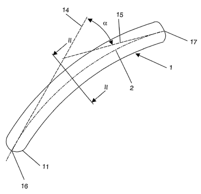
Fig. 1 A lateral view of the pin according to the invention;
Fig. 2 a cross section through the pin according to Fig. 1, along the line II -
II;
Fig. 3 a perspective view of a modification of the pin according to Fig. 1;
CA 02608286 2007-11-13
7
Fig. 4 a cross section of a toe with an endomedullarly inserted, bioresorbable
pin;
Fig. 6 a perspective view of another form of embodiment of the pin according
to the
invention;
Fig. 7 A lateral view in a direction orthogonal to a lateral surface of the
form of
embodiment shown in Fig. 6, and
Fig. 8 a cross section along the line II - III of Fig. 7.
The device for the temporary splinting of toes shown in the Figs. 1 and 2, in
particular
for treating hammer toes or other toe misalignments, essentially consists of a
curved pin
1 with a central axis 2 and a non-circular (in this case elliptical) cross
section 3, which
preferably consists of a self-reinforced poly-L,D-lactide (SR-PLA 96/4).
Copolymers
made of poly-L-lactide (PLLA) and poly-(DL-lactide-co-glycolic acid)(PLGA),
preferably
in a ratio of 4:1, are also suitable for this purpose. A mixture of 96% poly-L-
lactide
(PLLA) with 4% poly-D-lactide has also proved advantageous.
The pin 1 presents a curvature in the plane of the drawing, with a curving
radius of a
length of 10 cm. The tangents 14, 15 at both end points 16, 17 of the central
axis 2 of
the pin 1 are in this case intersecting at an angle a of 10 to 20 , typically
15 . The
length of the pin 1 amounts to 3.75 cm. The surface of the pin 1 is perfectly
smooth. The
one end 16 of the pin 1 destined for inserting into the toe is formed in a
tapering fashion,
so as to terminate in a rounded tip.
The Figs. 3 and 4 illustrate an alternative form of embodiment of the pin 1,
which differs
from the form of embodiment shown in Fig. 1 only in the fact that the non-
circular cross
section 3 is formed in a triangular rather than elliptical fashion, so that
the mantle
surface 10 of the pin 1 offers three longitudinal edges or longitudinal ridges
6, 7, 8. The
sides of the triangle have in this case a concave conformation so that the
longitudinal
CA 02608286 2007-11-13
8
edges or longitudinal ridges 6, 7, 8 are separated from each other by concave
depressions 9. The pin 1 presents a curvature with a curving radius 4 of a
length of 10 -
15 cm, typically of 12.5 cm. The triangular cross section 3 of the pin 1
tapers in this
case in the direction toward the centre 5 of the curving radius 4.
The borehole to be drilled into the marrow channel of the affected bones
presents a
bore diameter 18 which is advantageously smaller than the maximum diameter 12
of
the pin 1, so as to allow the longitudinal ridges 6, 7, 8 to intersect
themselves into the
walls of the drilled up marrow channel, so that a rotational stability of the
pin 1 results.
The core diameter 13 of the pin amounts to 1.0 - 2.5 mm, typically 1.6 mm.
The Fig. 5 illustrates a toe with a terminal phalanx 19, a middle phalanx 20,
a base
phalanx 21 and a metatarsal head 22. The insertion of the pin 1 occurs in the
previously
drilled-up marrow channels of the middle phalanx 20 and of the base phalanx
21.
The form of embodiment represented in the Figs. 6 to 8 comprises, apart from
the pin 1,
a head portion 20 coaxial with the central axis, where the pin 1 and the head
portion 20
are formed asymmetrically to a plane E orthogonal to the central axis 2. The
central axis
2, which is formed by the line connecting the points of gravity of the
successive axial
cross section surfaces extends, in the form of embodiment shown here along a
straight
line. The pin is formed in a prismatic shape, while the head portion 20 forms
a
longitudinal section widening in a direction toward the rear end 21.
The pin 1, which is limited by the front end 22 destined for inserting into
the toe and the
plane E, presents over its entire length L a triangular cross section 3, so
that the pin
mantle surface 10 shows three flat lateral surfaces 26, 27, 28 and three
longitudinal
edges or longitudinal ridges 6, 7, 8. The cross section 3 is limited by an
equilateral
triangle with a peripheral circle equal to the maximum outside diameter 12 of
the pin 1.
The head portion 20 is distinguished by the fact that the plane lateral
surfaces 26, 27,
28 of the pin 1 are curved in an axial direction in such a manner that the
distance a
CA 02608286 2007-11-13
9
between the central axis 2 and any one of the lateral surfaces 26, 27, 28
gradually
widens in a direction toward the rear end 21 of the head portion 20. The head
portion 20
also presents a length I and a circularly cylindrical enveloping surface with
a maximum
outside diameter 12 that the lateral surfaces 26, 27, 28 at the rear end 21 of
the pin 1
are opening out to, so that the front surface 23 orthogonal to the central
axis 2 at the
rear end 21 of the pin 1 is a circular surface.
For a better understanding of the device according to the invention, an
operating
sequence in key-word style follows:
1. The patient is laid down to a dorsal position.
2. A dorsal access with a longitudinal intersect from the middle phalanx 20 to
the
metatarsal head 22 follows.
3. Excision or simple longitudinal splitting of the long stretching tendon.
4. Opening up of the proximal interphalangeal joint.
5. Excision of the interphalangeal joint.
6. Curving of the distal toe.
7. Drilling out of the marrow channel of the middle phalanx 20, up to the
distal epiphysis.
8. Opening up of the metatarso-phalangeal joint along three-quarters of its
perimeter.
9. The articular surface of the arc of the foot is left over.
10. Curving of the base phalanx 21.
11. Drilling out of the marrow channel through the entire base phalanx 21.
12. Driving in the pin 1, from proximal to distal, through the drilled-out
hole with the
borehole diameter 18 in the marrow channel, up to the distal end of the middle
phalanx
20.
13. Intersecting off the pin 1 at the level of the articular surface of the
base phalanx 21.
14. Applying the skin suture.
Description for a joint fragment fixation
On the example of an osteochondritis dissecans tali:
1. Osteotomy of the median malleolus
CA 02608286 2007-11-13
2. Checking the instability of the osteochondral fragment or reduction
3. Drilling into the fragment and the talus body and measuring the depth
4. Driving in the pin
5. Sawing off the pin at the cartilage level.
5
The profile of the fragment provides the necessary rotational stability.
Inserting a
second pin is not needed. In any case, there is generally no room for this
purpose, and
such a second pin would also endanger the vitality (perfusion).