Note: Descriptions are shown in the official language in which they were submitted.
CA 02608426 2012-05-03
WO 2006/124455
PC=52006/018082
IMPROVED METHOD FOR SPECTROPHOTOMETRIC
BLOOD OXYGENATION MONITORM
Applicant hereby claims priority benefits under 35 U.S.C. 119(6) ofU.S.
Provisional
Patent Application No. 60/680,192 filed May 12 2005
BACKGROUND OF THE INVENTION
1. Technical Field.
(0001] This invention relates to methods for non-inva.sively determining
biological
tissue oxygenation in genemt, and to non-invasive methods utilizing near-
infrared
spectroscopy (N1RS) technisues kor determining the same in particular.
2, Background Information.
[00023 US. Patent No. 6,456,862 ancl US. Patent Application Serial No.
10/628,068,
both assigned to the assignee of the present application, disclose methods for
spectrophotometric blood oxygenation monitoring. Oxygen saturation within
blood is defined
as;
1802
02saturation% (mo2 Ho-*100% (Eqn. 1)
These methods, and others known within the prior art utiliYe variants of the
Beer-Lambert
law to account for optical attenuation in tissue at. a partieuktr wavelength.
Relative
concentrations of oxyhemoglobin (1.1b02) and deoxyhemoglobin (1-1h)õ and
therefore
oxygenation levels, within a tissue sample are determinable using changes in
optical
atteamation:
= ¨10gH2 = *z12*d* BA (Eqn.2)
A
SUBSTITUTE SHEET (RULE 26)
CA 02608426 2007-11-13
WO 2006/124455
PCT/US2006/018082
wherein "Ax" represents the optical attenuation in tissue at a particular
wavelength X, (units:
optical density or OD); "I" represents the incident light intensity (units:
W/cm2); "ax."
represents the wavelength dependent absorption coefficient of the chromophore
(units: OD *
cm-I * u1\4-1); "C" represents the concentration of chromophore (units: uM);
"d" represents
the light source to detector (optode) separation distance (units: cm); and
"13x," represents the
wavelength dependent light scattering differential pathlength factor
(unitless)
[0003] To non-invasively determine oxygen saturation within tissue
accurately, it is
necessary to account for the optical properties (e.g., absorption coefficients
or optical
densities) of the tissue being interrogated. In some instances, the absorption
coefficients or
optical densities for the tissue components that create background light
absorption and
=
scattering can be assumed to be relatively constant over a selected wavelength
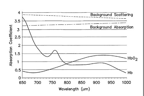
range. The
graph shown in FIG. 1, which includes tissue data plotted relative to a Y-axis
of values
representative of absorption coefficient values and an X-axis of wavelength
values, illustrates
such an instance. The aforesaid constant value assumption is reasonable in a
test population
where all of the subjects have approximately the same tissue optical
properties; e.g., skin
pigmentation, muscle and bone density, etc. A tissue interrogation method that
relies upon
such an assumption may be described as being wavelength independent within the
selected
wavelength range and subject independent. Our findings indicate that the same
assumption is
not reasonable, however, in a population of subjects having a wide spectrum of
tissue optical
properties (e.g., a range of significantly different skin pigmentations from
very light to very
dark) unless consideration for the wide spectrum of tissue optical properties
is provided
otherwise.
[0004] What is needed, therefore, is a method for non-invasively
determining the level
of oxygen saturation within biological tissue that accounts for optical
influences from the
specific tissue through which the light signal passes.
DISCLOSURE OF THE INVENTION
[0005] According to one aspect of the present invention, a method and
apparatus for
non-invasively determining the blood oxygen saturation level within a
subject's tissue is
provided. In one embodiment, the method includes the steps of: 1) providing a
near infrared
2
CA 02608426 2007-11-13
WO 2006/124455
PCT/US2006/018082
spectrophotometric sensor operable to transmit light along a plurality of
wavelengths into the
subject's tissue; 2) sensing the light transmitted into the subject's tissue
using the sensor, and
producing signal data representative of the light sensed from the subject's
tissue; 3)
processing the signal data, including accounting for physical characteristics
of the subject; and
4) determining the blood oxygen saturation level within the subject's tissue
using a difference
in attenuation between the wavelengths.
[0006] The
apparatus includes at least one sensor having at least one light source and
at least one light detector, which sensor is operably connected to a
processor. The light
source is operable to transmit light along a plurality of wavelengths into the
subject's tissue,
and to produce signal data representative of the light sensed from the
subject's tissue. The
algorithm selectively produces calibration constants for use with the sensor
that account for
the specific physical characteristics of the particular subject being sensed.
The calibration
constants are produced using the signal data.
[0007] According to another aspect of the present invention, a method for
calibrating a
NIRS sensor is provided that includes the steps of: 1) transmitting light into
a subject's tissue
using the sensor; 2) sensing the light using the sensor along a plurality of
wavelengths after
the light travels through the subject's tissue, and producing signal data from
the sensed light;
and 3) calibrating the sensor using the signal data.
[0008] The
present method and apparatus provides advantageous accuracy. All prior
art non-invasive devices and methods for detei _______________________ ining
blood oxygen saturation level within a
in_
subject's tissue, of which we are aware, do not consider the specific physical
characteristics of
the particular subject being sensed. The sensor is calibrated by use of
assumed constants and
/or relative to a source (e.g., a phantom sample, empirical data, etc.) other
than the subject
being sensed; i.e., calibrated in a "subject independent" manner. The present
device and
method, in contrast, considers the specific physical characteristics (e.g.,
tissue pigment,
muscle and bone density and mass, etc.) of the particular subject by initially
sensing the
subject's tissue, creating signal data based on the sensing, and accounting
for the specific
physical characteristics of the subject using the signal data. The sensor, now
calibrated in a
"subject dependent" manner, can be used determine the tissue blood oxygen
saturation level
3
CA 02608426 2007-11-13
WO 2006/124455
PCT/US2006/018082
of the subject tissue. As a result, the sensor is able to provide a more
accurate assessment of
the subject's blood oxygen saturation level within the tissue being sensed.
[0009] Another advantage of the present method and apparatus is that
accurate blood
oxygen saturation level information can be provided for a population of
subjects having a
wide range of physical characteristics. Physical characteristics (e.g., tissue
pigmentation,
thickness and density, etc.) naturally vary between subjects, and those
characteristics create
differences in light attenuation, background scattering and absorption. The
present method
and apparatus considers the physical characteristics of the specific subject
being tested, and
calibrates the sensor with signal data generated from sensing the tissue of
the specific subject.
Consequently, the present method and device accounts for the differences in
light attenuation
specific to that subject and enables the tissue blood oxygenation saturation
level of subjects
having a wide range of physical characteristics to be accurately sensed.
[0010] These and other objects, features, and advantages of the present
invention
method and apparatus will become apparent in light of the detailed description
of the
invention provided below and the accompanying drawings. The methodology and
apparatus
described below constitute a preferred embodiment of the underlying invention
and do not,
therefore, constitute all aspects of the invention that will or may become
apparent by one of
skill in the art after consideration of the invention disclosed overall
herein.
BRIEF DESCRIPTION OF THE DRAWINGS
[0011] FIG. 1 is a graph diagrammatically illustrating tissue data plotted
relative to a
Y-axis of values representative of absorption coefficient values, and an X-
axis of wavelength
values.
[0012] FIG. 2 is a diagrammatic representation of a NIRS sensor.
[0013] FIG. 3 is a diagrammatic representation of a NIRS sensor placed on
a subject's
head.
[0014] FIG. 4 is a diagrammatic view of a NIRS sensor.
[0015] FIG. 5 is a graph having values diagrammatically representative of
subject-
specific calibration coefficients plotted along a Y-axis, TOP index values
plotted along an X-
4
CA 02608426 2007-11-13
WO 2006/124455
PCT/US2006/018082
axis, and data representative of deoxyhemoglobin values and oxyhemoglobin
values plotted
therebetween with best-fit curves applied thereto.
[0016] FIG.6 is a flow chart illustrating steps according to one aspect of
the present
invention.
DETAILED DESCRIPTION THE INVENTION
[0017] The present method of and apparatus for non-invasively determining
the blood
oxygen saturation level within a subject's tissue is provided that utilizes a
near infrared
spectrophotometric (NIRS) sensor that includes a transducer capable of
transmitting a light
signal into the tissue of a subject and sensing the light signal once it has
passed through the
tissue via transmittance or reflectance. The present method and apparatus can
be used with a
variety of NIRS sensors, and is not therefore limited to any particular NIRS
sensor.
[0018] Referring to FIGS. 2-4, an example of an acceptable NIRS sensor
includes a
transducer portion 10 and processor portion 12. The transducer portion 10
includes an
assembly housing 14 and a connector housing 16. The assembly housing 14, which
is a
flexible structure that can be attached directly to a subject's body, includes
one or more light
sources 18 and light detectors 19, 20. A disposable adhesive envelope or pad
is preferably
used for mounting the assembly housing 14 easily and securely to the subject's
skin. Light
signals of known but different wavelengths from the light sources emit through
a prism
assembly. The light sources 18 are preferably laser diodes that emit light at
a narrow spectral
bandwidth at predetermined wavelengths. The laser diodes may be mounted remote
from the
assembly housing 14; e.g., in the connector housing 16 or within the processor
portion 12. In
these embodiments, a fiber optic light guide is optically interfaced with the
laser diodes and
the prism assembly that is disposed within the assembly housing 14. In other
embodiments,
the light sources 18 are mounted within the assembly housing 14. A first
connector cable 26
connects the assembly housing-14 to the connector housing 16 and a second
connector cable
28 connects the connector housing 16 to the processor portion 12. The light
detectors 19, 20
each include one or more photodiodes. The photodiodes are also operably
connected to the
processor portion 12 via the first and second connector cables 26, 28. Other
examples of
acceptable NIRS sensors are described in U.S. Patent Application No.
60/751,009 filed on
CA 02608426 2012-05-03
WO 2006/124455 PCIIUS2006/015082
December 16, 2005, and U.S. Patent Application No. 60/729,339 fled on October
21, 2005,
both of which applications are commonly assigned to the assignee of the
present application.
[0019) The processor portion 12 includes a processor for processing light
intensity
signals associated with the light sources 18 and the light detectors 19,20 as
described herein
-
A person of skill in the art will recognize that the processor may assame
varkars forms (e, g.,
digital signal processor, analog device, etc-) capable of performing the
functions described
herein. The processor utilizes an algorithm that characterizes a change in
attenuation as a
function of the difference in attenuation between cliare.ut wavelengths. Tho
algoritlun
accounts for the effects of pathlength and parameter 'E", which represents
energy losses
("Cr) due to light scattering within tissue, other background absorption
losses (q") from
biological compounds, and other unknown losses ("N") including measuring
apparatus
variability (E = (3 + F -3-144). As will be discussed below, the parameter "E"
reflects energy
losses not specific to the subject being tested with a calibrated sensor
(i.e., "subject-
indepe,ndentl,
[0020] The absorption At,,. detected from the deep light detector 20
includes
attenuation and energy losses from both the deep and shallow tissue, while the
absorption Axx
detected from the shallow light detector 19 includes attenuation and energy
losses from
shallow tissue. Absorptions Aba. and 40, can be expressed in the form of
Equation 3 and
Equation 4:
(/
(Eqn.3)
.r, a
1
* C,, * L., + Ec4 (Eqn..4)
In some applications (e.g., infants), a single light detector may be used, in
Mill ca.se
Equation 5 is used:
(Eqn 5)
6
CA 02608426 2007-11-13
WO 2006/124455 PCT/US2006/018082
If both the deep and shallow detectors are used, then substituting Equation 4
into Equation 3
yields A', which represents attenuation and energy loss from deep tissue only:
AA = Am¨ 4,2= ceA *CI, *Lb + (E 2 ¨E2) (Eqn.6)
From Equation 5 or Equation 6, L is the effective pathlength of the photon
traveling through
the deep tissue and A'1 and Av2 represent light attenuation at two different
wavelengths to
determine differential wavelength light attenuation AA '12:
Ai ¨ A A42 A42 (Eqn.7)
Substituting Equation 5 or 6 into Equation 7 for A' and A'2, AA'12 can be
expressed as:
42 '---- aat2 * C b * Lb + AFL (Eqn.8)
and Equation 8 can be rewritten in expanded form:
42 = ((ari ¨a7.2 )[Hblb + (a01¨ab2)[Hb021b) Lb + (g¨E;).
(Eqn.9)
(Acyr12 *[Hbib*Lb)+(Aa.12*,r
Hb02L *Lb) + AR; 2
where:
(Aar12 *[Hblb *Lb) represents the attenuation attributable to Hb; and
(A a o12*{HbO2L*Lb) represents the attenuation attributable to Hb02; and
AR'/2 represents energy losses due to light scattering within tissue, other
background
absorption losses from biological compounds, and other unknown losses
including measuring
apparatus variability.
[0021] The multivariate form of Equation 9 is used to deteimine [Hb02]b
and [Hb]b
with three different wavelengths:
_
_ -
A42 AFL
_
AA/I3 AFL _ (Lb a
) =_
-1 Aa Aa
r12 ol2 [11b]b
A1.13 Aa013 [Rh
2 _1
th (Eqn.1 0)
[
- - - _
Rearranging and solving for [Hb02]b and [Hb]b, simplifying the Aa matrix into
[Aa' ]:
_ AA
' -AR' - [Hb] _
___________________________ b 12 {Aar ]-1 I L 1-1 _ 12 rAaT1 (Lb i l ,
(Eqn.11)
_AA13_ k b ) AR13 L [HbO, ib _
- _
Then combined matrices [AA'] [Aa]i= [As] and [AE] [Aa']-1 =
7
CA 02608426 2007-11-13
WO 2006/124455
PCT/US2006/018082
[Al - -1 [41 lib] -1 - [HI)]b
(Lb) ¨ 1 (Lb) = (Eqn.12)
Alma, _ 4 [Hb02
The parameters AHb and AHb02 represent the product of the matrices [AA] and
[AaTI and the
parameters qjHb and THb02 represent the product of the matrices [AEix] and
[Ace']". To
determine the level of cerebral tissue blood oxygen saturation (Sn02),
Equation 12 is
rearranged using the form of Equation 1 and is expressed as follows:
(AHb02-41 Hb02)
S1702% = \* 100% (Eqn.13)
AHbo2 tiirybo, AHb¨T Hb )
Note that tissue blood oxygen saturation is sometimes symbolized as St02,
Sct02, CrS02, or
rS02. The effective pathlength Lb cancels out in the manipulation from
Equation 12 to
Equation 13.
[0022] The value for Sn02 is initially determined from an empirical
reference of
weighted combination of venous and arterial oxygen saturation (Sinv02) value,
for example
using:
Smv02 = Kv* Sv02+ Ka* Sa02 (Eqn.14),
and the empirically determined values for Sv02 and Sa02, where the term "Sv02"
represents
venous oxygen saturation, the term `µSa02" represents arterial oxygen
saturation, and the
terms Kv and Ka are the weighted venous and arterial contributions
respectively (Kv + Ka =
1). The empirically determined values for Sv02 and Sa02 are based on data
developed by
discrete sampling or continuous monitoring of the subject's blood performed at
or about the
same time as the sensing of the tissue with the sensor; e.g., blood samples
discretely collected
can be analyzed by blood gas analysis and blood samples continuously monitored
can be
analyzed using a fiber optic catheter inserted within a blood vessel. The
temporal and
physical proximity of the NIRS sensing and the development of the empirical
data helps
assure accuracy. The initial values for Kv and Ka within Equation 14 are
clinically reasonable
values for the circumstances at hand. The values for AHb02 and Am are
determined
mathematically using the values for /b2 and 40, for each wavelength sensed
with the NIRS
8
CA 02608426 2007-11-13
WO 2006/124455
PCT/US2006/018082
sensor (e.g., using Equation 3 & 4 for deep and shallow detectors or Equation
5 for a single
detector). The calibration parameters THb and THb02, which account for energy
losses due to
scattering as well as other background absorption from biological compounds,
are then
determined using Equation 14 and non-linear regression techniques by
correlation to different
weighted values of Sv02 and Sa02; i.e., different values of Ka and Ky.
Statistically acceptable
values of Kv and Ka and Ttib and IPHbO2 are converged upon using the non-
linear regression
techniques. Experimental findings show that with proper selection of Ka and
Kv, the
calibration parameters THb and THbo2 are constant within a statistically
acceptable margin of
error for an individual NIRS sensor used to monitor brain oxygenation on
different human
subjects.
[0023] The
above-identified process produces a NIRS sensor calibrated relative to a
particular subject using invasive techniques, or a NIRS sensor calibrated
relative to an already
calibrated sensor (or relative to a phantom sample). When these calibrated
sensors are used
thereafter on a different subject, they do not account for the specific
physical characteristics of
the particular subject being tested. The present method and apparatus as
described below
permits a NIRS sensor to be calibrated in a non-invasive manner that accounts
for specific
physical characteristics of the particular subject being sensed.
[0024] Certain
physical characteristics will vary from subject to subject, such as but
not limited to, tissue pigmentation and thickness and density of muscle and/or
bone. The
present method and apparatus accounts for background tissue's wavelength
dependent light
attenuation differences due to these subject-dependent physical
characteristics by sensing the
subject's tissue, creating signal data from the sensing, and using the signal
data to create one
or more "subject-specific" calibration constants that account for the specific
characteristics of
the subject. For example, during an initial phase of monitoring, light is
transmitted into and
sensed passing out of the subject's tissue. Signal data representative of the
sensed light is
analyzed to account for the physical characteristics of the subject, and one
or more subject-
specific calibration constants indicative of the specific physical
characteristics are created.
The subject-specific calibration constants are subsequently used to determine
properties such
as the blood oxygen saturation level, deoxyhemoglobin concentration,
oxyhemoglobin
concentration, etc.
9
CA 02608426 2007-11-13
WO 2006/124455 PCT/US2006/018082
[0025] The subject-specific calibration constants can be determined by
using the
sensed signal data to create a tissue optical property (TOP) index value. The
TOP index value
is derived from wavelength dependent light attenuation attributable to
physical characteristics
such as tissue pigmentation, thickness and density of tissue, etc. These
physical
characteristics are collectively considered in determining the TOP index value
because the
characteristics have absorption coefficients that increase with decreasing
wavelength from the
near-infrared region to the red region (i.e., from about 900nm to about 400
nm) mainly due to
the presence of melanin, the light absorbing pigmentation in skin and tissue.
For example, it
has been reported by S. L. Jacques et al., that light absorption in skin due
to melanin can be
described by the relationship: lta= 1.70x10¹² (wavelength in nm)-148 [cm-1] in
the wavelength
range from about 400nm to about 850 mn. If the overall light absorption
characteristics of
tissue are modeled to follow that of melanin, then the TOP light absorption
coefficients (amp)
can be determined using the same equation for the particular wavelengths of
light used in the
interrogation of the tissue (where A = 1.7 x 10¹² and T= -3.48):
aTOP = A* (wavelength)-T
(Eqn.15)
To determine the TOP index value, one or more of the wavelengths in the near-
infrared region
to the red region (i.e., from about 900nm to about 600 nm; e.g., 690 nm, 780
nm, 805 nm, 850
nm) are sensed. Red wavelengths are favored because red light is more
sensitive to the tissue
optical properties than infrared light. Lower wavelengths of light could also
be used, but
suffer from increased attenuation from the higher tissue and hemoglobin
absorption
coefficients, resulting in reduced tissue penetration, reduced detected light
signal strength, and
resultant poor signal to noise ratio.
[0026] To calculate the TOP index value (identified in Equation 16 as
"TOP"), a four
wavelength, three unknown differential attenuation algorithm (following
similarly to the
derivation shown by Equations 3-10), is used such as that shown in Equation
16:
AA1'2
Act".õ Aa0r12 AaT1 OP12¨ Hb
014 TOP14
CA 02608426 2007-11-13
WO 2006/124455
PCT/US2006/018082
Alternatively, Equation 17 shown below could be used. Equation 17 accounts for
energy
losses "E" as described above:
- - A42 AP'
-Aa".12 Aaoti2 A¨TOP12 Hb -
-
AA13 A Elf3 (Lb)' Acer'13 Aac,' ,3 AaTIOP13 Hb02 (Eqn. 17)
_644 AE14_ Aa A Aa'a'
014 T0P14 _ _TOP
[0027] The TOP index value determinable from Equations 16 or 17 accounts
for
subject tissue optical properties variability and can be converted to a
"corrective" factor used
to determine accurate tissue blood oxygen saturation Sn02. In some
embodiments, the TOP
index value can be used with a database to determine subject-specific
calibration constants
(e.g., ZHb and ZHb02). The database contains data, at least some of which is
empirically
collected, pertaining to oxyhemoglobin and deoxyhemoglobin concentrations for
a plurality
of subjects. The concentration data is organized relative to a range of TOP
index values in a
manner that enables the determination of the subject-specific calibration
constants. The
organization of the information within the database can be accomplished in a
variety of
different ways.
[0028] For
example, the empirical database may be organized in the form of a graph
having subject-specific calibration coefficients plotted along the y-axis
versus TOP index
values plotted along the x-axis. An example of such a graph is shown in FIG.
5, which
contains data 30 representing the differences between calculated
deoxyhemoglobin values
(Hb) values and empirically derived deoxyhemoglobin values (the differences
referred to in
FIG.5 as "Hb-offset2 data"), and a best fit curve 32 applied to a portion of
that data 30. The
graph also contains data 34 representing the differences between calculated
oxyhemoglobin
values (Hb02) values and empirically derived oxyhemoglobin values (the
differences referred
to in FIG.5 as "Hb02-offset2 data"), and another best-fit curve 36 applied to
a portion of that
data 34. In the example shown in FIG. 5, a statistically significant number of
the data 30, 34
for each curve lies within the sloped portion 32a, 36a (i.e., the portion that
does not have a
constant calibration constant value). At each end of the sloped portion 32a,
36a, the curves
CA 02608426 2012-05-03
WO 2006/124455 PCT/US2006/018082
32,36 are depicted as having constant calibration values 32b, 32; 361,, 36c
for convenience
sake. The values for the subject-specific calibration coefficients Zifb and
211602 are determined
by drawing a fine (e.g., see phantom line 38) perpendicular to the TOP index
value exit at the
determined TOP index value. The subject-specific calibration constant (41) for
deoXyhernoglobin is equal to the value on the calibration constant axis
aligned with the
intersection point between the perpendicalar line and the 9lb-offsd2" curve,
and the subject-
specific calibration constant (Zet,02) for oxyhemoglobin is equal te the value
on the
calibration constant axis aligned with the intersection point with the "111302-
oftsen'T curve.
[0029] Alternatively, the subject-spec calibration constant values may be
determined using an empirical database in a form other than a graph. For
example, a
mathematical solution can be implemented rather than the above-described
graph. The
mathematical solution may use linear equations representing the "1-1b-offset2"
and the 141102-
offset2" curves.
[0030] Once the subject-specific calibration constant values are
determined, they are
utilized -with a variation of Equation 13:
(.4/64 ¨1P1,60, +ZEb02)
Se02% ¨ _______________________________ , *100% (Bp_ 18)
¨14 ziThoz4ffb ¨Tim +46)
to determine the cerebral blood oxygen saturation level.
[0031] The above-described process for determining the subject-specific
calibration
constants can be performed one or more times in the initial period of sensing
the subject to
calibrate the sensor to that partitular subject, prefeably right after the
sensor is attached to the
subject The subject-dependent calibration constants can then be used with an
algorithm for
measurement of a subject's blood oxygen saturation level using the same or
different signal
data. The algorithm in which the subject-dependent calibration constants are
utilized may be
the same algorithm as used to determine the constants, or a different
algorithm for
determining the tissue oxygen sanitation level. For example, calibration
constants can be
used with the three wavelength method disclosed above in Equations 2 ¨ 14, and
in U.S.
Patent No. 6,456,862. Prior to the cerebral blood
12
CA 02608426 2007-11-13
WO 2006/124455 PCT/US2006/018082
oxygen saturation level being calculated, the subject-specific calibration
constants ZHb and
ZHb02 can be incorporated as corrective factors into the three wavelength
algorithm (e.g.,
incorporated into Eqn. 13). As a result, a more accurate determination of the
subject's tissue
oxygen saturation level is possible. FIG. 6 illustrates the above described
steps within a flow
chart.
[0032] In alternative embodiments, the TOP index methodology disclosed
above can
be used within an algorithm in a subject-independent manner. This approach
does not provide
all of the advantages of the above described subject¨dependent methodology and
apparatus,
but does provide improved accuracy by specifically accounting for subject skin
pigmentation.
For example, the TOP absorption coefficients can be determined as described
above and
utilized within Equation 16 or Equation 17. Regardless of the equation used,
the determined
values for deoxyhemoglobin (Hb) and oxyhemoglobin (Hb02) can subsequently be
used to
determine the tissue oxygen saturation level. For example, the Hb and Hb02
values can be
utilized within Equations 11 through 13.
[0033] Although the present method and apparatus are described above in
terms of
sensing blood oxygenation within cerebral tissue, the present method and
apparatus are not
limited to cerebral applications and can be used to determine tissue blood
oxygenation
saturation within tissue found elsewhere within the subject's body. If the
present invention is
utilized to determine the tissue blood oxygenation saturation percentage is
typically
symbolized as St02 or rS02.
[0034] Since many changes and variations of the disclosed embodiment of
the
invention may be made without departing from the inventive concept, it is not
intended to
limit the invention otherwise than as required by the appended claims.
[0035] What is claimed is:
13