Note: Descriptions are shown in the official language in which they were submitted.
CA 02608464 2010-06-03
SURGICAL DRILL GUIDE DEVICES FOR LIGAMENT REPAIR
FIELD OF THE INVENTION
The present invention generally relates to surgical methods and devices, and
more particularly to instruments and methods for use in knee reconstruction to
repair
torn ligaments and procedures for such reconstructions.
BACKGROUND
Joint injuries may commonly result in the complete or partial detachment of
ligaments, tendons and soft tissues from bone. Tissue detachment may occur in
many
ways, e.g., as the result of an accident such as a fall, overexertion during a
work-related activity, during the course of an athletic event, or in any one
of many
other situations and/or activities. These types of injuries are generally the
result of
excess stress or extraordinary forces being placed upon the tissues.
In the case of a partial detachment, commonly referred to under the general
term "sprain," the injury frequently heals without medical intervention, the
patient
rests, and care is taken not to expose the injury to undue strenuous
activities during
the healing process. If, however, the ligament or tendon is completely
detached from
its attachment site on an associated bone or bones, or if it is severed as the
result of a
traumatic injury, surgical intervention may be necessary to restore full
function to the
injured joint. A number of conventional surgical procedures exist for re-
attaching
such tendons and ligaments to bone.
One such procedure involves the re-attachment of the detached tissue using
"traditional" attachment devices such as staples, sutures, and bone screws.
Such
traditional attachment devices have also been used to attach tendon or
ligament grafts
(often formed from autologous tissue harvested from elsewhere in the body) to
the
desired bone or bones. In one procedure, a damaged anterior cruciate ligament
("ACL") is replaced in a human knee. Initially, bone tunnels are formed
through the
tibia and femur at the points of normal attachment of the ACL. Next, a
ligament graft
with a bone graft on one of its ends is sized so as to fit within the bone
tunnels.
Suture is then attached to the bone graft and thereafter passed through the
tibia and
femoral bone tunnels. The bone graft is then pulled through the tibial tunnel
and up
into the femoral tunnel using the suture. As this is done, the ligament graft
ligament
extends back out of the femoral tunnel, across the interior of the knee joint,
and then
CA 02608464 2010-06-03
-2-
through the tibial tunnel. The free end of the ligament graft ligament resides
outside
the tibia, at the anterior side of the tibia. Next, a bone screw is inserted
between the
bone graft and the wall of femoral bone tunnel so as to securely lock the bone
graft in
position by a tight interference fit. Finally, the free end of the ligament
graft ligament
is securely attached to the tibia.
In another ACL reconstruction procedure, aligned femoral and tibial tunnels
are initially formed in a human knee. A bone graft with a ligament graft
attached
thereto is passed through the tunnels to a blind end of the femoral tunnel
where the
block is fixed in place by an anchor. The ligament extends out of the tibial
tunnel,
and the end is attached to the tibia cortex by staples or the like.
Alternatively, the end
of the ligament may be fixed in the tibial tunnel by an anchor or by an
interference
screw. Various types of ligament and/or suture anchors for attaching soft
tissue to
bone are also well known in the art. A number of these devices are described
in detail
in U.S. Pat. Nos. 4,898,156, 4,899,743, 4,968,315, 5,356,413, and 5,372,599,
all of
which are commonly assigned to Mitek Surgical Products, Inc.. a Johnson &
Johnson
company_
Alternatively, an ACL reconstruction procedure may be performed where
instead of reconstructing the dual bundle structure of the native ACL with the
functional equivalent of a single band graft, two bone grafts can be used,
each graft in
an individual bone tunnel. Such an "anatomic" or double-tunnel procedure
traditionally involves freehandedly positioning the two tunnels in the tibia
and femur.
Forming the two tunnels in the correct positions can be technically
challenging, time
consuming, and have little consistency or reproducibility in tunnel placement
from
patient to patient.
Accordingly, there is a need for improved methods and devices for repairing
ligaments and for positioning and forming bone tunnels.
SUMMARY
In one embodiment, a surgical drill guide apparatus is provided. The
apparatus can include a first guide member having a longitudinal passageway
extending therethrough for aiming a first guide pin along a first path to form
a first
bone tunnel, and a second guide member having a longitudinal passageway
extending
therethrough for aiming a second guide pin along a second path to form a
second bone
CA 02608464 2007-10-29
-3-
tunnel. The first and second guide members can be coupled to one another via
an
elongate support such that the first and second guide members extend at an
angle
relative to one another and are offset such that axes of the guide members do
not
intersect. In one exemplary embodiment, the support is an arcuate support that
is
fixedly mated to the first guide member and slidably mated to the second guide
member. A plurality of markings can be formed on the arcuate support for
indicating
the angle between the first and second guide members.
In another embodiment, the apparatus can include a tibial engagement member
having a proximal end coupled to the first guide member and having a distal
end
adapted to indicate an exit location of first and second bone tunnels formed
along the
first and second paths. The tibial engagement member can include a slider arm
coupled to the first guide member and a probe arm slidably disposed along the
slider
arm. The distal end of the tibial engagement member can be formed on the probe
arm.
The apparatus can also include a first drill guide mounted in the longitudinal
passage of the first guide member and a second drill guide mounted in the
second
longitudinal passage of the second guide member. Markings can be formed on the
first and second drill guides. The markings can indicate a depth of a bone
tunnel
formed by drill bits disposed through the first and second drill guides. The
first and
second guide members can also each include a locking mechanism for locking a
drill
guide slidably disposed therethrough in a fixed longitudinal position.
In another embodiment, a surgical drill guide apparatus is provided having a
first guide member with a first longitudinal guide passage adapted to receive
a drill
guide pin therein to form a bone tunnel. A first support arm can be coupled to
and
can extend horizontally from the first guide member. In one embodiment, the
first
support arm can be fixedly mated to the first guide member. The first support
arm
can also have at least one arcuate section. The apparatus can also include a
second
guide member having a second longitudinal guide passage adapted to receive a
drill
guide pin to form a bone tunnel. The second guide member can be slidably
disposed
along the at least one arcuate section of the first support arm to form a
variable angle
relative to the first guide member. The second guide member can also include a
locking element adapted to lock the second guide member in a position on the
at least
CA 02608464 2007-10-29
-4-
one arcuate section. In some embodiments, the first and second guide members
each
have a longitudinal axis, and the axes do not intersect. The apparatus can
also include
a first drill guide mounted in the first longitudinal guide passage and a
second drill
guide mounted in the second longitudinal guide passage. In another embodiment,
the
apparatus can include a second support arm coupled to and extending vertically
from
the first guide member, and an engagement member 'slidably mounted to the
second
support arm. The second support arm can be fixedly mated to the first guide
member,
and it can have at least one arcuate portion. In one exemplary embodiment, the
engagement member has a distal tip adapted to indicate an exit location of
bone
tunnels formed by drill bits disposed through the first and second guide
members.
In other aspects, methods for repairing ligaments are provided. In one
embodiment, a method can include positioning a first guide member adjacent to
bone
such that the first guide member is configured to aim a first guide pin
through the
bone along a first pathway. The method can further include positioning a
second
guide member adjacent to the bone such that the second guide member can aim a
second guide pin through the bone along a second pathway. The method can also
include slidably moving the second guide member along an arcuate support arm
coupled to the first guide member to adjust an angular orientation of the
second guide
member relative to the first guide member. The method can also include
inserting
first and second guide pins through the first and second guide members to form
first
and second bone tunnels that converge toward one another but that have axes
that do
not intersect. In some embodiments, the method can also include locking the
second
guide member in a fixed position along the arcuate support after slidably
moving the
second guide member. The method can also include sliding at least one of the
first
and second guide pins longitudinally through at least one of the first and
second guide
members to adjust a depth of at least one of the first and second bone tunnels
to be
formed.
In other embodiments, positioning the first and second guide members can
include positioning a distal tip of an engagement member coupled to the first
guide
member at an intended exit location of first and second bone tunnels to be
formed
along the first and second pathways. In some embodiments, the distal tip is
formed
on a probe arm slidably coupled to a support arm mated to the first guide
member.
CA 02608464 2007-10-29
-5-
The method can further include slidably moving the probe arm along the support
arm
to adjust a position of the probe arm.
BRIEF DESCRIPTION OF THE DRAWINGS
The invention will be more fully understood from the following detailed
description taken in conjunction with the accompanying drawings, in which:
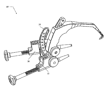
FIG. I is a perspective view of one exemplary embodiment of an instrument
useful in drilling tunnels in bone in a ligament reconstruction procedure;
FIG. 2 is a perspective view of another exemplary embodiment of an
instrument useful in drilling tunnels in bone in a ligament reconstruction
procedure;
FIG. 3 is a perspective side view of the instrument of FIG. 1;
FIG. 4 is a perspective view of the instrument of FIG. I engaging an upper end
of a tibia and showing a tip of the instrument's probe arm engaging a tibial
plateau;
FIG. 5 is top view of the tibia of FIG. 4 showing drill tips exiting an upper
end
of the tibia;
FIG. 6 is a table showing various tunnel offsets using various sized drill
guides
with the instrument of FIG. 1;
FIG. 7 is a cross-sectional schematic view of a portion of the instrument of
FIG. 1;
FIG. 8 is a side view of the tibia of FIG. 4 showing entrance holes of dual
tunnels drilled in an upper section of the tibia;
FIG. 9 is a schematic illustration of entrances to tunnels drilled in the
tibia of
FIG. 4, exits from the tunnels, tunnel trajectories, and a footprint in the
tibial plateau;
CA 02608464 2007-10-29
-6-
and
FIG. 10 is a perspective view of the instrument of FIG. 1 engaging the tibia
of
FIG. 4 and a femur.
DETAILED DESCRIPTION
Certain exemplary embodiments will now be described to provide an overall
understanding of the principles of the structure, function, manufacture, and
use of the
devices and methods disclosed herein. One or more examples of these
embodiments
are illustrated in the accompanying drawings. Those skilled in the art will
understand
that the devices and methods specifically described herein and illustrated in
the
accompanying drawings are non-limiting exemplary embodiments and that the
scope
is defined solely by the claims. The features illustrated or described in
connection
with one exemplary embodiment may be combined with the features of other
embodiments. Such modifications and variations are intended to be included
within
the scope of the present application.
The present invention generally provides methods and devices for repairing
ligaments and for positioning and forming bone tunnels. The various methods
and
devices disclosed herein can be used in a variety of surgical procedures,
however the
methods and devices are particularly useful for repairing an anterior cruciate
ligament
("ACL") in a human knee. In an ACL repair, the torn ACL can be replaced with
two
ligament grafts which are each anchored to the tibia and femur. The term
"ligament
graft," as used herein, is intended to include natural materials, such as
autografts,
allografts, and xenografts, including harvested ligaments and tendons, as well
as
synthetic materials. A ligament graft can also include an anchoring element
attached
thereto for anchoring the graft to the tibia and femur. For example, the
ligament graft
can include a bone graft, plug, or other member, attached to one or both
terminal ends
thereof. The term "bone graft," as used herein, in intended to include natural
materials, such as autografts, allografts, and xenografts, as well as
synthetic materials.
A person skilled in the art will appreciate that the various methods and
devices
disclosed herein can be used in a variety of surgical procedures, and that the
particular
configuration of the ligament grafts can vary depending on the intended use,
and
CA 02608464 2007-10-29
-7-
virtually any ligament grafts known in the art can be used with the devices
and
methods disclosed herein.
FIG. 1 illustrates one exemplary embodiment of a surgical drill guide
instrument 60. In general, the instrument 60 allows for placement of multiple
tibial
tunnels. As shown, the instrument 60 generally includes first and second guide
members 12, 30 connected together via a support or outrigger arm 26. The guide
members 12, 30 can be angularly adjustable relative to one another to form an
angle
Al therebetween that defines an insertion trajectory of two bone tunnels to be
formed.
The instrument 60 can also include first and second guide pin sleeves or drill
guides
40a, 40b slidably disposable through guide channels 13, 31 formed in the guide
members 12, 30. The drill guides 40a, 40b can be configured to receive drill
bits
therethrough for drilling bone tunnels in bone. As further shown in FIG. 1,
the
instrument 60 can also optionally include a tibial engagement member 17 that
is
coupled to one of the guide members, e.g., the first guide member 12, and that
is
adapted to engage a tibial plateau to indicate an exit location of the first
and second
bone tunnels. In use, the instrument 60 can allow for adjustable placement of
tunnel
entrance points on the tibial cortex and for adjustable offset of tunnel exit
points on
the tibial plateau. The placement and offset can also be recorded using the
instrument
60, thereby allowing time-saving reproduction of multiple tunnels.
The guide members 12, 30 can each have a variety of configurations, but in
the illustrated embodiment each guide member 12, 30 generally includes a
housing
and a pathway. The housings and the pathways can have a variety of shapes, but
they
are preferably configured to receive a drill guide or other drilling
apparatus. In the
illustrated embodiment each housing is generally rectangular and each pathway
13, 31
is generally cylindrical for slidably receiving a generally cylindrical drill
guides 40a,
40b, as will be discussed further below. The guide members 12, 30 can each
also
include a locking mechanism for locking the drill guides 40a, 40b in position
within
the guide members 12, 30. In one exemplary embodiment, each locking mechanisms
is in the form of a conventional ratchet having a pawl that engages teeth
formed on
the guide members. A person skilled in the art will appreciate that a variety
of other
locking mechanisms known in the art can be used to allow the guide members to
be
locked in a fixed positioned relative to the guide members 12, 30.
CA 02608464 2007-10-29
-8-
Each of the guide members 12, 30 can also be angularly adjustable relative to
one another. While various techniques can be used for allowing angular
adjustability,
in an exemplary embodiment, as shown, the guide members 12, 30 are coupled to
one
another via the support or outrigger arm 26. The outrigger arm 26 can be a
generally
elongate member, and in an exemplary embodiment all, or at least a portion of,
the
elongate member is arcuate. The outrigger arm 26 can be movably coupled to the
first
and second guide members 12, 30, or one of the guide members 12, 30 can be
fixed
relative to the outrigger arm 26 while the other guide member 12, 30 is
movable. In
the illustrated embodiment, the outrigger arm 26 is mated to the first guide
member
12 in a transverse, keyed thru-hole 12b located in the side of the first guide
member
12 such that the outrigger arm 26 can extend horizontally from the first guide
member
12. Preferably, the outrigger arm 26 is fixedly mated to the first guide
member 12 and
secured via a thumbscrew 22. In other embodiments, however, the outrigger arm
26
can be rotatably, but non-slidably mated to the first guide member 12.
The outrigger arm 26 can slidably couple to the second guide member 30 in a
slot 30a formed in the second guide member 30. The slot 30a can have virtually
any
configuration, and in this embodiment it is a generally rectangular arcuate
channel
that can removably seat the arcuate outrigger arm 26. The second guide member
30
can thus slidably move along the outrigger arm 26 until positioned at a
desired angle
Al relative to the first guide member 12. Once positioned as desired, the
second
guide member 30 can be secured in position using a conventional locking
mechanism,
such as a thumbscrew 32. As further shown in FIG. 1, the outrigger arm 26 can
optionally include external markings 28 that indicate the angle Al between the
first
and second guide members 12, 30.
As indicated above, the outrigger arm 26 can be oriented in a vertical
direction
about an axis AX1 relative to the first guide member 12 to achieve an offset
between
the longitudinal axes 13a, 31 a of the guide channels 13, 31 and hence the
drill guide
channels 41a, 41b and bone tunnels that maybe drilled following them. In an
exemplary embodiment, the instrument 60 can include multiple outrigger arms
(each
similar to the outrigger arm 26) that can be keyed to different rotational
positions
within the slotted thru-hole 12b on the first guide member 12. The multiple
outriggers can be keyed with a series of slot patterns to maintain the ability
to use a
CA 02608464 2007-10-29
-9-
single outrigger with a given offset on both the left and right side of the
first guide
member 12, or individual left and right outrigger arms can be constructed for
each
desired offset position. The multiple outrigger arms could be fixed in
position within
the thru-hole 12b. A locking mechanism, such as the thumbscrew 22 as shown,
can
optionally be used. In other embodiments, as previously explained, the
outrigger arm
26 can be rotatably but non-slidably mated to the first guide member 12. The
offset
of the outrigger arm 26 can be adjusted (i.e., either rotationally or by
selecting an
outrigger arm having a predetermined offset) and secured following advancement
of
the first guide pin 50a through the drill guide 40a (and prior to advancement
of a
second guide pin 50b, described further below) to help visually gauge a
desired offset,
or the offset is adjusted and secured at any point prior to placement of the
guide pin
50a in the guide sleeve 40a. An additional guide pin positioned through a hole
20 in
the outrigger arm 26 can be used as an offset positional reference and/or to
provide
additional support to the outrigger arm 26, as further described below.
FIG. 1 shows the outrigger arm 26 on a right side of the first guide member
12, but the outrigger arm 26 can be located on a left side of the first guide
member 12,
as shown in another embodiment of an instrument 60' in FIG. 2. The instruments
60,
60' are configured to accommodate the anatomies of the left and right knees,
respectively.
Referring still to FIG. 1, the first and second drill guides 40a, 40b that are
disposable through the guide channels 13, 31 in the guide members 12, 30 can
also
have a variety of configurations. In an exemplary embodiment, each drill guide
40a,
40b has a generally elongate tubular configuration, such as a cylindrical
shape for
receipt in the cylindrical guide channels 13, 31, as shown. This will allow
the drill
guides 40a, 40b to guide drill bits or any other bone drilling devices
disposed in the
drill guides 40a, 40b through the guide members 12, 30 and into bone. The
drill bits
or other bone drilling devices may be directly disposed in the guide channels
13, 31
without the drill guides 40a, 40b. In an exemplary embodiment, as shown, the
first
drill guide 40a has external markings 46 at its proximal end 48, external
teeth 45 or
other surface features at its mid-portion or adjacent to its distal end 53,
and a center
channel 41 a extending therethrough between the proximal and distal ends 48,
53
thereof. The first drill guide 40a can be disposed in the first guide member
12
CA 02608464 2007-10-29
-10-
through the channel 13, parallel to the first drill guide's channel 41 a. As
mentioned
above, the teeth 45 can be engaged by a locking mechanism within the channel
13 to
allow movement of the first sleeve 40a in a forward or distal direction
between a
plurality of fixed positions. The first sleeve 40a can also include a bone-
engaging
distal end, such as teeth 44 formed thereon that engage bone at a desired bone
tunnel
entrance location, as described further below. The external markings 46 on the
first
drill guide 40a can indicate an estimated depth of the tunnel with, for
example, depth
lines printed, embossed, etched, or otherwise marked on the first drill guide
40a that
can advance into the channel 13 with the sleeve 40a. As further shown in FIG.
1, the
first drill guide 40a can also include a first annular knob handle 49 mounted
or
otherwise coupled to the proximal end 48 of the first drill guide 40a for
grasping the
drill guide 40a and facilitating movement relative to the guide channel 13.
The knob
handle 49 can include an opening 49a through which a tool or device can be
inserted
to turn or otherwise manipulate the handle 49 to distally advance the sleeve
40a.
The second guide pin sleeve or drill guide 40b can be configured and
manipulated similar to the first drill guide 40a and it can include external
markings
46b at its proximal end 48b, external teeth 45b or other surface features at
its mid-
portion or adjacent to its distal end 42, a center channel 41b, and a knob
handle 49b
having an opening 49c. The second drill guide 40b is typically disposed in and
advanced through the second guide member 30 after the first drill guide 40a
has been
disposed in and advanced through the first guide member 12, but the drill
guides 40a,
40b can be disposed in and advanced through the guide members 12, 30 in any
order.
As previously indicated, the device 60 can also optionally include a tibial
engagement member 17 that is effective to engage the tibial plateau and
indicate an
exit location for guide pins 50a, 50b (and/or other devices used to form the
bone
tunnels) disposed through the drill guides 40a, 40b. The tibial engagement
member
17 can have a variety of configurations, but in the illustrated embodiment it
include a
slider arm 16 and a probe arm 10. The slider arm 16 can be arcuate or straight
and it
can include a plurality of connected, angulated straight and/or curved
segments. In
this embodiment, the slider arm 16 has a generally arcuate orientation
extending
vertically from the first guide member 12, i.e., offset 45 from the outrigger
arm 26.
The slider arm 16 can, however, have an arcuate portion and a straight
portion, or any
CA 02608464 2007-10-29
-11-
other orientation. The probe arm 10 generally includes an elongate body that
can be
adjustably or slidably mounted to the slider arm 16 through a passage I1 a
formed in a
handle portion 11 of the probe arm 10. The probe arm 10 can also include a
distally
extending arm portion 10a including a distal tip 18. The distal tip 18 is
preferably
angulated proximally (e.g., toward a direction of an approaching guide pin),
but it can
have a distal orientation, both proximal and distal angulations, or neither
proximal and
distal orientations.
A position of the probe arm 10, and hence the distal tip 18 and desired
endpoint of a bone tunnel, can be slidably adjusted on the slider arm 16 to
form an
angle A2 between the arm portion I Oa of the probe arm 10 and the first guide
pin 50a
(and the first drill guide 40a). FIG. 3 shows the slider arm 16 slidably
mounted to the
probe arm 10. External markings 54 on the slider arm 16, which can be printed,
embossed, etched, or otherwise marked on the slider arm 16, can indicate the
A2
angular position of the probe arm 10. The probe arm 10 can be fixed in
position to
form a desired angle A2 by, for example, using a conventional locking
mechanism
such as a thumbscrew 14 coupled to the handle portion 11 of the probe arm 10
and
adapted to engage the slider arm 16. The probe arm 10 is typically adjusted to
a
desired position on the slider arm 16 prior to advancing the guide pin 50a
through
bone, although the probe arm 10 can be adjusted at any point or points during
a
ligament repair procedure.
FIG. 1 also illustrates first and second drill tipper guide pins 50a, 50b
which
can be used with the guide device 60. The first drill tipped guide pin 50a can
be
disposed through the center channel 41 a of the first guide sleeve 40a and
advanced
distally beyond the teeth 44 of the first guide sleeve 40a and toward the
distal tip 18
of the probe arm 10. The first guide pin 50a can be advanced in any known
manner,
for example, by coupling the guide pin 50a to a conventional surgical drill or
driver
and advancing the guide pin 50a through the first drill guide 40a and into
bone. The
guide pin 50a can be advanced through bone, as further described below, until
a distal
tip 51a of the guide pin 50a is proximate to the tip 18 of the probe arm 10.
The drill
guide pin 50a can be removed from the drill chuck of the surgical drill and
left in
place. Similar to that described above regarding the first guide pin 50a, the
second
guide pin 50b can be advanced through the second drill guide 40b with its
distal tip
CA 02608464 2007-10-29
-12-
51c approaching the distal tip 18 of the probe arm 10. A person skilled in the
art will
appreciate that a variety of other drilling devices can be used, and guide
pins 50a, 50b
are merely shown for example.
In another aspect, a method for repairing ligaments can be performed using a
surgical drill guide apparatus, such as the instrument 60 of FIG. 1. FIG. 4
illustrates
one exemplary method for forming bone tunnels for ligament grafts in a
surgical
procedure, and in particular dual tibial tunnels of a human knee in an
arthroscopic
procedure. The first guide member 12 can be positioned adjacent to a bone,
e.g., the
tibia 100, such that the first guide member 12 can aim the first guide pin 50a
through
the tibia 100 along a first pathway, e.g., the first longitudinal axis 13a
extending
through the first guide member 12 and the first drill guide 40a. Positioning
the first
guide member 12 can include inserting the distal portion I Oa of the probe arm
10
through an arthroscopic portal (although the instrument 60 can also be used in
an open
procedure) and orienting it such that the distal tip 18 of the probe arm 10
can engage a
section of a tibial plateau 110 of the tibia 100 at an intended tibial tunnel
exit point.
The first drill guide 40a can be advanced until the guide sleeve distal teeth
44 engage
the cortical bone of the tibia 100 at a desired tibial tunnel entrance
location 122. The
drill-tipped guide pin 50a can be inserted through the center channel 41 of
the first
guide pin sleeve 40a, mounted to a conventional surgical drill driver, and
advanced
through the tibial bone 100 using the surgical drill/driver so that the tip 51
a of the pin
50a is proximate to the tip 18 of the probe arm 10 on the tibial plateau 110
and
indicates intended tunnel depth. The first guide pin 50a can be inserted
through the
first guide member 12 at any point during the procedure, but it is typically
inserted
after the first guide member 12 has been positioned at the tibia 100 and
before the
second guide member 30 has been positioned at the tibia 100.
To form a second bone tunnel, the second guide member 30 can be positioned
adjacent to the tibia 100. Positioning the second guide member 30 can include
inserting the outrigger arm 26 through the thru-hole 12b of the first guide
member 12.
Adjustment of the location of the second guide member 30 on the outrigger arm
26
determines the relative positions of the tibial tunnel entrance points to one
another,
while the vertical offset of the outrigger arm 26 determines the relative
vertical offset
of the tibial tunnel entrance points. A rotational position of the outrigger
arm 26
CA 02608464 2007-10-29
-13-
about the axis AX1 can be adjusted to orient the vertical offset between the
two tibial
tunnels. In this embodiment, an additional guide pin 21 has been inserted
through a
thru-hole 20 in the outrigger arm 26 at a 45 angle position on the outrigger
arm 26 to
act as a position reference with respect to the tibial crest and to help
correctly
determine the offset. Once at a desired vertical offset, the outrigger arm 26
can be
secured in position using the thumbscrew 22. If the second guide member 30 is
not
already secured to the outrigger arm 26, it can be secured to the outrigger
arm 26 at
the desired angle Al relative to the first guide member 12. The second guide
member
30 can be slidably moved along outrigger arm 26 toward and away from the first
guide member 12 to adjust the Al angular orientation of the second guide
member 30
relative to the first guide member 12, and once positioned as desired it can
be
tightened and locked in position using the thumbscrew 32. The second drill
guide 49b
and the second guide pin 50b can be inserted and advanced through the second
guide
member 30 as discussed above.
If desired, variations in the method of using the instrument 60 can be
employed depending upon surgeon preference. For example, the instrument 60 can
be
completely assembled prior to deployment and tibial engagement, the order of
drilling
the tunnels may be varied, the sleeves 40a, 40b may be removed after drilling
with
pins 50a and 50b or left in place, etc.
With the guide pins 50a, 50b in place in the tibia 100, the instrument 60 can
be
disengaged from the bone 100. Both of the ratcheted guide pin sleeves 40a, 40b
can
be rotated 180 to disengage the locking mechanism in the guide members 12, 30
so
that the sleeves 40a, 40b can be withdrawn from their respective guide members
12,
30. The outrigger arm 26 can be removed from the first guide member 12 by
releasing the thumbscrew 22 and withdrawing the still assembled outrigger 26
and the
second guide member 30 over the guide pin 50b. The first guide member 12,
along
with the attached slider arm 16 and probe arm 10, can be withdrawn over the
guide
pin 50a, leaving the two guide pins 50a, 50b placed in the tibial bone 100
along
intended tibial tunnel trajectories having respective tibial tunnel exit
points 106, 108
as shown in FIG 5.
Tibial tunnels of the desired diameter can then be drilled and reamed over
guide pin wires 50a, 50b using conventional cannulated reamers in a
conventional
CA 02608464 2007-10-29
-14-
manner using conventional surgical drills. FIG. 5 illustrates 5mm tunnels
having a
3mm offset, although any offset and any size reamers can be used. FIG. 6
shows, by
way of non-limiting example, a variety of possible tunnel offset positions
using either
a 5mm or 8mm reamer. In an exemplary embodiment, a kit is provided containing
a
plurality of outrigger arms, each having a predetermined offset that
corresponds to the
tunnel offset positions shown in FIG. 6. A person skilled in the art will
appreciate
that, while the axes of the pathways 13, 31 in the guide members 12, 30
preferably do
not intersect, the reamed tunnels can overlap as shown.
Because of the angle Al and vertical offset between the guide members 12,
30, the second guide pin 50b (and hence the second tibial tunnel) can converge
toward
the first guide pin 50a (and hence the first tibial tunnel), however the axes
will not
intersect. FIG. 7 illustrates a cross-section of FIG. 3, looking toward the
probe arm
10. A distance D can separate the guide pins 50a, 50b, and the distance D can
vary
based on the offset of the outrigger arm 26 and the position of the second
guide
member 30 on the outrigger arm 26. By preventing intersection of the axes,
overlap
between the tunnels formed is minimized or prevented. This is advantageous as
the
tunnels can each maintain a substantially constant diameter through the entire
length
thereof, thereby ensuring that the grafts disposed therein will have
sufficient freedom
to move without impingement by the tunnels. While the offset and the distance
D are
preferably set to prevent intersection of the axes, the instrument 60 enables
a surgeon
to allow intersection, such as if the offset is slight enough or is
nonexistent. In such a
case, it may be advisable for easier maneuverability to slightly withdraw the
second
guide pin wire 50b prior to reaming the tunnel over the first guide pin wire
50a.
The end result is first and second tibial tunnels 120, 130 that meet with an
offset at the tibial plateau 110 as shown in FIGS. 8-9. The first and second
tunnels
120, 130 form, respectively, first and second passageways 128, 138 that extend
between first and second entrance openings 122, 132 and first and second exit
openings 125, 135.
Once tibial tunnels 120, 130 are in place, completion of a dual bundle ACL
reconstruction can be performed based on individual surgeon preference. As
shown
in FIG. 10, two femoral tunnels can be created in a femur 200. A currently
available
conventional femoral offset guide can be used through one of the tibial
tunnels 120,
CA 02608464 2007-10-29
-15-
130 to locate a guide pin at the appropriate anatomic position within the
femoral
notch. A conventional cannulated acorn reamer can be used to create a femoral
tunnel
of the appropriate depth. A second guide pin, placed either through the second
tibial
tunnel 130 or an auxiliary arthroscopic portal, can be positioned adjacent to
the first
femoral tunnel location and a tunnel drilled to the appropriate depth in the
same
manner as the first.
Graft bundles can next be passed through the tibial tunnels 120, 130 into the
femoral tunnels in a conventional manner. A guide pin with an eyelet can be
re-introduced through one of the tibial tunnels 120, 130 into the
corresponding
femoral tunnel and extended along the tunnel axis until the guide pin tip
extended
through the distal tissue of the lateral thigh. One of the graft bundles can
be doubled
over a passing suture, the suture can be threaded through the eyelet of the
guide pin,
and the guide pin can be pulled until the passing suture exited from the skin
on the
thigh. The passing suture would be used to pull the graft through the tibial
tunnel
120, 130 into the femoral tunnel until appropriately seated. The second graft
bundle
can be passed and seated in the same manner as the first. In the case that an
arthroscopic auxiliary portal was used in the creation of the second femoral
tunnel, a
second guide pin can be reintroduced through the auxiliary portal and an
intermediate
step of capturing an attached passing loop with a crochet hook or suture
grasper
inserted through the tibial tunnel 120, 130 and pulling the loop though the
tunnel in a
retrograde fashion would be required prior to proceeding with passing the
second
bundle of the graft in the manner described above.
Fixation of the graft in the tibial and femoral tunnels can be accomplished
using any of a variety of conventional fixation methods/devices, including but
not
limited to interference screws, cross pins, sheaths with compression screws,
and
cortical buttons, posts and screws. One method for anchoring bone grafts in
bone
tunnels is through a "cross-pinning" technique in which a pin, screw, or rod
is driven
into the bone transversely to the bone tunnel so as to intersect the bone
graft and
thereby cross-pin the bone graft in the bone tunnel. In order to provide for
proper
cross-pinning of the bone graft in the bone tunnel, a drill guide is generally
used. The
drill guide can ensure that the transverse passage is positioned in the bone
so that it
will intersect the appropriate tunnel section and the bone graft. The femoral
bundles
CA 02608464 2007-10-29
-16-
of the graft can be fixed first using the chosen femoral fixation method. The
knee can
then be flexed to an appropriate position and the bundles can be tensioned to
an
appropriate amount prior to placement of the tibial fixation devices. With a
dual
tunnel approach, each of the graft bundles may be tensioned and fixed at
different
flexion positions and tension levels during the procedure.
A person skilled in the art will appreciate that the various methods and
devices
disclosed herein can be formed from a variety of materials. Moreover,
particular
components can be implantable and in such embodiments the components can be
formed from various biocompatible materials known in the art. Exemplary
biocompatible materials include, by way of non-limiting example, composite
plastic
materials, biocompatible metals and alloys such as stainless steel, titanium,
titanium
alloys and cobalt-chromium alloys, and any other material that is biologically
compatible and non-toxic to the human body.
One skilled in the art will appreciate further features and advantages based
on
the above-described embodiments. Accordingly, the description is not to be
limited
by what has been particularly shown and described, except as indicated by the
appended claims.