Note: Descriptions are shown in the official language in which they were submitted.
CA 02608517 2012-10-09
TISSUE PUNCTURE CLOSURE DEVICE WITH DISENGAGABLE
AUTOMATIC TAMPING SYSTEM
FIELD OF THE INVENTION
This invention relates generally to medical devices and more particularly to
devices for sealing punctures or incisions in a tissue wall.
BACKGROUND
Various surgical procedures are routinely oarried out intravascularly or
io intraluminally. For example,
in the treatment of vascular disease, such as =
arteriosclerosis, it is a common practice to invade the artery and insert an
instrument
(e.g., a balloon or other type of catheter) to carry out a procedure -within
the artery.
Such procedures usually involve the percutaneous puncture of the artery so
that an
insertion sheath can be placed in the artery and thereafter instruments (e.g.,
catheter)
can pass through the sheath and to an operative position within the artery.
Intravascular and intraluminal procedures unavoidably present the problem of
stopping the bleeding at the percutaneous puncture after the procedure has
been
completed and after the instruments (and any insertion sheaths used therewith)
have
been removed. Bleeding from puncture sites, particularly in the case of
femoral
arterial punctures, is typically stopped by utilizing vascular closure
devices, such as
those described in U.S. Patent Nos. 6,090,130; and
6,045,569 and related
patents
Typical closure devices such as the ones described in the above-mentioned
patents place a sealing plug at the tissue puncture site. Successful
deployment of the
CA 02608517 2007-11-14
WO 2006/124245
PCT/US2006/016383
sealing plug, however, requires that it be manually ejected from within a
device
sheath and tamped down to an outer surface of the tissue puncture using a
tamping
tube. The tamping procedure cannot commence until the device sheath (within
which
the tamping tube is located) has been removed so as to expose the tamping tube
for
manual grasping. Under certain conditions, removal of the sheath prior to
tamping
the sealing plug may cause the sealing plug itself to be displaced proximally
from the
tissue puncture, hindering subsequent placement of the sealing plug, and
resulting in
only a partial seal and associated late bleeding from the tissue puncture.
Accordingly, there is a need for improving the mechanism for deployment of the
sealing plug at the site of a tissue puncture.
SUMMARY
The present invention meets the above-described needs and others.
Specifically, the present invention provides methods and systems for closing
internal
tissue punctures. However, unlike prior systems, the present invention
provides
automatic tamping to a sealing plug as the closure device is retracted. In
addition,
the present invention allows the automatic tamping system to disengage,
facilitating
full retraction of the closure device and easy separation of the sealing plug
from the
remainder of the closure device.
In one of many possible embodiments, the present invention provides a tissue
puncture closure device for partial insertion into and sealing of an internal
tissue
wall puncture. The closure device includes a filament extending from a first
end of
the closure device to a second end of the closure device, an anchor for
insertion
2
CA 02608517 2007-11-14
WO 2006/124245
PCT/US2006/016383
through the tissue wall puncture attached to the filament at the second end of
the
closure device, a sealing plug slidingly attached to the filament adjacent to
the
anchor, and a selectably disengagable automatic driving mechanism for
automatically
tamping or cinching the sealing plug toward the second end upon withdrawal of
the
closure device from the internal tissue wall puncture. The device may include
a
tamping tube disposed adjacent to the sealing plug, such that the tamping tube
is
driven by the automatic driving mechanism to tamp the sealing plug.
According to some embodiments, the automatic driving mechanism includes a
transducer for effecting a tamping force on the sealing plug upon withdrawal
of the
closure device from the tissue wall puncture. The transducer may include a
first gear
and spool assembly with a portion of the filament wound thereon, and a tamping
tube
driver directly or indirectly driven by the first gear. The tamping tube
driver may
comprise a rack slidingly disposed about the filament. As the spool rotates in
response to retraction of the closure device, it drives the first gear in a
first direction,
and the first gear drives the tamping tube driver directly or indirectly in a
second
direction. The tamping tube driver or rack may also comprise the tamping tube.
According to some embodiments, the gear may in fact be a gear train with a
gear ratio of at least of 2.5:1 with respect to the spool. A torque-limiting
and/or
manually operable clutch may be disposed between the spool and the gear
according
to some embodiments. The gear train is capable of transducing a retraction
force in a
first direction into a distal force on the sealing plug in a second direction
upon
withdrawal of the closure device from the tissue wall puncture. The gear train
may
comprise the first gear and spool assembly on a first axis with a portion of
the
3
CA 02608517 2007-11-14
WO 2006/124245
PCT/US2006/016383
filament wound thereon, a second gear on a second axis adjacent to the first
gear, and
a third gear on a third axis adjacent to the second gear. At least one of the
first,
second, or third gears may be movable along its respective axis to operatively
connect and disconnect the first, second, and third gears. The clutch may
selectively
connect and disconnect the spool from the first gear.
According to some embodiments there may be a biasing member on the second
axis biasing the second gear into a meshed relationship with the first and
third gears
and an actuator coupled to the second gear for selectively overcoming the
biasing
member to move the second gear axially out of the meshed relationship with at
least
one of the first and third gears. According to some embodiments there is a
rack
meshed with the third gear, such that the rack also interlocks with the second
gear
and locks out the actuator in a first rack position. The rack allows the
actuator to
move when the rack is in a second rack position. The first rack position may
comprise an initial position and the second rack position may comprise a
deployed
plug position. The tamping tube may be disposed between the rack and the
sealing
plug.
Another aspect of the invention provides a tissue puncture closure device for
partial insertion into and sealing of a tissue puncture in an internal tissue
wall
accessible through a percutaneous incision, comprising an anchor for
disposition on a
distal side of the internal tissue wall, a sealing plug for disposition on a
proximal
side of the internal tissue wall, and a filament connected to and anchored at
a distal
end to the anchor and sealing plug for slidably cinching the anchor and
sealing plug
together about the tissue puncture. The sealing plug is slidably disposed on
the
4
CA 02608517 2007-11-14
WO 2006/124245
PCT/US2006/016383
filament proximal to the anchor and a tamping device is disposed on the
filament for
driving the sealing plug along the filament distally towards the anchor. A
proximal
end of the filament is wound on storage spool, which may share a common first
axis
of rotation with a first gear. The device may include a second gear having a
second
axis of rotation, the second gear selectively movable along the second axis of
rotation into engagement and disengagement with the first gear for providing a
tamping force to the tamping device. The embodiment may further comprise a
third
gear engaged with the second gear and a rack.
According to some embodiments, there is an actuator coupled to the second
gear, and a spring biasing the second gear to a first position. Applying a
force to the
actuator sufficient to overcome the spring moves the second gear along the
second
axis of rotation to a second position. However, there may be an interlocking
geometry between the rack and the second gear wherein the interlocking
geometry
prevents movement of the second gear in at least one axial direction along the
second
axis of rotation with the rack in a first rack position, but allows movement
of the
second gear in the at least one axial direction with the rack in a second rack
position.
Accordingly, there may be a second gear hub with an annular groove disposed
therein
such that the rack is at least partially disposed in the annular groove in a
first rack
position. The rack moves out of the annular groove to a second rack position
in
response to rotation of the third gear.
Another aspect of the invention provides a method of sealing a tissue puncture
in an internal tissue wall accessible through a percutaneous incision. The
method
includes withdrawing a closure device from the tissue puncture, automatically
5
CA 02608517 2007-11-14
WO 2006/124245
PCT/US2006/016383
transducing a motive force generated by withdrawal of the closure device in a
first
direction to a cinching or tamping force in a second direction, and manually
disabling
the tamping force in the second direction. The method may comprise applying
the
cinching or tamping force in the second direction to a sealing plug. The
motive force
may be transferred to a rack that is slidingly disposed about a filament, the
filament
being connected to the sealing plug. The transferring may include
automatically
unwinding the filament from a spool by deploying an anchor attached to the
filament
inside the tissue puncture, and withdrawing the closure device from the tissue
puncture. The transferring may further comprises driving a gear train meshed
with
.the rack and connected to the spool via the unwinding of the spool. Manually
disabling the tamping force in the second direction may comprise disengaging
at least
one gear of the gear train, for example by axially displacing at least one
gear out of
contact with an adjacent gear.
Another aspect of the invention provides a method of sealing a tissue puncture
in an internal tissue wall accessible through a percutaneous incision. The
method
comprises providing a tissue puncture closure device comprising a filament
connected at its distal end to an anchor and to a sealing plug located
proximal of the
anchor for disposition and anchoring about the tissue puncture, the tissue
puncture
closure device also comprising an automatic tamping device, inserting the
tissue
puncture closure device into the percutaneous incision, deploying the anchor
into the
tissue puncture, at least partially withdrawing the closure device from the
percutaneous incision, automatically tamping the sealing plug toward the
anchor
upon withdrawal of the closure device from the internal tissue wall puncture
with the
6
CA 02608517 2012-10-09
=
automatic tamping device, disengaging the automatic tamping device, retracting
the
tissue puncture closure device, exposing the filament, cutting the filament,
and
leaving the anchor and the sealing plug at the tissue puncture.
Additional advantages and novel features of the invention wiIl be set forth in
s the description which follows or may be learned by those skilled in the
art through
reading these materials or practicing the invention.
BRIEF DESCRIPTION OF THE DRAWINGS
The accompanying drawings illustrate various embodiments of the present
invention and are a part of the specification. The illustrated embodiments are
merely
examples of the present invention and do not limit the scope of the invention.
Fig. 1 is a partial cut-away view of a tissue closure device a..ccording to
the
prior art.
Fig. 2 is a side view of the tissue closure device of Fig. 1 engaged with an
artery according to the prior art.
Fig. 3 is a side view of the tissue closure device of Fig. 1 being withdrawn
from an artery according to the prior art to deploy a collagen sponge.
Fig. 4 is a side view of the tissue closure device of Fig. 1 illustrating
tamping
of the collagen sponge according to the prior art.
Fig. 5A is a perspective assembly view of a tissue puncture closure device
with an automatic tamping or driving mechanism according to one embodiment of
the
present invention.
7
CA 02608517 2007-11-14
WO 2006/124245
PCT/US2006/016383
Fig. 5B is a side view of the tissue closure device of Fig. 5A inserted into a
procedure sheath and shown engaged with an artery in a first position
according to
one embodiment of the present invention.
Fig. 5C is a detailed inset of Fig. 5B.
Fig. 5D is a side view of the tissue closure device of Fig. 5A shown engaged
with an artery in a second position retracting the procedure sheath according
to one
embodiment of the present invention.
Fig. 5E is a detailed inset of Fig. 5D.
Fig. 5F is a side view of the tissue closure device of Fig. 5A shown engaged
with an artery in a third position tamping a sealing plug according to one
embodiment of the present invention.
Fig. 5G is a detailed inset of Fig. 5F.
Fig. 6 is illustrates the driving mechanism of Fig. 5A in a perspective
assembly view with a carrier tube removed for clarity according to one
embodiment
of the present invention.
Fig. 7 is a side cross sectional view of the driving mechanism of Fig. 6
according to one embodiment of the present invention.
Fig. 8 is blown up perspective view of a portion of the driving mechanism and
handle of Fig. 5A according to one embodiment of the present invention.
Fig. 9 is a perspective assembly view of a tissue puncture closure device with
an automatic tamping or driving mechanism according to another embodiment of
the
present invention.
8
CA 02608517 2012-10-09
. . .
Througlaout the drawings, identical reference numbers designate similar, but
not necessarily identical, elements.
DETAILED DESCRIPTION
As mentioned above, vascular procedures are conducted throughout the world
and require access to an artery througb a puncture. Most often, the artery is
a
femoral artery. To close the puncture following completion of the procedure,
many
times a closure device is used to sandwich the puncture between an anchor and
a
sealing plug. However, sometim.es the sealing plug is difficult to eject from
the
sealing device and may not properly seat against an exterior situs of the
arteriotomy.
If the plug does not seat properly against the arteriotomy, there is a
potential for
elongated bleeding. The present invention describes methods and apparatus that
facilitate sealing plug ejection and proper placement of the sealing plug.
While the
vascular instruments shown and described below include procedure sheaths and
puncture sealing devices, the application of principles described herein are
not
limited to the specific devices shown. The principles described herein may be
used
with any medical device.
As used in this specification and the appended claims, the term "tamp" or
"tamping" is used broadly to mean packing down by one or a succession of blows
or
taps or smooth, steady pressure, but not by excessive force. "Engage" and
"engabable" are also used broadly to mean interlock, mesh, or contact between
two
9
CA 02608517 2007-11-14
WO 2006/124245
PCT/US2006/016383
devices. Likewise "disengage" or "disengagable" means to remove or capable of
being removed from interlock, mesh, or contact. A "spool" is a cylinder or
other
device on which something else is at least partially wound. A "tube" is an
elongated
device with a passageway. The passageway may be enclosed or open (e.g. a
trough).
A "lumen" refers to any open space or cavity in a bodily organ, especially in
a blood
vessel. "Slidingly mounted" means movable relative to an appropriate support.
A
"detent" is a catch or lever that locks, at least temporarily, the movement of
one part
of a mechanism. "Free floating" means able to move freely according to at
least one
degree of freedom, at least after overcoming any initial holder. "Free
floating"
movement is not necessarily unlimited, and may include free movement only
within a
specified range. "Transduce" means to convert a force or other input energy in
one
form into output energy or forces of another form or direction. The term
"effecting"
means producing an outcome, achieving a result, or bringing about. The words
"including" and "having," as used in the specification, including the claims,
have the
same meaning as the word "comprising."
Referring now to the drawings, and in particular to Figs. 1-4, a vascular
puncture closure device 100 is shown according to the prior art. The vascular
puncture closure device 100 includes a carrier tube 102 with a filament or
suture 104
extending at least partially therethrough. The closure device 100 also
includes a first
or proximal end 106 and a second or distal end 107. External to a second or
distal
end 107 of the carrier tube 102 is an anchor 108. The anchor is an elongated,
stiff,
low profile member including an eye 109 formed at the middle. The anchor 108
is
typically made of a biologically resorbable polymer.
CA 02608517 2007-11-14
WO 2006/124245
PCT/US2006/016383
The suture 104 is threaded through the anchor 108 and back to a collagen pad
110. The collagen pad 110 may be comprised of randomly oriented fibrous
material
bound together by chemical means. The collagen pad 110 is slidingly attached
to the
suture 104 as the suture passes distally through the carrier tube 102, but as
the suture
traverses the anchor 108 and reenters the carrier tube 102, it is securely
slip knotted
proximal to the collagen pad 110 to facilitate cinching of the collagen pad
110 when
the closure device 100 is properly placed and the anchor 108 deployed (see
Fig. 4).
The carrier tube 102 typically includes a tamping tube 112 disposed therein.
The tamping tube 112 is slidingly mounted on the suture 104 and may be used by
an
operator to tamp the collagen pad 110 toward the anchor 108 at an appropriate
time
to seal a percutaneous tissue puncture.
Prior to deployment of the anchor 108 within an artery, the eye 109 of the
anchor 108 rests outside the distal end 107 of the carrier tube 102. The
anchor 108
may be temporarily held in place flush with the carrier tube 102 by a bypass
tube 114
disposed over the distal end 107 of the carrier tube 102.
The flush arrangement of the anchor 108 and carrier tube 102 allows the
anchor 108 to be inserted into a procedure sheath such as insertion sheath 116
as
shown in Figs. 2-4, and eventually through an arterial puncture 118. The
insertion
sheath 116 is shown in Figs. 2-4 inserted through a percutaneous incision 119
and
into an artery 128. However, the bypass tube 114 (Fig. 1) includes an
oversized head
120 that prevents the bypass tube 114 from passing through an internal passage
of the
insertion sheath 116. Therefore, as the puncture closure device 100 is
inserted into
the insertion sheath 116, the oversized head 120 bears against a surface 122
of
11
CA 02608517 2007-11-14
WO 2006/124245
PCT/US2006/016383
insertion sheath 116. Further insertion of the puncture closure device 100
results in
sliding movement between the carrier tube 102 (Fig. 1) and the bypass tube
114,
releasing the anchor 108 from the bypass tube 114 (Fig. 1). However, the
anchor 108
remains in the flush arrangement shown in Fig. 1 following release from the
bypass
tube 114, limited in movement by the insertion sheath 116.
The insertion sheath 116 includes a monofold 124 at a second or distal end
126 thereof. The monofold 124 acts as a one-way valve to the anchor 108. The
monofold 124 is a plastic deformation in a portion of the insertion sheath 116
that
elastically flexes as the anchor 108 is pushed out through the distal end 126
of the
insertion sheath 116. Typically, after the anchor 108 passes through the
distal end
126 of the insertion sheath 116 and enters the artery 128, the anchor 108 is
no longer
constrained to the flush arrangement with respect to the carrier tube 102 and
it
deploys and rotates to the position shown in Fig. 2.
Referring next to Figs. 3-4, with the anchor 108 deployed, the puncture
closure device 100 and the insertion sheath 116 are withdrawn together,
ejecting the
collagen pad 110 from the carrier tube 102 into the incision tract 119 and
exposing
the tamping tube 112. With the tamping tube 112 fully exposed as shown in Fig.
4,
the collagen pad 110 is manually tamped, and the anchor 108 and collagen pad
110
are cinched together and held in place with the self-tightening slip-knot on
the suture
102. Thus, the tissue puncture is sandwiched between the anchor 108 and the
collagen pad 110, thereby sealing the tissue puncture 118. The suture 104 is
then cut
and the incision tract 119 may be closed. The suture 104, anchor 108, and
collagen
12
CA 02608517 2007-11-14
WO 2006/124245
PCT/US2006/016383
pad 110 are generally made of resorbable materials and therefore remain in
place
while the puncture 118 heals.
Using the typical tissue puncture closure device 100 described above,
however, it may be difficult to eject and tamp of the collagen pad 110. The
insertion
sheath 116 resists deformation as the collagen pad 110 is ejected from the
carrier
tube and tamping cannot commence until the sheath 116 has been removed so as
to
expose the tamping tube 112 for manual grasping. Under certain conditions,
removal
of the sheath 116 prior to tamping the collagen pad 110 causes the collagen
pad 110
to retract or displace proximally from the tissue puncture 118, creating an
undesirable gap 120 between the collagen pad 110 and the puncture 118. The gap
120 may remain even after tamping as shown in Fig. 4, and sometimes results in
only
a partial seal and bleeding from the tissue puncture 118.
Therefore, the present specification describes a medical device such as a
tissue
puncture closure device that is capable of retracting a procedural sheath
relative to a
closure device, exposing a distal end of the closure device prior to ejecting
a sealing
plug. The closure device also automatically drives the sealing plug toward a
tissue
puncture upon withdrawal of the tissue puncture closure device from the tissue
puncture site. The mechanism for automatically driving the sealing plug may be
selectably disengagable.
As described above, the general structure and function of tissue closure
devices used for sealing a tissue puncture in an internal tissue wall
accessible
through an incision in the skin are well known in the art. Applications of
closure
devices including those implementing principles described herein include
closure of
13
CA 02608517 2007-11-14
WO 2006/124245
PCT/US2006/016383
a percutaneous puncture or incision in tissue separating two internal portions
of a
living body, such as punctures or incisions in blood vessels, ducts or lumens,
gall
bladders, livers, hearts, etc.
Referring now to Figs. 5A-5G, a medical device, for example a tissue wall
puncture closure device 200, is shown according to one embodiment of the
present
invention. The closure device 200 is shown in an assembly view in Fig. 5A.
Figs.
5B-5G illustrate the closure device 200 assembled and inserted through a
procedure
sheath 216 and into a lumen 232. The closure device 200 has particular utility
when
used in connection with intravascular procedures, such as angiographic dye
injection,
cardiac catheterization, balloon angioplasty and other types of recanalizing
of
atherosclerotic arteries, etc. as the closure device 200 is designed to cause
immediate
hemostasis of the blood vessel (e.g., arterial) puncture. However, it will be
understood that while the description of the preferred embodiments below are
directed to the sealing off of percutaneous punctures in arteries, such
devices have
much more wide-spread applications and can be used for sealing punctures or
incisions in other types of tissue walls as well. Thus, the sealing of a
percutaneous
puncture in an artery, shown herein, is merely illustrative of one particular
use of the
closure device 200 of the present invention.
The closure device 200 includes a first or proximal end portion 206 and a
second or distal end portion 207. A carrier tube 202 extends from the proximal
end
portion 206 to the distal end portion 207 and includes an outlet 213 at the
distal end
portion 207. The distal end portion 207 may include a slit 209.
14
CA 02608517 2007-11-14
WO 2006/124245
PCT/US2006/016383
The carrier tube 202 may be made of plastic or other material and is designed
for insertion through the procedure sheath 216 (Fig. 5B). The procedure sheath
216
(Fig. 5B) is designed for insertion through a percutaneous incision 219 (Fig.
5B) in a
tissue layer 230 and into the lumen 232. According to Figs. 5B-5G, the lumen
232
comprises an interior portion of a femoral artery 228.
At the distal end portion 207 of the carrier tube 202 there is an anchor 208
and
a sealing plug 210 (Fig. 5B). The anchor 208 of the present embodiment is an
elongated, stiff, low-profile member arranged to be seated inside the artery
228 (Fig.
5B) against an artery wall 234 (Fig. 5B) contiguous with a puncture 218 (Fig.
5B).
The anchor 208 is preferably made of a biologically resorbable polymer. The
sealing
plug 210 (Fig. 5B) is formed of a compressible sponge, foam, or fibrous mat
made of
a non-hemostatic biologically resorbable material such as collagen, and may be
configured in any shape so as to facilitate sealing the tissue puncture 218
(Fig. 5B).
The sealing plug 210 and anchor 208 are connected to one another by a
filament or suture 204 that is also biologically resorbable. The anchor 208,
the
sealing plug 210, and the suture 204 are collectively referred to as the
"closure
elements" below. As shown in Fig. 5A, the anchor 208 is initially arranged
adjacent
to and exterior of the distal end portion 207 of the carrier tube 202, while
the sealing
plug 210 (Fig. 5B) is initially disposed within the carrier tube 202. The
anchor 208
is shown nested in its low profile configuration along the carrier tube 202 to
facilitate insertion into the lumen 232 in Fig. 5A, and deployed with a first
surface
236 abutting the artery wall 234 in Figs. 5B-5G. The suture 204 extends
distally
from the first end portion 206 of the closure device 200 through the carrier
tube 202.
CA 02608517 2007-11-14
WO 2006/124245
PCT/US2006/016383
The suture 204 may be threaded through one or more perforations in the sealing
plug
210, through a hole in the anchor 208, and proximally back toward the carrier
tube
202 to the sealing plug 210. The suture 204 is preferably threaded again
through a
perforation or series of perforations in the sealing plug 210. The suture 204
may also
be threaded around itself to form a self-tightening slip-knot. The suture 204
may
thus connect the anchor 208 and the sealing plug 210 in a pulley-like
arrangement to
cinch the anchor 208 and the sealing plug 210 together when the carrier tube
202 is
pulled away from the anchor 208 and the sealing plug 210. The anchor 208 and
the
sealing plug 210 sandwich and lock the anchor and plug together, sealing the
tissue
puncture 218.
The carrier tube 202 houses a tamping device, such as a tamping tube 212
(Fig. 5A), for advancing the sealing plug 210 along the suture 204 and toward
the
anchor 208. The tamping tube 212 is shown located partially within the carrier
tube
202 and proximal of the sealing plug 208. The tamping tube 212, however, also
extends through a handle 252 of the closure device 200. The tamping tube 212
is
preferably an elongated tubular or semi-tubular rack that may be rigid or
flexible and
formed of any suitable material. For example, according to one embodiment, the
tamping tube 212 is made of polyurethane. The suture 204 extends through at
least a
portion of the tamping tube 212. For example, as shown in Figs. 5A-5G, the
suture
204 extends along the tamping tube 212 between the first and second end
portions
206, 207. However, the suture 204 is not directly connected to the tamping
tube 212.
Accordingly, the suture 204 and the tamping tube 212 may slide past one
another.
16
CA 02608517 2007-11-14
WO 2006/124245
PCT/US2006/016383
According to the embodiment of Figs. 5A-5G, the suture 204 attaches to an
automatic tamping assembly. The automatic tamping assembly may include an
automatic driving mechanism 630 or other transducer, and the tamping tube 212.
The automatic driving mechanism 630 is located within the housing or handle
252 at
the first end portion 206 of the closure device 200. Embodiments of the
automatic
driving mechanism 630 are described in detail below with reference to Figs. 6 -
9.
The tamping tube 212 may comprise a rack receptive of gear teeth (discussed in
more
detail below).
In practice, the carrier tube 202 of the closure device 200 (containing the
closure elements described above) is inserted into the insertion sheath 216,
which is
already inserted within the artery 228 (Figs. 5B-5C). As the closure device
200 and
the associated closure elements are inserted into the procedure sheath 216,
the anchor
208 passes through and out of the distal end of the procedure sheath 216 and
is
inserted into the artery lumen 232. As mentioned above and shown in Fig. 5A,
the
anchor 208 is initially arranged substantially flush with the carrier tube 202
to
facilitate insertion of the anchor 208 through the percutaneous incision 219
and into
the lumen 232.
After the anchor 208 passes out of the distal end of the procedure sheath 216,
however, it tends to deploy or rotate to the position shown in Figs. 5B-5C.
The
closure device 200 may also be partially withdrawn from the insertion sheath
216,
catching the anchor 208 on the distal end of the insertion sheath 216 and
rotating it
to the position shown in Figs. 5B-5C. However, the closure device 200
preferably
includes a pair of biased fingers 215 that are lockingly received by a
matching pair of
17
CA 02608517 2007-11-14
WO 2006/124245
PCT/US2006/016383
recesses 217 in the procedure sheath 216. The locking arrangement between the
biased fingers 215 and matching recesses 217 preferably fixes the position of
the
handle 252 relative to the procedure sheath 216.
Following deployment of the anchor 208, the handle 252 and the insertion
sheath 216 are withdrawn together. Withdrawing the handle 252 causes the
anchor
208 to anchor itself within the artery 228 against the artery wall 234. With
the
anchor 208 anchored within the artery 228 at the puncture site 218, further
retraction
of the handle 252 and insertion sheath 216 tends to pull the sealing plug 210
out from
the distal end portion 207 of the carrier tube 202, thereby depositing the
plug 210
within the incision or puncture tract 219. The slit 209 (Fig. 5A) in the
carrier tube
202 allows the distal end portion 207 of the carrier tube to flex or open,
facilitating
ejection of the sealing plug 210. However, the slit 209 (Fig. 5A) at the
distal end
portion 207 of the carrier tube 202 may be prevented from opening or flexing
by the
procedure sheath 216, which is concentric with the carrier tube 202.
Therefore,
according to principles of the present invention, retraction of the handle 252
and
insertion sheath 216 causes the insertion sheath 216 to retract with respect
to the
carrier tube 202 to a second position shown in Figs. 5D-5E.
Referring to Figs. 5D-5E, the distal end portion 207 of the carrier tube 202
is
exposed (within the incision tract 219) as the handle 252 and the procedure
sheath
216 are retracted. The carrier tube 202 retains its position relative to the
puncture
218 until the handle 252 and the procedure sheath 216 have been retracted a
predetermined distance. Relative movement between the handle 252/procedure
sheath 216 and the carrier tube 202 is facilitated by a sliding mount
arrangement
18
CA 02608517 2007-11-14
WO 2006/124245
PCT/US2006/016383
between the automatic driving mechanism 630 and the handle 252. However,
according to some embodiments the automatic driving mechanism 630 is fixed to
the
handle 252.
As shown by the combination of Figs. 5B-5G, the automatic driving
mechanism 630 (which is attached to the carrier tube 202) is preferably free
floating
or displaceable and slides relative to the handle 252 as the handle 252 and
the
procedure sheath 216 are retracted. However, the automatic driving mechanism
630
may be initially held in a first position relative to the handle 252 as shown
in Figs.
5B and 8. For example, as shown in Fig. 8, the automatic driving mechanism 630
may comprise a temporary holder such as a stowage detent 255 slidingly mounted
in
a track. The track is shown in Fig. 8 as a webbing track 253. The webbing
track 253
is disposed in the handle 252. The webbing track 253 may have a first width W1
and
a second width W2. The stowage detent 255 may include a finger 257 with a
protrusion 259 biased to a third width W3 greater than the first width W1, but
less
than the second width W2. The finger 257 extends at least partially into the
webbing
track 253 at the second width W2 to at least temporarily hold the automatic
driving
mechanism 630 in the first position shown in Figs. 5B and 8, and prevent
premature
sliding within the handle 252.
Although the finger 257 tends to hold or temporarily lock the automatic
driving mechanism 630 in the first position shown in Figs. 5B and 8, the
finger 257
releases when a sufficient predetermined force is applied between the handle
252 and
the automatic driving mechanism 630. For example, with the anchor 208
deployed, a
retraction force provided by a user to the handle 252 causes the finger 257 to
deflect
19
CA 02608517 2007-11-14
WO 2006/124245
PCT/US2006/016383
inward and slide distally toward the first width W1 portion of the webbing
track 253.
When the protrusion 259 of the finger enters the first width Wl, the stowage
detent
255 is "released" and provides very little resistance to sliding movement
between the
automatic driving mechanism 630 and the handle 252. Accordingly, retraction of
the
handle 252 retracts the procedure sheath 216 (which is fixedly connected to
the
handle 252), but the automatic driving mechanism 630 and the carrier tube 202
slide
relative to the handle 252 and therefore remain in position with respect to
the
puncture 218. The automatic driving mechanism 630 may slide a predetermined
distance with respect to the handle 252 until the automatic driving mechanism
630
reaches a stop 261. The predetermined distance is preferably at least long
enough to
fully expose the slit 209 (Fig. 5A) in the carrier tube 202.
When the automatic driving mechanism 630 reaches the stop 261 (Fig. 5D),
further retraction of the handle 252 withdraws the carrier tube 202 as well,
ejecting
and tamping the sealing plug 210 automatically as shown in Figs. 5F-5G. Unlike
previous closure devices that require a separate, manual tamping procedure
following
the deposition of the sealing plug 210, the closure device 200 of the present
invention automatically tamps the sealing plug 210. The sealing plug 210 is
tamped
while the carrier tube 202 is being withdrawn, reducing or eliminating any
gaps that
may otherwise occur between the sealing plug 210 and the puncture 218 in the
femoral artery 228.
In addition, by placing tension on or pulling the suture 204 away from the
puncture tract 219, the suture 204 may cinch and lock (with a slip knot or the
like)
together the anchor 208 and the sealing plug 210, sandwiching the artery wall
234
CA 02608517 2007-11-14
WO 2006/124245
PCT/US2006/016383
between the anchor 208 and sealing plug 210. The force exerted by the tamping
tube
212 and the cinching together of the anchor 208 and sealing plug 210 by the
filament
204 also causes the sealing plug 210 to deform radially outward within the
puncture
tract 219 and function as an anchor on the proximal side of the tissue
puncture site
218 as shown in Figs. 5F-5G.
The tamping tube 212 is automatically driven toward the sealing plug 210 by
the automatic driving mechanism 630. One embodiment of the automatic driving
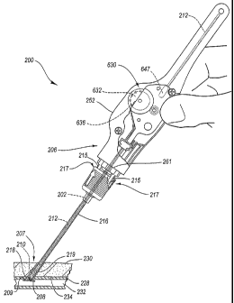
mechanism 630 is shown in detail in Fig. 6. The automatic driving mechanism
630
may comprise a gearbox assembly 629, and the gearbox assembly 629 may be
selectably disengagable. According to the embodiment of Fig. 6, once the
automatic
driving assembly 630 contacts the stop 261, further retraction of the closure
device
200 automatically effects tamping of the sealing plug 210 (Fig. 5F).
According to the gearbox assembly 629 of Fig. 6, the suture 204 is connected
to and partially wound about a spool 632 of a first gear an spool assembly
631. The
first gear and spool assembly 631 includes both the spool 632 and a first gear
636
arranged on a first axis 635. According to the embodiment of Fig. 6, the first
gear
636 is connected to the spool 632 and therefore they rotate together.
Withdrawal of
the closure device 200 (Fig. 5F) from the tissue puncture site 218 (if the
anchor 208
(Fig. 5F) is deployed and the gearbox assembly 629 has contacted the stop 261)
causes the suture 204 to unwind from the spool 632. The spool 632 rotates as
the
suture 204 unwinds and provides a torsional motive force that is transduced to
a
linear tamping force.
21
CA 02608517 2007-11-14
WO 2006/124245
PCT/US2006/016383
The torsional motive force provided by the spool 632 is transduced into the
linear tamping force by the gearbox assembly 629 according to the embodiment
of
Fig. 6. The gearbox assembly 629 includes the first gear 636 arranged
coaxially with
the spool 632. As shown in Fig. 6, the first gear 636 may be arranged adjacent
to a
second gear 642. The second gear 642, when assembled, engages the first gear
636.
The second gear 642 is arranged on a second axis 640. The second gear 642 may
be
a two-stage gear, with each stage engaging a different adjacent gear as shown.
The
first and second gears 636 and 642 may engage one another with a frictional
fit, or
with meshed gear teeth as shown. The second gear 642 is arranged adjacent to a
third gear 643 on a third axis 645. When assembled, the second gear 642
engages
and drives the third gear 643.
The tamping tube 212 is disposed between the third gear 643 and a guide 646.
The tamping tube 212 preferably includes the teeth shown, which mesh with
teeth of
the third gear 643. A concave holder 647 may support the tamping tube 212.
When
the spool 632 rotates, it drives the tamping tube 212, which in turn tamps the
sealing
plug 210 (Fig. 5F). Alternatively, the tamping tube 212 may not extend into
the
housing 252, and instead a separate rack may mesh with the third gear 643. The
separate rack would, in turn, drive the tamping tube 212.
The tamping tube 212 is preferably semi-tubular and partially disposed about
the suture 204 along its longitudinal axis. The semi-tubular shape of the
tamping
tube 212 has a generally U-shaped cross section, and provides an open channel
or
trough 648 through which the suture 204 may enter and exit. The open channel
648
permits the suture and the tamping tube 212 to merge as the spool 632 unwinds.
The
22
CA 02608517 2007-11-14
WO 2006/124245
PCT/US2006/016383
suture 204 and the tamping tube 212 are not fixedly connected to one another,
allowing each to slide freely past the other. Accordingly, with the anchor 208
(Fig.
5D) deployed, as the closure device 200 (Fig. 5F) is retracted in a first
direction with
the gearbox assembly 629 bearing against the stop 261 (Fig. 5F), the suture
204
unwinds from the spool 632, which drives the gearbox assembly 629. The gearbox
assembly 629 drives the tamping tube 212 in a second, opposite direction, and
the
tamping tube tamps the sealing plug 210 (Fig. 5F).
It may be desirable in some cases to increase the linear velocity of the
tamping
tube 212 relative to the linear velocity at which the closure device 200 (Fig.
5F) is
withdrawn. Increasing the linear velocity for the tamping tube 212 may better
assure
that the sealing plug 210 (Fig. 5F) is forced toward the anchor 208 (Fig. 5F)
when the
closure device 200 (Fig. 5F) is withdrawn in an opposite direction. Therefore,
according to some embodiments, the gearbox assembly 629 may have an overall
gear
ratio greater than 1:1. For example, the gear ratio may range between
approximately
1.5:1 and 3.0:1 for some embodiments, while the gear ratio is about 2.1:1 in
other
embodiments
However, it should be noted that the linear velocity of the tamping tube 212
should not be excessively greater than the linear velocity of withdrawal of
the closure
device, as excessive speed could potentially force the sealing plug 210 (Fig.
5F)
through the tissue puncture 218 (Fig. 5F) and into the lumen 232 (Fig. 5F) of
the
artery 228 (Fig. 5F). Likewise, an insufficient opposing force against the
anchor 208
(Fig. 5F) could potentially result in the anchor 208 (Fig. 5F) being pulled
out of
23
CA 02608517 2007-11-14
WO 2006/124245
PCT/US2006/016383
place from within the artery 228 (Fig. 5F). Therefore, according to some uses,
the
withdrawal force should not exceed approximately 2.5 pounds.
It will be understood by those of skill in the art having the benefit of this
disclosure that the gearbox assembly 629 configuration shown in Fig. 6 is
exemplary
in nature, and not limiting. Any gear configuration (including a single gear)
may be
used to transmit a motive force generated by retraction of the suture 204 from
the
closure device 200 (Fig. 5F) to provide an automatic driving force to the
sealing plug
210 (Fig. 5F) via the tamping tube 212.
As mentioned above, the gearbox assembly 629 may be selectable
disengagable. Therefore, one or more of the spool 632, first gear 636, second
gear
642, and third gear 643 may be movable to disengage or manually disable
adjacent
gears. For example, one or more of the first gear 636, second gear 642, or
third gear
643 may be movable along its respective axis to disengage from an adjacent
gear. As
shown in Fig. 6, a biasing member such as a spring 649 is disposed at the
second axis
640 biasing the second gear 642 into a meshed relationship with the first and
third
gears 636, 643. However, the second gear 642 is movable along the second axis
640
by operation of an actuator 651 coupled to the second gear 642. Therefore, a
force
may be applied to the actuator 651 (following sliding movement of the gearbox
assembly 629 to reach the stop 261, thereby aligning the actuator 651 with an
access
hole 253 in the handle 252) laterally with respect to the second gear 642, to
overcome a biasing force provided by the spring 649 and move or displace the
second
gear 642 axially out of the meshed or contacting relationship with at least
one of the
first and third gears 636, 643. According to the embodiment of Fig. 6, axial
24
CA 02608517 2007-11-14
WO 2006/124245
PCT/US2006/016383
movement of the second gear 642 only disengages the second gear 642 from the
first
gear 636. Disengaging the gearbox assembly 629 allows retraction of the
closure
device 200 (Fig. 5F) and unwinding of the suture 204 from the spool 632
without
driving the tamping tube 212. The advantages of this disengagement are
discussed
below with reference to the operation of the closure device 200.
However, as shown in Figs. 6-7, the tamping tube 212 may interlock with the
second gear 642 in a first rack position shown, preventing premature
activation of the
actuator 651. The interlocking geometry is seen more clearly in Fig. 7. The
second
gear 642 may include a second gear hub 653 with an annular groove 655. The
tamping tube 212 is disposed in the annular groove 655 in the first rack
position,
which locks out the actuator 651. The tamping tube rests on the concave holder
647.
Therefore, as long as the tamping tube 212 is disposed in the annular groove
655,
the actuator 651 may not be depressed. With the tamping tube 212 disposed in
the
annular groove 655, forces applied to the actuator 651 are transmitted to the
second
gear 642, but the second gear is prevented from moving axially by the rack
disposed
in the annular groove 655 and supported by the concave holder 647.
Nevertheless,
retracting the closure device 200 (Fig. 5F) results in rotation of the gears
of the
gearbox assembly 629, and linear movement of the tamping tube 212. When the
tamping tube 212 has moved a predetermined distance to a second tamping tube
position sufficient to cause effective tamping of the sealing plug 210 (Fig.
5F), the
tamping tube 212 also moves out of the annular groove 655 (See Fig. 5F).
Therefore,
the actuator 651 is no longer locked out, and the second gear 642 may be
disengaged
once the tamping tube 212 has moved linearly the predetermined distance.
CA 02608517 2007-11-14
WO 2006/124245
PCT/US2006/016383
Operation of the embodiment of Figs. 5A-8 is as follows. As the handle 252
of the closing device 200 is retracted from the puncture tract 219 as shown in
Fig.
5B, the detent 255 releases. The automatic tamping mechanism 630 and carrier
tube
202 remain stationary and therefore float relative to the handle 252. The
procedure
sheath 216 is retracted as the handle 252 is withdrawn, exposing the distal
end 207 of
the carrier tube 202. The automatic tamping mechanism 630 eventually contacts
a
stop 261, and further retraction causes the automatic tamping mechanism 630
and
carrier tube 202 to retract as well. As the automatic tamping mechanism 630
retracts, the suture 204, which is threaded through the anchor 208, unwinds
from and
causes rotation of the spool 632. The spool 632 drives the first gear 636 as
it rotates
via the coaxial connection between the spool 632 and the first gear 636. As
the first
gear 636 rotates, it drives the second gear 642. The second gear 642 drives
the third
gear 643, and the third gear 643 drives the tamping tube 212. The tamping tube
212
tamps the sealing plug 210. Therefore, as the closing device 200 is retracted
from
the puncture tract 219, the procedure sheath 216 is retracted (Figs. 5D-5E),
and the
sealing plug 210 is automatically tamped (Figs. 5F-5G). The sealing plug 210
is
more likely to create a sufficient arterial seal without a gap relative to the
anchor
208, as may otherwise occur with a separate manual tamping procedure.
Moreover, when the sealing plug 210 has been sufficiently tamped, the
selectably disengagable gearbox assembly 629 may be disengaged, enabling
further
retraction of the closure device 200 without additional tamping. With the
sealing
plug 210 fully tamped, there may be little or no portion of the suture 204
extending
outside of the tissue layer 230 and exposed to an operator. Therefore, it may
be
26
CA 02608517 2007-11-14
WO 2006/124245
PCT/US2006/016383
difficult for an operator to separate the sealing plug 210 and anchor 208 from
the
remainder of the closure device 200. In addition, too much retraction with the
selectably disengagable gearbox assembly 629 enabled could potentially
overtamp
the sealing plug 210 into the artery 228. Accordingly, the selectably
disengagable
gearbox assembly 629 may be advantageously disabled by activating the actuator
651
through the access hole 253. Activating the actuator 651 allows the suture 204
to
fully unwind from the spool 632 without driving the tamping tube 212.
Unwinding
the spool 632 exposes a sufficient length of the suture 204 to allow an
operator to
easily cut it and separate the sealing plug 210 and anchor 208 from the
remainder of
the closure device 200.
Referring next to Fig. 9, another embodiment of a selectably disengagable
automatic driving mechanism 930 is shown. The selectably disengagable
automatic
driving mechanism 930 of Fig. 9 may replace the selectably disengagable
gearbox
assembly 629 shown in Fig. 6 within the closure device 200 (Fig. 5A). Similar
to the
embodiment of Fig. 6, the selectably disengagable automatic driving mechanism
930
of Fig. 9 includes the suture 204 at least partially wound about a spool 932
of a first
gear and spool assembly 931. The first gear and spool assembly 931 includes
both
the spool 932 and a first gear 936 arranged on a first axis 935. However,
according
to the embodiment of Fig. 9, the first gear 936 and the spool 932 form a
manually
operated clutch therebetween. The clutch may be used to selectively connect
and
disconnect the first gear 936 from the spool 932. The clutch comprises a
plurality of
release fingers 961 in Fig. 9. The release fingers 961 are arranged
substantially in a
circle. A first component 963 of the release fingers 961 is cantilevered from
the first
27
CA 02608517 2007-11-14
WO 2006/124245
PCT/US2006/016383
gear 936 and extends normal to the first gear 936. A protrusion 965 of the
first
component 963 extends radially outward and is received by a mating recess 967
of
the spool 932. A second component 969 of the release fingers 961 arcs
substantially
normal to the first component 963 and the first gear 936. The second component
969
of each of the release fingers 961 extends through a central hole 971 of the
spool
932. An actuator button 951 fits over and contacts the second components 969
of
each of the release fingers 961.
The fit of the protrusions 965 of the first gear 936 with the mating recesses
967 of the spool 932 causes the first gear 936 and spool 932 to rotate
together at an
identical angular velocity. However, when the actuator button 951 is
depressed, the
actuator button slides along the arcs of the second component 969, forcing
each of
the release fingers 961 radially inward. The radial inward displacement of the
release fingers 961 at least partially removes the protrusions 965 from the
mating
recesses 967, allowing independent rotation of the spool 932 with respect to
the first
gear 936. Therefore, similar to the arrangement described above with reference
to
Figs. 5A-8, after the sealing plug 210 is driven toward the anchor 208, the
selectably
disengagable automatic driving mechanism 930 is disengaged or disabled,
allowing
the suture 204 to safely unwind without further tamping. The suture 204 is
then
exposed to the operator for convenient cutting.
The remaining components of the selectably disengagable automatic driving
mechanism 930 may be similar to the embodiment of Fig. 6. Transducing the
torsional motive force provided by the spool 932 to the linear tamping force
is
achieved by a gear train 934. The gear train 934 may include the first gear
936 and
28
CA 02608517 2012-10-09
=
second and third gears 942, 943. As shown, the second gear 942 engages and
drives
the third gear 943, and the third gear 943 drives a tamping tube 212 or other
sealing
plug driving device. The second gear 942 of Fig. 9 does not, however, include
an
annular groove interlocking with the tamping tube 212.
The scope of the claims should not be limited by the preferred embodiments set
forth in
the examples but should be given the broadest interpretation consistent with
the
description as a whole.
to
29