Note: Descriptions are shown in the official language in which they were submitted.
CA 02608995 2007-11-19
Received 29 August 2006
PAOPER(DM12768920 ip.I (wo ra)AOC-29/0&Ofi
~ -1-
ASSAY DEVICE
Field of the Invention
The present invention relates generally to an assay device, cantilevered
detector and/or
chemical assay method.
Background of the Invention
It is known to use compact discs (CDs) for chemical testing. The CDs are
provided with
micro-fluidic structure which defines various fluid input ports in
communication with
associated channels and fluid mixing chambers. In order to conduct a test,
fluid is
deposited into the input ports and the CD is spun so that the fluid is forced
by centrifugal
pumping through the relevant channels to the mixing chambers. Significantly
modified
CD optics and addressing technology can be used to capture images of specific
mixing
chambers to determine the test results of any chemical reaction within the
chambers.
It is also known that microcantilever beams have been considered as a means
for detecting
the results of chemical reactions, but the limited sensitivity of the beams
studied has not
resulted in any widespread application of the technology.
Summarv of the Invention
In accordance with the invention, there is provided an assay device having a
rotatable
platform with a test chamber and a sensor with a porous section, the sensor
undergoing
displacement when subject to a particular substance such as a chemical,
biological species
or other organism.
Preferably, the sensor is a cantilever beam.
Preferably, the device includes microfluidic paths in communication with
associated
Amended Sheet
IPEA/AU
CA 02608995 2007-11-19
Received 29 August 2006
P WPEROM I216l920 spo I(wo ra)doc-29iO8/06
-2-
channels and the test chamber or an associated plurality of test chambers.
Preferably, the or each test chamber includes one or more cantilever beams.
The porous section can give the sensor a sensitivity which enables the
presence of the
particular substance i.e., a selected chemical, species or organism, to be
detected.
Preferably, the sensor is functionalised with receptors, antibodies, antigens
or enzymes
which will selectively attract and bond with the particular substance to be
detected.
Preferably, the porous section is coated with a gold layer which attaches the
receptors to
the beam, functionalising the beam for bonding with preselected species or
organism
within the fluid in the test chamber.
Preferably, the sensor includes a surface to be monitored, which is subject to
displacement
upon movement of the sensor, the position of the monitored surface being
monitored by an
apparatus into which the assay device is loaded.
Preferably, the surface is a reflective surface.
Preferably, the apparatus is a CD drive connected to a computer allowing the
position of
the reflective surface to be determined and displayed by the computer.
Preferably, the assay device includes a micro-fluidic system for conveying a
test fluid from
an inlet port to the test chamber containing the cantilever beam and on to a
waste chamber.
Preferably, the waste chamber is separated from the test chamber by a micro-
mechanical
valve which is actuated above a threshold angular velocity of the device.
Preferably, the device can accept the whole fluid to be tested and includes a
filter for
filtering material from the whole fluid after insertion into the inlet port to
provide fluid in a
Amended Sheet
IPEA/AU
CA 02608995 2007-11-19
Received 29 August 2006
P\OPEROM 12768920 rpr t puo ra) Ax.29N8A6
-3-
form suitable for testing. More preferably, the filter is formed of a porous
silicon.
Preferably, the system includes provision for secondary chamber connected to
the test
chamber to allow the cycling of fluid between the secondary and test chamber.
Preferably, the device is in the form of a compact disc (CD).
In another aspect, there is provided a test apparatus for receiving an assay
device, as
described above, including a drive unit for rotating the device and a read
unit for
monitoring the sensor.
Preferably, the apparatus is adapted to display information derived from the
read unit.
More preferably, the apparatus is in the form of CD drive and the read unit
forms part of an
existing optical read/write head of the CD drive.
More preferably, the apparatus is connected directly to a computer, on which
is installed a
computer program which controls the operation of the CD drive to initiate the
filtering
process, the transfer of fluid between chambers and the optical reading system
to measure
the displacement of the sensor.
Preferably, the assay process is initiated by using the computer to input data
defining the
test to be performed and presenting the results with this same identification.
In another aspect, there is provided a chemical assay method including
introducing fluid to
a sensing chamber on a rotatable platform, wherein the sensing chamber
includes a sensor
with a porous section, the sensor being arranged for displacement upon
detection of a
particular substance, such as a selected molecule, within the chamber, and
monitoring the
sensor to detect the displacement.
In yet another aspect, there is provided a cantilever sensor, as described
above.
Amended Sheet
IPEA/AU
CA 02608995 2007-11-19
WO 2006/122360 PCT/AU2006/000656
-4-
Brief Description of the Drawings
The invention is now described, by way of non-limiting example only, with
reference to
the accompanying drawings in which:
Figure 1 is a diagrammatic representation of a plan view of an assay device;
Figure 2 is a diagrammatic cross-sectional view of a test apparatus;
Figure 3a is a diagrammatic side view of a microcantilever;
Figure 3b is a diagrammatic side view of the microcantilever, illustrating
deflection;
Figure 4 is a graph illustrating a relationship between resonant frequency and
porosity of a
cantilever;
Figure 5 is a diagrammatic perspective view of a cantilever sensor and a
read/write head of
a CD drive;
Figure 6 is a graph illustrating a relationship between intensity and time,
for the purpose of
detecting displacement of the sensor;
Figure 7 is a flow chart of a test procedure; and
Figure 8 is a graph illustrating comparative deflection of a porous and non-
porous
cantilever.
Detailed Description
An assay device 1 is illustrated in Figure 1 as including a rotatable platform
2, in the form
of a compact disc (CD), with a microfluidic system 3 including an inlet port
4, a secondary
chamber 5, a test chamber 6 and a waste chamber 7 interconnected by respective
channels
8,9,10. A filter 11 is provided in one of the channels 8, adjacent the inlet
port 4 for
filtering material such as cellular material from the test fluid introduced
into the inlet port
4. The filter 11 is preferably formed of porous silicon 12. A micro-mechanical
valve 13 is
also provided in the channel 10 separating the test chamber 6 and waste
chamber 7. The
valve 13 moves from a closed position, indicated by dashed lines 14, to an
open position,
indicated by arrow 15, when the angular velocity of the device 1 is above a
predetermined
threshold.
CA 02608995 2007-11-19
WO 2006/122360 PCT/AU2006/000656
-5-
In operation, fluid is introduced into the inlet port 4 and the device 1 is
rotated at a required
speed to effect centrifugal pumping so that the fluid is forced through the
channel 8 into
the secondary chamber 5 and subsequently the test chamber 6 where a sensor is
provided
for the purpose of detecting the presence of a particular substance, such as a
selected
chemical, biological species or other organisms within the fluid. The device 1
is then
rotated at higher angular velocity to open the valve 13 and allow the fluid to
exit the test
chamber 6.
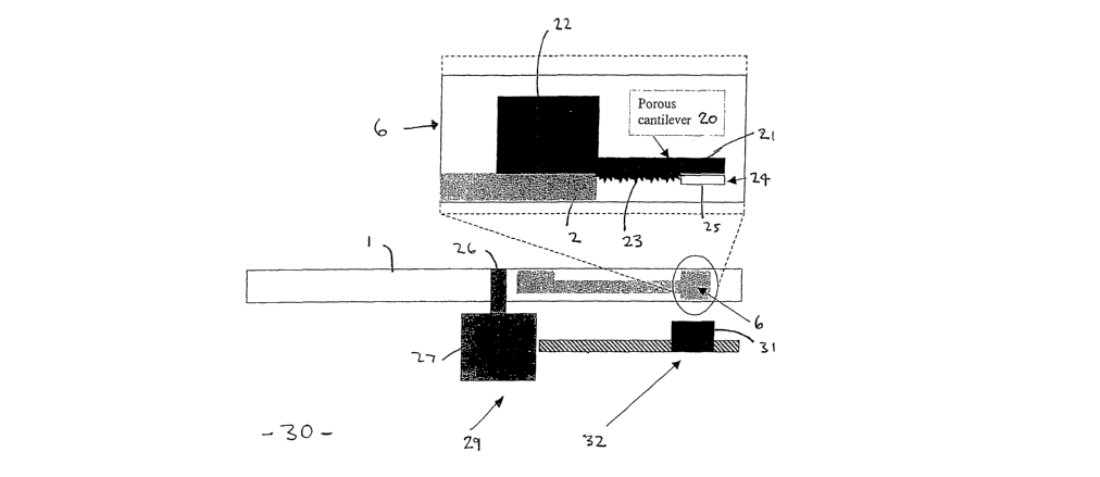
Referring now to Figure 2, the test chamber 6 of the device 1 is shown in
enlarged section
as including a cantilever sensor 20, which projects from the platform 2 of the
device 1.
More specifically, the porous cantilever sensor 20 is formed of a beam 21
which projects
from a silicon block 22 and includes a porous section 23 and a surface 24
formed of, for
example, a section of gold 25 or other suitable metallic or reflective
substance.
The device 1 is shown fitted on a spindle 26 of a drive unit 27 of a test
apparatus 30, which
is preferably in the form of a computer, with a CD drive 29 and the drive unit
27 forms
part of the drive 29, together with a read unit 31, which monitors any
displacement of the
reference surface 24 and thereby the cantilever sensor 20. The read unit 31
preferably
forms part of an existing read/write head 32 of the CD drive 31, without
modification.
The structure of the cantilever sensor 20 is now described in more detail with
reference to
Figure 3. Figure 3a shows an enlarged part 33 of the sensor 20 as including a
porous layer
35 and a silicon layer 34 both coated with gold which is provided with
antibody receptors
36 for capturing molecules 37 such as antigen ligands. The binding of the
molecules 37 to
the receptors 36 will lead to a deflection of the beam 21, as illustrated in
Figure 3b, which
can then be detected.
By forming the cantilever beam of porous material, the deflection is enhanced.
More
particularly, the characteristics of the cantilever sensor 20 rely on surface
processes such as
adsorption , desorption, surface reconstruction and reorganisation to induce a
surface stress
CA 02608995 2007-11-19
WO 2006/122360 PCT/AU2006/000656
-6-
in the active surface layer of the cantilever beam 39. Modifying the surface
stress on
surface 39 of the beam 21 will induce a differential stress across the
cantilever sensor 20,
causing it to bend.
The curvature of the beam 21 is proportional to the differential stress
gradient across the
beam. Increasing surface stress on surface 39 compared to the surface 40 or
layer 34
increases the differential stress gradient. Porous silicon at surface 39 can
be used as the
layer 35 to increase surface area and hence sensitivity. To the best of our
knowledge, no
research or development has been focussed on increasing the sensitivity of the
cantilever
based sensing technique by modifying the beam geometry or material structure.
The beam
21 increases the maximum surface stress that can be induced by the chemical
analyte by
introducing the porous layer 35 and modifying the beam geometry.
Analysis and tests have shown that by modifying the beam, geometry and
material
structures as described, the increased beam deflection for increased porosity
can be varied
as shown in Figure 8.
Accordingly, in Figure 5, the sensor 20 allows for increased deflection of the
cantilever
beam 21, as compared to a conventional beam of the same thickness and length,
by
fabricating a porous section on surface 23 of the beam 21. This has three
affects on the
mechanical response of the beam 21:
1. It reduces the effective thickness of the beam where it is porous, reducing
the second
moment of inertia of the beam, making the beam less rigid;
2. The spring constant of the beam is also reduced where it is porous; and
3. The surface area of the beam is also increased due to the increased
porosity of the
cantilever beam of the cantilever beam.
These three physical affects have a combined effect to increase the deflection
of the beam
and sensitivity to surface-combination events over current cantilever based
biosensors.
Increasing the differential stress induced between the layers 35,34 of the
beam, Figure 3b
CA 02608995 2007-11-19
Received 29 August 2006
P:WPfR1DM12268920'pa l p.o s)Acc2910tA6
-7-
leads to an increase in the deflection. Further to this, the surface area to
be functionalised,
i.e. provided with receptors for bonding with selected molecules, is
increased, allowing for
a greater density of functionalised groups to be attached to the surface,
thereby increasing
the sensitivity and induced surface stress for the same concentration of
chemical or
biological species.
This enables a more concentrated binding of the species and also enables less
variation in
deflection for the same chemical or species concentrate.
Another affect of modifying the geometry of the beam is that the resonant
frequency of the
beam is changed since the resonant frequency is a direct measure of the amount
of
porosity.
The resonant frequency change according to the beam geometry has the following
relationship: fo - 1 fk
;r where fo = resonance frequency
k = spring constant
m= mass of the beam
A change of porosity changes the resonant frequency of the cantilever beam 21
and is an
additional sensing capability of the sensor, which could be applied to
detection of
corrosion or chemical reaction caused by fluid, for example, measurement of
corrosion on
a marine vessel or detection of acid rain or similar events for environmental
monitoring.
The change in resonance frequency with porosity is illustrated in Figure 4
which indicates
that there is a minimum resonant frequency for a range of porosity levels. In
the apparatus
Amended Sheet
IPEA/AU
CA 02608995 2007-11-19
WO 2006/122360 PCT/AU2006/000656
-8-
30, however, it is only the deflection of the beam 21 that needs to be
monitored.
Conventional systems for detecting such deflection use a laser and a position
sensitive
detector to detect the deflection. The detection system is an external set-up
and requires the
laser to be optical aligned to the cantilever beam. The detection system used
in the
apparatus 30, on the other hand, uses the inherent optical detection system of
the CD drive
29. The read/write head (RWH) 32 of the drive 29 is used to interrogate the
cantilever
sensor 20 and monitor the position of the reference surface 24. In addition
the laser of the
RWH may be used to control the temperature of the test and secondary chambers
5,6.
More particularly, to sense the deflection of the sensor 20, the RWH is moved
over the
position of the porous cantilever beam 21, as illustrated in Figure S. The CD
device 1 can
be rotating while sensing the deflection. The laser of the RWH is focused onto
the
cantilever beam 21 and the reflected intensity from the reference surface 24
of the beam 21
is measured prior to loading a test fluid into the test chamber 4, for
calibration purposes.
The test fluid is then caused to enter the test chamber 6 and subsequently
exhausted to the,
waste chamber 7. The change in reflected intensity from the cantilever beam 21
after the
test fluid has been removed from the test chamber 6 is measured. The change in
reflected
intensity is a measure of the sensor deflection. Secondary to this, the
deflection can also be
measured as a change in focus. When the laser is initially focused onto the
beam 21 prior
to loading the test fluid into the test chamber 6, the focus position can be
measured. After
the test fluid has been removed from the test chamber the beam 21 will have
deflected and
the reflective surface 24 will have moved out of focus. A graphical
representation
illustrating the affect of a change in focus on the measured intensity of
reflected laser light
is illustrated in Figure 6. The change in focus is an indirect measure of the
deflection and
can be measured as a change in current or voltage output from the RWH.
Application of Assay Device to Testing of Blood.
A detailed example of use of the assay device 1 and apparatus 20 is described
with
reference to Figure 7. Specifically, a diagnostic test procedure 40 is shown
as including a
step 41 of drawing blood from a client and inserting the blood into the inlet
port 4 of the
CA 02608995 2007-11-19
WO 2006/122360 PCT/AU2006/000656
-9-
device 1 at step 42. The CD device 1 is then inserted into a computer at step
43 and disc
information is read from the CD. Relevant software is then employed at step 44
to initiate
testing which commences at step 45 with the reflected intensity from the
cantilever sensor
20 being measured for calibration purposes. The CD is then spun at step 46 to
force the
blood into the first channel 8 and through the filter 11, where cellular
material is removed.
The resulting serum is then passed through the secondary chamber 5 (if
required) and into
the test chamber 6. If required, the serum is then heated at step 47 by a
laser of the RWH
resulting in the serum being cycled back and forth between the test chamber 6
and
secondary chamber 5 to improve interaction with the receptors. The CD is then
spun at a
higher angular velocity at step 48, to move the valve 13 into the open
position so that the
serum may exit the test chamber 6 and pass into the waste chamber 7 at step
49. The
RWH may then be used to measure the reflected intensity of the displaced
cantilever beam
21 at step 50 and the output of the RWH is then returned at step 51 for
analysis at step 52,
where the measured intensity is read and compared with calibrated data to
determine the
presence of a relevant chemical or molecule. The test results are then logged,
a user
notified of the results at step 53, and the CD ejected at step 54, as
required. The CD may
then be disposed of or stored for the purpose of a permanent record of the
test result.
Other Applications
The technology enables near patient health pathology to be performed, avoiding
the need
for use of expensive laboratory equipment and the associated delay in
provision of results.
Examples of the range of applications include:
= Human Health Pathology
Detection of - Prostate Specific Antigen
- Cardiac Enzymes
- Infectious diseases (Hepatitis, HIV)
- Snake bite venom
= Environment Pathology
Detection of - Leionella bacteria
CA 02608995 2007-11-19
WO 2006/122360 PCT/AU2006/000656
-10-
- Hepatitis in water ways
- E-coli levels
= Animal Health Pathology
Detection of - Johne's disease
= Fluid Quality Measurement
DetB.ction of - Wine fermentation
= Industrial Measurement
Detection of electrical insulation deterioration.
The invention has been described by way of non-limiting example only and many
modifications and variations may be made thereto without departing from the
spirit and
scope of the invention described.