Note: Descriptions are shown in the official language in which they were submitted.
CA 02609022 2009-08-11
APPARATUS AND METHODS FOR REPAIRING
THE FUNCTION OF A
DISEASED VALVE AND METHOD FOR MAKING SAME
Field of the Invention
The present invention is directed to an apparatus and methods for repairing
the
function of a diseased valve, such as a cardiac or venous valve, via an
endovascular
technique, and is further directed to methods for making the apparatus.
Background of the Invention
It is known to implant prosthetic valves in various body passages to replace
native
valves that are diseased or otherwise defective in some manner. Blood
pressure, as
provided by heart activity via the arteries, is normally sufficient to
maintain the flow of
blood in one direction through the vasculature. The blood pressure in the
veins is much
lower than in the arteries and venous valves function to limit the backflow of
blood
through the veins. Numerous such venous valves are located throughout the
venous
system and are particularly important to maintaining directional blood flow in
the lower
extremities.
Venous valves can become incompetent and lead to chronic venous insufficiency.
Various surgical techniques have been developed for treating incompetent
venous valves
including valvuloplasty, transplantation, and replacement with a prosthetic
valve. These
known surgical techniques include both open and percutaneous approaches. As
with any
prosthetic, compatibility issues for prosthetic venous valves are important,
along with the
need to avoid thrombosis and platelet deposition.
Another common type of prosthetic valve is a prosthetic cardiac valve.
Prosthetic
cardiac valves have been used to replace all four of the native cardiac
valves. Cardiac
valve replacement has traditionally been done though an invasive
CA 02609022 2007-11-19
WO 2006/127412 PCT/US2006/019310
-2-
open surgical procedure, although endovascular (or percutaneous) approaches
are
being developed.
The four native cardiac valves (mitral, aortic, tricuspid, and pulmonary)
serve to direct the flow of blood through the two sides of the heart in a
forward
direction. On the left (systemic) side of the heart, the mitral valve is
located
between the left atriuin and the left ventricle, while the aortic valve is
located
between the left ventricle and the aorta. These two valves direct oxygenated
blood
coming from the lungs, through the left side of the heart, into the aorta for
distribution to the body. On the right (pulmonary) side of the heart, the
tricuspid
valve is located between the right atrium and the right ventricle, while the
pulmonary valve is located between the right ventricle and the pulmonary
artery.
These two valves direct de-oxygenated blood coming from the body, through the
right side of the heart, into the pulmonary artery for distribution to the
lungs, where
it again becoines re-oxygenated to begin the circuit anew.
All four of these native cardiac valves are passive structures that do not
themselves expend any energy and do not perform any active contractile
function.
The valves consist of moveable leaflets that open and close in response to
differential pressures on either side of the valve. The mitral and tricuspid
valves
are referred to as atrioventricular valves because they are situated between
an
atrium and a ventricle on each side of the heart. The mitral valve has two
leaflets
and the tricuspid valve has three leaflets. The aortic and pulmonary valves
are
referred to as semilunar valves because of the unique appearance of their
leaflets,
which are often termed "cusps" and which are shaped somewhat like a half-moon.
The aortic and pulmonary valves each have three cusps.
Cardiac valves can exhibit abnormal anatomy and function as a result of
congenital or acquired valve disease. Congenital valve abnonnalities may be so
severe that eniergency surgery is required within the first few hours of life,
or they
may be well-tolerated for many years only to develop a life-threatening
problem in
an elderly patient. Acquired valve disease may result from causes such as
rheumatic fever, degenerative disorders of the valve tissue, bacterial or
fungal
infections, and trauma.
CA 02609022 2007-11-19
WO 2006/127412 PCT/US2006/019310
-3-
The two major problems that can develop with cardiac valves are stenosis,
in which a valve does not open properly, and insufficiency (also called
regurgitation), in which a valve does not close properly. Stenosis and
insufficiency
may occur concomitantly in the same valve or in different valves. Both of
these
abnormalities increase the worlcload and stress placed on the heart. The
severity of
this increased stress on the heart, and the heart's ability to adapt to it,
determine
whether the abnormal valve will have to be surgically repaired or replaced.
In addition to stenosis and insufficiency of cardiac valves, surgery may also
be required for certain types of bacterial or fungal infections in which the
valve
may continue to function normally, but nevertlleless harbors an overgrowth of
bacteria on the leaflets of the valve that may flalce off (or embolize) and
lodge
downstream in a vital artery. If this occurs on the valves of the left side
(i.e., the
systemic circulation side) of the heart, einbolization results in sudden loss
of the
blood supply to the affected body organ and immediate malfunction of that
organ.
The organ most commonly affected by such embolization is the brain, in which
case the patient suffers a stroke. Thus, surgical replacement of either the
mitral or
the aortic valve may be necessary for this problem even though neither
stenosis nor
insufficiency of either valve is present.
If a cardiac valve must be replaced, there are currently several options
available, and the choice of a particular type of prosthesis (i.e., artificial
valve)
depends on factors such as the location of the valve, the age and other
specifics of
the patient, and the surgeon's experiences and preferences. Available
prostlieses
include mechanical valves, tissue valves, and homograft valves.
Mechanical valves include caged-ball valves, bi-leaflet valves, and tilting
disk valves. The main advantage of mechanical valves is their long-term
durability. Their main disadvantage is that they require the patient to talce
systemic
anticoagulation drugs for the rest of his or her life, because of the
propensity of
mechanical valves to cause blood clots to form on them.
Tissue valves are typically constructed either by sewing the leaflets of
porcine aortic valves to a stent (to hold the leaflets in proper position), or
by
constructing valve leaflets from porcine or bovine pericardial tissue and
sewing
them to a stent. The stents may be rigid or slightly flexible and are
typically
CA 02609022 2007-11-19
WO 2006/127412 PCT/US2006/019310
-4-
covered witli a fabric, such as the material sold under the trademarlc
DACRONTM,
and then attached to a sewing ring for fixation to the patient's native valve
amiulus.
The porcine or bovine tissue is chemically treated to alleviate any
antigenicity (i.e.,
to reduce the risk that the patient's body will reject the foreign tissue).
Tissue
valves may be used to replace any of the heart's four valves. The main
advantage
of tissue valves is that they do not cause blood clots to form as readily as
do the
mechanical valves, and therefore, they do not necessarily require systemic
anticoagulation.
Homograft valves are harvested from human cadavers. Homograft valves
are most commonly implanted in the aortic position, but are also occasionally
implanted in the pulmonary position. Homograft valves are specially prepared
and
frozen in liquid nitrogen, where they are stored for later use. The advantage
of
aortic homograft valves is that they appear to be as durable as mechanical
valves,
but do not promote blood clot formation and therefore do not require
anticoagulation. The main disadvantage of these valves is that they are not
available in sufficient numbers to satisfy the needs of patients who need new
aortic
or pulmonary valves. Homograft valves are also extremely expensive and can be
more difficult to implant than either mechaiiical valves or tissue valves.
Cardiac valve replacement using any of the aforementioned prostheses has
traditionally been done via an open surgical technique in which the thoracic
cavity
is opened. This exacting operation requires use of a heart-lung machine for
extenlal circulation of the blood as the heart is stopped and opened during
the
surgical intervention and the artificial cardiac valve is implanted under
direct
vision. This operation exposes the patient to many risks especially in the
elderly
population. Hence, an apparatus for repairing the function of a diseased
cardiac or
venous valve via an endovascular (or percutaneous) procedure, rather than an
open
surgical procedure, could offer tremendous benefits for these patients, many
of
whom have no options today.
Summary of the Invention
The present invention includes an apparatus for repairing the function of a
diseased valve. The apparatus comprises an annular first support member
CA 02609022 2007-11-19
WO 2006/127412 PCT/US2006/019310
-5-
expandable to a first diameter. An annular second support member is spaced
axially apart from the first support member and is expandable to a second
diameter
that is independent of the first dianieter. A tubular graft section
interconnects the
first and second support members. The graft section defines an annulus having
a
third diameter that is independent of each of the first and second diaineters.
A
prosthetic valve is secured within the annulus of the graft section. The
bioprosthetic valve has at least two valve leaflets that are coaptable to
pernnit the
unidirectional flow of blood.
In accordance with one aspect of the invention, an expandable ring
encircles the annulus of the graft section and supports the prosthetic valve
secured
within the annulus.
In accordance with another aspect of the invention, the apparatus further
coinprises at least one tubular conduit having first and second ends. The
second
end is received in a passage in the graft section and the first end is for
positioning
in a branch blood vessel.
In accordance with another aspect of the invention, a method for making an
apparatus to repair the function of a diseased valve is provided. According to
the
inventive method an annular first support member expandable to a first
diameter
and an annular second support member expandable to a second diameter that is
independent of the first diameter are provided. A tubular graft section is
also
provided. The graft section defines an annulus having a third diameter that is
independent of each of the first and second diameters. The first and second
support members are interconnected with the graft section such that the
support
members are spaced axially apart by the graft section. A bioprosthetic valve
having at least two valve leaflets that are coaptable to permit the
unidirectional
flow of blood is secured within the annulus of the graft section.
In accordance with another aspect of the invention, a minimally invasive
.
method for repairing the fiinction of a diseased valve is provided. According
to the
inventive method, an apparatus including annular first and second support
members that are spaced axially apart and are expandable to independent first
and
second diameters, respectively, is provided. The apparatus further includes a
prosthetic valve and a tubular graft section interconnecting the first and
second
CA 02609022 2007-11-19
WO 2006/127412 PCT/US2006/019310
-6-
support members. The graft section defines an annulus having a tliird diameter
that is independent of the first and second diameters. The prostlietic valve
is
secured within the aimulus of the graft section. The apparatus is collapsed
and
loaded into a sheath for intravascular delivery. The apparatus is inserted
into the
vasculature and advanced to a location witli the vasculature adjacent the
diseased
valve. The sheath is retracted and the first and second support members expand
into engagement with the vasculature at the respective first and second
diaineters
to form a seal between at least one of the first and second support members
and the
vasculature. The suspension of the prosthetic valve inside the graft section
at the
third diameter and within the vasculature adjacent the diseased valve assuines
the
function of the diseased valve.
In accordance with another aspect of the present invention, an apparatus for
repairing the function of a diseased valve coinprises an aimular support
member
having inner and outer surfaces. The support member is expandable to a first
dianieter. A tubular first graft section for sealing against a vessel wall
adjacent the
diseased valve is connected to the outer surface of the support meinber. A
tubular
second graft section is secured to the inner surface of the support member.
The
second graft section defines an annulus having a second diameter that is
smaller
than and independent of the first diameter of the support member. A prosthetic
valve is secured within the annulus of the second graft section. The
bioprosthetic
valve has at least two valve leaflets that are coaptable to permit the
unidirectional
flow of blood.
In accordance with anotller aspect of the present invention, a method for
making an apparatus to repair the function of a diseased valve is provided.
According to the inventive method, an annular support member expandable to a
first diameter is provided. The support member has inner and outer surfaces. A
tubular first graft section is connected to the outer surface of the support
member
for sealing against a vessel wall adjacent the diseased valve. A tubular
second
graft section is connected to the inner surface of the support member. The
second
graft section defines an annulus having a second diameter that is smaller than
and
independent of the first diameter. A bioprosthetic valve having at least two
valve
CA 02609022 2007-11-19
WO 2006/127412 PCT/US2006/019310
-7-
leaflets that are coaptable to permit the unidirectional flow of blood is
secured
within the aiuiulus of the second graft section.
In accordance wit11 another aspect of the present invention, a minimally
invasive method for repairing the function of a diseased valve is provided.
According to the inventive method, an apparatus including an annular support
member having inner and outer surfaces and which is expandable to a first
diameter is provided. The apparatus further includes a prosthetic valve and
first
and second tubular graft sections. The first graft section is connected to the
outer
surface and the second graft section is connected to the outer surface. The
second
graft section defines an annulus having a second diameter that is smaller than
and
independent of the first diameter. The prosthetic valve is secured within the
annulus of the second graft section. The apparatus is collapsed and loaded
into a
sheath for intravascular delivery. The apparatus is inserted into the
vasculature and
advanced to a location with the vasculature adjacent the diseased valve. The
sheath is retracted and the support member is expanded into engagement with
the
vasculature, forming a seal between the first graft section and the
vasculature. The
suspension of the prosthetic valve inside the second graft section at the
second
diameter and within the vasculature adjacent the diseased valve assumes the
function of the diseased valve.
Brief Description of the Drawings
The foregoing and other features of the present invention will become
apparent to those skilled in the art to which the present invention relates
upon
reading the following description with reference to the accompanying drawings,
in
which:
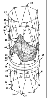
Fig. 1 is a schematic perspective view of an apparatus for repairing the
function of a diseased valve in accordance with the present invention;
Fig. 2 is a schematic side view of the apparatus of Fig. 1;
Fig. 3 is a schematic sectional view of the apparatus of Fig. 1;
Fig. 4 is a sectional view talcen along 4-4 in Fig. 3;
Fig. 5 is a side view similar to Fig. 2 illustrating an optional construction
for the present invention;
CA 02609022 2007-11-19
WO 2006/127412 PCT/US2006/019310
-8-
Fig. 6 is a schematic sectional view of a portion of a heart illustrating one
option for placement of the apparatus of Fig. 1 to repair the fiuiction of a
diseased
tricuspid valve;
Fig. 7 is a view similar to Fig. 6 illustrating another option for placeinent
of
the apparatus of Fig. 1 to repair the function of a diseased tricuspid valve;
Fig. 8 is a view similar to Figs. 6 and 7 illustrating yet another option for
placement of the apparatus of Fig. 1 to repair the function of a diseased
tricuspid
valve;
Fig. 9 is a view similar to Figs. 6-8 illustrating still another option for
placement of the apparatus of Fig. 1 to repair the function of a diseased
tricuspid
valve;
Fig. 10 is a schematic sectional view of an apparatus for repairing the
function of a diseased valve in accordance with a second embodiment of the
present invention;
Fig. 11 is a schematic side view of the apparatus of Fig. 10;
Fig. 12 is a sectional view taken along 12-12 in Fig. 10;
Fig. 13 is a schematic sectional view of a portion of a heart illustrating one
option for placement of the apparatus of Fig. 10 to repair the fiuiction of a
diseased
aortic valve;
Fig. 14 is a schematic sectional view of an apparatus for repairing the
function of a diseased valve in accordance with a third embodiment of the
present
invention;
Fig. 15 is a schematic sectional view of an apparatus for repairing the
function of a diseased valve in accordance with a fourth embodiment of the
present
invention;
Fig. 16 is a schematic perspective view of an apparatus for repairing the
function of a diseased valve in accordance with a fifth embodiment of the
present
invention;
Fig. 17 is a schematic side view of the apparatus of Fig. 16;
Fig. 18 is a schematic perspective view of an apparatus for repairing the
function of a diseased valve in accordance with a sixth embodiment of the
present
invention;
CA 02609022 2007-11-19
WO 2006/127412 PCT/US2006/019310
-9-
Fig. 19 is a schematic side view of the apparatus of Fig. 18;
Fig. 20 is a sectional view of an apparatus for repairing the fiinction of a
diseased valve in accordance with a seventh embodiment of the present
invention;
and
Fig. 21 is a sectional view of an apparatus for repairing the fiinction of a
diseased valve in accordance with an eighth embodiment of the present
invention.
Detailed Description of Embodiments
The present invention is directed to an apparatus and methods for repairing
the function of a diseased valve, such as a cardiac or venous valve, via an
endovascular teclmique, and is further directed to methods for making the
apparatus. As representative of the present invention, Figs. 1-4 illustrate a
first
einbodiment of an apparatus 10 for repairing the function of a diseased
cardiac
valve, such as a tricuspid valve 12 shown schematically in Figs. 6-8. It
should be
apparent, however, to those skilled in the art that the apparatus 10 disclosed
herein
can also be used to repair the function of other cardiac valves as well as
venous
valves. The apparatus 10 includes amlular first and second support members 14
and 16, a tubular graft section 18 interconnecting the support members, and a
prosthetic valve 20 secured within the graft section.
The first support member 14 coinprises a self-expanding or balloon
expandable stent made from stainless steel, but could alternatively be made
from
any suitable medical grade plastic or metal, including shape memory metals
such
as Nitinol. The first support member 14 has oppositely disposed proximal and
distal ends 22 and 24 connected by axially extending beams 26 having a
l~lown "M" or "Z" shape.
The axially extending beams 26 define generally cylindrical inner and outer
surfaces (not numbered) for tlie first support member 14. In the expanded
condition shown in Figs. 1-4, the outer surface of the first support member 14
has a
first diameter Dl (Fig. 2) that has been selected to exceed the largest
potential
venous diameter for a given patient.
Both the proximal and distal ends 22 and 24 of the first support member 14
include a plurality of eyelets 28 spaced circumferentially about the ends. The
first
CA 02609022 2007-11-19
WO 2006/127412 PCT/US2006/019310
-10-
support ineinber further includes a plurality of hooks (or barbs) 30 located
on the
outer surface of the beams 26. The hooks 30 extend radially outward and at an
angle to prevent migration of the support member 14 upon implantation. It
should
be understood that the location, quantity, configuration, and orientation of
the
hooks 30 may be altered depending on specific needs of the apparatus 10.
The second support member 16 resembles the first support meinber 14 and
comprises two conjoined self-expanding stents made from stainless steel, but
which could alternatively be made from any suitable medical grade plastic or
metal, including shape memory metals such as Nitinol. The two stents of the
second support member 16 together define oppositely disposed proximal and
distal
ends 32 and 34 connected by axially extending beams 36 having a known "M '
or "Z" shape.
The axially extending beams 36 define generally cylindrical inner and outer
surfaces (not numbered) for the second support member 16. In the expanded
condition shown in Figs. 1-4, the outer surface of the second support ineinber
16
has a second diameter D2 that has been selected to exceed the largest
potential
venous diameter for a given patient. It should be noted that, when implanted,
the
second support member 16 is fiee to expand to the second dianieter D2 (Fig. 2)
independent of the expansion of the first support member 14 to the first
diaineter D 1.
Both the proximal and distal ends 32 and 34 include a plurality of
eyelets 38 spaced circumferentially about the ends. The second support member
further includes a plurality of hooks (or barbs) 401ocated on the outer
surface of
the beams 36. The hooks 40 extend radially outward and at an angle to prevent
migration of the support member upon implantation. It should be understood
that
the location, quantity, and configuration of the hooks 40 may be altered
depending
on the specific needs of the apparatus 10.
It should also be understood that that the invention is not limited to the
particular configuration of the illustrated first and second support members
14
and 16, and that the first and second support members need not be similarly
configured. Further, it is contemplated that the lengtlis of the first and
second
support members 14 and 16 will be varied based on the needs of a particular
CA 02609022 2007-11-19
WO 2006/127412 PCT/US2006/019310
-11-
implantation. In addition, it should be noted that radiopaque inarlcers may be
attached at various locations on the first and second support members 14 and
16 to
aid witli placement of the apparatus 10 under fluoroscopy.
To enhance the biocompatibility of the apparatus 10, it is conteinplated that
at least a portion of the first and second support members 14 and 16 may be
coated
witll a therapeutic agent such as, for example, an anti-coagulant, an anti-
thrombogenic agent, an anti-proliferative agent, an anti-inflammatory agent,
an
antibiotic, an angiogenesis agent, a statin, a growth factor, or stem cells.
The
therapeutic agent may be loaded into a compound or polymer that is coated onto
the support meinbers 14 and 16 for a time-delayed release into surrounding
tissue.
The apparatus 10 further includes the tubular graft section 18
interconnecting the first and second support members 14 and 16. The graft
section 18 comprises a biocompatible material such as Dacrono', woven velour,
polyurethane, PTFE, or heparin-coated fabric. Alternatively, the graft section
18
may be a biological material such as bovine or equine pericardium, a
homograft, an
autograft, or cell-seeded tissue.
The graft section 18 has an hourglass shape defined by first and second end
portions 50 and 52 (Fig. 2) and a neck portion 541ocated between the ends. The
neck portion 54 of the graft section 18 is formed by a converging portion 56,
which
extends inward fiom the first end 50 to an annulus 58, and a diverging portion
60,
which extends outward from the annulus toward the second end 52. The
annulus 58 of the graft section 18 defines a third diameter D3 that is less
than and
independent of the first and second diameters Dl and D2 of the first and
second
support members 14 and 16, respectively. As may be seen in Fig. 1, one or more
axial seams 61 in the graft section 18 are used to create a smaller diameter
at the
annulus 58 than at either of the end portions 50 and 52.
The first end portion 50 of the graft section 18 is secured about the outer
surface of the distal end 24 of the first support member 14. As shown in Figs.
1-4,
the first end portion 50 may be sutured to the eyelets 28 at the distal end 24
of the
first support member 14. Alternatively, it is contemplated that first end
portion 50
may be sutured to other structure at the distal end 24 of the first support
member 14 depending on the configuration of the stent used. It is further
CA 02609022 2007-11-19
WO 2006/127412 PCT/US2006/019310
-12-
conteinplated that the first end portion 50 may be woven to the distal end 24
of the
first support menlber 14 or otherwise attached in another suitable mamier.
The second end portion 52 of the graft sectioii 18 is secured to the inner
surface of the proximal end 32 of the second support member 16. As shown in
Figs. 1-4, the second end portion 52 may be sutured to the eyelets 38 at the
proximal end 32 of the second support member 16. Alternatively, it is
contemplated that the second end portion 52 may be woveiz to the proximal end
32
of the second support member 16 or otherwise attached in another suitable
manner.
The second end portion 52 of the graft section 18 further includes a
plurality of extension flaps 62 that extend axially toward the distal end 34
of the
second support member 16. The extension flaps 62 are connected, such as by
sutures, to the beams 36 of the second support member 16. The number and
circumferential orientation of the extension flaps 62 correspond to the number
and
orientation of leaflets in the prosthetic valve 20.
The bioprosthetic valve 20 may be a homograft, an autograft, or made from
a harvested biological material including, but not limited to, bovine
pericardial
tissue, equine pericardial tissue or porcine pericardial tissue.
Alte.rnatively, the
bioprosthetic valve 20 may be made from a biocompatible synthetic material
including, but not limited to, polyurethane or expanded PTFE.
The bioprosthetic valve 20 is secured, by sutures or other suitable means,
within the aiuiulus 58 of the neck portion 54 of the graft section 18 so that
the
valve is suspended inside the graft section at the third diameter D3. In the
illustrated enlbodiments, the bioprosthetic valve 20 has three leaflets 64
that are
coaptable to permit the unidirectional flow of blood. However, it should be
understood that the prosthetic valve 20 could have less than three or more
than
three leaflets. Each of the leaflets 64 of the prosthetic valve 20 may be
sutured to a
respective one of the extension flaps 62 of the graft section 18 to create a
minor
amount of valve insufficiency in the apparatus 10 if so desired.
Fig. 5 illustrates an optional construction for the apparatus 10 in which an
expandable ring 66 encircles the annulus 58 of the graft section 18 to support
the
prosthetic valve 20. The ring 66 may comprise a single wire or a small stent.
The
ring 66 may be made from a shape memory metal, such as Nitinol, or any other
CA 02609022 2007-11-19
WO 2006/127412 PCT/US2006/019310
-13-
suitable medical grade plastic or metal. As shown in Fig. 5, the sutures that
are
used to secure the prosthetic valve 20 in the annulus 58 of the graft section
18 may
extend around the ring 66 to strengtlien the attachment of the valve 20 to the
armulus of the graft section. Further, the ring 66 can be used to positively
establish
the third diameter D3 at the aiululus 58 of the graft section 18.
One application for the present invention is to repair the function of the
diseased tricuspid valve 12 (Fig. 6). To enable delivery and deployment of the
apparatus 10, the apparatus is radially collapsed annd loaded into a sheath
(not
shown) over a catlieter (not shown). After de-airing of the assembly, the
apparatus 10 is delivered via a venotomy into the femoral vein and may be
assisted
with access through an internal jugular vein to establish through-and-through
access. In the application of the apparatus 10 illustrated in Fig. 6, the
apparatus is
delivered to a desired location in the inferior vena cava (IVC) just below the
right
atrium (RA), but above the hepatic veins, under fluoroscopic and/or
transesophageal echocardiographic guidance.
Once the apparatus 10 is advanced to the desired location, the sheath is
retracted to allow the first and second support members 14 and 16 to expand
radially outward into engagement with the IVC wall as shown in Fig. 6. It
should
be noted that a balloon (not shown) may be used to assist with the expansion
or
stabilization of one or both of the support meinbers 14 and 16. As the support
members 14 and 16 expand into the IVC wall, the hooks 30 and 40 on the
beams 26 and 36 of the support meinbers embed into the vessel wall to secure
the
apparatus 10 from migration in the IVC or right atrium.
Significantly, in the implanted condition shown in Fig. 6, the second
support member 16 expands to the second diameter D2, which is the diameter of
the IVC at that specific vascular location, and is able to independently
expand and
contract with the IVC in accordance with fluctuations in venous pressure or
capacitance. Furthermore, the first support member 14 expands to the first
diameter D1, which is the diameter of the IVC at that specific vascular
location,
and is able to independently expand and contract with the IVC in accordance
with
fluctuations in venous pressure or capacitance. In addition, the first end 50
of the
CA 02609022 2007-11-19
WO 2006/127412 PCT/US2006/019310
-14-
graft section 18 that encircles the distal end 24 of the first support member
14 seals
against the wall of the IVC to prevent any blood leakage around the apparatus
10.
Notwithstanding the flexibility of the diameters of the first and second
support members 14 and 16, the diameter of the prosthetic valve 20 is
predetermined by the third dianieter D3 of the annulus 58 of the graft section
18
and is functionally independent of the diameters of the first and second
support
members. This functional independence of the diameter of the prosthetic valve
20
suspended within the graft section 18 helps to prevent antegrade and
retrograde
blood lealcs around the prosthetic valve and ensures proper valvular function.
Further, the extra-cardiac location of the apparatus 10 reduces potentially
detrimental effects of cardiac contraction and provides an anatomically
favorable
region for fixation and sealing. Finally in the location shown in Fig. 6, the
apparatus 10 eliminates systolic flow through the hepatic veins and IVC.
Fig. 7 illustrates placement of the apparatus 10 for repairing the function of
the tricuspid valve in the superior vena cava (SVC). The apparatus 10 is
delivered
to a desired location in the SVC just above the right atrial junction, but
below the
azygos vein, under fluoroscopic and/or transesophageal echocardiographic
guidance. The apparatus 10 is then deployed in the same basic manner as
described above with regard to placement in the IVC.
In the implanted condition shown in Fig. 7, the second support member 16
expands to the second diameter D2, which is the diameter of the SVC at that
specific vascular location, and is able to independently expand and contract
with
the SVC in accordance with fluctuations in venous pressure or capacitance.
Furthermore, the first support member 14 expands to the first diameter D1,
which
is the diameter of the SVC at that specific vascular location, and is able to
independently expand and contract with the SVC in accordance with fluctuations
in venous pressure or capacitance. In addition, the first end 50 of the graft
section 18 that encircles the distal end 24 of the first support member 14
seals
against the wall of the SVC to prevent any blood leakage around the apparatus
10.
Notwithstanding the flexibility of the diameters of the first and second
support members 14 and 16, the diameter of the prosthetic valve 20 is
predetermined by the third diameter D3 of the annulus 58 of the graft section
18
CA 02609022 2007-11-19
WO 2006/127412 PCT/US2006/019310
-15-
and is functionally independent of the diameters of the first and second
support
members. This fimctional independence of the diameter of the prosthetic valve
20
suspended within the graft section 18 helps to prevent antegrade and
retrograde
blood lealcs arotind the prosthetic valve and ensures proper valvular
function.
Fig. 8 illustrates repairing the function of the tricuspid valve by placing a
first apparatus 10 in the IVC and a second apparatus 10 in the SVC. The first
apparatus 10 is placed in the IVC just below the right atrium but above the
hepatic
veins, and the second apparatus 10 is placed in the SVC just above the right
atrial
junnction but below the azygos vein. Alternatively, it is contemplated that
the
apparatus may be formed by a single second support member 16 that spans fiom
the SVC to the IVC.
Both the first apparatus 10 and the second apparatus 10 are deployed and
function in the same basic manner as previously described. In the implanted
condition shown in Fig. 8, the second support member 16 of the first apparatus
10
expands to the second diameter D2, which is the diameter of the IVC at that
specific vascular location, and is able to independently expand and contract
with
the IVC in accordance with fluctuations in venous pressure or capacitance.
Furthermore, the first support member 14 of the first apparatus 10 expands to
the
first diameter D1, which is the diameter of the IVC at that specific vascular
location, and is able to independently expand and contract with the IVC in
accordance with fluctuations in venous pressure or capacitance. In addition,
the
first end 50 of the graft section 18 of the first apparatus 10 that encircles
the distal
end 24 of the first support member 14 seals against the wall of the IVC to
prevent
any blood lealcage around the apparatus.
Similarly, the second support member 16 of the second apparatus 10
expands to the second diameter D2, which is the diameter of the SVC at that
specific vascular location, and is able to independently expand and contract
with
the SVC in accordance with fluctuations in venous pressure or capacitance.
Furthermore, the first support member 14 of the second apparatus 10 expands to
the first diaineter D1, which is the diameter of the SVC at that specific
vascular
location, and is able to independently expand and contract with the SVC in
accordance with fluctuations in venous pressure or capacitance. In addition,
the
CA 02609022 2007-11-19
WO 2006/127412 PCT/US2006/019310
-16-
first end 50 of the graft section 18 of the second apparatus 10 that encircles
the
distal end 24 of the first support member 14 seals against the wall of the SVC
to
prevent any blood leakage around the apparatus.
Notwithstanding the flexibility of the diameters of the first and second
support members 14 and 16 of each apparatus 10, the diameter D3 of each of the
prosthetic valves 20 is predetermined by the third diameter D3 of the annulus
58 of
the respective graft section 18 and is functionally independent of the
diameters of
the first and second support members. This fiinctional independence of the
diameter of each of the prosthetic valves 20 helps to prevent antegrade and
retrograde blood leaks around the prostlietic valves and ensures proper
valvular
function.
Fig. 9 illustrates another option for repairing the function of the diseased
tricuspid valve 12 using a modified version of the apparatus. In Fig. 9,
coiuponents of the apparatus that are similar, but not identical, to
previously
described components carry the suffix "a". The apparatus I Oa includes lower
and
upper support sections 14a, a valve section 80 suspended between the support
sections, and a graft enclosure 82.
The lower support section 14a of the apparatus l0a is placed in the IVC just
below the right atrium but above the hepatic veins, and the upper support
section 14a is placed in the SVC just above the right atrial junction but
below the
azygos vein. The valve section 80 includes a support member 16a a.nd the
bioprosthetic valve 20 secured therein. The valve section 80 is deployed in
the
right atrium at a location adjacent the tricuspid valve 12. The graft
enclosure 82
extends over the valve section 80 and the majority of the lower and upper
support
sections 14a to form a lining in the riglit atrium between the valve section,
the IVC,
and the SVC.
In the implanted condition shown in Fig. 9, the lower support section 14a
expands to the diameter of the IVC and is able to independently expand and
contract with the IVC in accordance with fluctuations in venous pressure or
capacitance. In addition, the portion of the graft enclosure 82 that covers
the lower
support section 14a seals against the wall of the IVC to prevent any blood
leakage
around the apparatus 10a. Similarly, the upper support section 14a expands to
the
CA 02609022 2007-11-19
WO 2006/127412 PCT/US2006/019310
-17-
diaineter of the SVC and is able to independently expand and contract with the
SVC in accordance with fluctuations in venous pressure or capacitance. The
portion of the graft enclosure 82 that covers the upper support section 14a
seals
against the wall of the SVC to prevent any blood leakage around the apparatus
10a.
Notwithstanding the flexibility of the diameters of the lower and upper
support sections 14a, the diameter of the prosthetic valve 20 is predetennined
by
the third diameter D3 of the annulus 58 of the valve section 80 and is
functionally
independent of the diameters of the support sections.
The apparatus 10 and 10a and associated methods described above help to
protect the lower and/or upper body from elevated venous pressures caused by a
diseased tricuspid valve. Probleins such as ascites, liver dysfunction, edema
and
cardiac cirrhosis that are often associated with severe tricuspid valve
regurgitation
can be treated using the apparatus and methods according to the present
invention.
Further, the apparatus 10 and 10a and methods of the present invention provide
a
minimally invasive, endovascular approach to treat severe valvular disease,
which
is particularly important for high risk patients.
Figs. 10-12 illustrate an apparatus for repairing the function of a diseased
valve in accordance with a second embodiment of the present invention. In the
second einbodiinent of Figs. 10-12, components of the apparatus that are
similar,
but not identical, to previously described components carry the suffix "b".
The graft section 18b of the apparatus 10b includes first and second
passages 90 and 92 extending axially through the neck portion 54b. Each of the
first and second passages 90 and 92 terminates at openings in the converging
portion 56b and the diverging portion 60b, respectively. As shown in Fig. 12,
the
passages 90 and 92 are spaced circumferentially apart and may have an
elliptical
shape in cross-section. It should be understood that the spacing and quantity
of
passages 90 and 92 may be varied based on the specific application for the
apparatus l Ob. The apparatus lOb of Figs. 10-12 is configured for repairing
the
function of a diseased aortic or other type of valve (not shown).
The apparatus 10b further includes first and second tubular conduits 94
and 96 that are receivable in the first and second passages 90 and 92,
respectively.
The first and second conduits 94 and 96 are made of a biocompatible material
such
CA 02609022 2007-11-19
WO 2006/127412 PCT/US2006/019310
-18-
as Dacronwoven velour, polyurethane, PTFE, or heparin-coated fabric.
Alternatively, the conduits 94 and 96 may be made from a biological material
such
as bovine or equine pericardium, a homograft, an autograft, or cell-seeded
tissue.
It should be understood that the quantity of conduits 94 and 96 may be varied
based on the specific application for the apparatus l Ob.
Each of the first and second conduits 94 and 96 has oppositely disposed
first and second ends 98 and 100. The first eiid 98 of each of the conduits 94
and 96 may have a cylindrical configuration supported by a stent 102 for
securing
the first end in a branch vessel. The second end 100 of each of the conduits
94
and 96 may have an elliptical configuration for mating witli the elliptical
passages 90 and 92. The second end 100 of the conduits 94 and 96 will be
larger
in diameter than the first end 98 so that the conduits taper in diameter fiom
the
second end to the first end. This taper assists in inserting the conduits 94
and 96
into the passages 90 and 96 and in ensuring a sealed connection between the
conduits and the graft section 18b. As shown in Fig. 11, it is conteinplated
that the
Nitinol ring 66, previously described with regard to Fig. 5, could also be
used in
the embodiment of Figs. 10-12.
Fig. 13 illustrates placement of the apparatus l Ob in accordance with the
second embodiment to repair the function of a diseased aortic valve. It should
be
noted that it may be desirable to excise the native aortic leaflets prior to
implantation of the apparatus l Ob. To enable delivery and deploylnent of the
apparatus, the apparatus 10b is radially collapsed and loaded into a sheath
(not
shown) over a catheter (not shown). Carotid or subclavian access may be used
to
cannulate the aorta (AO) and each of the two coronary arteries (CA).
After de-airing of the assembly, the apparatus I Ob is introduced into the
aorta. Under fluoroscopic and/or transesophageal echocardiographic guidance,
the
Is apparatus I Ob is advanced to the desired location above the annulus of the
native
aortic valve. Wires placed within the coronary arteries may be loaded through
guides (not shown) in the conduits 94 and 96 to ensure proper orientation. The
sheath is retracted to allow the first and second support inembers 14 and 16
to
expand radially outward into engagement with the aortic wall as shown in Fig.
13.
It should be noted that a balloon (not shown) may be used to assist with the
CA 02609022 2007-11-19
WO 2006/127412 PCT/US2006/019310
-19-
expansion of one or both of the support members 14 and 16. As the support
members 14 and 16 expand into the vessel wall, the hooks 30 and 40 on the
beams 26 and 36 of the support members embed into the vessel wall to secure
the
apparatus 10b from migration in the aorta.
The first and second conduits 94 and 96 are then inserted into the
passages 90 and 92 in the graft section 18b and the first end 98 of eacll of
the
conduits is placed into the coronary arteries. The placement of the conduits
94
and 96 into the coronary arteries bridges the prosthetic valve 20 with the
conduits
and allows the arteries to be perfused during diastole, or systole (depending
on the
valve structure).
In the implanted condition shown in Fig. 13, the second support member 16
expands to the second diameter D2, which is the diameter of the ascending
aortic,
and is able to independently expand and contract with the aorta in accordance
with
fluctuations in venous pressure or capacitance. Furthermore, the first support
member 14 expands to the first diameter D1, which is the diameter of the
aortic
root, and is able to independently expand and contract with the aorta in
accordance
with fluctuations in venous pressure or capacitance. In addition, the first
end 50 of
the graft section 18b that encircles the distal end 24 of the first support
member 14
seals against the wall of the aorta to prevent any blood leakage around the
apparatus lOb.
Notwithstanding the flexibility of the diameters of the first and second
support members 14 and 16, the diameter of the prosthetic valve 20 is
predetermined by the third diazneter D3 of the annulus 58 of the graft section
1 8b
and is functionally independent of the diameters of the first and second
support
members. This functional independence of the diameter of the prosthetic valve
20
suspended within the graft section 18b helps to prevent antegrade and
retrograde
blood leaks around the prosthetic valve and ensures proper valvular function.
Fig. 14 illustrates an apparatus for repairing the function of a diseased
valve
in accordance with a third embodiment of the present invention. In the third
embodiment of Fig. 14, components of the apparatus that are similar, but not
identical, to previously described coinponents carry the suffix "c".
CA 02609022 2007-11-19
WO 2006/127412 PCT/US2006/019310
-20-
The primary difference between the apparatus 10c of Fig. 14 and the
apparatus of Figs. 1-4 is the use of a single support meinber 110 that is
expandable
to a first diameter D t. A first graft section 120 is secured to the inner
surface of
the support member 110. The prosthetic valve 20 is suspended within the
aimulus 58 of the neck portion 54 of the graft section 120 at a second
diameter D2
that is smaller than and independent of the first diameter Dl of the support
member. A second graft section 122 extends from the first graft section 120
and
wraps around a first end 112 of the support member 110, althougli it should be
understood that the first and second graft sections could be made of separate
pieces
of material. It is contemplated that the Nitinol ring 66, previously described
witli
regard to Fig. 5, could also be used in the embodiment of Fig. 14.
The apparatus 10c may be deployed in the same basic manner as described
above with regard to the other embodiments to repair the fiuiction of a
diseased
tricuspid valve. Once implanted in either the SCV or the NC, the support
member 110 expands to the first diameter D1, which is the diameter of the
vasculature at that specific location, and is able to independently expand and
contract with the vasculature in accordance with fluctuations in venous
pressure or
capacitance. In addition, the second graft section 122 that encircles the
first
end 112 of the support member 110 seals against the wall of the vasculature to
prevent any blood leakage around the apparatus l Oc.
Notwithstanding the flexibility of the first dianieter Dl of the support
member 110, the diaineter of the prosthetic valve 20 is predetermined by the
second diameter D2 of the annulus 58 of the graft section 120 and is
functionally
independent of the diameter of the support member. This functional
independence
of the diameter of the prosthetic valve 20 suspended within the graft section
120
helps to prevent antegrade and retrograde blood Iealcs around the prosthetic
valve
and ensures proper valvular function.
Fig. 15 illustrates an apparatus for repairing the function of a diseased
valve
in accordance with a fourth embodiment of the present invention. In the fourth
embodiment of Fig. 15, components of the apparatus that are similar, but not
identical, to previously described components carry the suffix "d". The
apparatus l Od of Fig. 15 is similar to the apparatus of Fig. 14, but includes
the first
CA 02609022 2007-11-19
WO 2006/127412 PCT/US2006/019310
-21-
and second passages 90 and 92 and corresponding first and second tubular
conduits 94 and 96 of Figs. 10-12 so that the apparatus can be used to repair
the
ftinction of a diseased aortic valve. The first and second conduits 94 and 96
are
inserted'into the passages 90 and 92 in the graft section 120 following
placement
of the apparatus l Od above the native aortic valve, and the first end 98 of
each of
the conduits is placed into the coronary arteries. The placement of the
conduits 94
and 96 into the coronary arteries allows the arteries to be perfused during
diastole.
When implanted, the support member 110 expands to the first diameter D1,
which is the diameter of the aorta at that specific location, and is able to
independently expand and contract with the aorta in accordance with
fluctuations
in venous pressure or capacitance. In addition, the second graft section 122
that
encircles the first end 112 of the support member 110 seals against the wall
of the
aorta to prevent any blood leakage around the apparatus.
Notwithstanding the flexibility of the first diaineter D I of the support
meinber 110, the dianieter of the prosthetic valve 20 is predetennined by the
second diameter of the annulus 58 of the graft section 120 and is functionally
independent of the diameter of the support member. This fiuictional
independence
of the diameter of the prosthetic valve 20 suspended within the graft section
120
helps to prevent antegrade and retrograde blood leaks around the prosthetic
valve
and ensures proper valvular function.
Figs. 16-17 illustrate an apparatus 10e for repairing the function of a
diseased valve in accordance with a fifth embodiment of the present invention.
In
the fiftll embodiment of Figs. 16-17, components of the apparatus that are
similar,
but not identical, to previously described components carry the suffix "e".
Description of common elements and operation similar to those in the
previously
described embodiments will not be repeated with respect to the fifth
embodiment.
The primary difference between the apparatus 10e of Figs. 16-17 and the
apparatus 10 of Figs. 1-4 is that the second end portion 52 of the graft
section 18
further includes a plurality of extension stents 62e that extend axially
toward the
distal end 34 of the second support member 16, in lieu of the extension flaps
62 of
the previously described apparatus 10. The extension stents 62e are each
connected, such as by sutures, to one of the leaflets 64 of the bioprosthetic
valve 20
CA 02609022 2007-11-19
WO 2006/127412 PCT/US2006/019310
-22-
and may act to support the leaflets 64 in a desired manner. The number and
circumferential orientation of the extension stents 62e sllould correspond to
the
number and orientation of leaflets 64 in the bioprosthetic valve 20. It is
contemplated that the Nitinol ring 66, previously described witli regard to
Fig. 5,
could also be used in the fifth einbodiment of Figs. 16-17 to anchor the
extension
stents 62e. Conversely, the extension stents 62e could be directly anchored,
via
sutures or the like, to the second end portion 52 of the graft section 18, as
shown in
Figs. 16-17.
Figs. 18-19 illustrate an apparatus for repairing the function of a diseased
valve in accordance with a sixth embodiment of the present invention. In the
sixth
embodiment of Figs. 18-19, coniponents of the apparatus that are similar, but
not
identical, to previously described components carry the suffix "f'.
Description of
common elements and operation similar to those in the previously described
embodiments will not be repeated with respect to the sixth embodiment.
The primary difference between the apparatus 10f of Figs. 18-19 and the
apparatus 10e of Figs. 16-17 is that the plurality of extension stents 62f,
extending
axially toward the distal end 34 of the second support member 16, are
connected
together at a distal end (not numbered) thereof by a stent ring 124. The
extension
stents 62f are each connected, such as by sutures, to one of the leaflets 64
of the
bioprosthetic valve 20 and to the stent ring 124. The extension stents 62f may
act
to support the leaflets 64 in a desired manner in cooperation with the stent
ring 124. As in the aforementioned embodiments, the number and circumferential
orientation of the extension stents 62f of the sixth embodiinent should
correspond
to the nLunber and orientation of leaflets 64 in the bioprosthetic valve 20.
Fig. 20 illustrates an apparatus for repairing the function of a diseased
valve
in accordance with a seventh embodiment of the present invention. In the
seventh
embodiment of Fig. 20, components of the apparatus that are similar, but not
identical, to previously described components carry the suffix "g".
Description of
coinmon elements and operation similar to those in the previously described
embodiments will not be repeated with respect to the seventh embodiment.
The primary difference between the apparatus l Og of Fig. 20 and the
apparatus 10 of Figs. 1-4 is that the bioprosthetic valve 20g is secured, by
sutures
CA 02609022 2007-11-19
WO 2006/127412 PCT/US2006/019310
-23-
or other suitable means, in an off-center orientation within a cross-section
of the
graft section 18, as shown in Fig. 20. One or more biasing sutures 126 is
placed to
draw the annulus 58g of the neck portion 54g of the graft section 18, to which
the
bioprosthetic valve 20g is secured, toward a chosen side of the aiu-iular
first or
second support meinber 14 or 16. This off-center placement of the
bioprosthetic
valve 20g witli respect to the graft section 18, as seen in cross-section,
allows the
apparatus 10g to have a desired directionality. The directionality, or radial
orientation, of the off-center bioprosthetic valve 20g of the apparatus I Og
according to the seventh embodiment may be readily selected by one of ordinary
skill in the art for a particular application of the present invention.
Fig. 21 illustrates an apparatus for repairing the function of a diseased
valve
in accordance with an eighth embodiment of the present invention. In the
eighth
embodimeiit of Fig. 21, coinponents of the apparatus that are similar, but not
identical, to previously described components carry the suffix "h".
Description of
common elements and operation similar to those in the previously described
embodiments will not be repeated with respect to the eighth embodiment.
The primary difference between the apparatus l Oh of Fig. 21 and the
apparatus l Ob of Figs. 10-12 is that the bioprosthetic valve 20h is secured,
by
sutures or other suitable means, in an off-center orientation witliin a cross-
section
of the graft section 18, as shown in Fig. 21 and in a similar manner to the
apparatus l Og of the seventh embodiment. One or more biasing sutures 126 is
placed to draw the annulus 58h of the neclc portion 54 of the graft section
18, to
which the bioprosthetic valve 20h is secured, toward a chosen side of the
annular
first or second support member 14 or 16. This off-center placement of the
bioprosthetic valve 20h with respect to the graft section 18, as seen in cross-
section, allows the apparatus 10h to have a desired directionality. The
directionality, or radial orientation, of the off-center bioprosthetic valve
20h of the
apparatus 10h according to the eighth embodiment may be readily selected by
one
of ordinary skill in the art for a particular application of the present
invention. For
example, the directionality of the off-center bioprosthetic valve 20h may be
chosen
to bias the bioprosthetic valve 20h away from the first and second conduits 94
CA 02609022 2007-11-19
WO 2006/127412 PCT/US2006/019310
-24-
and 96, as shown in Fig. 21, aild thereby avoid crushing or other fluid
obstnzction
of the first and second conduits.
From the above description of the invention, those skilled in the art will
perceive improvements, changes and modifications. As mentioned previously, it
should be understood by those skilled in the art that the apparatus and
methods
disclosed above could be adapted for repairing the function of a venous valve.
Such improvements, changes and modifications within the skill of the art are
intended to be covered by the appended claims.