Note: Descriptions are shown in the official language in which they were submitted.
CA 02609653 2011-07-25
OPTICAL MICROSCOPY WITH
PHOTOTRANSFORMABLE OPTICAL LABELS
BACKGROUND
A paper by one of the inventors, E. Betzig, Opt. Lett. 20, 237 (1995)
described a
method to improve the m-dimensional spatial resolution in the image of a
sample that
includes a dense set of discrete emitters (e.g., fluorescent molecules) by
first isolating each
discrete emitter in an (m+n)-dimensional space defined by the m spatial
dimensions and n
additional independent optical properties (e.g., excitation or emission
polarization or
wavelength of the illumination light, fluorescence lifetime of the fluorescent
molecules,
etc.). After isolation, the in spatial coordinates of each emitter can be
determined with an
accuracy dependent upon the signal-to-noise-ratio (SNR) of the imaging
apparatus, but
generally much better than the original spatial resolution defined by the m-
dimensional
diffraction limited resolution volume ("DLRV") of the imaging optics. The map
of all
spatial coordinates determined in this manner for all emitters then yields a
superresolution
image of the sample in the in-dimensional position space.
Successful isolation of each emitter by this approach requires a mean volume
per
emitter in m+n space that is larger than the (m+n)-dimensional point spread
function PSF.
Consequently, a high molecular density of emitters (e.g. fluorescent
molecules) in the
sample requires high (m+n)-dimensional resolution by the imaging optics. In
the 1995
paper by Betzig, it was estimated that emitting molecules having molecular
density of about
1 molecule per cubic nanometer nm could be isolated with near-field
microscopy/spectroscopy at cryogenic temperatures (e.g., 77 K) if the
molecules were
located in a matrix that introduced sufficient inhomogeneous spectral
broadening. However,
26 with conventional optical microscopy and the broad molecular spectra
that exist under
ambient conditions, the density of most target molecular species would be far
too high for
this approach to be used.
SUMMARY
In a first general aspect, a method includes providing first activation
radiation to a
sample that includes phototransformable optical labels ("PTOLs") to activate a
first subset
of the PTOLs in the sample. First excitation radiation is provided to the
first subset of
40281543.2
CA 02609653 2011-07-25
PTOLs in the sample to excite at least some of the activated PTOLs, and
radiation emitted
from activated and excited PTOLs within the first subset of PTOLs is detecting
with
imaging optics. The first activation radiation is controlled such that the
mean volume per
activated PTOLs in the first subset is greater than or approximately equal to
a diffraction-
limited resolution volume ("DLRV") of the imaging optics.
In another general aspect, a method of imaging with an optical system
characterized
by a diffraction-limited resolution volume is disclosed. In a sample including
a plurality of
PTOL distributed in at least a portion of the sample with a density greater
than an inverse of
the diffraction-limited resolution volume of the optical system, a first
subset of the PTOLs in
the portion of the sample are activated, such that the density of PTOLs in the
first subset is
less than the inverse of the diffraction-limited resolution volume. A portion
of the PTOLs in
the first subset of PTOLs is excited, and radiation emitted from the activated
and excited
PTOLs in the first subset of PTOLs with the imaging optics is detected.
Locations of
activated and excited PTOLs in the first subset of PTOLs are determined with a
sub-
diffraction-limited accuracy based on the detected radiation emitted from the
activated and
excited PTOLs.
In another general aspect, a method includes providing activation radiation to
a
sample that includes phototransformable optical labels PTOLs to activate a
first subset of the
PTOLs in the sample. Deactivation radiation, having a spatially-structured
radiation field
including intensity minima, is provided to the sample to transform activated
PTOLs to an
unactivated state, such that a second subset of PTOLs located substantially at
the minima of
the resetting radiation remain activated, while activated PTOLs exposed to the
resetting
radiation outside the minima are substantially transformed in an unactivated
form.
Excitation radiation is provided to the sample to excite at least a portion of
the activated
PTOLs in the sample, and radiation emitted from the activated and excited
PTOLs is
detected with imaging optics. The intensity of the first activation radiation
is controlled and
at least one of the intensity and the spatial structure of the deactivation
radiation is
controlled such that the mean volume per activated PTOL in the first subset is
greater than
or approximately equal to DLRV of the imaging optics.
In another general aspect, an apparatus includes a position-sensitive detector
adapted
for detecting intensities of radiation as a fix-teflon of position on the
detector, an optical
system characterized by a diffraction-limited resolution volume and adapted
for imaging
light emitted from a plurality of activated and excited phototransformable
optical labels
2
40281543.2
CA 02609653 2011-07-25
("PTOLs") in a sample onto the position sensitive-detector. The PTOLs are
distributed in at
least a portion of the sample with a density greater than an inverse of the
diffraction-limited
resolution volume of the optical system. The apparatus also includes a first
light source
adapted for providing first activation radiation to the sample to activate a
first subset of the
PTOLs in the portion of the sample, a second light source adapted for
providing first
excitation radiation to the sample to excite a portion of the PTOLs in the
first subset of the
PTOLs, and a controller adapted for controlling the activation radiation
provided to the
sample such that a density of PTOLs in the first subset of activated PTOLs is
less than the
inverse of the diffraction-limited resolution volume.
BRIEF DESCRIPTION OF DRAWINGS
Fig. 1 is a schematic diagram of interactions between light and fluorescent
dyes and
between light and PTOLs.
Fig. 2 is a schematic diagram of an optical imaging system, e.g., a
microscope, that
illustrates how a single fluorescent emitter or multiple ones can create
diffraction limited
images.
Fig. 3 is a schematic diagram illustrating how a sparse subset of activated
PTOLs can
be imaged and localized to sub-diffractive accuracy in one spatial dimension
without
to interfering emission from neighboring PTOLs. The lower half of Fig. 3
illustrates how a
second or subsequent activation can image a sparse subset of remaining PTOLs
which in-
turn can also be localized to better than diffraction-limited accuracy.
Repeated application
of this procedure can resolve many individual PTOLs that are otherwise too
close to resolve
by conventional fluorescence.
Fig. 4 is a schematic diagram illustrating how a sparse subset of activated
PTOLs can
be imaged and localized to sub-diffractive accuracy in two spatial dimensions
without
interfering emission from neighboring PTOLs. The images of sparse diffraction-
limited
spots are on the left side of Fig. 4, and the localized centers of the spots
are rendered as
corresponding images on the rights side of Fig. 4. An accumulation of such
images on the
right gives the super resolution images of the lower right corner.
Fig. 5 is a schematic diagram illustrating how different types of proteins
labeled with
different PTOL species can be co-localized and how relative distances and
positions within a
3
40281543.2
CA 02609653 2011-07-25
DRLV of each of the label types can be extracted. Potential uses are in
protein co-
localization tests, or affinity tests or affinity mapping, e.g., for synthetic
drug design.
Fig. 6 is a schematic diagram of an apparatus that can localize PTOL locations
to
better than diffractive resolution even if their spacing is less than a DRLV.
The components
include the PTOL-labeled sample, an activation subsystem for the PTOLs, an
excitation
system for PTOLs, an imaging/detection system for the emitted light, and a
control system
for sequencing these tasks and acquiring the data.
Fig. 7 is a flow chart outlining a process in which PTOLs in sample
iteratively are
activated, excited, and emit radiation that is detected.
Fig. 8A is a schematic diagram illustrating the use of widefield microscopy
for the
detection of radiation emitted by PTOLs near the focal plane of a lens. Fig.
8B is a
schematic diagram illustrating the widefield detection of radiation emitted by
PTOLs over a
region large compared to the depth of focus of a detection lens by translating
the sample
relative to the lens. Fig. 8C is a schematic diagram illustrating the use
structured excitation
in a widefield system to preferentially excite and then detect the radiation
emitted from
PTOLs in multiple planes. Fig. 8D is a schematic diagram illustrating the
different patterns
at the detector of a widefield system arising from PTOLs at different planes.
Fig. 9A is a schematic diagram of an exemplary superresolution microscope
showing
the subsystem used to deliver excitation and activation radiation via total
internal reflection
to the sample. Fig. 9B is a schematic diagram of the subsystem used to detect
the radiation
emitted by PTOLs in the exemplary superresolution microscope of Fig. 9A.
Fig. 10A is a schematic diagram illustrating the use of excitation radiation
structured
in a plane parallel to the focal plane of a lens in order to provide improved
localization
precision for individual PTOLs. Fig. 10B compares detection-based and standing
wave
excitation-based point spread functions useful for localizing individual
PTOLs. Fig. 10C illustrates the generation of a standing wave at a total
internal reflection
interface between a sample and a substrate by using two counter-propagating
coherent
beams that pass through an imaging objective.
Fig. 11A is a conventional total internal reflection image of a thin section
through
several lysosomes in a cell, made visible by fluorescence from a PTOL-tagged,
lysosome-
specific transmembrane protein. Fig. 11B is a superresolution image of the
same area of the
same section, obtained by isolation and precise localization of individual
PTOLs.
4
40281543.2
CA 02609653 2011-07-25
Fig. 12A is a conventional total internal reflection image of points of
adhesion of a
whole fixed cell to a substrate, made visible by fluorescence from a PTOL-
tagged version of
the attachment protein vinculin. Fig. 12B is a supeuesolution image of the
same region of
the whole fixed cell, obtained by isolation and precise localization of
individual PTOLs.
Fig. 13A is a plot of an activation optical lattice at an activation
wavelength for a
given PTOL species. Fig. 13B is a plot of an excitation optical lattice at an
excitation
wavelength for the given PTOL species. Fig. 13C is an effective overall signal
producing
lattice based on the overlap of the activation and excitation lattices in
Figs. 13A and B,
respectively. Fig. 13D is a plot of a single intensity maximum within the
activation lattice in
Fig. 13A. Fig. 13E is a plot of a single intensity maximum within the
excitation lattice in
Fig. 13B. Fig. 13F is a plot of a single effective overall signal generating
region within the
overall signal producing lattice in Fig. 13C.
Fig. 14A is a plot of an activation optical lattice at an activation
wavelength for a
given PTOL species. Fig. 14B is a plot of a deactivation optical lattice at a
deactivation
wavelength for the given PTOL species, consisting of a deactivating intensity
shell with a
central node at each lattice point. Fig. 14C is a plot of an excitation
lattice at an excitation
wavelength for the given PTOL species. Fig, 14D is an effective overall signal
producing
depletion lattice based on the overlap of the activation, deactivation, and
excitation lattices
in Figs. 14A-C, respectively. Fig. 14E is a virtual image of a 3D test object
obtained by the
depletion lattice in Fig. 14D. Fig. 14F is a virtual image of the same 3D test
object obtained
by conventional confocal microscopy.
Fig. 15 is a schematic diagram of how a sub-diffractive latent image can be
rendered
using PTOLs. In this example PTOLs are embedded in a chemically amplified
resist.
Exposure of part of the area of the resist to a patterning beam can release
acids in that area.
Such acids in turn can change the optical properties of the neighboring PTOLs.
DETAILED DESCRIPTION
1. Overview
a. Superresolution via Isolation and Localization of Transformable Labels
The advent of photo activated or photoswitched optical labels, such as, for
example,
photoactivated or photoswitched fluorescent proteins ("FPs"), provides a
variable control
parameter (colloquially, a "knob") with which to control the density of
activated molecules
that contribute to the signal that is detected in the imaging apparatus and
that is used to
5
402815432
CA 02609653 2011-07-25
generate an image of the sample that contains the F1's by this process of
molecular isolation
and localization. Thus, the density of the FPs that contribute to the signal
can be tailored to
the PSF of the imaging optics to provide an image at the necessary low
molecular density at
any given time.
More generally, a sample can include many optical labels transformable from an
inactive state (wherein the labels do not produce significant detectable
radiation when
excited) to an activated state (wherein the labels can emit radiation when
excited) by virtue
of the interaction of the transformable labels with their environment. With
sufficient control
over at least one activating environmental parameter, a controllable, sparse
subset of the
labels can be activated. These activated labels can then be excited into
excited states, from
which they can emit fluorescence radiation that can be imaged by an optical
system. By
controlling the activation environment and exciting radiation, the mean volume
per activated
and excited label that emits radiation can be greater than the DLRV
characteristic of the
optical system. By detecting radiation from such a sparse subset of emitting
labels, the
location of the activated and excited PTOLs can be determined with
superresolution
accuracy. Then, the activated labels can be deactivated, and another subset of
transformable
labels, statistically likely to be located at different positions within the
sample, can be
activated by controlling at least one activating environmental parameter, and
fluorescence
from the second subset of activated labels can be imaged, and their locations
can be
determined with superresolution accuracy. This process can be repeated to
determine the
location of more transformable labels within the sample with superresolution
accuracy. The
determined locations of all the transformable labels from the different images
can be
combined to build up a superresolution image of the sample.
In the specific case of the photoactivatable or photoswitchable fluorescent
proteins,
the labels are transformed with light, and therefore these labels represent
one class of
phototransformable optical label ("PTOL"). The activating environmental
parameter is
then an activation radiation at an activation wavelength that can transform
the labels to an
activated state, and at least one of the intensity or the duration of the
activation radiation can
be controlled to activate only a sparse subset of these PTOLs within the
sample. However,
other forms of energy other than electromagnetic or other environmental
parameters might
be used to achieve controllable activation of other types of transformable
labels.
6
40281543.2
CA 02609653 2011-07-25
b. Enhanced Resolution via Overlapped Spatially Structured Activation and
Excitation
In another example, a sample can include many PTOLs, and a subset of PTOLs
located at controlled locations can be activated when the sample is
illuminated with
spatially-structured activation radiation. The activated PTOLs then can be
excited with
spatially-structured exciting radiation. The overlap of the structure of the
activation
radiation with the structure of the exciting radiation is controlled, such
that at least one
overlap region of fluorescing PTOLs comparable to or smaller than the DLRV can
be
produced. Fluorescence from the subset of the activated and excited PTOLs then
can be
detected and recorded. The activated PTOLs then can be deactivated, and a
second subset of
PTOLs can be activated with spatially-structured activation radiation and
excited with
spatially-structured excitation radiation, to generate at least one overlap
region of
fluorescing PTOLs in a second subset at a different location than the first
overlap region,
and fluorescence from the second overlap region can detected and recorded.
This process
can be repeated at multiple locations in the sample to build up a
superresolution image of the
sample.
c. Superresolution via Spatially Structured Partial Deactivation
In a further example, a sample can include many PTOLs, and the PTOLs can be
activated with spatially-structured activation radiation. A spatially-
structured deactivation
radiation field having one or more nodes can then be applied to the activated
PTOLs, with
nodes of the deactivation radiation overlapping one or more regions of
activated PTOLs.
The deactivation radiation is controlled so that substantially all the
activated PTOLs are
deactivated, except for those activated PTOLs near each node. Thus, the
remaining
activated PTOLs are confined to one or more regions substantially smaller than
the DLRV.
The activated PTOLs that remain after the application of the deactivation
radiation lattice
can be excited by an exciting radiation field, and fluorescence from the
excited PTOLs can
be detected and recorded. The remaining activated PTOLs are then deactivated
with another
deactivating field. This process can be repeated to build up a superresolution
image of the
sample.
d. Properties of Phototransfonnable Optical Labels
Figure 1 is a schematic diagram illustrating how light interacts with
fluorescent dyes
and with PTOLs. A fluorescent molecule 101 can be stimulated by excitation
radiation 102
7
40281543.2
CA 02609653 2011-07-25
from a ground state into an excited state 103 that emits a portion of the
energy of the excited
state into a fluorescence radiation photon 104. A wavelength of the excitation
radiation can
correspond to the energy difference between the ground state and the excited
state, The
molecule 101 then reverts to the ground state 105. This cycle of excitation of
the molecule
101 by radiation 102 and emission of fluorescence radiation 104 can be
repeated many times
106, and the fluorescence radiation can be accumulated by a microscope camera
or detector.
If there are many such fluorescent molecules 101 within a diffraction limited
resolution
volume ("DLRV") it might seem difficult to distinguish the fluorescence
radiation of one
molecule from another molecule.
In the case of a phototransformable optical label ("PTOL") molecule or emitter
111,
the ability of the PTOL to absorb excitation radiation and therefore to emit
fluorescence
radiation can be explicitly turned on by an activating, and in certain cases,
can be turned off
by a de-activating signal. In an inactivated state, a PTOL 111 can be exposed
to excitation
radiation 112 having a characteristic wavelength, but it will radiate little,
if any,
16 fluorescence radiation at a wavelength characteristic of an activated
and excited PTOL.
However, when the PTOL 121 is irradiated with activation radiation 122, the
PTOL 121 can
be transformed into an excitable state 123. The activation radiation 122 often
has a different
wavelength than the wavelength of the excitation radiation, but for some PTOLs
activation
radiation and excitation radiation have the same wavelength and are
distinguished by their
intensities. After a PTOL is transformed into an excitable state 123,
subsequent illumination
of the activated PTOL 123 by excitation radiation 124, which generally has a
different
wavelength than the wavelength of the activation radiation 122 ,generally
results in
detectable emission of fluorescence radiation 126 that has a different
wavelength than the
wavelength of the excitation radiation 124. This process of excitation and
emission can be
26 repeated numerous times 128 for an activated PTOL 127 until the PTOL
eventually bleaches
or deactivates, at which point the PTOL 129 can no longer be excited and can
no longer emit
fluorescence radiation. Thus, a PTOL 121 can be illuminated with activation
radiation 122
having an activation wavelength, thereby transforming the PTOL into an
activated state 123.
The activated PTOL 123 can be illuminated with excitation radiation 124 having
an
excitation wavelength that is generally different from the wavelength of the
activation
radiation 122 to excite the PTOL into an excited state 125, from which the
PTOL 125 can
emit radiation 126 at an emission wavelength that is generally longer that the
wavelength of
the excitation wavelength 124. For some species of PTOL, the PTOL can be
transformed
8
40281543.2
CA 02609653 2011-07-25
from an activated state 123 back to an unactivated state 121, either through
spontaneous
decay to the unactivated state or through the application of de-activation
radiation.
Several photoactivatable fluorescent proteins useful for superresolution
microscopy
are described below. An FP is a particular kind of phototransformable optical
label
("PTOL") or substance whose optical properties can be altered by light and
that can be used
to label a portion of a sample to image optically the portion of the sample.
As used herein
"fluorescence" and "fluorescent" generally designate an optical response of
the PTOL. In
addition to the common understanding of fluorescence (e.g., emission of a
photon from a
substance in response to excitation by a more energetic photon) we include
other properties
that can characterize the PTOL. For example, we include emission of a photon
in response
to multi-photon excitation, or a large elastic optical cross section that can
be activated or
deactivated.
On type of PTOL is a variant of the Aequorea victoria photoactivated green
fluorescent protein ("PA-GFP") ¨ a variant of a protein derived from the
Aequorea genus of
jellyfish by genetic modification, as described in G.H. Patterson and J.
Lippincott-Schwartz,
Science 297, 1873 (2002). This variant can include a isoleucine mutation at
the 203 position
(T203) (e.g,, a histidine substitution at the 203 position) of wild-type GFP
and results in a
molecule that has a primary absorption peak in its unactivated state at about
400 nm and a
secondary emission peak with an absorption peak that is about 100x weaker
centered around
about 490 nm. Radiation is emitted from the excited GFP in a spectrum that
centered
approximately around a wavelength of about 509 nm. After intense illumination
of the PA-
GFP with radiation having a wavelength of about 400 nm, the 400 nm absorption
peak
decreases by about 3x, while the about 490 nm absorption peak increases by
about 100x.
Therefore, excitation of the PA-GFP with 490 urn excitation radiation to
create fluorescence
radiation will predominantly show only those PA-GFP molecules that have been
locally
activated with prior irradiation with intense 400 nm light. Other forms of
photoactivatable
GFP can also be used.
Photoswitchable cyan fluorescent protein ("PS-CFP"), as described in D.M.
Chudakov, et al., Nature Biotechnol. 22, 1435 (2004) has properties that are
similar to those
of PA-GFP, except that for PS-CFP, weak illumination with radiation having a
wavelength
of about 400 nm can yield fairly bright emission at about 470 nm when the PS-
CFP is in its
unactivated state, which allows initial set-up and targeting to be readily
performed. Intense
excitation of the PS-CFP at the same wavelength, about 400 urn, causes
photoswitching of
9
40281543.2
CA 02609653 2011-07-25
the protein to a version having a peak in the absorption spectrum of
excitation radiation at
about 490 urn and an emission peak at about 511 nm. Therefore, imaging PS-CFP
labels
within a sample by exciting the sample with about 490 nm excitation radiation
and detecting
the about 510 nm fluorescence radiation will predominantly image only those PS-
CFP
molecules that have been activated with prior about 400 run excitation. PS-CFP
emission is
somewhat weaker than that for PA-GFP, due to its lower quantum yield, although
fluorescence emission at about 510 nm increases by about 300x after activation
with the
approximately 400 nm radiation, as compared with an increase of about 100x at
the emission
peak for PA-GFP.
Kaede, as described in R. Ando, H. Hama, M. Yamamoto-Hino, H. Mizuno, and A.
Miyawaki, Proc. Natl. Acad. Sci. USA 99, 12651 (2002) is like PS-GFP, in that
Kaede shifts
its emission band upon activation. However, unlike PS-GFP, activation occurs
at a different
wavelength (350 - 400 nm) than the peak absorption wavelength in the
unactivated state
(508 nm), so that the unactivated protein can be observed at length without
causing
photoconversion to the activated state. The fluorescence emission spectrum in
the
unactivated state peaks at about 518 nm, while in the activated state the
absorption spectrum
(of excitation radiation) and emission spectrum (of fluorescence emission)
peak at about 572
nm and about 582 nm, respectively. Hence, with Kaede, excitation at either the
activated or
unactivated peak may cause unintended excitation of molecules in the opposite
state. An
even brighter protein with an even greater spread in unactivated/activated
emission peaks is
commercially available as Kikume Red-Green, and a monomeric type, PA-mRFP1
also has
been developed, as described in V. Verldrusha and A. Sorkin, Chemistry and
Biology 12,
279 (2005). The long wavelength excitation/emission in the activated state of
these proteins
may help reduce background for single molecule detection.
Kindling fluorescent proteins ("KFP"), which are described in D.M. Chudakov,
et
al., Nature Bioteclurol. 21, 191(2003) have several distinguishing
characteristics relative to
the others FPs described above. First, for KFPs, activation occurs at longer
wavelengths
(525-570 nm), which can inflict less damage on a sample and which can be
easier to
generate.
Second, activation under low intensity illumination naturally reverses with a
half-life of
about 50 seconds. Third, activation under low intensity is reversible under
illumination with
blue light. Fourth, activation under high intensity at 525-570 nm is
irreversible, even under
illumination in blue light. Thus, molecules not only can be "turned-on", but
"turned-off' as
40281543.2
CA 02609653 2011-07-25
well, or set permanently "on". However, KFP1 currently has a relatively low
quantum yield
and is tetrameric.
Dronpa is a bright, monomeric fluorescent protein, described in R. Ando, H.
Mizuno,
and A. Miyawaki, Science 306, 1370 (2004) that can be activated/deactivated
over many
cycles. Activation of Dronpa occurs at about 400 nm, with the activated
molecules having
an about 490 nm absorption peak, and an about 510 nm emission peak. The
molecules
revert to the unactivated state under continued exposure to the about 490 nm
excitation.
This cycle of activation/deactivation can be repeated at least about 100
times, with only a
relatively low loss in total fluorescence during such cycling. However, during
such cycling
observation of the activated molecules can lead to their deactivation,
possibly before the
deactivation of the molecules is desired.
Given the diversity of PTOL species with different activation, excitation, and
emission wavelengths and time constants, it is possible to construct separate
images for each
species of PTOLs. Thus, different components of a sample can be tagged with
distinct
labels, and each labeled object can then be independently identified in a
super-resolution
image that can be constructed as disclosed herein.
It is possible to label specific sample features of interest with PTOLs, such
that the
PTOL, and therefore the specific sample features, can be imaged. For PTOLs
that can be
genetically expressed (e.g., the photoactivable fluorescent proteins), DNA
plasmids can be
created and inserted into the cell by transient transfection, so that
fluorescent protein PTOLs
are produced fused to specific proteins of interest. Likewise, stable
transfections that
permanently alter the genetic makeup of a cell line can be created, so that
such cells produce
fluorescent protein PTOLs. PTOLs also can be tagged to specific cellular
features using
immumolabeling techniques, or high-specificity small molecule receptor-ligand
binding
systems, such as biotin ligase.
2. Superresolution via Isolation and Localization of Phototransformable
Optical
Labels
a. General Concepts
Radiation from molecules or emitters can be used for sub-diffractive
localization of
PTOLs when the radiating molecules or emitters are isolated and spaced further
apart from
each other than the diffraction limited length scale of the imaging optics.
For example, as
11
40281543.2
CA 02609653 2011-07-25
shown in Fig. 2, excitation radiation 201 can excite an isolated emitter 202
into an excited
state 203. Outgoing radiation 204 emitted from the excited emitter 203 can be
collected by
microscope optics 205 and refocused 206 onto a diffraction limited spot 207.
This spot
profile is shown plotted on the axis of position 208 versus emission intensity
209 in the
image plane 208. The image and object plane are scaled by the magnification M.
In the
image plane 208, the minimum spatial width of this spot is characterized by
fundamental
limitation of resolution of microscopes and is given by the Abbe criteria
Dx.4.5*EIW NA,
where 9 is the wavelength of emission radiation 204 and NA is the numerical
aperture of
the objective 205. One can use this magnified image of the isolated emitter to
localize the
emitter to sub-diffractive precision by measuring the distribution of the
emission at a
detector such as a CCD camera. This data can then be fit or otherwise
processed to find the
center of the detected signal. For example, the emission intensity profile of
light emitted
from a PTOL and detected on a detector can be characterized by the discrete
data set, { n, },
where Ili are the number of photons detected in the ith pixel of the detector
located at position
xi. This data can be fit to a peaked function to determine a location of the
PTOL. For
example, a Gaussian function,
(x,-:,)2
n, 20-2
can be used to perform the fit. A least squares fit of the data to the peaked
function, for
example, can find a value for the peak center location xc. In addition other
parameters, such
as, for example, the total number of photons detected, N, and the peak width,
I=! (which can
be generally on the order of Ox) can also be deduced from the fit. Errors in
ni can be
expressed by a value, On', and likewise the uncertainty in the center
position, xc, can be
expressed as through a parameter, Ox. In particular, when the system noise is
limited by
photon shot noise statistics (meaning On; = art(n1) ) arising from the
detected signal and N
is the number of photons detected, then the accuracy to which this center can
be localized is
given by Ox = Ox / sqrt(N). To the extent that N is much larger than unity,
the localization
accuracy 210 can be significantly better than the diffraction limit 221. The
data also can be
fit to other functions than the Gaussian function to determine a center
location and width of
the position of a PTOL.
However, it can be difficult to apply this technique to a set of continuously-
emitting
fluorescent molecules 212 that are spaced so closely together that they are
within Dx of each
12
4 028 1 5 4 3.2
CA 02609653 2011-07-25
other. In this case, the diffractive spots are highly overlapped, such that
fitting of the image
of a molecule to obtain a position of the molecule with superresolution
accuracy is difficult.
Thus, in this situation the resolution limit generally is given by standard
Abbe criterion 221,
i.e. the width of the diffractive limited spot.
However, by selectively activating and de-activating subsets of PTOLs within a
dense set of PTOLs this localization concept can be used even when the optical
labels are
closely spaced. As shown in Fig. 3, weak intensity activation radiation 301
can bathe
closely spaced PTOLs 302. A small, statistically-sampled fraction 303 of all
the PTOLs
absorbs the activation radiation and is converted into a state 303 that can be
excited by the
excitation radiation 304. The emission radiation 305, 307 from this activated
and excited
subset is focused to a set of isolated, diffraction limited spots 308 whose
centers can be
localized to sub-diffractive resolution 309 as illustrated previously in
Figure 2. After
enough photons are collected to generate sufficiently resolved images of the
PTOLs that are
members of the activated and excited subset, the activated PTOLs are either
deactivated to
return to an activatable state 302 (as in the case of Dronpa or KFP) or are
permanently
photobleached to a dark form 313, effectively removing them from the system.
Another
cycle of weak intensity activation radiation 311 is then applied to activate a
new subset 316
of the remaining activatable PTOLs 312. The PTOLs in this second subset in
turn can be
put into the excited state 317 by excitation 315. The radiated light 318, 320
is refocused by
the microscope lens 319 on to well separated diffractive resolution limited
spots 321. Once
again, fitting of each peak can define the sub-diffractive locations 322 of
the PTOLs in the
second subset. Further cycles will extract sub-diffractive locations of other
PTOLs, such as
PTOL image locations 323.
As shown in Figure 4, multiple sub-diffractive resolution images in two
spatial
26 dimensions, x and y, of individual PTOLs in a sample can be generated,
and then the multiple
images can be combined to generate a sub-diffraction limited resolution image
of the sample.
Images shown in Figure 4 were generated from experimental data taken with a
system as
described herein. An initial image of a few discrete PTOLs emitting at a
wavelength that is
imaged by imaging optics is shown in frame 401. After a subset of PTOLs is
activated with an
activation pulse of radiation having an activation wavelength different form
the wavelength of
radiation is imaged, more PTOLs are detected, as shown in frame 402. Several
such frames are
recorded until many of these initially-activated PTOLs bleach and can no
longer emit, as shown
in frame 403. At this point, a new activation pulse can convert a new subset
of PTOLs into an
activated state, and this new subset of PTOLs can emit radiation at the
imaging wavelength
13
40281543.2
CA 02609653 2011-07-25
when the newly-activated PTOLs are excited, which results in the image of
frame 404. This
cycle can be repeated to generate several hundred or thousands of such image
frames, which can
be considered to represent a 3D data stack 405 of PTOL images, with the
coordinates, x and y,
on the horizontal plane and the time, t, on the vertical axis. Then all these
individual image
frames in the data stack can be summed to generate a total image that is
equivalent to a long
time exposure of a diffraction-limited image from a microscope, as shown in
frame 406.
However, if activated PTOLs are sufficiently sparse in the sample, the raw
signal from
each activated PTOL (e.g., the intensity of the signal on individual pixels of
a CCD detector), as
shown in frame 407, can be fitted with an approximate point spread function
(e.g., a Gaussian)
to generate a smoothed, fitted signal, as shown in frame 408, and the center
x,y coordinates of
the PTOL can be determined. The location of the PTOL can then be rendered in a
new image as
a Gaussian centered at the measured localization position, having a width
defmed by the
uncertainty to which this location is known. This uncertainty can be
significantly less than the
original radius of the original, diffraction-Ihnited PTOL image 407 (typically
by an approximate
factor of sqrt(N), when N is the number of photons detected to generated the
image of the
PTOL). For example, if there were 400 photons in the pixels of the image spot
of a PTOL, the
uncertainty of the fitted central location can be 1/20 of the size of the
original diffraction limited
image of that PTOL
Applying this process to images of all the activated PTOLs in frames 401, 402,
403, and
404 leads to the corresponding narrow rendered peaks in frames 410, 411, 412,
and 413. The
widths of these rendered peaks are given by their localization uncertainty.
Applied to all activated
PTOLs in all frames of the data stack 405, this localization process results
in a list of coordinates
for many PTOLs within the sample. Alternatively, the rendered peaks can be
accumulated (e.g.,
summed) to give a superresolution image 414 of a dense set of PTOLs. The
emission of any
activated PTOL may persist over several frames until it is bleached or
otherwise deactivated. For
such a case, an implementation of this accumulation is to identify the
coordinates across several
frames of what is likely to be a common PTOL. This set of coordinates can be
averaged or
otherwise reduced to obtain a single, more accurately localized coordinate
vector of that PTOL. A
comparison of the diffraction limited image 406 and the superresolution image
414 illustrates the
higher resolution achievable by this process.
This process of serial activation of different isolated PTOL subsets allows an
effective way
of localizing the positions of a dense set of PTOLs, such that superresolution
images in 1, 2, or 3
spatial dimensions can be generated, as described in more detail herein.
Furthermore, this process
can also be independently repeated for different species of PTOLs within a
sample, which have
different activation, excitation, and/or emission wavelengths. Separate or
combined
14
40281543.2
CA 02609653 2011-07-25
supeuesolution images can then extracted using each PTOL species. The
extracted positional
information of two or more different PTOLs that label two different binding
proteins can describe
co-localization and relative binding positions on a common or closely
connected target. This can
be useful for determining which proteins are related to each other.
An example of how multiple PTOL species can be used to provide molecular
binding
(e.g., co-localization) information and molecular structural information is
illustrated in Fig 5. For
example, two different PTOL species 501 and 502 can label two different
molecules proteins 503
and 504, for example, when the PTOL species 501 selectively binds to protein
503, and the PTOL
species 502 selectively binds to protein 504. If these two proteins 503 and
504 bind to each other
to form a molecular complex 506, then the two PTOLs 501 and 502 can be located
at a short
distance 505 from each other and therefore radiate in close proximity to each
other. The distance
505 between such co-localized molecules (e.g., proteins 503 and 504) can be
less than the size of
the molecular complex 506. Because the PTOL species can be imaged, and their
locations
determined, independently with the methods and systems described herein, PTOLs
501 and 502
5 can be distinguished even when their locations are determined to be
within the diffraction limit.
Indeed, if the distance 505 is larger than the localization resolution of the
systems described
herein, then the quantitative value of the distance 505 between the PTOLs 501
and 502 can
provide additional information about how and where these proteins 503 and 504
are bound
to each other. Furthermore, the spatial orientation of each the PTOLs 501 and
502 can be
deduced by the methods described herein (e.g., by observing the polarization
of dipole
radiation emitted from the PTOL), which in turn can also provide positional
and
orientational data on the relative attachment between proteins 503 and 504. In
one
implementation, radiation emitted from activated and excited PTOLs in a sample
can be
passed through a polarization filter to discriminate the emitted radiation on
the basis of the
emitted radiation's polarization. Because the polarization of emitted
radiation is indicative
of the dipole orientation of the PTOL from which the radiation is emitted, the
polarization-
sensitive signal detected at the detector provides information about the
orientation of the
emitting PTOL. This method can be extended to a larger multiplicity of various
PTOL
species 507, 508, 509, and 510. Co-localization experiments could determine
which PTOL
species 507, 508, 509, and 510 bind to each other or, for example, to another
target 511.
Relative distances 512 between PTOLs 507, 508, 509, and 510 bound to a target
513 can be
derived from the localization methods described herein and can be used to map
the type and
position of the binding sites on the target 513.
One implementation of these principles of affinity identification and co-
localization
measurements is in drug discovery. In particular for synthetic drug design
there is interest in
40281543.2
CA 02609653 2011-07-25
mapping where and how strongly a library of smaller molecules can bind to
various parts of
the surface of a target. If a collection of such low affinity fragments can be
identified and
tethered together then as a group they will have high affinity for the target.
There are
several techniques utilized by companies in identifying such drug fragments
out of a library.
Such a structure activity relationship and proximity sensing can be identified
by several
techniques, for example, NMR, X-ray Crystallography, chemical ligation with
mass
spectroscopy, or surface plasmon resonance.
A similar approach can identify structural activity relationships using
multiple PTOL
labeled drug fragments and can identify and localize them with the
phototransformable
optical localization approaches described herein. For example, various PTOL
species can
label a library of different molecules (e.g., drug fragments) 507, 508, 509,
and 510. The co-
localization of a molecule 507, 508, 509, or 510 with a target 514 could
confirm attachment
of drug fragments to the target 514 and map the binding affinity of the
surface of the target
514 using the methods described herein. The resulting co-localization and
positional
information along with any dipole information can be used to design a
synthetic drug 514
that would have a high binding affinity to a target such as 511.
b. General Hardware and Software Requirements
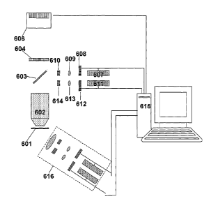
Fig. 6 is a schematic view of a PTOL microscope. A sample 601 that has been
labeled with PTOLs emits radiation that is collected with an imaging lens
(e.g., a
microscope objective lens) 602 and that can be filtered with one or more
filters 604. Images
of currently activated PTOLs are formed at detector 606, which in one
implementation can
detect single photons. Optical elements for providing activation radiation to
the sample can
include a light source 607, a shutter 608, a lens 609, and a filter 610. The
light source 607
(e.g. one or more lasers, light emitting diodes, or broadband sources) can
emit radiation at an
activation wavelength that causes a PTOL to be transformed from an inactivated
to an
activated state. The light source 607 can be directly modulated, or modulated
via the shutter
608. The shutter 608 can operate to admit or prevent activation radiation from
passing from
the light source 607 to the sample 601. In one implementation, the shutter can
be a
mechanical shutter that moves to selectively block the beam path. In another
implementation, the shutter can be a material that can be modified
electronically or
acoustically to admit or prevent light from passing or to alter a beam path
from the light
16
40281543.2
CA 02609653 2011-07-25
source 607. The filter 610 can block certain wavelengths of radiation while
passing other
wavelengths. For example, if the sample 601 contains several species of PTOLs,
each
having different activation wavelengths, the light source may emit light at
each of the
activation wavelengths but various filters 610 can be inserted in the beam
path between the
light source 607 and the sample to block some activation wavelengths while
passing other
wavelengths, such that only one (or a selected few) species of PTOL is
excited. Radiation
from the light source 607 can be deflected by a partial reflector 603 (e.g., a
beam splitter, a
dichroic mirror, a spotted mirror, or a diffractive structure and directed
through the imaging
lens 602 onto the sample 603. Similarly, excitation radiation that causes an
activated PTOL
to be transformed from a de-excited state to an excited state can also be
passed from an
excitation light source 611, through a shutter 612, a lens 613, and a filter
614 and off a
partial reflector 603 to the sample 601. A controller 615 (e.g., a general or
special purpose
computer or processor) can control parameters of the activation and excitation
pulses (e.g.,
the wavelength, intensity, polarization, and duration of pulses of various
radiation beams
that reach the sample 601; and the timing of activation radiation pulses and
excitation
radiation pulses) during an image acquisition sequence. Of course, the optical
elements 607
¨ 614 can be arranged in other configurations. For example, the activation
optics 607 ¨ 610
and/or the excitation optics 611 ¨614 can be configured, as in the module 616,
to direct
radiation to the sample 601 from outside of the lens 602, or the excitation
radiation can be
directed onto the sample from a different partial reflector than the
activation radiation, etc.
Furthermore, there can be a multiplicity of components so that PTOLs of a
different species
can also be imaged either in parallel or a separate sequential acquisition.
For example, there
can be additional cameras, filters, shutters, activation sources, or
excitation sources, of
different wavelengths associated with the characteristics of different PTOL
species. Data
from images formed at the detector 606 are communicated to the controller 615
for storage
and processing. For example, the controller 615 can include a memory for
recording or
storing intensity data as a function of position on the detector for different
image frames.
The controller 615 can also include a processor for processing the data (e.g.,
a general or
special purpose computer or processor), for example, to fit the data recorded
for an image of
an individual PTOL to determine a location of the PTOL to sub-diffraction
limited
resolution, or to combine the data about the locations of multiple PTOLs that
are determined
with superresolution accuracy to generate an image of the sample based on the
locations of
multiple PTOLs that have been located with superresolution accuracy.
17
40281543.2
CA 02609653 2011-07-25
Fig. 7 is a flow chart of a process 700 for creating an image of a sample
containing
multiple relatively densely-located PTOLs. An activation pulse of radiation
having an
activation wavelength is directed onto a sample to transform a subset of PTOLs
in the
sample from an unactivated to an activated state (step 702). Excitation
radiation is applied
to activated PTOLs in the sample at the excitation wavelength, and radiation
that is emitted
from activated and excited PTOLs and incident onto the imaging and detecting
optics is
acquired and saved (step 703). Images of a set of activated PTOLs can be
acquired and
saved multiple times. For example, the controller can require that N images of
a set of
activated PTOLs are acquired, such that if N images have not yet been acquired
(step 704)
image acquisition (step 703) is repeated. The excitation radiation can be
applied to the
sample continuously or can be switched off between acquisitions of images.
After N images of the subset of activated PTOLs are acquired, and if more
images
are to be obtained from the sample (step 705) another activation pulse can be
applied to the
sample to activate another set of PTOLs (step 702). Excitation radiation can
be applied to
16 this other set of activated PTOLs, and radiation emitted from the
activated and excited
PTOLs can be acquired and saved (step 703). Multiple sets of PTOLs can be
activated. For
example, the controller can require that M sets PTOLs be activated, such that
if M sets have
not yet been activated (step 705) another activation pulse is applied (step
703). Thus, the
process of activating a set of PTOLs, exciting PTOLs within the activated set,
and acquiring
images from the activated and excited PTOLs can be repeated multiple times,
for example,
until the total pool of available PTOLs becomes exhausted or until a desired
number of
images of a desired number of different PTOLs within a spatial area or volume
is achieved.
While applying the activation and excitation radiation, the number of
iterations N
between activation pulses, along with the intensity of the activation and
excitation radiation
can be controlled such that the mean volume per imaged PTOL in an individual
image is
generally more than the DLRV of the optical imaging system used to detect and
localize the
individual PTOLs. The density of activated PTOLs that are capable of emitting
radiation is
generally highest in images acquired immediately after the activation pulse
and generally
decreases as more PTOLs photobleach during the acquisition of the N image
frames.
Furthermore, as the process 700 progresses, and the number of activation
pulses increases
from 1 to M, PTOLs within the sample may photobleach, such that fewer and
fewer PTOLs
within the sample are available to be activated, excited, and imaged. Thus, in
one
implementation, the intensity and time length of individual activation pulses
and the
intensity and time length of excitation radiation can be controlled, to reduce
the variation in
18
40281543.2
CA 02609653 2011-07-25
density of activated PTOLs as the process progresses. For example, using less
excitation
radiation (possibly with fewer frames N between activation pulses) can reduce
the decrease
in imaged PTOLs from the first frame after an activation pulse to the Nth
frame just
preceding the next activation pulse. In another example, the intensity of
individual
activation pulses can increase as the process 700 progresses from the first to
the Mth
activation pulse. This would reduce the decrease in the number of imaged PTOLs
in the
first acquisition frame after the Mth activation pulse relative to the number
of imaged
PTOLs in the first acquisition frame after the first activation pulse, thereby
compensating
for the reduction in the number of activable PTOLs as the sequence of
activation and image
acquisition progresses. Thus, in the first example, the variation of activated
and excitable
PTOLs during an excitation sequence is reduced and in the second example the
variation of
activated and excitable PTOLs during the activation sequence is reduced. The
reduced
variation of activated and excitable PTOLs allows operation, where more PTOLs
can be
localized per unit time, while not exceeding the density criteria of more than
one imaged
PTOL per DLRV.
In one implementation, multiple species of PTOLs within the sample can be
activated, excited, and imaged. For example, steps of applying the activation
pulses (702)
and of exciting and imaging (703) can include applying pulses of activation
radiation and
excitation radiation, respectively, having wavelengths corresponding to the
different
activation and excitation wavelengths of different PTOL species. A
multiplicity of detectors
and/or filters can also be used in the imaging step 703 to image different
wavelengths of
radiation emitted from different PTOL species, In this manner, multiple
independent data
sets of images can be acquired. These independent data sets in turn can be
reduced to
corresponding super-resolution images of each PTOL species within a sample.
c. Exemplary Excitation and Detection Geometries
The process of activating a subset of PTOLs in a sample, exciting some or all
of
those activated PTOLs, and imaging the activated and excited PTOLs can be
applied in any
optical imaging mode, for example, in widefield microscopy, total internal
reflection
fluorescence (TIRF) microscopy, confocal microscopy, and multifocal lattice
microscopy.
As shown in Figs. 8a, 8b, 8c, and 8d, widefield microscopy permits many
individual
PTOLs 800 within a sample 810 that reside near the plane of focus 801 of a
lens 802 to be
localized simultaneously, when the PTOLs are activated at a low enough density
that their
19
40281543.2
CA 02609653 2011-07-25
separations in the plane 801 are generally larger than the diffraction limited
2D resolution
defined by the lens 802. The magnification of the imaging optics (e.g.,
including lens 802)
is chosen relative to the size of individual pixels 803 in a detector 804
(e.g., an electron
multiplying charge coupled device (EMCCD) camera) that images the PTOLs 800,
so that
the image 805 from each PTOL is dispersed over several pixels to optimize the
localization
accuracy for each PTOL. Of course, if radiation emitted from a particular PTOL
were
detected by only one pixel it would be difficult to determine the location of
the PTOL with
sub-diffraction limited accuracy, but if radiation from the PTOL falls on
multiple pixels the
signals from the different pixels can be fitted, such that the PTOL can be
localized with sub-
to diffraction limited accuracy. However, if radiation from a particular
PTOL falls on very
many pixels, then it may overlap with the radiation from another PTOL, or the
background
noise from the greater number of pixels involved may be increased. In either
case, such
that the localization accuracy would be relatively low. Thus, a compromise
between having
an image of a PTOL fall on too many or too few pixels can be obtained.
Widefield microscopy is easily used with the processes described herein to
achieve
2D localization of PTOLs in thin samples (i.e., samples having a thickness
comparable to or
smaller than the depth of focus 806 characterized by the numerical aperture of
the lens and
the wavelength of the fluorescence light emitted from the PTOLs). Application
to such thin
samples can: a) limit background signal from autofluorescence or unresolved
PTOLs in
areas away from the focal plane 806 (since such background can degrade the
accuracy with
which PTOLs are localized); b) reduce the number of potentially
photoactivatable molecules
within the 2D PSF; and c) when the activating energy is delivered through the
imaging lens,
insure that the PTOLs that are activated are generally within the focal plane
of the lens, and
therefore produce minimally sized spots at the detector and corresponding
optimal
localization.
One example of such thin sections is the lamellipodial regions of cultured
cells.
Another class of thin samples suitable for widefield detection is thin
sections cut from a
larger sample using the microtome techniques common to transmission electron
microscopy
(either cryosections or sections from resin-embedded cells or tissues). Such
solid, cut
sections insure that the PTOLs remain immobile for accurate localization, and
permit deeply
buried sample features to be imaged, without the problems of out-of-plane
autofluorescence,
aberrations, and light scattering that potentially exist when trying to image
the same features
by widefield microscopy in the original, thicker sample.
40281543.2
CA 02609653 2011-07-25
As shown in Fig. 8b, in cases where widefield detection of PTOLs can be
applied to
samples that are thick compared to the depth of focus of the lens,
localization of PTOLs in
3D can be performed by translating the focal plane along the optical axis 807
of the lens
(e.g., by changing the separation between the lens and the sample) for each
activated subset
of PTOLs that is imaged to create 2D images of multiple planes of the sample.
These
multiple 2D images can be combined digitally to build an image stack 808 such
that a 3D
image of each imaged PTOL in the sample is obtained. Then the 3D image of each
PTOL
can be fitted to obtain a sub-diffraction limited position of the PTOL
positions in 3D, by
direct analogy to the 2D case described above. A complete 3D superresolution
image can be
thereby constructed from many subsets of localized PTOLs.
Another approach to providing position information for the PTOLs in the
direction
defined by the axis 807 of the lens is to apply the excitation light in a form
that is spatially
structured primarily along this direction, and substantially uniform parallel
to the focal plane
(so that the advantage of simultaneous detection in 2D is retained). The
spatially structured
field can then be scanned in the axial direction for each subset of
individually resolvable,
activated PTOLs, thereby permitting the axial excitation PSF to be measured at
each. The
known PSF of the axially structured excitation can then be fit to this data to
find the relative
locations of the PTOLs in the axial direction with nanometric precision. The
data can then
be combined with the localized coordinates of the same PTOLs in the focal
plane, and
further combined with similar results from other subsets of activated PTOLs to
build a dense
superresolution 3D image.
As shown in Fig. 8c., such an axially structured excitation field can be
created by
impinging the excitation light on the sample 810 in two coherent beams 811 and
812 from
directions that are mirror imaged with respect to the detection plane. The
beams 811 and
812 within the sample 810 to produce a standing wave ("SW") intensity profile
813 in the
axial direction 807. The beam 811 approaching the sample from the same side of
the focal
plane as the lens 802 can pass through the lens, if desired. For samples
sufficiently thin such
that only a single SW plane 814 of maximum intensity resides within the sample
810,
detection and localization can proceed by axially scanning the maximum
intensity plane as
described above. For moderately thicker samples, the period 815 of the SW,
which can be
expressed as p = sin(0) 2 (where p is the period, X is the wavelength of the
excitation
radiation, and 0 is angle each beam makes with the focal plane) can be
increased by
decreasing the angle, 0, until only a single, wider SW plane of maximum
intensity
intersects the sample 810. Alternatively, as shown in Fig. 8d, if several SW
maxima reside
within the sample 810, PTOLs 800 excited in planes corresponding to different
intensity
21
40281543.2
CA 02609653 2011-07-25
maxima 816, 817, and 818 can produce different patterned spots (e.g., spots
819 and 820
from maximum 816, spot 821 from maximum 817, and spots 822 and 823 from
maximum
818) at the detector due to the differences in 2D detection point spread
function that exists in
different planes parallel to the plane of focus of the lens 802. For example,
an image of a
PTOL on the detector due to emission from the PTOL at the focal plane of the
imaging
optics will be smaller than a image of the PTOL due to emission from the PTOL
from a
plane that does not correspond to the focal plane. This information can be
used to
discriminate from which SW maximum a given PTOL originates. Also, the detected
light
can be split between M detectors in the case where M standing wave maxima
reside within
the sample, and corrective optics (e.g., a phase mask) can be placed between
the lens 802
and each detector, such that the focal plane for each detector is coincident
with a different
SW maximum. Those PTOLs in focus at a given detector then can be localized in
either 2D
or 3D using the information recorded at that detector.
A total internal reflection ("TIRF") geometry also permits simultaneous
detection
and 2D localization of multiple photoactivated PTOLs in a plane. In TIRF
microscopy, the
intensity of excitation radiation that illuminates the sample exponentially
decreases with
increasing distance from the sample/substrate interface. Because of the
exponential
decrease of the excitation radiation as a function of distance from the
sample/substrate
interface, excitation that is highly localized in the z direction can be
achieved with relatively
little autofluorescence, especially when thick specimens are imaged. Also with
TIRF
microscopy, relatively few PTOLs (both activated and deactivated) are excited
simultaneously for a given molecular density, so a larger density of target
molecules can be
initially prepared in the sample. Further, evanescent illumination at multiple
angles can be
used to localize the PTOLs in the z direction as well to a high degree of
accuracy.
Additionally, the wavelength of activation radiation as well as the wavelength
of excitation
radiation can be applied via an evanescent field to further reduce the extent
of activated,
excited PTOLs in the z direction.
Excitation radiation and activation radiation for TIRF microscopy can be
delivered to
the sample/substrate interface external to the objective lens using a prism
that is optically
coupled to the substrate. Alternatively, excitation and activation radiation
can be applied to
the sample/substrate interface in an epi configuration, with the excitation
radiation entering
at the rear pupil of the same objective lens that is used to collect
fluorescence radiation
emitted from PTOLs in the sample, as long as the numerical aperture ("NA '9 of
the lens
yields a maximum illumination angle, > sin
-I (NA I nsõb) , that is greater than the critical
22
40281543.2
CA 02609653 2011-07-25
angle for total internal reflection ("TIR") (where nsub is the refractive
index of the substrate),
and the excitation radiation enters the rear pupil in the outer annular region
that supports
TIR of the excitation radiation.
Figs. 9a and 9b are schematic diagrams of a system that can use through-the-
objective TIRF excitation radiation to excite sparsely-populated activated
PTOLs in a
sample, such that radiation emitted from the activated, excited PTOLs can be
imaged to
produce superresolution images of the sample via phototransforrnation,
isolation, and
localization of multiple subsets of discrete PTOLs within the sample. For
continuous
excitation of activated PTOLs, light having a wavelength of 561 nm emitted
from a 10 mW
to diode-pumped solid-state laser (available from Lasos GmbH, Jena,
Germany) is
fiber-coupled to an excitation collimator 900 and provides an excitation input
beam 901 that
can be focused at the rear pupil plane internal to a 60X, 1.45NA total
internal reflection
fluorescence ("TIRF") oil immersion objective 902 (available from Olympus
America,
Melville, NY). A narrow bandwidth laser line filter 903 (available from
Semrock, Inc.,
16 Rochester, NY) is used to reject both emission noise from the laser and
autofluorescence
generated in the optical path prior to the objective 902. For pulsed
activation of the PTOLs,
a second diode laser (available form Coherent Inc., Santa Clara, CA) that can
yield about 50
mW of power at an activation wavelength, Xact, of about 405 nm can be fiber-
coupled
through an intermediate galvanometer-based switch (not shown) to an activation
collimator
20 904 to create a focused activation input beam 905 that is similarly
filtered by a bandpass
filtered 906 (available from CVI Optical, Covina, CA) before being combined
with the
excitation input beam 901 at a dichroic mirror 907 (available from Semrock,
Inc.). This
combined input beam 908 then can be reflected from an elliptical spot on a
custom-
patterned, aluminized mirror 909 (available from Reynard Corp., San Clemente,
CA) into
25 the objective 902. The radius, p, at which the combined beam 908 enters
objective 902 can
be controlled to be (nsaõ,ple I NA)* 4.35 ::.=== 4.14 mm p 4.35 mm (for ;amp!,
:k11.38), such
that the resulting refracted ray transverses a low autofluorescence immersion
oil (e.g.,
Cargille type FF, available from Structure Probe Inc., West Chester, PA) and
is incident at
the interface between the sample and a cover slip 913 (e.g., a #2 thickness
cover slip
30 available from Fisher Scientific, Hampton, NH) at greater than the
critical angle,
0,sin-1(nõõ,/,/, / nõ,,,õ10,), for which total internal reflection ("TIR")
occurs, An
evanescent field can be thereby established within the sample, exciting only
those molecules
within the short decay length of the evanescent field. A substantial
proportion of the
23
40281543.2
CA 02609653 2011-07-25
incident energy of the excitation and activation beams, however, can be
reflected at the
interface to yield a combined output beam 910 that emerges from the objective
902, and that
is then reflected from a second elliptical spot on mirror 909 diagonally
opposite the first
elliptical spot on the mirror. This beam 910 is then divided at dichroic
mirror 907 into an
excitation output beam 911 and a separate activation output beam 912 that are
finally
directed to respective beam dumps.
For typical molecular cross-sections (e.g., approximately 1046 cm2), the
reflected
excitation beam energy may be 10" -fold more intense than a PTOL signal beam
914 that
emerges from the objective 902, as shown in Fig. 9b. Therefore, a challenge in
this through-
to the-objective TIRF geometry is the isolation of the molecular signal
from both the interface-
reflected excitation beam and any autofluorescence generated by this beam in
the optics
encountered thereafter. The mirror 909 aids in this isolation because the
mirror has an
elliptical, anti-reflection coated, transmissive aperture whose projection
perpendicular to the
objective axis matches the 8.7 mm diameter of the rear pupil, and therefore
passes signal
beam 914 to the detection optics with high efficiency. Also, for an elliptical
reflective spot
D times larger than the gaussian width of the reflected beam at the spot, only
about erfc(D)
of the excitation energy is passed onto the detection optics, or ¨ 2 .10-5 to
¨ 2 .10-8 for D
3 or 4, respectively. Furthermore, since the spots occlude only a small
fraction of the
periphery of the rear pupil, they do not substantially degrade the detection
numerical
aperture. Consequently, the PSF standard deviation, s, that factors into sub-
diffraction
limited localization of PTOLs is not substantially degraded. Furthermore, the
mirror 909 is
wavelength insensitive, and therefore can be used with different excitation
lasers and
different PTOLs without replacement. The mirror 909 can include multiple spots
to support
multi-angle, multi-polarization and/or standing wave TIRF excitation.
After passage through custom spotted mirror 909, the largely collimated signal
beam
914 emerging from the infinity-corrected objective 902 can be reflected by a
first mirror 915
(as shown in Fig. 9b) to travel along the axis of the detection optics. Any
remaining
excitation light (as well as much of the remaining activation light) traveling
substantially
along this axis can be removed by a Raman edge filter 916 (available from
Semrock, Inc.).
However, because the optical density of this filter 916 decreases rapidly with
increasing
deviation from normal incidence, baffles 917 can be placed on either side of
the filter to
remove scattered light at higher angles of incidence generated elsewhere
within the system.
The filtered signal beam can be focused into a focused beam 918 with an
acromatic tube
24
40281543.2
CA 02609653 2011-07-25
lens 919 (available from Edmund Optics, Barrington, MI) onto the face of a
back-
illuminated, thermoelectrically cooled (e.g., to -50 C), electron multiplying
CCD camera
920 (available from Andor Scientific, South Windsor, CT) to create the desired
image of
isolated single molecules. A 405 nm notch filter 921 (available from Semrock,
Inc.) also
can be included to further insure that the camera 920 is not saturated when
the activation
beam is applied.
To further increase the localization accuracy in the plane of the
sample/substrate
interface in the TIRF configuration the substrate can be used as a waveguide
to support the
propagation of two or more intersecting excitation beams. These beams then can
form a
structured excitation field within this plane that is evanescent perpendicular
to the interface.
For example, as shown in Fig. 10a, two such excitation beams 1000 and 1001 can
create a
standing wave ("SW") intensity profile 1002 along one axis 1003 parallel to
the interface
between the sample 1004 and the substrate 1005. Scanning this SW over one
period along
this axis (e.g., at phases, A = 0 (as illustrated in frame 1006), A =120 (as
illustrated in
16 frame 1007), and A = 240 (as illustrated in frame 1008)) and capturing
images (e.g., as
shown in frames 1009, 1010, and 1011) of the activated PTOLs at each SW
position then
can allow the PTOLs to be localized on the basis of an effective excitation
PSF 1012 as
shown in Fig. lob, having a width ¨ 2excl(4n51b), where Xeõ, is the wavelength
of the
excitation radiation and nsub is the index of refraction of the substrate,
which is lower than
the detection PSF 1013 having a width ¨ /Ls 1(2NA) present at the CCD, where
Xems is the
wavelength of signal radiation emitted from PTOLs. The PSF is especially
improved when
high ns.õ1, substrates can be used. A second SW orthogonal to the first then
can be generated
and scanned over the same subset of activated PTOLs to localize them along the
other axis
within the plane.
The beams 1000 and 1001 forming a TIRF excitation field structured in the
plane of
the interface also can be transmitted to the interface either through a TIRF-
capable signal
collection objective (as shown in Fig. 10c), or with optical elements (e.g.,
prisms) on the
side of the substrate opposite the interface.
Widefield molecular localization is well suited to thin samples (to reduce out-
of-
focal plane fluorescence), and TIRF molecular localization is suited to
portions of the
sample near the sample/substrate interface, by the evanescent field. On the
other hand,
confocal microscopy can be used to localize PTOLs in 3D in thick samples, such
as whole
cells. The 3D confocal overall PSF, defined by the product of the excitation
PSF
40281543.2
CA 02609653 2011-07-25
(determined by focusing of the excitation) with the 3D detection PSF (defined
by the
confocal pinhole and the numerical aperture of the detection objective), can
be
volumetrically larger than in the thin sample widefield or TIRF cases.
Therefore,
autofluorescence may be larger in such a case, which can reduce the
localization accuracy or
suggest the use of PTOLs having higher intrinsic brightness.
However, activation and excitation energy outside the focal plane in the
incoming
and outgoing focal cones of the confocal microscope can prematurely activate
and then
photobleach PTOLs, adding to the out-of-focus background and reducing the
population of
PTOLs that can be accurately localized (i.e, those near the focal plane)).
Since many of the
to photoswitchable FPs (e.g., PA-GFP and Kaede) are activated by violet or
near-UV light, this
problem can be lessened by using multiphoton excitation to activate the
molecules, since
this nonlinear process generally results in low PTOL activation outside the
effective
multiphoton depth of focus. The multiphoton focus could either precede the
confocal
excitation focus during the latter's path through the scan volume, or
molecules across the
current focal plane could first be activated by a 2D scan of the multiphoton
focus until the
desired density of individually resolvable activated molecules is reached, to
be followed by
a similar 2D scan of the confocal focus to detect and localize the molecules
so activated.
Also, when multifocal activation is used damage to the specimen from the short
wavelength
of the activation beam is likely to be greatly reduced.
Confocal molecular localization is a serial process, and therefore relatively
slow.
For example, confocal molecular localization is a triply serial process
because it provides
activation, followed by acquisition of multiple serially scanned 3D images
until all currently
activated molecules are bleached, followed again by activation and multiple 3D
scanning ¨
over and over again, until a 3D map of molecular positions of the desired
density is
obtained. This is obviously a slower proposition than 2D imaging by widefield
or TIRF
molecular localization. Multifocal microscopy utilizing Nipkow disk technology
can be
used to increase the speed of the process to some extent, but it creates
multiple foci only in a
single plane, and still generates significant out-of-focal plane excitation
leading to premature
bleaching of target molecules and increased autofluorescence-induced
background, even
with pinhole filtering. On the other hand, 3D lattice excitation can provide
many excitation
maxima simultaneously in 3D, as described in PCT Patent Application Serial No.
PCT/US2005/042686 November 23, 2005, and entitled "OPTICAL LATTICE
MICROSCOPY ,"with unintended photobleaching and associated background
significantly
26
40281543.2
CA 02609653 2011-07-25
reduced by the improved confinement of the excitation to predominantly these
maxima
alone. Furthermore, if the lattice is created with constituent beams spread
across a greater
solid angle than that covered by a single microscope objective, the
confinement of the
excitation at each lattice maximum (e.g., as defined by the full volume at
half peak intensity)
can be greater than that in either single focus or traditional multifocal
microscopy, further
reducing the background signal significantly, and permitting more accurate
localization of
each PTOL, due to the tighter initial PSF. Of course, an optimal SNR and
initial PSF is
expected when all beams of the maximally symmetric composite lattice are used.
As in the
confocal case, multiphoton activation can be locally applied, such as with a
multiphoton
o lattice, either scanned ahead of the fluorescence excitation lattice, or
scanned to create a
series of parallel planes of activated PTOLs prior to simultaneous scanning of
these planes
by the fluorescence excitation lattice.
d. PTOL Properties
is PTOLs useful for superresolution via localization of isolated PTOLs
generally have
one or more of the following distinguishing characteristics: a relatively high
brightness (as
defined by its excitation cross section and the quantum efficiency); a
relatively high contrast
ratio between luminescence generated in the activated state to that generated
in the
inactivated state (which might be improved through a judicious choice of the
excitation
20 wavelength and detection filter set); an excitation wavelength that
reduces autofluorescence
from other cellular material exposed to the excitation; an emission wavelength
that is
sufficiently different from the spectral range over which most
autofluorescence occurs; and
photostability that is large enough that a sufficient number of photons are
collected from
each PTOL to achieve the desired localization accuracy prior to irreversible
bleaching, yet,
25 for PTOLs other than the kindling proteins and Dronpa that can switch
back to the
deactivated state, is nevertheless still finite, so that a new population of
individually
resolvable activated PTOLs can be created after the current set is largely
bleached. Indeed,
to reduce possible phototoxicity related to irreversible photobleaching, an
ideal PTOL would
remain in the activated state until it is deactivated by choice using other
means (e.g.,
30 illumination at a separate deactivation wavelength).
Superresolution via localization has been demonstrated with the tetrameric
PTOLs
Kaede and Kikume, as well as the monomeric, dimeric, and tandem dimer forms of
EosFP.
These PTOLS have the common advantages of large wavelength spread between the
27
40281543.2
CA 02609653 2011-07-25
inactivated and activated absorption and emission maxima, high brightness, and
longer
wavelength emission, where autofluorescence is typically lower. Monomeric
EosFP has the
added advantage of smaller physical size than tetrameric Kaede or Kikume, and
may
therefore be less perturbative of cellular structure and function. In
practice, a particular FP
could be selected from a number of different FPs based on a user's criteria
for optimization
for a given application.
e. Background Reduction
If the contrast ratio between activated and inactivated PTOLs is too low at a
given
initial density of target PTOLs to achieve the desired SNR and consequent
localization
accuracy, the contrast ratio can be improved by irreversibly bleaching a
portion of the target
PTOLs until the effective molecular density and resulting SNR is as desired.
Other
autofluorescent material in the sample can also be pre-bleached using the
excitation light
without affecting the bulk of the inactivated PTOLs. Further discrimination
with respect to
background might be obtained via appropriate spectral filtering, fluorescence
lifetime
measurements, or polarized excitation and/or polarization analyzed detection.
In widefield microscopy, spatially structured activation energy concentrated
near the
focal plane (e.g., from an axial standing wave) can be used to reduce the
background from
activated, out-of-focus PTOLs away from this plane. In confocal or lattice
microscopy,
similar background reductions can be achieved with standing wave or other
means of planar,
axially structured activation rather than the 3D confined foci traditionally
applied by these
methods.
f Polarized Excitation / Detection
Light from PTOLs having an electric dipole moment located in a sample can be
detected and
imaged using the techniques describe herein. When such PTOLs have a fixed
spatial
orientation in the sample, the can be selectively activated, excitated, and/or
imaged with
polarized light. By analyzing the polarization of the light emitted from such
PTOLs (e.g., by
passing light emitted from such PTOLs in the sample through a polarization
filter prior to
detecting the light such that only light having a desired polarization is
detected, allows the
dipole orientations of these PTOLs to be determined. For a plurality of fixed
dipole PTOLs
that are randomly oriented in a sample, the PTOLs can be activated and/or
excited with
equal probability by using activation and/or excitation radiation polarized in
all three
28
40281543.2
CA 02609653 2011-07-25
orthogonal directions, rather than with the unequal weightings that would
result from using
activation and/or excitation radiation having a single excitation
polarization. Because the
electric field of a polarized light lies in a plane orthogonal to its
direction of propagation,
polarized excitation in all three directions requires at least two independent
excitation
beams. For example, the through-the-objective TIRF system described herein,
inter alia,
with respect to Fig. 9 is capable of delivering four independent beams at 90
intervals in the
plane of the sample/substrate interface, by using four excitation collimators
900, creating
four input beams 901 that are reflected from four spots on mirror 909 and sent
into the rear
pupil of objective 902. Polarizing the beams at 00 and 90 radially with
respect to the rear
pupil and similarly polarizing the beams at 180 and 270 azimuthally results
in two
interfacial waves, polarized orthogonally with respect to one another in the
plane of the
interface, and two interfacial waves polarized orthogonal to the interface.
These beams can
be turned on either sequentially or simultaneously ¨ although in the latter
case, beams
having common polarization vectors will mutually interfere. In fact, as shown
in Fig. 10,
by turning on the beams in pairs with like polarization, a standing wave can
be formed to
provide enhanced localization accuracy due to the sharp excitation PSF of the
standing
wave. Thus, the features of accurate localization, dipole determination, and
equal excitation
probably of fixed dipoles can be combined.
g Exemplary Superresolution Images
Fig. 11 compares a diffraction-limited image (Fig. 11a) of a lysosomal
structure in a
COS7 cell and superresolution image (Fig. 11b) of the same lysosomal structure
in the same
COS7 cell , which was obtained using the apparatus and techniques of TIRE.'
isolation /
localization described herein. The sample containing the COS7 cell was
prepared by
transient transfection with a plasmid designed for the expression of the
photoactivatable
protein Kaede fused to the lysosomal transmembrane protein 0363. Cells were
pelletized
and then sectioned with a microtome, using the techniques common to
transmission electron
microscopy, to create the approximately 80 nm thick section that was imaged.
20,000
frames of single molecule images were taken, with activation energy applied in
a brief pulse
after every 20 frames to restore the number of activated molecules to a
higher, but still
individually resolvable level. The superresolution image shown in Fig. 1 lb
was formed
from more than 51,000 isolated molecules, with each molecule localized with an
uncertainty
of 24 nm or less redrawn in Fig, llb as a spot having an intensity image
profile given by a
29
40281543.2
CA 02609653 2011-07-25
Gaussian distribution with a standard deviation equal to the position
uncertainty. The
profiles of the spots for each molecule were normalized to provide the same
integrated
intensity for each molecule. Thus, more highly localized molecules appear as
bright, sharp
dots, and less well localized ones appear broad and dim. The diffraction
limited image was
formed by summing the diffraction limited images of the same set of isolated
molecules, and
was verified to be indistinguishable from the conventional TIRF image.
Fig. 12 compares a diffraction-limited image (Fig. 12a) obtained at the
interface of a
whole, fixed fox lung fibroblast cell and a glass cover slip in phosphate
buffered saline and a
superresolution (Fig. 12b) image of the same fox lung fibroblast cell. The
cell was
transiently transfected to express the photoactivatable protein dEosFP fused
to the cell
attachment protein vinculin. The images were created in the same manner as
described in
conjunction with Fig. 11. The diffraction limited image highlights a single
focal adhesion
region at the periphery of the cell, and the superresolution image by PTOL
localization
shows a magnified view of the structure within the box in Fig. 12a.
3. Enhanced Resolution via Overlapped Spatially Structured Activation and
Excitation
The overall PSF of any form of optical microscopy (e.g., widefiekl, TIRF,
confocal,
or lattice) is typically given by the product of the excitation PSF with that
of the detection
PSF (i.e., PSFõ = JPSFexatotion x P8FrIelection)= Widefield microscopy offers
no excitation
contribution to the resolution, traditional TIRF microscopy offers very high z-
axis excitation
resolution, but none in the x- and y-axes, and both confocal and lattice
microscopy
contribute excitation resolution by concentration of the excitation field to
either a single
focus, or a lattice of intensity maxima.
PTOLs offer a way of contributing a third component to the overall PSF by
confining the activation illumination to a localized region in a manner
similar to that used to
confine the excitation energy itself (i.e., PSE
- PSFactivatton x P8Facriattoto X
PSFelerection).
Thus, for example, a focused activation beam can be temporarily applied at the
focal point
of a confocal microscope, followed by a focused beam at the excitation
wavelength for the
activated PTOLs, with the resulting emission being detected confocally in a
spatially
localized manner. This process can then be repeated over many voxels (i.e., a
3D pixel) to
create a complete superresolution 3D image. One caveat is that the number of
activated
PTOLs in a focal volume should decline significantly (either by irreversible
photobleaching,
40281543.2
CA 02609653 2011-07-25
or reversion to the unactivated state) before activation and excitation is
applied to an
immediately neighboring voxel, or else the effective activation PSF will be
reflective of the
larger region defined by the overlapping, neighboring activation foci, thereby
degrading the
effective overall PSF. Dronpa appears to be a particularly good candidate for
this method of
superresolution, because the activated molecules are returned to the
deactivated state by the
process of their excitation, thereby providing a natural means to depopulate
the activated
ensemble while simultaneously determining when the scan should proceed to the
next
voxel., If the deactivation occurs too quickly, multiple
activation/(deactivation and
measurement) cycles can be performed at the same position before proceeding to
the next
position. Using Dronpa as the PTOL in this process allows more than about 100
such cycles
to be performed at each position.
Because the activation wavelength is typically short (e.g., about 400 rim) for
Dronpa,
the activation PSF can provide most of the resolution benefit in the overall
PSF. If cellular
damage from this short of a wavelength is a concern, multiphoton activation
can be used, at
the cost of a slightly larger activation PSF than is possible with linear
(i.e., single photon)
activation. In addition, because the density of emitting molecules is given by
PSF.1.110.x PSFex
citation 2 the emitting molecules will be confined to at least as tight a
focal
region as in conventional two-photon excitation, thereby leading to greatly
reduced out-of-
plane photobleaching and background, even using linear, confocal excitation.
Of course,
further gains in both spatial and temporal resolution are possible if sparse
composite lattices
of the same or commensurate periods are used for both the activation radiation
(as shown in
Fig. 13a, and in the close up view of Fig. 13a shown in Fig. 13d) and for the
excitation
radiation (as shown in Fig. 13b, and in the close up of Fig. 13b in Fig. 13e),
leading to an
overall lattice (shown in Fig. 13c) that achieves activation and excitation of
PTOLs and that
has having sharper maxima (shown in Fig. 130 than the maxima in the lattices
for the
activation and excitation radiation. Point spread function engineering and
relative
displacement of the activation and excitation PSFs might be used to further
increase the
resolution by reducing the region of their effective overlap. If the
contribution of the
detection PSF to the overall resolution is negligible, it might be
advantageous to simply omit
pinhole filtering (as in most embodiments of multiphoton microscopy) in order
to maximize
the collected signal. Finally, we note that this method of superresolution, is
well suited to
dynamic superresolution imaging in living cells (particularly with lattice
microscopy),
because potentially many more molecules would be emitting photons at a given
time from
31
40281543.2
CA 02609653 2011-07-25
each focus (when confocal radiation is used for activation and excitation) or
excitation
maximum (when radiation patterned in a lattice is used for activation and
excitation).
4. Superresolution via Saturated Deactivation
By exploiting the saturation of the deactivation of PTOLs over a sub-portion
of a
diffraction-limited focal volume in which a portion of the PTOLs were
previously activated,
one can collect emission from a sub-diffraction limited region, and then
repeat at multiple
locations to generate a sub-diffraction limited image. This concept is
described in Fig. 14 in
reference to activation, deactivation, and excitation with optical lattices,
although other
means (e.g., single focused beams) can also be employed.
As shown in Fig. 14a, a lattice of confined intensity maxima can be first
created at
the activation wavelength of the PTOLs to create an array of localized regions
of activated
PTOLs. Next, a depletion lattice (as shown in Fig. 14b) having a central low
intensity node
within a shell of high intensity located at each lattice point, can be applied
at a wavelength
that returns the PTOLs outside each node to their unactivated state. Next, an
excitation
lattice (as shown in Fig. 14c) can be applied at the excitation wavelength of
the activated
PTOLs, so that the small (e.g., having dimensions that can be less than the
wavelength of the
emission radiation) volume of PTOLs near each node of the depletion lattice is
excited and
then emits photons, resulting in the desired lattice of superresolution foci
(as shown in Fig.
14d). Next, the remaining activated PTOLs are deactivated, such as by exciting
them until a
substantial fraction of them photobleach, or by applying a deactivtion
radiation until a
substantial fraction of them are returned to the unactivated.state. This
process of activation,
partial deactivation with a nodal pattern, excitation, and nearly complete
deactivation can
then be repeated at a different points to create a lattice of superresolution
foci offset from the
26 first. By repeating the process further at a multiplicity of points
across each primitive cell of
the lattice, and detecting emission radiation from individual superresolution
foci in a given
cycle of activation/nodal deactivation/excitation/complete deactivation at
separate detection
elements (e.g., the pixels of a CCD detector), a complete 3D image can be
constructed (as
shown in Fig. 140, at considerably higher resolution than is possible, for
example, by
conventional confocal microscopy (as shown in Fig. 14e). All three lattices
(i.e., the lattices
of the activation radiation, the depletion radiation, and the excitation
radiation) can be
chosen at wavelength-normalized periodicities, such that the ratios of their
absolute
periodicities form simple integer fractions (i.e., i/ j), or ideally, have the
same absolute
32
40281543.2
CA 02609653 2011-07-25
periodicity (i/ j = 1), so that many of the activation maxima, deactivation
depletion shells,
and excitation maxima overlap. The completely deactivating radiation can also
be applied
in the form of a lattice, or as a substantially uniform deactivation field.
Again considering more general radiation patterns, it is important to note
that only
the nodal deactivation radiation pattern needs to be spatially structured
(specifically, with at
least one low intensity node), and that even uniform activation, excitation,
or complete
=
deactivation radiation fields may be applied. However, it may be beneficial to
spatially
structure either or both of the activation and excitation fields as well, in
order to increase the
contrast between the desired remaining activated PTOLs near the nodes after
deactivation
relative to undesired remaining activated PTOLs elsewhere, and to reduce the
potential
damage to reversible PTOLs by repeated activation and deactivation cycles.
More thorough
final deactivation near the nodes may also be attained by spatially
structuring the complete
deactivation radiation as well, to concentrate it at these points of residual
activation. Also
note that the density and spatial confinement of the activated PTOLs remaining
after
application of the deactivation energy is improved if the deactivation field
is closer to zero
intensity at the nodes, and if the rate of decrease in deactivation intensity
near the nodes is
high.
Specific photoactivatable FPs can be used in this technique. For example,
kindling
proteins, such as KFP1 and dronpa, can be used because they both can be
photoswitched
back to an unactivated state. KFP1 requires low intensity activation to insure
that the
molecules are not irreversibly activated, has a relatively low quantum
efficiency, and
deactivation of KFP1 occurs at a different wavelength than the excitation.
Dronpa exhibits
high brightness and is demonstrably switchable over many cycles, but time
gating of the
detection signal is required, because the depletion wavelength is the same as
the excitation
wavelength, so the fluorescence generated during depletion of the activated
state must be
rejected, or collected separately from the later emission near the depletion
nodes. On the
other hand, the emission collected during depletion can be used to generate a
high SNR
diffraction-limited image, since many more molecules would contribute to the
emission
during depletion.
5. PTOL Imaging at Reduced Temperatures
Biological samples labeled with PTOLs that are alive or at room temperature
pose
special challenges for this localization microscopy. The PTOL labels might
diffuse or be
transported beyond the localization accuracy during the relatively long multi-
frame
33
40281543.2
CA 02609653 2011-07-25
acquisition time. In addition, other properties such as the PTOL orientation
could be
varying during the acquisition resulting in the loss of potentially useful
microscopic
information. Thus, cooling a sample below room temperature can reduce the
movement of a
sample while it is imaged.
In addition, at reduced temperatures, the brightness and spectral line widths
of certain
PTOLs improve, so that more photons can be acquired more quickly for better
resolved
localization images, and contrast of the PTOLs relative to autofluorescence
background may
be reduced. Included here is an implementation where the sample or the sample
and parts of
the microscope is cooled below freezing temperatures to mitigate these
limitations. In
particular, rapid freezing can prepare samples in a vitreous state so that no
potentially
damaging ice crystals are formed within the sample
6. PTOL Microscopy of Latent Images
In lithography, nanometer scale patterns can be written with photon, electron,
ion or
atom beams. Typically the pattern is written onto a beam sensitive material
such as a resist.
In the cases of optical or electron beams, photoresist or e-beam resists can
be used. For
optimal lithographic performance it is useful to characterize the precise
shape of the beam
and the exposed pattern at an early stage before subsequent processing
transfers the exposed
resist pattern onto other materials. Thus, a resist can contain PTOLs or be
labeled with
PTOLs on the top or bottom surfaces of the resist layer. In this case,
contrast can be
imposed by the exposing beam by several kinds of exposure beams, and the
exposure beam
can have a detectable effect on the PTOLs in the resist, such that imaging the
PTOLs after
exposure can reveal the pattern of the exposure beam in the resist. For
example, such an
exposure beam can: destroy a PTOLs ability to radiate (e.g. by electron beam
ionization, or
UV induced bond breaking, etc.); shift the emission wavelength of the PTOL
(e.g., in a
manner similar to the wavelength shift in Kaede due to activation radiation);
or catalyze the
release of an acid in the resist, as is common in the case of chemically
activated resists,
which then changes the photophysical properties of the exposed PTOLs. Thus, as
shown in
Fig 15a, ta resist can include a number of PTOLs. As shown in Fig. 15h, when a
portion
1502 of the resist is exposed to exposure radiation in a lithography process,
photo-
lithographically activated acids 1503 can catalyze further cleavage or
polymerization of
resist. In addition, the acids 1503 can also shift the emission wavelength of
the PTOLs (e.g.,
when Eos is used as the PTOL 1501), where an activated state of the PTOL 1506
in the
34
40281543.2
CA 02609653 2011-07-25
presence of the acid 1503 can emit more strongly at a different wavelength
than when the
PTOL 1501 is not in the presence of the acid 1503. A PTOL microscope could
image the
acid transformed PTOLs 1506 or the nontransformed PTOLs 1501 as a latent image
on the
resist. This in turn could provide a measure of the exposure properties and
profiles at the
resolution of the PTOL localization length scale.
Unless otherwise defined, all technical and scientific terms used herein have
the
same meaning as commonly understood by one of ordinary skill in the art to
which this
invention belongs. In case of conflict, the present specification, including
definitions, will
control. In addition, the materials, methods, and examples are illustrative
only and not
limiting.
A number of implementations have been described. Nevertheless, it will be
understood that various modifications may be made. Accordingly, other
implementations
are within the scope of the following claims.
40281543.2