Note: Descriptions are shown in the official language in which they were submitted.
CA 02610409 2007-11-30
EYE MOVEMENT SENSOR
DESCRIPTION
OBJECT OF THE INVENTION
The present invention is applied to the field of ophthalmology, in
particular for the detection, monitoring and optimization of the further
treatment of pathologies related to certain ocular motility anomalies, such as
those caused by facial paralysis, strabismus (eye deviation), blepharospasm
(spastic closure of the eyes), nystagmus (accelerated eye vibration), ptosis
(drooping upper eyelid), as well as the examination of eye prosthesis, etc.
In particular, the eye movement sensor recommended comprises a
helmet, applicable to both adult and children patients, which incorporates
means of recording the eyes, preferably a video camera for both eyes or one
for each eye, which record them in images viewed from an angle which
permits capturing all their movements, which is the angle of the gaze in
maximum infraduction, i.e. downward. Additionally, the helmet has a light
projector which emits light spots towards a screen, which the patient is made
to look at to undergo a very precise eye examination of said anomalies and
optimize the diagnosis.
The object of the invention is for the patient to be able to freely move
his/her head when looking at the visual tests that are normally undergone
during an ophthalmologic examination, permitting a more pleasant and
shorter examination, whose results are not affected by said voluntary or
involuntary head movements.
Likewise, an object of the invention is to collect the eye movements in
any of their possible positions, recording them in images by the means of
recording incorporated in the helmet, for their subsequent processing and
analysis by means of appropriate computer equipment.
BACKGROUND OF THE INVENTION
Normally, when a person fixes their attention on an object and
follows it with his/her gaze, both eyes move in unison keeping them focussed
on the same point. But, if certain pathologies are present in the subject,
this
does not happen and some people, largely children, suffer a deviation of one
CA 02610409 2007-11-30
2
eye with respect to another, so that whilst one eye remains fixed looking at a
point (fixating eye), the other does not do so, instead it remains in a
deviated
position (deviated or strabismic eye). This ophthalmologic disease is often
known with the name of strabismus.
There are many ways of dividing the classes of strabismus; one of the
most important is that it depends on the variability of the eye deviation:
- Concomitant: the deviation of the eyes does not vary or varies little
in the different gaze positions. This is the most frequent
strabismus in a child.
- Incomitant: the deviation of the eyes varies according to the gaze
positions or is only manifested in some of them. This group
includes the strabismus produced by paralysis of the nerves which
are directed towards the muscles or by lesions of the muscles
(degeneration or citatrization).
Furthermore, depending on the direction of the eye deviation,
strabismus can be classified as:
- Esotropia (inward)
- Exotropia (outward)
- Hypertropia (upward)
- Hypotropia (downward)
- Cyclotropia (inward or outward rotation)
From an epidemiological perspective, although strabismus was a
pathology that saw its frequency in developed countries decrease from the
decade of the seventies until the end of the century, due to the narrowing in
the population base, this trend has reversed in recent years due to different
factors present in current society:
1. The new increase in birth rate
2. Greater susceptibility of suffering the disease in new rising population
groups: immigrants, adopted children from less developed countries
and premature babies with the increase in assisted fertilization
techniques (in vitro, etc.).
Therefore, it is estimated that the incidence of strabismus in
developed countries is approximately 4%, which means an increase in
children undergoing said ophthalmologic diagnosis and treated for this type
of pathology could be expected during one or two decades.
Specifically, in the province of Barcelona around 5000 ocular motility
CA 02610409 2007-11-30
3
examinations are carried out on children annually, also exceeding one
thousand examinations in adults, which gives rise to a high number of
strabismus surgical procedures and more particularly, in the Hospital Sant
Joan of Deu, around 500 children are operated on for strabismus per year.
Taking the overall statistics into consideration, the need arises for a
constant adaptation of ophthalmologic medicine to the populations' changing
health requirements, placing emphasis on the first phases of the disease and
the prevention with diagnostic systems better adapted to patients and which
cover the greatest possible number of pathologies in its detection
functionality, which can be extended to the collaboration in treatment and
monitoring of eye disease.
To date, a multitude of systems are known which permit determining
and measuring possible ocular mobility alterations, but they all have one or
several of the following problems
- Active collaboration is required from the patient, which is difficult to
achieve for children under the age of 6 and especially in mentally
handicapped patients.
- The lack of precision in measuring the ocular parameters leads to
the excessively subjective interpretation of the results obtained with
most of the existing diagnosis systems.
- The strabismus detecting means known do not permit measuring
deviation of the eyes at all points in the field of ocular motility of a
person.
In this regard, we can quote International Patent application PCT WO
93/19661 which provides an apparatus for testing ocular motility, which
incorporates a light projector controlled by a computer and a screen where a
light spot is projected, which the subject undergoing said eye examination
should follow with his/her gaze. Therefore, the patient should be sitting down
staring at the screen, if he/she is a child, there is usually little
collaboration and
means of fixing the child's head or controlling his/her movements needs to be
included in the system to be able to obtain valid eye motility tests with the
method set down in WO 93/19661. Furthermore, this method and the
apparatus that executes it are based on the use of a pair of video cameras
disposed frontally, in the visual axis of each eye respectively, but their
position
does not permit recording their entire visual field, and, in particular,
prevents
recording a downward gaze, since the upper lid covers the eye. Another
CA 02610409 2007-11-30
4
deficiency is that the video cameras are fixed only recording the eyes, but
means for capturing movements of the head are not provided, which means the
ocular motility detected with this method and apparatus is not correct if the
patient, as is typical in children or the mentally handicapped, moves his/her
head.
Another example of apparatus to evaluate the alignment of the eyes is
that disclosed in US patent US 5094521, which involves a drastic solution to
the problem posed if the patient makes voluntary or involuntary head
movements, avoiding this by immobilizing the head with tapes which are fixed
to a support wherein the person should place his/her face, with the chin
resting
on a chinrest, so that the eyes remain perfectly aligned, through which he/she
is made to look at a screen with light spots. Evidently, its seems very
improbable that a young person or people with mental problems would tolerate
the rigid fixing of his/her head, which in any case is uncomfortable even
simply
using velcro tapes. Furthermore, the fixed screen, which is positioned in
front of
the subject and has multiple light spots, incorporates a video camera in the
centre to record both eyes at the same time. Nor can the camera, therefore, as
in the previous apparatus, of document WO 93/19661, capture the movement
of the eyes when the patient looks down, being very sensitive to small head
movements which, even with the fixing tapes, the person makes on rotating the
eyes in a sideward gaze, which falsifies the evaluation of ocular deviation.
An alternative which resolves the possible head movements, voluntary
or involuntary, connected to eye movement, is the unit disclosed in European
Patent EP 0940117, where the patient is free to move his/head, since it has
means of control which detects its position, calculating the coordinates on
the
three axes (x, and, z) of the head by infrared. Infrared is also used to
measure
ocular motility, having an infrared detector for each eye, but these detectors
do
not move together with the head, but the captured measurements of the eye
movements are independent from those taken concerning whether the patient
moves his/her head. Children or even adults with any mental or physical
disorders which causes chaotic movements of the head may cause too large
movements of their head position, it being practically impossible to make
reliable calculations of the eye movements, with respect to the point at which
the gaze is directed, as it is easily possible to go outside the capturing
area
marked by the infrared detecting means. For these and other motives, such as
the fact that the patient should be in continuous communication with the
CA 02610409 2007-11-30
, .
,
examiner by means of a helmet with microphone and headphones, the
functioning of this system entails its exclusive application to very
collaborative
adults for it to be really effective, at least in the conceptual aspect.
5 DESCRIPTION OF THE INVENTION
The present invention resolves, among others, the aforementioned
problems, in a fully satisfactorily manner in each and every one of the
different aspects commented, offering interesting advantages compared with
the references.
In particular, the invention described is devised to capture eye
movements, in view of its application in the detection of anomalies derived
from strabismus or other diseases of the eyes and eyelids, in all types of
patients, from adults in full possession of their mental and physical
faculties
to children under the age of six who do not provide sufficient collaboration
in
an ophthalmologic examination.
The eye movement sensor which is recommended comprises a
helmet, in short a device designed ergonomically for the head, which
incorporates means of recording of the eyes, which basically consists of at
least one camera, preferably a video, to record images of each one of the
eyes.
In this way, since the means of capturing eye movements, i.e. the
cameras that record the eyes, are associated in fixed manner by the helmet
which the subject being ophthalmologically examined has to put on; although
he/she moves his/her head, the movements of the eyes continue to be
recorded without decentering the stills, irrespective of these involuntary
head
movements that normally occur.
Depending on the patient's age, the ergonomic eye movement sensor
is designed according to different variants:
- For adults, the helmet has two cameras positioned at the height
of the cheeks being able to record the mobility of each eye separately and
both simultaneously.
- For a child patient, the helmet is equipped with a single camera
at the front, with the camera lens directed downward, which captures the eye
movements reflected by means of different mirrors close to the eyes,
provided in the same helmet; thus, its movements are reflected in a tiny
CA 02610409 2007-11-30
6
mirror for each eye and are recorded by a single central camera.
This second alternative of embodiment of the invention is also
applicable for ocular examination in adults.
To adapt the device to the size of the head, another helmet of smaller
size is provided, which, in turn, is attached to the main helmet that
incorporates the camera, the smaller one normally fitting inside the first,
which is made with the dimensions corresponding a large diameter adult
cranium.
The helmet construction may be of comprehensive type, which is fitted
directly to the head, or have conventional means of fixing, such as an elastic
tape, tapes with velcro or other known fastening or an adjustable arc of the
type with which the indirect ophthalmoscopes are currently supported on the
ophthalmologist's head. Preferably the rear third of the helmet is left open
so
that when the patient is sitting for a more comfortable examination, the
support of the head on the back of a seat does not affect the helmet's
position with respect to the head.
To record eye movements, there is only one angle of observation
which allows the eye to be seen in any of its rotation positions. Said angles
correspond to the direction of the gaze in maximum infraduction, i.e.
downward. This means that the camera or the two cameras used in the
possible embodiments of the device, positioned in the described form, have
the capacity to record the eyes, moving in a coronal plane, at 270 (6h), the
patient's face looking ahead, as well as in the sagital plane, at
approximately
240 , looking from the right profile of the patient.
The cameras incorporated in the helmet are high resolution and with
the possibility of capturing images with high colour discrimination, in the
precise light conditions required for the ophthalmologic examination, in order
to subsequently permit digital processing of said images in a computer
system to which the recordings may be transmitted.
Optionally, the helmet of the eye movement sensor has a support for
corrective lenses, when the patient needs them due to his/her visual
deficiency, so that said support with the optical correction lenses does not
hamper the recording carried out by the helmet cameras.
The helmet of the invention, designed to be used as an integral part of
a unit for ophthalmologic examination, will incorporate in its upper part a
light
projector, preferably a laser emitter, whereby the projector of the light spot
at
CA 02610409 2007-11-30
7
which the patient is made to look during the examination is displaced in
unison with the head if the patient moves it.
In this way, the eye recording cameras and the projector of the
objective light spot on which is gaze is fixed incorporated in the same
device,
which is the helmet, attached to the subject's head, manage to avoid the
problem of involuntary movements of the head on rotating the eyes towards
the objective point. Here, both the projector and the cameras capturing the
eye movements are jointly displaced with the head, minimizing the
misalignments that may arise from taking the images that should be
processed in a later phase by a computer whereto the recorded images are
transferred.
Integrated in an ophthalmologic unit, which usually comprises at least
one seat, so that the patient takes the examination sitting down, and a screen
where the objective light spot is projected, the device disclosed has
important
improvements with respect to the references seen.
With respect to the two first documents quoted in the references, WO
93/19661 and US 5094521, the main advantage of the device of the
invention is that the patient is free to move his/her head, within the
reasonable limits set by the situation in which the person sat down to be
examined is in, whilst in the documents mentioned, it is essential for the
head
to be still so that the measurements resulting from the examination are
reliable and, specifically, the apparatus of US 5094521 specifies the use of
tapes to force the head to be in a fixed position, which makes these
apparatus of the state of the art technically unsuitable, especially for use
with
child patients.
On the other hand, although documents WO 93/19661 and US
5094521 speak of the use of video cameras to record the patient's eyes, its
fixed location on the subject's forehead does not allow them to be captured
when he/she looks down, whilst the eye movement capturing means
incorporated in the helmet, as is proposed in the device of the invention, in
the positions that have been previously described, with the cameras under
the eyes or the central camera focussing towards the mirrors provided in the
helmet under the eyes recording their reflection, can always capture a
downward gaze and, in short, they are capable of viewing both eyes in all
their positions.
With regard to the last document mentioned in the references, EP
CA 02610409 2011-01-17
27395-196
8
0940117, the improvement of the invention, which is also observed on comparing
it with the other documents, stems from the fact that the eye movement sensors
or
the set of camera plus mirrors integrated in the helmet, move with the head.
Nevertheless, in EP 0940117, where it also speaks of a helmet and infrared
movement sensors, both of the eyes and of the head which is not forced in a
fixed
position, said eye movement sensors are separated from the helmet, which
means that when the head moves chaotically in the space, the calculation of
the
eye movements with the other infrared sensors outside the helmet would lack
reliability.
Additionally, in the embodiment of the invention, wherein the device
incorporates a light projector in the helmet with the means of eye movement
capturing or recording, the tests with light spots which are made in the
ophthalmologic examination are also connected to head movement. In contrast,
in the references seen these tests with light spots are projected in a manner
unconnected to any possibility of the patients moving his/her head, nullifying
the
reference measures of the position of the gaze and the persons objective
point.
In addition to the detection, measuring and monitoring of strabismus,
the invention has other uses in ophthalmology, such as other eye diseases
which
can be evaluated by capturing images of the eye movements for later computer
analysis which may appear in a still of an eye. For example:
- Determination of the rotation angles of an ocular prosthesis.
- Analysis of the dynamics of the upper eyelid in ptosis (dropping of
the upper eyelid).
- Evaluation of the defect of voluntary and involuntary eye closure in
facial paralysis (impossibility of closing the eye).
- Capturing blepharospasm (spastic closure of the eyes).
- Study of nystagmus, which is a high-speed involuntary eye
vibration, with cameras in the helmet capable of recording more than 25 stills
per
second.
CA 02610409 2011-11-17
27395-196
8a
According to one aspect of the present invention, there is provided eye
movement sensor, for the detection, monitoring and evaluation in ophthalmology
of
eye movement anomalies, designed to be applied in an ophthalmologic
examination
unit which comprises a projection screen of light spots located at an
appropriate
distance for the ophthalmologic examination and which a patient is made to
look at
during the examination, the eye movement sensor comprising a helmet with a
light
projector centered at an upper front part of the helmet and which emits a
light spot
towards the projection screen and the helmet comprising: means of image
recording
of each one of the eyes which records the eye movements seen from an angle
which
allows the eye to be seen in any of its positions, a pair of cheek-extending
portions
defining a central open area for leaving eyes of the patient unobstructed by
the
helmet, wherein the means of image recording consists of a video camera
disposed
on the upper central part of the helmet, under its light projector disposed
fixedly in an
upper front portion of the helmet at the height of the patient's forehead,
focusing
downwards to a pair of mirrors, each mirror disposed on a respective cheek-
extending portion which, when the helmet is worn by the patient, extends from
proximal to an ear of the patient laterally over a cheek portion of the
patient, and each
mirror is angled, under each eye respectively, to reflect a respective image
of the
respective eye into the videocamera, where the eye movements are reflected
from an
angle of maximum infraduction.
According to another aspect of the present invention, there is provided
eye movement sensor, for the detection, monitoring and evaluation in
ophthalmology
of eye movement anomalies, designed to be applied in an ophthalmologic
examination unit which comprises a projection screen of light spots located at
an
appropriate distance for the ophthalmologic examination and which a patient is
made
to look at during the examination, the eye movement sensor comprising a helmet
with
a light projector centered at an upper front part of the helmet and which
emits a light
spot towards the projection screen and the helmet comprising: means of image
recording of each one of the eyes which records the eye movements seen from an
angle which allows the eye to be seen in any of its positions, a pair of cheek-
CA 02610409 2011-11-17
27395-196
8b
extending portions defining a central open area for leaving eyes of the
patient
unobstructed by the helmet, wherein the means of image recording consists of
two
video cameras, each videocamera disposed on a respective cheek-extending
portion
which, when the helmet is worn by the patient, extends from proximal to an ear
of the
patient laterally over a cheek portion of the patient, and each videocamera is
positioned at an angle of maximum infraduction, focusing directly a respective
eye
from below, to record the respective eye movement directly without
intermediate
reflection means.
DESCRIPTION OF THE DRAWINGS
To complement the description being made and with the object of
helping towards a better understanding of the characteristics of the
invention, in
accordance with a preferred example of practical embodiment thereof, a set of
CA 02610409 2007-11-30
9
figures is attached as an integral part of the description, wherein the
following
has been represented with an illustrative non-limitative character:
Figure 1 Shows a schematic representation of the unit for
ophthalmologic
examinations where the eye movement sensor of the invention is
applied, illustrating the helmet the device comprises, according
to a preferred embodiment, placed on the patient's head.
Figure 2 Shows schematic detailed representations of the laser
light
projector incorporated in the helmet in a preferred embodiment.
Figure 3 Shows a front view of the helmet which incorporates a
video
camera below the light projector and two mirrors under the eyes
to reflect the eye movements which are recorded by said camera,
in accordance with the object of the invention.
Figure 4 Shows a detailed side view of the helmet camera and one
of the
mirrors under the eye, in accordance with the embodiment of the
helmet illustrated in the previous figure.
Figure 5 Shows a top view and another side view of the helmet
according
to another possible embodiment, wherein it has two video
cameras, one under each eye to record their respective
movements, in accordance with the object of the invention.
Figure 6 Shows a front view of the patient's face, where it indicates the
situation of a coronal plane, where the eye movement capturing
means are positioned, seen from the front.
Figure 7 Shows a side view of a patient's face, where it indicates
the
situation of the sagital plane, where the eye movement capturing
means are positioned, seen from the right side
Figure 8 Shows an alternative embodiment of the invention,
essentially
applicable to children, which consists of incorporating a second
smaller helmet, adapted to the size of the head, which can be
attached to the helmet, which has the image recording means
represented in figures 1, 3 or 5.
PREFERRED EMBODIMENT OF THE INVENTION
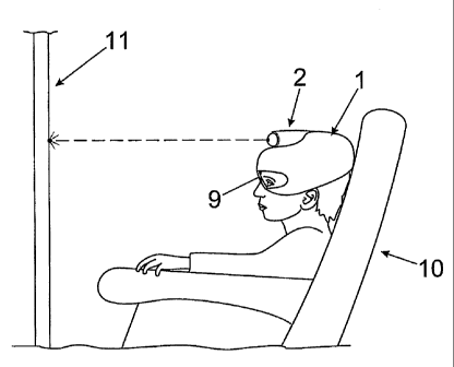
As represented in figure 1, the eye movement sensor comprises a
helmet (1) which is fitted to the head of a subject who is undergoing an
ophthalmologic examination in a unit, where the subject sits comfortably in a
CA 02610409 2007-11-30
seat (10) and in front of which there is a light spot projection screen (11),
situated at the appropriate distance for the examination, having oriented the
seat (10) conveniently to place the subject in the position required for a
correct
examination.
5 To
project the light spots at which the examiner duly makes the subject
look, the helmet (1) incorporates a light projector (2), located in its upper,
front and central part, which emits a light spot towards the projection screen
(11).
As illustrated in figure 2, a preferred embodiment can be described
10 which
consists of incorporating a light projector (2) in the helmet (1), which
emits a laser beam, so that it is projected on the projection screen (11) as a
circular image or a light spot. Said laser light projector (2) comprises a
rotatory
sphere with a laser pointer (3) which shoots the beam over the surface of the
sphere forward in a specific direction, upward, downward, right and left,
covering angles of up to 60 in the rotation of the sphere with the pointer
(3),
which is achieved by the action of an electric motor (4), incorporated in the
helmet (1).
Below the light projector (2), as shown in figure 3, in the upper and
central part of the helmet (1), at the height of the subject's forehead, said
helmet (1) incorporates a video camera (6) which focuses its lens downward,
having provided, in the lower part of the helmet (1), a pair of mirrors (7),
under each eye respectively on one side of the helmet (1), where the
different eye movements are reflected which are recorded by the video
camera (6). This configuration enables the recording of all the possible
positions of the eyes as the patient moves them following the light spot
produced by the light projector (2), on capturing the eyes seen from below, at
an angle of approximately 45 to 50 , reflected in the respective mirrors (7),
as shown in figure 4.
Another alternative embodiment is that represented in figure 5, where
the helmet (1) has two video cameras (5), instead of one, disposed
respectively (1) under the eyes, at the height of the subject's cheeks,
instead
of being positioned on the forehead as in the previous embodiment of the
helmet (1). In this configuration, the indirect recording by means of specular
reflections, as in the embodiment of the aforementioned invention, is not
necessary but instead the video cameras (5) directly capture all the eye
movements as they are located under each one thereof.
CA 02610409 2007-11-30
11
In any case, the video cameras (5, 6) are light, around a weight of 60-
90 grams, with high resolution, since at least between 200 and 400 pixels
resolution is required to capture all the positions of one extreme to the
other
of an eye, considering its angle of maximum movement, which is around
100 .
Both options of embodiment seen, with two video cameras (5) or just
one camera (6) which records the reflections of a pair of mirrors (7) manage
to record the images of all possible positions of either of the two eyes, seen
from the angle of maximum induction, corresponding to the downward
direction of the gaze and which, as has previously been explained,
constitutes the angulation from which all eye movements can be viewed,
without the eyelids getting in the way. Said angle of maximum induction
covers the intersection of the coronal plane, in a position of 6 hours, the
patient's face looking forward according to figure 6, plus the sagital plane
at
around 235 5 , the eyes looking from the left profile as indicated in
figure
7.
Optionally, the helmet (1) in any of its possible configurations
incorporates in its front part a support for sight correcting lenses (9),
which
are integrated in the helmet (1) outside the focal path of the cameras (5, 6),
so that the correcting lenses (9) do not get in the way when recording the
eyes.
In order to anatomically adapt the helmet (1) to the patient's head size,
it is planned to make 2 or 3 helmets of different size, taking into
consideration
the different ages and cranial perimeters. Furthermore, another helmet of
less size between the previous and the head exercises the functions of
adjustment of the size to perfection (8, 10). There should be several sizes of
this second helmet which is interposed between the main one and the head.