Note: Descriptions are shown in the official language in which they were submitted.
CA 02610703 2007-11-30
WO 2006/130755 PCT/US2006/021258
- 1 -
SIDE BRANCH STENT GRAFT
Description
Technical Field
This invention relates to a side branch stent graft and in the preferred
embodiments to the construction and deployment of a stent graft for the iliac
artery.
Background of the Invention
Stent grafts are used for treatment of vasculature in the human or animal
body to bypass a repair or defect in the vasculature. For instance, a stent
graft
may be used to span an aneurism which has occurred in or is associated with
the
iliac artery. In many cases, however, such a damaged or defective portion of
the
vasculature may include a branch vessel such as an internal iliac artery.
Bypassing
such a branch vessel without providing blood flow into it can cause problems
and
hence it has been proposed to provide a side branch on a stent graft which
when
deployed is positioned over the opening to the internal iliac artery and then
another stent graft can be deployed through the side branch into the internal
iliac
artery to provide a blood flow path to the internal iliac artery.
Generally, when deploying an endovascular stent graft into a body lumen, it
is possible to obtain access to such a body lumen from each end of the lumen
where necessary, thereby facilitating placement of a device in the lumen. The
internal iliac artery, which extends from the common iliac artery below the
aortic
bifurcation, is to all intents and purposes a blind vessel because there is no
practical way of performing an endovascular minimally invasive procedure into
that vessel other than by entry from the common iliac artery. The term blind
vessel is used herein to describe such a vessel.
There have been proposals to deploy a branched stent graft into the
common iliac artery via a femoral artery from a femoral incision with the
branched
stent graft having a side arm to extend into or at least adjacent the internal
iliac
artery. However, the use of such devices is very dependent upon favourable
layout of the arteries and in many cases access is extremely difficult. Access
over
CA 02610703 2011-05-31
- 2 -
the aortic bifurcation has been proposed but as there is limited distance
between
the aortic bifurcation and the iliac bifurcation special construction of a
stent graft
for this region is desirable.
Summary of the Invention
The present invention seeks to provide an improved side branch stent
graft and an improved stent graft for deployment particularly into the iliac
arteries
or at least to provide a physician with a useful alternative.
According to an aspect of the present invention, there is provided a side
branch stent graft.
The teachings herein will be generally discussed with reference to a
branched stent graft which can be deployed so that its branch is directed
towards
an internal iliac artery from the common iliac but is not so limited and may
be
used with deployment into any branched vessel but is particularly applicable
where the vessel into which the device is deployed is a blind vessel, that is,
access is not available from an end of the vessel remote from the bifurcation
from
a main vessel.
Throughout this specification the term distal with respect to a portion of
the aorta, a deployment device or a prosthesis is the end of the aorta,
deployment device or prosthesis further away in the direction of blood flow
from
the heart and the term proximal means the portion of the aorta, deployment
device or end of the prosthesis nearer to the heart. When applied to other
vessels, similar terms such as caudal and cranial should be understood.
Preferably there are one or two external zig-zag stents proximal of the
tubular side branch and from one to three external zig-zag stents distal of
the
tubular side branch.
The reinforcing ring around the proximal end of the tubular body can
comprise at least two turns of nitinol wire, each end of the nitinol wire
terminating
in a loop.
Preferably the tubular side branch comprises a connection socket for an
extension stent. In one embodiment the connection socket comprises a first
resilient ring around the tubular side branch at a distal end thereof, a
second
CA 02610703 2007-11-30
WO 2006/130755
PCT/US2006/021258
- 3 -
resilient ring spaced apart along the tubular side branch from the first ring
and a
zig zag resilient stent between the first and second rings. The zig zag
resilient
stent and the first and second rings are preferably on the outside of the
tubular
body and the first and second rings preferably comprise shape memory wire. The
first and second rings preferably comprise at least two turns of wire with
each
end terminating in a loop.
The zig zag resilient stent of the connection socket preferably comprises a
shape memory wire and the zig zag resilient stent defines a cylindrical form
having
a diameter less than that of the tubular side branch whereby to define a self
contracting stent.
The invention also encompasses a side arm stent graft comprising a main
tubular body of a biocompatible material, a fenestration in the main tubular
body
defined by a peripheral edge and a side arm being a tube of biocompatible
material
being joined around the peripheral edge at a joined region by stitching and
extending from the main tube at an acute angle thereto and being in fluid
communication with the main tubular body, at least one external zig-zag stent
on
the main tubular body proximal of the side arm, at least one external zig-zag
stent
on the main tubular body distal of the side arm, one internal zig-zag stent at
the
distal end of the main tubular body, and a reinforcing ring around the
proximal end
of the main tubular body and stitched thereto.
Preferably the fenestration is selected from the group comprising an
elongate aperture, a substantially rectangular aperture, a substantially
elliptical
aperture and a substantially triangular aperture. The acute angle may be in
the
range of from 15 to 60 degrees and preferably 15 to 45 degrees.
The or each external zig-zag stents proximal of the tubular side branch can
have a length of 14 mm and are spaced apart by about 2 mm and the or each
external zig-zag stents distal of the tubular side branch has a length of 12
mm and
are spaced apart by about 5 mm.
There may be further a central external zig-zag stent around the main
tubular body and the tubular side branch. The central external zig-zag stent
can
have a length of 14 mm and is spaced apart from the stent proximal to it by
about
CA 02610703 2007-11-30
WO 2006/130755
PCT/US2006/021258
- 4 -
2 mm and from the stent distal of it by about 5 mm.
The main tubular body can have a diameter of from 12 mm in a central
portion and remaining at 12 mm or expanding to 20 mm at a proximal end with a
frustoconical portion between the 12 mm and 20 mm portions and the tubular
side branch has a diameter of about 8 mm. Alternatively the main tubular body
can have a diameter of about 12 mm proximal of the tubular side branch and in
a
central portion and a diameter distal of the tubular side branch of from about
10
mm to 16 mm frustoconical portion between the 12 mm and 16 mm portions.
The main tubular body can have a length of about 69 mm to 119 mm and
the tubular side branch has a length of about 25 mm. The stent graft can have
a
length from its proximal end to the distal end of the side arm of about 45 mm
to
61 mm. The stent graft can have a length from its distal end to the distal end
of
the side arm of about 24 mm to 58 mm.
Brief Description of the Drawing
Embodiments of the present invention are described below, by way of
example only, with reference to the accompanying drawings, in which:
Figure 1 shows a first embodiment of stent graft intended for use in the
iliac artery;
Figure 2 shows a second embodiment of stent graft intended for use in the
iliac artery;
Figure 3 shows a third embodiment of stent graft intended for use in the
iliac artery;
Figure 4 shows a fourth embodiment of stent graft intended for use in the
iliac artery;
Figure 5 shows a fifth embodiment of stent graft intended for use in the
iliac artery;
Figure 6 shows a sixth embodiment of stent graft intended for use in the
iliac artery;
Figures 7A to 7F show schematically a first range of preferred
configurations of stent grafts intended for use in the iliac artery;
Figures 8A to 8F show schematically an alternative range of preferred
CA 02610703 2007-11-30
WO 2006/130755 PCT/US2006/021258
- 5 -
configurations of stent grafts intended for use in the iliac artery;
Figures 9A to 9D show schematically an alternative range of preferred
configurations of stent grafts intended for use in the iliac artery;
Figure 10 shows a detail of the proximal end of an embodiment of stent
graft, and
Figure 11 shows schematically a cross section of an aneurysed aorta and
common iliac artery with a composite stent graft system deployed into it
including
an embodiment of iliac stent graft.
Detailed Description
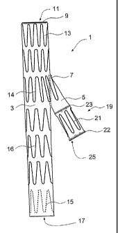
Now looking more closely at the drawings and in particular Figure 1, an
embodiment of stent graft 1 comprises a tubular body 3 with a side arm 5, also
a
tubular body, stitched into an aperture in the main body 3 at 7 so as to allow
fluid
communication from the lumen of the main body into the lumen of the side arm
5.
The main tubular body 3 has a reinforcing ring arrangement 9 at its proximal
end
11. The construction of the reinforcing ring 9 will be discussed in relation
to
Figure 10. The tubular body 3 also has a number of external zig-zag self-
expanding stents 13, 14 and 16 along its length and an internal self-expanding
zig-zag stent 15 at its distal end 17.
PCT Patent Publication No. WO 2006/034276 entitled "Side Branch Stent
Graft" includes discussion of one method of connection of a side arm to a main
tubular body in a stent graft which could be used in the embodiments of stent
graft taught herein, if desired.
On the side arm 5 there is a connection socket arrangement 19. The
connection socket arrangement includes a self-expanding stent 21 between two
reinforcing rings 22 and 23.
PCT Patent Publication No. WO 2006/034340 entitled "Stent Graft
Connection Arrangement" discloses a stent graft connection arrangement of the
type suitable for the side arm of the stent graft of the type taught herein,
although
other connection arrangements apparent to the skilled person could instead be
used.
CA 02610703 2011-05-31
- 6 -
In the side arm stent graft shown in Figure 1 there are two external
stents 13 proximal of the connection of the side arm, one central external
stent 14
which also encompasses the side arm and three external stents 16 and one
internal stent 15 distal of the connection with the side arm 5. The internal
and
external stents are fastened to the tubular body by known methods including by
stitching, adhesive or the like.
The stent graft depicted in Figure 1 would normally have a diameter of the
main tubular body of 12mm and a diameter of the side arm of 8mm. The stent
graft shown in Figure 1 is the longest that would normally be used in
deployment
into the iliac artery of a patient as will be discussed below.
Figure 2 shows the shortest practical stent graft which can be deployed
into a common iliac artery. The stent graft 29 depicted in Figure 2 has a
tubular
body 30 and a side arm 32. In this embodiment there is one external stent 13
proximal of the junction with the side arm, one central external stent 14
which
also encompasses the side arm and one external stent 16 and one internal
stent 15 distal of the junction with the side arm. The stent graft depicted in
Figure 2 has a diameter of the main tubular body 30 of 12mm and a diameter of
the side arm 32 of 8mm. The construction of the connection socket 33 on the
stent graft shown in Figure 2 is similar to that shown in Figure 1.
For placement of a side branch stent graft into the internal iliac artery, the
critical dimension is the distance from the proximal end 11 of the stent graft
to the
end 25 of the side arm 5 as this is the distance which must fit between the
aortic
bifurcation and the entrance to the internal iliac artery as will be discussed
below.
Figure 3 shows an alternative embodiment of stent graft 35 substantially
similar to that shown in Figure 1 except that the tubular body 40 is tapered
to a
diameter of 10mm distal of the connection with the side arm 41 whereas the
diameter of the tubular body 40 proximal of the connection is 12mm.
Similarly in Figure 4 the stent graft 36 has a tubular body 37 which is
tapered distal of the side arm connection 38 to give a diameter of lOmm
whereas
the diameter of the tubular body 37 proximal of the connection is 12mm.
CA 02610703 2011-05-31
- 7 -
Figure 5 shows another embodiment of stent graft 40 substantially
similar to that shown in Figure 1 except that the tubular body 41 is expanded
to a diameter of 20 mm at its proximal end 42 proximal of the connection with
the side arm 44 with a frusto-conical portion 41a joining the central portion
to
the proximal portion and also expanded to a diameter of 16mm at its distal
end 43 with a frusto-conical portion 41b joining the central portion to the
distal
portion.
Similarly in Figure 6 the stent graft 45 has a tubular body 46 which is
expanded distal of the side arm 47 connection to give a diameter of 16mm at
its distal end 48 with a frusto-conical portion 46a joining the central
portion to
the distal portion whereas the diameter of the tubular body proximal of the
connection is 12mm.
Figures 7A to 7F show schematically a range of sizes of stent grafts for
the common iliac artery in stylised form. The position of stents is
represented
by a box on the stylised form.
Figure 7A shows a schematic stent graft 50 with a proximal reinforcing
ring 51, two external stents 52 proximal of the side arm 53, central external
stent 54, three external stents 55 distal of the side arm 53 and one internal
distal-most stent 56. The stents 52 and 54 each have a length of 14mm and
are separated by a gap 57 of 2mm. The stents 55 each have a length of
12mm and are separated by a gap 58 of 5mm. The stent 56 has a length of
17mm and is separated from the stent 55 by a gap of 5mm.
Figures 7B, 7C, 7D, 7E and 7F all show the various configurations of
stent graft with varying numbers of stents proximal and distal of the side
branch.
Table 1 shows a range of sizes of stent grafts and numbers and
sizes of stents as depicted in Figures 7A to 7F. The overall length is the
dimension 60 shown in Figure 7A, the proximal length is the length 61 shown
in Figure 7A and the distal length is the length 62 shown in Figure 7A.
CA 02610703 2007-11-30
WO 2006/130755
PCT/US2006/021258
- 8 -
Table 1
Fig Fig Fig Fig7D Fig7E Fig7F
_ 7A 7B 7C
Overall Length (mm) 119 102 85 103 86 69
Proximal length (mm) 61 61 61 45 45 45
Distal length (mm) 58 41 24 58 41 24
Proximal Diameter (mm) 12 12 12 12 12 12
Central Diameter (mm) . 12 12 12 12 12 12
Distal Diameter (mm) 12 12 12 12 12 12
Number of Stents on main 7 6 5 6 5 4
body
Number of stents on main 2 2 2 1 1 1
body proximal of side arm
Length of stents on main 14 14 14 14 14 14
body proximal of side arm
(mm)
Spacing of stents on main 2 2 2 2 2 2
body proximal of side arm
(mm) ,
Number of central external 1 1 1 1 1 1
stents
Length of central external 14 14 14 14 14 14
stents (mm)
Number of stents on main 4 3 2 4 3 2
body distal of side arm
Length of external stents 12 12 12 12 12 12
on main body distal of side
arm (mm) ,
Length of distal most 17 17 17 17 17 17
internal stent on main
body (mm)
Spacing of stents on main 5 5 5 5 5 5
body distal of side arm
(mm) .
Number of Stents on side 1 1 1 1 1 1
arm ....
12 mm Ring at proximal Yes Yes Yes Yes Yes Yes
end _
7 mm Rings on side arm Yes Yes Yes Yes Yes Yes
As discussed above, the proximal length is important as a stent graft must
be able to be deployed with its distal end below the aortic bifurcation and
the
CA 02610703 2007-11-30
WO 2006/130755
PCT/US2006/021258
- 9 -
distal end of its side arm proximal of the iliac bifurcation between the
internal and
external iliac arteries.
Similarly Figures 8A to 8F show schematic configurations of stent grafts
similar to those of the corresponding drawings in Figures 7A to 7F except that
in
Figures 8A to 8F the proximal end 70 of the stylised stent graft has a
diameter of
12mm and the distal end 71 is tapered to a diameter of lOmm. The lesser
diameter of the distal end of the stent graft in the embodiments of stent
graft
shown in Figures 8A to 8F allow for sealing into a smaller non-aneurysed
region of
the external iliac artery without exerting excessive pressure onto the walls
of the
vasculature.
Table 2 shows a range of sizes of stent grafts as depicted in Figures 8A to
8F. The overall length is the dimension 60 shown in Figure 7A, the proximal
length is the length 61 shown in Figure 7A and the distal length is the length
62
shown in Figure 7A.
CA 02610703 2007-11-30
WO 2006/130755 PCT/US2006/021258
- 10 -
Table 2
Fig Fig Fig8 Fig Fig Fig
8A 8B C 8D 8E 8F
Overall Length (mm) 119 102 85 103 86 69
Proximal length (mm) 61 61 61 45 45 45
Distal length (mm) 58 41 24 58 41 24
Proximal Diameter (mm) 12 12 12 12 12 12
Central Diameter (mm) 12 12 12 12 12 12
Distal Diameter (mm) 10 10 10 10 10 10
Number of Stents on 7 6 5 6 5 4
main body
Number of stents on main 2 2 2 1 1 1
body proximal of side arm
Length of stents on main 14 14 14 14 14 14
body proximal of side arm
(mm)
Spacing of stents on main 2 2 2 2 2 2
body proximal of side arm
(mm)
Number of central 1 1 1 1 1 1
external stents
Number of stents on main 4 3 2 4 3 2
body distal of side arm
Length of central external 14 14 14 14 14 14
stents (mm)
Length of external stents 12 12 12 12 12 12
on main body distal of
side arm (mm)
Length of distal most 17 17 17 17 17 17
internal stent on main
body (mm)
Spacing of stents on main 5 5 5 5 5 5
body distal of side arm
(mm)
Number of Stents on side 1 1 1 1 1 1
arm
12 mm Ring at proximal Yes Yes Yes Yes Yes Yes
end
7 mm Rings on side arm Yes Yes Yes Yes Yes Yes
Figures 9A to 9D show schematic configurations of stent grafts similar to
those of the corresponding drawings in Figures 7A to 7F except that in Figures
9A
CA 02610703 2007-11-30
WO 2006/130755
PCT/US2006/021258
- 11 -
to 9D the proximal end 70 of the stylised stent graft has a diameter of 20 mm
and the distal ends 71 have diameters of 12 mm and 16 mm.
Table 3 shows a range of sizes of stent grafts as depicted in Figures 9A to
9D. The overall length is the dimension 60 shown in Figure 7A, the proximal
length is the length 61 shown in Figure 7A and the distal length is the length
62
shown in Figure 7A.
CA 02610703 2007-11-30
WO 2006/130755
PCT/US2006/021258
- 12 -
Table 3
Fig Fig Fig Fig
9A 98 9C 9D
Overall Length (mm) 119 119 102 102
Proximal length (mm) 61 61 61 61
Distal length (mm) 58 58 41 41
Proximal Diameter (mm) 20 20 20 20
Central Diameter (mm) 12 12 12 12
Distal Diameter (mm) 12 16 12 16
Number of Stents on main 7 7 6 6
body
Number of stents on main 2 2 2 2
body proximal of side arm
Length of stents on main 14 14 14 14
body proximal of side arm
(mm)
Spacing of stents on main 2 2 2 2
body proximal of side arm
(mm)
Number of central external 1 1 1 1
stents
Length of central external 14 14 14 14
stents (mm)
Number of stents on main 4 4 3 3
body distal of side arm
Length of external stents 12 12 12 12
on main body distal of side
arm (mm)
Length of distal most 17 17 17 17
internal stent on main
body (mm)
Spacing of stents on main 5 5 5 5
body distal of side arm
(mm)
Number of Stents on side 1 1 1 1
arm
20 mm Ring at proximal Yes Yes Yes Yes
end
7 mm Rings on side arm Yes Yes Yes Yes
Figure 10 shows the construction of a preferred embodiment of stent graft with
a
proximal reinforcing ring. The same reference numerals as used in Figure 1 are
used for Figure 10 for the corresponding components. The tubular body 3 of the
= CA 02610703 2013-05-30
- 13 -
stent graft 1 has a proximal-most external stent 13 stitched onto the tubular
body by
means of stitches 75. At the proximal end 78 of the stent graft 1 a
reinforcing ring 9 is
provided. The reinforcing ring 9 comprises two turns of a shape memory wire
74,
such as NitinolTM wire, around the proximal end 78 and loops 76 at each
terminal end
of the NitinolTM wire 74. The loops 76 are provided to prevent the ends of the
Nitinol TM wire causing damage to the vasculature in which they are deployed.
The
two turns of NitinolTM wire 74 are stitched by means of stitching 77 to the
proximal
end of the tubular body 3.
The NitinolTM reinforcing ring 9 provides reinforcement for the proximal end
of
the stent graft so that when a catheter or other device is being deployed into
the
proximal end of the stent graft the end of the stent graft cannot be damaged.
The
NitinolTM reinforcing ring 9 provides a fixed diameter into which another self
expanding or balloon expandable stent graft can expand or be expanded within
the
proximal end of the stent graft 1.
Figure 11 shows a full assembly of a composite stent graft system into an
aneurysed aortic and common iliac artery using the preferred embodiment of
stent
graft for the common iliac artery.
The vasculature illustrated generally consists of a descending aorta 152
extending down to renal arteries 153 and further extending as the infra-renal
aorta
150 down to an aortic bifurcation 178 and into the iliac and contra-iliac
arteries 154
and 187. The common iliac artery 154 further bifurcates into an external iliac
artery
164 and an internal iliac artery 168. It is this internal iliac artery 168
which is
described above as a blind artery as endovascular access is only available via
the
common iliac artery. An aneurysed region 151 of the aorta 150 extends down
into
the common iliac artery 154.
In Figure 11 the aorta 150 has an aneurysm 151 which extends from below
the renal arteries 153 into the common iliac artery 154. The aorta 152
proximal of the
renal arteries 153 is not aneurysed and provides a suitable landing zone for a
proximally extending external stent 156 of a bifurcated stent graft 157.
The general order of placement of such a stent graft assembly for an
aneurysed aorta is first that the iliac branch stent graft 160 is placed first
with its
CA 02610703 2007-11-30
WO 2006/130755
PCT/US2006/021258
- 14 -
distal end 162 placed into a non-aneurised region of the external iliac artery
164.
The side arm 166 extends towards the internal iliac artery 168. Subsequent to
placement of the iliac branched stent graft 160 a covered leg extension 170 is
placed extending from the side branch 166 so that it extends into and seals in
the
internal iliac artery 168. The covered leg extension 170 can be a covered
balloon
expandable stent or a covered self expanding stent.
The main bifurcated stent graft 157 is then deployed through the contra-
lateral iliac artery 187 so that its longer leg 176 extends down the contra-
lateral
iliac artery 187 and its shorter leg 172 terminates proximal of the
reinforcing ring
174 on the iliac stent graft 160 and proximal of the aortic bifurcation 178.
The
proximal end of the bifurcated stent graft 157 seals into a landing zone in
the
region of aorta 182 just distal of the renal arteries 153 and the proximally
extending supra-renal external stent 156 extends over the renal arteries to
the
aorta region 152 to provide good support for the bifurcated stent graft. A leg
extension 190 can then be deployed to connect the shorter leg 172 of the main
bifurcated stent graft 157 to the reinforcing ring 174 on the iliac stent
graft 160.
In a final deployment stage a leg extension 185 can deployed into the longer
leg
176 to seal into a non-aneurised portion of the contra-lateral common iliac
artery
187 if the longer leg does not already seal onto such a zone.
By this arrangement a stent graft is effectively bridging the aneurised region
by sealing in the non-aneurised portion of the aorta as well as in the non-
aneurised
portions of the iliac arteries.
The critical dimension in relation to deployment of a stent graft into an
aneurised iliac artery 154 is the distance shown as 135 in Figure 11. This is
the
distance from the iliac bifurcation 178 down to the bifurcation of the
external iliac
artery 164 and the internal iliac artery 168. The proximal length of a stent
graft of
the type discussed in relation to Figures 1 to 6 and as shown in Figures 7 to
9 is
less than the distance 135 so that access over the aortic bifurcation is
possible to
endovascularly enter the proximal end 174 of the stent graft 160 and the end
167
of the side branch 166 of the stent graft 160 is placed so that a guide wire
(not
shown) can extend out of the side branch 166 and be manipulated into the
CA 02610703 2007-11-30
WO 2006/130755
PCT/US2006/021258
- 15 -
internal iliac artery 168.
The length of the distal portion 162 of the stent graft 160 is such that it
can extend to non-aneurised region of the external iliac artery 164 so that a
seal
may be obtained with the distal end of the stent graft 160. Where the aneurism
extends some distance down the external iliac artery a further leg extension
may
be placed so that the stent graft assembly extends through non-aneurised
region
of the external iliac artery.
Throughout this specification various indications have been given as to the
scope of this invention but the invention is not limited to any one of the
disclosed
features but may reside in two or more of these combined together. The
examples are given for illustration only and not for limitation.
Throughout this specification and the claims that follow, unless the context
requires otherwise, the words 'comprise' and 'include' and variations such as
'comprising' and 'including' will be understood to imply the inclusion of a
stated
integer or group of integers but not the exclusion of any other integer or
group of
integers.