Note: Descriptions are shown in the official language in which they were submitted.
CA 02610856 2007-12-04
WO 2006/138261 PCT/US2006/022908
SYSTEM AND METHOD FOR FLUORESCENCE EXCITATION AND DETECTION
HAVING DISTINCT OPTICAL PATHS
RELATED APPLICATIONS
This application claims the benefit of U.S. Provisional Application Serial
Number
60/689,903, filed June 13, 2005, the entirety of which is hereby incorporated
herein by
reference for the teachings therein.
FIELD
The embodiments disclosed herein relate to fluorescence excitation and
detection, and
more particularly to a system and method for fluorescence excitation and
detection having
distinct optical paths.
BACKGROUND
Techniques for thermal cycling of DNA samples are known in the art. By
performing
a polymerase chain reaction (PCR), DNA can be amplified. It is desirable to
cycle a specially
constituted liquid biological reaction mixture through a specific duration and
range of
tenlperatures in order to successfully amplify the DNA in the liquid reaction
mixture.
Thermocycling is the process of melting DNA, annealing short primers to the
resulting single
strands, and extending those primers to make new copies of double stranded
DNA. The
liquid reaction mixture is repeatedly put through this process of melting at
high temperatures
and annealing and extending at lower temperatures.
In a typical thermocycling apparatus, a biological reaction mixture including
DNA
will be provided in a large number of sample wells on a thermal block
assembly.
Quantitative PCR (qPCR) uses fluorogenic probes to sense DNA. Insth-umentation
designed
for qPCR must be able to detect approximately 1 nM of these probes in small
volume
samples (e.g., approximately 25 l). The detection method must be compatible
with the
thermal cycling required for qPCR. The detection method must also be capable
of
distinguishing multiple fluorogenic probes in the same sample.
Enhancing the sensitivity of fluorescence detection of a qPCR instrument or
method
improves the usefulness of that instrument or method by enabling detection of
DNA sooner,
that is, after fewer thermal cycles.
1
CA 02610856 2007-12-04
WO 2006/138261 PCT/US2006/022908
Prior art systems use the same light path for excitation and detection. In
those
systems excitation light is directed to a beam splitter, which transmits
typically about one-
half of the excitation light to the sample. Some of the emitted light from the
saniple comes
back, to the beam splitter and a portion of that light, typically about one-
half, is directed to a
detector. By using beam splitters, only about one-half of the light is
reflected and
transmitted; therefore, only about one-quarter of the signal is measured.
U.S. Patent No. 5,757,014 to Bruno et al. discloses an optical detection
device for
analytical measurements of chemical substances. The Bruno et al. device
includes an
excitation light guide and an emission light guide that share the same optical
light path. U.S.
Patent No. 6,563,581 to Oldham et al. discloses a system for detecting
fluorescence emitted
from a plurality of samples in a sample tray. The Oldham et al. device
includes a plurality of
lenses, an actuator, a light source, a light direction mechanism and an
optical detection
system. U.S. Patent No. 6,015,674 to Woudenberg et al. discloses a system for
measuring in
real time polynucleotide products from nucleic acid amplification processes,
such as
polymerase chain reaction (PCR). The Woudenberg et al. device includes a
sample holder,
an optical interface, a lens, and a fiber optic cable for delivering an
excitation beam to a
sample and for receiving light emitted by the sample.
Other prior art methods use fiber optics to deliver the excitation light to
and collect
the fluorescence from the sample. These methods may either use independent
fiber optics for
each sample or scan the same fiber optics over all the samples. Some methods
illuminate the
entire collection of samples simultaneously and detect the fluorescence with
large area
detectors.
SUMMARY
A system and method for fluorescence excitation and detection having distinct
optical
paths is disclosed. According to aspects illustrated herein, there is provided
a system for
detecting fluorescence comprising a light source that emits an excitation
light into an
illumination tube; a plurality of collection optics located around an aperture
in the
illumination tube for collecting fluorescence; and a detector for determining
the amount of
fluorescence.
According to aspects illustrated herein, there is provided a detection system
for
detecting fluorescence from a plurality of samples comprising an illumination
tube for
2
CA 02610856 2007-12-04
WO 2006/138261 PCT/US2006/022908
receiving an excitation light from a light emitting diode; a plurality of
collection optics
located around an aperture in the illumination tube for collecting
fluorescence; and a
photodiode for detecting the amount of fluorescence.
According to aspects illustrated herein, there is provided a system for
detecting
fluorescence coinprising a tube for collecting fluorescence; a light source
that emits an
excitation light into a plurality of optics located around an aperture in the
tube; and a
photodiode for detecting the amount of fluorescence.
According to aspects illustrated herein, there is provided a method for
detecting
fluorescence comprising emitting an excitation light from a light source into
an illumination
tube; directing the excitation light to an excitation filter; illuminating a
sample with the
excitation light to generate an emission light; and detecting the optical
characteristics of the
emission light using a plurality of collection optics located around the
illumination tube.
BRIEF DESCRIPTION OF THE DRAWINGS
The presently disclosed embodiments will be further explained with reference
to the
attached drawings, wherein like structures are referred to by like numerals
throughout the
several views. The drawings are not necessarily to scale, the emphasis having
instead been
generally placed upon illustrating the principles of the presently disclosed
embodiments.
FIG. 1 is a perspective view of an optical module having collection optics
located
around an illumination tube.
FIG. 2 is a front view of an optical module having collection optics located
around an
illumination tube.
FIG. 3 is a sectional perspective view of an optical module having collection
optics
located around an illumination tube taken along line A-A in FIG. 2.
FIG. 4 is a sectional view of an optical module having collection optics
located
around an illumination tube taken along line A-A in FIG. 2 and showing traces
of the light
paths.
3
CA 02610856 2007-12-04
WO 2006/138261 PCT/US2006/022908
FIG. 5 is a perspective view of an optical module having collection optics
located
around an illumination tube mounted to an assembly that shows the path as the
optical
module is scanned over a plurality of sample tubes.
While the above-identified drawings set forth presently disclosed embodiments,
other
embodiments are also contemplated, as noted in the discussion. This disclosure
presents
illustrative embodiments by way of representation and not limitation. Numerous
other
modifications and embodiments can be devised by those skilled in the art which
fall within
the scope and spirit of the principles of the presently disclosed embodiments.
DETAILED DESCRIPTION
A system and method for fluorescence excitation and detection having distinct
optical
paths is disclosed. A system for fluorescence excitation and detection having
separate and
distinct optical paths is shown generally at 30 in FIG. 1. The system has one
optical light
path for the illumination (excitation), and a different optical light path for
the detection of
fluorescence. The optical path for excitation light is free space optics
without any collection
optics. The optical path for the detection of emitted fluorescence involves
collection optics
guiding light to a detector. The optical path for detection is outside and
around the optical
path for excitation.
A light source shines excitation light through a central illumination tube and
onto a
sample. Illuminating through the central illumination tube allows a compact
design and
concentrates the light on the sample, minimizing the amount of scattered
light. The sample
then emits fluorescent light that is detected by a plurality of collection
optics located around
the illumination tube. Collecting the emitted light at locations around the
illumination tube
obviates the need for a beam splitter thereby reducing the complexity of the
design,
eliminating losses from the beam splitter, and reducing the size of the
design. The system is
compact, and the detected light has both high quality (small amount of
scattered light) and
quantity (no losses from beam splitters).
When the system is applied to qPCR, the PCR amplification scheme used is not
critical, but generally qPCR requires the use of either a nucleic acid
polymerase with
exonuclease activity or a population of double stranded DNA that increases
during the course
of the PCR being monitored. Thermal cyclers used in qPCR are typically
programmable
heating blocks that control and maintain the temperature of the sample through
the
4
CA 02610856 2007-12-04
WO 2006/138261 PCT/US2006/022908
temperature-dependent stages that constitute the cycles of PCR: template
denaturation,
primer annealing, and primer extension. These temperatures are cycled up to
forty times or
more to obtain amplification of the DNA target. Thermal cyclers use different
technologies
to effect temperature change including, but not limited to, peltier heating
and cooling,
resistance heating, and passive air or water heating.
As used herein, "optical module" refers to the optics of systems for thermal
cycling
known in the art including, but not limited to, modular optics, non-modular
optics, and any
other suitable optics. The optical module can be used for scanning a plurality
of sanlples of
biological material after thermal cycling of DNA to accomplish a polymerase
chain reaction
(PCR), during thermal cycling of DNA to accomplish a quantitative polymerase
chain
reaction (qPCR), after thermal cycling of DNA after a reverse transcriptase
reaction to
accomplish a reverse transcription-polymerase chain reaction (RT-PCR), during
thermal
cycling of DNA after a reverse transcriptase reaction to accomplish a reverse
transcription-
quantitative polymerase chain reaction (RT-qPCR), or for fluorescence
detection during other
nucleic acid amplification types of experiments. The optic module controls the
illumination
light and the detection of fluorescence.
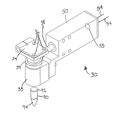
FIG. 1 shows an illustrative optical module 30 having collection optics
located around
an illumination tube above one of a plurality of sample tubes 90. The optical
module 30 is
used for detecting fluorescence from a plurality of samples 94 in the
plurality of sample tubes
90. The optical module 30 includes at least an optics housing 35, a plurality
of collection
optics 39, a detector housing 50, and a detector 53. The plurality of
collection optics 39
extends down within the optics housing 35 and is located around an
illumination tube 44
(shown in FIG. 3). The optical module 30 illuminates from the inside,
directing excitation
light through the central illumination tube 44, and collects fluorescence from
the outside
around the illumination tube 44. The plurality of collection optics 39 extends
into the
detector housing 50. A plurality of leads 54 extend from detector housing 50
connecting the
detector 53 to electronics. The electronics both powers a light source and
detects the signal
from detector 53 in the detector housing 50. The electronics may be remotely
attached to the
optical module 30. The electronics may be under computer control. The optical
module 30
may be a single component or composed of a plurality of assembled parts.
FIG. 2 is a front view of an optical module 30 having collection optics
located around
an illumination tube. The optical module 30 is compact, being comparable in
size to the
5
CA 02610856 2007-12-04
WO 2006/138261 PCT/US2006/022908
sample holders 90 that hold the samples 94 that the optical module 30
measures. The small
size of the optical module 30 allows use of a few, small optics, which keeps
the overall size
and cost of the device low. Use of the same optical module 30 for all samples
reduces
measurement variability from different samples compared to using different
optics for
different samples, including optics that illuminate and detect from multiple
samples
simultaneously.
FIG. 3 is a sectional perspective view taken along line A-A in FIG. 2 of the
optical
module 30 having collection optics located around an illumination tube. The
optical module
30 collects the fluorescence from the samples through the plurality of
collection optics 39
arrayed concentrically around the illumination tube 44. The plurality of
collection optics 39
may be either a fiber optic bundle or multiple fiber optic bundles that
surround the
illumination tube 44. Alternatively, the plurality of collection optics 39 may
be fused optical
fibers, light pipes, or individual opiical fibers that surround the
illumination tube 44.
Alternatively, the plurality of collection optics 39 may be any type of light
guide or guides
including, but not limited to, fluid filled fibers or molded plastics. Those
skilled in the art
will recognize that other types of solid state optics known in the art are
within the scope and
spirit of the presently disclosed embodiments.
Individual fiber optics can be used to collect light as a concentric ring of
collection
optics 39 around the illumination tube 44 by packing the individual fiber
optics around the
illumination tube 44 such that one end of each fiber optic is flush with an
aperture 47 of the
illumination tube 44. The other ends of the individual fiber optics can be
bundled into a
ferrule 52 that directs the output of the light collected from the collection
ends into a
detection module 50. Other methods for collecting light from a concentric ring
around an
illumination tube may be used, but fabrication of an arrangement of individual
fiber optics in
a specialized ring may be difficult, expensive, and the fibers may break.
Using fiber optic bundles rather than individual fiber optics could alleviate
some of
the fabrication and handling concerns. Rather than forming a ring from
individual fiber
optics, the individual fiber optics could be collected into cylindrical fiber
bundles that are
arrayed around the aperture 47 of the illumination tube 44. Bundling the fiber
optics may
provide strength and stability and minimize handling damage.
6
CA 02610856 2007-12-04
WO 2006/138261 PCT/US2006/022908
An assembly of fused fiber optics may also be reliable and easier to handle
when
manufacturing an instrument that uses collection optics around an illumination
tube than an
assembly of individual fiber optics. Fused fiber optics consist of individual
fiber optics or
optical tubes that are bonded together to form a stiff and sturdy monolithic
part. Because the
fibers or tubes are bonded together over their entire lengths, no stray
individual fibers have
the possibility to break during handling of the fused fiber optics. In
addition, the optical
tliroughput of fused fiber optics is larger than that of individual fiber
optics or fiber optic
bundles because fused fibers can be packed more tightly than individual
fibers, whicli
increases the fraction of the area of the collection optics 39 that actually
collects light.
A molded liglit pipe may also be used for collection of fluorescence. A light
pipe is a
single, solid piece of optically clear material. The light pipe can be molded
from a bulk
material, for example, plastic, making fabrication simpler than that of
individual fiber optics,
fiber optic bundles, or fused fiber optics all of which require assembly from
many parts.
Because the entire structure of the light pipe transmits light, the fraction
of the light pipe
collection area that actually collects light is nearly 100%, which is a larger
fraction than even
fused fiber optics. Because the light pipe consists of essentially only one
part, it may be more
reliable than individual fiber optics, fiber optic bundles, and fused fiber
optics.
An excitation light is produced by a light source 40 mounted to a mounting
board 34.
A plurality of excitation light rays is emitted from the light source 40 into
the illumination
tube 44. In FIG. 3, the excitation from the light source 40 is in a downward
direction. The
light from the light source 40 travels through a lens 72, an excitation filter
62, and then
toward the sample tube 90. The light is focused on the inside the sample tube
90, but aiming
and focusing the light from the light source 40 onto a cap 92 of the sample
tube 90 is
effective. Using free space optics for the illumination tube instead of fiber
optics enables
more compact design because optics for coupling the excitation light into the
fiber optics and
optics for collimating the excitation light before it reaches the excitation
filter are not
required. In addition, the excitation light in the free space design can be
converging on the
sample rather than diverging as it does from fiber optics, which helps reduce
scattered light
that lowers sensitivity by increasing background.
The lens 72 and/or the illumination tube 44 confines the excitation light into
a
narrow beam that is coupled preferentially to the sample tube 90. Because the
excitation light
is focused into the sample tube 90 and the sample 94, there is minimal stray
light reflecting
7
CA 02610856 2007-12-04
WO 2006/138261 PCT/US2006/022908
throughout the rest of the optical system, which helps keep the background low
and the
sensitivity high.
The plurality of collection optics 39 surrounds the illumination tube and may
cover
the remaining area of the opening of the sample tube 90. The plurality of
collection optics 39
is designed to maximize collection of light that is emitted from the sample
94. The
integration of the plurality of collection optics 39 around the illumination
tube 44 provides an
efficient two optic system that excites the sample with a small light source,
and detects a
large area of emitted light. Having the plurality of collection optics 39
located around the
illumination tube 44 allows a large detection area for emitted light.
The illumination tube 44 in the center of the plurality of collection optics
39
minimizes the scattering of the excitation light. By ensuring that as much
light as possible
enters the sample tube 90, less excitation light reflects off the corners and
edges of the sample
cap 92. Thus, most of the excitation light is coupled into the sample tube 90,
wasting only a
small amount of the excitation light that does not enter the sample tube 90
and is reflected
into the atmosphere. Coupling more ligllt into the sample improves the
sensitivity of the
module by increasing the signal from the sample. Coupling a higher fraction of
the light into
the sample improves the sensitivity of the module by increasing the signal
while at the same
time reducing the background, which limits sensitivity.
The light travels through the cap 92 and into the sample tube 90 where it
excites
fluorogenic probes typically used in qPCR that are within the sample 94 in the
sample tube
90, causing the sample 94 to fluoresce. A biological probe can be placed in
each DNA
sample so that the amount of fluorescent light emitted as the DNA strands
replicate during
each thermal cycle is related to the amount of DNA in the sample.
Emitted fluorescent light from the sample 94 passes through the cap 92, and is
collected by the plurality of collection optics 39. The fluorescent light
travels through the
plurality of collection optics 39 around the illumination tube 44. The
plurality of collection
optics 39 is drawn up and around the illumination tube 44 and grouped in a
bundle 38 to
converge and enter the detector housing 50. The bundle 38 of collection optics
39 is mounted
in the ferrule 52 for attachment to the detector housing 50. The light
collected by the
collection optics 39 leaves the bundle 38 of collection optics 39 at the
ferrule 52 and passes
through an emission filter 64, which preferentially transmits signal light and
blocks scattered
8
CA 02610856 2007-12-04
WO 2006/138261 PCT/US2006/022908
light collected by the collection optics 39. After being transmitted by the
emission filter 64,
the light collected by the collection optics 39 can be condensed by
appropriate optics, in this
case a lens 84, onto the detector 53. Reflection optics can also be used to
condense the light
from the collection optics. The detector 53 converts the intensity of the
light into a voltage
that is a function of the light intensity. The sense and control electrics for
the detector 53 are
connected to the detector 53 by the leads 54. By detecting the amount of
emitted fluorescent
light, the detection system measures the amount of DNA that has been produced.
Data can be
collected from each sample tube 90 and analyzed by a computer.
In an alternative embodiment, a reflector collects the light exiting the
emission filter
64 and reflects the light onto the detector 53. In this embodiment, the
reflector is used
instead of the lens 84. By replacing the lens 84 with the reflector, the
detector 53 could be
moved closer to the emission filter 64, resulting in a more coinpact detector
housing 50. The
reflector may be conical, toroidal or other geometries known in the art that
collect and reflect
light.
FIG. 4 is a sectional view of the optical module 30 having the plurality of
collection
optics 39 located around the illumination tube 44 taken along line A-A in FIG.
2 and showing
the area of illumination by an excitation light 48 to the sample 94 and cones
of collection 58
of the collection optics 39 for the light emitted from the sample 94. The
light source 40
supplies the excitation light 48 for the illumination tube 44. The excitation
light 48 travels
through the lens 72 that focuses and collimates the excitation light 48. The
excitation light 48
then passes through the excitation filter 62, which selects the wavelength of
light to excite the
sample 94. The excitation light 48 continues through and exits the
illumination tube 44
through the aperture 47 and travels toward the plurality of samples 94.
Some of the light transmitted by the cap 92 of the sample tube 90 is absorbed
by the
sainple 94 and excites the fluorogenic probes within the sample, re-emitting
light through
fluorescence. The re-emitted light (fluorescence) that travels up the sample
tube 90, exits
through the cap 92, and falls within the cones of collection 58 of the
plurality of collection
optics 39 concentrically arranged around the illumination tube 44. After the
plurality of
collection optics 39 collects the fluorescence from the sample 94, the
plurality of collection
optics 39 transmits the light to the emission filter 64, after which the light
is focused by the
lens 84 or other suitable optics onto the detector 53.
9
CA 02610856 2007-12-04
WO 2006/138261 PCT/US2006/022908
FIG. 4 illustrates the range of emitted light that is accepted into the
plurality of
collection optics 39. The plurality of collection optics 39 can accept light
incident from a
range of angles so long as the light travels in a direction toward the
plurality of collection
optics 39. Each of the plurality of collection optics 39 can accept light
incident from a range
of angles defined by the cone of collection 58 in FIG. 4. Because the
plurality of collection
optics 39 are located around the illumination tube 44, the plurality of
collection optics 39
form the cone of collection 58 to collect fluorescence from the sample 94. The
plurality of
collection optics 39 may be located around 360 degrees around the illumination
tube 44.
In an embodiment, the plurality of collection optics 39 partially surrounds
the aperture
47 of the illumination tube 44. The plurality of collection optics 39 may be
located at distinct
positions around the aperture 47 of the illumination tube 44 to maximize the
collection of
emitted light. In this embodiment, the plurality of collection optics 39 does
not completely
surround the aperture 47 of the illumination tube 44, and gaps may exist
between adjacent
collection optics. For example, collection optics may be located every 90
degrees around the
excitation light opening, every 45 degrees around the excitation light opening
or continuously
except for one gap. The spacing between adjacent collection optics may be
uniform, varied,
or random. Those skilled in the art will recognize that the any number of
collection optics
and any type of spacing between adjacent collection optics is within the
spirit and scope of
the disclosed embodiments.
As best shown in FIG. 3 and FIG. 4, the optics housing 35 encloses a portion
of the
plurality of collection optics 39 and positions the plurality of collection
optics 39 around the
illumination tube 44. The plurality of collection optics 39 is preferably
individual fiber optics
arranged in a circular fashion around the illumination tube 44. The plurality
of collection
optics 39 is placed inside the optics housing 35 in a circular fashion,
wrapping around the
illumination tube 44. An engagement surface 37 on the interior of the optics
housing 35
engages the plurality of collection optics 39. The interior shape of the
optics housing 35
positions the plurality of collection optics 39 to arrange and secure the
collection optics 39 in
a layer on an outside surface 42 of the illumination tube 44. The engagement
surface 37 of
the optics housing 35 is slanted to create the conical shape of the plurality
of collection optics
39 around the illumination tube 44. The optics housing 35 ensures that the
plurality of
collection optics 39 surround the illumination tube 44 and maintains the
conical orientation of
the plurality of collection optics 39 around the illumination tube 44.
CA 02610856 2007-12-04
WO 2006/138261 PCT/US2006/022908
The illumination tube 44 has a top end 45 that is closed by the light source
40
mounted to the mounting board 34. The illumination tube 44 has a wider
diameter at the top
end 45 than at a bottom end 46, which allows the illumination optics (the
light source 40, the
excitation filter 62, and the lens 72) to be contained inside the collection
optics 39 and allows
the ends of the collection optics to be as close to the center of the
illumination tube 44 as
possible so as to improve their collection efficiency. The illumination tube
44 acts as a taper
so the plurality of collection optics is near to the center of the
illumination tube 44. In the
conical design, the diameter of the illumination tube 44 decreases from the
light source 40 to
the aperture 47. The aperture 47 allows the excitation light to exit the
illumination tube 44
and flow toward the plurality of sample tubes 90.
As shown in FIG. 3, the plurality of collection optics engage the outer
surface 42 of
the illumination tube 44 to form an approximately conical shape around the
illumination tube
44 . A diameter of the plurality of collection optics 39 is greater toward the
light source 40
and smaller toward the aperture 47 of the illumination tube 44. The conical
design of the
plurality of collection optics 39 around the illumination tube 44 allows the
plurality of
collection optics 39 to be close to the illumination tube 44 at the aperture
47, creating a
compact optical module 30. A cylindrical illumination tube is also possible,
although it
would likely be neither as compact nor as efficient at collecting light from
the samples.
The light source 40 is mounted to the underside of the mounting board 34 that
contains one or more mounting holes 36. The mounting board 34 may be a circuit
board.
The light source 40 may be broad band or narrow band, and it must be bright
enough
for the optical module 30 to be able to detect the concentration of probes
used in the reaction,
for example, qPCR.
A light emitting diode (LED) or a plurality of LEDs are particularly suited as
the light
source 40 because LEDs stabilize quickly, have a compact size, and are
available at various
wavelengths. An LED is a semiconductor device that emits light through
electroluminescence. An LED is a special type of semiconductor diode. Like a
normal
diode, an LED consists of a chip of semiconducting material impregnated, or
doped, with
impurities to create a structure called a pn junction. Charge-carriers
(electrons and holes) are
created by an electric current passing through the junction. When an electron
meets a hole, it
falls into a lower energy level, and releases energy in the form of light.
11
CA 02610856 2007-12-04
WO 2006/138261 PCT/US2006/022908
LEDs emit incoherent quasi-monochromatic light when electrically biased in the
forward direction. The color of light emitted depends on the semiconducting
material used
and can be near-ultraviolet, visible, or infrared. The wavelength of the light
emitted, and
therefore its color, depends on the bandgap energy of the materials forming
the pn junction.
A normal diode, typically made of silicon or germanium, emits invisible far-
infrared light,
but the materials used for an LED have bandgap energies corresponding to near-
infrared,
visible, or near-ultraviolet light.
Alternative light sources include, but are not limited to, one or a plurality
of laser
diodes, lasers, flash lamps, incandescent sources, tungsten halogen lights, or
arc sources.
Size, heat dissipation, and power limitations, among other factors, should be
considered when
using alternative light sources.
A laser diode generally refers to the combination of the semiconductor chip
that does
the actual lasing along with a monitor photodiode chip (used for feedback
control of power
output) housed in a package. Diode lasers use nearly microscopic chips of
Gallium-Arsenide
or other exotic semiconductors to generate coherent light in a very small
package. The
energy level differences between the conduction and valence band electrons in
these
semiconductors provide the mechanism for laser action. Laser diodes have
desirable
characteristics such as compactness (the active element is about the size of a
grain of sand),
low power and voltage requirements, high efficiency (especially compared to
gas lasers),
high reliability, and long lifetimes with proper treatment.
Unlike LEDs, laser diodes require much greater care in their drive electronics
or else
they cease operation instantly. There is a maximum current that must not be
exceeded for
even a microsecond, which depends on the particular device as well as junction
temperature.
The light source 40 may be pulsed as disclosed in Assignee's co-pending
application
serial no. 60/677,747, filed May 4, 2005, and application serial no.
11/416,886, filed May 2,
2006, the disclosures of which is hereby incorporated herein by reference in
its entirety.
The lens 72 focuses the light on the sample tube 90. The optical design should
take
into account the positions and sizes of the light source 40, the lens 72, the
aperture 47, and
the sample tube 90. For example, more light can be coupled into the sample
tube 90 with a
bigger aperture 47, but a bigger aperture means the collection optics 39 are
farther from the
optical axis of the illumination tube 44, and therefore, collect less emitted
light. In addition,
12
CA 02610856 2007-12-04
WO 2006/138261 PCT/US2006/022908
the excitation filter 62 performs best when light incident on it is nearly
parallel. Thus, the
excitation filter 62 should be positioned on the side of the lens having the
more nearly
parallel light rays.
If used, the filters 62, 64 are preferably narrow band-pass filters that
attenuate
frequencies above and below a particular band. The filters are preferably a
matched pair of
filters, consisting of the excitation filter 62 and the emission filter 64.
The excitation filter 62
transmits light that excites a particular fluorogenic probe of interest and
effectively blocks
light that excites other probes or is the same or nearly the saine wavelength
as the
fluorescence emitted by the fluorogenic probes. The emission filter 64
transmits light from
the same, excited fluorgenic probe efficiently, but blocks light from other
probes and the
excitation light effectively. The specifications of the filters depend on the
light source. For
example, because an incandescent source has a broader spectrunl than an LED
source, the
filters used with an incandescent source need to attenuate a larger range of
wavelengths than
the filters used with an LED source.
For the emission filter 64 to select the correct wavelength of light for
detection, the
light should be parallel or at least not diverging by more than about a 20
half-angle upon
entering the emission filter 64. The divergence of the light exiting the
collection optics 39 is
determined by the numerical aperture (NA) of the optics. The lower the NA, the
less the light
diverges. If the collection optics 39 consists of fiber optics, those fiber
optics can be chosen
to have a low NA. Alternatively, other optics, for examples lenses, can reduce
the divergence
of the light from the collection optics 39 before the light reaches the
emission filter 64.
After the light passes through the emission filter 64, the lens 84 condenses
the light on
the detector 53. Because the ratio of signal light to background light is
determined primarily
by the pair of filters 62 and 64, once the light emitted by the sample is
transmitted by the
emission filter 64, as much of it as possible should be detected by the
detector 53. The lens
84 or other condensing optics should be chosen to maximize the light reaching
the detector
53, without regard for image quality.
The detector 53 is capable of determining the fluorescence from the
fluorogenic
probes in the sample by converting that fluorescence to a voltage. The
detector 53 preferably
comprises a photodiode for detecting the fluorescent light. Photodiodes tend
to be the
smallest and least expensive detection methods. A photodiode detector may be a
silicon
13
CA 02610856 2007-12-04
WO 2006/138261 PCT/US2006/022908
diode that is photo sensitive. Over a wide range, the amount of light directed
into the
photodiode detector is directly proportional to the current that the
photodiode detector emits.
Electronics attached to the photodiode can convert the current to a voltage
for input into an
analog digital converter, which converts the signal from the detector into a
number that may
be human or computer readable.
With careful design of the light source, optics, and electronics, photodiodes
may be
used in the optical module 30. The optical module 30 minimizes the electronics
noise though
circuit design, cable routing and shielding, using a large electronics gain
for the signal from
the photodiode, choosing the highest power LEDs available that meet the size
constraints of
the optical module 30, and optical design that directs as much light as
possible to the sample
and collects as much light as possible from the sample while simultaneously
minimizing the
scattered light that is unrelated to the sample.
In other embodiments, other detectors known in the art could be used
including, but
not limited to, an avalanche photodiode (APD), a photomultiplier tube (PMT), a
charge-
coupled device (CCD), or similar photodetectors. Avalanche photodiodes
typically have
faster responses to signals than photodiodes, but require higher voltages to
operate and are
more expensive. Photomultiplier tubes are typically the most sensitive and the
most
expensive, and photomultiplier tubes require the highest voltage power
supplies. Charge-
coupled devices have sensitivity comparable to photodiodes, they provide
spatial resolution
to the detected light, and they are more expensive than photodiodes.
The electronics of the optical module 30 should be optimized so that its
contribution
to the noise that limits the sensitivity of the module is as low as possible.
Design guidelines
that help reach this goal include locating a preamplifier as close as possible
to the detector,
shielding the optical module from electromagnetic interference, increasing the
total
electronics gain, and RC filtering the signal.
Optimization of the electronics should occur in concert with optimization of
the light
source. The light source should produce as stable an illumination as possible.
Once the electronics and light source generate as little noise as possible,
the intensity
of the light source should be optimized. At low light levels, the detection
and electronics
noise limits the sensitivity. This noise is independent of light intensity,
and because the
signal from the optical module 30 increases with increasing light intensity,
increasing the
light intensity will increase the sensitivity of the optical module 30. At
some light intensity
14
CA 02610856 2007-12-04
WO 2006/138261 PCT/US2006/022908
level, however, the optical noise (inherent in the generation and detection of
the light) will
become larger than the electronics noise, and once that intensity is reached,
more light
intensity will not increase the sensitivity of the optical module 30. The
light intensity should
be raised as high as possible until the sensitivity of the module no longer
increases.
Limitations on how high the light intensity can be raised are set by the
physical properties of
the light source and the space available, as higher power light sources are
bigger, require
more volume for heat dissipation, and require larger power supplies. Although
theoretical
modeling helps understand the noise and signal sources, the optimum light
intensity is most
often deterinined empirically.
The optical module 30 has the plurality of collection optics 39 completely or
partially
around the illumination tube 44. The plurality of collection optics 39
surround the aperture '
47 of the illumination tube 44 which is located in the center of the plurality
of collection
optics 39. The plurality of collection optics 39 are located continuously or
discretely around
the illumination tube 44 to collect and detect the fluorescence in a circular
pattern and direct
the signal to the detector housing 50 having the detector 53.
As shown in FIG. 5, the optical module 30 can be used for scanning over the
samples
of a 96 well (8x 12 array) thermal cycler that allows optical access to the
samples through
caps. FIG. 5 shows a serpentine method for scanning an optical module over an
array of
samples. The optical module 30 is shown attached to a two-axis motion system
80 that can
be controlled by a computer. A path 82 traversed by the optical module 30 can
be defined by
blind stepping (driving the axes for predefined time periods). Alternatively,
the path 82 can
be defined through feedback from a sensor or sensors (not shown). Such sensors
could be,
for example, scales used for measuring the absolute position of the optical
module 30 or limit
switches set to sense when the optical module 30 is over or at the end of a
particular row or
column. The path 82 is serpentine and takes the optical module 30 along each
row of
samples, starting to the left of the left-most sample of a row and ending to
the right of the
right-most sample of every other row. The motion system 80 then moves the
optical module
to the next row before scanning the optical module 30 in the opposite
direction as the
previous row. Although FIG. 5 shows the optical module path over a 96 well
thermal cycler,
30 those skilled in the art will recognize that 48 well, 384 well, 1536 well,
and other multiple
well thermal cyclers are within the spirit and scope of the disclosed
embodiments.
CA 02610856 2007-12-04
WO 2006/138261 PCT/US2006/022908
In an embodiment, multiple optical modules 30 are packaged together in single
unit to
scan samples for multiplexing (detection of different fluorogenic probes from
the same
sample). Each optical module 30 can represent a separate optics channel for a
different
fluorophore. As the unit with multiple optical modules 30 moves across a
plurality of
samples, each individual optical module 30 scans the samples sequentially,
producing several
readings. Having the illumination tube located around the plurality of
collection optics
minimizes the scattering of light from one optical module 30 into another and
increases the
combinations of fluorophores that can attain optimal performance, including
pairs of
fluorophores, one of which has an excitation wavelength close to or the same
as the emission
wavelength of the other. The multiple optical modules 30 can be connected to a
two-axis
motion system (shown in FIG. 5) to move across a two-dimensional array of
samples. Two,
three, four, five, or more optical modules 30 can be packaged together as
single unit to
interrogate the individual samples. The multiple optical modules can be
arranged in straight
line one behind each other, in a square, in a parallelogram, in a diamond or
other patterns and
be within the spirit and scope of the disclosed embodiments.
In an embodiment, the locations of the light source 40 and detector 53 can be
switched so fluorescence from the sample is collected in the center of the
optical module 30
along the illumination tube 44, and the excitation light reaches the sample
from the collection
optics. In this embodiment, the excitation light is directed to the sample
from the outside,
and the fluorescent light emitted from the sample is detected on the inside,
along the optical
axis. Collecting primarily along the optical axis of the tube could permit
preferential selection
of a fluorescence from the sample over scattered light from elsewhere,
increasing sensitivity.
Directing the excitation light from the outside may allow some of the
excitation light to not
be directed to the sample and escape to be reflected off the corners and edges
of the sample
cap. The arrangement of the excitation light surrounding the central tube may
necessitate
using more excitation light to excite the sample.
The optical module having collection optics located around an illumination
tube can
be used with qPCR instruments of various makes and models, and is not limited
to use in an
optical module as exemplified in FIGS. 1-5. Other qPCR instruments, systems,
and methods
of detecting the fluorescence from a qPCR reaction could also benefit from an
optical module
having collection optics located around an illumination tube. For example, the
optical
module having collection optics located around an illumination tube could be
used with the
16
CA 02610856 2007-12-04
WO 2006/138261 PCT/US2006/022908
apparatus for thermally cycling samples of biological material described in
assignee's U.S.
Patent No. 6,657,169, and the entirety of this patent is hereby incorporated
herein by
reference. The optical module having collection optics located around an
illumination tube
could also be used with the Mx3000P Real-Time PCR System and the Mx4000
Multiplex
Quantitative PCR System (commercially available from Stratagene California in
La Jolla,
CA) using a tungsten halogen bulb that sequentially probes each sample,
detecting
fluorescence with a photomultiplier tube. In addition, the optical module
having collection
optics located around an illumination tube could be used with qPCR instruments
incorporating any or all of the following: a tungsten halogen bulb that
sequentially probes
each sample; a scanning optical module; stationary samples, light sources, and
detectors;
stationary LEDs and a detector to probe spinning samples sequentially; and
other qPCR
instruments known in the art.
The samples of biological material are typically contained in a plurality of
sample
tubes. The sample tubes are available in three common forms: single tubes;
strips of eight
tubes attached to one another; and tube trays with 96 attached sample tubes.
The optical
module 30 is preferably designed to be compatible with any of these three
designs.
Each sample tube may also have a corresponding cap for maintaining the
biological
reaction mixture in the sample tube. The caps are typically inserted inside
the top cylindrical
surface of the sample tube. The caps are relatively clear so that light can be
transmitted
through the cap. Similar to the sample tubes, the caps are typically made of
molded
polypropylene; however, other suitable materials are acceptable. Each cap has
a thin, flat,
optical window on the top surface of the cap. The optical window in each cap
allows
radiation such as excitation light to be transmitted to the fluorogenic probes
in the samples
and emitted fluorescent light from the fluorogenic probes in the samples to be
transmitted
back to an optical detection system during cycling.
Other sample holding structures such as slides, partitions, beads, channels,
reaction
chambers, vessels, surfaces, or any other suitable device for holding a sample
can be used
with the disclosed embodiments. The samples to be placed in the sample holding
structure
are not limited to biological reaction mixtures. Samples could include any
type of cells,
tissues, microorganisms, or non-biological materials.
17
CA 02610856 2007-12-04
WO 2006/138261 PCT/US2006/022908
The optical module having collection optics located around an illumination
tube can
be used for detecting fluorescence in other biological applications including,
but not limited
to, green fluorescent protein, DNA microarray chips, protein microarray chips,
flow
cytoinetry, and similar reactions known to those skilled in the art.
All patents, patent applications, and published references cited herein are
hereby
incorporated by reference in their entirety. It will be appreciated that
various of the above-
disclosed and other features and functions, or alternatives thereof, may be
desirably combined
into many other different systems or applications. Various presently
unforeseen or
unanticipated alternatives, modifications, variations, or improvements therein
may be
subsequently made by those skilled in the art which are also intended to be
encompassed by
the following claims.
18