Note: Descriptions are shown in the official language in which they were submitted.
CA 02610934 2010-11-23
54106-387
,=
Interferometer for quantitative phase contrast imaging and tomo-
graphy with an incoherent polychromatic x-ray source
The present invention relates to an interferometer for X-rays,
in particular hard X-rays, for obtaining quantitative phase con-
trast images and measuring wavefront shapes.
Background of the Invention
For hard X-rays, the cross section for absorption, which gener-
ates the contrast in conventional radiography, is usually much
smaller than that for elastic scattering. The elastic scattering
causes a phase shift of the wave passing through matter. If, for
example, 17.5-key x-rays (which are commonly applied for mammog-
raphy) pass through a 50-pm-thick sheet of biological tissue,
the attenuation will only be a fraction of a percent, while the
phase shift is close to n. Thus, the possibility to record the
phase shift of x-rays opens the potential for greatly enhanced
contrast and, in consequence, reduction of the applied x-ray
dose. Reduction of the dose is desirable i) because of health
risks for patients exposed to x-rays, and ii) because of the re-
duced exposure times.
Several methods to detect phase variations in the radiation be-
hind the sample were developed in the past years. They can be
classified into interferometric methods, techniques using an
analyzer crystal, and free-space propagation methods. These me-
thods differ in the nature of the signal recorded, the experi-
mental setup, and the requirements on the illuminating radiation
(especially its spatial coherence and monochromaticity).
Although some of them yield excellent results for specific prob-
lems, none is very widely used. In particular, none of them has
so far found medical diagnostics applications, which require a
large field of view of many centimeters, the efficient use of
broadband radiation as provided by laboratory x-ray generators
and a reasonably compact setup. In addition to medical applica-
tions, any investigation of low contrast objects, e.g., in biol-
ogy or materials science could benefit from exploiting phase
CA 02610934 2007-12-04
WO 2006/131235 PCT/EP2006/005119
2
contrast. It should be mentioned that an object embedded in a
matrix of the same absorption coefficient is completely invi-
sible in absorption contrast, while for the same sample signifi-
cant differences in phase shift may occur.
The use of gratings as optical elements in hard X-ray phase im-
aging has the potential of overcoming the problems that so far
impair the wider use of phase contrast in X-ray radiography and
tomography. Several different geometries of grating-based inter-
ferometers for hard x-rays have been investigated recently. The
following contains a list of topics and results that have alrea-
dy been published, patented or made available in the past:
The international patent application WO 2004/071298 Al describes
the use of three gratings (two phase gratings and one amplitude
grating) to obtain phase contrast images using a polychromatic,
incoherent x-ray source. Further publications contain a descrip-
tion of results obtained with a grating based interferometer us-
ing two phase gratings and a Bragg analyzer crystal, or a phase
grating together with an amplitude grating.
The experimental results known in the prior art were obtained at
synchrotron x-ray sources, which are highly expensive
installations and are only available at distinct scientific fa-
cilities.
At optical or x-ray wavelengths, the phase of a wave cannot be
measured directly, so any conversion of a phase shift into an
intensity modulation has to depend on interference of two (or
more) waves. In order to be able to interfere constructively or
destructively, the waves need to have a well-defined phase rela-
tion in time and space, i.e. sufficient temporal (longitudinal)
coherence and spatial (transverse) coherence.
CA 02610934 2010-11-23
54106-387
3
The commercial impact of an invention in context with radiogra-
phy will greatly depend on whether an x-ray tube is suitable as
radiation source or whether the method is restricted to use at
synchrotron radiation facilities because of the required degree
of coherence. Thus, a thorough understanding of the relevant
terms and relations is essential to appreciate the advantages of
the interferometer set-up according to the present invention.
Temporal coherence is related to the monochromaticity of the ra-
diation. For radiation of a bandwidth of ö2 around a central wa-
velength A, the longitudinal coherence length is A,2/45.i. When con-
sidering two beams originating from the same source point that
are superimposed after taking different paths through an optical
set-up, these beams only have a well-defined phase relation if
the difference in optical path lengths is shorter than the lon-
gitudinal coherence length. While for visible laser light 22/SA
can extend over1 many meters, it is only in the order of a micron
at hard x-ray wavelengths even when a crystal monochromator (A/54
= 104) is used.
Spatial coherence is related to the size and distance of the
source. When considering an intrinsically incoherent and chaotic
source (e.g. a light bulb or a conventional x-ray tube) of
transverse size c emitting at a wavelength A, then, at a distance
/ from the source, the wave-front phase difference between two
points lying in a plane normal to the optical axis and separated
by a distance r is well defined only if the condition r <A. 1/c
is fulfilled. t=A. 1/c is called the transverse coherence length.
Most importantly, interference effects such as those used in the
grating-based interferometers can only occur when the coherence
length is approximately equal to or larger than the relevant
length scale of the diffraction aperture or phase mask. For an
x-ray tube source with a spot size of 0.1 mm emitting at X=0.1
nm, the transverse coherence length at a distance of 1 m from
the source is again in the order of only one micron.
Summary of the Invention
It is the object of the present invention to provide an appara-
tus for phase contrast x-ray images which does not rely on syn-
CA 02610934 2007-12-04
WO 2006/131235 PCT/EP2006/005119
4
chroton x-ray sources and which requires only little effort in
adjusting the optical arrangements.
This objective is achieved in the present invention by an inter-
ferometer for X-rays, in particular hard X-rays, for obtaining
quantitative phase contrast images, comprising:
a) an X-ray source, preferably a standard polychromatic X-ray
source,
b) a diffractive optical element, preferably in transmission ge-
ometry, that acts as a beam splitter, and
c) a position-sensitive detector with spatially modulated detec-
tion sensitivity. This modulation acts as an analyzer for the
interference pattern formed by the beam splitter; it can be in-
tegrated in the detector or separate from it.
This interferometer unifies all the requested requirements in
using only a conventional X-ray source instead of a synchrotron
source and in using only two different grating structures, one
for diffracting the x-rays having passed through the object to
be investigated, and the other one for delivering the spatially
modulated detection sensitivity.
In a preferred embodiment of the present invention the beam
splitter is a phase grating, that is, a line grating or a two-
dimensionally structured grating with low X-ray absorption, but
considerable X-ray phase shift, preferably with a phase shift of
n or odd multiples thereof. The phase grating may be made by
deep etching into silicon or a polymer.
Further, the modulation of the detector sensitivity can be
achieved by a one- or two-dimensional grating structure with
high X-ray absorption contrast, placed immediately in front of
the detector. This analyzer grating may serve as an anti-scatter
grid, or an anti-scatter grid can be used as the analyzer grat-
ing. The absorption grating can be made by deposition of a heavy
metal into the gaps of a low-absorbing structure.
CA 02610934 2007-12-04
WO 2006/131235 PCT/EP2006/005119
The periodicity p2 of the detector sensitivity modulation is best
chosen in such way that it is related to the period pi of the
beam-splitter grating and to the radius / of the incident wave-
5 front by the relation p2= 1p1i+1)
, where d is the distance be-
tween the beam splitter and analyzer. Preferably, the distance
between beam splitter and analyzer is chosen to be an odd frac-
tional Talbot distance, given by the equation
1.dn 1.11=19121(8/1)
ds h = = , wherein n = 1, 3, 5 ,....
n
'P. l-dn /-n=pi2/(8A)
Furthermore, the phase shift of the beam-splitter grating and
the distance between beam splitter and analyzer can be adapted
to a photon energy corresponding to an emission line of the X-
ray generator used as the source.
In order to be able to observe moire fringes of any desired
period, or to minimize or altogether suppress moire fringes, a
mechanism can be included to vary the angular orientation around
the optical axis of the beam splitter with respect to the
analyzer.
For the purpose of increasing the lateral spatial resolution and
to separate the information on X-ray phase shift from that of x-
ray absorption, a one- or two-dimensional phase stepping scan
can be implemented by lateral transverse movement of the radia-
tion source and/or an aperture located in front of the radiation
source, and/or of the beam splitter, and/or of the analyzer,
and/or by a tilt of the interferometer with respect to the X-ray
source.
To ensure fast data acquisition, the detector can be designed as
being position-sensitive in two dimensions, whereby the lateral
dimensions of the beam splitter and the analyzer cover a sig-
nificant portion of the active area of the detector.
CA 02610934 2007-12-04
WO 2006/131235 PCT/EP2006/005119
6
Alternatively, to keep production costs down and alleviate
scattering artifacts in the interferograms, a collimator can be
placed between the source and the beam splitter that limits the
spatial extent of the illuminating X rays to a fan beam; a line-
array detector and a mechanism can be comprised that allows a
linear scan of the sample relative to the rest of the apparatus,
perpendicular to the opening angle of the fan.
In both of the configurations named in the two previous para-
graphs, the flux can be increased and data acquisition time thus
reduced, by using, instead of a single line or spot source, a
one- or two-dimensional array of individual physical or virtual
sources that may be mutually incoherent and whose lateral sepa-
ration Po is preferably given by po=p2x¨/. . This array of
sources may be generated by an aperture mask with line- or dot-
shaped openings. Alternately, the array of sources is generated
by electron optics that creates an array of electron line or dot
foci on the anode of the X-ray source. This can be achieved i.e.
by generating the array of sources using electron optics that
either scans a single line or dot focus across the anode surface
of the X-ray source or projects an array of lines or dots onto
the anode surface. Alternately or additionally, the array of
sources can be generated by using an X-ray source comprising an
anode which is structured either topographically or has assem-
bled in a mosaic manner from different materials.
Again in order to improve the scanning properties of the inter-
ferometer, means for rotating the sample relatively to the re-
maining components can be comprised in order to perform data
collection for a tomography scan.
In a preferred embodiment the scanning techniques can be carried
out by implementing an analysis procedure for phase-stepping
scan data that comprises the steps of calculating for each ele-
ment of the detector the Fourier transform or at least one Fou-
rier component of the intensity curve measured in the element,
CA 02610934 2013-07-09
54106-387
7
and then retaining the phase of one or more Fourier components as a signal for
further processing.
Alternately, an analysis procedure can be implemented for phase-stepping scan
data
that comprises the steps of fitting for each element of the detector the
intensity curve
measured in the element to an intensity curve modeled or measured separately
without the beam distortions under study, where at least one of the fit
parameters is
the shift of the curve along the position axis of the scan.
According to one aspect of the present invention, there is provided an
interferometer
for X-rays, in particular hard X-rays, for obtaining quantitative phase
contrast images,
comprising: a) a standard polychromatic X-ray source b) a diffractive optical
beam
splitter other than a Bragg crystal in transmission geometry, and c) a
position-
sensitive detector with spatially modulated detection sensitivity, wherein a
period of
an analyzer grating and a period of a grating of the beam splitter are matched
to a
1 (d + 1)
radius of curvature of an incident wavefront by a relation r) =
where p1 is
2 2
the period of the beam splitter; p2 is the period of the analyzer grating; d
is a distance
between the beam splitter and the analyzer grating; and I is a distance
between the
X-ray source and the beam splitter.
Brief Description of the Several Views of the Drawings
Preferred examples and embodiments of the present invention are described
hereinafter with reference to the accompanying drawings, which depict in:
Figure 1 a schematical view on a two-grating interferometer according
to the
present invention;
Figure 2 a schematical view on Talbot self-imaging of a linear phase
grating and
moire fringe formation using the interferometer according to Figure 1;
CA 02610934 2012-06-21
54106-387
7a
Figure 3 a schematic view on Moire interferometry;
Figure 4 a schematic view on the principles of the phase stepping technique
used with the interferometer according to Figure 1;
Figure 5 tomography images obtained with phase stepping interferometry;
CA 02610934 2010-11-23
54106-387
8
Figure 6 a schematic view on four different options to obtain
phase stepping movement for the two grating interfer-
ometer according to Figure 1;
Figure 7 a schematic view on the cone-beam problem and a num-
ber of solutions for it;
Figure 8 a schematic view of a two grating interferometer im-
plemented in a scanning x-ray imaging system, whereby
two possible orientations of the grating lines with
respect to the fan beam are shown;
Figure 9 x-ray images of a test sample containing aluminium
and plastic spheres;
Figure 10 a schematic view on a grating interferometer with an
array of line sources;
Figure 11 a schematic view on a grating interferometer with a
source grating; and
Figure 12 X-ray images of a test sample containing aluminum and
plastic spheres, recorded with polychromatic radia-
tion from a standard laboratory x-ray source.
Detailed Description of the Invention
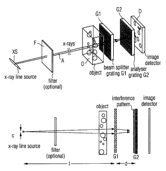
The basis of the present invention is the two-grating interfer-
ometer as it is shown in figure 1. It comprises the following
components: an x-ray source XS (e.g. a stationary or rotating
anode with a typical electron energy in the range of 10 to 100
key, or any other X-ray source), an optional set of appropriate
filters F, an object 0, a beam splitter phase grating Gl, an
analyzer amplitude grating G2, and a spatially resolving x-ray
detector D. The source XS provides some degree of spatial coher-
ence at least in one direction perpendicular to the optical axis
A. If the source is an X-ray tube, then the anode material
CA 02610934 2007-12-04
WO 2006/131235 PCT/EP2006/005119
9
should be chosen to have appropriate lines in the energy range
well suited for the specific application. For mammography these
could be Nb, Mo, Rh, Pd, Ag, which have K-emission lines in the
15 to 25-key region. For other applications with thinner or less
absorbing samples (e.g. desktop tomography setups) the energy
would be in the range of 5-10 key (e.g. a Cu anode). The spec-
trum of the source XS may be filtered to reduce the source band-
width. The coherence requirements are defined in more detail
further below.
Regarding the image formation process the beam splitter grating
G1 with a period /0/ should be constructed such that it divides
the incoming beam essentially into the two first diffraction or-
ders. The conditions for this are that the grating lines have
little absorption and shift the phase of the radiation passing
through the grating lines by an odd multiple of n, and that the
width of the phase-shifting structures is close to half of the
grating period, i.e., that the grating has a duty cycle close to
0.5. Since the wavelength of the illuminating hard x-rays is on
the order of 10-1 ...10-11m, and thus much smaller than the grating
pitch (10-5...10-6 m), the angle between the two diffracted beams is
so small that they overlap almost completely. In the overlap re-
gion downstream of the beam splitter grating Gl, the diffracted
beams interfere and form linear periodic fringe patterns in
planes perpendicular to the optical axis A at a distance d down-
stream of G1. For a spherical incident wave, i.e. for radiation
coming from a source at a distance / the period of the interfe-
rence pattern is equal to
1 (d + 1)
P2 = 2 Pi (1)
If d 1, which is usually the case, the period of the interfe-
rence fringes is about half of the period of Gl. Neither the pe-
riod nor the lateral position of these fringes depends on the
wavelength of the radiation used. Perturbations of the incident .
wave front, such as those induced by refraction on a phase ob-
ject in the beam, lead to local displacement of the fringes. A
fundamental idea for the operation of the grating-based interfe-
rometer described here is to detect the positions of the fringes
and determine from these the shape of the wave front. However,
CA 02610934 2007-12-04
WO 2006/131235 PCT/EP2006/005119
since the periodicity of the phase grating (and thus the spacing
of the interference fringes) does not exceed a few microns, an
area detector placed in the detection plane will generally not
have sufficient resolution to resolve the fringes, let alone the
5 exact position of their maxima. Therefore, an analyzer grating
G2 with absorbing lines and the same periodicity p2 and orienta-
tion as the fringes is placed in the detection plane, immedi-
ately before the detector. The transmission of the absorber li-
nes should be as low as possible, and the width of the absorber
10 lines should be close to the width of the gaps in between them
(duty cycle close to 0.5) to give optimum performance. This ana-
lyzer grating G2 acts as a transmission mask for the detector D
and transforms local fringe position into signal intensity
variation. The detected intensity signal contains quantitative
information about the phase shift induced by the object 0. At
least three methods can be applied to extract this phase infor-
mation:
1) Simplest method - When the analyzer grating G2 is placed into
the interference fringe pattern of the empty set-up (meaning the
case without sample) with exactly the same orientation and pe-
riodicity of the grating lines and the interference fringes,
then the transmission of the transmitted intensity depends on
the relative position of fringes and grating lines in the direc-
tion perpendicular to the grating lines. In the case when the
maxima of the interference fringes coincide with the absorbing
grating lines, the transmitted intensity reaches a minimum whe-
reas it will reach a maximum when the maxima of the interference
fringes coincide with the transmitting gaps between the absor-
bing lines. If this position is chosen such that the transmitted
intensity is between these extreme values, and ideally close to
the average between the maximum and minimum transmission, then a
local distortion of the interference fringes will cause a change
in the local transmission proportional to fringe distortion. A
phase object introduced upstream of the gratings will cause a
distortion of the transmitted wave front that in turn will cause
a distortion of the interference fringe pattern. It can be
shown, that this distortion is proportional to the derivative of
the phase shift induced by the object 0. The recorded image is
therefore a differential phase contrast image. If required, a
CA 02610934 2007-12-04
WO 2006/131235 PCT/EP2006/005119
11
phase contrast image can be obtained by integration of the dif-
ferential phase contrast image in the direction perpendicular to
the grating lines. The disadvantages of the methods mentioned
above are the following: i) the recorded image contains possible
absorption contrast and contrast from diffraction of edges in-
side the object 0. Both are difficult to distinguish from the
(differential) phase contrast contribution. ii) The method re-
quires an exact alignment of the interference pattern with re-
spect to the analyzer grating lines over the whole field of
view. This may be difficult to obtain especially for large
fields of view, and even impossible when either the interference
fringes or the grating lines are distorted due to mechanical im-
perfections of the set-up or fabrication errors of the gratings.
2) Moire interferometry - In this mode, the beam-splitter grat-
ing G1 and the analyzer grating G2 are not aligned with their
lines perfectly parallel to each others', but rather with a de-
liberate, small rotation angle a of the two gratings with re-
spect to one another, about the optical axis. This results in a
set of moire fringes as shown in Fig. 2. For an incident plane
wave (Fig. 2a), the moire fringes have a spacing
Pm = .1:12 / a Go
and are perpendicular to the lines of the two gratings (Fig.
2b). For an inclined wave that is still plane, the fringes are
displaced with respect to the plane wave parallel to the optical
axis (Fig. 2c, d). For a converging or diverging (Fig. 2e) wave,
the moire fringes are inclined (Fig. 2f). From the local positi-
ons of the moire-fringe maxima or from the local moire-fringe
inclination, the derivative of the wave-front phase profile can
be obtained relatively simply. The relationship between the lat-
eral moire-fringe displacement 837 and the wavefront phase 0 is
acb Ly
¨ = a- (3).
ax
Standard fringe-analysis methods and software can be used to ob-
CA 02610934 2007-12-04
WO 2006/131235 PCT/EP2006/005119
12
tam n the moire-fringe positions .837. Figure 3 shows an example of
experimental data and a preliminary analysis. The choice of the
inclination angle a of the two gratings is determined by a tra-
deoff between spatial resolution and sensitivity. The spatial
resolution in the direction normal to the moire fringes cannot
be much better than one moire-fringe period pm. Since pm decrea-
ses with increasing a (Eq. 2), stronger tilt means better spati-
al resolution. But Eq. 3 shows that the sensitivity decreases
with increasing tilt, i.e., for a given phase gradient the moire
fringes are less displaced for stronger a. The main advantage of
moire operation of the interferometer is that pure phase infor-
mation, without any unwanted absorption contrast, can be obtai-
ned from a single image. The main disadvantage is that the reso-
lution in the direction parallel to the grating lines (normal to
the moire fringes) is at least one order of magnitude poorer
than the pixel size of the detector.
3) Phase stepping - A third method that avoids the disadvantages
of the approaches described above is the following: to separate
the phase information from other contributions to the signal,
such as absorption in the sample, inhomogeneous illumination or
imperfections of the gratings, the phase-stepping approach used
in visible-light interferometry was adapted to this setup. It is
illustrated in Figure 4: one of the gratings is scanned along
the transverse direction perpendicular to the grating lines over
one period of the grating, and for every point of the scan an
image is taken (Figure 4a to d). The intensity signal I(x,y) in
each pixel (x,y) in the detector plane oscillates as a function
of xg (Figure 4e). The phases of the intensity oscillations in
each pixel, 9(x,y) (Figure 4f) are related to the wavefront pha-
se profile 0(x,y) by
49(x, = Ad acto(x, y) . (4)
P2 aX
p contains no other contributions, particularly no absorption
contrast. The phase profile of the object can thus be retrieved
from p(x,y) by a simple one-dimensional integration, as shown in
CA 02610934 2007-12-04
WO 2006/131235 PCT/EP2006/005119
13
Figure 4g. In the general case where the wavefront impinging on
the object 0 already shows some distortion, the background phase
distribution Obacax,Y). should be measured with the object 0 re-
moved from the beam and then subtracted. Even in cases where the
range of phase values exceeds 2n by far, such as in the example
in Figure 4g, phase unwrapping is generally not necessary becau-
se the measured quantity cp, essentially the first derivative of
0 (Eq. 4), will not exceed n as long as the phase gradients in
the sample are not too steep.
Another quantity contained in the information obtained by a
phase-stepping scan, the average signal for each pixel over an
entire oscillation, a(x,y) (Figure 4h), is identical to the
transmission radiography signal as it would be measured in a ra-
diograph taken without the interferometer. It contains the pro-
jected absorption coefficient and, depending on experimental ge-
ometry and detector resolution, edge-enhancing Fresnel diffrac-
tion contrast. A single phase-stepping scan thus yields both the
phase and the absorption image. The full resolution of the de-
tection system can be used in both images. The main disadvantage
is the fact, that several images have to be acquired to obtain a
phase image. This however does not necessarily mean, that the
applied x-ray dose or exposure time has to be increased.
Figure 5 shows processed radiographs a(x,y) and co(x,y) obtained
by phase stepping, the integrated phase shift 0(x,y), as well as
reconstructed tomograms of both the absorption and the phase
signal and a three-dimensional rendering of the tomographically
reconstructed refractive index of the sample, a small spider.
The edge-enhanced absorption image a(x,y) shows strong contrast
for fine details both in projection (fig. 5a) and tomogram (fig.
5d). But low spatial frequencies of the phase distribution are
lost, so that image segmentation, i.e., the assignment of each
pixel or voxel to one of the constituent materials of the sam-
ple, is not easily possible. The phase tomogram (fig. 5f) exhi-
bits a clear advantage: its pixel values are a direct measure of
the decrement of refractive index 6 in the sample, and are the-
refore directly accessible to quantitative analysis. Moreover
the edge enhancing inline Fresnel diffraction contrast in Figure
5a and d would not be observable at lower resolutions of the de-
CA 02610934 2007-12-04
WO 2006/131235 PCT/EP2006/005119
14
tector, whereas the interferometric phase contrast is not sub-
ject to such a limitation. This is of crucial importance when
the interferometric method is scaled up to large fields of view,
as the limited number of pixels will inherently limit the spa-
tial resolution.
It should be mentioned that there are several possibilities to
perform the phase stepping: the most obvious is to move either
the grating G1 according to Fig. 6a or the grating G2 according
to Fig. 6b. The disadvantage is that the movement has to be done
with sub-micron accuracy and that the exact parallel orientation
of the lines of both gratings may be lost, especially when large
gratings are used to provide a large field of view. Instead it
is also possible to rotate the grating G1 and the grating G2 to-
gether around an axis oriented along the direction of the grat-
ing lines by an angle a as indicated in Fig. 6c. This again al-
lows to keep the gratings G1 and G2 in an aligned position with
respect to each other or even to mechanically fix both gratings
together. The most advantageous possibility is to move the sour-
ce in the direction perpendicular to the optical axis and the
grating lines as shown in Fig. 6d. This may be easier as the mo-
vement is coarser and has to be performed with 1/d times less
precision as compared to the first two solutions a and b. Again
it allows to keep the gratings G1 and G2 in an aligned position
and to mechanically fix them so that the alignment cannot be not
lost during the phase stepping movement.
The source can be moved either mechanically or by deflecting the
electron beam of the x-ray source to move the position of the
electron impact on the anode. In case of the usual geometry of
an x-ray tube where the x-rays are extracted under a shallow
angle from the anode surface, the movement of the source point
also has a larger component along the optical axis. Here only
the component perpendicular to the optical axis has an effect on
the phase stepping. The total deflection of the beam on the ano-
de surface is in this case larger reducing the requirements on
the accuracy of the electron beam movement.
CA 02610934 2007-12-04
WO 2006/131235 PCT/EP2006/005119
As mentioned above and illustrated in Fig. 4, a phase-stepping
scan yields, for each pixel in the detector, a series of inten-
sity values from which different quantities can be extracted,
especially the phase clo of the intensity oscillation in each pi-
5 xel, which is related to the derivative of the wavefront phase 0
by eq. (2), and the average intensity a over an oscillation pe-
riod, which essentially corresponds to the non-interferometric
X-ray image. Hereinafter it is discussed how many phase steps
need to be made in one phase-stepping scan, and how the phase-
10 stepping scan is analyzed to extract 9.
The range of the phase stepping scan should cover (at least) one
period of the oscillation. In conventional phase-stepping inter-
ferometry (i.e. with visible light), the optical components are
15 usually designed such that the contrast curve (as shown, e.g.,
in Fig. 4e) is sinusoidal. In that case it is sufficient to take
three points in a scan. The tangent of the phase 9 of the oscil-
lation is then easily calculated by just taking the ratio of
signal differences.
The situation in the X-ray interferometer differs from the con-
ventional one in that the shape of the contrast curve is not si-
nusoidal. (For the ideal case of box-profile gratings and fully
coherent illumination, it would, for example, be triangular.)
There are at least two ways to cope with this problem:
1. Perform phase-stepping scans with a larger number of points
per oscillation period. To analyze the phase-stepping scan
signal for each pixel, its Fourier transform can be calcu-
lated. The phase of the first component of the Fourier
transform represents the oscillation phase 9 (wrapped into
a half-open interval of width n). The minimum number of
points in the phase-stepping scan for this type of analysis
should be the number of the highest Fourier component of
non-negligible intensity, plus 2.
2. In a calibration measurement without any sample, measure
the contrast curve (in each pixel) by taking a phase-
CA 02610934 2007-12-04
WO 2006/131235 PCT/EP2006/005119
16
stepping scan with very fine steps. In imaging of samples,
phase-stepping scans can be performed with few points in
the scan, and the phase p determined by using the calibra-
ted contrast curve, for example with a fitting procedure.
When comparing the two methods, it should be noted that the
first method has the drawback of more necessary points per
phase-stepping scan, but the advantage that higher Fourier com-
ponents can potentially be used to extract additional informa-
tion.
The distance d between the two gratings can be chosen freely,
although there are values of d for which the contrast is best
and others for which it has a minimum (see below). As can be
seen from equation (2), the sensitivity of the method increases
linearly with d. The choice of larger d may result in a loss of
spatial resolution due to penumbral blurring, as the distance
between the object and the detector is increased. Moreover a
higher degree of lateral coherence is required (see below).
When choosing the inter-grating distance d, the contrast of the
interference fringes changes periodically as a function of d.
For a plane incoming wave and a pure phase grating of pitch Pi
whose lines shift the phase by n, the contrast is strongest for
odd multiples of d = m2 / (8A) and vanishes for even multiples
of d = pi2 / (8A), - a phenomenon related to the Talbot self-
imaging effect. In the following, one refers to any distance di, =
n RI2 / (8A) as the "n-th fractional Talbot distance" and to the
planes at a distance di, from the phase grating as the "n-th
fractional Talbot plane". d should be set to values giving maxi-
mum contrast, i.e. to an odd fractional Talbot distance (n = 1,
3, 5, _). For a spherical incoming wave instead of a plane wave,
the expression above for the fractional Talbot distances has to
be modified to
/ = dn = 1 = n = pi2 I (8 A)
(5) =
d n ,sph. =
1 d n 1 - n = p12 48/1)
CA 02610934 2007-12-04
WO 2006/131235 PCT/EP2006/005119
17
where 1 denotes the distance between the source and G1. For /
an,, the difference between dn and dri, spii. is small.
As already mentioned, neither the period nor the lateral posi-
tion of these fringes depends on the wavelength of the radiation
used. In this sense the described setup is achromatic allowing
for the use of broadband radiation from an x-ray tube without
the need of monochromators that would only cut out a small frac-
tion of the used radiation and would therefore not be photon ef-
ficient.
However, two aspects of the proposed set-up do depend on the
photon energy. Firstly, the phase shift of the lines of the
grating G1 depends on the photon energy due to the dispersion of
the grating material. The condition to induce a phase shift e-
qual to odd multiples of n is not strict, and a radiation within
a certain energy band in the order of 10% around the design e-
nergy can be accepted. A deviation from n only affects the
contrast - but it does not change the interference pattern qua-
litatively. Secondly, the position of the Talbot planes depends
on the photon energy. But again the condition that the grating
G2 has to be placed in a Talbot plane is also not very strict,
allowing the use of radiation within a certain energy band in
the order of 10% for the grating G2 placed in the first Talbot
plane. This condition becomes stricter for higher grating di-
stances.
It should be mentioned that also the refractive index of the ob-
ject depends on the photon energy used. Nevertheless, re-
constructed phase projections and tomograms will still be quan-
titative under the condition that the elements contained in the
sample have no absorption edges in the range of the used photon
energy band. In this case, all parts of the sample have the same
dispersion relation, i.e. the real part of the refractive index
changes with E-2.
CA 02610934 2007-12-04
WO 2006/131235 PCT/EP2006/005119
18
The considerations of the previous sections show that the phase
shift induced by the structures of the grating G2 should be
close to odd multiples of n and that the distance between the
grating G1 and the grating G2 should be close to an odd frac-
tional Talbot distance d, sph . Both, phase shift and fractional
Talbot distance, depend on the wavelength of the radiation used.
It is therefore important that the grating structures and the
distance chosen between the grating G1 and the grating G2 are
matched to each other, meaning that both fulfill the conditions
for the same wavelength. Moreover, it might be useful if this
wavelength coincides with an emission line of the anode material
of the used x-ray source. As mentioned above, the acceptable
bandwidth is wide compared to other interferometric methods. In
particular, it is wide enough to accept a series of emission
lines (e.g. the Cu-K(1, Cu-K0 and Cu-K0 - lines emitting at
8.048, 8.028 and 8.905 key photon energy, or the Mo-K(1, Mo-K0
and Mo-K0 - lines emitting at 17.479, 17.374 and 19.608 key pho-
ton energy, or the Ag-K(1, Ag-K(2 and Ag-K - lines emitting at
22.163, 21.990 and 24.942 key photon energy).
The setup described requires spatial coherence only in the di-
rection perpendicular to the optical axis A and to the grating
lines. In this direction the minimum required coherence length t
-,
is
A
t = 4d = ¨ (6) .
A
If the grating G2 is placed in the first fractional Talbot
plane, then t=p1/2. For grating periods in the range of several
microns, t is therefore in the order of one micron. As described
before, the size of a source emitting at wavelength A = 0.1 nm
and placed at a distance / = 1 m from the beam-splitter grating
G1 should be smaller than 0.1 mm along the direction perpendicu-
lar to the grating lines. The use of a smaller source will inc-
rease the spatial coherence, resulting in an improvement of the
phase contrast. As there are no limitations in terms of coher-
ence in the direction along the grating lines, a line source can
be used.
CA 02610934 2007-12-04
WO 2006/131235 PCT/EP2006/005119
19
In medical x-ray imaging, where samples with thicknesses of many
cm are viewed, scattering of x-rays in the sample generates dif-
fuse background intensity on the detector that reduces the ob-
tained contrast and image quality. Therefore, usually, an array
of collimating absorbers, called an anti-scatter grid, is moun-
ted in front of the detector to block radiation that has been
deflected by a significant angle. This grid can be either a two-
dimensional array of absorbing structures or a one-dimensional
array. In the grating interferometer setup described here, the
grating G2 comprises structures that are 5 to 10 microns thick -
depending on the photon energy used -to provide sufficient x-ray
absorption and that have, at the same time, widths on the order
of one micron. The aspect ratio of the structures is therefore
usually much greater than unity. The grating G2 can therefore
also act as an anti-scatter grid. Likewise, in principle, the
anti-scatter grid of an x-ray imaging system may be used as the
analyzer in a grating based interferometer, although this would
require that the period of the anti-scatter grid be smaller than
is the case in commonly available systems.
In many X-ray imaging applications that require a field of view
of many centimeters (such as in medical diagnostics) the diver-
gence angle of the x-ray beam is considerable, as the source
cannot be placed at distances too far from the detector. Due to
the high aspect ratio of the grating structures in the interfer-
ometer, especially of the grating G2, the beams in certain re-
gions close to the edges of the field of view may pass through
the gratings under too large angles. Only the angular component
within the drawing of the lower part of figure 1 has an effect.
To completely avoid this problem, the grating structures have to
be bent or curved as depicted in Fig. 7a to d, or a scanning
scheme with appropriate orientation of the gratings has to be
applied. To cope with this problem that the rays far from the
center of the beam cone or fan will be obstructed by the lines
of a flat deep grating as shown in Fig. 7a) different solutions
can be applied. Fig. 7b) illustrated a solution by bending the
grating lines on a flat substrate. Fig. 7c) schematically de-
picts the grating lines being placed on a curved surface. Fi-
CA 02610934 2007-12-04
WO 2006/131235
PCT/EP2006/005119
20
=
nally, Fig. 7d) shows a way that segments of a flat grating ap-
proximating the curved or bent geometry.
Besides full-field radiography systems, in which a two-
dimensional X-ray detector is used to obtain projection images
of a sample, scanning systems are also applied in medical X-ray
imaging. As illustrated in Figure 8, a slit S placed between the
source XS and the sample 0 generates a fan-shaped beam, and a
one-dimensional line detector D1 can be used, with its elements
aligned to the fan. The two dimensional projection of the sample
0 is obtained by either scanning the sample through the fan-
beam, or by scanning the beam across the sample and moving the
line detector D1 with the beam. The detector D1 is read out at
different positions during the scan and a two-dimensional image
is created line by line.
Although the mechanical setup is more complicated, the scanning
scheme has some important advantages compared to a two-
dimensional setup: i) a suppression of diffuse scattering with-
out an anti-scatter grid, since the slit S, together with the
limited size of the source spot, acts as a collimator, ii) lower
cost and higher detection efficiency of one-dimensional detec-
tors in comparison with two-dimensional detectors. The grating
based phase contrast can also be applied to a scanning scheme,
as shown in Figure 8. The advantage is that only gratings over a
smaller area are required compared to a full-field imaging sche-
me, which will significantly reduce the cost. Two possible ori-
entations of the grating lines with respect to the fan beam are
shown, either with the grating structures along the slit direc-
tion (above) or with the grating lines perpendicular to the slit
direction (below). The arrangement show in the upper part of Fi-
gure 8 has the advantage that it avoids the cone beam problem
(see previous section).
Figure 9 shows x-ray images of a test sample containing poly-
methylmethacrylate (PMMA, 1.5 mm diameter) and Aluminum (1.0 mm
diameter) spheres. The images were recorded using a standard,
sealed x-ray tube source with a Mo anode operated in line focus
CA 02610934 2007-12-04
WO 2006/131235 PCT/EP2006/005119
21
mode (8 mm x 0.04 mm) at an electron energy of 40 key and an an-
ode current of 25 mA, placed at a distance / = 1.7 m from the
beam splitter phase grating G1. For the Mo-K(emission lines, the
coherence length along the smaller source dimension (c = 0.04
mm) is t= A. 1/C 3 pm at the position of Gl. The two gratings
G1 and G2 had periods of m = 3.94 pm and m.2.0 pm. They were
spaced by the first fractional Talbot distance, d1 = 28.4 mm.
Apart from the 500-pm-thick silicon substrates supporting the
gratings, no additional filter was inserted in the x-ray beam.
This means that the whole Mo K-emission series and large parts
of the bremsstrahlung spectrum contributed to image formation.
The detector was a fiber-coupled CCD (model "Hystar", manufac-
turer Photonic Science) with an effective pixel size of approxi-
mately 30 pm. The data were acquired by phase stepping with 17
steps over 1 oscillation period, with an exposure time of 50 s
for each CCD frame.
In the conventional amplitude contrast image, the aluminum
spheres are clearly visible (Figure 9a). The absorption coeffi-
cient of the plastic spheres is about 20 times lower than that
of Al. In the phase gradient and integrated-phase images (Figure
9b,c) the contrast for the plastic spheres is strongly in-
creased.
Due to the requirements of spatial coherence described above,
the choice of the source geometry is limited to relatively nar-
row line sources placed at large distances from the sample. This
results in low x-ray flux density and thus in long exposure ti-
mes. The following section describes a setup that can record
phase contrast images with much higher flux density. The schema-
tic setup of the proposed design is shown in Figure 10. The dif-
ference to the previously described interferometer shown in Fi-
gure 1 is the x-ray source that consists of an array of N line
sources. The line sources are oriented along the grating lines
and the width of each source line is chosen narrow enough to
provide sufficient spatial coherence in the direction perpendi-
cular to the grating lines. Thus, each of the line sources is
intrinsically coherent, whereas the lines can be mutually inco-
CA 02610934 2007-12-04
WO 2006/131235 PCT/EP2006/005119
22
herent. The spacing of neighboring source lines, i.e. the period
Po of the source array is chosen such that the shift of the in-
terference patterns created by two neighboring virtual line
sources along the direction perpendicular to the grating lines
is exactly p2. This condition is fulfilled for:
Po = P2X = (7)
The relationship between the periods Po, PI, and p2 of the source
array, the beam-splitter grating and the analyzer grating is
P2
pl= 2P0. 00
Po +1;12
Another useful relationship is that for operation in the n-th
fractional Talbot distance, the distance / between the source
array and the beam-splitter grating is
n po2p2
l= ---x _________ , (9)
22 po+p2
which is particularly useful when solved for po:
2
p0=-111 +.11(¨ (10)
111)+-2111.
N/2 N32
The arrangement also works for source array periods that are in-
teger multiples of Po. The size c of the source array can be much
bigger than the width s of an individual line source. The duty
cycle DC = S / Po of the source array determines the degree of
transverse coherence in the direction perpendicular to the in-
terferometer grating lines. The interference patterns of all N
virtual line sources add up incoherently (as the virtual line
sources are mutually incoherent) but preserving contrast.
Therefore, the flux density is increased by a factor N compared
to a single line source. It is also useful to note that the se-
tup results in a decoupling of the resolution limit due to pe-
numbral blurring, 5pen = c d /, from the coherence requirement
in equation (6), which, with t = A 1 / s, becomes s < /pi / (4d).
For plane-wave case, where p2 = pi/2, it is obvious from Equation
CA 02610934 2007-12-04
WO 2006/131235 PCT/EP2006/005119
23
(7) that this results in a maximum duty cycle of DC = 0.5, bey-
ond which contrast will be limited by spatial coherence.
A source array as described above can be obtained in various
ways:
1) One can generate an array of electron line foci on an anode
surface.
2) One can use an anode surface that is structured either to-
pographically or that consists of regions of different materi-
als.
3) It is also possible to generate a single line focus or spot
focus that is scanned across the anode surface to produce - av-
eraged over time - an array of lines. One advantage of such a
scheme in comparison to keeping the single spot on the same pla-
is that, due to improved heat dissipation, more power can be
deposited without damaging the anode. Another advantage is that
the period of the source array can be tuned via the electron
beam deflection to match Eq. (7) for different values of l/d.
Moreover the beam deflection could be used to shift the source
array across the anode to perform phase stepping.
4) The simplest method is to put an array of slits, i.e. an amp-
litude grating in front of a large x-ray source to generate an
array of virtual line sources. A schematic view of such a set-up
is shown in figure 11. Here the open fraction, or duty cycle, DC
of the source grating determines the degree of spatial coheren-
ce. For smaller values of DC, the contrast of the phase images
is improved at the cost of reduced flux density. As an example a
typical set of parameters is calculated: For a wavelength of A. =
0.1 nm, a period pi = 4 pm, and choosing the first fractional
Talbot distance for the distance between G1 and G2 we obtain dn
P/2/(8A) = 20 mm. For a distance / = 1 m between source and beam-
splitter grating G1, we obtain p2 = 2.04 pm and po= 102 pm. If
the source size is 1 mm x 1 mm, the slit array generates about N
= 10 virtual line sources, which results in a flux density 10
times higher than in a setup with a single, unstructured line
source of 0.1 x 1 mm.
CA 02610934 2007-12-04
WO 2006/131235 PCT/EP2006/005119
24
The considerations described in the previous sections in context
with the two grating interferometer with a single source are al-
so valid for the setup with an arrayed source. The possibility
to perform a phase stepping scan by moving the source has to be
generalized in this case: it is the entire array of sources that
should be moved for phase stepping. When an array of slits is
used to create the source array, then that slit-array aperture
should be moved together with the source, or it may even be
moved alone, with the source remaining fixed in space.
Figure 12 shows images obtained under the similar conditions as
those shown in figure 9, i.e., with a laboratory X-ray source.
The important difference is that this time a source with a size
of 0.8 mm x 0.4 mm was used, which is far to large to provide
sufficient transverse coherence for interferometric phase
contrast. By mounting an additional source grating with a period
of p0=127 pm and a duty cycle of approximately DC = 0.25, an ar-
ray of virtual line sources with sufficient transverse coherence
was created.
In this section, configurations particularly suited for phase
contrast x-ray imaging are listed in detail. They are designed
for two different applications: desktop microtomography and mam-
mography. The latter can be implemented either with a full-field
setup or a scanning setup.
A grating based interferometer for phase contrast desktop tomo-
graphy system may consist of the following components:
= An X-ray source of either the rotating-anode or the sealed-
tube type with emission lines in the range between 5 and 10
key. Suitable anode materials include Ti, V, Cr, Mn, Fe,
Co, Ni, Cu, and Ge. In this example a design wavelength was
chosen to X = 0.154 run, which corresponds to the Cu-1c,, emis-
sion line, at an energy of 8.0 key. Both of the K lines (Cu
Kai at X = 0.1540 nm, Cu Kea at X = 0.1544 nm) can be used
simultaneously in an efficient way.
CA 02610934 2007-12-04
WO 2006/131235 PCT/EP2006/005119
= A two-dimensional digital x-ray detector, placed at a di-
stance of 0.3 - 1.5 m, typically 0.8 m from the source. The
number of pixels of the detector should be between 500x500
and 4000x4000, typically 2000x2000. The field of view of
5 the detector should be between 5 and 30 mm, typically 10
mm. The pixel size of the detector is thus typically 5 pm x
5 pm.
= The center of the detector and the center of the source de-
fine the optical axis. The optical axis can be in any di-
10 rection, but a horizontal axis is most advantageous.
= A rotatable sample holder with the axis of rotation perpen-
dicular to the optical axis is placed between the source
and the detector so that a sample can be mounted with its
center of gravity close to the optical axis. The distance
15 between the sample and the source is called a, The distance
between the sample and the detector is called b.
= The phase grating and the absorption grating are placed di-
rectly in front of the detector. The period m of the analy-
zer grating should be chosen smaller than the detector pi-
20 xel size, so that the spatial resolution of the detector
can be fully used. For a pixel size of 5 pm, the pitch P2
should therefore be less than 3 or 4 pm, typically p2 = 1
pm. In this case, and with A = 0.154 nm and 1 = 0.8 m, the
period of the phase grating should be pi = 1.992 pm (Equati-
25 ons 8 and 10) for operation in the first fractional Talbot
distance, i.e., at an inter-grating distance d = 3.23 mm
(Equation 5). If silicon is used as the material for the
beam-splitter grating, then a grating depth of 10.2 pm is
required to obtain a phase shift of n. The intensity trans-
mission of 10.2 pm of Si is 86.6 %, so that the transmissi-
on of an Si grating with that thickness and a duty cycle of
0.5 would be 93.3 %. If poly-methylmethacrylate (PMMA) is
used as the material for the beam-splitter grating, then
the ideal grating depth is 18.7 pm. The intensity transmis-
sion of such a grating would be 99.3 % (for a duty cycle of
0.5). The use of other polymers would result in similar fi-
gures. For the absorption grating, if gold is used as the
material, a thickness of 6 pm would ensure an absorption of
CA 02610934 2007-12-04
WO 2006/131235 PCT/EP2006/005119
26
more than 90 %, and 12 pm thickness would result in more
than 99 % of absorption in the grating lines.
= An arrayed source, technically realized, e.g., by slit-
array aperture. In this case, the slit-array period should
be Po = 0.248 mm (obtained from Equation 10), max. size of
array (in direction perpendicular to slits) c = 1.24 mm,
given by the condition that penumbral blurring should not
exceed the pixel size, max. gap size of each slit 0.124 mm
(i.e., DC=0.5), given by coherence requirement.
A grating based interferometer for a phase contrast full-field
digital mammography (FFDM) system could consist of the following
components:
= An x-ray source of either the rotating anode or a sealed
tube type with an anode with emission lines in the 15-25
key region. Anode materials could be Nb, Mo, Rh, Pd, or Ag.
In this example we choose a design wavelength A = 0.056 nm
corresponding to the Ag-K(emission at 22 key energy (Ag Kcy1
at A = 0.0559 nm, Ag Kea at A = 0.0564 nm). The design of
the entire apparatus can be based on that of common (ab-
sorption-contrast) FFDM systems, with the source array,
beam splitter and analyzer as new, different elements.
= A filter of Pd or Rh or another material with an x-ray ab-
sorption edge in the 23-30 key energy range and a thickness
between 20 and 100 pm, typically 50 pm may be used to sup-
press unwanted parts of the spectrum.
= A gantry with compression paddle as commonly used in mam-
mography screening systems comprises a two-dimensional
digital flat-panel x-ray detector, placed at a distance of
0.5-1.0 m from the source, just below (i.e., downstream of)
the gantry. Here 1.0 m as the distance was used. The field
of view of the detector should be not much smaller than
(200 mm)2', preferably around 240 x 300 mm2 (i.e., the size
of the largest commonly used film cassette in conventional
mammography). The pixel size of the detector is typically
between 50 and 100 pm in each dimension, so that the detec-
CA 02610934 2007-12-04
WO 2006/131235 PCT/EP2006/005119
27
tor has between (2500)2 and (5000)2 pixels. (These are the
characteristics of most commercially available detectors
for FFDM.) Here, a pixel size of 70 pm is assumed.
= A suitable set of parameters for the grating periods, sour-
ce array dimensions, and distances is p2 = 3 pm (period of
the analyzer grating), A. = 8.4 pm (period of the phase gra-
ting), Po = 26.6 pm (spacing of the virtual line sources).
With a distance / = 1.0 m between the source array and the
phase grating, the first fractional Talbot distance is d1 =
74.8 mm. The total size of the source array should, with
these parameters, not exceed 0.4 mm, i.e., the maximum num-
ber of virtual line sources is 15. For the phase grating,
if nickel is used as the material, the thickness required
to achieve a phase shift of n is 7.7 pm, resulting in an
average intensity transmission of 92.6 % (for a duty cycle
of 0.5). The analyzer grating, if made of gold, should have
a minimum thickness of 20 pm, which yields absorption of 90
% in the grating lines.
In a slot-scanning mammography setup (possibly based on existing
non-interferometric slot-scanning systems), the source material,
distances and grating periods can be chosen similar or identical
to the FFDM example above. The following items would be diffe-
rent or additional with respect to the FFDM setup:
= A first collimator (one slit or a sequence of slits), made
of a strongly absorbing material, should be mounted between
the source and the object position. Together with the spots
of an arrayed virtual source, this collimator defines a
fan-shaped beam.
= A second collimator slit, made of a strongly absorbing ma-
terial, should be mounted just above (i.e., upstream of)
the interferometer gratings.
= The detector can be a line-array detector, for example a
direct-detection detector. The detector elements should ha-
ve non-square shape, with a size between 25 and 100 pm
(corresponding to the desired resolution) along the line of
detector elements and several millimeters (corresponding to
the width of the collimator slits) in the other dimension.
CA 02610934 2007-12-04
WO 2006/131235 PCT/EP2006/005119
28
= The collimator slits, the interferometer gratings and the
detector should be parallel to each other. The lines of the
interferometer gratings should be parallel to the fan-
opening plane.
= The collimators and the detector can be rotated around the
source array, perpendicular to the fan-opening plane, over
an angular range large enough to cover one dimension of the
field of view mentioned in the FFDM setup. For the acquisi-
tion of a mammogram, these components will be scanned to-
gether.
All of the considerations above can be applied to an interfer-
ometer that resembles the one presented, with the only differ-
ence that the beam-splitter grating G1 is not a phase grating,
but an absorption grating, i.e., its lines strongly absorb X
rays. The use of an absorption grating as the beam splitter is
less advantageous than that of a phase grating because even a
perfect absorption grating (completely opaque lines, duty cycle
exactly 0.5) will only deliver 20 % of the incoming intensity
into the negative and positive first orders. 50 % of the inci-
dent power are absorbed in the grating, and one-half of the re-
maining 50 % go into the zeroth order, or undiffracted portion
of the beam, contributing to background signal and/or radiation
dose deposited in the object or patient. A phase grating is four
times more efficient. Nonetheless, for a number of applications
where these drawbacks can be tolerated and/or where a phase gra-
ting is not available, the use of an absorption grating as beam
splitter can make sense.
In this case, all of the considerations for interferometer de-
sign laid out above remain valid with the following modificati-
ons:
The relation between the periods of G1 and G2 is different. E-
quation 1 (section 3.1) becomes
(c1+1)
P2 =P1 )
1
The Talbot distances at which the contrast is best (section 3.6)
are a,' = n pi2 / A for a plane illuminating wave and
CA 02610934 2007-12-04
WO 2006/131235 PCT/EP2006/005119
29
/ = d' 1=n=p1212
dflSPh =' - __________ " (5')
1-d 'n 1-n.p,212
for a spherical wave, which replaces Equation (5).
The relationship between Po Pi, and A (Equation 8) changes to
PoP2
p1= . (8').
Po + P2