Note: Descriptions are shown in the official language in which they were submitted.
CA 02611211 2007-12-06
WO 2006/134325 PCT/GB2006/002103
1
FIXATOR
This invention relates to the field of external bone fixators for use in the
treatmenfi of
fractured bones.
BACKGROUND
Bone is capable of self-healing at a fracture site by the formation of callus
which is able
to reunite the ends of the fractured bone. Callus formation is triggered and
maintained
by relative movement of the fractured bone ends and occurs during a specific
and limited
time period following occurrence of the fracture.
If allowed to heal completely naturally, a fractured bone would heal in a
poorly aligned
condition, resulting in consequential future problems. Therefore the fractured
bone ends
are more usually manipulated into a well-aligned condition (fracture
reduction) before
callus formation and the natural healing process occurs. Once reduced, the
fracture
needs to be supported or fixed in order to maintain the desired alignment.
Rigid fixation of the fractured bone ends means that they are kept well
aligned but may
lead to a reduction or prevention of the formation of callus, the'rrefore
prolohging the
-natural healing process. - - Treatment of a bone fracture by providing
external support (e.g. a plaster of Paris cast)
allows relative movement of the fractured bone ends to occur, which promotes
callus
formation. However, such external supports may not be suitable to assist with
the need
to accurately align the fractured bone ends, particularly with unstable or
metastable
fractures.
To alleviate these. problems, external bone fixators have been developed which
hold the
fractured bone ends together sufficiently rigidly to maintain accurate
alignment and yet
at the same time allow sufficient relative movement between the fractured bone
ends to
promote callus formation. Such external fixators are applied externally to the
injured
limb and are attached to the fractured bone ends by bone pins or screws which
pass
through the soft tissue of the limb and into the bone.
CONFIRMATION COPY
CA 02611211 2007-12-06
WO 2006/134325 PCT/GB2006/002103
2
Bridging the gap between the pins in the two ends of the fractured bone is a
support
mechanism, which holds the bone ends in alignment. To promote callus
formation, the
external fixator can be adapted to allow specific and controlled types of
movement
between the fractured bone ends. Such movement is generally effected by a
corresponding movement in the fixator itself, e.g. relative axial movement
between two
component parts of the fixator can lead to corresponding relative axial
movement of the
fractured bone ends.
An example of an external fixator of this type is described in US 5,320,622
(Orthofix Sri).
Other examples of prior art fixators are described in EP1351613 (Mitkovic) and
EP1434531 (Langmaid et al).
The prior art fixators are relatively complex, having numerous component parts
and
joints which are necessary in order to ailow the fixator to provide selective
retention of a
wide variety of angular relations so that positional and angular adjustment
can readily be
applied to the fractured bone.
However, this complexity makes the fixator relatively expensive, heavy and
bulky; the
- latter two factors being particularly undesirable from the point of view of
a patient who
may need to wear the fixator for many weeks.
Furthermore, most surgeons reduce fractures by manipulation, apply bone screws
and
the external fixator and then manipulate the fracture again to improve the
reduction
before locking the fixator. This second stage of reduction often results in
the bone
screws in the respective ends of the fractured bone no longer being
longitudinally
aligned, so that the fixator has to be locked in a position which is no longer
in line with
the longitudinal axis of the bone, resulting in unpredictable mechanical
properties and
movement. Furthermore, a consequence of the need to design joints capable of
accommodating ail potential eventualities may result in a final configuration
of the device
that is mechanically unsound.
It is therefore an object of the present invention to provide an external
fixator which
seeks to alleviate the problems of the above-described prior art.
CA 02611211 2007-12-06
WO 2006/134325 PCT/GB2006/002103
3
SUMMARY OF THE INVENTION
According to a first aspect of the present invention there is provided a bone
fixator for
use in the treatment of a fractured bone comprising a support beam having
means for
attaching each end thereof to the respective ends of a fractured bone, the
support beam
being configured so as to permit predetermined relative movement between the
respective ends of the support beam and thereby,transmitting said relative
movement to
the respective ends of the fractured bone.
Preferably, the support beam is a one-piece support beam with no articulated
joints
therein. This simplifies the manufacture of the fixator and reduces the
complexity of its
operation.
Preferably, the means for attaching each erid of the support beam to the
respective ends
of a fractured bone comprise apertures for receiving therein bone pins, bone
screws,
wires or the like. The apertures can be very accurately positioned on the
fixator so as to
contribute to the predictable nature of the predetermined movement,
reproducible from
one fixator to another of the same type.
Preferably, the apertures can be used as location guides for the drilling of
holes for the
bone pins, bone screws, wires or the like.
Preferably, the fixator further comprises bone screw sleeves, each of which
locate in one
of said apertures, each bone screw sleeve having a bore therethrough for
receiving
therein bone pins, bone screws, wires or the like. If desired, one or more of
said bone
screw sleeves can have an angled bore therethrough in order to direction a
bone screw
to a particular location.
In a preferred form, at least one of said apertures can be used as a healing
indicator by
observing the possible positions of a bone pin or bone screw located therein
relative to
the periphery of said aperture.
Preferably said bone fixator is disposable i.e. for single-use. This is made
feasible by
the greatly simplified (and hence cheaper) construction of the fixator when
compared
with prior art fixators.
CA 02611211 2007-12-06
WO 2006/134325 PCT/GB2006/002103
4
In a preferred form, the predetermined relative movement between the
respective ends
of the support beam is a property of the material of the support beam.
Alternatively, or in addition, the predetermined relative movement between the
respective ends of the support beam is a property of the shape of the support
beam.
Preferably, said support beam is made from a titanium alloy. Alternatively,
said support
beam is made from a layered composite material.
In one embodiment, the support beam includes one or more actuators which, in
use,
generate or contribute to said predetermined movement.
Preferably, said fixator is adapted to receive three bone pins, bone screws,
wires or the
like at each end thereof.
Preferably, the fixator is adapted to support a hybridisation component such
as a T-bar
attachment or (ng attachment at one end thereof.
According to a second aspect of the invention, there is provided a bone
fixation system
comprising
a) a bone fixator as claimed in any of the preceding paragraphs; and
b) bone pins, bone screws, wires or the like for attaching the fixator to the
respective ends of a fractured bone.
Preferably, thea bone fixation system further comprises a hybridisation
component such
as a T-bar attachment or ring attachment.
Preferably, the bone fixation system further comprises comprising drilling
guides for
locating a drill with respect to said fixator.
BRIEF DESCRIPTION OF THE DRAWINGS
Preferred embodiments of the present invention will now be more particularly
described,
by way of example only, with reference to the accompanying drawings wherein:
CA 02611211 2007-12-06
WO 2006/134325 PCT/GB2006/002103
Figure 1 is a perspective view of an external fixator embodying the first
aspect of the
present invention;
Figure 1A is a side view of a bone screw locating sleeve;
5
Figure 2 is a side view of the fixator with the bone screws and one bone screw
locating
sleeve in piace;
Figure 3 is a perspective view of the fixator of Figure 2, with all bone screw
locating
sleeves in place;
Figure 4 is a perspective view of the fixator of Figure 1, with the bone screw
locating
sleeves in place;
Figure 5 is a side view of the fixator of Figure 4, indicating possible
alternative cross-
sectional shapes;
Figure 6 is a top view- of an alternative embodiment of the fixator which is
adapted for
use with a T-bar attachment;
20-
Figures 6A-6C are further views of the T-bar attachment of Figure 6, showing
alternative
configurations;
Figure 7 is a side view of an alternative embodiment of the fixator which is
adapted for
use with a ring attachment;
Figure 7A is a top view of the ring attachment of Figure 7;
Figure 8 is a side view of the fixator in use as a healing indicator;
Figure 8A is a top view of a bone screw within an aperture in an unloaded
condition; and
Figure 8B is a top view of a bone screw within an aperture in a loaded
condition.
DETAILED DESCRIPTION
CA 02611211 2007-12-06
WO 2006/134325 PCT/GB2006/002103
6
Throughout the description and claims of this specification, the words
"comprise" and
"contain" and variations of the words, for example "comprising" and
"comprises", means
"including but not limited to", and is not intended to (and does not) exclude
other
components, integers or steps.
Throughout the description and claims of this specification, the singular
encompasses
the plural unless the context otherwise requires. In particular, where the
indefinite article
is used, the specification is to be understood as contemplafiing plurality as
well as
singularity, unless the context requires otherwise.
Features, integers, characteristics or groups described in conjunction with a
particular
aspect, embodiment or example of the invention are to be understood to be
applicable to
any other aspect, embodiment or example described herein unless incompatible
therewith.
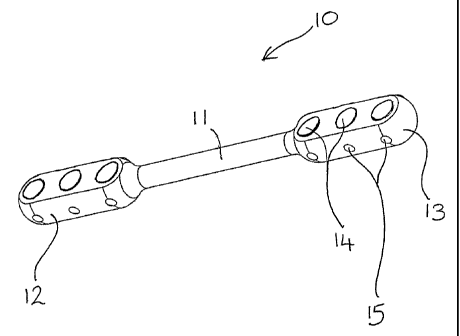
Referring to Figure 1, the fixator 10 comprises an elongate support beam 11
having
enlarged heads 12, 13 at either end thereof. The support beam is formed from
one
piece of a titanium alloy (for example) and has no joints, hinges or other
articulation
therein.
Each of the enlarged heads 12, 13 has three carefully positioned and aligned
apertures
14 therethrough, which serve as the means for attaching the fixator 10 to the
respective
ends of a fractured bone.
Referring to Figure 1A, bone screw locating sleeves 20 are provided, having a
generally
cylindrical shape and of suitable diameter to fit into one of the apertures
14. A flange 21
at the upper end of the bone screw locating sleeve 20 prevents it from falling
through the
aperture 14, in use. Each bone screw locating sleeve 20 and aperture 14 is
provided
with a lateral hole 22, 15. The lateral holes 22, 15 are aligned when the bone
screw
locating sleeve 20 is placed in an aperture 14 so that a fixing such as a grub
screw or
the like (not shown in Figures 1 or IA) can be inserted therein.
Figure 2 shows the fixator 10, bone screw locating sleeves 20 and bone screws
30
partially assembled- together. An alternative embodiment of the bone screw
locating
sleeves 20 is shown in which no upper flange 21 is present. In Figure 2, the
rightmost
bone screw locating sleeve is shown in place in the fixator, held in place by
a grub screw
CA 02611211 2007-12-06
WO 2006/134325 PCT/GB2006/002103
7
23. The exterior surfaces of the bone screw locating sleeves 20 are preferably
not
entirely in contact with the interior surface. of apertures 14 and may have
detents,
grooves or other surface markings in order to reduce the adhesive effect of
blood or
general detritus at the interface between the sleeves 20 and apertures 14.
Figures 3 and 4 show the fixator 10 and bone screw locating sleeves 20
assembled
together. Figure 3 additionally shows the bone screws 30, fixings 23 and the
fractured
bone 50. As shown in Figure 3, each aperture 14 contains a bone screw locating
sleeve
20 and a bone screw 30. Fixings 23 fit into lateral holes 22, 15 to fix the
bone screws 30
with respect to the fixator 10. Each set of three bone screws is fixed into
the respective
end of a fractured bone 50.
Since the bone screws 30 are rigidly fixed with respect to the bone screw
locating
sleeves 20 and enlarged heads 12, 13, relative movement between the respective
ends
of the fractured bone is only possible by means of corresponding movement of
the
support beam 11.
Movement of the support beam 11 is critical to the effectiveness of the
present invention.
The nature of the movement is predetermined by careful selection of the
properties of
the support beam so that the movement is both predictable and reproducible.
For example, the support beam 11 may be made predictably flexible, rigid, or
even
internally actuated in order to impart the desired type of movement according
to a
particular patient's requirements.
The predetermined movement may be as a result of the material from which the
support
beam 11 is made, for example a flexible layered composite may be used which
has a
predetermined range of deflection.
Alternatively, or in addition, the predetermined movement may be as a result
of the
shape of the support beam 11. Many possible cross-sectional shapes for the
support
beam 11 may be envisaged, for example circular, elliptical, octagonal etc (see
Figure 5,
in which suggested alternatives are indicated). Each shape of support beam
imparts
different types of relative movement, the surgeon being able to select a
fixator having a
cross-section appropriate to the patient's particular needs.
CA 02611211 2007-12-06
WO 2006/134325 PCT/GB2006/002103
8
Alternatively, or in addition, the predetermined movement may be generated by
or
contributed to by one or more actuators embedded within the support beam 11.
Other
transducers may also be incorporated into the support beam 11, for example
sensors for
monitoring the predetermined movement or other physical property.
In some circumstances it is desirable to prevent relative movement of the
respective
ends of a fractured bone (for example in the treatment of hypertrophic non-
union) and it
is possible to have a rigid support beam in order to achieve this. The support
beam 11
may be made from a memory alloy which is usually flexible but which, upon
application
of heat for example, may become rigid so as to prevent relative movement of
the bone
ends in order to treat such conditions. Consequently, the term "predetermined
relative
movemenfi" encompasses the possibility of "no relative movement".
The bone screw locating sleeves 20 each have a bore therethrough, through
which a
bone screw or the like can be inserted. In one embodiment, the bore of at
least one
bone screw locating sleeve 20 may be angled so that a bone screw inserted
therein is
directed towards a specific position with respect to the other bone screws.
Hybridisation components such as ring fixators or T-bar attachments can be
used readily
with the fixator of the present invention and may by usefui when fixating near
a knee
joint, for example. Examples of hybridisation components are illustrated in
Figures 6-7A.
Figure 6 is a top view of a fixator in which a T-bar attachment 40 has been
attached to
one end 12 of the fixator 10. The T-bar attachment enables bone screws to be
fixed in
an orientation that is perpendicular to the longitudinal axis of the fixator
10. In use, the
bone screws (not illustrated) are located in apertures 41 of the T-bar
attachment 40.
The short leg 42 of the T-bar attachment is fixed into the underlying aperture
14 of the
fixator so that the T-bar attachment and fixator cannot move with respect to
one another.
As illustrated in Figures 6A-6C, various alternative configurations are
possible, according
to the patient's needs. In all cases, a further bone screw or pin can
optionally be
inserted into the unused aperture in the fixator end 12, in addition to the
three bone
screws/pins in the T-bar attachment apertures 41.
Figure 7 is a side view of a fixator 10 to which is attached a ring attachment
43. A top
view of the ring atfachment 43 is shown in Figure 7A. The ring attachment 43
is
attached to one end 12 of the fixator 10 by means of fixings 45 through any
one of the
CA 02611211 2007-12-06
WO 2006/134325 PCT/GB2006/002103
9
apertures 14. Tension wires 44 and the bone 50 to which the fixator and ring
attachment are attached are iilustrated in Figure 7A.
Referring to Figure 8, the apertures 14 in the enlarged heads of the fixator
may be used
as a healing indicator with which the stiffness of the bone to which the
fixator is attached
can be tested to determine whether it is sufficiently healed. This is done by
removing
the bone screw locating sleeves 20 from one end of the fixator (hereafter
called the
"loose end", 13), leaving the other end (the "fixed end", 12) of the fixator
properly
attached to the bone screws (with bone screw locating sleeves still in place).
The
support beam 11 is then deflected by hand or by ambulation by as much as the
bone 50
(to which the "fixed end" is still attached) will allow. During this
deflection, the bone
screw 30 located within aperture 14 at the "loose end" will move within the
aperture 14.
Figures 8A and 8B show the unloaded and loaded conditions respectively. It may
be
deemed that, if the possible deflection is sufficient to cause the bone screw
to touch the
periphery of aperture 14 at the "loose end" (as shown in Figure 8B), the bone
50 is not
yet sufficiently stiff to be properly healed. The build-up of callus at the
junction between
the bone fragments is indicated by reference numeral 51 in Figure 8.
The fixator described herein is preferably disposable. The simplicity of the
fixings
means that the fixator can be easily removed and replaced on the bone screws,
for
example, to allow testing of the degree of healing or to heat a memory alloy
fixator to
make it rigid (see above).
The precision of the placing of the bone screws, selection of the shape and/or
materials
for the support beam etc mean that a range of fixators can be made, each
capable of
differerit (but predictable and reproducible) predetermined movement so that
each
fixator can be selected according to a particular patient's needs in order to
minimise
healing time. In addition, the fixator is much lighter, smaller and easier to
fit and remove
than prior art fixators so that patient discomfort is reduced and theatre time
minimised.
As an example of how the fixator can be used in practice, here follows a
description of
how the fixator can be applied to a fractured tibia once the fracture has been
reduced,
for example using the STAFFORDSHIRE ORTHOPAEDIC REDUCTION MACHINE
(described in PCT/GB98/00884).
CA 02611211 2007-12-06
WO 2006/134325 PCT/GB2006/002103
It is necessary that fracture reduction is complete (i.e. no further reduction
required)
when the fixator is to be applied, if the fixator of the present invention is
to be used. The
STAFFORDSHIRE ORTHOPAEDIC REDUCTION MACHINE (described in
PCT/GB98/00884) provides reduction of suitable accuracy.
5
After reduction, it is expected that the respective ends of the fractured bone
will be held
by means of bone screws associated with the reduction machine. Importantly,
these
reduction machine bone screws are not necessarily the bone screws to which the
fixator
will be applied.
There are six bone screws 30 for the fixator 10, each having an outside
diameter of
6mm, and which are inserted into the antero-medial surface of the tibia. Three
screws
are placed in the proximal fragment and three in the distal fragment.
Uniquely, the fixator 10 also acts as the drilling guide for the bone
screw/pin sites. The
normal operative technique now follows where suitable drill guides are used in
conjunction with apertures 14 to pre-drill the six holes for the bone
screws/pins.