Note: Descriptions are shown in the official language in which they were submitted.
CA 02611851 2007-12-11
Bosch Graf von Stosch Jehle
Patent Attorneys
Unser Zeichen/Our Ref. Datum/Date
1101P018W0 02 Juni 2006
1
Applicant:
IMI Intelligent Medical Implants AG
Extraocular Epiretinal Implant
The present invention relates to a device for implantation in a human eye,
having
an electrode array or a microcontact structure for contacting nerve tissue in
the
visual system of the human eye. The present invention relates in particular to
a
visual prosthesis having a device for generating stimulation impulses, which
are
used to stimulate living tissue or nerves.
One frequent cause of the partial or full loss of eyesight is destruction of
the
photoreceptor layer in the retina of the human eye, after which incident
photons
are not converted into a corresponding stimulation of the ganglion cells. The
ganglion cells are only partly affected by this pathology, so that an external
stimulation of the ganglion cells still existing in the retina can generate a
visual
perception. On the basis of this, developments which involve the implantation
of a
microcontact structure for contacting intact ganglion cells have been carried
out
for some time.
Devices have already been developed in the form of implants for the retina of
the
human eye, which are intended for the treatment of patients who have partially
or
fully lost their eyesight owing to defects in the retina. A microelectronic
device is in
this case implanted in the region of the retina with a multiplicity of
photosensitive
pixel elements, via which an image projected onto the retina through the still
intact
lens of the eye is captured. In other visual prostheses, the image capturing
is
carried out using an external camera, in particular a video camera. The image
CA 02611851 2007-12-11
-
- -
2
captured by the pixel elements or the camera is converted into electrical
signals
and delivered via stimulation electrodes by means of electrical stimulation
impulses to the ganglion cells of the retina or to the optic nerve, so as to
restore or
improve the eyesight of the blind or partially blind patient.
For epiretinal transmission of the stimulation impulses to the cells of the
retina or
to the cells of the optic nerves, microcontact structures are used which
essentially
consist of a support material that carries electrically conductive contact
elements
designed in the form of pins or needles on one side, which protrude from the
plane of the support sheet and are distributed uniformly with a constant area
density over the surface of the implant. The known visual prostheses, however,
have the disadvantage that they entail a large space requirement. Owing to the
particular sensitivity of the human eye and the extremely limited space inside
the
eye, it is in principle desirable to accommodate stimulation systems or the
implants of the visual prostheses in as small a space as possible.
Another problem with known visual prostheses consists in supplying energy to
the
implants and their microcontact structure, or the surface of the electrodes.
According to the present state of knowledge, an average power of about 40 mW
is
necessary for the energy supply of a retinal implant. Such a energy supply
cannot
be provided over a prolonged period of time by means of an implanted battery,
since this would entail too great a space requirement.
Active retinal implants therefore require a energy supply unit which is
independent
of the system for generating the visual impression, lies outside the eye and
operates without a wire connection to the retinal implant. DE 19705988 C2
discloses a subretinal implant, the implant being provided with a photovoltaic
layer
which is effective for light outside the visible spectrum. The energy supply
is in this
case carried out using infrared light. The retinal implant is provided with a
surface
tightly attached to the retina, the surface being provided with electrodes for
stimulating cells of the retina. The current supply of the components of the
CA 02611851 2010-11-23
3
implants inside the eye using infrared light may, however, entail the risk of
thermal damage to the eye due to local heating inside the eye.
It is therefore an object of the present invention to provide a visual
prosthesis
in the form of a retinal implant, which is distinguished by the least possible
space requirement inside the eye. It is another object of the present
invention
to provide an implant system whose current supply impedes the eye's
freedom of movement in the eye socket as little as possible.
The present invention achieves the aforementioned object by a visual
prosthesis with a stimulation system for implantation in a human eye, having
an electrode array for contacting and stimulating living tissue or nerves in
the
visual system of the eye, which generates stimulation impulses by means of
an electrical circuit, the stimulation system comprising at least one
intraocular
implant and at least one extraocular implant, which supplies the intraocular
implant with energy.
The present invention provides a neurostimulation device for the stimulation
of
still existing ganglion nerve cells, which can improve eyesight if there is
degenerate retinal damage but there are still intact optic nerves. By
separating the implant into an epiretinal part and an extraocular part, a
multiplicity of the necessary components and the greatest volume of the
implant can be located in the outer, extraocular part of the implant. With the
aid of the implant according to the invention, potential damage to the retina
or
other sensitive structures of the eye when arranging the stimulation system
can be minimised.
The visual prosthesis according to the invention therefore
offers the advantage that virtually all the electronic components, which
do not necessarily need to be accommodated with the intraocular
implant inside the eye, can be arranged outside the eyeball, for
example on the so-called sclera. In
this way, the space
CA 02611851 2007-12-11
4
requirement of the stimulation system inside the eye is reduced and the
operative
intervention for implanting the stimulation system inside the eye can be kept
as
small as possible. Another advantage of the visual prosthesis according to the
invention is that the current supply of the intraocular implant can be carried
out via
the extraocular implant, without impeding the eye's freedom of movement in the
eye socket. The visual prosthesis according to the invention furthermore
allows
substantially non-injurious maintenance or replacement of the stimulation
system,
for example when the extraocular implant is intended to be replaced by a more
modern version.
The extraocular part of the implant is arranged on the sclera at the outer
periphery
of the eye, so that the movement of the eyeball is compromised as little as
possible. It is particularly advantageous, if the extraocular part of the
implant is
placed in the adipose tissue surrounding the eye between two muscles, which
are
used for moving the eye. The extraocular implant may be sutured externally
onto
the sclera of the eye. In this way, unimpeded and painless movement of the eye
inside the eye socket is possible.
The individual implant parts inside and outside the eye may preferably be
coupled
to one another via a wire connection (with or without a plug connector). When
the
implant according to the invention is in the implanted state, this wire
connection is
preferably fed through the eye in the region of the pars plane, in the
vicinity of the
iris where no retina is present. The transfer both of the energy, i.e. the
current
supply, and the image data between the extraocular implant outside the eye and
the further electronics may be carried out wirelessly by inductive means. The
wireless transmission of energy and image data from the electronics remote
from
the eye to the implant avoids cable movements and concomitant impediments or
damage.
The other electronics of the visual prosthesis, which are required for
processing
and preparing the image data captured by an external camera, may be arranged
remotely from the eye outside the body. The electronic components may for
CA 02611851 2007-12-11
. -
_ -
example be accommodated in a so-called pocket computer, which may be carried
in a separate pocket on the body. The electronic components are particularly
advantageously accommodated in a spectacle frame, which also contains the
camera that captures the image data.
5
Since the electronic components, which are required for image-processing the
signals delivered by the video camera, are located outside the body, their
maintenance or replacement by a more modern version of the electronic
interface
is straightforward. The components of the electronic interface may be adapted
individually to the respective electronic stimulation level of the implant
system. In
this way, it is possible to ensure a minimal level of electrical charge for
all the
electrodes in the electrode array, so that the tissue or nerve cells
stimulated by
the electrical stimulation impulses are stressed as little as possible. It is
thus
possible to avoid damage on the retina of the eye in the vicinity of the
electrodes
due to an elevated charge level, as well as painful sensations for the
patient.
In principle, image acquisition in the stimulation system according to the
invention
is carried out by an external camera, the image signals of which are delivered
after electronic preprocessing via the extraocular implant and the epiretinal
implant to the retina of the eye. The epiretinal implant comprises an
integrated
electrode array which stimulates the ganglion cells or the cells of the retina
by
electrical signals in a position-resolved way according to the received image
data,
and thereby forwards the image captured by the external camera to the nerves
of
the visual system. A particular advantage of the active epiretinal implant is
that it
can be adapted to various conditions in respect of the ambient luminance.
The intraocular implant comprises an electrode array having a number of
stimulation electrodes, which are preferably arranged close together in a
matrix.
The electrode array comprises a microcontact structure with a number of
contact
sites, via which the stimulation electrodes are in contact with the retinal
cells or
ganglion cells and stimulate the contacted retinal cells or ganglion cells by
means
of stimulation impulses. The outer region of the microcontact structure for
CA 02611851 2007-12-11
-
6
epiretinal contacting of the ganglion cells is adapted to the outer contour of
the
foveal region of the retina, and may have a spherical shape. The microcontact
structure, or the electrode array, of the epiretinal implant is in this case
preferably
arranged in the region of the macula of the eye. The macula is the place
inside
the eye, or on the retina, which receives the greatest amount of light; it is
therefore often referred also referred to as the "place of sharpest vision".
The extraocular implant is equipped with an electrical control unit, which is
preferably designed as a digital control unit with analogue auxiliary
functions and
generates stimulation impulses with the aid of the image data captured by an
external camera. To this end, the electrical control unit comprises at least
one
current or voltage source and at least one impulse generator that generates
electrical stimulation impulses, which are amplified by the current/voltage
source
to form stimulation impulses or stimulation currents and are forwarded to the
stimulation electrodes in the electrode array in the intraocular implant. The
electrical control unit may also be equipped with electronic storage means, in
which the calculated duration and intensity of the stimulation impulses to be
generated are stored and can be called in response to a particular
instruction.
Expediently, the electronic components of the electrical control unit are
accommodated at least partially in an integrated circuit by being
photolithographically microstructured, and preferably on a chip in the
extraocular
implant. The extraocular implant has at least one counter-electrode, which
serves
as a return current path for the stimulation electrodes.
The electrical control unit has a contact pad for each stimulation electrode,
i.e. a
connection surface via which a stimulation electrode can respectively be
contacted by a separate wire connection. The wire connection is designed as a
flexible implant and is fed between the extraocular implant and the
intraocular
implant into the interior of the eye, preferably in the region of the pars
plana where
no retina is present so as to avoid compromising the retina.
CA 02611851 2007-12-11
. '
7
Feeding the wire connection between the epiretinal implant and the extraocular
implant through the sclera of the eye in the region of the pars plana
represents an
intervention with the least outlay and the least possible damage to the eye.
The
danger of complications and the infection risk during the operation are
therefore
also reduced. If the flexible implant of the wire connection is fastened
together
with the inner and outer parts of the implant on the eye, these execute the
same
movements as the eye so that the eye's freedom of movement is not
compromised either by the wire connection or by the inner and outer parts of
the
implant.
The wire connection for coupling the extraocular implant to the intraocular
implant
comprises at least one line for transmitting the operating current and at
least one
signal line for transmitting image data and/or electrical stimulation impulses
from
the digital control unit to the intraocular implant. According to a preferred
embodiment of the present invention, besides the electrical lines for
transmitting
the operating current, the wire connection also comprises at least as many
lines
for transmitting electrical stimulation impulses as there are stimulation
electrodes
provided in the intraocular implant. The wire connection may furthermore
comprise one or more optical fibers for unidirectional or bidirectional data
transmission by means of light signals between the extraocular part and the
intraocular part of the implant.
In order to ensure reliable fixing of the flexible implant with the electrode
array or
the microcontact structure and the wire connection between the microcontact
structure and the extraocular implant, the intraocular implant and/or the
flexible
implant of the wire connection may be fixed inside the eye with the aid of a
nail, a
so-called tack. To this end, the tack is operatively fitted from inside the
eye and
extends through the flexible implant and the retina into the choroid or the
sclera of
the eye, where it is anchored by its retaining hooks.
The intraocular implant comprises a number of photosensitive elements, which
drive the contact sites of the electrode array via the electrical circuit as a
function
CA 02611851 2007-12-11
_ -
8
of light incident on the intraocular implant. At least one light receiver of
the
intraocular implant is in this case capable of receiving light signals of a
light
transmitter from outside the eye. According to a preferred embodiment, the
light
receiver of the intraocular implant is designed as an infrared receiver which
receives infrared signals of an infrared transmitter from outside the eye,
preferably
via the natural light path of the eye.
In this way, the interface between the light transmitter outside the eye and
the
photosensitive elements, or the light receiver, of the intraocular implant can
transfer image data captured by an external camera via light signals from the
light
transmitter outside the eye to the photosensitive elements or the light
receiver of
the intraocular implant. Infrared light is preferably used for transmitting
the image
data, since it lies outside the visible light spectrum and therefore does not
irritate
any remaining eyesight of the patient and the transmission of the image data.
Signal processing of the received image data takes place in the extraocular
implant, including signal amplification, for which reason external energy
input is
necessary. This energy input is carried out wirelessly in the visual
prosthesis
according to the invention through the inductive interface between an external
radiofrequency transmitter coil and the radiofrequency receiver coil of the
extraocular implant. To this end, according to another preferred embodiment of
the visual prosthesis according to the invention, an antenna remote from the
stimulation system is provided for an inductive interface, which can transmit
electromagnetic signals preferably in the radiofrequency range. The
extraocular
implant is furthermore equipped with an antenna, which can receive
electromagnetic signals preferably in the radiofrequency range.
The radiofrequency antenna of the extraocular implant receives the
radiofrequency electromagnetic signals emitted by the transmitter antenna of
the
electronics outside the body. This creates an inductive current which supplies
the
implant on the eye with sufficient energy. The current resulting from the
induction
is transferred from the outer part of the implant via the wire line to the
inner part of
CA 02611851 2007-12-11
-
9
the implant, in order to supply the electrode array and the infrared receiver
with
current.
The inductive interface between the antenna outside the eye and the antenna of
the extraocular implant may also be designed bidirectionally, in that the
antenna
remote from the stimulation system can receive electromagnetic signals
preferably
in the radiofrequency range and the antenna of the extraocular implant can
transmit electromagnetic signals preferably in the radiofrequency range. In
this
preferred embodiment, the extraocular implant is designed so that it can
transfer
information, for example about operating parameters of the stimulation system,
via
the inductive interface. According to another particular embodiment of the
invention, the data rate of the signals received by the antenna of the
extraocular
implant is different from the data rate of the signals transmitted by the
antenna of
the extraocular implant.
In the case of a bidirectional inductive interface, both the outer part of the
implant
on the eye and the electronics outside the body are therefore equipped with a
transmission unit and a reception unit, which can respectively transmit and
receive
electrical signals preferably in the radiofrequency range. Signals generated
by the
epiretinal implant inside the eye can therefore also be transferred via the
wire line
to the outer part of the implant, and forwarded from there via the
transmission unit
in the form of radiofrequency signals to the electronics outside the body.
The electronics outside the body receive the radiofrequency signals from the
transmitter of the extraocular implant via their receiver unit, and feed them
to a
central computation unit where the signals are evaluated. In this way,
signals,
which provide information for example about a sufficient current supply of the
internal implant, the quality of the received image signals, the function of
the
stimulation electrodes in the electrode array, the efficiency of the inductive
interface or the contact of the stimulation electrodes with the ganglion
cells, can
be transferred from the epiretinal implant inside the eye.
CA 02611851 2007-12-11
. -
. -
The intraocular implant may furthermore comprise at least one light-emitting
element, which radiates light signals as a function of operating parameters of
the
stimulation system. To this end, the light signals emitted by the light-
emitting
element are encoded as a function of operating parameters of the intraocular
5 implant, for example by modulating the duration and/or intensity of the
light
signals. The light signals emitted by the light-emitting element may for
example
contain information about the position of the intraocular implant, about the
quality
of the image data received by the intraocular implant, about the quality of
the
current supply of the intraocular implant and/or about the impedance or the
10 electrical resistance of the stimulation electrodes. The light signals
emitted by the
light-emitting element may furthermore contain information about the function
of
the stimulation electrodes in the electrode array and about the contact of the
stimulation electrodes with the ganglion cells.
This light-emitting element is preferably arranged inside the eye so that the
light
signals emitted by the light-emitting element can be detected by an observer
via
visual contact into the interior of the eye. The light-emitting element is
preferably
designed as a diode (status diode) that radiates light, in particular infrared
light,
which can be detected by a light receiver, in particular by an infrared light
receiver
outside the eye.
According to another aspect of the invention, the aforementioned objects are
furthermore achieved by a method for operating the device according to the
invention, comprising at least the following steps:
= capturing an image using an external camera,
= generating position-resolved image data from the captured image,
= calculating diagnostic instructions, control instructions or stimulation
instructions with a particular duration and intensity as a function of the
image
data,
CA 02611851 2007-12-11
= =
. -
11
= transferring the diagnostic instructions, control instructions or
stimulation
instructions to a stimulation system having an intraocular implant and an
extraocular implant,
= calculating and generating electrical stimulation impulses or stimulation
currents with a particular duration and intensity in the extraocular implant
or
carrying out diagnostic tasks according to the diagnostic instructions,
control
instructions or stimulation instructions,
= transferring the electrical stimulation impulses or stimulation currents
to the
intraocular implant, and
= applying
the electrical stimulation impulses or stimulation currents to at least
one stimulation electrode in the intraocular implant so that at least one
retinal
cell or ganglion cell, which is in contact with the relevant stimulation
electrode, is stimulated.
In order to prepare the image data captured by the external camera for use in
the
stimulation system, before transfer to the stimulation system they are
electrically
evaluated or processed in the electrical control unit in order to generate
corresponding electrical stimulation impulses or stimulation currents. In this
case
the components of the electrical control unit may on the one hand be part of
the
extraocular implant, or on the other hand accommodated in an external
computation unit which the patient carries with them separately, or
accommodated
in spectacles on which the external camera and/or the light transmitter for
the
infrared interface is also arranged.
As described above, in the method for operating the device according to the
invention, the current required for operation of the extraocular implant and
the
intraocular implant is transmitted wirelessly via an inductive interface
between the
radiofrequency transmitter antenna outside the eye and the radiofrequency
receiver antenna of the extraocular implant, while the image data captured by
the
external camera are transmitted wirelessly via an infrared interface between
the
infrared transmitter outside the eye and the infrared receiver inside the eye.
As an
CA 02611851 2007-12-11
,
_
12
alternative, the image data captured by the external camera may likewise be
transmitted wirelessly via the inductive interface between the radiofrequency
transmitter antenna outside the eye and the radiofrequency receiver antenna of
the extraocular implant.
The image data captured by the external camera or the diagnostic instructions,
control instructions or stimulation instructions may be transmitted as a
serial data
stream from the infrared receiver inside the eye via the wire connection to
the
digital control unit in the extraocular implant. In this case, the serial data
stream
from the infrared receiver inside the eye via the wire connection to the
digital
control unit in the extraocular implant contains information about the
electrode
address, for example from 1 to 250, and about the amplitude associated with
the
electrode address, for example from 0 to 1000 pA, of the stimulation impulses
for
the relevant stimulation electrode. With the aid of the information relating
to the
electrode address and the amplitude of the stimulation impulses, stimulation
impulses with a particular duration and intensity are calculated and generated
by
the electrical control unit of the extraocular implant for each stimulation
electrode.
The shape or the profile of the electrical stimulation impulses is adapted to
the
ganglion cells to be stimulated. Using a multiplicity of current generators in
the
extraocular implant, electrical current with a particular intensity and
duration is
applied to the stimulation electrodes.
The stimulation impulses or stimulation currents are transferred as a parallel
signal stream from the electrical control unit of the extraocular implant via
parallel
wire connections to the stimulation electrodes in the intraocular implant. To
this
end, the electrical control unit of the extraocular implant, or the retinal
stimulator
chip, has for example 250 connection pads to which wires for 250 stimulation
electrodes in the electrode array of the intraocular implant can respectively
be
connected.
In a preferred embodiment of the method, it is also possible for the
intraocular
implant to transfer diagnostic data relating to operating parameters of the
CA 02611851 2007-12-11
. -
13
intraocular implant via the wire connection to the extraocular implant, for
example
as a serial data stream. The serial diagnostic data stream is subsequently
forwarded inductively, for example using load modulation, from the extraocular
inductive coil to external diagnostic means which are for example accommodated
in the spectacles. As an alternative to the inductive feedback path described
above, the status light-emitting diode in the intraocular part of the implant
may
also be used as an optical return channel with a digital reception unit and
digital
evaluation unit in the spectacles.
Other preferred embodiments of the implant according to the invention
As already described above, electronic components of the visual prosthesis
according to the invention may in particular be accommodated in a module
outside the body, preferably in spectacles which the patient may wear like a
normal visual aid. The electronic components accommodated in a module outside
the body will be referred to below as the extracorporeal part of the visual
prosthesis according to the invention, while the components of the visual
prosthesis according to the invention which are arranged inside the body,
comprising the components implanted intraocularly in the eye and those
implanted extraocularly in the eyeball, will be summarised as the
intracorporeal
part of the visual prosthesis according to the invention.
Between the extracorporeal part (for example in the spectacles) of the visual
prosthesis and the intracorporeal part in or on the patient's eye, there is a
wireless
inductive interface via which both the energy is input and also data
transmission is
carried out. According to a preferred embodiment of the present invention
which
has already been described, this inductive interface between the
extracorporeal
part and the intracorporeal part is designed bidirectionally. To this end,
both the
extracorporeal part and the intracorporeal part are equipped with a
transmitter coil
which can transmit electrical signals preferably in the radiofrequency range,
and
with a receiver coil or an antenna which can transmit electrical signals
preferably
CA 02611851 2007-12-11
-
14
in the radiofrequency range. In this way, electrical signals can be
transferred both
from the extracorporeal part to the intracorporeal part and in the opposite
direction, from the intracorporeal part to the extracorporeal part of the
visual
prosthesis according to the invention (bidirectionality). Both in the
extracorporeal
part and in the intracorporeal part, it is also possible to provide only a
transmitter-
receiver coil which respectively fulfils both functions of transmitting and
receiving
electrical signals.
In a refinement of this preferred embodiment of the present invention, the
bidirectional data line between the extracorporeal part and the intracorporeal
part
of the visual prosthesis comprises at least two preferably wireless
transmission
channels. In this case, at least one wireless transmission channel extends
from
the extracorporeal part (for example in the spectacles) to the intracorporeal
part of
the visual prosthesis in the eye, also referred to below as the "forth
transmission
channel" (up-link), and at least one wireless transmission channel extends
from
the intracorporeal part of the visual prosthesis in the eye to the
extracorporeal part
in the spectacles, also referred to below as the "back transmission channel"
(down-link).
The data transmission between the extracorporeal part and the intracorporeal
part
of the visual prosthesis is preferably carried out simultaneously, i.e. data
are
transmitted at the same time both on the forth transmission channel (up-link)
and
on the back transmission channel (down-link). The back transmission channel
(down-link) may be used in particular to transfer data about the status of the
intracorporeal part of the visual prosthesis. This provides an additional
safety
factor, in that the status and the functionality of the intracorporeal part of
the visual
prosthesis can be constantly monitored and a corresponding malfunction can be
signalled in the event of failure of the visual prosthesis or the back
transmission
channel (down-link).
In another preferred embodiment of the present invention, the data
transmission
between the extracorporeal part and the intracorporeal part of the visual
CA 02611851 2007-12-11
prosthesis may take place alternately. For example, the forth transmission
channel (up-link) from the extracorporeal part to the intracorporeal part may
be
active during particular time periods, and the back transmission channel (down-
link) from the intracorporeal part to the extracorporeal part of the visual
prosthesis
5 may
be active during other particular time periods. With this alternating data
transmission, it is also possible to provide only one transmission channel
since
this can then be used alternately either as an forth transmission channel (up-
link)
or as a back transmission channel (down-link).
10
During normal operation, the data transmission between the extracorporeal part
and the intracorporeal part of the visual prosthesis predominantly takes place
by
means of the forth transmission channel (up-link), i.e. the image data
captured
and processed by the extracorporeal part of the visual prosthesis (for example
in
the spectacles) are transferred or transmitted via the forth transmission
channel
15 (up-
link) to the intracorporeal part of the visual prosthesis (in the eye). On the
back transmission channel (down-link), conversely, transmission is carried out
only in the event of a feedback from the intracorporeal part to the
extracorporeal
part of the visual prosthesis, for example when the intention is to transfer
data
about the status of the intracorporeal part of the visual prosthesis or an
error
message.
Various types of data may be transmitted on the forth transmission channel (up-
link) from the spectacles to the implant in the eye. For example, stimulation
instructions are transferred from the extracorporeal part of the visual
prosthesis to
the stimulator chip of the intracorporeal part in the eye via the forth
transmission
channel (up-link). Such stimulation instructions may comprise the following
information:
- electrode addresses, i.e. the addresses of the electrodes arranged in the
electrode array which are used to stimulate the ganglion cells in the retina
of
the eye,
CA 02611851 2007-12-11
16
- current amplitudes, i.e. the information for the stimulator chip which
specifies
the current strength of the stimulation impulses to be generated,
- phase duration, i.e. the information for the stimulator chip which contains
the
phase duration of the stimulation impulses to be generated,
-- - phase ratio, i.e. the information for the stimulator chip which specifies
the
phase ratio of the stimulation impulses to be generated,
- polarity sign of the stimulation impulses, i.e. the information for the
stimulator
chip which contains the polarity sign of the stimulation impulses to be
generated.
Measurement instructions, for example, may furthermore be transferred via the
forth transmission channel (up-link) from the extracorporeal part of the
visual
prosthesis to the intracorporeal part. Measurement instructions are
instructions
from the extracorporeal part of the visual prosthesis to the intracorporeal
part, to
-- carry out particular measurements and transfer the ascertained measurement
value via the back transmission channel (down-link) to the extracorporeal part
of
the visual prosthesis. Such measurement instructions may comprise the
following
information:
-- - voltage measurement on an electrode during the stimulation,
- voltage measurement on an electrode outside the stimulation,
- measurement of nerve action potentials with the aid of one or more
stimulation
electrodes,
- measurement of nerve action potentials with the aid of special measurement
electrodes.
Status instructions, for example, may furthermore be transferred via the forth
transmission channel (up-link) from the extracorporeal part to the
intracorporeal
part of the visual prosthesis. Such status instructions contain requests for
the
-- intracorporeal part of the visual prosthesis to record particular status
parameters
and transfer them via the back transmission channel (down-link) to the
extracorporeal part of the visual prosthesis. The status instructions may, for
CA 02611851 2007-12-11
_
17
example, contain requests for the intracorporeal part of the visual prosthesis
to
record the following status parameters and transfer them on the back
transmission
channel (down-link) to the extracorporeal part of the visual prosthesis:
- the identification number (ID number) of the implant,
- a status report of the implant, for example
o about the status of the charge balancing systems or
o about the status of the energy supply of the implant, i.e. for example
whether it has too much energy or too little energy,
- the temperature of the stimulation chip or of implant parts,
- the moisture sensor measurement value.
According to another preferred embodiment of the visual prosthesis according
to
the invention, at least the forth transmission channel (up-link) from the
extracorporeal part to the intracorporeal part of the visual prosthesis is
configured
in the form of optical data transmission. The data may in this case be
transmitted
via light signals by means of light-emitting diodes (LEDs) or by means of
lasers,
for example with infrared light. The natural light path of the eye may be used
at
least partially for the optical data transmission, by the light signals of the
light-
emitting diodes or the laser outside the eye being directed through the lens
aperture of the eye onto an optical reception element inside the eye.
The data transmission between the intracorporeal part and the extracorporeal
part
of the visual prosthesis may be carried out with any desired coding on the
back
transmission channel (down-link). Balanced coding, which contains
approximately
the same number of zero-states and one-states, is preferably used so as to
avoid
driving the optical reception element to saturation. For example Manchester
coding, so-called 4PPM coding, 4PPM+ coding or other suitable coding methods
may be used.
According to another preferred embodiment of the visual prosthesis according
to
the invention, the forth transmission channel (up link) and/or back
transmission
CA 02611851 2007-12-11
_
18
channel (down-link) between the intracorporeal part and the extracorporeal
part of
the visual prosthesis is configured in the form of electromagnetic data
transmission, in which the carrier frequency of the transmitter is
correspondingly
modulated in order to transmit data. The electromagnetic data transmission may
in this case be designed actively, in which case for example the 13.56 MHz ISM
frequency band, the 27.12 MHz ISM frequency band, the 125 kHz ISM frequency
band or another suitable frequency band is used as the carrier frequency of
the
transmitter. Instead of frequency modulation for the electromagnetic data
transmission between the intracorporeal part and the extracorporeal part of
the
visual prosthesis, it is also possible to use amplitude modulation, phase
modulation of the carrier frequency or other suitable modulation methods.
According to another modulation method which may be used for the visual
prosthesis according to the invention, a separate data carrier frequency is
used,
for example in the 433 MHz ISM frequency band or in another suitable frequency
range, which is preferably different to from the frequency for the energy
input via
the inductive interface. This separate data carrier frequency may in turn be
modulated by one of the following methods:
- amplitude modulation of the data carrier frequency,
- frequency modulation of the data carrier frequency,
- phase modulation of the data carrier frequency,
- other suitable modulation methods.
Various types of data may be transmitted via the back transmission channel
(down-link) from the intracorporeal part to the extracorporeal part of the
visual
prosthesis. In particular, diagnostic data about the status of the
intracorporeal part
of the visual prosthesis or about the status of the implant may be transferred
via
the back transmission channel (down-link), for example:
- measurement values for the electrode impedance of particular stimulation
electrodes,
CA 02611851 2007-12-11
19
- measurement values for the electrical voltage which is applied to
stimulation
electrodes,
- monitoring data of the status of particular stimulation electrodes.
Information or diagnostic data about the system status of the process control
in
the intracorporeal part of the visual prosthesis or in the implant may also be
transferred via the back transmission channel (down-link), for example
information
about the following control details:
- data correctly transferred from the extracorporeal part to the
intracorporeal part
of the visual prosthesis (yes or no),
- intracorporeal part or implant correctly initialised (yes or no),
- system status reset carried out (yes or no),
- status of the energy supply of the implant (power status), for example
o status of the analogue component or components of the stimulator chip,
o send status of the so-called power-down stage of the intracorporeal part
to the extracorporeal part of the visual prosthesis,
- fault in the stimulation of the retina of the eye, for example
o maximum stimulation current reached,
o charge balance between stimulation electrodes not achieved,
o charge balance between stimulation electrodes takes too long,
- stimulation was carried out even though not requested, for example
owing to a
fault in the output stage of a current source,
- status of the electrical energy supply, for example
o operating voltage too low,
o operating voltage too high,
- voltage on stimulation electrodes at particular measurement times.
- Diagnostic data about the patient's physiology, for example
o reading of the action potentials of individual nerve cells, in particular
ganglion cells,
CA 02611851 2007-12-11
O reading of the sum action potentials of nerve cells, in particular
ganglion
cells, so that information can be inferred about the stimulability of the
contacted nerve cells
- diagnostic data about the general status of the intracorporeal part of
the visual
5 processes or the implant, for example
o temperature in the region of the electronics of the implant,
O temperature at a particular point in the eye,
o temperature at a plurality of positions in the eye,
o measurement value for the internal eye pressure,
10 o acceleration measurement of the implant,
o moisture measurement inside the housing of the implant.
As already described in conjunction with the forth transmission channel (up-
link),
the back transmission channel (down-link) from the intracorporeal part to the
15 extracorporeal part of the visual prosthesis may also be designed as an
optical
data transmission path. To this end, as in the case of the forth transmission
channel (up-link), the data may also be transmitted using light signals in the
back
transmission channel (down-link) by means of light-emitting diodes (LEDs) or
by
means of lasers, for example with infrared light. The natural light path of
the eye
20 may likewise be used at least partially in this case, by the light
signals of a light-
emitting element arranged inside the eye being directed through the lens
aperture
of the eye onto an optical reception element outside the eye. The optical
reception
element records the encoded light signals of the light-emitting element and
forwards them in the form of electrical signals to electronics for evaluation.
According to another preferred embodiment of the visual prosthesis according
to
the invention, the back transmission channel (down-link) from the
intracorporeal
part to the extracorporeal part of the visual prosthesis is configured in the
form of
passive electromagnetic data transmission, in which the carrier frequency of a
transmitter is correspondingly modulated in order to transmit data. For
example,
load modulation of the energy transmission frequency may be carried out as a
modulation method in this case. The load modulation of the carrier frequency
may
CA 02611851 2007-12-11
=
21
be carried out by connecting and disconnecting a resistive load, a capacitive
load
or an inductive load according to the data stream to be transmitted. A
combination
or partial combination of the said methods is also possible for the
electromagnetic
data transmission between the intracorporeal part and the extracorporeal part
of
the visual prosthesis.
In addition or as an alternative, the data transmission via the back
transmission
channel (down-link) from the intracorporeal part (in the eye) to the
extracorporeal
part (for example in the spectacles) of the visual prosthesis according to the
invention may be carried out by using an error correction method. Likewise, in
addition or as an alternative, the data transmission via the forth
transmission
channel (up-link) from the extracorporeal part (in the spectacles) to the
intracorporeal part of the visual prosthesis (in the eye) may be carried out
by using
an error correction method.
For example, a method which can recorrect a defined number of incorrectly
transmitted data bits out of the data bits transmitted overall, by redundancy
in the
coding of the data transmission, may be used as an error correction method.
One
of the following methods may be used as an error correction method both for
the
forth transmission channel (up-link) and for the back transmission channel
(down-
link):
- Hamming coding,
- convolution coding,
- repetition coding or
- other suitable error correction methods.
In addition or as an alternative to using an error correction method, the data
transmission via the forth transmission channel (up-link) from the
extracorporeal
part (in the spectacles) to the intracorporeal part of the visual prosthesis
(in the
eye) may be also carried out by using an error detection method. Likewise, in
addition or as an alternative, the data transmission via the back transmission
CA 02611851 2007-12-11
_
22
channel (down-link) from the intracorporeal part (in the eye) to the
extracorporeal
part (in the spectacles) of the visual prosthesis according to the invention
may
likewise be carried out by using an error detection method. Such an error
detection method may be implemented by various coding methods, for example:
- cyclic redundancy check (CRC) coding,
- parity check coding,
- repetition coding or
- other suitable error detection methods.
A data rate in the range of from 100 kilobits/second to 10 megabits/second,
preferably a data rate in the range of from 1 megabit/second to 10
megabits/second, particularly preferably from 1 to 5 megabits/second and even
more preferably from 1 to 2 megabits/second, may be used for the data
transmission via the forth transmission channel (up-link) from the
extracorporeal
part (in the spectacles) to the intracorporeal part (in the eye) of the visual
prosthesis according to the invention.
A data rate in the range of from 1 kilobit/second to 100 kilobits/second,
preferably
a data rate in the range of from 5 to 20 kilobits/second, may be used for the
data
transmission via the back transmission channel (down-link) from the
intracorporeal
part (in the eye) to the extracorporeal part (in the spectacles) of the visual
prosthesis according to the invention. The data rates used for the data
transmission via the forth transmission channel (up-link) and for the data
transmission via the back transmission channel (down-link) may in this case be
different.
Further details, preferred embodiments and advantages of the present invention
will be found in the following description with reference to the drawing, in
which:
CA 02611851 2007-12-11
23
Figure 1
shows a schematic representation of the cross section through a
human eye with a visual prosthesis according to a preferred
embodiment of the present invention;
Figure 2 shows a
perspective view of a stimulation system comprising
spectacles and a human eye with a visual prosthesis according to
the invention;
Figure 3
shows a schematic representation of the cross section through a
human eye with a visual prosthesis according to a second preferred
embodiment of the present invention;
Figure 4
shows a schematic representation of the cross section through a
human eye with a visual prosthesis according to a third preferred
embodiment of the present invention;
Figure 5
shows a schematic representation of the cross section through a
human eye with a visual prosthesis according to a fourth preferred
embodiment of the present invention; and
Figure 6 shows a schematic representation of the cross section through
a
human eye with a visual prosthesis according to a fifth preferred
embodiment of the present invention.
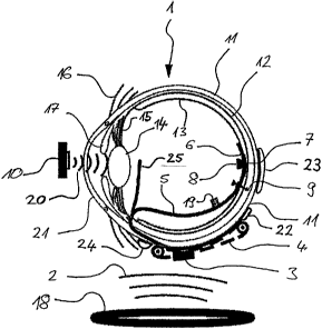
Figure 1 shows a schematic representation in cross section through a human eye
with a visual prosthesis according to a preferred embodiment of the present
invention. The eyeball 1 of the human eye has an essentially round shape, the
transparent cornea 21 having a more pronounced curvature on its anterior side.
The region of the eyeball 1 held in the eye socket is constructed from a
plurality of
layers, the outermost layer constituting the so-called sclera 11. The sclera
11 is
followed in the direction of the interior of the eye by the choroid 12, on
which the
CA 02611851 2007-12-11
24
so-called retina 13 with photosensitive cells or photoreceptors (cones, rods
and
ganglion cells) rests.
In a healthy human eye, the natural light path travels via the transparent
cornea
21 in the anterior region of the eyeball 1 through the iris 17 and the
biconvex lens
14, the shape or refractive energy of which can be modified by tensioning the
ciliary muscle 15. The incident light enters the interior of the eye while
being
optically refracted by the cornea 21 and the eye lens 14, and is projected
onto the
retina 13 in the posterior region of the eyeball 1. The light-sensitive
The purpose of the visual prosthesis according to the invention is to restore
or
improve a visual process impaired or destroyed owing to degenerative
modifications on the retina 13. A prerequisite for using the visual prosthesis
according to the invention is that the ganglion cells contained in the retina
13 are
substantially intact and are capable of forwarding nerve impulses via the
optic
nerve to the brain.
According to the preferred embodiment represented in Figure 1, the visual
prosthesis according to the invention comprises a stimulation system with an
intraocular implant 6, 8, which is arranged inside the eyeball 1, and an
extraocular
implant 3, 4 which is arranged outside the eyeball 1. The intraocular implant
is
The intraocular implant is coupled to the extraocular implant a via a wire
connection 5. The wire connection 5 is designed as a flexible implant, which
CA 02611851 2007-12-11
. _
implant. The wire connection 5 comprises electrical lines in order to provide
the
current supply of the intraocular implant via the extraocular implant. The
wire
connection 5 furthermore comprises electrical lines in a sufficient number to
allow
transfer of image data or diagnostic instructions, control instructions or
stimulation
5 instructions in the form of serial data streams and/or parallel data
streams or
signal streams between the intraocular implant and the extraocular implant.
The intraocular implant comprises an electrode array 6, which bears
epiretinally
on the retina 13 and has a number of stimulation electrodes, for example
10 arranged in a matrix. The stimulation electrodes of the electrode array
6 are
connected to ganglion cells and can stimulate them by means of stimulation
impulses or stimulation currents. The electrode array 6 of the epiretinal
implant is
centred in the region of the macula of the eye, where the greatest amount of
light
arrives on the retina 13 via the natural light path. In order to ensure a
secure
15 position of the intraocular implant on the retina, it is fastened inside
the eye with
the aid of a so-called nail or tack 9 which extends through the intraocular
implant
and the retina 13 and is anchored by retaining hooks in the sclera 11.
An infrared receiver 8, which can receive light signals from an infrared
transmitter
20 10 outside the eye via the natural light path, is arranged on the
intraocular
implant. An image is captured by an external camera (not shown), and its
preprocessed image data are transferred via the infrared transmitter 10 along
the
natural light path of the human eye to the infrared receiver 8 of the
intraocular
implant. These image data are forwarded from the intraocular implant to the
25 extraocular implant via the wire connection 5, preferably in the form of
the serial
data stream.
Any position along the wire connection 5 is conceivable for the infrared
receiver 8,
although it preferably lies in the region of the nail or tack 9. As an
alternative, the
infrared receiver 8 may lie on a branch 25 of the wire connection 5 in order
to
adjust the reception properties favourably. This branch 25 departs from the
wire
connection 5 and expediently protrudes into the eye in the beam path of the
CA 02611851 2007-12-11
_
-
26
natural light path. In this way, the infrared signals incident in the eye via
the
natural light path from outside the eye can arrive directly on the infrared
receiver 8
arranged on the branch 25 of the wire connection 5.
The image data are evaluated in the retinal stimulator chip 3 of the
extraocular
implant and converted into the stimulation impulses or stimulation currents.
The
stimulation impulses or stimulation currents are subsequently transferred in
the
form of a parallel signal stream via the wire connection 5 to the stimulation
electrodes in the electrode array 6 of the intraocular implant, and flow back
via the
counter-electrode 22, 23 and/or 24 into the respective current source. The
stimulation electrodes stimulate the ganglion cells in the retina via the
microcontact structure according to the position-resolved stimulation
impulses,
and thereby generate a visual impression with nerve signals, which corresponds
to the image captured by the external camera.
The stimulation system of the visual prosthesis according to the invention
furthermore comprises an extraocular implant, which is arranged outside the
eyeball 1 on the sclera 11. All those components of the stimulation system
which
do not necessarily need to be arranged on the intraocular implant inside the
eye
are accommodated in the extraocular implant. The extraocular implant comprises
a retinal stimulator chip 3, which can calculate and generate stimulation
impulses
or stimulation currents for the stimulation electrodes of the intraocular
implant on
the basis of received image data. To this end, the retinal stimulator chip 3
comprises electronic components for calculating the intensity and duration of
the
stimulation impulses with the aid of the received image data, current
generators
for generating the required stimulation currents and electronic storage means,
in
which the parameters of the stimulation impulses and the coordinates of the
corresponding stimulation electrodes are buffered and can be called up or
released in response to a particular command.
The extraocular implant furthermore comprises at least one counter-electrode
which, for example, may be arranged in the positions which are denoted by the
CA 02611851 2007-12-11
27
references 22, 23 and 24 in Figure 1. The counter-electrodes 22, 23, 24 are
used
as a return current path for the stimulation current sources, in order to
close the
current path to the stimulation electrodes in the electrode array 6 via the
tissue of
the sclera 11, choroid 12 and the retina 13.
The extraocular implant furthermore comprises a radiofrequency antenna 4, via
which radiofrequency signals 2 that are emitted by a radiofrequency antenna 18
arranged remotely from the eyeball 1 can be received. Via the inductive
interface
between the radiofrequency antenna 4 of the extraocular implant and the
external
radiofrequency antenna 18, inductive energy which is required for operation of
the
extraocular implant and the intraocular implant can be transferred.
The external radiofrequency antenna 18 may, for example, be accommodated
together with other electronic components outside the body in an
extracorporeal
part of the visual prosthesis according to the invention, for example in
spectacles
which the patient may wear like a normal visual aid. Conversely, the
intraocular
implant 6, 8 and the extraocular implant 3, 4 constitutes an intracorporeal
part 3,
4, 6, 8 of the visual prosthesis according to the invention. Via the inductive
interface, wireless contact can be established between the extracorporeal part
and the intracorporeal part of the visual prosthesis according to the
invention.
Via this inductive interface between the extracorporeal part and the
intracorporeal
part, the image data captured by an external camera can also be transferred to
the retinal stimulator chip 3 which generates stimulation impulses with the
aid of
the received image data and forwards them via the wire connection 5 to the
stimulation electrodes in the intraocular implant. The inductive interface
between
the radiofrequency antenna of the intraocular implant and the external
radiofrequency antenna 18 may also be designed bidirectionally, so that the
retina
stimulation chip 3 can transfer information about operating parameters of the
intraocular implant and/or the extraocular implant inductively via the
radiofrequency antenna 4 to the external radiofrequency antenna 18, and these
may then be evaluated by external electronics (not shown).
CA 02611851 2007-12-11
28
In order to set up the bidirectional inductive interface between the
extracorporeal
part and the intracorporeal part of the visual prosthesis according to the
invention,
the extracorporeal part outside the eye 1 may have an antenna 18 which can
both
transmit and receive electromagnetic signals 2, preferably in the
radiofrequency
range. The extraocular implant 3, 4 and/or the intraocular implant 6, 8 may
likewise have an antenna 4 which can both transmit and receive electromagnetic
signals 2, preferably in the radiofrequency range.
As an alternative, the extracorporeal part of the visual prosthesis according
to the
invention may comprise at least two antennas 18 for the bidirectional
inductive
interface, of which a first antenna can transmit electromagnetic signals 2 and
a
second antenna can receive electromagnetic signals 2. The extracorporeal part
of
the visual prosthesis according to the invention, i.e. the extraocular implant
3, 4
and/or the intraocular implant 6, 8, may comprise at least two antennas 4 for
the
bidirectional inductive interface, of which a first antenna can transmit
electromagnetic signals 2 and a second antenna can receive electromagnetic
signals 2.
The intraocular implant furthermore comprises a light-emitting element 19,
which
generates light signals as a function of operating parameters of the
intraocular
implant. This light-emitting element 19 is designed for example as an infrared
diode, the infrared light signals of which can be perceived by an observer or
a
corresponding infrared receiver outside the eye. With the aid of the light
signals
emitted by the light-emitting element 19, for example, it is possible to
establish the
optimal position of the intraocular implant on the retina 13 during the
operative
implantation. The light-emitting element 19 may therefore also be referred to
as a
status display. Any position of the light-emitting element 19 over the region
of the
wire connection 5 is conceivable, although it preferably lies in the region of
the
nail or tack 9. As an alternative, the light-emitting element 19 may also lie
on the
branch 25 of the wire connection 5 in order to adjust the emission properties
favourably.
CA 02611851 2007-12-11
29
For the transmission of information, the electromagnetic signals 2 may be
encoded during the data transmission via the bidirectional inductive interface
and
the light signals may be encoded during the data transmission via the optical
interface, by using one of the methods described above. One of the error
correction and error detection methods described above may also be employed in
this case.
The present invention achieves the aforementioned object by a visual
prosthesis
with an epiretinal implant, which is supplied with current via an extraocular
device,
the extraocular device receiving the current from a radiofrequency transmitter
via
an inductive interface and therefore wirelessly. The radiofrequency
transmitter
may be arranged inside or in the vicinity of the eye socket, for example in
spectacles, or remotely from the human eye provided with the implant.
The present invention furthermore achieves the aforementioned object by a
bidirectional inductive interface between a transmitter/receiver or antenna
arranged outside the eye and the body, and a transmitter/receiver or antenna
arranged inside the body, on or in the eye, via which bidirectional data
transmission can be carried out between the extracorporeal part and the
intracorporeal part of the visual prosthesis.
Figure 2 shows a perspective view of a stimulation system comprising
spectacles
and a human eye with a visual prosthesis according to the invention. In the
stimulation system represented in Figure 2, the extracorporeal components of
the
visual prosthesis according to the invention are accommodated in spectacles or
a
spectacle frame 26, which the patient may wear like conventional spectacles.
The
spectacles 26 comprise two spectacle side-arms 27 for arranging the spectacles
26 conventionally on the patient's head, and two spectacle lens holders 28 for
receiving spectacle lenses, which may be without an optical function and serve
merely for the natural appearance of the spectacles.
CA 02611851 2007-12-11
The spectacle side-arms 27 may for example accommodate the external camera,
in particular a video camera (not shown), which captures the image or
successive
sequences of images in front of the patient's field of view. Electronic
components
of the visual prosthesis, which are needed for processing and preparing the
image
5 data captured by the external camera, may likewise be accommodated in the
spectacles or in the spectacle frame 26. As an alternative or in addition,
electronic
components of the visual prosthesis may be accommodated in a so-called pocket
computer, which the patient may carry in a separate pocket on their body.
10 The spectacles 26, in particular the spectacle side-arms 27, may also
accommodate the receiver coil and the transmitter coil 18 of the
extracorporeal
part of the visual prosthesis, which can respectively transmit and receive
electromagnetic signals preferably in the radiofrequency range. The inductive
interface between the extracorporeal part and the intracorporeal part of the
visual
15 prosthesis is formed bidirectionally owing to the transmission and
reception
functions of the transmitter and receiver coil 18 in the spectacles and the
transmitter and receiver coil 4 of the extraocular intracorporeal part of the
visual
prosthesis. The receiver coil and/or the transmitter coil 18 or the
transmitter/receiver coil of the extracorporeal part of the visual prosthesis
are
20 advantageously accommodated in the spectacle lens holder 28, for example by
the loop of the spectacle lens holder 28 constituting the coil per se.
In the embodiment of the stimulation system according to the invention as
represented in Figure 2, an image is initially captured during operation by
the
25 external camera in the spectacles 26, the image signals of which are
transferred
inductively after electronic preprocessing via the transmitter and receiver
coil 18 in
the spectacle side-arm 27 to the transmitter and receiver coil 4 of the
intracorporeal part, and are forwarded from there via the wire connection 5 to
the
epiretinal electrode array 8 of the intraocular implant. The electrode array 8
30 stimulates the cells of the retina by electrical signals according to
the received
image data, and thus forwards the image captured by the external camera to the
nerves of the visual system. In this way, the image captured by the camera is
CA 02611851 2007-12-11
31
converted into electrical signals, transmitted from the extracorporeal part
via the
bidirectional inductive interface to the intracorporeal part of the visual
prosthesis,
and delivered via stimulation electrodes by means of electrical stimulation
impulses to the ganglion cells of the retina, or to the optic nerve, so as to
restore
or improve the eyesight of a visually handicapped patient.
Figure 2 also shows a dashed line S, which extends centrally through the eye 1
and represents the section plane of Figures 3 to 6. Figure 3 shows a schematic
representation of the cross section, along the section plane S shown in Figure
2,
through a human eye with a visual prosthesis according to a second preferred
embodiment of the present invention. In this second preferred embodiment of
the
visual prosthesis according to the invention, the intraocular part comprises
the
electrode array or microcontact structure 6, the nail or tack 9 for epiretinal
fastening, the infrared receiver 8 and the wire connection 5 between the
intraocular part and the extraocular components of the visual prosthesis. A
transmitter/receiver coil 4, which can both transmit and receive
electromagnetic
waves 2, is represented as an extraocular intracorporeal component of the
visual
prosthesis.
Arranged below the eye 1, there is a transmitter coil 18 which lies outside
the
body and transfers signals inductively to the transmitter/receiver coil 4 via
electromagnetic waves 2, preferably in the radiofrequency range. The signals
received by the extraocular transmitter/receiver coil 4 are then forwarded via
the
wire connection 5 to the intraocular part of the visual prosthesis, as
described
above. Arranged on the right-hand side of the eye, there is a receiver coil 18
which also lies outside the body and inductively receives the electromagnetic
signals 2 emitted by the extraocular transmitter/receiver coil 4. In this way,
signals
or data can be transferred inductively from outside the eye 1 to the
intraocular part
of the visual prosthesis and, in the other direction, signals or data can be
transmitted inductively in parallel from inside the eye 1 to the
extracorporeal part
of the visual prosthesis, as described above.
CA 02611851 2007-12-11
32
An infrared transmitter/receiver 8, 10 may also be provided inside the eye 1,
which
transfers data from the intraocular part of the visual prosthesis by infrared
signals
20 that radiate outwards via the natural light path of the eye through the
pupil and
are recorded by an infrared receiver 8 arranged outside the body. The
extracorporeal infrared receiver 8 may also have the function of an infrared
transmitter, or an infrared transmitter 10 separate from the infrared receiver
8 may
be provided, which transfers data by infrared signals 20 that radiate from
outside
the body via the natural light path of the eye through the pupil into the eye,
and
are recorded by the infrared transmitter/receiver 8, 10 of the intraocular
part of the
visual prosthesis. In this way, signals or data can be transferred by means of
infrared signals 20 from outside the eye 1 to the intraocular part of the
visual
prosthesis and, in the other direction, signals or data can be transmitted by
means
of infrared signals 20 from inside the eye 1 to the extracorporeal part of the
visual
prosthesis.
Figure 4 shows a schematic representation of the cross section, along the
section
plane S shown in Figure 2, through a human eye with a visual prosthesis
according to a third preferred embodiment of the present invention. Like the
embodiment shown in Figure 3, this third preferred embodiment of the visual
prosthesis according to the invention also comprises an intraocular part with
an
electrode array or microcontact structure 6, the nail or tack 9, the infrared
transmitter/receiver 8, 10 and the wire connection 5 between the intraocular
and
extraocular parts of the visual prosthesis. A transmitter/receiver coil 4,
which can
both transmit and receive electromagnetic waves 2, is again represented as an
extraocular component of the visual prosthesis.
In contrast to the visual prosthesis represented in Figure 3, in the third
embodiment shown in Figure 4 arranged on the right-hand side of the eye there
is
a transmitter coil 18 which lies outside the body and transfers signals
inductively
to the intracorporeal transmitter/receiver coil 4 via electromagnetic waves 2.
The
signals received by the intracorporeal transmitter/receiver coil 4 are then
forwarded via the wire connection 5 to the intraocular part of the visual
prosthesis.
CA 02611851 2007-12-11
. =
_
33
Arranged below the eye 1, there is a receiver coil 18 which also lies outside
the
body and inductively receives the electromagnetic signals 2 emitted by the
extraocular transmitter/receiver coil 4. In this way, signals or data can be
transferred inductively from outside the eye 1 to the intraocular part of the
visual
prosthesis and, in the other direction, signals or data can be transmitted
inductively in parallel operation from inside the eye 1 to the extracorporeal
part of
the visual prosthesis, as described above.
As in the visual prosthesis represented in Figure 3, in the third preferred
embodiment as shown in Figure 4 an infrared transmitter/receiver 8, 10 may
also
be provided inside the eye 1, which transfers data from the intraocular part
of the
visual prosthesis by infrared signals 20 that radiate outwards via the natural
light
path of the eye and are recorded by an infrared receiver 8 arranged outside
the
body. The extracorporeal infrared receiver 8 may also have the function of an
infrared transmitter, or a separate infrared transmitter 10 may be provided
further
to the infrared receiver, which transfers data by infrared signals 20 that
enter the
eye from outside the body via the natural light path of the eye and are
recorded
there by the infrared transmitter/receiver 8, 10 of the intraocular part of
the visual
prosthesis. In this way, signals or data can be transferred by means of
infrared
signals 20 from outside the eye 1 to the intraocular part of the visual
prosthesis
and, in the other direction, signals or data can be transmitted by means of
infrared
signals 20 from inside the eye 1 to the extracorporeal part of the visual
prosthesis.
Figure 5 shows a schematic representation of the cross section, along the
section
plane S shown in Figure 2, through a human eye with a visual prosthesis
according to a fourth preferred embodiment of the present invention. As in the
embodiments described above, the electrode array 6, the nail or tack 9, the
infrared transmitter/receiver 8, 10 and the wire connection 5 between the
intraocular part and the extraocular components of the visual prosthesis are
represented inside the eye 1 in this fourth preferred embodiment. The
transmitter/receiver coil 4 is again arranged intracorporeally but outside the
eye 1,
and can both transmit and receive electromagnetic waves 2.
CA 02611851 2007-12-11
_
_
34
Arranged on the right hand side of the eye 1, there is a transmitter/receiver
coil 18
which lies outside the body and, via electromagnetic waves 2, transfers
signals
inductively to the transmitter/receiver coil 4, which are forwarded from the
extraocular part via the wire connection 5 to the intraocular part of the
visual
prosthesis. The signals received by the extraocular transmitter/receiver coil
4 are
then forwarded via the wire connection 5 to the intraocular part of the visual
prosthesis. In contrast to the embodiments described above, a separate
receiver
coil is not provided in this fourth preferred embodiment, but the
transmitter/receiver coil 18 can both transmit and receive electromagnetic
waves 2
like the extraocular transmitter/receiver coil 4. Via this bidirectional
interface
between the transmitter/receiver coil 4 and the transmitter/receiver coil 18,
signals
or data can be transferred inductively from outside the eye 1 to the
intraocular part
of the visual prosthesis and, in the other direction, signals or data can be
transmitted inductively in an alternating operation mode from inside the eye 1
to
the extracorporeal part of the visual prosthesis, as described above.
An infrared transmitter/receiver 8, 10 is also be provided inside the eye 1 in
the
fourth preferred embodiment as represented in Figure 5, which transfers data
from
the intraocular part of the visual prosthesis by infrared signals 20 that
radiate
outwards via the natural light path of the eye and are recorded by an infrared
receiver 8 arranged outside the body. The extracorporeal infrared
transmitter/receiver 8, 10 may also have the function of an infrared
transmitter, or
a separate infrared transmitter 10 may be provided further to the infrared
receiver,
which transfers data by infrared signals 20 that that enter the eye from
outside the
body via the natural light path of the eye and are recorded by the infrared
transmitter/receiver 8, 10 of the intraocular part of the visual prosthesis.
In this
way, signals or data can be transferred by means of infrared signals 20 from
outside the eye 1 to the intraocular part of the visual prosthesis and, in the
other
direction, signals or data can be transmitted by means of infrared signals 20
from
inside the eye 1 to the extracorporeal part of the visual prosthesis.
CA 02611851 2007-12-11
. .
According to the embodiments shown in Figures 3, 4 and 5, the infrared
transmitter/receiver 8, 10 and the extracorporeal transmitter/receiver coil 18
may
be combined together in one device. The infrared transmitter/receiver 8, 10
preferably comprises a parabolic photosensitive surface, so that the infrared
5 signals 20 travelling outwards from the intraocular infrared transmitter
10 via the
natural light path of the eye 1 can be recorded reliably.
Figure 6 shows a schematic representation of the cross section, along the
section
plane S shown in Figure 2, through a human eye with a visual prosthesis
10 according to a fifth preferred embodiment of the present invention. As
in the
embodiments described above, the electrode array 6, the nail or tack 9, the
infrared transmitter/receiver 8, 10 and the wire connection 5 between the
intraocular part and the extraocular components of the visual prosthesis are
also
arranged inside the eye 1 in this fifth preferred embodiment. The
15 transmitter/receiver coil 4 is arranged intracorporeally but outside the
eye 1, and
can both transmit and receive electromagnetic waves 2.
Arranged on the right hand side of the eye 1, there is a transmitter/receiver
coil 18
which lies outside the body and, via electromagnetic waves 2, transfers
signals
20 inductively to the transmitter/receiver coil 4, which are forwarded from
the
extraocular part via the wire connection 5 to the intraocular part of the
visual
prosthesis. Similarly as in the embodiments represented in Figure 5, a
separate
receiver coil is likewise not provided in this fifth embodiment, but the
extracorporeal transmitter/receiver coil 18 can both transmit and receive
25 electromagnetic waves 2 like the intracorporeal transmitter/receiver
coil 4. Via this
bidirectional interface between the transmitter/receiver coil 4 and the
transmitter/receiver coil 18, signals or data can be transferred inductively
from
outside the eye 1 to the intraocular part of the visual prosthesis and, in the
other
direction, signals or data can be transmitted inductively in an alternating
operation
30 mode from inside the eye 1 to the extracorporeal part of the visual
prosthesis, as
described above.
CA 02611851 2007-12-11
36
An infrared transmitter/receiver 8, 10 is also be provided inside the eye 1 in
the
fifth preferred embodiment as represented in Figure 6, which transfers data
from
the intraocular part of the visual prosthesis by infrared signals 20 that
radiate
outwards via the natural light path of the eye and are recorded by an infrared
receiver 8 arranged outside the body. The extracorporeal infrared
transmitter/receiver 8 also has the function of an infrared transmitter, which
transfers data by infrared signals 20 that enter the eye from outside the body
via
the natural light path of the eye and are recorded by the infrared
transmitter/receiver 8, 10 of the intraocular part of the visual prosthesis.
In this
way, signals or data can be transferred by means of infrared signals 20 from
outside the eye 1 to the intraocular part of the visual prosthesis and, in the
other
direction, signals or data can be transmitted by means of infrared signals 20
from
inside the eye 1 to the extracorporeal part of the visual prosthesis.
In contrast to the embodiments represented in Figures 3, 4 and 5, the infrared
transmitter/receiver 8, 10 and the extracorporeal transmitter/receiver coil 18
are
not combined together in one device in this fifth embodiment as shown in
Figure
6, but they are arranged separately from one another. The infrared
transmitter/receiver 8, 10 again comprises a parabolic surface with
photosensitive
sensors, so that the infrared signals 20 travelling outwards from the
intraocular
infrared transmitter 10 via the natural light path of the eye 1 can be
recorded
reliably.
CA 02611851 2007-12-11
37
List of References
1 human eye or eyeball
2 electromagnetic radiofrequency signals
3 retinal stimulator chip or RS chip
4 radiofrequency transmitter/receiver coil
5 wire connection between RS chip 3 and electrode array 6
6 electrode array or microcontact structure
7 macula or place of sharpest vision
8 infrared receiver
9 nail or tack
10 infrared transmitter
11 sclera
12 choroid
13 retina
14 eye lens
15 ciliary muscle
16 connective tissue
17 iris
18 radiofrequency transmitter/receiver coil
19 light-emitting element or infrared diode
20 infrared signals
21 cornea
22 counter-electrode
23 counter-electrode
24 counter-electrode
25 branch of the wire connection 5
26 spectacles or spectacle frame
27 spectacle side-arm
28 spectacle lens holder
section plane