Note: Descriptions are shown in the official language in which they were submitted.
CA 02612056 2007-12-13
WO 2006/136297 PCT/EP2006/005531
1
Methods and Systems for Adding a Reagent to an Analyte in a Gel
The present invention relates to methods and systems for adding a reagent to
an
analyte in a gel, in particular methods and systems for adding a reagent to
gels which
have been used to separate biological molecules such as peptides, proteins,
carbohydrates or nucleic acids.
Background to the Invention
The separation of biological molecules, such as proteins, peptides and nucleic
acids,
prior to or in parallel with their identification and quantification, can be
achieved by a
variety of techniques. Gel electrophoresis is a technique which is commonly
used to
separate these biological molecules on the basis of their size and/or their
charge.
Mass spectrometry has today become the method of choice for the determination
of
the identity and composition of proteins and peptides. To allow collection of
the
informatiori' required a protein is in a first step cut up into peptides by
either enzymatic
or chemical means. The most common approach is enzymatic digestion using
enzyme(s) which cut the protein at specific amino acid residues, a typical
example
being trypsin which hydrolyses the protein after lysine or arginine residues.
It is,
when tryptic digestion is carried out on a sample containing a very limited
number of
proteins, possible to determine the identity of the protein present from the
masses of
the peptides resulting from the digestion. A second approach used for
identification
purposes is the generation of a collision induced secondary mass spectra ion
from
ions separated in a primary mass spectrum. As the secondary mass spectra
contains
information on the masses of the amino acid residues constituting a peptide,
these
masses in combination with the mass of the ion selected in the primary
spectrum can
be used for identification of the tryptic peptide and the protein
corresponding to this
peptide. Evidently MS/MS spectra can be used not only for the identification
and
characterisation of enzymatically digested peptides, but also for peptides
originally
CA 02612056 2007-12-13
WO 2006/136297 PCT/EP2006/005531
2
present in the biological sample. In proteomic studies it is common to use MS
or
MS/MS not only for identification of protein but also for relative
quantification
(Aebersold et al; Nature, 2003, 422, 198-207).
A sample applied to a MALDI-MS target is only allowed to contain a limited
number of
peptides and similarly ESI-MS can only accept a limited number of peptides per
time
unit. The sample is normally a very complex mixture containing many thousand
of
proteins which after digestion could easily correspond to one hundred thousand
to
more than one million peptides. There is therefore a need for rigorous
separation of
the peptides prior to MS characterisation and quantification. A variety of
different
separation methods including electrophoretic and chromatographic methods can
be
used; normally multiple separation steps are required.
Separation can be conducted solely at the protein level prior to tryptic
digestion. A
typical example of this approach is two-dimensional (2-D) electrophoresis.
Alternatively, separation can be carried out at the protein level in the first
step,
followed by digestion and finally separation of the resulting peptides prior
to MS. One
example of this approach uses reverse phase chromatography (RPC) at the
protein
level followed by digestion and reverse phase chromatography separation of
resulting
peptides prior to ESI MS/MS. Another approach described is SDS-electrophoresis
at
the protein level followed by digestion and RPC (Breci et al; Proteomics,
2005, 5,
2018-2028). Finally tryptic digestion can be carried out prior to
multidimensional
separation at the peptide level. Approaches of this type include MudPit
(Washburn et
al; Nat Biotechnol., 2001, 19, 242-247), more conventional ion-exchange
chromatography followed by RPC (Peng et al; Journal of Proteome Research,
2003,
2, 43-50) as well as peptide isoelectric focusing (IEF) followed by RPC
(Cargile et al;
Electrophoresis, 2004, 25, 936-945).
When tryptic digestion is the first step, an altemative approach is to
decrease the
complexity of the sample by the use of methods which allow the selection of a
small
CA 02612056 2007-12-13
WO 2006/136297 PCT/EP2006/005531
3
fraction of the peptides (e.g. iCAT [Aebersold et al; Proteomics, 2005, 5, 380-
387] alt
COFRADIC [Vandekerckhove et al; Nat Biotechnol., 2003, 21, 566-569]).
Generally electrophoretic techniques like IEF and SDS electrophoresis give,
when
used at the protein level in gel, much better resolution and protein yields
than
chromatographic alternatives. 2-D electrophoresis based on the combination of
these
two techniques, IEF and SDS, is also a commonly used approach when separation
of
very complex samples is conducted at the protein level. The disadvantages with
electrophoretic techniques are however that they are labour intensive, often
demand
craftsmanship and that they are hard to automate.
Problems can also be encountered extracting the analyte from the gel.
The processing of gel fractions containing peptides, proteins, carbohydrates
or
nucleic acids from electrophoretic gels in order to facilitate further
separation or to
enable analyte analysis presents significant difficulties to the operator.
Where the gel
is present on a glass or plastic plate, individual bands or fractions must be
blotted or
scraped from the plate, typically with a spatula or sharp knife, and carefully
transferred either to a second gel or a reaction vessel for further analysis.
In the
situation where the gel is supported on a plastic sheet, as with an IPG strip,
the strip
must be carefully cut with scissors or a sharp blade into a series of pieces
which can
then be transferred to another gel or reaction vessel for further
processing/analysis.
Automatic sampling systems are known for removing bands or spots from gels,
such
as those described in WO 02/071072. In fact, 2-D electrophoresis frequently
employs
automatic spot pickers in which gels are generally stained to detect the
protein or
peptide samples. However, these systems usually involve aspiration of the gel
into a
pipette which leads to losses due to gel sticking to the outside or inside of
the pipette.
Furthermore, these systems are labour intensive and time consuming, involving
CA 02612056 2007-12-13
WO 2006/136297 PCT/EP2006/005531
4
protein/peptide staining and careful use of the apparatus to avoid losses and
contamination.
It will be understood by the skilled person that the process of removing bands
or
fractions of gel manually from a plate or strip is time consuming as
painstaking care
must be taken in order to ensure that the gel is divided evenly into the
appropriate
number of fractions, that there is quantitative recovery of the analyte from
the gel, and
that cross-contamination from 'dirty instruments used in the transfer process
is
avoided. The problem of cross-contamination is particularly significant where
the
analyte has been separated using IPG strips and scissors or a scalpel is used
to cut
the strip into bands for further processing/analysis, as the blades of these
instruments
must be thoroughly cleaned before the next band of gel is excised from the
strip.
Furthermore, such processes generally involve the additional step of pre-
staining the
gel in order to detect peptides or proteins, such systems are extremely labour
intensive.
It will also be understood by the skilled person that the problems described
above
experienced in removing and transferring gel bands from a plate or IPG strip
to a
second gel or reaction vessel for further processing will be exacerbated with
an
increasing number of bands or fractions. Thus, for example, where an IPG strip
has to
be divided into some 50 pieces and each of the 50 pieces transferred to
another gel
or a reaction vessel, there is an increasing likelihood of cross-contamination
and poor
recoveries.
To avoid the problem with sample extraction from gels, isoelectric focusing
separation
can be carried out in liquid phase (Zuo et al.; Methods Mol Biol., 2004, 244,
361-75).
The equipment used by Zuo et al. comprises a series of chambers separated by
membranes titrated to specific pH-values. However, one disadvantage of this
approach is that peptides and proteins have low solubility in the vicinity of
their
isoelectric points; the resulting precipitation and aggregation can lead to
CA 02612056 2007-12-13
WO 2006/136297 PCT/EP2006/005531
problems of poor resolution of the peptides and proteins during the
isoelectric
focusing.
5 Michel et al. (Electrophoresis, 2003, 24, 3-11) describe a technique which
allows the
fractionation of complex biological samples according to their isoelectric
point (pl) as
well as the direct recovery of the compounds for further analysis. The
technique,
termed 'off-gel IEF, involves dividing IPG strips into a series of wells using
a multiwell
device which is open at both ends, adding protein sample in an IPG buffer and
then
conducting electrophoresis to separate the protein mixture. The content of
each well
is then removed for protein analysis by mass spectrometry and the technique
shown
to effect a resolution of 0.1 pH units. However, as in the approach of Zuo et
al.
discussed above, the proteins are present in liquid phase during focusing
which
increases the risk of precipitation and aggregation. With the geometry
resulting from
the approach of Michel et al., the proteins will be present in a region with
much lower
electric field than would be the case if the focusing was done solely in the
gel in the
absence of any solution added in the multiwell device. Compared to
conventional gel
focusing the result is lower resolution and a demand for longer focusing
times.
The same group (Heller et al.; Electrophoresis, 2005, 26, 1174-1188) has
recently
reported the use of 'off-gel IEF' for the separation and identification of
proteins and
their isoforms by use of a two-stage process, the first involving separation
of the
proteins and their isoforms on the basis of their pl's and the second the
separation
and identification of the trypsinized peptide fragments.
IEF can also be carried out in configurations where separated proteins are
collected
in solution in chambers separated with membranes (Righetti et al; J. Biochem.
Biophys. Meth., 1987, 15, 199-206). This approach is also limited by the fact
that
proteins close to their isoelectric point tend to aggregate and precipitate.
CA 02612056 2007-12-13
WO 2006/136297 PCT/EP2006/005531
6
Other systems have been disclosed which describe methods for processing
proteins
in gels wherein gel fragments containing proteins are isolated from the gel,
subjected
to proteolytic digestion and then the cleavage peptides produced are
identified. Such an automated system is described in WO 02/071072, in which
isolated protein-gel fragments are directly transferred to a corresponding
number of
reaction vessels of a first microtitre plate by a robotic arm device, the base
of the
microtitre plate having a hydrophobic filter membrane, and incubated with a
protease.
Following hydrolysis, the peptide products are filtered through the
hydrophobic filter
membrane into a second microtitre plate and concentrated for subsequent
analysis.
Thus electrophoretic separation in gel provides outstanding resolution but, as
discussed above, often involves problems with sample transfer from the gel to
liquid
phase and is difficult to automate.
It is therefore an object of the present invention to provide methods and
systems
which facilitate the preparation of gel fractions and enable the further
processing and
manipulation thereof while ameliorating the problems encountered in the prior
art.
Another object of the invention is to provide such methods and systems without
the
need to pre-stain gels for the detection of such analytes. A further object of
the
present invention is to provide methods and systems for adding reagents to gel
fractions and for eluting analyte, either prior to or following chemical or
enzymatic
modification, from a gel.
Summary of the Invention
According to a first aspect of the present invention, there is provided a
method for
adding a reagent to an analyte in a gel comprising the steps of
i) moving a multiwell template onto said gel,
CA 02612056 2007-12-13
WO 2006/136297 PCT/EP2006/005531
7
wherein said multiwell template comprises a body having a
plurality of open-ended chambers, each said chamber being
defined by one or more walls,
to form a plurality of wells between the gel and the one or more said
walls, and
ii) optionally, subjecting said analyte to a chromatographic or
electrophoretic separation within the gel, and
iii) adding a liquid reagent to the one or more of said wells to form a
liquid analyte reagent mixture,
wherein the reagent is capable of solubilising the analyte or modifying the
analyte or
its environment.
Suitably, the reagent is a buffer; the pH of the buffer may be varied
depending upon
the particular analyte.
Suitably, the reagent is an acid or an alkali. The acid or alkali may modify
the analyte
or the environment in which it is present.
Suitably, the reagent is an enzyme. Preferably, the enzyme is a hydrolase.
More
preferably, the enzyme is a nuclease or a protease. Most preferably, the
enzyme is a
protease.
Suitably, the reagent is a detectable moiety. Such detectable markers may, for
example, have an isotopic or fluorescent label.
Suitably, the gel is supported on a sheet.
CA 02612056 2007-12-13
WO 2006/136297 PCT/EP2006/005531
8
Suitably, the method additionally comprises the step of positioning the gel or
said
sheet onto a base plate. This base plate could, for example, correspond to a
cooling
plate of an horizontal electrophoretic apparatus.
Suitably, the method additionally comprises the step of placing the gel or
sheet within
a retainer.
Suitably, the method additionally comprises the step of positioning said gel
or sheet
or retainer onto said base plate in a predefined position. Preferably, the
base plate
additionally comprises one or more recesses and/or protusions on a single
surface for
locating the gel or the sheet or retainer for the gel or the sheet on said
surface.
Suitably, said retainer comprises one or more recesses or protusions on one
surface
for receipt of the gel or the sheet thereon. Preferably, said one or more
recesses or
protusions of the retainer additionally comprises locating means for
positioning the
sheet thereon.
Suitably, the method additionally comprises the step of inserting the
multiwell
template into an opening in a top plate.
Suitably, the method additionally comprises the step of affixing a securing
strip over
the end of the multiwell template located within the top plate, said strip
comprising a
plurality of openings corresponding to the positions of the open-ended
chambers in
the template.
Preferably, the base plate and/or the top plate and/or the securing strip
additionally
comprise fastening means for positioning the plurality of wells formed on the
gel in a
predefined position relative to the base plate and the top plate. More
preferably, said
fastening means comprises a threaded screw bore in the base plate and an
opening
suitable for a screw in the top plate and the securing strip.
CA 02612056 2007-12-13
WO 2006/136297 PCT/EP2006/005531
9
Preferably, the gel is a polyacrylamide gel. More preferably, the gel is a SDS
gel or
an isoelectric focusing gel.
Suitably, the body of the multiwell template is divided into a first portion
and a second
portion, said first portion being shaped for insertion into the opening in the
top plate
and a second portion being tapered to a base for moving onto the gel.
Preferably, the
first and second portion are separated by a flange for supporting the
multiwell
template within the opening in the top plate.
Suitably, the analyte is a peptide, protein, nucleic acid or carbohydrate.
Preferably,
the analyte is a protein or peptide.
Preferably, the multiwell template is moved onto the gel following the
chromatographic or electrophoretic separation of the analyte within the gel.
However, under some circumstances it may be preferable to move the multiwell
template onto the gel prior to subjecting the analyte to a chromatographic or
electrophoretic separation within the gel. In this situation, it will be
understood that the
chromatographic or electrophoretic separation will have been completed prior
to the
addition of the liquid reagent to one or more of the wells generated by the
multiwell
template and the gel.
Suitably, the liquid reagent is added by manual means such as by use of a
pipette.
Suitably, the method further comprises the step of transferring the liquid
analyte
reagent mixture to a second vessel by either manual or automatic means. An
example of manual means includes manually operated pipettes, whilst examples
of
automatic means include automated or programmable liquid handling devices.
Preferably, said second vessel is a well in a microtitre plate.
CA 02612056 2007-12-13
WO 2006/136297 PCT/EP2006/005531
Suitably, the reagent is added by an automatic liquid handling device and/or
the liquid
analyte reagent mixture is transferred by an automatic liquid handling device.
Preferably, the automatic liquid handling device is under the control of a
computer.
5
According to a second aspect of the present invention, there is provided a
system for
carrying out the method as hereinbefore described, comprising
i) a multiwell template comprising a body having a plurality of open-
10 ended chambers, each said chamber being defined by one or more
walls; and
ii) an automatic liquid handling device.
Preferably, the system is under the control of a computer.
Brief Description of the Invention
The method and system of the invention will now be described by reference to
the
following Figures in which:
Figure 1 shows an apparatus which can be used to carry out the method of the
invention.
Figure 2 shows an apparatus which can be used for adding a reagent to an
analyte
present in a SDS gel.
Figure 3 shows an apparatus for adding a reagent to an analyte in an
isoelectric
focusing gel which is in the form of an IPG strip.
Figure 4 is a plan perspective of the apparatus of Figure 3.
CA 02612056 2007-12-13
WO 2006/136297 PCT/EP2006/005531
11
Figures 5a & b illustrate different features of a multiwell template used in
the method
of the invention wherein Figure 5a is an underside view showing the base of
the
template featuring a plurality of open-ended chambers and Figure 5b is the
same
view but with an IPG strip in position on the base of the template.
Figure 6 shows a top plate of the apparatus illustrated in Figures 3 & 4 in
which the
multiwell templates have been inserted.
Figures 7a is a plan view giving details of a top plate and securing strip for
use in the
method of the invention. Figure 7b is an underside view of a top plate with
the
multiwell template positioned within it.
Figure 8 shows an automatic eluting system according to the present invention.
Figures 9a & b are fluorescence intensity scans of an IPG strip which has been
used
to separate fluorescently labelled peptides before (Figure 9a) and after
(Figure 9b)
elution of the gel by the method of the invention. Additional fluorescence
scans of the
IPG strip before and after extraction are seen in Figure 9c, together with a
scan of a
microtitre plate containing the fractions eluted from the strip.
Figure 10 is a graphical illustration of the distribution of identified
peptides present in
only one or several fractions extracted from a gel using the method of the
invention.
Detailed Description of the Invention
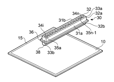
Reagent can be added to an analyte present in a gel (10), in accordance with
the
claimed method, by using the multiwell template (30) shown in Figure 1. The
diagram
shows a multiwell template (30), made of a suitable material such as plastic
or metal,
positioned above a polyacrylamide gel (10) which is supported on a sheet (15)
which
may be made, for example, of plastic or glass. The gel (10) contains an
analyte
CA 02612056 2007-12-13
WO 2006/136297 PCT/EP2006/005531
12
which has typically undergone electrophoretic separation. Thus, for example,
the gel
may contain proteins or peptides. The multiwell template (30) consists of an
elongated body (32) having two elongated side walls (31a, b) joined at their
ends by
two end walls (33a, 33b). A plurality of open-ended chambers (34 i-n) are
arranged
along the longitudinal axis of the body (32), side chambers being separated
from their
neighbour(s) by intermediate walls (35a-35n-1), each of which extends from
side wall
31 a to side wall 31 b. The shape of each chamber (34) may vary, for example
being
circular, oval, polygonal, square or rectangular (as shown).
The multiwell template (30) is moved onto the gel (10), such that the tapered
portion
(32b) of the body of the template compresses the gel (10) so that the base
(38) of the
template comes into close proximity to the sheet supporting the gel (10). In
this
position, a well is defined with the gel or plastic sheet forming the base and
the walls
of the template defining the walls of the well.
A liquid reagent, such as a buffer or a protease enzyme solution, is added to
one or
more of the wells (34), to form a liquid analyte reagent mixture. The reagent
may
solubilise the analyte, as for example in the case of a buffer, or it may
modify the
analyte (as, for example, in the case of a protease and a protein), or it may
modify the
environment in which the anaiyte is present (as for example in the case of an
acid).
A diagram of another apparatus which can be used to add a reagent to an
analyte in
a gel is shown in Figure 2. The gel (110), such as an SDS gel, is present on
the
surface of the sheet (115). The sheet (115) is positioned on a base plate
(120), made of a plastic or metal material, which has a recess (122) for
locating the
sheet in a predefined position relative to the plate (120). Fastening means,
in the form
of threaded screw bores (124 a-c, 124d not shown), are located at each corner
of the
plate (120) to allow affixing by screws (not shown) of the base plate (120) to
a top
plate (140) in a predefined position. It will be understood that other forms
of fastening
means can be used (e.g. clasps, clamps, pins and holes, snap fastening).
CA 02612056 2007-12-13
WO 2006/136297 PCT/EP2006/005531
13
A plurality of wells is formed on the gel by means of a multiwell template
(130) which
may be made of any suitable material such plastic, a metal, ceramic or
composite
material. The multiwell template (130) consists of an elongated body (132)
having
two elongated side walls (131 a, 131 b) joined at their ends by two end walls
(133a,
133b). A plurality of open-ended chambers (134 i-n) are arranged along the
longitudinal axis of the body (132), side chambers being separated from their
neighbour(s) by intermediate wall (135a-135n-1), each of which extends from
side
wall 131 a to side wall 131 b. Each chamber (134) may take any appropriate
shape,
for example circular, oval, polygonal, square or rectangular (as shown).
The body (132) of the multiwell template (130) is divided by a flange (136)
into a first
(132a) and second (132b) portion; the first (132a) portion being shaped for
insertion
into an opening (142) in the top plate (140) and a second portion (132b) being
tapered to a base (138) for compressing the gel. It will be understood that
when the
template (130) is lowered or pushed onto the gel (110) and contacts, or comes
into
close proximity with, the sheet (115) supporting the gel (110), each chamber
(134) forms a well with the gel or sheet forming a base and the walls of the
chamber
(not shown) defining the walls of the well. In this way a plurality of wells
are created
on the gel. The multiwell template (130) may be inserted into an opening (142)
in a
top plate (140), which is composed of a plastic or metal (e.g. stainless
steel) material,
either before or after it has been lowered or pushed onto the gel to form a
plurality of
wells thereon. In the example shown, the flange (136) supports the template
(130) on
the ledge (144) of the top plate (140). It should be noted that the template
does not
cut the gel but rather compresses it to form a plurality of wells. The wells
are held in a
predefined position relative to the base plate (120) and the top plate (140)
by affixing
the top plate (140) to the base plate (120) by fastening means in the top (146
a-d)
and base (124 a-d) plates. In the example of Figure 2, screws (not shown) are
used
to secure the top plate (140) to the base plate (120) by insertion through
openings in
the top plate (146 a-d) and into the screw bores (124a-d) in the base plate
(120). In
this way the plurality of wells formed in the gel is held in a predefined
position relative
CA 02612056 2007-12-13
WO 2006/136297 PCT/EP2006/005531
14
to the top and bottom plate. A liquid reagent can then be added to the one or
more
wells to form a liquid analyte reagent mixture as described above.
Figure 3 is a perspective view of another apparatus which can be used to carry
out
the method of the invention. The apparatus shown in Figure 3 is suitable for
use with
isolectric focusing gels, in particular IPG strips such as ImmobilineT""
DryStrip gels
(GE Healthcare). The IPG strip (not shown), consisting of a plastic base sheet
supporting a coating of polyacrylamide gel (210), is placed within a recess
(252) of a
retainer (250) which is an electrophoresis manifold. The retainer (250), which
is
typically made of a plastic material, may consist of a plurality of recesses
(252 i - n),
twelve being shown in the example of Figure 3, such that a plurality of IPG
strips may
be processed at the same time. Following electrofusing of an analyte in the
IPG strip,
the retainer (250) together with the strip is located in a predefined position
within a
recess (222) in the top surface of the base plate (220). The base plate may be
made
of a plastic or metal material. A multiwell template (230), similar in
construction to that
described above in Figures 1 & 2, comprises a plurality of open-ended chambers
(234i-n) and is inserted in an opening (242) in a top plate (240) such that it
supported
by its flange (236) on a ledge (not shown) surrounding the opening (242). The
multiwell template (230) and the top plate (240) are typically made of a
plastic
material but may be made of other materials such as a metal. It will be
understood
that a plurality of multiwell templates (230) may be positioned in the top
plate (240) in
the manner described; thus, for example, in the example shown, twelve
multiwell
templates (230) can be positioned within the top plate (240).
Once it is positioned within the top plate (240), the multiwell template (230)
is lowered
or moved onto the surface of the gel (210), such that the tapered portion
(232b) of the
body of the template compresses the gel (210) such that the base (238) of the
template comes into close proximity to the plastic sheet supporting the gel
(210). In
this position, a well is defined with the plastic sheet or gel forming the
base and the
walls of the template defining the walls of the well.
CA 02612056 2007-12-13
WO 2006/136297 PCT/EP2006/005531
It will be understood that the multiwell template (230) may be lowered or
moved onto
the gel (210) to form a plurality of wells thereon before the template (230)
is inserted
into the top plate (240). The multiwell template can then be secured into
position
5 relative to the top (240) and bottom (220) plates by use of the fastening
means in the
top (246 a-d) and bottom (224 a-d; d not shown) plates; for example, in Figure
3,
screws (not shown) could be used to affix the plates together.
A securing strip (260) is positioned over the top of the multiwell template
(230) such
10 that the openings (264i- n) in the strip (260) overlap and correspond to
the positions
of the open ended chambers (234 i-n) in the template (230). The securing strip
(260)
may then lock the template (230) into a predefined position by affixing it to
the top
plate (240) by use of the fastening means in the strip (266 a-b) and the top
plate (248
i-n); such fastening means may take the form of openings in the securing strip
(266a-
15 b), screw bores in the top plate (248 i - n) and the use of one or more
screws of
appropriate bore. Altematively the securing strip may be formed integrally
with a
multiwall template.
Figure 4 is a plan perspective of the apparatus of Figure 3, where each of the
component parts has the same features as described above for Figure 3. Thus
the
apparatus consists of a base plate (320) having a recess (322) and fastening
means
(324 a-d, d not shown). A retainer (350) in the form of an isoelectric
focussing
manifold holds a number of IPG strips (not shown) within a series of recesses
(352 i-
n) consisting of a plastic sheet supporting a polyacrylamide gel (310). The
top plate
(340), made of a plastic material, consists of a plurality of openings (342 i-
n)
corresponding to the positions of the IPG strips within the retainer (310).
Fastening
means (346 a-d; and 348 i - n), corresponding to those present in the base
plate (324
a-d) are present in the top plate (340). The multiwell template (330)
comprises a
plurality of open ended chambers (334 i-n). The securing strip (360) consists
of a
number of openings (364 i-n) corresponding to the position of the open-ended
chambers (334i-n) in the template (330) and fastening means (366 a & b).
CA 02612056 2007-12-13
WO 2006/136297 PCT/EP2006/005531
16
The apparatus of Figure 4 may be used in the same way as described above in
connection with Figure 3 to add a reagent to an analyte in a gel.
Figure 5a is a view of the base (438) of a multiwell template (430) which is
used
according to the method of the invention. The open-ended chambers (434 i-n)
are
defined by a series of walls (433) throughout the body (432) of the template
(430).
Recesses or notches (437) on the base of the template (430) are used to place
the
template (430) onto protrusions in the retainer (not shown) which holds the
IPG strips,
and thus to locate the template (430) in a predefined position relative to the
IPG strip.
Figure 5b shows the plastic sheet (415) of an IPG strip positioned on the base
(438)
of the multiwell template (430). In the perspective view shown, the gel cannot
be seen
because it is on the underside of the sheet (415) and is in contact with the
base (438)
of the template (430). In this position, the base of the sheet (415) within
each
chamber (434) forms the base of a well and the walls of the chamber act as the
walls
of a well.
Figure 6 shows a plan perspective of a top plate (540) which is made of steel.
The
openings (542 i-n) for receipt of the multiwell template (shown in position),
together
with fastening means for affixing to the base plate (546 a-d) and for affixing
to the
securement strip (548 i-n), are illustrated in the diagram.
Figure 7a is a plan view showing details of a top plate (640) used in the
method of the
invention in which the securing strip (660) has been positioned to affix the
multiwell
template (not shown) to the top plate (640). The fastening means (666), in the
form of
openings, are shown and co-locate with those of the retainer (not shown) in
the top
plate (see 548 i-n in Figure 6).
Figure 7b is an underside view showing details of the arrangement given in
Figure 7a.
The base (638) of the tapered second portion of the multiwell template, which
CA 02612056 2007-12-13
WO 2006/136297 PCT/EP2006/005531
17
protudes from the lower surface of the top plate (660), is seen clearly from
this angle.
It is this base (638) which compresses the gel, each open-ended chamber (634i-
n)
forming a well with the gel or the base sheet (not shown) of the gel.
Figure 8 shows an automatic eluting system according to the present invention.
Following electrophoresis of a sample on a gel, for instance an IPG strip, a
plurality of
wells is formed and buffer added to each well using the method of the
invention as
described above. The gel in each well is then eluted with the buffer to
extract the
analyte (such as a peptide) and the resulting eluant transferred to a reaction
vessel
for further processing/analysis. Figure 8 shows an eight channel eluting probe
(770)
in the process of transferring eluant from the wells present in the top plate
(740) of the
apparatus of the invention to wells (782 i-n) in a microtitre plate (780). The
system is
under the control of a computer (not shown). The number of wells formed in the
IPG
strip typically correspond to the number of wells across the length or breadth
of the
microtitre plate (e.g. they are a multiple of 8 or 12 for a 96 well microtitre
plate) or a
fraction of these numbers (e.g. 2, 3, 4, 6).
Specific Examples
Isoelectric focusing, fluorescence analysis and extraction of peptides
0.5mg of a tryptic digest sample from Saccharomyces cerevisiae, Type II, was
mixed
with 5pg of each of the pl-markers '3.73', '4.25' and '4.54'. A'pl-marker' is
a
fluorescently labelled peptide with known isoelectric point that can be
detected by
fluorescence scanning. The fluorescent label used was Cy5TM (available from
Amersham Biosciences AB; Sweden) which emission spectrum is taken at -660nm
(Ettan DIGE System - User Manual, Amersham Biosciences AB, Sweden). A 24cm
IPG peptide strip (pH 3.4-4.8) was rehydrated overnight (-15 hours, room
temperature) in 350NI of 8M urea and sample solution. The rehydrated strip was
CA 02612056 2007-12-13
WO 2006/136297 PCT/EP2006/005531
18
transferred to an EttanTM IPGphorTM manifold and isoelectric focusing was run
using
the following program: Gradient 500 V 1 minute, Gradient 4000 V 1.5 hours,
Gradient
6000 V 1.5 hours, Gradient 10000 V 1.5 hours, Step 10000 V 12 hours (total
-150kVhrs). Ettan IPGphor II was used as the focusing unit and the focusing
was
performed at 20 C.
After focusing, the IPG strip was scanned in a fluorescence scanner (Typhoon
9400
scanner, Amersham Biosciences, Sweden) at 660 nm, to determine the exact
position
of the fluorescent pl-markers. The Typhoon pictures were evaluated in
ImageQuant
and fluorescence intensity graphs established.
After scanning, the peptides in the strip were extracted from the gel into
liquid
fractions using the multiwell template of the invention. Thereby the pH
gradient is
divided into a series of discrete fractions along the strip. In this manner,
the IPG strip
was divided into 72 fractions at about 3 mm intervals. 50pI water was added to
each
of the 72 wells, incubated at room temperature for 60 minutes and extracted
peptides
were then transferred to a microtitre plate in an automated manner. The
elution
process was repeated three times to ensure extraction and transfer of all
peptides
from each well. After extraction, the multiwell template was removed from the
IPG
strip and the device can be reused following cleaning in consecutive
experiments. In
the described experiment, the IPG peptide strip was once more scanned in a
Typhoon scanner and the pictures were evaluated in ImageQuant.
Figure 9 shows the fluorescent intensity of the peptide IPG strip before (Fig
9a) and
after (Fig 9b) extraction. Figure 9c shows the scanned microtitre plate with
extracted
peptide samples and the strips before and after extraction, demonstrating high
and
low levels of fluorescence, respectively. From the Figures it is clear that
the peptides
have been effectively extracted from the IPG strip and are now present in the
wells of
the microtitre plate.
CA 02612056 2007-12-13
WO 2006/136297 PCT/EP2006/005531
19
Figure 10 shows the result of a comparison between all identified peptide
sequences
in seven fractions next to each other on the basic end of the IPG strip. Of a
total of
719 identified peptides in the seven compared fractions, 82% of the peptides
were
present in only one fraction and 16% in two fractions. The results of this
experiment
not only underline the high resolution in the IPG strip but also that there is
no problem
with leakage between the wells formed using the multiwell template of the
invention.