Note: Descriptions are shown in the official language in which they were submitted.
CA 02612247 2007-12-14
WO 2007/005201 PCT/US2006/022807
MEDICAL-IMAG.ING 'DEVICE HAVING A FORWARD LOOKING FLOW DETECTOR
FIELD OF THE INVENTION
The field of the invention relates to medical devices, and more particularly
to medical
imaging devices for placement in catheters and guidewires.
BACKGROUND OF THE INVENTION
Intraluminal, intracavity, intravascular, and intracardiac treatments and
diagnosis of
medical conditions utilizing minimally invasive procedures are effective tools
in many areas of
medical practice. These procedures are typically performed using imaging and
treatinent
catheters that are inserted percutaneously into the body and into an
accessible vessel of the
vascular system at a site remote from the vessel or organ to be diagnosed
and/or treated, such
as the femoral artery. The catheter is then advanced through the vessels of
the vascular system
to the region of the body to be treated. The catheter may be equipped with an
imaging device,
typically an ultrasound imaging device, which is used to locate and diagnose a
diseased portion
of the body, such as a stenosed region of an artery. For example, U.S. Pat.
No. 5,368,035,
issued to Hamm et al., the disclosure of which is incorporated herein by
reference, describes a
catheter having an intravascular ultrasound imaging transducer. These are
generally known in
the art as Intravascular Ultrasound ("IVUS") devices.
Fig. 1 shows an example of an inlaging transducer assembly 1 known in the art.
The
imaging transducer 1 is typically within the lumen 10 of a guidewire or
catheter (partially
shown), having an outer tubular wall member 5. To obtain an image of a blood
vessel the
imaging transducer assembly 1 may be inserted into the vessel. The transducer
assembly 1
may then interrogate the cross-sectional plain of the vessel from the inside
by rotating while
simultaneously emitting energy pulses, e.g., ultrasound pulses, and receiving
echo signals.
It may be desirable to obtain not only a cross-sectional plane of the vessel,
but also
information on blood flow within the vessel. Accordingly, an improved imaging
catheter
would be desirable.
SUMMARY OF THE INVENTION
The present invention generally relates to medical devices, and more
particularly to an
improved medical imaging device. In one embodiment, an imaging device includes
a drive
shaft having proximal and distal ends received within the lumen; an imaging
transducer
coupled to the distal end of the drive shaft and positioned at the distal
portion of the elongate
member; and a flow detector coupled to the imaging transducer. The flow
detector may
include a forward facing ultrasound transducer configured to emit energy in
the direction of the
1
CA 02612247 2007-12-14
WO 2007/005201 PCT/US2006/022807
longitudinal axis-of the iinaging device and detect a Doppler shift from the
received echoes.
The imaging device may be configured to be placed in a catheter or guidewire.
Other systems, methods, features and advantages of the invention will be or
will
become apparent to one with skill in the art upon examination of the following
figures and
detailed description. It is interided that all such additional systems,
methods, features and
advantages be included within this description, be within the scope of the
invention, and be
protected by the accompanying claims.
BRIEF DESCRIPTION OF THE DRAWINGS
In order to better appreciate how the above-recited and other advantages and
objects of
the present inventions are obtained, a more particular description of the
invention briefly
described above will be rendered by reference to specific embodiments thereof,
which are
illustrated in the accoinpanying drawings. It should be noted that the
components in the figures
are not necessarily to scale, emphasis instead being placed upon illustrating
the principles of
the invention. Moreover, in the figures, like reference numerals designate
corresponding parts
throughout the different views. However, like parts do not always have like
reference
numerals. Moreover, all illustrations are intended to convey concepts, where
relative sizes,
shapes and other detailed attributes may be illustrated schematically rather
than literally or
precisely.
Fig. 1 is a cross-sectional side view of an imaging transducer assembly known
in the
art.
Fig. 2 is a perspective view of an imaging device in accordance with a
preferred
embodiment of the present invention.
Fig. 3a is a perspective view illustrating a construction of an imaging device
in
accordance with a preferred embodiment of the present invention.
Fig. 3b is a perspective view illustrating an construction of an imaging
device in
accordance with a preferred embodiment of the present invention.
Fig. 4 is a cross-sectional view of an imaging wire in accordance with a
preferred
embodiment of the present invention.
Fig. 5 is a diagram of a medical imaging system in accordance with a preferred
embodiment of the present invention.
DETAILED DESCRIPTION OF THE PREFERRED EMBODIMENTS
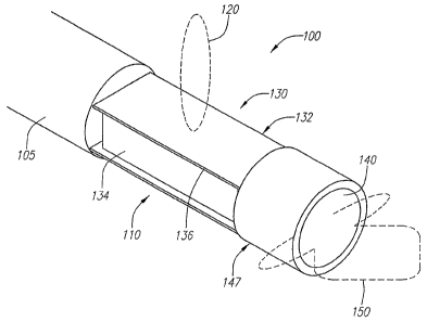
Turning to Fig. 2, an imaging device 100 is shown in accordance with a
preferred
embodiment of the present invention. The imaging device 100 includes a
transducer housing
2
CA 02612247 2007-12-14
WO 2007/005201 PCT/US2006/022807
110 having an imaging trari"sducer 130 known in the art, such as that shown in
Fig. 1. In the
case where the imaging transducer 130 is an ultrasound transducer, the
transducer 130 may
include a layer of electrode coated piezoelectric crystal ("PZT") 136,
"sandwiched" between a
conductive acoustic lens 132 and a conductive backing material 134, formed
from an
acoustically absorbent material (e.g., an epoxy substrate having tungsten
particles). During
operation, the PZT layer 136 is electrically excited by both the backing
material 134 and the
acoustic lens 132, causing energy, e.g., acoustic pulses 120, to be emitted
from the lens 132,
which faces a direction generally perpendicular to the longitudinal axis of
the imaging device
100. As described above, a cross-sectional image of a vessel may be obtained
from the
imaging transducer 130 as it rotates about the longitudinal axis of the
imaging device 100.
The transducer 130 is coupled to the distal end of a cable 105 that includes a
drive shaft
(not shown) and conductors (not shown) that electrically couple the transducer
130 to a
processing unit (not shown). An additional transducer 140 is coupled to the
distal end of the
imaging transducer 130. The additional transducer 140 may be positioned within
a housing
socket 147 that is mounted to the distal end of the imaging transducer 130.
The additional
transducer 140 is forward facing, i.e., the transducer 140 faces a direction
generally parallel to
the longitudinal axis of the imaging device 100. The additional transducer
140, herein referred
to as the Doppler transducer 140, can be configured to send narrow band burst
energy signals,
e.g., acoustic signals, and receive the echoes. From the received echoes, the
velocity of blood
flow within a vessel may be calculated. Due to the red cell's movement in the
blood, the
emitted energy signals scatter, resulting in scattered echoes. The scattered
echoes will have a
frequency shift from the original signals, known in the art as a Doppler
shift. Generally, the
Doppler shift is proportional to the blood velocity and cosine of the Doppler
angle, which is the
angle between the blood flow and the energy beam, e.g., ultrasound beam. For
ultrasound
beams, the Doppler shift can be within audio range, so a user can determine
the direction of
blood flow by listening for the Doppler shift. Such information, i.e.,
direction and velocity of
the blood flow, is invaluable in locating and evaluating the existence or
effect of stenosis in a
patient.
In the case where the Doppler transducer 140 is an ultrasound transducer 140,
the
Doppler frequency shift information can be detected by using demodulation
methods on the
received echoes. The Doppler frequency shift fd is quantitatively related to
the blood velocity
that it encounters:
2vcos6 .fo (1)
.f~t - c
where fo is the center frequency of the transmitted acoustic beam emitted from
the Doppler
3
CA 02612247 2007-12-14
WO 2007/005201 PCT/US2006/022807
transducer 140, c is the sound velocity in the tissue, v is the velocity of
the blood flow, and A
is the angle between the flow of the blood and the ultrasound beam 150.
Equation (1) shows
that the Doppler shift has a maximum value when the Doppler transducer 140 is
parallel to the
blood flow direction, i.e., cos 0 . Thus, the Doppler transducer 140 can serve
as a forward-
facing guide for the imaging catheter or guidewire. The user can simply search
for the Doppler
shift. Such a transducer 140 may include a thin PZT layer, similar to the
ultrasound transducer
described above. Other single crystal and/or piezofilm materials may be used,
or any kind of
composite materials using piezomaterials. The transducer 140 may be a single
beam, an
annular array, or multi-beam device.
Further, other imaging devices may be used, instead of, or in addition to
imaging
transducers 130, such as light based apparatuses for obtaining images through
optical
coherence tomography (OCT). Image acquisition using OCT is described in Huang
et al.,
"Optical Coherence Tomography," Science, 254, Nov. 22, 1991, pp 1178-1181,
which is
hereby incorporated by reference in its entirety. A type of OCT imaging
device, called an
optical coherence domain reflectometer (OCDR) is disclosed in Swanson U.S.
Pat. No.
5,321,501, which is incorporated herein by reference. The OCDR is capable of
electronically
performing two- and three-dimensional image scans over an extended
longitudinal or depth
range with sharp focus and high resolution and sensitivity over the range. In
addition, other
devices may be used instead of, or in addition to, ultrasound transducers 140,
such as light
based apparatuses.
The Doppler transducer 140 may have a beam pattern 150 that is wider than the
imaging transducer 130. Further, because the Doppler transducer's 140 surface
is generally
perpendicular to the longitudinal axis of the imaging device 100, the rotation
of the imaging
device 100 and/or the imaging transducer 130 will have little effect on the
Doppler shift signal,
which is generally only sensitive to the relative movement between the Doppler
transducer 140
and the scattered echoes.
Where ultrasound transducers are used for the imaging transducer 130 and the
Doppler
transducer 140, the Doppler transducer 140 can operate at a relatively narrow
bandwidth,
different from the imaging transducer 130, allowing both the transducers 130
and 140 to
operate in parallel. For example, the imaging transducer 130 may operate at 40
MHz with a
bandwidth of 80%, i.e., where the low frequency band edge will be at 24 MHz.
In such a case,
a Doppler transducer 140 may operate at 20 MHz. Assuming a maximum flow
velocity for the
blood is approximately 1 meter per second (m/s) and the sound velocity is
approximately 1500
m/s, from equation (1), the maximum Doppler shift is lower than 26 kHz. Thus,
in the
4
CA 02612247 2007-12-14
WO 2007/005201 PCT/US2006/022807
irequency ctomain, the two~signals, i.e., the imaging signals, and the Doppler
signals, are
substantially different.
Preferably, for efficiency purposes, the electrical impedance between the
imaging
transducer 130 and the Doppler transducer 140 are configured to be different.
Thus, one of the
transducers 130 and 140 is configured to have a high impedance at the
operating frequency of
the other transducer 130 and 140, and the operating energy will travel to the
corresponding
load. With inductor tuning methods known in the art, the imaging transducer
130 can have an
impedance as high as 1 kg at 20 MHz and the Doppler transducer 140 can have an
impedance
of 37052 at 40 MHz.
To construct an imaging device 100 having both an imaging transducer 130 and a
Doppler transducer 140, a round socket 149 is attached to the distal end of
the imaging
transducer 130 and configured to receive the Doppler transducer 140, which is
covered in an
isolation ring 145, as shown in Fig. 3a. The round socket 149 functions as a
housing for the
Doppler transducer 140. The round socket 149 can be conductive, serving as a
ground for both
transducers 130 and 140. A signal wire for the Doppler transducer 140 (not
shown) can be
directly connected from the imaging transducer surface 130 or be located on
the side of the
imaging transducer 130. Of course, the shape and size of the socket 149 need
not be round as
the socket should be adapted to accommodate the shape and size of the Doppler
transducer
140.
In another embodiment, an isolation ring 142 configured to cover the Doppler
transducer 140 can be constructed to also function as the housing, as shown in
Fig. 3b. The
isolation ring 142 and the Doppler transducer 140 may be attached to the
imaging transducer
housing 110 with conductive epoxy. The Doppler transducer 140 may have any
shape, such as
round, square, hexagon, or octagon. Further, to increase the beam 150 diameter
of the Doppler
transducer 140, the transducer 140 can include a convex surface and/or a
separate lens (not
shown).
Turning to Fig. 4, the imaging device 100 may be used in a catheter, as
described
above, and can also be placed in a distal portion 520 of a guidewire 500. The
guidewire 500
may comprise a guidewire body 302 in the form of a flexible, elongate tubular
member, having
an outer wall 301. The guidewire body 302 may be formed of any material known
in the art
including nitinol hypotube, metal alloys, composite materials, plastics,
braided polyimide,
polyethylene, peek braids, stainless steel, or other superelastic materials.
The Doppler transducer 140 and the imaging transducer 130 may utilize two
different
wiring systems for electrical coupling to one or more processing devices
(shown below). For
5
CA 02612247 2007-12-14
WO 2007/005201 PCT/US2006/022807
example, the cable 105 attached to the proximal end of the imaging transducer
130 may include
two coaxial cables, each servicing a transducer 130 and 140. Alternatively,
the coupling may
be indirect, capacitive, or inductive coupling as known in the art.
Turning to Fig. 5, a proximal portion 510 of the guidewire 500, shown in Fig.
4, may be
adapted to connect to circuitry 600 that processes imaging signals from the
imaging transducer
130 and/or electrical signals from the Doppler transducer 140, such circuits
being well known.
In the foregoing specification, the irivention has been described with
reference to
specific embodiments thereof. It will, however, be evident that various
modifications and
changes may be made thereto without departing from the broader spirit and
scope of the
invention. For example, the reader is to understand that the specific ordering
and combination
of process actions described herein is merely illustrative, and the invention
can be performed
using different or additional process actions, or a different combination or
ordering of process
actions. As a further example, each feature of one embodiment can be mixed and
matched
with other features shown in other embodiments. Additionally and obviously,
features may be
added or subtracted as desired. Accordingly, the invention is not to be
restricted except in light
of the attached claims and their equivalents.
6