Note: Descriptions are shown in the official language in which they were submitted.
CA 02612450 2007-12-17
WO 2007/000048 PCT/CA2006/001065
Membrane Array and Analytical Device
Field of the Invention
This invention relates to analytical devices and methods useful for analytical
assays of fluid samples. More specifically, the invention is directed to a
novel
membrane array and analytical devices incorporating same. The invention is
useful
for rapidly determining the presence of one or more analytes in small volumes
of
sample.
Background of the Invention
Immunoassay devices and procedures currently exist for detecting the
presence of an analyte in a sample of biological fluid. Typically,
immunochemical
reactions involving antigen/antibody reactions take place on dry porous
carriers such
as cellular membranes through which the sample to be analyzed flows by
capillary
action. The presence of an analyte in the sample can be detected either
visually or
by using reflectance or fluorescence based detection systems and instruments.
Oftentimes, the label is an enzyme label or a particulate direct label, for
instance a
gold sol label.
Typical innmunochromatographic devices of this nature are described in the
following U.S. Patents: 4,094,647; 4,235,601; 4,361,537; 4,703,017, 4,774,192;
4,839,297; 4,861,711; 4,885,240; 4,960,691; 5,075,078; 5,079,142; 5,110,724;
5,120,643; 5,135,716; 5,468,648; 5,591,645; 5,607,863; 5,622,871; 5,648,274;
5,656,503; 5,846,838; 5,869,345; 5,877,028; 5,998,220; 6,017,767; 6,168,956;
6,171,870; 6,187,598; 6,214,629131; 6,228,660; 6,528,321; and 6,534,320.
U.S. Patent No. 5,290,678 describes an analytical test kit incorporating a dry
chemistry membrane comprising antibody pairs to multiple analytes observed
during
cardiovascular events. In operation of the device, multiple transfer steps are
required before analysis and further this device is only designed for
receiving
samples of serum or plasma and as such is not suitable for analyses using
whole
blood.
CA 02612450 2007-12-17
WO 2007/000048 PCT/CA2006/001065
U.S. Patent No. 5,559,041 discloses an immunoassay device with a
membrane array comprising an overlapping arrangement of a reservoir pad,
numerous membrane filters and a wicking membrane all with an equal range of
pore
sizes. In the use of this device, rapid and high sensitivity analysis of
analyte
concentrations cannot be achieved with small sample sizes.
PCT/182003/005088 describes a membrane array and analytical device
designed for the sensitive detection of analytes from a sample of fluid as
small as a
drop. The membrane array comprises a two membrane system including a first
separation membrane and an analytical capture membrane. However, rapid and
high sensitivity detection of analytes using whole blood is not achievable in
all
circumstances and membrane array constructions with small sample volumes
without background interference caused by hemolysis (the liberation of
hemoglobin
from the red blood cell).
While the aforementioned devices are generally useful for detecting analytes
in a sample, it is desirable to provide an analytical device which has greater
sensitivity using smaller sample volumes and at the same time provides a rapid
test
result. Thus, there is a need to develop an analytical device that is designed
to
obviate some of the deficiencies of the prior art devices.
Summary of the Invention
The present invention is an improved membrane array and analytical device
that is used to rapidly detect one or more analytes from small volume samples
in
one step with high efficiency and high sensitivity compared to any type of
membrane
arrays of the prior art. In aspects, the invention is especially suitable for
use with
small samples of whole blood with minimal hemolysis.
According to an aspect of the present inventiqn is an improved membrane
array that accommodates small sample volumes and provides rapid, highly
efficient
and highly sensitive detection of one or more analytes in the small sample
volume.
According to another aspect of the present invention is an improved
membrane array that accommodates small whole blood sample volumes and is
capable of rapid, highly efficient and highly sensitive detection of one or
more
analytes in the whole blood sample volumes with substantially minimal or
negligible
hemolysis of the whole blood.
2
CA 02612450 2007-12-17
WO 2007/000048 PCT/CA2006/001065
According to another aspect of the present invention is an improved
membrane array that accommodates small sample volumes and is capable of rapid,
highly efficient and highly sensitive detection of one or more analytes in the
sample,
the membrane array comprising three or more porous membranes, each of which is
arranged in a manner to be in non-planar contact with respect to adjacent
ones.
According to another aspect of the present invention is an improved
membrane array that accommodates small whole blood sample volumes and is
capable of rapid, highly efficient and highly sensitive detection of one or
more
analytes in the whole blood sample volumes with minimal or negligible
hemolysis of
the whole blood, the membrane array comprising three or more porous membranes,
each of which is arranged in a manner to be in non-planar contact with respect
to
adjacent ones.
According to another aspect of the present invention there is provided a
membrane array, said membrane array comprising:
- at least three overlapping porous membranes arranged in stair step
configuration comprising;
- a first step adapted to receive a fluid sample and containing a detection
reagent;
- a second step having a lower porosity than said first step; and
- a third step having a lower porosity than said second step and containing
a
capture reagent.
According to yet another aspect of the present invention there is provided a
membrane array, said membrane array comprising:
- at least three overlapping porous membranes arranged in stair step
configuration comprising;
- a first step adapted to receive a small sample of whole blood, said first
step
being retardant of red blood cells and containing a detection reagent;
- a second step having a porosity that is further retardant of red blood cells
compared to said first step with minimal hemolysis of the sample; and
- a third step having a lower porosity than said second step and containing a
capture reagent.
3
CA 02612450 2007-12-17
WO 2007/000048 PCT/CA2006/001065
According to another aspect of the present invention is an analytical device
comprising a membrane array of the present invention.
According to still another aspect of the present invention is an analytical
device for the detection of an analyte in a small volume of sample, the device
comprising:
- a membrane array having at least three overlapping porous membranes
arranged in stair step configuration, the membrane array comprising;
- a first step adapted to receive a fluid sample and containing a detection
reagent;
- a second step having a lower porosity than said first step; and
- a third step having a lower porosity than said second step and containing
a
capture reagent.
According to yet another aspect of the present invention is an analytical
device useful for the rapid and highly sensitive detection of at least one
analyte in a
drop of sample; the analytical device comprising:
- a membrane array of at least three overlapping porous membranes
arranged in stair step configuration the membrane array comprising; a first
step
adapted to receive a fluid sample and containing a detection reagent; a second
step
having a lower porosity than said first step; and a third step having a lower
porosity
than said second step and containing a capture reagent;
- the membrane array being enclosed in a platform formed with sample
application means and a sample flow channel, where said sample flow channel
directs flow of sample from sample application means to the first step wherein
the
sample flows substantially horizontally into the first step through a
thickness of the
membrane edge and wherein the sample flows by capillarity through the second
step
to the third or subsequent steps of said membrane array.
According to yet another aspect of the present invention is an analytical
device comprising;
- a membrane array supported within an analytical device housing having a
first and a second end, wherein one end is adapted to receive a removable cap,
said
4
CA 02612450 2007-12-17
WO 2007/000048 PCT/CA2006/001065
cap facilitating the application of a sample to said membrane array via a
sample flow
channel.
In aspects, the membrane array can be a two membrane or a three or more
membrane stair step configuration as described herein. In further aspects, the
application of a sample may be done via a pipette for example.
In yet another aspect of the present invention there is provided an analytical
device, the device comprising;
- a membrane array supported within an analytical device housing having a
first and a second end, wherein one end is adapted to be immersed in a sample
such
that sample is provided to said membrane array.
In aspects, the membrane array can be a two membrane or a three or more
membrane stair step configuration as described herein.
According to another aspect of the present invention is a method for
determining the amount of analyte in a small volume fluid sample, said method
comprising;
- providing a membrane array comprising;
a first step adapted to receive a fluid sample and containing a
detection reagent;
a second step having a lower porosity than said first step; and
a third step having a lower porosity than said second step and
containing a capture reagent; and
- applying a fluid sample to one end of said first step via a sample flow
channel, wherein said fluid sample moves into the first step horizontally
through the
thickness of a membrane edge via capillary flow and moves to said second step
and
then to said third step where capture of said analyte occurs.
In aspects, the membrane array has more than three steps and the fluid
sample moves from first, to second, to third and subsequent steps as herein
described.
In aspects of the invention, the fluid sample is a small volume of whole
blood.
CA 02612450 2007-12-17
WO 2007/000048 PCT/CA2006/001065
According to yet another aspect of the present invention is a one step method
for detecting an analyte in a fluid sample, the method comprising;
applying a fluid sample to a membrane array comprising;
a first step adapted to receive a fluid sample and containing a
detection reagent; a second step having a lower porosity than said first step;
and a third step having a lower porosity than said second step and containing
a capture reagent;
wherein said fluid sample is applied via a sample flow channel, horizontally
through a thickness of the membrane edge, to one end of said first step, said
fluid
sample moving via capillary flow to said second step and then to said third
step
where detection of said analyte occurs.
According to another aspect of the present invention there is provided an
improved membrane array that accommodates small sample volumes and provides
rapid, highly efficient and highly sensitive detection of one or more analytes
in the
sample, the membrane array comprising three or more porous membranes which are
non-planar with respect to adjacent ones.
According to another aspect of the present invention there is provided an
improved membrane array that accommodates small sample volumes and provides
rapid, highly efficient and highly sensitive detection of one or more analytes
in the
sample, the membrane array comprising three or more porous membranes each of
which has a different porosity, said porous membrane being non-planar with
respect
to adjacent ones.
According to another aspect of the present invention there is provided an
analytical device for the rapid detection of a component in a small volume of
sample,
the analytical device comprising:
- a membrane array having at least three overlapping porous membranes
arranged in stair step configuration comprising;
- a first step adapted to receive a fluid sample and containing a detection
reagent;
-a second step having a lower porosity than said first step; and
- a third step having a lower porosity than said second step and containing
a
capture reagent.
6
CA 02612450 2007-12-17
WO 2007/000048 PCT/CA2006/001065
According to another aspect of the present invention there is provided an
analytical device useful for the rapid and highly sensitive detection of at
least one
analyte in a drop of sample; the analytical device comprising:
- a membrane array of at least three overlapping porous membranes
arranged in stair step configuration the membrane array comprising; a first
step
adapted to receive a fluid sample and containing a detection reagent; a second
step
having a lower porosity than said first step; and a third step having a lower
porosity
than said second step and containing a capture reagent;
- the membrane array being enclosed in a platform formed with a sample
application means and a sample flow channel, where said sample flow channel
directs flow of sample from the sample application means to the first step
where the
sample flows by capillarity into and through the second step to the third step
of said
membrane array.
According to another aspect of the present invention there is provided an
analytical device comprising;
- a membrane array supported within an analytical device housing having a
first and a second end, wherein one end is adapted to receive a removable cap,
said
cap facilitating the application of a sample using a sample transfer means to
said
membrane array.
According to another aspect of the present invention there is provided an
analytical device, the device comprising;
- a membrane array supported within an analytical device housing having a
first and a second end, wherein one end is adapted to be immersed in a sample
such
that the sample is provided to said membrane array through the immersed end of
said analytical device.
According to another aspect of the present invention there is provided a
method for determining the amount of analyte in a small volume fluid sample,
said
method comprising;
- providing a membrane array comprising;
a first step adapted to receive a fluid sample and containing a
detection reagent;
7
CA 02612450 2007-12-17
WO 2007/000048 PCT/CA2006/001065
a second step having a lower porosity than said first step; and
a third step having a lower porosity than said second step and
containing a capture reagent; and
- applying a fluid sample to one end of said first step, wherein said fluid
sample moves via capillary flow into and through said second step and then to
said
third step where detection of said analyte occurs.
According to another aspect of the present invention there is provided an
improved membrane array that accommodates small sample volumes and is capable
of rapid, highly efficient and highly sensitive detection of analytes in the
sample, the
membrane array comprising at least three porous membranes which are non-planar
with respect to adjacent membranes.
According to another aspect of the present invention there is provided an
analytical device for the rapid detection of a component in a small volume of
sample,
the analytical device comprising:
- a membrane array having at least three overlapping porous membranes
arranged in stair step configuration comprising;
- a first separation membrane adapted to receive a fluid sample and
containing a detection reagent;
- a second separation membrane downstream from said first
separation membrane and having a lower porosity than said first separation
membrane; and
- an analytical membrane downstream from said second separation
membrane having a lower porosity than said second separation membrane
and containing a capture reagent.
According to another aspect of the present invention there is provided an
analytical device useful for the rapid and highly sensitive detection of at
least one
analyte in a drop of sample; the analytical device comprising:
- a membrane array of at least three overlapping porous membranes
arranged in stair step configuration, the membrane array comprising;
- a first separation membrane adapted to receive a fluid sample and
containing a detection reagent;
8
CA 02612450 2007-12-17
WO 2007/000048 PCT/CA2006/001065
- a second separation membrane downstream from said first
separation membrane and having a lower porosity than said first separation
membrane; and
- an analytical membrane downstream from said second separation
membrane and having a lower porosity than said second separation
membrane and containing a capture reagent.
- the membrane array being housed in a platform formed with a sample
application means and a sample flow channel, wherein said sample flow channel
directs flow of sample from the sample application means to the first
separation
membrane where the sample flows into through the second separation membrane to
the analytical membrane of said membrane array.
According to another aspect of the present invention there is provided an
analytical device for the rapid detection of an analyte in a small volume of
sample,
the analytical device comprising:
- a membrane array having at least three overlapping porous membranes
arranged in stair step configuration;
- a sample application means for receiving said sample;
- a sample flow channel in fluid communication with said sample application
means, said sample flow channel dimensioned to provide for substantially
uniform
horizontal flow of said sample into said membrane array.
According to another aspect of the present invention there is provided an a
method for detecting an analyte in a fluid sample, the method comprising;
- applying a fluid sample to a membrane array comprising;
a first separation membrane adapted to receive a fluid sample and
containing a detection reagent; a second separation membrane downstream
of said first separation membrane and having a lower porosity than said first
separation membrane; and a analytical membrane downstream from said
second separation membrane having a lower porosity than said second
separation membrane and containing a capture reagent; wherein said fluid
sample is applied via a sample flow channel to one end of said first
separation
membrane, said fluid sample moving via capillary flow to said second
separation membrane and then to said analytical membrane where detection
of said analyte occurs.
9
CA 02612450 2012-08-16
In accordance with an aspect of the present invention, there is provided an
improved
membrane array for rapid and efficient detection of one or more analytes in a
small sample
volume, said membrane array comprising three or more porous membranes which
are non-
planar with respect to adjacent ones, wherein said three or more porous
membranes are
overlapping and arranged in stair step configuration, said three or more
porous membranes
comprising:
- a first step adapted to receive a fluid sample and containing a detection
reagent, said
first step having an apex at an upstream end, said apex for receiving said
sample through its
thickness by capillary flow said first step overlapping a second step;
- said second step having a lower porosity than said first step, wherein
said second
step has a pore size that accommodates red blood cells without substantial
hemolysis, said
second step overlapping a third step; and
- said third step having a lower porosity than said second step and
containing a
capture reagent.
In accordance with a further aspect of the present invention there is provided
an
analytical device for the rapid detection of one or more components in a small
volume of
sample, the analytical device comprising:
- a membrane array having at least three overlapping porous membranes
arranged in
stair step configuration comprising:
- a first step adapted to receive a fluid sample and containing a detection
reagent, said
first step having an apex at an upstream end, said apex for receiving said
sample through its
thickness by capillary flow, said first step overlapping a second step;
- said second step having a lower porosity than said first step; wherein
said second
step has a pore size that accommodates red blood cells without substantial
hemolysis, said
second step overlapping a third step; and
- said third step having a lower porosity than said second step and
containing a
capture reagent.
In accordance with a further aspect of the present invention there is provided
a
method for detecting an analyte in a small volume fluid sample, said method
comprising:
- to a membrane array comprising three or more porous membranes which are
non-
planar with respect to adjacent ones, wherein said three or more porous
membranes are
9a
CA 02612450 2012-08-16
overlapping and arranged in stair step configuration, said three or more
porous membranes
comprising:
- a first step adapted to receive a fluid sample and containing a detection
reagent, said
first step having an apex at an upstream end, said apex for receiving said
sample through its
thickness by capillary flow said first step overlapping a second step;
- said second step having a lower porosity than said first step, wherein
said second
step has a pore size that accommodates red blood cells without substantial
hemolysis, said
second step overlapping a third step; and
- said third step having a lower porosity than said second step and
containing a
capture reagent; and
- applying a fluid sample to one end of said first step, wherein said fluid
sample
moves via capillary flow into and through said second step and then to said
third step where
detection of said analyte occurs.
In accordance with a further aspect of the present invention there is provided
an
improved membrane array for rapid and efficient detection of one or more
analytes in a
sample volume of about 10 il to about 50 [II, said membrane array comprising a
first porous
membrane having an apex at an upstream end, said apex for receiving said
sample through its
thickness by capillary flow; a second porous membrane having a lower porosity
than said first
porous membrane, wherein said second porous membrane has a pore size that
accommodates
red blood cells without substantial hemolysis; and a third porous membrane
having a lower
porosity than said second porous membrane, said first, second and third porous
membranes
being non-planar with respect to adjacent ones, and being overlapping and
arranged in stair
step configuration such that said first porous membrane overlaps said second
porous
membrane and said second porous membrane overlaps said first porous membrane,
wherein said sample volume of about 1 OW to about 50u1 is applied at said apex
and
moves horizontally and uniformly through the thickness of said first porous
membrane to said
second porous membrane and then to said third porous membrane where detection
of said
analytes occurs.
In accordance with a further aspect of the present invention there is provided
a
membrane array for detecting one or more analytes in a fluid sample containing
red blood
9b
CA 02612450 2013-04-16
cells, the membrane array comprising a separation membrane and an analytical
membrane in
fluid communication with one another, wherein:
- the separation membrane has a pore size of greater than a red blood cell to
about
8i_tm such that it retards red blood cells without substantial hemolysis and
overlaps at least a
portion of the analytical membrane; and
- the analytical membrane has a lower porosity than the separation membrane,
wherein a small volume of about 10111 to 501.11 of sample is used.
In accordance with a further aspect of the present invention there is provided
an
analytical device for detecting one or more analytes in a fluid sample
containing red blood
cells, the analytical device comprising a membrane array comprising a
separation membrane
and an analytical membrane in fluid communication with one another, wherein:
- the separation membrane has a pore size of greater than a red blood cell
and about
8pm such that it retards red blood cells without substantial hemolysis and
overlaps at least a
portion of the analytical membrane; and
- the analytical membrane has a lower porosity than the separation
membrane,
wherein a small volume of about lOjfl to 500 of sample is used.
9c
CA 02612450 2007-12-17
WO 2007/000048 PCT/CA2006/001065
Other features and advantages of the present invention will become apparent
from the following detailed description. It should be understood, however,
that the
detailed description and the specific examples while indicating embodiments of
the
invention are given by way of illustration only, since various changes and
modifications within the spirit and scope of the invention will become
apparent to
those skilled in the art from the detailed description.
Brief description of the drawings
Embodiments will now be described, by way of example only, with reference to
the attached figures, wherein:
Figure 1 is a perspective view of the membrane array of the invention;
Figure 2 is an exploded view of an analytical device incorporating the
membrane array of the invention;
Figure 3 is an exploded view of another embodiment incorporating the
membrane array of the invention in an analytical device in which the flow
channel is
formed with an indent in the top surface of the lower half that incorporates
the
membrane array of the invention;
Figure 4 is a top perspective view of a cap designed to facilitate application
of
a fluid sample to the analytical device and to protect the user from any
contamination from the fluid sample;
Figure 5 is a bottom perspective view of the cap of figure 4 and the
analytical
device of figure 2 or 3;
Figure 6 is a top perspective view showing the cap of figure 4 for reversible
engagement with the analytical device;
Figure 7 is a top perspective view of showing the cap of figure 4 fitted on
the
analytical device; and
Figure 8 is a top perspective view of another embodiment of the cap designed
to facilitate application of a fluid sample to the analytical device and to
protect the
user from any contamination from the fluid sample;
Figure 9 is a bottom perspective view of the cap of figure 8 and another
embodiment of the analytical device of invention;
Figure 10 is an exploded view of another embodiment featuring the
membrane array of the invention in an analytical device configured for dipping
into a
reservoir of sample.
CA 02612450 2007-12-17
WO 2007/000048 PCT/CA2006/001065
Detailed Description of the Invention
Glossary
The following terms have the following general meaning as they are employed
in the description of the invention and in the claims.
"Analytical device" is a combination of a membrane array and a support
platform comprising upper and lower halves which are brought into registry to
hold
and support the membrane array. These halves are generally prepared from a
rigid
plastic such as but not limited to polyacrylate or polymethacrylate. They may
be
formed into the desired configuration for cooperation with the dry porous
carriers to
form channels by molding, stamping, machining or any equivalent type process.
The
channels may be formed in the dry porous layers by stamping from a strip of
the
selected porous material or with a hydrophobic substance such as wax or ink.
"Antigen" is a molecule which, in a mammal, induces the production of an
antibody. The devices of this invention are useful for determining the
presence of
antigens or antibodies in whole blood or any other type of body fluids.
Antigens are
often referred to as "analytes" because they are characteristic of specific
physiological conditions such as infections, cancer or pregnancy.
"Capture reagent" is a material, often a second antibody to the analyte which
is to be detected in the liquid sample. It is fixed to the carrier downstream
of the
detecting reagent. It reacts with and concentrates the complex on the carrier
to
form a product which is visible to the naked eye or readable with the aid of a
suitable
instrument.
"Cardiac analytes" are analytes which are released into the blood as a result
of cardiac tissue deterioration.
"Channel" is any formed conduit in the analytical device through which the
fluid sample under analysis flows. Channels are said to be in operative
communication when a fluid in one channel flows substantially directly into
another.
"Control reagent" is any reagent that reacts with either the detection reagent
or another component separate from an analyte in a sample to provide a visible
product and thereby advise the operator that the sample has reached analytical
membrane.
"Detection reagent" is a material, often an antibody to the analyte which is
to
be detected in the liquid sample. It is typically releasably bound to the dry
porous
carrier at or downstream of the application point for the liquid sample. For
most
11
CA 02612450 2007-12-17
WO 2007/000048 PCT/CA2006/001065
immunochemical analyses, it is labeled with a detectable label such as
colloidal gold
and forms a complex with the analyte to be determined.
"Efficient" means that a detectable product can be formed with a low/small
volume of fluid, e.g. just one drop of whole blood (about 10 pl to about 50
pl),
utilizing small amounts of reagents even when the antigen is present in very
low/small concentrations as is usually the case with most analytes such as for
example with the cardiac analyte troponin I (cTnI).
"Membrane array" refers to a cellular product through which the sample to
be analyzed moves by capillary action. As will be seen by the figures and
understood by description of the invention, an array of three or possibly more
membrane segments are arranged in stair step configuration for capillary flow.
"Rapid" means that a detectable product forms within a sufficiently short
period of time relative to detection times of current technologies measuring
the same
analyte, e.g. within about 2 to about 30 minutes, to permit the medical
attendant to
draw meaningful and useful conclusions. Furthermore, it can be appreciated
that the
time required for the analysis will vary depending on the particular analytes
in
question.
The present invention is a novel membrane array and analytical devices
incorporating such, the membrane array and analytical devices permitting
rapid,
highly efficient and highly sensitive detection of a desired analyte(s). In
aspects, the
detection may be qualitative, semi-quantitative or substantially quantitative.
This
membrane array comprises at least three membrane layers arranged in a stair-
step
configuration where the pore size decreases in each successive step. This
membrane
array is particularly suited for the rapid analysis of analytes and components
of fluid
samples and in particular the analysis of small volumes of fluid samples. In
aspects,
the invention is particularly suited for the rapid analysis of components of
whole
blood using a one step procedure. The analysis is conducted with minimal
invasiveness as only a small amount of blood is required to obtain high
sensitivity
detection without background interference and with minimal hemolysis. Small
volumes of whole blood can readily be provided with a any type of finger
lancet.or
pin prick to the finger for example. Furthermore, the membrane array of the
invention can be adapted for use in a variety of analytical device
configurations. The
membrane array and analytical device incorporating such are easy to
manufacture,
do not require separate sample collection or transfer devices for capillary
blood
samples and may require a separate timing device. Furthermore, the test result
is
12
CA 02612450 2007-12-17
WO 2007/000048 PCT/CA2006/001065
relatively stable for a long time period. Rapid and accurate diagnoses using
small
volumes of sample is provided by the present invention.
The invention is now herein described with reference to Figure 1 which shows
a membrane array designated generally as reference numeral 10. The membrane
array 10 comprises three overlapping porous membranes arranged in a stair-step
configuration, that is, the layers are non-planar with respect to adjacent
ones. The
first step is a first separation membrane 18, the second step is a second
separation
membrane 20 and the third step is an analytical membrane 22. The first
separation
membrane has an upstream end 18a,a downstream end 18c and first separation
membrane edge 19. The first separation membrane 18 performs the initial
filtration
of the sample, and in the case of a drop of whole blood, the first separation
membrane 18 acts to hinder the downstream movement of the whole red blood
cells.
The first separation membrane 18 also contains at least one detection reagent
for
the analyte of interest such as for example a labeled antibody to an epitope
on the
analyte to form a labeled antigen/antibody complex. The second separation
membrane 20 is selected to have a smaller pore size than the first separation
membrane 18 and a larger pore size than the analytical membrane 22. The second
separation membrane 20 has an upstream end 20a and a downstream end 20c.
When the sample applied is a drop of whole blood, the second separation
membrane
20 serves to further retard the downstream movement of whole red blood cells
in the
sample with minimal hemolysis. The analytical membrane 22 contains the capture
reagent and has an upstream end 22a and a downstream end 22c.
The first separation membrane 18 is formed from any type of porous
membrane material that is blood compatible and in general, body fluid
compatible.
Such material may be selected for example but not limited to nitrocellulose,
PVDF
(polyvinylidene difluoride), glass fiber such as Whatman F87-14, synthetic
fiber
membranes available from Pall Corporation (Long Island, New York) and
polyethersulfone and pyrrolidone membranes available from Spectral Diagnostics
(Toronto, Canada). One of skill in the art would understand that any similar
type of
such materials as disclosed herein would be suitable for use in the invention.
The
pore size of the first separation membrane 18 is selected so that it is
greater than
the pore size of the second separation membrane 20. In aspects of the
invention the
pore size of the first separation membrane 18 may be selected from a pore size
of
about 8 pm to about 60 pm (and any range there-in-between). Such ranges may
include but not be limited to from about 8 pm to about 10 pm, from about 8 pm
to
13
CA 02612450 2007-12-17
WO 2007/000048 PCT/CA2006/001065
about 20 pm, from about 8 pm to about 30 pm, from about 8 pm to about 40 pm
and from about 8 pm to about 50 pm. This also includes sub-ranges of these
ranges.
The second separation membrane 20 is similarly formed from any type of
porous membrane material that is blood compatible and in general body fluid
compatible as would be understood by one of skill in the art. In aspects, the
second
separation membrane is formed from nitrocellulose selected with a pore size
that is
smaller than the pore size of the first separation membrane 18. In preferred
aspects
of the invention, the pore size is selected to accommodate red blood cells
without
substantial hemolysis. In an aspect of this invention this pore size is about
greater
than the size of a red blood cell up to about 8 pm or so. The second
separation
membrane 20, by virtue of being of a smaller pore size than the first
separation
membrane 18 is a further retardant to the movement of red blood cells.
The analytical membrane 22 is formed from any porous membrane material
that binds protein with high affinity as is understood by one of skill in the
art such as
but not limited to nitrocellulose and PVDF (polyvinylidene difluoride). In
aspects of
the invention nitrocellulose is used and is selected to have a pore size that
is less
than that of the second separation membrane 20. In an aspect of the invention
this
membrane has a smaller porosity than the second separation membrane 20.
Because of its small pore size, the analytical membrane 22 can bind a large
amount
of capture reagent, for example, an antibody which reacts with a second
epitope on
the analyte forming a detectable labeled antibody-analyte/antigen product at
the
capture line 24. The capture reagent may also be an antigen. The increased
amount of capture reagent results in high sensitivity of the analytical
devices of the
invention. Analytical membrane 22 may optionally contain a control line 26
that
may contain a control reagent which reacts with either the detection reagent
or
another component separate from the analyte in the sample to provide a visible
product and thereby advise the operator that the sample has passed through the
second separation membrane 20 and reached analytical membrane 22.
In embodiments of the invention, the membrane array 10 may be optionally
provided with a backing strip otherwise known as a backing card for support
(not
shown). Typically the backing card is a polystyrene tape with an appropriate
adhesive that will not migrate in the membrane array 10. One such polystyrene
tape
is Super White polystyrene tape (G & L Precision Die Cutting, Inc, San Jose,
California). A transparent cover tape may also be utilized over each or all of
the
14
CA 02612450 2007-12-17
WO 2007/000048 PCT/CA2006/001065
membranes 18, 20 and 22 to inhibit evaporation of the sample. A
typical
transparent cover tape suitable for use with the invention is ARcare which is
a
polyester film about 50 pm thick (Adhesives Research, Glenn Rock,
Pennsylvania).
The membrane array of the present invention may be fabricated in a variety
of sizes and shapes and is not limited to that specifically shown in figure 1
as is
understood by one of skill in the art. The fabrication of the membrane array
may be
accomplished to be accommodated in a variety of analytical devices as desired.
Furthermore, while the membrane array is shown to comprise three steps, it is
understood by one of skill in the art that each step may be provided as more
than
one membrane so long as the function of that step and thus the function of the
entire membrane array, remains the same. Furthermore, the membrane array as
provided with more than three steps should maintain a decreasing porosity size
from
the first step at one end of the membrane array to the last step of the
membrane
array.
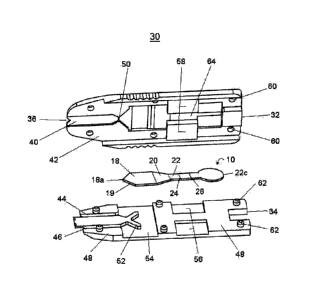
In one embodiment of the present invention shown in figure 2, is an analytical
device 30 for use with the membrane array of the present invention. The device
has
an upper half 32 and a lower half 34 that cooperate to enclose the membrane
array
10. Fluid sample entry into the analytical device 30 is from sample
application
means in registry with a sample flow channel formed from an indent 40 in the
bottom surface 42 of upper half 32 of the device to include indent 36 in the
upper
half 32 of the device. There are open areas 44 and 46 where the bottom surface
42
of upper half 32 comes into contact with the top surface 48 of the lower half
34 of
the device. Open areas 44 and 46 act to inhibit the flow of sample from the
sample
flow channel into the space between the two halves of the device. The open
areas
44 and 46 extend to area 54 to prevent sample from flowing out of the membrane
array 10 into the space between the two surfaces 42 and 48.
The sample flow channel terminates at the apex of the upstream end 18a of
first separation membrane 18 which is supported by a Y-shaped protrusion 52
extending from the top surface 48 of lower half 34. It will be noted that the
downstream end of the sample flow channel has a constriction 50 so that the
sample
flows horizontally and uniformly into the first separation membrane 18 at the
apex
18a through the thickness of the first separation edge 19. There is also
surface-to-
surface contact between the first separation membrane edge 19 and the
sidewalls of
the widening area downstream of constriction otherwise known as the crosswise
channel 50 of upper half 32. It will be noted that the first separation
membrane 18
CA 02612450 2007-12-17
WO 2007/000048 PCT/CA2006/001065
is shaped and placed so that the sample enters first separation membrane 18 at
the
upstream end 18a, through the thickness of the first separation edge 19 by
capillary
flow. From the above passages, it will be readily apparent to one skilled in
the art
that the greater capillary forces of the membrane array 10 than that of the
sample
flow channel of the analytical device 30 ensures that the analytical test only
begins
when a sufficient volume of sample is received.
To assist in holding the halves of the device 30 together, the top surface 48
of
lower half 34 of the device contains rectangular indents 56 and cylindrical
pillars 62
that are in register with rectangular protrusions 58 and cylindrical indents
60
respectively, in the bottom surface 42 of upper half 32 of the device.
Features 56
and 58 also serve the additional function of holding the membrane array 10 in
place.
It is shown in figures 1 and 2 that the upstream end 18a of first separation
membrane 18 contacts the downstream end of the crosswise channel 50 of the
analytical device.
Similarly the downstream end 18c of the first separation
membrane 18 slightly overlaps the upstream end 20a of a second separation
membrane 20. The downstream end 20c of the second separation membrane 20 in
turn slightly overlaps the upstream end 22a of the analytical membrane 22. The
overlapping membranes configured in a stair-step manner permit continuous flow
of
the sample from the sample flow channel to the crosswise channel 50 and
through
the membrane array 10 to its closed end 22c. To obtain rapid movement of the
sample through the membrane segments 'and yet retain the capture line 24 and
control line 26 sufficiently distanced from each other so as to be visible
through the
viewing window 64, the analytical membrane 22 is shaped with a narrow upstream
end 22a and a circular downstream end 22c. The completion of the analytical
test is
indicated by the sample flow to the downstream end 22c of the analytical
membrane
22 and is visibly evident through viewing window 64 in upper half 32, thus a
timing
device is not needed to determine test completion. Accordingly, when the fluid
sample has completed its capillary flow to the downstream end 22c, the sample
flow
channel is substantially empty. This arrangement serves as a control to
determine
and to limit the volume of the sample used in the test. In
this preferred
embodiment, the total dimensions of the first separation membrane 18, second
separation 20 and analytical membrane 22 are determined by the total
absorption
volume occupied by a single drop of blood which is about 10 pl to about 50 pl.
In use, a single drop of whole blood sample of sufficient quantity (about 10
I
to about 50 I) is readily obtained with a finger lancet procedure. The blood
sample
16
CA 02612450 2012-08-16
is brought into contact with the membrane array 10 at the upstream end 18a of
the
first separation membrane 18 via the sample flow channel formed when the upper
and lower halves of the analytical device 30 are assembled. The sample flows
horizontally through the thickness of the first separation membrane edge 19 by
capillary action into and through the first separation membrane 18 where the
red
blood cells are initially retarded within the first separation membrane 18.
The
sample then flows by capillary action towards the second separation membrane
20
where the red blood cells in the sample are further retarded and the plasma
continues to flow to the downstream analytical membrane 22.
As is readily apparent, upon contact with the first separation membrane 18,
the red blood cells of the sample will begin to separate from plasma and in
the
course of its flow the analyte will encounter a detection reagent, typically
but not
limited to a labeled antibody directed to an epitope of the analyte to form an
analyte-detection reagent complex. The analyte-detection reagent complex then
moves to the second separation membrane 20, where red blood cell migration
will be
further hindered/retarded. The analyte-detection reagent complex then moves
toward the analytical membrane 22 and encounters a fixed capture reagent,
typically
but not limited to an antibody directed to a separate epitope of the analyte.
The
reaction of the analyte-detection reagent complex with the fixed capture
reagent
forms a concentrated capture line 24 visible to the naked eye or appropriate
instrumentation. The optional control line 26 downstream of the capture line
24 will
contain the control reagent. In aspects of the invention, the control reagent
may be
an anti-animal IgG. Alternatively, in place of a control line 26, variations
in the
length of the transparent cover tape over the membranes 18, 20 and 22 of the
membrane array 10 can cause the sample when it reaches the end 22c of the
analytical membrane 22 to evaporate in a controlled manner revealing a readily
detectable signal.
Another embodiment incorporating the membrane array of the present
invention is shown in figure 3. This analytical device generally described as
130 is
similar to the analytical device 30 shown in figure 2, except the sample flow
channel
is formed from a protrusion 138 in a bottom surface 142 of an upper half 132
that
registers with an indent 140 in a top surface 148 of a lower half 134.
Downstream
from the sample flow channel is a crosswise channel 150 that widens into an
open
area 154. Alternatively, the sample flow channel can also be formed by a
registry of
indents in the bottom surface of the upper half with protrusions in the top
surface of
17
CA 02612450 2012-08-16
the lower half. This variation will be readily obvious to the skilled artisan
and is not
shown. To assist in holding the halves of the device 130 together, the top
surface
148 of lower half 134 of the device 130 contains cylindrical pillars 162 that
are in
register with cylindrical indents 160, in the bottom surface 142 of the top
half 132 of
the device 130. As can be readily seen in figure 3, the top surface 148 of
lower half
134 is provided with indents formed in the shape of the membrane array 10
which
function to hold the array 10 in place in the device 130.
Another embodiment incorporating the membrane of the present invention is
a removable cap designated generally as reference numeral 200 is shown in
figure 4
and is designed to facilitate the application of a small volume of sample
using a
micropipette to the analytical device and to protect the user from
contamination with
a fluid sample. In an embodiment, the cap is provided and fitted to serve as a
guide
for the placement of a sample transfer device such as micropipette tip prior
to
ejection of a small volume of fluid towards the sample application means of
the
analytical device 30. The cap 200 is formed by two guide arms 212 and 214
connected by a central body 216. Within one side of the central body 216 is an
opening with side surfaces 218 and 220 and a sloping surface 222 that form a
channel 236 designed to facilitate flow of fluid ejected from a pipette to the
sample
application means of analytical device 30. From central body 216 is a
protrusion 224
with an indentation 226 that is in registry with a corresponding member 78 on
the
lower surface 76 of bottom half 34 at one end of analytical device 30 as shown
in
figure 5. The indentation 226 and protrusion 78 provide a snap or latch fit
engagement and generally cooperate to prevent the unintentional separation of
cap
200 with device 30 when fully attached as is evident from figures 6 and 7.
Figure 6 shows a perspective view of the cap 200 in register with an
analytical
device 30. The cap 200 has slots 228 and 230 that engage with corresponding
guide
protrusions 68 and 70, respectively, on the analytical device 30. The cap 200
can be
readily engaged to the analytical device 30 by an operator's hands by using
holding
surfaces 232 and 234 of cap 200 to form a combination analytical device
generally
described as 300 which is shown in figure 7. As shown in figure 7, the central
body
216 of cap 200 is abutted against analytical device 30 whereby the channel 236
of
cap 200 is in operative communication with the sample application means of the
analytical device 30. In use, when a fluid sample is applied via a
micropipette tip,
the sample flows continuously through the channel 236 to the sample
application
means and through the sample flow channel of the analytical device 30 to reach
the
18
CA 02612450 2012-08-16
membrane array 10. It is also understood and will be appreciated by those
skilled in
the art that the cap 200 may also be fitted to the analytical device 130 or
any similar
analytical device. Furthermore, it will appreciated by those skilled in the
art that
since the cap 200 is releasably bound to the device 30, the cap 200 may
reattached
after performing the finger lancet procedure to further protect the user from
contamination with a fluid sample.
Figure 8 shows a perspective view of another embodiment of the removable
cap designated generally as reference numeral 200'. In this embodiment, the
cap
200', similar to cap 200 in that cap 200' also serves as a guide for the
placement of
a micropipette tip prior to ejection of a small volume of fluid towards the
sample
application means of an analytical device 30'. The cap 200' is formed by two
guide
arms 212' and 214' connected by a central body 216'. Within one side of the
central
body 216' is an opening with side surfaces 218' and 220' and a sloping surface
222'
that forms a channel 236' designed to facilitate flow of fluid ejected from a
micropipette to the sample application means of the analytical device 30'.
From
central body 216' is a protrusion 224' having a member 226' that is adapted to
be in
registry with a corresponding indentation 78' on the lower surface 76' of
bottom half
34' at one end of analytical device 30' as shown in figure 9. The member 226'
and
indentation 78' when engaged, provide a snap or latch fit engagement and
generally
cooperate to prevent the unintentional separation of cap 200' with device 30'
when
the cap 200' and the device 30' are attached. The cap 200' has slots 228' and
230'
that engage with corresponding guide protrusions 68' and 70', respectively on
the
cap 200'. To further provide for a more secure fit between the cap 200' and
the
device 30', the guide arms 212' and 214' of the cap 200' may also have
protrusions
238' and 240', respectively, that engage in a snap or latch fit manner with
corresponding indentations 242' and 244' on device 30'. It will be readily
apparent
to those skilled in the art what other known means to provide for a releasable
attachment of the cap 200 or 200' to any of the analytical devices of the
present
invention.
In the embodiments shown to have a cap as part of the analytical device, the
analytical device may be fabricated to contain the membrane array of the
present
invention or alternatively, the two part membrane shown and described in
Applicant's
PCT IB/2003/005088. Briefly, the two part membrane comprises an upstream
19
CA 02612450 2012-08-16
first separation membrane that contains a detection reagent and a downstream
capture membrane containing a capture reagent.
Another embodiment of an analytical device is shown in figure10, in which the
device is designed for dipping into a reservoir containing a fluid sample. In
this
embodiment the analytical device 430 is configured at one with an elongated
portion
431 but functions in the same manner as the device of figure 2 or 3 except
that the
sample entry is provided by elongated portion 431 which can be dipped into the
fluid sample, such as urine for example. The fluid sample enters into the
analytical
device 430 from the elongated sample flow channel formed from notch 436 in the
upper half 434. The elongated sample flow channel terminates at the upstream
end
18a of first separation membrane 18 of the membrane array 10. It will be noted
that the downstream end of the sample flow channel is a constriction 450 so
that
sample flows uniformly into first separation membrane 18 at the apex 18a.
There is
also surface-to-surface contact between the first separation membrane edge 19
and
the sidewalls of the widening area downstream of constriction 450 of upper
half 434.
It will be noted that the first separation membrane 18 is shaped and placed so
that
the sample enters through the first separation edge 19 of the first separation
membrane 18 by capillary flow. To assist in holding the halves together, the
bottom
surface 448 of upper half 434 contain cylindrical indents 460 that are in
register with
cylindrical pillars 462 respectively, in the upper surface 442 of lower half
432. The
membrane array 10 is held in position by resting on support structures 476 and
478
and is enclosed by rectangular protrusions 458 on the bottom surface 448 of
the
upper half 434 function and rectangular protrusion 466 on the top surface 442
of the
lower half 432. There is also a viewing window 464 in the upper half 434 of
analytical device 430 that is in registry with the analytical membrane 22 of
the
membrane array 10. In the embodiments shown to have elongated portion of the
analytical device, the analytical device may be fabricated to contain the
membrane
array of the present invention or alternatively, the two part membrane shown
and
described in Applicant's PCT IB/2003/005088. Briefly, the two part membrane
=
comprises an upstream first separation membrane containing a detection reagent
and a downstream capture membrane containing a capture reagent.
It is within the scope of the present invention to detect an analyte or even
multiple analytes in the fluid sample at one time. Accordingly, it will be
appreciated
by one skilled in the art that one or more detection reagents and/or one or
more
CA 02612450 2012-08-16
capture reagents can be deposited on the membrane array 10 of the present
invention.
Any of a variety of labeled antibodies in the membrane array of the present
invention available to the skilled artisan may be utilized. Metal and enzyme
labels
are commonly used. Metal labels are especially preferred due to their
remarkable
sensitivity. Amongst the metals, gold is most preferred principally because it
is so
widely employed for this type of reaction and its characteristics are so well
understood. The preferred particle size for gold labeled antibodies employed
in the
invention is from about 20 to 65 nm, although appreciable variation can be
tolerated
depending on well understood factors such as the clinical cut off of the
analyte and
the affinity of the reactants. Additionally, a gold signal can be enhanced to
become
readily visible by the use of reducible silver salt which deposits as visible
product. A
typical reactive salt is silver lactate, which serves as the source of
reducible silver
ions, employing hydroquinone as a reducing agent. The metallic silver forms a
readily discernible black deposit around each particle of gold.
Alternatively, if an enzyme label such as horseradish peroxidase is employed
the reaction may be detected by the addition of hydrogen peroxide and a dye
such
as ortho phenylenediamine in accordance with standard procedures. Additional
labels that may be used well within the scope of this invention are
paramagnetic
labels such as described in U.S. patent 6,046,585, which enable an even
greater
sensitivity for analyte detection.
The numerous analytes that may be detected in accordance with this
invention are cardiac analytes associated with cardiovascular events such as
myoglobin, troponins T (cTnT) and I (cTnI) and creatinine kinase MB (CK-MB).
Furthermore, hormones associated with pregnancy or ovulation such as human
chorionic gonadotropin (hCG) and luteinizing hormone (LH), respectively may
also be
detected using this invention or various embodiments thereof. It is also
within the
scope of this invention that other antigens for diseases such as cancer,
specifically
prostate cancer antigens (prostate serum antigen, PSA) may also be detected
using
this invention.
Additional applications of this invention include the recognition of
analytes associated with viral infections such as hepatitis, bacterial and
fungal
infection including Helicobacter pylori for gastrointestinal ulcers, other
infections
caused by Bacillus anthracis, Pediculus humanis, Siphonaptera and gram
positive
bacteria as Streptococcus pyognes, Streptococcus pneumoniae and Streptococcus
21
=
CA 02612450 2007-12-17
WO 2007/000048 PCT/CA2006/001065
faecalis are all non-limiting examples. This invention may also useful for
detecting
drugs including drugs of abuse. Enzymatic assays such as those that determine
levels of glucose and in blood are also contemplated by the present invention.
It will
be recognized that the use of the devices is not limited to these specific
analytes or,
indeed, to whole blood but is equally applicable to other analytical
procedures such
as those mentioned above.
Although the invention will be described principally as applied to the so
called
sandwich assay, the skilled artisan will recognize that it is also applicable
to other
types of assays such as the competitive assay. In a competitive assay, an
additional
inclusion of a labeled antigen as the detection reagent in the first
separation
membrane 18 will compete with the analyte (antigen) in the sample for binding
to
the capture reagent such as for instance, an antigen binding molecule. In
aspects of
the invention, the antigen binding molecule may be a polyclonal or monoclonal
antibody.
In an embodiment of the invention where the analyte is an antigen binding
molecule such as an antibody the invention, the detection reagent may be a
labeled
antihuman IgG and the capture reagent is any a suitable immobilized antigen
(or
antigens) to the antibody (or antibodies) in the fluid sample. The numerous
types of
natural or synthetic antigens that may be employed and would be suitable for
use
with the present invention are well known to those of skill in the art.
Examples of
suitable antigens which can be immobilized include, but are not limited to,
Human
Immunodeficiency Virus (HIV) and hepatitis virus. Similarly, one skilled in
the art
would readily understand that in another embodiment of the invention, the
detection
reagent may also be a labeled antigen to an antibody in the fluid sample.
The above disclosure generally describes the present invention. A more
complete understanding can be obtained by reference to the following specific
Examples. These Examples are described solely for purposes of illustration and
are not
intended to limit the scope of the invention. Changes in form and substitution
of
equivalents are contemplated as circumstances may suggest or render expedient.
Although specific terms have been employed herein, such terms are intended in
a
descriptive sense and not for purposes of limitation.
Examoles
Without intending to be limiting in scope, the following example serves to
illustrate various embodiments of the invention.
22
CA 02612450 2007-12-17
WO 2007/000048 PCT/CA2006/001065
Example 1
A human cardiac troponin I test (TnI) device using one drop of whole blood
sample is prepared according to current invention. For the analytical
membrane,
nitrocellulose (Whatman) with a pore size of about 5 pm was impregnated with
both
control and capture solutions using a conventional liquid dispenser. Control
solution
contains 1 mg/mL of goat anti-mouse IgG polyclonal antibodies (Arista
Biologicals),
and capture solution contains 2 mg/mL of an anti-troponin I monoclonal
antibody
(HyTest). Impregnated nitrocellulose was incubated at 37 C for 30 minutes to
immobilize the antibodies. The first separation membrane (Whatman) was sprayed
with colloidal gold conjugate solution and then freeze dried to remove the
water.
The colloidal gold conjugate with a final OD of 2.2 at 540 nm was prepared
from 40
rim gold particles (Arista Biologicals) and a monoclonal antibody specific to
human
cardiac troponin I (HyTest). An 8 pm nitrocellulose membrane (Whatman) was
used
as the second separation membrane. The membrane array is covered by a 25 pm
transparent polyester tape (Adhesive Research) and supported by polystyrene
backing tape (G & L Precision Die Cutting, Inc). The membrane array was
assembled
as shown in figure 1 and housed in an analytical device as shown in figure 2.
The
shape of the membrane array was obtained using a die-cutting tool. Testing of
this
analytical device using 35 pL of blood or serum demonstrated excellent plasma
separation and sample flow in a testing procedure requiring approximately 10
minutes. The test achieved a sensitivity of 1 ng/mL of TnI.
Example 2
A human procalcitonin (PCT) test device using one drop of whole blood
sample is prepared according to current invention. For the analytical
membrane,
nitrocellulose (Millipore) with a pore size of 5pm was impregnated with both
control
and capture solutions using a conventional liquid dispenser. Control solution
contains
1 mg/mL of goat anti-mouse IgG polyclonal antibodies (Arista Biologicals), and
capture solution contains 2 mg/mL of anti-calcitonin sheep polyclonal
antibodies
(Brahms). Impregnated nitrocellulose was incubated at 37 C for 30 minutes to
immobilize antibodies. Detection membrane or plasma separator (Whatman) was
sprayed with colloidal gold conjugate solution and then freeze dried to remove
water.
Gold conjugate, prepared from 40 nm gold particles (Arista Biologicals) and a
monoclonal antibody specific to PCT (Brahms), had a final OD 1.5 at 540 nm. A
8
23
CA 02612450 2012-08-16
pm nitrocellulose membrane (Whatman) was used as the separation membrane. The
test strip is covered by a 25 pm thick transparent polyester tape (Adhesive
Research) and supported by polystyrene tape available from G & L Precision Die
Cutting, Inc. Test strip was assembled as indicated in Fig 1. The shape of the
test
strip was obtained using a die-cutting tool. Testing of this device using 35
pL of
blood or serum demonstrated excellent plasma separation and sample flow. The
testing procedure took approximately 25 min to complete. A sensitivity of 0.1
ng/mL
of PCT was achieved.
Although preferred embodiments of the invention have been described herein
in detail, it will be understood by those skilled in the art that variations
may be made
thereto. The scope of the claims should not be limited by the preferred
embodiments set forth in the examples but should be given the broadest
interpretation consistent with the description as a whole.
24