Note: Descriptions are shown in the official language in which they were submitted.
CA 02612548 2013-02-12
APPARATUS AND METHODS FOR TREATING BONE
Field of the Invention
[0002] The invention relates to surgical implants, and more particularly to
minimally invasive apparatus and methods for treating (filling, augmenting,
and/or
reposition bone) bone, preferably vertebrae and/or restoring spinal lordosis.
Background of the Invention
[0003] Vertebral compression fractures, as illustrated in FIG. 1, represent
a
generally common spinal injury and may result in prolonged disability. F.
Margerl et al:
A comprehensive classification of thoracic and lumbar injuries, Eur Spine J184-
201,
1994. These fractures involve collapsing of one or more vertebral bodies 12 in
the spine
10. Compression fractures of the spine usually occur in the lower vertebrae of
the
thoracic spine or the upper vertebra of the lumbar spine. They generally
involve fracture
of the anterior portion 18 of the affected vertebra 12 (as opposed to the
posterior side
16). Spinal compression fractures can result in deformation of the normal
alignment or
curvature, e.g., lordosis, of vertebral bodies in the affected area of the
spine. Spinal
compression fractures and/or related spinal deformities can result, for
example, from
metastatic diseases of the spine, from trauma or can be associated with
osteoporosis.
Until recently, doctors were limited in how they could treat such compression
fractures
and related deformities. Pain medications, bed rest, bracing or invasive
spinal surgery
were the only options available.
[0004] More recently, minimally invasive surgical procedures for treating
vertebral compression fractures have been developed. These procedures
generally
involve the use of a cannula or other access tool inserted into the posterior
of the effected
vertebral body through the pedicles. The most basic of these procedures is
- 1 -
CA 02612548 2007-12-17
WO 2007/002108
PCT/US2006/024009
vertebroplasty, which literally means fixing the vertebral body, and may be
done without
first repositioning the bone.
[0005] Briefly, a cannula or special bone needle is passed slowly through
the soft
tissues of the back. X-ray image guidance, along with a small amount of x-ray
dye,
allows the position of the needle to be seen at all times. A small amount of
polymethylmethacrylate (PMMA) or other orthopedic cement is pushed through the
needle into the vertebral body. PMMA is a medical grade substance that has
been used
for many years in a variety of orthopedic procedures. Generally, the cement is
mixed
with an antibiotic to reduce the risk of infection, and a powder containing
barium or
tantalum, which allows it to be seen on the X-ray.
[0006] Vertebroplasty can be effective in the reduction or elimination of
fracture
pain, prevention of further collapse, and a return to mobility in patients.
However, this
procedure may not reposition the fractured bone and therefore may not address
the
problem of spinal deformity due to the fracture. It generally is not performed
except in
situations where the kyphosis between adjacent vertebral bodies in the
effected area is
less than 10 percent. Moreover, this procedure requires high-pressure cement
injection
using low-viscosity cement, and may lead to cement leaks in 30-80% of
procedures,
according to recent studies. Truumees, Comparing Kyphoplasty and
Vertebroplasty,
Advances in Osteoporotic Fracture Management, Vol. 1, No. 4, 2002. In most
cases, the
cement leakage does no harm. In rare cases, however, polymethymethacrylate or
other
cement leaks into the spinal canal or the perivertebral venous system and
causes
pulmonary embolism, resulting in death of the patient. J.S. Jang: Pulmonary
Embolism
of PMMA after Percutaneous Vertebroplasty, Spine Vol. 27, No. 19, 2002.
[0007] More advanced treatments for vertebral compression fractures
generally
involve two phases: (1) reposition, augmentation or restoration of the
original height of
the vertebral body and consequent lordotic correction of the spinal curvature;
and (2)
filling or addition of material to support or strengthen the fractured bone.
[0008] One such treatment, balloon kyphoplasty (Kyphon, Inc.), is
illustrated in
FIGS 2A-D. A catheter having an expandable balloon tip is inserted through a
cannula,
sheath or other introducer into a central portion of a fractured vertebral
body comprising
relatively soft cancellous bone surrounded by fractured cortical bone (FIG.
2A).
Kyphoplasty then achieves the reconstruction of the lordosis, or normal
curvature, by
inflating the balloon, which expands within the vertebral body restoring it to
its original
height (FIG. 2B). The balloon is removed, leaving a void within the vertebral
body, and
- 2 -
CA 02612548 2013-02-12
PMMA. or other filler material is then injected through the cannula into the
void (FIG.
2C) as described above with respect to vertebroplasty. The cannula is removed
and the
cement cures to fill or fix the bone (FIG. 2D).
[0009] Disadvantages of this procedure include the high cost, the
repositioning of
the endplates of the vertebral body are lost after the removal of the balloon
catheter, and
the possible perforation of the vertebral endplates during the procedure. As
with
vertebroplasty, perhaps the most feared, albeit remote, complications related
to
kyphoplasty are related to leakage of bone cement. For example, a neurologic
deficit
may occur through leakage of bone cement into the spinal canal. Such a cement
leak may
occur through the low resistance veins of the vertebral body or through a
crack in the
bone which had not been appreciated previously. Other complications include;
additional adjacent level vertebral fractures, infection and cement
embolization. Cement
embolization occurs by a similar mechanism to a cement leak. The cement may be
forced
into the low resistance venous system and travel to the lungs or brain
resulting in a
pulmonary embolism or stroke. Additional details regarding balloon kyphoplasty
may
be found, for example, in U.S. Patent Nos. 6,423,083, 6,248,110, and 6,235,043
to Riley
et al.; Gantis et al., Balloon kyphoplasty for the treatment of pathological
vertebral
compression fractures, .Eur Spine J14:250-260, 2005; and Lieberman et al.,
Initial
outcome and efficacy of Kyphoplasty in the treatment of painful osteoporotic
vertebral
compression fractures, Spine 26(14):1631-1638, 2001.
[0010] Another approach for treating vertebral compression fractures is the
Optimesh system (Spineology, Inc., Stillwater, MN), which provides minimally
invasive
delivery of a cement or allograft or autograft bone using an expandable mesh
graft
balloon, or containment device, within the involved vertebral body. The
balloon graft
remains inside the vertebral body after its inflation, which prevents an
intraoperative loss
of reposition, such as can occur during a kyphoplasty procedure when the
balloon is
withdrawn. One drawback of this system, however, is that the mesh implant is
not well
integrated in the vertebral body. This can lead to relative motion between the
implant
and vertebral body, and consequently to a postoperative loss of reposition.
Additional
details regarding this procedure may be found, for example, in published U.S.
Patent
Publication Number 20040073308.
- 3 -
CA 02612548 2013-02-12
NOM Still another procedure used in the treatment of vertebral
compression
fractures is an inflatable polymer augmentation mass known as a SKy Bone
Expander.
This device can be expanded up to a pre-designed size and Cubic or Trapezoid
configuration in a controlled manner. Like the Kyphon balloon, once optimal
vertebra
height and void are achieved, the SKy Bone Expander is removed and PMMA cement
or
other filler is injected into the void. This procedure therefore entails many
of the same
drawbacks and deficiencies described above with respect to kyphoplasty.
[0012] A proposed improved procedure for repositioning and augmenting
vertebral body compression fractures is vertebral body stenting, for example
as described
in Piirderer et aL, "Vertebral body stenting", Orthopade 31:356-361, 2002;
European
Patent Application publication number EP1308134A3; and United States Patent
Application publication number US2003/0088249 .
Veterbral body stenting, as described herein
generally involves inserting into a vertebral body a balloon-tipped catheter
(e.g., such
as a kyphoplasty balloon) surrounded by a stent (e.g., such as those used in
angioplasty).
After insertion of the balloon and stent, the balloon is inflated, e.g., using
fluid pressure,
thereby expanding the stent within the vertebral body. After expansion of the
stent, the
balloon may be deflated and removed, with the stent remaining inside the
vertebral body
in an expanded state to fill the vertebral body.
[0013] There remains a need for implants and related methods for
repositioning
and augmenting fractured vertebral bodies and other bones.
Summon, of the Invention
[0014] The present invention provides a bone treatment system, preferably a
minimally invasive bone treatment system for filling, augmenting and/or
repositioning
bone, which may include a body or bobbin and a band configured to contact and
coil
around the bobbin and increase the diameter of the bone implant. In one
embodiment the
present invention provides an implant and method for correction of vertebral
fractures
and other disorders of the spine. For example, a cylindrical body or bobbin
may be
inserted into a vertebral body damaged by a vertebral compression fracture.
After
insertion of the bobbin, a wire, string, thread or band, collectively referred
to herein as a
"band", is coiled preferably multiple times around the bobbin. The band may
have any
profile or shape and may be comprised of any biocompatible material. During
coiling,
the diameter of the bobbin/band complex, sometimes referred to as a coiled
bobbin
- 4 -
CA 02612548 2007-12-17
WO 2007/002108
PCT/US2006/024009
assembly, may increase. Such increase in diameter can push against the inner
side of the
endplates of the vertebral body, and restore the vertebral body to its
original height.
Additionally, bone fragments or segments around the bobbin/band complex can be
compacted during coiling of the band.
[0015] In some embodiments, features for controlling rotation of the
bobbin head
and/or coiling of the band can include a shaft, rod or cannula for rotating
the bobbin, and
a guide, guide conduit, cannula, tub for controlling and/or providing the
band. The
bobbin can have various configurations, multiple joints and/or can be
bendable.
[0016] In other embodiments, minimally invasive implants for distracting
spine
segments include an elongated body having an end dimensioned for implantation
in a
space between two or more vertebral features, and a band associated with and
configured
to coil around the body to increase a diameter of the end and thereby increase
the space
between the two or more vertebral endplates, bone segments, or spinous
processes.
[0017] In another embodiment, a kit comprises various combinations of
assemblies and components according to the present invention. For example, a
kit may
include, for example, an insertion device, a bobbin, and a band according to
the present
invention.
[0018] In a further embodiment, a system for bone treatment (filling,
augmenting, and/or reposition bone), preferably minimally invasive osteopathic
treatment may comprise a bobbin having a diameter, and a first and second end,
the
bobbin configured for implantation within a bone, and a band having a length
substantially larger than its width or height, the band configured to contact
and coil
multiple times around the bobbin between the first and second end to increase
the
diameter of the bobbin when the bobbin is implanted within bone. The system
may
further comprise an elongated body having a proximal end and a distal end, the
proximal
end configured for manipulation by a user outside the patient to place the
distal end in a
desired position within the bone, and a joint disposed between the second end
of the
bobbin and the distal end of the elongated body. The bobbin is cylindrical and
comprises
a hole through which a portion of said band passes.
[0019] The elongated body may comprise at least a portion of a drive line
assembly to rotate the bobbin when the bobbin is implanted within bone. Tthe
elongated
body is configured to rotate, and the joint is configured to transfer rotation
from the
elongated body to rotate the bobbin. The joint is releasable so that the
elongated body
can be detracted from the bobbin.
- 5 -
CA 02612548 2007-12-17
WO 2007/002108
PCT/US2006/024009
[0020] The system may further comprise a guide mechanism configured to
control the position of the band between the first and second end of the
bobbin when the
band is coiled around the bobbin, a drive line assembly configured to
releasably attach to
and rotated the bobbin having a proximal end and a distal end, the proximal
end
configured in use to extend from the patient and be manipulated by a user to
place the
distal end in a desired position in the bone, the distal end releasably
connected to the
bobbin. The drive line assembly is rotatable to rotate the bobbin when the
bobbin is
located in the bone. The guide mechanism may comprise a guide moveable with
respect
to the bobbin to control the position of the band between the first and second
ends when
the band is coiled around the bobbin.
[0021] The drive line assembly may comprise a rotatable shaft and the
guide
mechanism may comprise an elongated tube coaxial with the rotatable shaft. The
elongated tube is moveable in an axial direction relative to the bobbin and
contacting the
band to position the band between the first and second ends of the bobbin.
[0022] The system may also comprise a knob connected to the elongated
tube
and containing a guide hole for the band.
[0023] The bobbin may have threads to assist in guiding the band.
[0024] In an alternative, the drive line assembly may comprise a
rotatable shaft
having a proximal end and a distal end, where the distal end of the shaft is
releasably and
rotatably connectable to the first end of the bobbin. The shaft being capable
of
transferring torque to the bobbin. The guide mechanism, in the alternative may
comprise
a guide tube having a proximal end and a distal end through which the band
moves, the
guide tube movable axially with respect to the shaft.
[0025] The rotatable shaft and guide tube may be located in a needle,
such that
the rotatable shaft is axially fixed with respect to the needle and the guide
tube moves
axially with respect to the needle. A portion of the needle may be positioned
along side
and adjacent the bobbin.
[0026] The system may also comprise a drive train to convert the rotary
motion
of the drive line assembly to an axial motion, the drive train connectable to
the drive line
assembly and the guide tube to move the guide tube axially. The drive train
may
comprise a gear connectable with the rotatable drive train, a rotatable cam
disk and an
axially moveable but non-rotatable follower. The cam disk having a groove
along its
outer surface and the follower including a projection, where the projection
extends into
the groove.
- 6 -
CA 02612548 2007-12-17
WO 2007/002108
PCT/US2006/024009
[0027] The follower may be a spool holder and the projection a dowel pin
insertable through a hole in the spool holder.
[0028] The band may be coated with or form part of a matrix with other
materials
to include osteo-inductive materials, osteo-conductive materials, antibiotics,
tricalcium
phosphate, bone morpho genetic proteins.
[0029] In an another embodiment a system for minimally invasive bone
treatement, filling, augmenting, and/or reposition bone, may comprise an
elongated body
having a first end and a second end, the body having a length along its
longitudinal axis,
configured for implantation within a bone, and an insertion device for
inserting the
elongated body within a bone, the insertion device comprising a band and
configured to
cause the band to coil multiple times around the elongated body between the
first and
second end to increase the diameter of the body and band assembly, the
insertion device
releasably connectable to the body. The insertion device may comprise a drive
line
assembly to apply a rotational force to the elongated body to cause the
elongated body to
rotate about its longitudinal axis to coil the band around the elongated body,
to increase
the diameter of the elongated body and band assembly implanted within the
region of
bone.
[0030] The system may further comprise an axial guide mechanism movable
axially with respect to the elongated body, the axial guide mechanism in
connection with
and controlling the position of the band along the length of the elongated
body as it
rotates.
[0031] The drive line assembly may comprise a rod connected to the
elongated
body, the rod being rotatable which in turn rotates the elongated body. The
guide
mechanism may comprise an outer cannula, causing the band to reposition along
the
length of the elongated body. Continued rotation of the elongated body causes
the
diameter of the elongated body and band assembly to increase due to the
coiling of the
band around the elongated body.
[0032] In the alternative, the drive line assembly may comprise a drive
mechanism, a drive shaft, and a flexible shaft connected serially to the
elongated body,
and the guide mechanism may comprise a band guide conduit, a spool holder, and
a
rotatable cam disk. The cam disk and spool holder converts the rotational
force of the
cam disk to an oscillating force applied to the band guide conduit causing the
band guide
conduit to move forward and backward relative to the elongated body. The band
guide
conduit may comprise an interior passage way having a proximal and distal
opening and
- 7 -
CA 02612548 2007-12-17
WO 2007/002108
PCT/US2006/024009
wherein the band is positioned in and moves through the interior passage way
of the
band guide conduit out the distal opening where it coils around the body.
[0033] The insertion device may further comprise a drive train to couple
the
drive shaft of the drive line assembly to the cam disk of the guide mechanism
so as to
have the cam disk rotate at a different velocity than the drive shaft, where
the drive train
comprises a drive gear, a sprocket, and another gear.
[0034] In a further embodiment, a system for minimally invasive bone
treatment,
filling, augmenting, and/or repositioning, may comprise an elongated body
having a first
end and a second end. The body having a length along its longitudinal axis, is
configured for implantation within a bone. The system may further include an
insertion
device for inserting the elongated body within a bone. The insertion device
may
comprise a band and configured to cause the band to coil multiple times around
the
elongated body between the first and second end to increase the diameter of
the body and
band assembly, the insertion device releasably connectable to the body. The
insertion
device may further comprise a drive line assembly to apply a rotational force
to the
elongated body to cause the elongated body to rotate about its longitudinal
axis to coil
the band around the elongated body, so as to increase the diameter of the
elongated body
and band assembly implanted within the region of bone. The drive line assembly
may
comprise a drive mechanism, a drive shaft and a flexible shaft connected
serially to the
first end of the elongated body. The insertion device may further include an
axial guide
mechanism movable axially with respect to the elongated body. The axial guide
mechanism in connection with and controls the position of the band along the
length of
the elongated body as it rotates. The guide mechanism may comprise a band
guide
conduit, a spool holder, and a rotatable cam disk, where the cam disk has a
groove along
its outer surface and the spool holder including a projection where the
projection extends
into the groove of the cam disk. The insertion device may also include a drive
train
coupling the drive shaft of the drive line assembly to the cam disk of the
guide
mechanism so as to have the cam disk rotate at a different velocity than the
drive shaft.
The drive train may comprise a drive gear, a sprocket, and another gear. The
cam disk
and spool holder converts the rotational force of the cam disk to an
oscillating force
applied to the band guide conduit causing the band guide conduit to move
forward and
backward relative to the elongated body, and the band guide conduit comprises
an
interior passage way having a proximal and distal opening and wherein the band
is
positioned in and moves through the interior passage way of the band guide
conduit out
- 8 -
CA 02612548 2007-12-17
WO 2007/002108
PCT/US2006/024009
the distal opening where it coils around the body. The flexible shaft and band
guide
conduit are located in a needle, such that the flexible shaft is axially fixed
with respect to
the needle and the band guide conduit moves axially with respect to the
needle. A
portion of the needle is positioned along side and adjacent the bobbin, and
continued
rotation of the elongated body causes the diameter of the elongated body and
band
assembly to increase due to the coiling of the band around the elongated body.
[0035]. In a further embodiment, the present invention provides an implant
and
method for correction of vertebral fractures and other disorders of the spine.
For
example, one or more wool bales or fibrous masses/bodies may be inserted into
a
vertebral body damaged by a vertebral compression fracture. As the fibrous
bodies are
inserted into a vertebral body, they may fill a central portion of the
vertebral body and
may push against the inner sides of the endplates of the vertebral body,
thereby providing
structural support and tending to restore the vertebra to its original height.
Optionally,
the fibrous masses may comprise a shape-memory alloy or other material that
expands or
changes configuration after implantation, which may lead to a thorough
integration of the
implant into the bone and/or help restore the height of the damaged vertebral
body.
After implantation, a bone cement (e.g., PMMA or tricalcium phosphate), bone
chips,
demineralized bone, or other filler material or implant may be added with or
without the
implanted fibrous mass to aid in stabilizing the bone and securing the implant
in place
within the bone.
[0036] The fibrous masses may be comprised of a thread or other
relatively thin
structure, for example a fiber or strand, of any biocompatible material having
desired
characteristics, for example a shape memory alloy (e.g., nitinol or other
nickel-titanium
alloy, copper-based alloys, iron-based alloys, etc.), titanium, stainless
steel, a
biocompatible polymer, another metal or metal alloy, a ceramic, a composite or
any
combination thereof. The, strand, thread or other fiber may be coiled, woven,
matted,
tangled or otherwise formed into a wool-like mass or body having a desired
configuration. The bodies may be individually inserted into a bone, or may be
joined or
linked in series to form a chain having desired characteristics of
flexibility, strength, and
the like. In some embodiments, the bodies and/or links may be resorbable.
[0037] In another embodiment, a kit may comprise various combinations of
components according to the present invention. A kit may include, for example,
a
cannula and one or more fibrous body implants. A kit may additionally include
a syringe
- 9 -
CA 02612548 2007-12-17
WO 2007/002108
PCT/US2006/024009
or other apparatus for injecting a cement or other filler into a vertebral
body. Optionally,
one or more other implants, devices may be included in a kit.
[0038] Another embodiment provides implants for minimally invasive
osteopathic treatment (filling, augmenting, and/or reposition bone), which may
include a
body comprising a sheet coiled multiple times. The body, having a first
diameter, is
configured for implantation within a bone. The body is also configured to
expand to a
second diameter by uncoiling the sheet when the body is implanted in the bone.
Insertion of the body into bone can be accomplished using a sheath or cannula.
[0039] The sheet may comprise any of stainless steel, a nickel titanium
alloy, a
cobalt alloy, another metal alloy, a polymer, or a combination thereof.
[0040] The system may further include an axial member, having a first end
and
second end that is substantially cylindrical, where the body may be coiled
around the
axial member. The axial member comprises a lumen through which a filler
material can
be injected into the bone.
[0041] In a further embodiment, the sheet may comprise a plurality of
holes,
where the holes are dimensioned to allow the filler material to penetrate the
body
[0042] In still a further embodiment, the axial member may be rotated in
a
direction opposite of the coiling of the body to expand the body by partially
uncoiling the
coiled sheet.
[0043] In still another embodiment, the present invention provides an
implant
and method for correction of vertebral fractures and other disorders of the
spine. For
example, a coiled sheet may be inserted into a vertebral body damaged by a
vertebral
compression fracture. After insertion into a damaged vertebral body, the
coiled sheet can
be uncoiled to expand its diameter. Such increase in diameter can push against
the inner
side of the endplates of the vertebral body, and tend to restore the vertebral
body to its
original height. Additionally, uncoiling of the sheet can compact the bone
around the
implant, which can lead to a better integration of the implant in the bone.
The coiled
sheet may be comprised of any biocompatible material having desired
characteristics, for
example stainless steel, aluminum, a metal alloy, e.g., a cobalt alloy, a
nickel titanium
alloy or another alloy, a polymer, or any combination thereof.
[0044] In some embodiments, a method of treating bone can include
inserting
inside a fractured bone, for example a vertebrae, a device comprising a sheet
of material
coiled around an axial member, causing the coiled sheet to partially uncoil
from around
the axial member to increase the diameter of the device and to apply a radial
force to
- 1 0 -
CA 02612548 2007-12-17
WO 2007/002108
PCT/US2006/024009
move the fractured bone into a desired position. After repositioning, the
implanted
device can be removed from the bone, or some or all of the device can be left
inside the
bone to maintain the desired position. In addition, a bone cement or other
filler may be
added with or without the implanted device to aid in stabilizing the bone.
[0045] In some embodiments, an expandable body comprises a fenestrated
sheet
coiled about a shaft, or axial member. The fenestrated sheet includes holes
that can
allow passage of bone cement or other material injected into the expandable
body to
further treating a vertebral body or other bone into which the expandable body
is
inserted. The bone cement or other filler material can be injected, for
example, through a
lumen of the axial member, for example using a syringe or other device.
[0046] In other embodiments, a coiled sheet includes perforations, hinge
features
or other joints that define the sheet into a plurality of adjacent planes, or
segments. Such
joints can provide incremental increases in diameter of the coiled body.
Moreover, the
joints allow use of more rigid materials for the sheet, and the joints provide
discrete
locations for the sheet to bend, e.g., between segments. In some embodiments,
use of
substantially rigid or stiff materials can provide for increased radial (e.g.,
outward) forces
during uncoiling in a confined area such as within a vertebral body.
[0047] In other embodiments, minimally invasive implants for distracting
spine
segments include a coiled body having a first diameter dimensioned for
implantation in a
space between two or more vertebral features, wherein the coiled body is
configured to
uncoil to a second diameter that is larger than the first diameter, and
thereby increase the
space between the two or more vertebral features.
[0048] In another embodiment, a kit comprises various combinations of
assemblies and components according to the present invention. A kit may
include, for
example, a cannula and a coiled body according to the present invention. In
other
embodiments, a kit may include a cannula, a coiled body, and a syringe or
other
apparatus for injecting a cement or other filler into a vertebral body.
Brief Description of the Drawings
[0049] The invention is explained in even greater detail and may be
better
understood by the following exemplary drawings, wherein like references
numerals
represent like elements. The drawings are merely exemplary to illustrate
certain features
that may be used singularly or in combination with other features and the
present
invention should not be limited to the embodiments shown.
- 11 -
CA 02612548 2007-12-17
WO 2007/002108
PCT/US2006/024009
[0050] FIG. 1 is an illustration of a spine having a vertical compression
fracture
in one vertebral body;
[0051] FIGS. 2A-D are illustrations of a prior art method for treating a
vertical
compression fracture;
[0052] FIGS. 3A and B are side view illustrations of a bobbin and band
apparatus
according to an embodiment of the present invention;
[0053] FIG. 4A and B are cross-sectional side view illustrations of an
apparatus
and method for minimally invasive osteopathic treatment of a vertebral body
according
to an embodiment of the present invention;
[0054] FIGS. 5A and B are cross-sectional side view illustrations of
another
embodiment of an apparatus and method according to the present invention;
[0055] FIGS. 6A and B are cross-sectional side view illustrations of
another
embodiment of an apparatus and method according to the present invention;
[0056] FIGS. 7A and B are cross-sectional side top view illustrations of
an
apparatus and method employing a band guide according to an embodiment of the
present invention;
[0057] FIG. 8 is a detailed cross-sectional side view of an apparatus of
FIGS. 7A
and B;
[0058] FIG. 9 is a cross-sectional top view illustration of another
embodiment of
an apparatus and method according to the present invention;
[0059] FIG. 10 is an illustration of an embodiment of an insertion device
and
bobbin according to the present invention;
[0060] FIG. 11 is an illustration of another embodiment of an insertion
device
and bobbin according to the present invention;
[0061] FIG 12 is a cross-sectional view of the insertion device and
bobbin
depicted in FIG. 11;
[0062] FIGS 13 is cross-sectional view of the insertion device and bobbin
of FIG.
11 depicting the use of small band spools;
[0063] FIGS. 14A and B are cross-sectional views of the housing of the
insertion
device depicted in FIG. 11;
[0064] FIGS. 15A and B are an end view and a side view of the end cap of
the
insertion device depicted in FIG. 11;
[0065] FIG. 16 is a cross-sectional view of the sprocket of the insertion
device
depicted in FIG. 11;
- 12 -
CA 02612548 2007-12-17
WO 2007/002108
PCT/US2006/024009
[0066] FIGS. 17A and B are cross-sectional views of the drive shaft of
the
insertion device depicted in FIG. 11;
[0067] FIG. 18 is a cross-sectional view of the cam disk of the insertion
device
depicted in FIG. 11;
[0068] FIG. 19A is a side view of the spool holder of the insertion
device
depicted in FIG. 11
[0069] FIG. 19B is a cross-sectional view of the spool holder of the
insertion
device depicted in FIG. 11;
[0070] FIG. 20A is a cross-sectional top view of a large band spool of
the present
invention;
[0071] FIG. 20B is perspective view of a large band spool of the present
invention;
[0072] FIG. 21A is a cross sectional view of a small band spool of the
present
invention;
[0073] FIG. 21B is a perspective view of a small band spool of the
present
invention;
[0074] FIGS. 22A and B are cross-sectional side views of an embodiment of
a
needle of the present invention;
[0075] FIGS. 23A and B is a cross-sectional view and an end view,
respectively
of an embodiment of a needle of the present invention;
[0076] FIGS. 24A-C are cross-sectional side views and a end view,
respectively
of an embodiment of a needle of the present invention;
[0077] FIG. 25 is a cross-sectional view of a band guide conduit of the
present
invention;
[0078] FIG. 26 is a cross-sectional view of a flexible shaft of the
present
invention;
[0079] FIGS. 27A-C are detailed cross-sectional side views and an end
view,
respectively of a bobbin of the present invention;
[0080] FIG. 28 is a cross-sectional top view of another embodiment an
insertion
tool and bobbin of the present invention, depicting three bands;
[0081] FIG. 29 is a cross-sectional view of the present device depicting
the
flexible shaft having four joints;
[0082] FIG. 30 is a cross-sectional view of the present device depicting
the
flexible shaft having two joints;
- 13 -
CA 02612548 2007-12-17
WO 2007/002108
PCT/US2006/024009
[0083] FIGS. 31A and B are cross-sectional end views of another embodiment
of
an expandable osteopathic augmentation apparatus according to an embodiment of
the
present invention;
[0084] FIG. 32 is a cross-sectional top view of a flexible bobbin according
an
embodiment of to the present invention;
[0085] FIGS. 33A-C are illustrations depicting different bobbin
configurations
according to embodiments of the present invention;
[0086] FIGS. 34A and B are cross sectional views of an expandable
osteopathic
augmentation apparatus in use in a proximal femur according to an embodiment
of the
present invention;
[0087] FIGS. 35A and B are illustrations depicting wool bales (or fibrous
masses/bodies) of different shapes;
[0088] FIGS. 36A and B are illustrations depicting wool bales before
expansion
and after expansion;
[0089] FIG. 37 is an illustration depicting insertion of fibrous masses
with a
cannula and insertion device;
[0090] FIG. 38 is an illustration depicting fibrous masses expanded after
insertion into the vertebral body;
[0091] FIG. 39 is an illustration depicting expanded fibrous masses in the
vertebral body;
[0092] FIG. 40 is an illustration depicting a vertebral body being filled
with bone
cement or other bone filler material;
[0093] FIG. 41 is an illustration depicting a vertebral body filled with
expanded
fibrous masses and bone cement;
[0094] FIGS. 42A-C are illustrations depicting linked wool bales or fibrous
masses according to an embodiment of the present invention;
[0095] FIGS. 43A-D are illustrations depicting different configuration of
wool
bales or fibrous masses/bodies of shape-memory alloy;
[0096] FIGS. 44A-C are illustrations depicting wool bales or fibrous
masses/bodies of the present invention augmenting other bones, e.g. a proximal
femur;
[0097] FIGS. 45A and B are perspective view illustrations of a coiled body
of an
expandable augmentation device according to an embodiment of the present
invention;
[0098] FIG. 46A and B are cross-sectional side view illustrations of an
augmentation device in a vertebral body according to a method of the present
invention;
- 14 -
CA 02612548 2007-12-17
WO 2007/002108
PCT/US2006/024009
[0099] FIG. 47 is a perspective view illustration of an expandable
augmentation
device according to an embodiment of the present invention;
[00100] FIG. 48 is a perspective view schematic illustration of a
fenestrated sheet
for use in an augmentation device according to the present invention;
[00101] FIG. 49 is a cross-sectional side view of an embodiment of an
augmentation device having a coiled fenestrated sheet in use within a
vertebral body;
[00102] FIGS. 50A and B are perspective views of another embodiment of an
augmentation device according to the present invention;
[00103] FIG. 51 is a cross-sectional top view illustration of another
embodiment
of an apparatus and method according to the present invention; and
[00104] FIGS. 52A and B are cross sectional views of an expandable
osteopathic
augmentation device in use in a proximal femur according to an embodiment of
the
present invention.
Detailed Description
[00105] A vertebral body may be filled, augmented, or repositioned through
insertion of one or more implants into an internal portion of the vertebral
body, between
the endplates of two adjacent vertebral bodies, or in other bone, e.g., a
femur. In one
embodiment, a vertebral body implant may comprise a bobbin with a thread,
string, or
wire, collectively referred to herein as a band 310, coiled around the bobbin
to create a
larger diameter mass or body, sometimes referred to as a coiled assembly,
coiled band
assembly, coiled body, final implant, or implant. The bobbin with the attached
band may
be inserted into the vertebral body through, for example, transpedicular
access canals
having a diameter of about 5 mm.
[00106] Referring to FIG. 3A, an elongated member 300, which may be
cylindrical and is hereinafter referred to as a bobbin 300, has a certain
diameter dl 302.
After insertion of bobbin 300 into a collapsed vertebral body, band 310 is
coiled around
bobbin 300 to create a larger bobbin/band mass 312 also referred to as final
implant 312
as shown in FIG. 3B. Such coiling may be performed by rotation of bobbin 300,
by
movement of band 310 around bobbin 300, or by any combination thereof. Band
310
can have different shapes and different sizes, and may be made of any
biocompatible and
preferably pliable material. The length of the band may be substantially
larger than its
width or height. One or more particular shapes of bands can be used to create
any
desired shape configuration of the coil band assembly depending upon the
desired
- 15-
CA 02612548 2007-12-17
WO 2007/002108
PCT/US2006/024009
application. The band 310 may be comprised of different materials, such as
nylon,
polymers, metals, and the like. Band 310 may be radio opaque, such as suture
material,
metal, metal coated with bone cement, metal with a bone cement surface, metal
with a
polymer surface to allow welding the bobbin and the surrounding tissue
together for
reduction of bone micro-movement after augmentation, and natural fiber
including a
metal wire. The band may be woven, twisted, solid, tubular, or any other known
type.
The band preferably should be able to resist tension, preferably above 100 N
and
preferably withstand transversal pressure without significant deformation. The
band
preferably should further be able to slide along the tissue within the
vertebral body but
not along the bobbin. The band 310 may be coiled with or form part of a matrix
with
other materials, such as osteo-inductive materials, osteo-conductive
materials,
antibiotics, bone cement, bone chips, hypoxiapitate, tricalcium phosphate,
bone
morphogenetic proteins (BMG), etc. For spinal stenosis, the band preferably
may
preferably be coated with a material that inhibits bone fusion, while for
application in
between vertebral endplates the band, as an interbody fusion device, may
preferably
promote bone growth and integration.
[00107] Heating mechanisms for melting the band coating may be ultra-sound
or
an electric current, although other methods for melting the band coating are
contemplated.
[00108] During coiling, diameter 302 of coiled band assembly 312 is
increased to
a desired size d2. Moreover, the diameter 302 of the implant can be varied
along the
length of the bobbin to tailor the size and shape of the implant.
[00109] Referring to FIGS. 4A and 4B, bobbin 300 is inserted into a
central
portion 400 of vertebral body 12, for example through a cannula or other
introducer.
Suitable procedures and materials used for inserting a cannula through which
bobbin 300
may be introduced are known in the art and may be similar to those described
above for
kyphoplasty and other procedures. For example, bobbin 300 may be introduced
through
the posterior portion 20 of the vertebral body 12. After bobbin 300 is
inserted, a band
310 may be wound around bobbin 300 to form coiled band assembly 312. By
increasing
the diameter 302 of bobbin 300, endplates 402, 404 of vertebral body 12 may be
pushed
apart and the vertebral body may be restored to its original height (FIGS. 4A
and 4B).
Additionally, the bone around bobbin 300 may get compacted during the coiling
of the
thread or band 310.
-16-
CA 02612548 2007-12-17
WO 2007/002108
PCT/US2006/024009
[00110] In some embodiments, the bobbin can include one or more joints to
provide the surgeon the possibility to insert and/or arrange the bobbin at any
angle a or
other orientation. FIG. 5A depicts a bobbin with one joint 514 before
augmentation of
body 12, and FIG. 5B depicts a bobbin with one joint 514 after augmentation.
Band 310
may pass through a passageway 522 in shaft 520 of bobbin 500 before coiling
into coiled
band assembly 512.
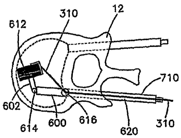
[00111] FIGS. 6A and B illustrate the use of a bobbin 600 having two
joints 614,
616 for positioning and orienting bobbin 600 within vertebral body 12. FIG. 6A
shows
articulated bobbin 600 before augmentation and FIG. 6B shows enlarged bobbin
implant
312 after coiling of band 310 around bobbin 600 within vertebral body 12. In
some
embodiments, as shown in FIGS. 5 and 6, band 310 may be inserted through a
central
passage or lumen in shaft 520, 620 of bobbin 500, 600, respectively.
[00112] Referring to FIGS. 7A and 7B, coiling of band 310 may be
facilitated
using a guide 700, also referred to herein as a slider 700. For example, as
shown in FIG.
7A, bobbin 500 having a slider 700 may be inserted into vertebral body 12
through a
cannula 710. Bobbin 500 may have a joint 514 that allows articulation of the
end 512 of
bobbin 500, with respect to shaft 520. Slider 700 can move, either in a
uniform or
predefined pattern, or manually as controlled by a user, with respect to
bobbin 500 to
guide coiling of band 310 over bobbin to expand size of implant 512 and define
its
shape.
[00113] As shown in FIG. 7B, guide 700 may be incorporated with a bobbin
assembly having two or more joints, for example bobbin 600 having two joints.
Bobbin
600 can be inserted into a vertebral body 12 through a cannula 710, and band
310 may
pass through shaft 620 of bobbin 600. Band 310 engages slide 700 which
cooperates
with bobbin 600 to guide band 310 around bobbin 600 and increase the size of
coil 612.
Joints 614 and 616 allow articulation of bobbin 600 into a desired position
and
orientation for augmentation of vertebral body 12.
[00114] In some embodiments, head 602 of bobbin 600 and/or coil 612
remains in
vertebral body 12 after the cannula 710 and shaft 620 of bobbin are removed
from the
patient to augment the vertebra and maintain proper lordosis. In other
embodiments,
PMMA or another cement or filler is inserted into vertebral body 12 along with
bobbin
coil 612 to further enhance fixation or repair of the damaged region. In other
embodiments, bobbin 600 and/or bobbin coil 612 are removed after repositioning
the
bone and PMMA or another filler is injected into a void created by coil 612.
-17-
CA 02612548 2007-12-17
WO 2007/002108
PCT/US2006/024009
[00115] FIG. 8 is a detailed cross-sectional view of a bobbin, e.g.,
bobbin 500,
having a guide or slider 700 to facilitate coiling of band 310. As described
above,
bobbin 500 may be inserted through cannula 710 and may include one or more
joints 514
to facilitate positioning of coil 512. Slider 700 may include a head 703 that
manipulates
band 310 and cooperates with bobbin 500 to control the wrapping of band 310 to
enlarge
coil 512. Slider 700 may employ a screw or other mechanism to provide uniform
coiling
or a predefined coil pattern, and/or slider may be configured to be
manipulated by a user
to provide any desired pattern and shape of bobbin coil 512, e.g., to optimize
engagement of coil 512 with inner walls of vertebral body 12. Slider 700 may
pivot,
bend or slide, or a combination of movements to facilitating placing of band
310 about
bobbin 500. Slider 700 may pass through cannula 710 outside of bobbin, as
shown in
FIG. 8. In other embodiments, slider 700 may attach to and/or pass through
shaft 520 of
bobbin 500, may attach to cannula 710, or may be introduced into vertebra
through
another cannula or introducer.
[00116] Band 310 may pass through cannula 710 as shown in FIG. 8. In other
embodiments, band 310 may pass through bobbin shaft 520. In other embodiments
band
310 can be inserted through a cannula different than the cannula through which
the
bobbin is inserted. For example, as shown in FIG. 9, bobbin 600 having two
joints 614
and 616 and slide 700 can be inserted into vertebral body 12 through a first
cannula 710,
while thread 310 can be inserted into vertebral body 12 through a second
cannula 900.
[00117] FIG. 10 depicts in more detail various components of an embodiment
of a
bobbin 800 attached to an insertion device 810. In this embodiment, the band
310 may
spool about a bobbin 800 due to rotational forces created by a drive line
assembly 840 of
the insertion device 810. The drive line assembly 840 of the insertion device
810 is
composed of a drive mechanism 814 and a rod 825. Further, a manual axial force
applied to an axial control assembly 830, in this embodiment comprising an
outer
cannula 811 and knob 813 of the insertion device, positions the band 310 along
the axial
length of the bobbin 800. By controlling the drive line assembly 840 and the
axial
control assembly 830 a user can create different shapes, such as conical, or
"egg" shape,
of the coiled band assembly 812.
[00118] The axial control assembly 830, e.g., outer cannula 811 and knob
813,
may guide the drive line assembly (rod 812 and drive mechanism 814) and bobbin
800
into the vertebral body as well as move the band 310 axially over the bobbin
800. The
knob 813 may be attached to a midsection of the outer cannula 811 in a fixed
manner,
-18-
CA 02612548 2007-12-17
WO 2007/002108
PCT/US2006/024009
such that the ends 817, 818 of the outer cannula 811 project past the knob
813. The knob
813 may have an opening 815 through which the band 310 is inserted through.
The knob
813 may be cylindrical in shape as shown, or may have other shapes. The knob
813 may
be made from different materials such as metal, plastic, and rubber. The outer
cannula
811 has an inner diameter which is larger than the outer diameter of the rod
825, which
may be inserted through the outer cannula 811. The distal end of the outer
cannula 811
has attachment 816 which aids in guiding the band 310 onto the bobbin 800. The
attachment 816 is preferably fixed to the outer cannula 811, but may also
move, e.g.,
translate or rotate, relative to the outer cannula 811. The attachment 816,
together with
the manual axial force applied to the outer cannula 811, controls the position
of the band
310 on the bobbin 800. Moving the attachment 816 axially back and forth causes
the
band 310 to move forward or rearward on the bobbin 800. The band 310 is coiled
about
the bobbin 800 underneath the attachment 816. The attachment 816 limits the
diameter
of the band mass about the bobbin 800. Although attachment 816 is shown have a
curvature, preferably with a radius, other shapes are possible for attachment
816. The
shape of attachment 816 can influence and limit the final shape of the coiled
band
assembly 812. While the axial control assembly has been described as
comprising
multiple pieces it can also comprise a single piece and is not limited to the
form
illustrated.
[00119] The drive line assembly 840 of the insertion device 810 may be
comprised of the rod 825 and the drive mechanism 814. The rod 825 has a
proximal end
819 and a distal end 820. At the proximal end 819, the rod 825 preferably has
an
attachment interface 822 that is compatible with the attachment interface 821
on the
drive mechanism 814. One example of an attachment interface could be a ball
detent
mechanism such as those found on ratchet set drives, a bayonet nut connector,
thread,
conical connections, or hexagonal connector. A hex, star or other shaped male
and
female respective connectors may also be used. Other forms of interface
attachments are
contemplated. The distal end 820 of the rod 825 has another attachment
interface 823
which is compatible with the attachment interface 824 on the bobbin 800 to
preferably
releasably attach the bobbin 800 to the rod 825. The drive mechanism 814
rotates the
rod 825 about its longitudinal axis. The drive mechanism 814 may be hand
driven, such
as with a T-handle as depicted in FIG. 10, or mechanized, such as a motorized
drill (not
shown). Operation of the drive mechanism 814 causes the rod 825 to rotate, and
the rod
825 causes the bobbin 800 to rotate. Alternatively, the drive mechanism 814
can be
-19-
CA 02612548 2007-12-17
WO 2007/002108
PCT/US2006/024009
integral with the rod 825, or the drive mechanism preferably can releasably
connect
directly to the bobbin 800 such that rotation of drive line mechanism rotates
the bobbin.
[00120] The bobbin 800 may comprise a cylindrical shaft, having a threaded
exterior and whose outer diameter is smaller than the inside diameter of the
outer cannula
811. The threaded exterior may aid in guiding the coiling band 310 about the
bobbin
800. The bobbin 800 is attached to one end, the distal end, of the rod 825 and
inserted
through the outer cannula 811 such that the bobbin 800 projects from the
distal end of
the outer cannula 811. The bobbin 800 may also include a hole 801 through the
cylindrical shaft so that an end of the band 310 may be fed through the hole
801 to
preferably attach and hold the band 310 in place on the bobbin 800. Other
means of
attaching the band 310 to the bobbin 800 may be utilized.
[00121] Operation of the insertion device 810 and the bobbin 800 to create
a final
implant will now be discussed. The bobbin 800, having a band 310 inserted into
the hole
801 of the cylindrical shaft is attached to the rod 825. The insertion device
810,
comprising the drive line assembly 840 and the axial control assembly 830,
with the
attached bobbin 800 may be introduced into a collapsed vertebral body (not
shown). The
insertion device 810 is preferably inserted through a cannula but may be
inserted through
an open incision or percintaneous by piercing the skin and soft tissue with
the bobbin
800 positioned in the vertebrae. The drive mechanism 814, connected to the rod
825,
rotates the rod 825 such that the band 310 coils about the bobbin 800 to
create a larger
bobbin/band mass, also referred to as the final implant. The user may move the
outer
cannula 811 forward and backward, with respect to the collapsed vertebral
body, to allow
the band 310 to coil around the full axial length of the bobbin 800. Further,
moving the
outer cannula 811 forward and backward allows the user to create different
shapes of the
larger coiled band assembly. During coiling, the diameter of the coiled band
assembly is
increased to a desired size. The attachment 816 on the outer cannula 811 aids
in creating
different shapes of the larger coiled band assembly, as well as limiting the
diameter of
the coiled band assembly. The diameter of the implant can be varied along the
length of
the bobbin 800 to tailor the size and shape of the implant. Once the desired
size and
shape of the coiled band assembly has been achieved, the user cuts the band
310. This
may occur outside the vertebral body. After the band 310 has been severed, the
user
rotates the rod 825 and bobbin 800 using the drive mechanism 814 to coil the
end of the
band about the bobbin 800, such that the band 310 is completely coiled about
the bobbin
800 within the vertebral body. Once this is achieved, the user pulls on the
rod 825 and
-20 -
CA 02612548 2007-12-17
WO 2007/002108
PCT/US2006/024009
drive mechanism 814, causing the rod 825 to detach from the bobbin 800,
leaving the
bobbin 800 in the vertebral body. The rod 825 may then be completely removed
from
the patient. Further, the outer carmula 811 is also removed after the rod 825
and bobbin
800 have been detached. PMMA or another cement or filler may be inserted into
vertebral body containing the bobbin/band mass to further enhance fixation or
repair of
the damaged region.
[00122] Another embodiment of a bobbin implant is depicted in FIGS. 11-30.
FIG. 11 depicts a bobbin 1160 with an insertion device 1000. Similar to the
previous
embodiment, the bobbin 1160 is rotated about its longitudinal axis and one or
more
bands 310 are coiled about the shaft of the bobbin 1160 to create a larger
coiled band
assembly. The insertion device 1000 transfers rotational motion from a drive
mechanism
(not shown) to rotate the bobbin 1160. When coiling a single band 310, the
insertion
device 1000 also preferably moves the band 310 axially along the shaft of the
bobbin
1160. Whereas, when coiling multiple bands 310 about the bobbin 1160, no axial
movement may be necessary. When various multiple bands 310 coil about the
bobbin
1160, the bands 310 preferably coil about the bobbin 1160 at different
locations along
the shaft of the bobbin 1160. In a single band configuration, the insertion
device 1000
preferably functions to rotate the bobbin 1160 about the bobbin's longitudinal
axis, as
well as provide axial movement of the band 310 with respect to the bobbin
1160.
Rotation of the bobbin 1160 is accomplished through a drive line assembly 1300
comprising a drive shaft 1110, a flexible shaft 1070 and preferably a rotating
a drive
mechanism (not shown). While the drive line assembly 1300 is illustrated and
described
as comprising multiple pieces it may be a single component or comprise
components
other than those shown and described. Axial movement of the band 310 relative
to the
bobbin 1160, is accomplished through guide mechanism 1600 comprising a band
guide
conduit 1120, a spool holder 1030 and a cam disk 1010. While the guide
mechanism
1600 is illustrated and described as comprising multiple pieces, it may also
comprises a
single component or additional components other than those shown and
described.
Moreover, while insertion device 1000 axially fixes the location of the bobbin
1160 and
axially moves the band 310 along the axial length of the bobbin 1160, the
position of the
band 310 could be fixed and the bobbin 1160 could move axially.
[00123] Referring to the drive line assembly 1300, the drive shaft 1110 is
generally cylindrical in shape and may have a length of about 115 mm, although
other
lengths are contemplated. A section 1116 of the drive shaft 1110, near the
proximal end
-21-
CA 02612548 2007-12-17
WO 2007/002108
PCT/US2006/024009
1111, may have gear teeth about the circumference of the drive shaft 1110. The
proximal end 1111 of the drive shaft 1110 extends from a proximal end 1023 of
a
housing 1020 of the insertion device 1000. As depicted in FIGS. 17A and B, the
drive
shaft 1110 proximal end 1111 has a quick coupling feature 1114 which is
compatible
with a drive mechanism, preferably a rotating drive mechanism that provides
torque to
drive shaft 1110, such as for example a T-handle or drill (not shown). A lock
washer
1230 and stop disk 1170 are attached near the proximal end 1111 of the drive
shaft 1110
(FIG. 12) preventing the drive shaft 1110 from moving in a distal direction.
The lock
washer 1230 and stop disk 1170 may abut an end cap 1100 (FIGS. 15A and B)
attached
to the proximal end 1023 of the housing 1020. The end cap 1100 is secured to
the
housing 1020 by shear stress pins 1260, preferably three shear stress pins
1260. Other
means for securing the end cap 1100 to the housing 1020 may be used. The drive
shaft
1110 projects through center hole 1102 in the end surface 1104 of the end cap
1100. The
distal end 1112 of the drive shaft 1110 extends into the housing 1020 and has
an
attachment interface 1113 which may be compatible with an attachment interface
1075
on the proximal end 1074 of the flexible shaft 1070. One example of an
attachment
interface could be a ball detent mechanism such as those found on ratchet set
drives, a
bayonet nut connector, thread, or conical connections. Another example of an
attachment interface could be a hexagonal or other shaped protrusion seating
in a
corresponding hexagonal or other shaped recess. The drive shaft 1110 connects
to the
flexible shaft 1070 in the interior of the housing 1020.
[00124] The flexible shaft 1070 (FIG. 26), connected to the drive shaft
1110
through attachment 1075 at its proximal end 1074, extends through a needle
1040. The
flexible shaft 1070 preferably may comprise two components, a straight
optionally rigid
rod 1071 and a flexible component 1072 distal of the rigid rod 1071. The
distal end
1073 of the flexible shaft 1070 connects to the bobbin 1160 preferably by a
quick
disconnect attachment 1076. The quick disconnect attachment 1076 may take any
number of forms, such as a ball detent mechanism such as those found on
ratchet set
drives, a bayonet nut connector, thread, conical connections, hexagonal
connector, etc.
The attachment 1076 also allows the flexible shaft 1070 to disconnect from the
bobbin
1160 when a user pulls the drive shaft 1110 together with the flexible shaft
1070 in a
proximal direction along the longitudinal axis of the housing 1020 and drive
shaft 1110.
A drive mechanism rotates the drive shaft 1110 which in turn rotates the
flexible shaft
- 22 -
CA 02612548 2013-02-12
1070 which rotates the bobbin 1160. The flexible shaft 1070 rotates within
main bore
1042 formed in the needle 1040.
[00125] The needle 1040 assists in inserting the bobbin into the vertebral
body,
similar to the outer cannula 811 of the previous embodiment. The needle 1040
(FIGS.
22-24) is preferably fixed to and extends from the distal end 1026 of the
housing 1020.
The needle 1040, in particular the configuration of the distal end 1041 of the
needle
1040, can take various shapes arid forms some of which are illustrated in
FIGS. 22-24.
The needle 1040 is generally cylindrical in shape with a half cylinder shape
at its distal
end 1041. The needle 1040 may have a length of about 126 mm with an outside
diameter of about 5 mm, although these length and diameter are only exemplary
and
other lengths and diameters are contemplated. The distal end 1041 may have a
flat, open
surface 1047 on which the bobbin 1160 rests. In the alternative, the flat
surface 1047
may be inclined such that the distal end 1041 has a larger dimension, causing
the flexible
shaft 1070 to bend (FIG. 23A). A threaded pin 1270 through the hole 1022 at a
distal
end 1026 of the housing 1020 holds the needle 1040 to the housing. Other means
of
fixing the needle 1040 to the housing are contemplated.
[00126] The bobbin 1160 (FIGS. 27A-C) may be cylindrical, having a length
of
about 16 mm and a diameter of about 2.5 mm, although this length and diameter
are
merely exemplary and other lengths and diameters are contemplated which would
depend, in part, upon the desired final implant shape. The bobbin 1160 may
have a
rough surface providing friction to the surrounding band so as to ease force
transfer
between the rotating bobbin 1160 and the coiled band 310 The proximal end 1161
includes a quick disconnect attachment 1062 complementing the attachment 1076
on the
flexible shaft 1070. The distal end 1163 of the bobbin 1160 may be hollow. At
the
distal most end of the bobbin 1160 a hole 1164 may extend through the diameter
of the
bobbin 1160. Two additional holes 1165, 1166 may be included. These holes
extend
only through one side of the outer circumference of the bobbin 1160. The holes
1164,
1165, and 1166 may assist in attaching the band 310 to the bobbin 1160. With
the band
310 attached to the bobbin 1160, rotation of the bobbin 1160 will cause the
band 310 to
wrap around the bobbin 1160. Rotational motion of the bobbin 310 pulls the
band 310
from band spools 1140, 1150 located inside the housing 1020. The cone shape
about the
bobbin 1160 in FIGS. 11 and 12 depicts one exemplary shape the coiled bobbin
may
form when implanted in a vertebrae.
-23 -
CA 02612548 2007-12-17
WO 2007/002108
PCT/US2006/024009
[00127] Axial movement of the band 310 relative to the bobbin 1160 is
accomplished through guide mechanism 1600 comprising the band guide conduit
1120
(FIG. 25), spool holder 1030, cam disk 1010, and a gear train 1700. In the
insertion
device 1000 the rotary motion of the drive mechanism is converted to an axial
motion
which is transferred to the band guide conduit 1120 to control the axial
location where
the and 310 is coiled onto the bobbin 1160. The gear train 1700 may comprise
drive
gear 1080 and sprocket 1130 on which gear 1090 is mounted. The gear train 1700
functions to transfer the rotational force of the drive shaft 1110 to the cam
disk 1010,
such that the cam disk 1010 may rotate at a different speed than the drive
shaft 1110, for
example the cam disk 1010 preferably may rotate once for every five rotations
of the
drive shaft 1110. The cam disk 1010 converts the rotational force of the drive
shaft 1110
to an axial, preferably oscillating force due to cause the spool holder 1030
to move
axially, preferably oscillate back and forth. The spool holder 1030 moves the
band guide
conduit 1120 similarly in an axial direction, preferably in an oscillating
back and forth
motion. The purpose of the axial movement is to have the band 310 move
axially,
relative to the bobbin 1160.
[00128] More specifically, the teeth of drive gear 1080 meshes with the
teeth 1116
of the drive shaft 1110 and with the teeth of gear 1090, which rotates about
sprocket
1130. The teeth of gear 1090 also meshes with teeth 1016 located at the
proximal end
1017 of the cam disk 1010. That is, the drive gear 1080 interacts with both
the drive
shaft 1110 (FIGS. 17A and B) and the gear 1090, causing the cam disk 1010 to
rotate at
a different speed, preferably slower speed, than the drive shaft 1110. Gear
1090 is seated
over sprocket 1130 (FIG. 16) which is held in place by a lock washer 1240. The
gear
train 1700 can be configured to cause the cam drive at the desired speed to
control the
axial motion and depending upon the gearing, cam disk 1010 can rotate faster
or slower
than the drive shaft 1110.
[00129] The cam disk 1010 (FIG. 18) is cylindrical in shape having a length
of
about 41 mm and an external diameter of about 31.5 mm, which is smaller than
an inner
diameter of the housing 1020. Other sizes, lengths and diameters can be
utilized for the
cam disk 1010. The cam disk 1010 is structured with a cross member 1011 and
center
core 1012 having an inner passage 1013 allowing the drive shaft 1110 to pass
through
the center of the cam disk 1010. The inner passage 1013 helps maintain
alignment of the
drive shaft 1110. The drive shaft 1110 is free to rotate in inner passage 1013
relative to
- 24 -
CA 02612548 2007-12-17
WO 2007/002108
PCT/US2006/024009
the cam disk 1010. The external diameter of the cam disk 1010 has a groove
1014 about
its circumference.
[00130] The spool holder 1030 (FIGS. 19A and B) is also cylindrical in
shape,
having a distal end 1037 and a proximal end 1034. The outer diameter of the
spool
holder 1030 is smaller than the inner diameter of the housing 1020, but larger
than the
distal end of the cam disk 1110. Similar to the cam disk 1010, the spool
holder 1030 has
a cross member 1031 and center core 1032 having an inner passage 1033. The
drive
shaft 110 and flexible shaft 1070 are free to rotate in inner passage 1033
relative to spool
holder 1030. The proximal end 1034 of the spool holder 1030 has a cavity 1037
which
receives the distal end 1015 of the cam disk 1010 such that the proximal end
1034 of the
spool holder 1030 overlaps the cam disk 1010 and is between the housing 1020
and the
cam disk 1010. The overlap is sufficiently4arge so that as the spool holder
1030 moves
towards the distal end 1026 of the housing 1020 (discussed later) the proximal
end 1034
of the spool holder 1030 continues to overlap the distal end 1015 of the cam
disk 1010.
[00131] The spool holder 1030 is prevented from rotating about its
longitudinal
axis within the housing 1020 by dowel pin 1250 extending through hole 1035 in
the
proximal end 1034 of the spool holder 1030. The dowel pin 1250 extends into
the
groove 1014 of the cam disk 1010 and through slot 1024 at the proximal end
1023 of the
housing 1020. The gear train 1700 rotates the cam disk 1010 and as the cam
disk 1010
rotates, dowel pin 1250 moves along slot 1024 due to the inclination of the
groove 1014
in the cam disk 1010. As the dowel pin 1250 moves axially along slot 1024 it
moves
spool holder 1030 axially as well. Thus, the spool holder 1030 undergoes the
same
motion as the dowel pin 1250 and provides a visual indicator of the location
of the spool
holder and the position of the band 310. If the dowel pin 1250 moves back and
forth in
slot 1024 then the spool holder 1030 will likewise move axially back and
forth.
[00132] Either a large band spool 1140 (FIGS. 20A and B) or a small band
spool
1150 (FIGS. 21A and B) or multiple band spools can be placed over the center
core 1032
of the spool holder 1030. The band spool 1140, 1150 may be held in place by a
displacement disk 1180 and lock washer 1220. Between the band spool 1140, 1150
and
the cross member 1031 of the spool holder 1030 a spring 1190, such as leaf
spring,
maintains the band spool 1140, 1150 against the displacement disk 1180. As the
bobbin
1160 rotates, it unspools the band 310 from the band spool 1140, 1150. A
pressure
spring 1200, such as a helical spring, is located between the cam disk 1010
and the spool
holder 1030, coaxial with the drive shaft 1110. The pressure spring 1200
maintains
- 25 -
CA 02612548 2007-12-17
WO 2007/002108
PCT/US2006/024009
tension on the band, so as the spool holder 1030 moves axially backward, the
band 310
does not get entangled. The pressure spring 1200 also aids in maintaining the
components in place.
[00133] The band 310 from the band spool 1140, 1150 passes through hole
1038
in the spool holder 1030 and is inserted into the interior conduit 1123 at the
proximal end
1121 of band guide conduit 1120. The band guide conduit 1120 is located in
groove
1043 of the needle 1040. The proximal end 1121 of the band guide conduit 1120
is
attached to the spool holder 1030 through a hole 1036 at the distal end 1037
of the spool
holder 1030. Thus, as the spool holder 1030 moves axially within the housing
1020, the
band guide conduit 1120 moves similarly, preferably axially back and forth in
the needle
1040. The distal end 1122 of the band guide conduit 1120 terminates in the
groove 1043
of the distal end 1041 of the needle 1040. The band guide conduit 1120 moves
axially
relative to the bobbin 1160, thus controlling the position along the axial
length of the
bobbin 1160 where band 310 is placed.
[00134] Attached to the drive shaft 1110 between the spool holder 1030 and
cam
disk 1010, is a second stop disk 1170 and a lock washer 1230. Connected to the
second
stop disk 1170 and the cam disk 1010 is a pressure spring 1210. This pressure
spring
1210, similar to pressure spring 1200 aids in keeping the components in a
defined
position.
[00135] The housing 1020 (FIGS. 14A and B) is cylindrical in shape having
a
proximal end 1023 and a distal end 1026, and houses the above described
components.
The diameter of the housing 1020 at the distal end 1026 tapers at
approximately 45
degrees to a smaller diameter forming a nozzle 1021. Other shapes and sizes
for the
housing are contemplated. Another slot 1025, perpendicular to the housing's
longitudinal axis is located near the distal end 1026 of the housing 1020.
This slot 1025
provides access to band(s) 310 proximate the band spool 1140, 1150 to allow a
user to
cut the band(s) 310 once the bobbin/band mass has obtained its desired size.
[00136] In a multiple band configuration of the insertion device 1000
(FIG. 28), a
band guide conduit 1120, a cam disk 1010 preferably may not be employed. No
axial
movement may be required when coiling multiple bands 310 about the bobbin
1160,
however, if desired axial movement may be provided. One reason that axial
movement
may be unnecessary is that each band 310, passing through holes 1038, 1039,
and 1191
in the spool holder 1030 and through bores 1044, 1045, and 1046 of needle
1040, are
attached to the bobbin 1160 at a different location along the shaft of the
bobbin 1160,
-26 -
CA 02612548 2007-12-17
WO 2007/002108
PCT/US2006/024009
through holes 1164-1166. In the multi-band embodiment, bands of different
sizes,
thickness and shapes may be utilized to configure the final implant shape.
Preferably,
the thickest band coils about the distal end 1163 of the bobbin 1160, with
success thinner
bands coiling next to the previous band 310. Thus, as the bobbin 1160 rotates,
multiple
bands 310 coil about the bobbin. And as the bobbin 1160 rotates more, the
bands will
naturally spread out along the shaft of the bobbin, creating a coiled band
assembly.
Thus, in a multiple band configuration, the insertion device 1000 may function
to rotate
the bobbin 1160 about the bobbin's longitudinal axis with little or no axial
movement
about the insertion device's longitudinal axis to the band 310 with respect to
the bobbin
1160. Accordingly, the internal components of the housing 1020 may not need to
move
axially in a multiple thread configuration. Similarly, the cam disk 1010 does
not rotate
either.
[00137] To disengage the axial guide mechanism 1600, the drive shaft 1110
is
displaced in a proximal direction until a narrower section 1115 of the drive
shaft
corresponds with the location of drive gear 1080. Because this section 1115 of
the drive
shaft is narrower and has no gear teeth, the gear teeth of drive gear 1080 not
do interact
with the drive shaft 1110. Accordingly, as the drive shaft 1110 rotates, there
is no
corresponding interface between the drive shaft 1110 and the gear train 1700.
Thus, the
gear train 1700 does not rotate, and therefore no rotational force is applied
to the cam
disk 1010. Because the cam disk 1010 does not rotate, the spool holder 1030
does not
move axially.
[00138] The needle 1040 (FIGS 24A-C) may include three additional bores
1044,
1045, 1046 instead of the groove 1043. Each additional bore allows for a band
310 to be
threaded through. The additional bores may have different diameters to
accommodate
different sizes of the band. Where no axial movement of the guide mechanism
1600 is
required, the band guide conduit 1120 may be unnecessary and the bands may be
inserted directly through bores 1044, 1045, and 1046 as shown in FIG. 28.
[00139] The operation of the bobbin 1160 and insertion device 1000 will
now be
described. A band spool 1140, 1150 containing a band 310 is secured to the
spool holder
1030 inside the housing 1020. The band 310 is threaded from the band spool
1140, 1150
through hole 1038 in the spool holder 1030 and through the band guide conduit
1120
along the needle 1040 such that the band 310 exits the needle 1040 at its
distal end 1041
and is attached to the bobbin 1160 through one of the holes 1064, 1065, and
1066 of the
bobbin 1160. Other means may be used to attach the band 310 to the bobbin
1160. The
- 27 -
CA 02612548 2007-12-17
WO 2007/002108
PCT/US2006/024009
bobbin 1160 with the band 310, together with the needle 1040, is inserted into
the
collapsed vertebral body, for example either down a cannula or an open
surgical field.
[00140] The user rotates a drive mechanism causing the drive shaft 1110 to
rotate.
This rotation causes the cam disk 1010 and flexible shaft 1070 to rotate. The
dowel pin
1250 seated in the groove 1014 of the cam disk 1010 causes the spool holder
1030 to
move axially forward and back, depending on the shape of the groove 1014. The
axial
movement of the spool holder 1030 is transferred to the band guide conduit
1120,
causing the band guide conduit 1120 to move axially in needle 1040. This axial
movement determines the location where the band 310 is coiled about the bobbin
1160.
Meanwhile, the rotation of the flexible shaft 1070 causes the bobbin 1160 to
rotate which
causes the band 310 to uncoil from the spool 1140, 1150 and be fed through the
band
guide conduit 1120 and wrap around the bobbin 1160. When the bobbin 1160 has
obtained the desired mass and shape, the user stops rotation of the drive
shaft 1110 and
cuts the band 310 through the perpendicular slot 1025 in the housing 1020. The
user
then rotates the drive shaft 1110 so that the band 310 remaining in the band
guide
conduit 1120 is coiled about the bobbin 1160. Once completed, the user pulls
on the
drive shaft 1110, disconnecting the bobbin 1160 from the flexible shaft 1070,
leaving the
bobbin 1160 in the vertebral body. The user then removes the needle 1040 from
the
vertebral body. Alternatively, the user can pull on the housing 1020
simultaneously
disconnecting the bobbin 1160 from the flexible shaft 1070 and removing the
insertion
device 1000 from the patient PMMA or another cement or filler may be inserted
into
vertebral body along with bobbin to further enhance fixation or repair of the
damaged
region. Alternatively, the band 310 may be coated with or contain bone cement
or other
material that may be activated after or during insertion of the bobbin, coiled
body.
[00141] As discussed previously, multiple threads, of varying diameter may
be
coiled onto the bobbin to create the final implant (FIG. 28). This requires
multiple band
spools to be inserted into the housing 1020 of the insertion device 1000. The
bands from
these spools are threaded through the bores 1044, 1045,and 1046 of needle 1040
depicted
in FIGS 24A-C. Preferably no band guide conduit 1120 is employed in a multiple
band
configuration (although one or more band guide conduits may be utilized), and
the
flexible shaft 1070 and bobbin 1160 preferably do not move axially. Preferably
the
largest band is inserted in bore 1045 such that it exits at the most distal
end of the needle
1040. The next largest band is preferably inserted in bore 1044, such that it
exits needle
1040 proximal to bore 1045, as shown in FIG. 24B.
-28-
CA 02612548 2007-12-17
WO 2007/002108
PCT/US2006/024009
[00142] Prior to performing a procedure, a user may be provided with a
number of
insertion devices for implanting a bobbin 1160. Each insertion device may have
a
different cam disk having a different groove configuration. As discussed
previously, the
groove configuration in part determines the shape of the bobbin. In addition,
each
insertion device may include different band configurations or sizes. Thus,
user can chose
the best insertion device to provide the desired coiled band assembly
configuration.
[00143] In the alternative, a user may remove the end cap 1100 to gain
access to
the inner components of the housing 1020. In this manner, the user can
exchange cam
disks 1010 and add spools of band after the existing spool of band is
exhausted. Once
the proper configuration of cam disk 1010 and size(s) of band is inserted into
the
housing, the user re-attaches the end cap 1100, securing it with the shear
stress pins
1260. Other means of accessing the internal component of the insertion device,
such as
quick release mechanism may also be used. The insertion device may be
configured for
single or multiple uses. For example, a single use device may be made of
plastic and
may be made such that it cannot be reused. For example, the plastic housing
could be
molded so that the spool of band material can not be replaced without breaking
the
housing, or such that the band material can not be re-threaded in the band
guide
conduit/needle once the band has been cut and coiled onto the bobbin.
Alternatively, or
in addition to, the connection between the bobbin and the flexible shaft could
be
damaged when the bobbin is disengaged from the flexible shaft such that a new
bobbin
will not mate or attach to the flexible shaft. If the insertion device is
configured to be
reusable consideration should be made to sterilize the device for the next use
which may
require different materials than single use applications.
[00144] FIG. 29 illustrates the use of a flexible shaft having four joints
1500. The
joints 1500 may be in the form of universal joints. This configuration of the
flexible
shaft 1070 allows the bobbin to remain parallel with the needle 1040. FIG. 30
illustrates
the use of a flexible shaft having two universal joints 1500. This
configuration allows
the flexible shaft to support the bobbin at a predetermined angle.
[00145] In some embodiments, band 310 may be wound inside a bendable sheet
2000, such as a sheet of metal, plastic, Teflon or other material, wrapped
around a
bobbin. For example, FIG. 31A shows a transverse cross sectional view taken
across an
end of a bobbin, e.g., bobbin 600, having a sheet 2000 wrapped around bobbin
600
before winding of band 310. Referring to FIG. 31B, as band 310 is wound around
bobbin 600, sheet 2000 expands as coil 612 increases in diameter. Such
expansion can
- 29 -
CA 02612548 2007-12-17
WO 2007/002108
PCT/US2006/024009
force sheet 2000 outward, for example against structures within vertebral body
12. In
other embodiments, an expandable or elastic bag, balloon, or other structure,
which may
or may not have a predefined shape, can be used instead of or in addition to
sheet 2000
around coil 612 which can expand within vertebral body 12 as the coil
increases in size.
[00146] Referring to FIG. 32, a bobbin 2100 may be bendable instead of or
in
addition to having joints as described above. Such a bendable bobbin 2100 may
help
optimize repositioning and augmentation of damaged bone by conforming within
vertebral body 12 and applying a stabilizing force against inner walls of the
vertebral
body 12. Such bendable bobbins may be comprised of various materials, such as
polymers, metals, biomaterials, or any other compatible materials.
[00147] As mentioned above, a variety of types of bobbins having various
geometries and other features and characteristics may be used. For example,
the bobbin
may be straight bobbin 300 or a curve bobbin such as bobbin 2100, and may
comprise a
variety of materials. In other embodiments, a bobbin 600 can have joints such
as joints
614 and 616 to allow movement of each portion 601, 603, and 620 to provide a
desired
orientation and configuration of bobbin 600 within a vertebral body or other
structure to
be repaired, distracted, or otherwise manipulated.
[00148] Although the apparatus and methods described herein thus far have
been
described in the context of repositioning and augmenting vertebral bodies
following
vertebral compression fractures, various other uses and methods are
envisioned. For
example, in some embodiments, a bobbin such as bobbin 300 and band 310 may be
used
to reposition and/or augment other damaged bone regions such as a fractured or
weak
proximal femur 1400 as shown in FIGS. 34A and B. In such embodiments, bobbin
300
may be inserted through a cannula, and may include an extension 1800 to which
it is
releasably connected. bobbin 300 may have various features as described
elsewhere
herein. After band 310 is coiled about the bobbin, the coiled band body 312
may be
detached from the extension and left in position in the femur. The cavity left
by the
extension and the volume surrounding the coiled band body may be filled with
bone
cement, bone chips, or other material to integrate the coiled band body into
the bone.
[00149] In another embodiment, the implants may be, for example, wool
bales or
fibrous masses/bodies comprising a shape memory metal or metal alloy (e.g., a
nickel
titanium alloy such as nitinol) or another material that may or may not have
shape
memory characteristics. The fibrous masses may have any desired shape, e.g.,
cylindrical, spherical, or another shape (e.g., see FIG. 35).
- 30 -
CA 02612548 2007-12-17
WO 2007/002108
PCT/US2006/024009
[00150] In some embodiments, the wool bales or fibrous masses/bodies
(generally
referred to herein as "fibrous masses") may comprise a continuous or segmented
thread,
wire, strand or other elongated member (generally referred to herein as a
"fiber"). The
one or more fibrous masses may be comprised of any biocompatible material
having
desired characteristics of shape memory, flexibility, strength, and/or other
characteristics.
For example, in some embodiments the fibrous masses may be comprised of a
fiber,
wire, thread or other relatively thin structure of a biocompatible shape
memory alloy,
stainless steel, titanium, polymer, tricalcium phosphate, or any other
material having
desired characteristics. In some embodiments, the fibrous bodies may be
resorbable.
[00151] As shown in FIGS. 36A and B, fibrous masses comprising a shape
memory alloy (e.g., nitinol) may expand when the fibrous mass is heated to a
temperature over an actuation temperature, for example as the shape memory
alloy
undergoes a phase transformation between a Martensite state (e.g., at a low
temperature)
and an Austensite state (e.g., at a higher temperature). The actuation
temperature of
shape-memory alloy fibers within the wool bales may be, for example, between
about
28 C and about 36 C. Alternatively, a fibrous mass may expand, contract, or
otherwise
change shape or configuration when it is activated by an energy source (e.g.,
an
ultraviolet light, ultrasonic radiation, radio waves, heat, electric or
magnetic field).
[00152] The wool bales or fibrous masses may have an initial diameter that
is
small enough to allow one or more bodies to be inserted into a vertebral body
or other
bone thorough a cannula or other introducer as shown in FIG. 37. For example,
the wool
bales 4000 may have an initial diameter that is between about lmm and about
lOmm.
Other sizes may be used.
[00153] As shown in FIG. 37, a minimally invasive method of augmenting a
damaged vertebral body 12, e.g., following a vertebral compression fracture,
may
comprise implanting one or more wool bales or fibrous masses/bodies into an
inner
portion of a vertebral body between the upper and lower endplates of the
vertebral body.
An access may be formed in the outer cortical shell of vertebral body by a
trocar, drill or
other instrument. The wool bales or fibrous masses/bodies may then be
implanted, for
example, through a cannula 4100 or introducer inserted into vertebral body as
shown in
FIG. 37. Suitable procedures and materials for inserting a cannula through
which the
masses may be introduced are known in the art, and may be similar to those
described
above for kyphoplasty and other procedures. For example, the cannula 4100 may
be
-31 -
CA 02612548 2007-12-17
WO 2007/002108
PCT/US2006/024009
introduced using a posterior approach as shown in FIG. 2, e.g., through a
pedicle and
into the interior of the vertebral body.
[00154] The fibrous masses 4000 may be sized to be inserted into the
vertebral
body 12 through the cannula 4100 and may expand after being implanted in the
vertebral
body 12 to compact or compress the bone material inside the vertebral body.
Alternatively, after the passageway is formed in the vertebral body,
instruments such as,
for example, currettes or balloon catheter may be used to compress and compact
the bone
inside the vertebral body to create a cavity. The cavity in the vertebral body
also may be
formed by removing bone material as opposed to compacting the bone. For
example, a
reamer or other apparatus could be used to remove bone material from the
inside of the
vertebral body.
[00155] As shown in FIGS. 37 and 38, the fibrous masses may be inserted
through
the cannula and into the central portion of the vertebral body using a
displacement device
4200 or other apparatus. For example, a plunger, pusher or other displacement
member
inserted within the cannula 4100 may be used to displace or push the fibrous
masses
4000 through the cannula 4100 and into the vertebral body 12. The displacement
device
4200 may be driven, for example, by pressure, e.g., from a syringe, rod, or
other
apparatus that forces the displacement device 4200 into the cannula 4100 and
towards
the vertebral body 12.
[00156] When the fibrous masses 4000 are inside the vertebral body, they
may be
heated, for example by the body temperature of the patient, to a temperature
that is over
the actuation temperature (e.g., about 28 C to about 36 C). At the actuation
temperature,
the fibrous masses may expand as shown in FIG. 38. The expanded fibrous masses
may
help restore the height of the vertebral body, and may help restore lordosis
of the spine.
[00157] As shown in FIG. 39, as more fibrous bodies are inserted into the
vertebral body, and/or as the fibrous masses are expanded, they may fill a
central portion
of vertebral body and can push against the inner sides of the endplates of the
vertebral
body. Outward forces imparted by the fibrous masses within the vertebral body
may
spread the endplates apart and tend to restore vertebral body toward its
original height,
and may provide structural support to stabilize the vertebral body.
[00158] Insertion and expansion of the fibrous masses into a vertebral
body may
also compact the cancellous and osteoporotic bone inside the collapsed
vertebral body.
Any desired number of fibrous masses may be inserted, and each fibrous mass
may have
a desired configuration in order to augment the bone.
- 32 -
CA 02612548 2013-02-12
[00159] As shown in FIG. 40, after insertion of one or more wool bales 4000
or
other fibrous masses/bodies, a bone cement (e.g., PMMA or iricalcium
phosphate), bone
chips, demineralized bone, or other bone filler or implant may be inserted to
further
augment or stabilize the vertebral body.
[00160] After injection or insertion of the filler material, the cannula or
other
introducer may be removed. As shown in FIG. 41, the fibrous masses within the
vertebral body may help limit or prevent leakage of the cement or other
filler, and may
increase the shear strength of the bone cement or filler material.
[00161] In some embodiments a pedicle screw, a chain, or other elongated
member may attach to and/or extend from the vertebral body to provide a
tensioning
member to reposition the vertebral body.
[00162] The fibrous masses may be of a uniform or non-uniform size, shape
and
materials. One or more fibrous masses may be implanted into a bone or other
cavity
individually or linked together in a desired configuration. As shown in FIGS,
42 and 43,
fibrous bodies described herein may have any desired geometry, configuration
and/or
shape memory characteristics.
[00163] For example, as shown in FIG. 42, an implant of fibrous bodies may
comprise substantially spherical fibrous bodies which may be joined by and/or
threaded
upon one or more other fibers. In other embodiments a chain may have different
configurations of fibrous bodies that may or may not be joined.
[00164] As shown in FIGS. 43A-D, different configurations of wool bales or
fibrous masses/bodies may be used. For example, the fibrous masses may be
configured
as substantially spherical, ellipsoidal, cylindrical, or any other shape or
dimension that
may fit through a cannula or other introducer. A fibrous mass may also have a
different
size, shape or configuration after activation or expansion, for example as
shown in FIGS.
43A-D.
[00165] One or more fibrous masses may be inserted, possibly with one or
more
other implants, and may become tangled and/or convoluted with the other
fibrous bodies
or implants within the vertebral body. For example, after insertion, the
fibrous masses
may become further attached together and/or tangled such that that the masses
become a
larger mass filling the spaces within the vertebral body. Such larger mass may
lock into
-33-
CA 02612548 2007-12-17
WO 2007/002108
PCT/US2006/024009
the vertebral body and resist removal through the insertion opening. In some
embodiments, one or more fibrous masses or other implants may be coated with
an
adhesive, such that the implant may inserted into vertebral body in a
substantially
flexible state and may become more rigid and/or tangled or convoluted during
or after
insertion.
[00166] Although the apparatus and methods described herein thus far have
been
described in the context of repositioning and augmenting vertebrae in the
context of
vertebral compression fractures and deformations in spinal curvature, various
other uses
and methods are envisioned. For example, in some embodiments, one or more
implants
comprising fibrous masses may be used to reposition and/or augment other
damaged
bone regions such as a fractured or weak proximal femur as shown in FIG. 44A-
C.
[00167] In such embodiments, for example, one or more fibrous masses or
chains
of fibrous masses may be inserted into a head of a femur, e.g., through a
cannula or other
introducer. Once inserted, the fibrous mass may compact material within the
head of the
femur and provide solid support to augment the head. In some embodiments, the
fibrous
mass may comprise a shape memory alloy and expand or otherwise change its
configuration after insertion (e.g., after heating to a temperature above an
activation
temperature). A bone cement or other filler may also be used to aid
augmentation. In
other embodiments, for example as shown in FIG. 44C, another implant such as a
screw
or other device may be inserted in addition to or instead of one or more
fibrous masses.
[00168] In some embodiments, the implants and methods described herein may
be
used in conjunction with other apparatus and methods to restore lordosis and
augment a
vertebral body. For example, one or more fibrous mass implants or coiled
bodies may be
used in conjunction with known procedures, e.g., a balloon kyphoplasty, which
may be
used to begin repositioning of a vertebral body and/or create a space within
the body for
the implant. In other embodiments, one or more implants described herein may
be used
in conjunction with other tools or devices, e.g., an external fixation
apparatus for helping
to manipulate or fix the vertebrae or other bones in a desired position.
[00169] In another embodiment, a kit may comprise various combinations of
implant assemblies, components and insertion instruments. A kit may comprise,
for
example, various combinations of fibrous implants, coiled bodies, coiled
sleeve,
insertion devices, other implants, and/or a cannula or other introducer.
Optionally, a kit
may include, for example, a syringe or other container of a cement or other
bone filler
material. A kit may also optionally include an external fixation apparatus or
other tool
- 34 -
CA 02612548 2007-12-17
WO 2007/002108
PCT/US2006/024009
for repositioning or fixing vertebrae or other bones or body segments. One
skilled in the
art will appreciate that various other combinations of devices, components and
assemblies can be made and are intended to fall within the scope of the
present
invention.
[00170] In another embodiment, the vertebral body implant may comprise a
coiled
sleeve. Referring to FIG. 45A, a coiled body 3300, also referred to herein as
coil 3300 or
body 3300, comprises one or more sheets 3312 coiled about a central axis 3316.
Body
3300 may be approximately cylindrical, and can have an inner surface 3302, an
outer
surface 3304, and a diameter 3310, which may have an initial value of dl. Body
3300
can be dimensioned to fit within any desired cavity or space. For example, in
some
embodiments, coiled body 3300 can be dimensioned to fit within a collapsed
vertebral
body. Diameter dl 3310 may be less than a diameter of a cannula or other
device having
a lumen through which coil 3300 can be passed. After insertion of coiled body
3300 into
a collapsed vertebral body, a bone cavity, an intervertebral space, or another
location,
body 3300 can be partially uncoiled as shown in FIG. 45B. Such uncoiling can
cause a
widening of spaces 3314 between coils of sheet 3312, and a concomitant
increase in
diameter 3310 from dl to d2. Such coiling may alternatively and/or
additionally cause
an increase in the diameter of the coiled body. Uncoiling of body 3300 may be
performed, for example, by rotation of body 3300 with respect to axis 3316,
e.g., in a
direction opposite the direction of coiling of sheet 3312 about axis 3316.
[00171] Sheet 3312 can comprise one or more metals, composite materials,
polymers, or the like, and preferably has a stiffness and an outward bias when
coiled.
Such outward bias can be sufficient to provide a desired radial force when
body 3300 is
partially uncoiled as shown in FIG. 45B. In some embodiments, coiled sheet 312
can
comprise stainless steel, aluminum, a metal alloy, e.g., a cobalt alloy, a
nickel titanium
alloy or another alloy, a polymer, or any combination thereof. Body 3300 can
be
radiopaque or non-radiopaque. In certain embodiments, sheet 3312 can comprise
a
nickel titanium alloy having shape memory characteristics.
[00172] Referring to FIGS. 46A and B, an augmentation device 5400
comprises
coiled body 5300 coiled around an axial member, or shaft 5410. As shown in
FIG. 46A,
a minimally invasive method of augmenting a damaged vertebral body, e.g.,
following a
vertebral compression fracture, comprises inserting a augmentation device 5400
into a
portion 5402, preferably central portion 5402, of vertebral body 12, for
example through
a cannula or other introducer (not shown). Suitable procedures and materials
for
- 35 -
CA 02612548 2007-12-17
WO 2007/002108
PCT/US2006/024009
inserting a cannula through which coil 5300 may be introduced are known in the
art, and
may be similar to those described above for kyphoplasty and other procedures.
For
example, device 5400 may be introduced through the posterior portion 20 of the
vertebral body 12, e.g., through pedicle 24 (e.g., transpedicular approach).
[00173] After coil 5300 is inserted into central portion 5402 of vertebral
body 12,
shaft 5410 may be rotated to partially uncoil body 5300. Uncoiling body 5300
can
increase its diameter 5310, for example from dl to d2 as shown in FIG. 46B. As
diameter 5310 of coil 5300 increases, surface 5304 of coil 5300 can impart a
radial force
against endplates 5402, 5404 of vertebral body 12. Such radial force can push
endplates
5406, 5408 apart, which can restore vertebral body 12 to its original height
and augment
or stabilize the vertebral body 12. Additionally, the radial force imparted by
expanding
coil 5300 can compact the bone inside vertebral body 12, which may aid in
integration of
the device 5400 within cavity 5402 of vertebral body 12.
[00174] FIG. 47 illustrates a close-up perspective view of an augmentation
device
5400 according to an embodiment of the present invention. Expandable body 5300
can
comprise a rigid or semi-rigid sheet 5312, which can be tightly coiled around
axial
member, or shaft, 5410 as shown. Axial member 5410 can, for example, be a
solid
member or can have a lumen through which objects may be passed. In some
embodiments, sheet 5312 is attached to shaft, for example at inner surface
5302 of coil
5300. Body 5300 can be expanded by partially uncoiling sheet 5312, which can
create or
increase spaces 5314 between layers of sheet 5312, and/or increase the space
in the
center of the body 5300. Such uncoiling can be performed, for example, by
rotating
axial member 5400. In one embodiment, body is expanded by rotating axial
member
5410 in a direction 5502 opposite the direction of coiling of sheet 5312,
which can result
in an outward displacement 5504 of one or more outer coils 5506 of sheet 5312.
[00175] In other embodiments, body 5300 can be held in a tightly coiled
position
within the lumen of a cannula or other sheath, and such sheath may be removed
from coil
5300 after implantation within a bone 12. In such embodiments, sheet 5312 may
be
uncoiled by rotation of shaft 5502 as described above, or sheet 5312 may have
sufficient
stiffness, rigidity or memory that upon removal of the sheath the sheet 5312
partially
uncoils within bone 12, e.g., with sufficient force to impart a desired radial
force to
augment the bone 12.
[00176] In some embodiments, coil 5300 and axial member 5410, or a portion
of
axial member 5410, can remain in vertebral body. In other embodiments, coil
5300 can
-36-
CA 02612548 2007-12-17
WO 2007/002108
PCT/US2006/024009
remain in vertebral body 12 after the axial member 5410 is removed from the
patient,
e.g. along with a cannula, sheath, or other introducer. Keeping coil 5300 in
vertebral
body 12 can, for example, help continue augmenting the vertebra and maintain
proper
lordosis. In other embodiments, PMMA or another cement or filler can be
inserted into
vertebral body 12, e.g., through shaft and/or a cannula, along with coil 5300
to further
enhance fixation or repair of the damaged region. In other embodiments, coil
5300
and/or shaft 5410 can be removed after repositioning the bone, and PMMA or
another
filler can be injected into a void created by coil 5300.
[00177] In other embodiments, body 5300 can include multiple sheets 5312
and/or
can include an expandable bag, balloon, or other device between one or more
coils of
sheet 5312 and/or within lumen 5316 to facilitate expansion of body 5300
within
vertebral body 12.
[00178] FIG. 48 is a perspective view schematic illustration of a
fenestrated sheet
5600 for use in an expandable augmentation device according to the present
invention.
In some embodiments sheet 5600 may comprise a rigid or semi-rigid material
5612 that
can be formed into a coil. Suitable materials may include, for example,
stainless steel,
aluminum, a metal alloy, e.g., a cobalt alloy, a nickel titanium alloy or
another alloy, a
polymer, or any combination thereof. Sheet 5600 can include a plurality of
holes 5614,
for example through which a cement or other filler material may pass.
[00179] Referring to FIG. 49, an augmentation device 5700 may comprise a
coil
5710 of fenestrated sheet material 5612, which may include holes 5614, coiled
around an
end 5716 of an axial member 5712. Axial member 5712 can include a lumen 5714,
through which a needle 5722 or other device or material may be passed. End
5716 of
axial member 5712 may be open and/or axial member 5712 may include side holes
near
end 5716 to allow material within lumen 5714 to pass into body 5710.
[00180] In use, coil 5710 can be inserted into a central portion 5402 of a
vertebral
body 12, e.g., using a transpedicular approach. Body 5710 may be uncoiled to a
desired
diameter 5310, e.g., to impart a radial force on endplates 5406, 5408 and
restore anterior
portion of vertebral body 12 to its original height. Such uncoiling of body
5710 may be
performed, for example, by rotating axial member 5712 in a direction opposite
of the
direction of coiling of sheet material 5612. The filler material can uncoil or
otherwise
expand, or assist in the expansion of body 5710 in combination with other
expansion
means. In other embodiments, body 5710 uncoils without rotating shaft, e.g.,
due to the
stiffness or rigidity of material 5612. A needle 5722 or other extension can
pass through
-37-
CA 02612548 2007-12-17
WO 2007/002108
PCT/US2006/024009
lumen 5714 toward end 5716 of axial member 5712 to inject or otherwise
transfer bone
cement or other filler material into coiled body 5710 from a syringe or other
injection
apparatus 5720. In other embodiments, injection apparatus 5720 can be coupled
with an
exposed end 5718 of axial member 5712. The injected bone cement or other
filler
material 5724 can flow from end 5716 of axial member, and through holes 5614
in
fenestrated sheet material 5612, e.g., to surround coils of body 5710 and
preferably aid in
augmenting vertebral body 12. In some embodiments, after injection of filler
material
5724, axial member 5712 can removed, leaving coiled body 5710 and filler
material
5724 inside vertebral body 12. In other embodiments, axial member 5712 is not
detached from body 5710. The coiled body 5710 can be releasably attached to
the axial
member 5712.
[001811
Referring to FIGS. 50A and B, an augmentation device 5800 according to
another embodiment of the invention comprises a coiled body 5810 comprising
one or
more sheets 5812 coiled around an axial member 5410. As described above with
respect
to augmentation devices 5400 and 5700, sheet 5812 can comprise materials
having any
desired dimensions, strength, rigidity, stiffness, elasticity, thickness,
flexibility or other
characteristics. In some embodiments, sheet 5812 comprises stainless steel,
aluminum, a
metal alloy, e.g., a cobalt alloy, a nickel titanium alloy or another alloy, a
polymer, or
any combination thereof. Sheet 5812 can further comprise perforations, hinge
features or
other joints 5816 between adjacent panels or segments 5818 of sheet 5812. Such
joints
can facilitate coiling (collapse) and/or uncoiling (expansion) of sheet 5812
and allow for
use of substantially rigid sheet material 5812.
[00182] In
use, axial member 5410 can be rotated, for example in a direction 5502
opposite the direction of coiling of sheet 5812 around axial member. Such
rotation can
cause uncoiling or expansion of sheet 5812, increasing or creating space 5814
between
coils and causing an outward expansion 5504 of body 5810. Thus, the coiled
body can
be implanted in a first size and thereafter to a second size inside the
vertebral body.
Joints 5816 provide discrete locations for the sheet to bend or pivot, e.g.,
between
segments 5818. Joints 5816 can thus provide for incremental increases in
diameter of
coiled body 5810. By having joints 5816, substantially rigid materials can be
employed
in segments 5818 of sheet 5812, which may help optimize outward, radial forces
imparted by body 5810 as it is expanded within a bone or other confined area
such as
within a vertebral body.
- 38 -
CA 02612548 2007-12-17
WO 2007/002108
PCT/US2006/024009
[00183] Referring to FIG. 51, a method of augmenting a vertebral body or
other
bone can comprise inserting multiple augmentation devices 5400-1 and 5400-2
into a
vertebral body 12, e.g., using a posterior 20 tanspedicular approach, e.g.
through
pedicles 5902, 5904. In one embodiment, augmentation devices 5400-1 can
include a
body 5300-1 coiled about an axial member 5410-1, and can be inserted into a
central
portion 5402 of a vertebral body 12, e.g., adjacent to a pedicle 5902. A
second
augmentation device, for example 5400-2 which can also include a body 5300-2
coiled
about an axial member 5410-2, 2 or wool bales, or coiled bobbin can also be
inserted
into central portion 5402, e.g., through an opening adjacent to opposite
pedicle 904. One
or both of devices 5400-1 and 5400-2 can have some or all of the features
described
above with respect to augmentation devices 5400, 5700 and/or 5800, e.g., rigid
or semi-
rigid materials, fenestrated sheets, joints, and/or various other features
described herein.
After insertion of devices 5400-1 and 5400-2, one or both bodies 5300-1 and
5300-2 can
be uncoiled or otherwise expanded as described above to increase the height of
vertebral
body 12. Devices 5400-1 and 5400-2 can then be removed, or some or all of one
or both
devices 5400-1 and 5400-2 (e.g., bodies 5300-1 and 5300-2) can remain to
augment
vertebral body. In some embodiments, a bone cement or other filler material
can be
inserted into space 5402, with or without coiled bodies 5300-1 and 5300-2
remaining in
the vertebral body, to facilitate augmentation. The bone cement or other
filler material
may alternatively, or additionally, be inserted into the vertebrae so that it
plugs the
opening into the vertebrae to prevent expulsion of the implant.
[00184] Although the apparatus and methods described herein thus far have
been
described in the context of repositioning and augmenting vertebral bodies
following
vertebral compression fractures, various other uses and methods are
envisioned. For
example, in some embodiments, an expandable device 5400 comprising, for
example a
coiled sheet 5300, may be used to reposition and/or augment other damaged bone
regions such as a fractured or weak proximal femur 6000 as shown in FIGS. 52A
and B.
In such embodiments, device 5400 may be inserted into a head 6002 of femur
6000, e.g.,
through a cannula or other introducer. Once inserted, coiled body 5300 of
device 5400
can be partially uncoiled to expand the diameter of body 5300 as shown in FIG.
52B, and
to provide a radial force to reposition and/or augment damaged head 6002. Such
uncoiling can be performed, for example, by rotating axial member 5410 as
described
above, or by removing a restraining sheath or other device from coiled body. A
bone
-39-
CA 02612548 2007-12-17
WO 2007/002108
PCT/US2006/024009
cement or other filler can also be used to aid augmentation, or plug the
insertion bore in
the bone.
[00185] In some embodiments, the implants and methods described herein can
be
used in conjunction with other apparatus and methods to restore lordosis and
augment
vertebral body. For example, one or more augmentation devices 5400, 5700, 5800
may
be used in conjunction with known procedures, e.g., a balloon kyphoplasty,
that can be
used to begin repositioning of a vertebral body and/or create a space within
the body for
device 5400, 5700 or 5800. In other embodiments, augmentation devices 5400,
5700,
5800 may be used in conjunction with an external fixation device that attaches
to one or
more vertebral bodies to aid in repositioning the vertebral the vertebral
bodies before or
after insertion of the devices 5400, 5700, 5800. In other embodiments, one or
more axial
members, e.g. members 5410 or 5712, can be manipulated by a user or attached
to a
fixation frame or other apparatus to facilitate repositioning of vertebral
bodies.
[00186] In another embodiment, a kit comprises various combinations of
assemblies and components according to the present invention. A kit may
include, for
example, a cannula and one or more wool bales, coiled bodies, coiled bobbins
according
to the present invention. The one or more wool bales, coiled bodies, coiled
bobbins may
be provided in different sizes, e.g., different lengths and/or diameters
(widths). In other
embodiments, a kit may include a carmula and/or sheath, one or more wool
bales, coiled
bodies, coiled bobbins, and a syringe or other apparatus for injecting a
cement or other
filler into a vertebral body. One skilled in the art will appreciate that
various other
combinations of devices, components and assemblies can be made and are
intended to
fall within the scope of the present invention.
[001871 In other embodiments, various minimally invasive implants and
methods
for alleviating discomfort associated with the spinal column may employ an
augmentation device, such as wool bales, coiled bodies, coiled bobbins, having
one or
more of the features described herein. For example, coiled sheet with or
without an axial
member can be implanted between spinous processes of adjacent vertebrae to
distract the
processes and alleviate pain and other problems caused for example by spinal
stenosis,
facet arthropathy, and the like. For example, augmentation systems described
herein
may be used instead of or in addition to expandable interspinous process
apparatus and
methods described in U.S. Patent Publication number 2004/018128 and U.S.
Patent
Application 6,419,676 to Zucherman et al.
- 40 -
CA 02612548 2013-02-12
[00188] The augmentation devices/systems described herein may be used for
other
purposes other than the femur and vertebrae.
[00189] While the foregoing description and drawings represent the
preferred
embodiments of the present invention, it will be understood that various
additions,
modifications and substitutions may be made therein. In particular,
it will be clear to those skilled in the art that the present invention may be
embodied in
other specific forms, structures, arrangements, proportions, and with other
elements,
materials, and components
One skilled in the art will appreciate that the invention may be used with
many
modifications of structure, arrangement, proportions, materials, and
components and
otherwise, used in the practice of the invention, which are particularly
adapted to specific
environments and operative requirements.
In addition, the features described herein can be used singularly or in
combination with other features. The presently disclosed embodiments are
therefore to
be considered in all respects as illustrative and not restrictive
- 41 -