Note: Descriptions are shown in the official language in which they were submitted.
CA 02612611 2007-12-14
WO 2007/005710 PCT/US2006/025770
CONCAVE PHASED ARRAY IMAGING CATHETER
FIELD OF THE INVENTION
The field of the invention relates to medical devices, and more particularly
to phased
array imaging catheters.
BACKGROUND OF THE INVENTION
Intraluminal, intracavity, intravascular, and intracardiac treatments and
diagnosis of
medical conditions utilizing minimally invasive procedures are effective tools
in many areas of
medical practice. These procedures are typically performed using imaging and
treatment
catheters that are inserted percutaneously into the body and into an
accessible vessel of the
vascular system at a site remote from the vessel or organ to be diagnosed
and/or treated, such
as the femoral artery. The catheter is then advanced tlirough the vessels of
the vascular system
to the region of the body to be treated. The catheter may be equipped with an
imaging device,
typically an ultrasound imaging device, which is used to locate and diagnose a
diseased portion
of the body, such as a stenosed region of an artery. For example, U.S. Pat.
No. 5,368,035,
issued to Hamm et al., the disclosure of which is incorporated herein by
reference, describes a
catheter having an intravascular ultrasound imaging transducer.
An imaging transducer generally includes an imaging element configured to emit
energy pulses. During operation, the imaging element is electrically excited,
thus causing an
energy pulse to be emitted. The pulse is directed to a surface where iniaging
is desired and
reflected back to the transducer. Two desirable features of the emitted energy
pulse are that the
energy pulse be focused and steerable. One known approach known in the art to
obtain these
features is to utilize an array of imaging elements instead of just one
element. Fig. 1 shows an
array 10 of two imaging elements, A and B, side-by-side. As is known in the
art, if both
elements, A and B, are excited simultaneously, then the energy pulses are
combined to form a
beam that is parallel to the direction that the elements, A and B, are facing,
so that the beam
travels directly away from the array 10. However, if a linear timing
excitation gradient (a time
delay based on a coherence theory) is used across the array 10, the beam can
be steered in the
azimuthal direction. By sending a short acoustic pulse and receiving the echo
at each
azimuthal direction, the array 10 may scan a sector area and construct an
image. The image
resolution is primarily determined by the beam 20 width in the lateral
direction and the
acoustic pulse length in the axial direction.
To focus the beam, i.e., adjust the beam width, time delays for each element
may also
be utilized. At a certain spatial location, the acoustic pulses from all
elements may be
coherently enlianced when they are in phase. The phase of the pulse is
determined by the
1
CA 02612611 2007-12-14
WO 2007/005710 PCT/US2006/025770
dilsi~~rzc'i;,l~+~ni tls~.~letiierit~-:t~h the destination location. To focus
the beam at a spatial point,
appropriate time delays are applied to all of the elements, A and B. These
compensating delays
ensure that the arrival of the acoustic pulses from different elements, A and
B, are in
coincidence at the desired spatial location.
The array of imaging elements configured to enable a beam to be focused and
steered is
known in the art as a "phased array." Though only two imaging elements, A and
B, are shown
in Fig. 1, a typical phased array may include as many as 256 elements. In the
case of
ultrasound imaging elements, each element, A and B, is generally small enough
to be treated as
an acoustic point source that generates a propagating wave with a spherical
front. Collectively,
the elements, A and B, form an acoustic field that can be enhanced when the
elements, A and
B, are in phase at a certain spatial location.
The elements, A and B, are typically rectangular and are typically evenly
spaced across
a flat plane. For ultrasound elements, each element has a pitch size equal to
half a wavelength
at the working ultrasound frequency. The pitch size is defined as the distance
between two
adjacent elenient, A and B, centers. With this typical configuration, when the
beam 20 is
steered and focused to a particular point F, the beams of the individual
elements, A and B, are
emitted at different angles, a and (3, with respect to the flat plane. This
will cause the beams of
the individual elements, A and B, to have different amplitudes, which can
undesirably result in
a widened beam 20, even if an accurate time delay compensation is used. This
is particularly
so when the beam 20 is steered to the maximum azimuthal direction.
Accordingly, an
improved phased array imaging catheter would be desirable.
SUMMARY OF THE INVENTION
The present invention generally relates to medical devices, and more
particularly to an
improved intravascular device. In one embodiment, an intravascular device
includes a catheter
having proximal and distal portions, and a phased array of imaging elements
located in the
distal portion of the catheter, wherein the phased array is concaved.
Other systems, methods, features and advantages of the invention will be or
will
become apparent to one with skill in the art upon examination of the following
figures and
detailed description. It is intended that all such additional systems,
methods, features and
advantages be included within this description, be within the scope of the
invention, and be
protected by the accompanying claims.
BRIEF DESCRIPTION OF THE DRAWINGS
In order to better appreciate how the above-recited and other advantages and
objects of
the present inventions are obtained, a more particular description of the
invention briefly
2
CA 02612611 2007-12-14
WO 2007/005710 PCT/US2006/025770
dVIX,Tqnttered by reference to specific embodiments thereof, which are
illustrated in the accompanying drawings. It should be noted that the
components in the figures
are not necessarily to scale, emphasis instead being placed upon illustrating
the principles of
the invention. Moreover, in the figures, like reference numerals designate
corresponding parts
throughout the different views. However, like parts do not always have like
reference
numerals. Moreover, all illustrations are intended to convey concepts, where
relative sizes,
shapes and other detailed attributes may be illustrated schematically rather
than literally or
precisely.
Fig. 1 is a cross-sectional side view of a phased array of imaging transducers
known in
the art.
Fig. 2 is a cross-sectional side view of a phased array of imaging transducers
in
accordance with a preferred embodiment of the present invention.
Fig. 3 is a cross-sectional view of an imaging wire in accordance with a
preferred
embodiment of the present invention.
Fig. 4 is a diagram of a medical imaging system in accordance with a preferred
embodiment of the present invention.
DETAILED DESCRIPTION OF THE PREFERRED EMBODIMENTS
As described above, prior art phased array assemblies 10 consist of a series
of small
rectangular elements, A and B, that are evenly spaced in a flat plane. This
configuration causes
the elements to emit energy beams at different angles when the phased array
beam 20 is steered
and focused. This can undesirably cause a widened phased array beam 20.
By contrast, the improved phased array in this patent specification reduces
the
undesired wideness by placing a phased array in a concaved configuration, as
shown in Fig. 2.
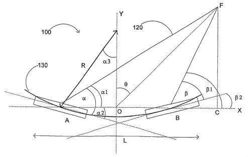
Fig. 2 shows a phased array 100 of imaging elements, A and B. The elements, A
and B, are
situated along a concave 130 path having a radius R. The phased array 100 can
have additional
imaging elements along the concave path 130 (not shown). The phased array 100
has a length,
L, and a maximum steering angle in the azimuthal direction, 0. F represents a
focal point at the
maximum steering angle in the azimuthal direction. The phased array 100 is
shown focusing
an energy beam, such as an acoustic pulse, 120 at focal point F. a and (3,
represent the
tangential angles for elements A and B to the focal point F respectively. al
and (31, represent
the flat plane angles for elements A and B to the focal point F respectively.
Point 0 represents
the origin of the horizontal and vertical axis, X and Y.
In the case of a phased array 100 having a large number of imaging elements,
wherein
elements A and B represent the edge elements of the array 100, at focal point
F, element B will
3
CA 02612611 2007-12-14
WO 2007/005710 PCT/US2006/025770
hAVI d,IH'elllWIrg,6srC::Ahgld' P1,1Itt.4 the focal point F, whereas element A
will have the smallest angle
a 1, to the focal point F. The concaving of the array 100 will decrease the
tangential angle (3,
for element B by (32, and increase the tangential angle a for element A by a2.
One of ordinary
skill in the art would appreciate that by concaving the array 100, elements A
and B will have
the most significant impact on the resulting beam 120 as compared to any
imaging elements in
between elements A and B, and the center imaging elements will be least
affected.
The following is an approach to calculating the radius of curvature R of a
concave
phased array 100 that enables the tangential angles a and (3, to be equal at
the maximum
azimuthal direction 0. From Fig. 2, the following is true:
a = al +a2, (1)
and
R = R l - R2 (2)
and because of geometric symmetry, we have:
a2= (32=a3, (3)
To obtain a=(3, the following is deduced:
al+a3= (3l-a3, (4)
which means:
a3= ((31 -al) /2, (5)
Since:
sin (a3) = (L/2)/R (6)
and:
tan (al) = OF cos (0) / (OF sin (0) + L/2) (7)
and:
tan ((31) = OF cos (0) / (OF sin (0) - L/2) (8).
Combining equations (5) - (8), the radius of curvature R can be expressed as:
R = L / 2 (9) 11 tan-' OFcosB tan ' OFcos9
sin (OFsinO-L/2) (OFsinO+L/2)
2
4
CA 02612611 2007-12-14
WO 2007/005710 PCT/US2006/025770
111bIih:c aa9e;;nV'a,,V6E3IHz, 64 element phased array, wherein the length of
the array is
5mm, if the array's maximum angle in the azimuthal direction is 45 , and the
focal depth is
5mm, then the radius of curvature R is preferably approximately 7mm.
Equation (9) expresses the radius of curvature of a concave array, R, as a
function of a
desired focal depth if the other parameters in the array design, such as image
range, have been
provided. For an image range from OF 1 to OF2, an average radius of curvature
Ra may be
determined by:
R" F_ rF2R(ooF 10 .
Fl OF2-OFl ( )
The concave array 100 may have a uniform pitch, wherein the space from element
center to element, A and B, is uniform; however, it can be non-uniform as
well. An element's
width can be varied to maximize the beam quality, as one of ordinary skill in
the art would
appreciate. For example, the element width can conform to a Gaussian, Bessel,
or sinusoidal
function using an element index number calculated from the array 100 center to
an edge
element, e.g., A or B.
For an ultrasound phased array 100, the array 100 can be fabricated by a
variety of
available active acoustic material, such as piezo-ceramics, piezo-films (thin
or thick), 2-2 or 1-
3 piezoceramic composites, 2-2 or 1-3 piezocrystal materials, or c1V1UT.
Further, other
imaging devices may be used, instead of, or in addition to imaging
transducers, such as light
based apparatuses for obtaining images through optical coherence tomography
(OCT). Image
acquisition using OCT is described in Huang et al., "Optical Coherence
Tomography," Science,
254, Nov. 22, 1991, pp 1178-1181, which is hereby incorporated by reference in
its entirety. A
type of OCT imaging device, called an optical coherence domain reflectometer
(OCDR) is
disclosed in Swanson U.S. Pat. No. 5,321,501, which is incorporated herein by
reference. The
OCDR is capable of electronically performing two- and three-dimensional image
scans over an
extended longitudinal or depth range with sharp focus and high resolution and
sensitivity over
the range.
Such an array 100 is useful for intracardiac applications, and may be used for
other
applications, such as any kind of B-scanner medical applications, ophthalmic
ultrasound, HIFU
and/or NDT.
Turning to Fig. 3, the phased array 100 may be used in a catheter, as
described above,
and can also be placed in a distal portion 520 of a guidewire 500. The
guidewire 500 may
comprise a guidewire body 302 in the form of a flexible, elongate tubular
member, having an
outer wa11301. The guidewire body 302 may be formed of any material known in
the art
5
CA 02612611 2007-12-14
WO 2007/005710 PCT/US2006/025770
plastics, braided polyimide, polyethylene, PEEK braids,
stainless steel, other superelastic materials, or metal alloys, such as a
nitinol hypertube,
Turning to Fig. 4, a proximal portion 510 of the guidewire 500, shown in Fig.
3, may be
adapted to connect to circuitry 600 that processes imaging signals from the
phased array 100,
such circuits being well known.
In the foregoing specification, the invention has been described with
reference to
specific embodiments thereof. It will, however, be evident that various
modifications and
clianges may be made thereto without departing from the broader spirit and
scope of the
invention. For example, the reader is to understand that the specific ordering
and combination
of process actions described herein is merely illustrative, and the invention
can be performed
using different or additional process actions, or a different coinbination or
ordering of process
actions. As a further example, each feature of one embodiment can be mixed and
matched
with other features shown in other embodiments. Additionally and obviously,
features may be
added or subtracted as desired. Accordingly, the invention is not to be
restricted except in light
of the attached claims and their equivalents.
6