Note: Descriptions are shown in the official language in which they were submitted.
CA 02612751 2007-12-18
WO 2007/001744 PCT/US2006/021755
METHODS AND COMPOSITIONS FOR ENHANCING
VASCULAR ACCESS
Related Application Data
[0001] This non-provisional patent application filed on June 5, 2006, claims
the
benefit under 35 U.S.C. Section 119(e) of provisional patent application,
U.S.S.N.
60/692,708 filed on June 21, 2005; and, claims priority under 35 U.S.C.
Sections
120, 363 and/or 365 to co-pending international application PCT/US05/43967
filed
December 6, 2005; which claims priority to provisional patent application,
U.S.S.N.
60/634,155 filed on December 8, 2004; provisional patent application, U.S.S.N.
60/663,859 filed on March 21, 2005; and provisional patent application,
U.S.S.N.
60/682,054 filed on May 19, 2005; and the entire contents of each of the
foregoing
are incorporated by reference herein.
Background of the Invention
[0002] Vascular access failure is the major complication in providing care to
patients on hemodialysis to treat end stage renal disease (ESRD). The rate of
existing ESRD cases in the United Sates has increased each year since 1980. In
2001 the prevalent rate reached almost 1,400 patients per million population,
a 2.4
percent increase from the previous year. Based on demographic changes in age,
race, ethnicity and diabetic status, the prevalent ESRD population in the US
is
expected to grow to 1.3 million by 2030. Currently, approximately 65% of the
CA 02612751 2007-12-18
WO 2007/001744 PCT/US2006/021755
prevalent ESRD population are treated with hemodialysis (approximately 264,710
patients). Between 1997 and 2001, the prevalent hemodialysis population grew
4.5% per year. Using Medicare data, it has been determined that by 2001 the
total
ESRD costs reached $15.5 billion, 6.4 % of the entire Medicare budget of $242
billion (total costs reached $22.8 billion from all sources). Indeed, the
annual cost of
vascular access related morbidity in the US currently exceeds 1 billion
dollars per
year.
[0003] Vascular access failure is the single most important cause of morbidity
in the hemodialysis population. A recent report analyzing US Renal Data System
(USRDS) data found an overall primary unassisted access patency rate of only
53%
at 1 year. The 1-year primary unassisted access patency rates were 49% for
vascular
access structures such as arteriovenous grafts involving ePTFE prosthetic
bridges
and 62% for arteriovenous (AV) fistulae. Cumulative patency rates for first
time
accesses at 1, 3 and 5 years were 54%, 46% and 36% for lower-arm fistulae and
54%, 28% and 0% for AV grafts, respectively. Currently, the use of grafts
involving
ePTFE prosthetic bridges accounts for 70% of all hemodialysis access
procedures in
the United States, the National Kidney Foundation currently recommends that AV
fistula be the preferred method of vascular access. It is expected that there
will be
an increase in the proportion of new AV fistulae in the US in the future.
[0004] Autogenous arteriovenous fistulae have historically been regarded as
the
best choice for vascular access in hemodialysis patients. When an AV fistula
successfully matures after surgical creation, it may function for years with a
low risk
of complications and a low incidence of revisions. However, the reported rates
of
2
CA 02612751 2007-12-18
WO 2007/001744 PCT/US2006/021755
AV fistula non-maturation vary widely, but remain about 20-50%. Non-maturation
is generally defined as the inability to permit repetitive cannulation of the
fistula for
dialysis or to obtain sufficient dialysis blood flow within 12 weeks after
surgical
creation. The occurrence of AV fistula non-maturation can depend, in part, on
the
quality and size of the vessels used to form the AV fistula. Preoperative
assessment
of vessel characteristics has been shown to have beneficial effects in
identifying
suitable vessels for AV fistula creation.
[0005] Failure of vascular access structures is attributable to the cumulative
effect of a variety of distinct acute and chronic phenomena, especially at the
so-
called "toe" of the anastomosis and its downstream surrounds. For example, AV
grafts may develop graft-associated stenoses and graft-associated occlusions
at the
anastomoses on the venous anastomotic side. In one published report,
histological
examination of segments removed from patients with graft-associated,
anastomotic
stenosis revealed intimal hyperplasia consisting of smooth muscle cells and
extracellular matrix. Graft thrombosis may also contribute to vascular access
dysfunction in ePTFE dialysis grafts. Moreover, generally isolation of veins
and
arteries followed by exposure of the vein segment to arterial blood flow and
pressure
can cause unavoidable ischemia and reperfusion injury. Surgical manipulation
such
as suturing can also result in direct trauma to the endothelium and smooth
muscle
cells of the media in both veins and arteries. Injury to the artery and vein
endothelium during the creation of a native or graft anastomoses can influence
patency and occlusion rates. In addition to the physical trauma associated
with
cutting and suturing veins and arteries during formation of a vascular access
structure, increased wall stress and shear force can also cause physical
and/or
3
CA 02612751 2007-12-18
WO 2007/001744 PCT/US2006/021755
biochemical injury to the endothelium. It has been suggested that arterial
pressure
may alter the normal production of endothelial growth regulatory compounds as
well
as produce morphological and biochemical changes in the media of the vein.
[0006] The current therapy for vascular access failure is either surgical
revision
or angioplasty with or without stenting. Surgical treatment can be risky in
these
typically multimorbid patients and the long-term results of angioplasty and
stenting
are generally disappointing due to failure rates of their own. The goal of
improved
vascular access for hemodialysis purposes as well as for peripheral
circulation
therefore is to maintain the anatomical integrity of the original graft site
to allow for
blood flow rates to support dialysis treatment or sufficient blood flow at
peripheral
bypass sites.
[0007] Other factors contributing to successful maturation of a newly created
vascular access structure or prolonged maturation of an already-existing
vascular
access structure remain elusive. Moreover, relatively few randomized clinical
trials
have been conducted in the field of vascular access failure prevention.
Studies that
have evaluated the causes of vascular access failure have reached inconsistent
conclusions. In fact, at the present time, despite the enormity of this
problem, no
effective surgical, therapeutic or pharmacologic measures for the prolonged
survival
of functioning dialysis access fistula are available to clinicians. Clearly a
need
exists to move ahead in this vital area of patient care.
Summary of the Invention
[0008] The present invention exploits the discovery that an implantable
material
comprising cells and a biocompatible matrix, w11en provided locally to a
vascular
4
CA 02612751 2007-12-18
WO 2007/001744 PCT/US2006/021755
access structure, can promote formation and/or enhance maturation of the
structure
as well as prolong the structure in a mature, functional state. In accordance
with the
present invention, the implantable material is located on an exterior surface
of a
blood vessel at or adjacent or in the vicinity of the vascular access
structure. The
present invention can effectively promote integration and/or enhance
maturation of a
newly created vascular access structure; promote and sustain the functional
lifetime
of an existing, functioning structure; and, can aid in the salvage of a failed
or failing
structure.
[0009] In one aspect, the invention is a method for treating a vascular access
structure in a patient comprising the step of locating at, adjacent or in the
vicinity of
the vascular access structure an implantable material comprising cells and a
biocompatible matrix, wherein the implantable material is effective to promote
functionality of said structure. According to certain embodiments described
below,
the vascular access structure is for dialysis.
[0010] According to various embodiments, the vascular access structure is an
arteriovenous native fistula, an arteriovenous graft, a peripheral graft, a
venous
catheter or an in-dwelling port. In one embodiment, the arteriovenous graft
comprises a prosthetic bridge. In other embodiments, the catheter is an
indwelling
dual lumen catheter and treating the indwelling dual lumen catheter promotes
clinical stability sufficient to permit hemodialysis.
[0011] In one embodiment, treating the vascular access structure promotes
normal or near-normal blood flow through and downstream of the structure. For
example, normal or near-normal blood flow is blood flow at a rate sufficient
to
5
CA 02612751 2007-12-18
WO 2007/001744 PCT/US2006/021755
prevent re-circulation during hemodialysis. According to additional
embodiments,
treating the vascular access structure promotes normal or near-normal vessel
diameter and reduces flow re-circulation during hemodialysis.
[0012] In the case of an arteriovenous native fistula, treating the
arteriovenous
native fistula enhances clinical maturation sufficient to permit hemodialysis,
reduces
delay in maturation of the arteriovenous native fistula and promotes
repetitive
cannulation. In the case of an arteriovenous graft, treating the arteriovenous
graft
promotes clinical stability sufficient to restore normal or near normal
circulation. In
various of the embodiments, the implantable material reduces the occurrence of
revision in a patient having an access structure.
[0013] In one embodiment, enhancing maturation is characterized by an ability
to repetitively cannulate the fistula for dialysis. According to another
embodiment,
enhancing maturation is characterized by an ability to obtain sufficient blood
flow
during dialysis. Preferably, sufficient blood flow comprises a rate of about
350
ml/min. According to various embodiments, application of the biocompatible
material to the arteriovenous fistula is preceded by or coincident with
administration
of a therapeutic agent, physical dilatation or stenting. The arteriovenous
fistula is
radiocephalic, brachiocephalic, or brachiobasilic.
[0014] In one preferred embodiment, the invention is a method for preventing
an arteriovenous fistula from failing to mature in a human comprising the step
of
locating an implantable material comprising a biocompatible matrix and
vascular
endothelial cells at, adjacent to or in the vicinity of the fistula thereby to
prevent a
fistula from failing to mature. In one embodiment, failing to mature is
characterized
6
CA 02612751 2007-12-18
WO 2007/001744 PCT/US2006/021755
by an inability to repetitively cannulate the fistula for dialysis or by an
inability to
obtain sufficient blood flow during dialysis, wherein the sufficient blood
flow
comprises a rate of about 350 ml/min. In other embodiments, failing to mature
is
characterized by an arteriovenous fistula that can not be cannulated at least
2
months, at least 3 months, or at least 4 months after creation.
[0015] In another embodiment, the invention is a method of maintaining a
blood flow rate of an arteriovenous graft sufficient to permit dialysis
comprising the
step of providing an implantable material comprising cells and a biocompatible
matrix wherein said implantable material is disposed on an exterior surface of
said
arteriovenous graft at, adjacent or in the vicinity of a prosthetic bridge of
a venous
outflow region of said arteriovenous graft in an amount effective to maintain
blood
flow rate of the graft sufficient to permit dialysis. In one embodiment, the
blood
flow rate at the venous outflow region of said arteriovenous graft is
substantially
similar to the blood flow rate upstream of said outflow region.
[0016] In another embodiment, the invention is a metllod of maintaining normal
blood flow of a peripheral bypass graft sufficient to maintain peripheral
circulation
comprising the step of providing an implantable material comprising cells and
a
biocompatible matrix wherein said implantable material is disposed on an
exterior
surface of said bypass graft at, adjacent or in the vicinity of a prosthetic
bridge in an
amount effective to maintain blood flow rates of the bypass graft sufficient
to
maintain peripheral circulation. In one embodiment, an inflow blood rate and
an
outflow blood rate are substantially similar.
7
CA 02612751 2007-12-18
WO 2007/001744 PCT/US2006/021755
[0017] In another embodiment, the invention is a method of maintaining a
blood pressure of an arteriovenous graft sufficient to permit dialysis
comprising the
step of providing an implantable material comprising cells and a biocompatible
matrix wherein said implantable material is disposed on an exterior surface of
said
arteriovenous graft at, adjacent or in the vicinity of a prosthetic bridge of
a venous
outflow region of said arteriovenous graft in an amount effective to maintain
blood
pressure sufficient to permit dialysis. In one embodiment, the blood pressure
at the
venous outflow region of said arteriovenous graft is substantially similar to
the
blood pressure upstream of said outflow region. The prosthetic bridge is
selected
from the group consisting of: saphenous vein; bovine heterograft; umbilical
vein;
dacron; PTFE; ePTFE, polyurethane; bovine mesenteric vein; and cryopreserved
femoral vein allograft. According to a preferred embodiment, the prostlletic
bridge
is ePTFE.
[0018] In another embodiment, the invention is a method of promoting tissue
integration of a prosthetic bridge of an arteriovenous graft or a peripheral
bypass
graft comprising the step of providing an implantable material comprising
cells and
a biocompatible matrix wherein said implantable material is disposed on an
exterior
surface of said arteriovenous graft or said peripheral bypass graft at,
adjacent or in
the vicinity of a prosthetic bridge in an amount effective to promote tissue
integration of said bridge. In certain embodiments, the implantable material
promotes smooth muscle cell proliferation or migration within or in the
vicinity of
an interior lumen surface of said prosthetic bridge or promotes endothelial
cell
proliferation or migration within or in the vicinity of an interior lumen
surface of
said prosthetic bridge. In certain other embodiments, the implantable material
8
CA 02612751 2007-12-18
WO 2007/001744 PCT/US2006/021755
promotes smooth muscle cell and/or endothelial cell proliferation at, adjacent
or in
the vicinity of the graft.
[0019] In another embodiment, the invention is a method of preventing or
reducing the incidence of dehiscence of an arteriovenous fistula or
arteriovenous
graft comprising the step of providing an implantable material comprising
cells and
a biocompatible matrix wherein said implantable material is disposed on an
exterior
surface of said fistula or arteriovenous graft at, adjacent or in the vicinity
of a
prosthetic bridge of a venous outflow region of said arteriovenous graft in an
amount
effective to prevent or reduce the incidence of dehiscence.
[0020] According to other embodiments, the providing step is performed as an
interventional therapy following failure of a native arteriovenous fistula or
following
failure of a native or saphenous vein peripheral bypass.
[0021] In another aspect, the invention is an implantable material comprising
cells and a biocompatible matrix suitable for treating a vascular access
structure.
The cells are endothelial cells or cells having an endothelial-like phenotype.
The
biocompatible matrix is a flexible planar form or a flowable composition. In a
particularly preferred embodiment, the cells are vascular endothelial cells.
The
flexible planar form is configured for implantation at, adjacent or in the
vicinity of a
vascular access structure. In certain embodiments, this form defines a slot.
According to one embodiment of the flowable composition, the flowable
composition is a shape-retaining composition.
9
CA 02612751 2007-12-18
WO 2007/001744 PCT/US2006/021755
[0022] In other embodiments, the invention is an implantable material
comprising cells and a biocompatible matrix suitable for use with methods for
enhancing maturation of an arteriovenous fistula by preventing an
arteriovenous
fistula from failing to mature. The cells are endothelial cells or cells
having an
endothelial-like phenotype and the biocompatible matrix is a flexible planar
form or
a flowable composition. In one embodiment, the flexible planar form is
configured
for implantation at, adjacent or in the vicinity of a native fistula. In
certain
embodiments, this form is configured such that it defines a slot or series of
slots.
With respect to the flowable composition, it is a shape-retaining composition.
[0023] In another embodiment, the invention is an implantable material
comprising cells and a biocompatible matrix wllerein the implantable material
is
disposed on an exterior surface of a blood vessel at, adjacent or in the
vicinity of a
prosthetic bridge. The prosthetic bridge is situated at or near a venous
outflow
region of an arteriovenous graft or is situated at or near an outflow of
[0024] In another aspect, the invention is a transport media composition for
storing an implantable material comprising a biocompatible matrix and
engrafted
cells. The transport media composition comprising an amount of VEGF sufficient
to maintain cell viability or an inhibitory phenotype and for the cells to
remain
viable for an extended period of time when stored in said transport media
composition at temperatures below the cells' standard cell culture
temperature.
[0025] In another aspect, the invention is a method for storing an implantable
material comprising a biocompatible matrix and engrafted cells for an extended
period of time at a temperature below the cells' standard cell culture
temperature.
CA 02612751 2007-12-18
WO 2007/001744 PCT/US2006/021755
The method comprises the steps of bathing the implantable material in a
transport
media composition comprising an amount of VEGF sufficient to maintain cell
viability or an inhibitory phenotype during storage. The cells remain viable
for an
extended period of time when stored in said transport media composition at a
temperature below the cells' standard cell culture temperature.
[0026] According to one embodiment, the transport media composition contains
an amount of VEGF sufficient to maintain cell viability or an inhibitory
phenotype
at a temperature below the cells' standard cell culture temperature and
greater than
the amount of VEGF required at the cells' standard cell culture temperature.
According to one embodiment, the amount of VEGF is about 4 ng/mL.
[0027] According to additional embodiments, the implantable material is stored
in said transport media composition at a temperature below 37 C or at ambient
temperature for an extended period of about 1 week, about 2 weeks, or about 3
weeks. Tthe cells are endothelial cells or endothelial-like cells that are
near-
confluent, confluent, or post-confluent at the time of storage, and at least
80%
viable.
[0028] In another aspect, the invention is a cryopreservation media
composition
for cryopreserving an implantable material comprising a biocompatible matrix
and
engrafted cells. The cryopreservation media composition comprising a
cryopreservative, a polysaccharide and serum. Cell viability or an inhibitory
phenotype and matrix integrity are maintained for an extended period of time
when
stored at at least about -4 C.
11
CA 02612751 2007-12-18
WO 2007/001744 PCT/US2006/021755
[0029] In one embodiment, the amount of serum in the cryopreservation media
composition exceeds the amount of serum for routine culturing of the cells,
for
example, 20% serum or 50% serum. In one embodiment, the serum is fetal bovine
serum. In one embodiment, the polysaccharide in the cryopreservation media
composition exceeds the amount of polysaccharide for routine culturing of the
cells,
for example at least about 4% polysaccharide or at least about 4.5%
polysaccharide.
In one embodiment, the polysaccharide is dextran. In another embodiment, the
cryopreservation media composition further comprises about 10% DMSO.
[0030] According to one embodiment, the cryopreservation media composition
is used for storage at at least about -80 C, at least about -140 C, or at at
least about -
160 C. According to various embodiments, the extended period of time is about
1
month, about 6 months or about 1 year. In one embodiment, cell viability is at
least
about 80%.
[0031] In another aspect, the invention is a cryopreserved implantable
material
comprising a biocompatible matrix engrafted with cells and a volume of
cryopreservation media composition sufficient to maintain cell viability or an
inhibitory phenotype and matrix integrity while cryopreserved, wherein the
cryopreservation media composition comprises a cryopreservative, a
polysaccharide
and serum.
[0032] In another aspect, the invention is a method of preparing an
implantable
material comprising a biocompatible matrix and engrafted cells. The method
comprises the steps of providing a working cell bank comprising cells,
providing a
hydrated biocompatible matrix material, seeding the hydrated biocompatible
matrix
12
CA 02612751 2007-12-18
WO 2007/001744 PCT/US2006/021755
material with cells from the working cell bank, placing the cell seeded
biocompatible matrix material in an incubator to facilitate cell attachment,
placing
the cell seeded biocompatible matrix material in an incubator until the cells
are near-
confluent, confluent, or post-confluent, and assessing cell count, cell
viability and
cell functionality of the cell seeded biocompatible matrix material.
[0033] In one embodiment, the method further comprises the steps of placing
the cell seeded biocompatible matrix material in a vial suitable for
cryopreservation,
introducing to the near-confluent, confluent, or post-confluent cell seeded
biocompatible matrix material a volume of cryopreservation media composition
comprising a cryopreservative, a polysaccharide and serum sufficient to
preserve
cell viability or an inhibitory phenotype and matrix integrity while the
material is
cryopreserved, placing the vial containing the cell seeded biocompatible
matrix
material and cryopreservation media composition in a freezing container,
introducing an agent which controls the freezing rate to the freezing
container,
placing the freezing container containing said agentl in a freezer at at least
-4 C,
removing the freezing container from the at least about -4 C freezer, and
placing the
freezing container in a freezer at at least about -80 C.
[0034] In another embodiment, the method further comprises the steps of
removing the freezing container from the at least -80 C freezer and placing
the
freezing container in a freezer at at least -160 C.
[0035] In another embodiment, the method further comprises the steps of
removing the vial from the freezer, placing the vial in ambient temperature
air for
about 15 minutes followed by placing the vial in ambient temperature water
bath for
13
CA 02612751 2007-12-18
WO 2007/001744 PCT/US2006/021755
about 15 minutes, removing the implantable material from the vial, rinsing the
implantable material in rinse media composition for about 5 minutes, and
placing the
implantable material in cell culture media for about 48 hours.
[0036] In another embodiment, the method further comprises the steps of
removing the vial from the freezer, placing the vial in ambient temperature
air for
about 15 minutes followed by placing the vial in ambient temperature water
bath for
about 15 minutes, removing the implantable material from the vial, and rinsing
the
implantable material in a rinse solution composition for about 30 minutes.
[0037] In another embodiment, the method further comprises the steps of
placing the cell seeded biocompatible matrix material in a vial suitable for
storage
and introducing to the near-confluent, confluent, or post-confluent cell
seeded
biocompatible matrix material a volume of transport media composition
comprising
an amount of VEGF sufficient to maintain cell viability or an inhibitory
phenotype
while the material is stored in said composition.
[0038] In another embodiment, the method further comprises the steps of
preparing the cell seeded biocompatible matrix material for cryopreservation
according to a disclosed method or for storage according to a disclosed
method,
preparing the vial for transport and transporting the outer box to a clinical
site for
administration to a patient.
[0039] In another embodiment, the method further comprises the steps of
placing the vial containing the cell seeded biocompatible matrix material into
an
inner box, placing the inner box into an insulated outer box, and providing
product
documentation.
14
CA 02612751 2007-12-18
WO 2007/001744 PCT/US2006/021755
[0040] In one embodiment of the method, the cell seeded biocompatible matrix
material is clinical trial material and the patient is a participant in a
clinical trial. In
another embodiment of the method, the implantable material is prepared on a
commercial scale. In another aspect, the invention is a robotic system to
perform
any of the disclosed methods.
[0041] In another aspect, the invention is a method of manufacturing an
implantable material comprising cells and a biocompatible matrix, the method
comprising the step of contacting the biocompatible matrix with the cells
using
reagents and conditions suitable therefor, wherein the cells are in an amount
sufficient to populate the matrix and grow to a confluent, near-confluent or
post-
confluent population and further wherein the matrix is populated with cell
typing-
independent, non-compatibility tested, non-matched cells. In another aspect,
the
invention is a method of providing an implantable material manufactured
according
to this method. In a further aspect, the invention is an implantable material
comprising cells and a biocompatible matrix manufactured according to this
method.
Brief Description of the Drawings
[0042] In the drawings, like reference characters generally refer to the same
parts throughout the different views. Also, the drawings are not necessarily
to scale
or proportion, emphasis instead generally being placed upon illustrating the
principles of the invention.
[0043] FIG.1 is a schematic perspective view of a flexible planar form of
implantable material for administration to an exterior surface of a tubular
anatomical
structure according to an illustrative embodiment of the invention.
CA 02612751 2007-12-18
WO 2007/001744 PCT/US2006/021755
[0044] FIG. 2A is a schematic perspective view of a contoured flexible planar
form of implantable material for administration to an exterior surface of a
tubular
anatomical structure according to an illustrative embodiment of the invention.
[00451 FIGS. 2B, 2C, 2D, 2E, 2F and 2G are schematic perspective views of a
contoured flexible planar form of the implantable material comprising a slot
according to various illustrative embodiments of the invention.
[0046] FIGS. 3A and 3B are representative cell growth curves according to an
illustrative embodiment of the invention.
[0047] FIGS. 4A, 4B and 4C illustrate a series of steps for administering
multiple flexible planar forms of implantable material to an exterior surface
of a
vascular anastomosis from a top perspective view according to an illustrative
embodiment of the invention.
[0048] FIG. 5 is a top perspective view of a contoured form of implantable
material administered to an exterior surface of a vascular anastomosis
according to
an illustrative embodiment of the invention.
[0049] FIG. 6 is a top perspective view of a flexible planar form of
implantable
material administered to a tubular anatomical structure according to an
illustrative
embodiment of the invention.
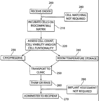
[0050] FIG. 7 is a flow chart of a method of preparing, storing and
transporting
an implantable material for administration to a recipient according to an
illustrative
embodiment of the invention.
16
CA 02612751 2007-12-18
WO 2007/001744 PCT/US2006/021755
Detailed Description of the Invention
[0051] As explained herein, the invention is based on the discovery that a
cell-
based therapy can be used to treat vascular access structures. The teacllings
presented below provide sufficient guidance to make and use the materials and
methods of the present invention, and further provide sufficient guidance to
identify
suitable criteria and subjects for testing, measuring, and monitoring the
performance
of the materials and methods of the present invention.
[0052] Accordingly, a cell-based therapy for clinically managing vascular
access complications and/or failures has been developed. An exemplary
embodiment of the present invention comprises a biocompatible matrix and cells
suitable for use with the treatment paradigms described herein. Specifically,
in one
preferred embodiment, the implantable material comprises a biocompatible
matrix
and endothelial cells or endothelial-like cells. In one embodiment, the
implantable
material is in a flexible planar form and comprises endothelial cells or
endothelial-
like cells, preferably human aortic endothelial cells and the biocompatible
matrix
Gelfoam gelatin sponge (Pfizer, New York, NY, hereinafter "Gelfoam matrix").
According to another preferred embodiment, the implantable material is in a
flowable form and comprises endothelial cells or endothelial-like cells,
preferably
human aortic endotlielial cells and the biocompatible matrix Gelfoam gelatin
particles or powder (Pfizer, New York, NY, hereinafter "Gelfoam particles").
[0053] Implantable material of the present invention comprises cells engrafted
on, in and/or within a biocompatible matrix. Engrafted means securedly
attached
via cell to cell and/or cell to matrix interactions such that the cells
withstand the
17
CA 02612751 2007-12-18
WO 2007/001744 PCT/US2006/021755
rigors of the preparatory manipulations disclosed herein. As explained
elsewhere
herein, an operative embodiment of implantable material comprises a near-
confluent, confluent or post-confluent cell population having a preferred
phenotype.
It is understood that embodiments of implantable material likely shed cells
during
preparatory manipulations and/or that certain cells are not as securedly
attached as
are other cells. All that is required is that implantable material comprise
cells that
meet the functional or phenotypical criteria set forth herein.
[0054] The implantable material of the present invention was developed on the
principals of tissue engineering and represents a novel approach to addressing
the
above-described clinical needs. The implantable material of the present
invention is
unique in that the viable cells engrafted on, in and/or within the
biocompatible
matrix are able to supply to the vascular access structure and associated
vasculature
multiple cell-based products in physiological proportions under physiological
feed-
back control. As described elsewhere herein, the cells suitable for use with
the
implantable material are endothelial or endothelial-like cells. Local delivery
of
multiple compounds by these cells and physiologically-dynamic dosing provide
more effective regulation of the processes responsible for maintaining a
functional
vascular access structure and diminishing vascular access complications and/or
failure. Importantly, the endothelial cells, for example, of the implantable
material
of the present invention are protected from the erosive blood flow within the
vessel
lumen because of its placement at a non-luminal surface of the vessel, for
example,
at the adventitia or contacting an exterior surface of a vessel. The
implantable
material of the present invention, when wrapped, deposited or otherwise
contacted
with such an exterior target site, i.e., the anastomosis and/or its surrounds,
serves to
18
CA 02612751 2007-12-18
WO 2007/001744 PCT/US2006/021755
reestablish homeostasis. That is, the implantable material of the present
invention
can provide an environment which mimics supportive physiology and is conducive
to vascular access structure formation, maturation, integration and/or
stabilization.
[0055] For purposes of the present invention, contacting means directly or
indirectly interacting with an extraluminal or non-luminal surface as defined
elsewhere herein. In the case of certain preferred embodiments, actual
physical
contact is not required for effectiveness. In other embodiments, actual
physical
contact is preferred. All that is required to practice the present invention
is
extraluminal or non-luminal deposition of an implantable material at, adjacent
or in
the vicinity of an injured or diseased site in an amount effective to treat
the injured
or diseased site. In the case of certain diseases or injuries, a diseased or
injured site
can clinically manifest on an interior lumen surface. In the case of other
diseases or
injuries, a diseased or injured site can clinically manifest on an
extraluminal or non-
luminal surface. In some diseases or injuries, a diseased or injured site can
clinically
manifest on both an interior lumen surface and an extraluminal or non-luminal
surface. The present invention is effective to treat any of the foregoing
clinical
manifestations.
[0056] For example, endothelial cells can release a wide variety of agents
that
in combination can inhibit or mitigate adverse physiological events associated
with
acute complications following vascular access structure creation. As
exemplified
herein, a composition and method of use that recapitulates normal physiology
and
dosing is useful to enhance vascular access structure formation, maturation,
integration and/or stabilization, as well as promote long-term patency of such
19
CA 02612751 2007-12-18
WO 2007/001744 PCT/US2006/021755
vascular access structures. Typically, treatment includes placing the
implantable
material of the present invention at, adjacent or in the vicinity of the
vascular access
structure site, for example, in the perivascular space external to the lumen
of the
artery and vein involved in the procedure. When wrapped, wrapped around,
deposited, or otherwise contacting an injured, traumatized or diseased blood
vessel,
the cells of the implantable material can provide growth regulatory compounds
to
the vasculature, for example to the underlying smooth muscle cells within the
blood
vessel. It is contemplated that, when situated at an extraluminal site, the
cells of the
implantable material provide a continuous supply of multiple regulatory
compounds
which can penetrate vessel tissue and reach the lumen, yet the cells are
protected
from the adverse mechanical effects of blood flow in the vessel(s). As
described
herein, one preferred extraluminal site is an exterior surface of a blood
vessel.
[0057] Treatment with a preferred embodiment of the present invention can
encourage normal or near normal healing and normal physiology. On the
contrary,
in the absence of treatment with a preferred embodiment of the present
invention,
normal physiological healing is impaired, e.g., native endothelial cells and
smooth
muscle cells can grow abnormally at an exuberant or uncontrolled rate
following
creation of a vascular access structure, leading to adverse clinical
consequences,
including vascular access structure failure. Accordingly, as contemplated
herein,
treatment with the implantable material of the present invention will improve
the
healing of native tissue at the anastomotic site(s) to maintain vascular
access
structure patency.
CA 02612751 2007-12-18
WO 2007/001744 PCT/US2006/021755
[0058] For purposes of the present inveiition, vascular access structures may
be
formed in a variety of configurations. Vascular access structures can include
naturally occurring or surgically created arteriovenous fistula, arteriovenous
grafts,
peripheral bypass grafts, in-dwelling venous catheters, in-dwelling vascular
ports, or
other vascular anastomotic structures created to improve vascular access in a
patient.
Additionally, various embodiments of vascular access structures are formed in
a
variety of configurations including side-to-side, end-to-side, end-to-end and
side-to-
end anastomoses. Vascular access structures can also be placed in a variety of
anatomical locations.
[0059] The implantable material of the present invention can be placed in a
variety of configurations at the vascular access structure to be treated.
According to
certain embodiments, the implantable material of the present invention can be
placed
both at the anastomotic juncture and also placed on the proximal vein segment,
distal to the anastomosis. In other embodiments, the implantable material of
the
present invention can be placed on the arterial segment, on the proximal vein
segment, on the distal vein segment, and/or bridging the vascular access
structure.
In another embodiment, the implantable material also can be placed on the
graft
material or a portion of the graft material at the anastomotic junction. The
vessels
can be contacted in whole or in part, for example, the implantable material of
the
present invention can be applied to the vessels circumferentially or in an arc
configuration. A vessel and/or vascular access structure need only be in
contact
with an amount of implantable material sufficient to improve formation,
maturation,
integration and/or stabilization of the vascular access structure.
21
CA 02612751 2007-12-18
WO 2007/001744 PCT/US2006/021755
[0060] Arteriovenous Fistula. According to certain embodiments, an
arteriovenous fistula ("AV fistula") created for vascular access can be
treated with
the implantable material of the present invention. An AV fistula can be placed
in a
variety of locations within the patient, including, for example, placement in
the
neck, wrist, upper arm and lower arm. Clinical AV fistula configurations
include
radiocephalic (between the radial artery and the cephalic vein), Brescia-
Cimino (a
side-to-side anastomosis of the radial artery and the cephalic vein within the
wrist),
brachiocephalic (between the brachial artery and the cephalic vein), brachial-
antecubital (between the brachial artery and the antecubital vein),
brachiobasilic (a
transposed basilic vein), ulnarcephalic (between the ulnar artery and the
cephalic
vein), and saphenous loop (saphenous vein and the right side of the femoral
artery)
fistula.
[0061] Complications from AV fistula surgery typically occur during three
phases. These phases are an acute phase which is often characterized by
thrombosis,
an intermediate phase whose clinical signature is a failure of the fistula to
mature,
and finally a more chronic failure of an already-established, functioning
fistula
which, for example, can be due to progressive venous stenosis.
[0062] Characteristics of AV fistula maturation include, for example, the
ability
to repetitively cannulate the fistula for dialysis. Another characteristic of
AV fistula
maturation is the ability to obtain sufficient dialysis blood flow useful for
hemodialysis. Adequate blood flow is at least a flow rate adequate to support
dialysis using a dialysis machine such that recirculation does not occur. A
sufficient
dialysis blood flow for purposes of the present invention is a blood flow of
at least
22
CA 02612751 2007-12-18
WO 2007/001744 PCT/US2006/021755
about 350 mL/min at a time point no more than 24 weeks, preferably more than
20
weeks, more preferably 16 weeks, and most preferably twelve weeks after the
creation of the fistula. The mechanisms of AV fistula failure to mature are
currently
understood to include, for example, early thrombosis of the fistula vessels,
stenoses
at or near the anastomotic site, the presence of accessory veins, including
collateral
or venous side branches, inadequate vein size, including inadequate vein
internal
diameter, and late fistula failure due to progressive stenosis. Accessory
veins can
prevent the development of the fistula by diverting blood flow and by not
allowing
for the vein associated with the fistula to become of adequate size to allow
for
cannulation. It is currently thought that accessory veins may develop, for
example,
in response to the presence of a stenosis in the fistula. The mechanisms of AV
fistula maturation are multimodal and generally require assessment of multiple
clinical indicia. The absence of stenosis alone is generally an insufficient
indication
of a mature AV fistula.
[0063] Moreover, an AV fistula that is considered adequate for the purpose of
dialysis requires both maturation of the fistula, those changes that occur in
the vein
segment of the fistula which allow the fistula to be repetitively cannulated;
and,
blood flow sufficient to support dialysis. An AV fistula can remain patent
even
when there is very little blood flow, but a patent AV fistula may not be
clinically
adequate for dialysis. For purposes of the present invention, clinically
adequate
blood flow for dialysis is about 150-500 mL/minute, preferably about 300-500
mL/minute, and most preferably about 350-400 mL/minute; suitable blood
pressures
are about 50-180 mmHg, preferably about 50-120 mmHg.
23
CA 02612751 2007-12-18
WO 2007/001744 PCT/US2006/021755
[0064] For purposes of the present invention, it is believed that treatment
with
the implantable material of the present invention provides a beneficial
homeostatic
environment such that complications common in AV fistula maturation, for
example, thrombosis, stenosis, clotting and/or the growth of accessory veins
are
reduced when placed adjacent to or in the vicinity at the fistula whether at
the time
of fistula creation or at a later stage. This type of beneficial environment
allows an
AV fistula to proceed to maturation and/or remain in a mature state. For
example,
maturation is functionally established when the AV fistula vein thickens and
is able
to conduct high flow, high pressure blood. Treatment of an AV fistula with the
implantable material provided for herein enhances maturation of the fistula
and/or
prevents the fistula from failing to mature. It is understood for purposes of
the
present invention that enhancement of AV fistula maturation includes any
improvement in the functioning of the fistula, including its formation, time
required
to reach a functional state as well as maintenance of the fistula in a mature
form.
[0065] Immediate post-operative thrombosis can prevent the formation of a
patent AV fistula and lead to early failure of the fistula. As explained
herein,
treatment with the implantable material of the present invention at the time
of
surgery can prevent the AV fistula from immediate failure due to post-
operative
thrombosis. For example, the implantable material releases anti-thrombotic
mediators that reduce thrombosis and can maintain a patent fistula through the
stages of fistula formation and maturation.
24
CA 02612751 2007-12-18
WO 2007/001744 PCT/US2006/021755
[0066] Placement of the implantable material at or in the vicinity of the site
of
the AV fistula at the time of surgery can also enhance maturation by reducing
stenosis at or near the fistula anastomoses, allowing the fistula to become of
adequate size to provide sufficient blood flow to support dialysis,
facilitating venous
and arterial dilatation, decreasing the forination of parasitic accessory
veins,
maintaining native accessory veins to enhance maturation of the fistula, and
improving the size of the vein.
[0067] Additionally, an AV fistula requires a longer period of time to reach
maturation than an AV graft. During the period of fistula maturation,
hemodialysis
is generally conducted using a percutaneous or indwelling catheter, leading to
an
increased risk of infection and compromising central vein patency. Placement
of the
implantable material at the site of an AV fistula at the time of fistula
creation can
reduce the time required for fistula maturation, thereby reducing the
associated risks
of infection and compromised central vein patency. Placement of the
implantable
material at the site of an indwelling catheter can reduce the risk of
thrombosis,
intimal hyperplasia and restenosis associated with the indwelling catheter,
thereby
reducing the associated risks of infection and compromised central vein
patency.
[0068] Finally, use of the implantable material described herein can decrease
late failure of a mature AV fistula. A mature fistula may experience decreased
blood flow and increased venous stenosis due to late progressive stenosis.
Stenosis
or occlusion of a fistula to a degree sufficient to reduce blood flow below a
level
necessary for dialysis may require interventional angioplasty or stenting of
the
fistula to restore adequate blood flow levels. Such interventional therapies
increase
CA 02612751 2007-12-18
WO 2007/001744 PCT/US2006/021755
the risk of venous stenosis and occlusion, further preventing the formation of
a
mature fistula. Furthermore, stenting can result in occlusive thrombosis or
restenosis of the treated vessel in the portion of the vessel distal and
proxiinal to the
stent, often referred to as edge effects.
[0069] Application of the implantable material can result in positive
remodeling
(a combination of vascular dilatation with a simultaneous inhibition of venous
neointimal hyperplasia) thereby preventing late progressive stenosis,
increasing
blood flow of the mature fistula, reducing the need for rehabilitative
angioplasty or
stenting of an occluded fistula, preventing stent-associated edge effects, and
prolonging the lifetime and usability of the mature fistula.
[0070] The implantable material of the present invention can be provided to
the
fistula at any of a number of distinct stages. For example, treatment at the
time of
surgery can prevent the AV fistula from failing to mature and/or can enhance
maturation of the fistula. The implantable material can also be provided after
the
initial surgery to hasten healing generally, as well as after a mature AV
fistula has
formed to maintain it in a clinically stable state. Additionally, the
implantable
material can also rescue a mature AV fistula that subsequently fails and/or
can
extend the lifetime of a mature fistula. These situations are non-limiting
examples
of enhancement of AV fistula maturation. Accordingly, it is contemplated that
the
implantable material can be used not only at the time of initial surgery to
create the
AV fistula, but also at subsequent time points (e.g., for maintaining a mature
fistula
or rescuing a mature fistula from failing). Subsequent administrations can be
accomplished surgically or non-invasively.
26
CA 02612751 2007-12-18
WO 2007/001744 PCT/US2006/021755
[0071] Arteriovenous Graft. According to additional embodiments, an
arteriovenous graft ("AV graft") created for vascular access can be treated
with
implantable material of the present invention. An AV graft can be in the form
of a
forearm straight graft, a forearni loop graft or an upper arm graft. Arterial
inflow
sites include, but are not limited to, the common carotid artery, the radial
artery at
the wrist, the brachial artery in the antecubital fossa, the brachial artery
in the lower
portion of the arm, the brachial artery just below the axilla, the axillary
artery and
the femoral artery. Venous outflow sites include, but are not limited to, the
median
antecubital vein, the proximal cephalic vein, the distal cephalic vein, the
basilic vein
at the level of the elbow, the basilic vein at the level of the upper arm, the
axillary
vein, the jugular vein and the femoral vein. Additional arterial and venous
locations
suitable for formation of an AV graft include the chest wall (axillary artery
to the
subclavian vein), the lower extremities (femoral artery / vein, saphenous
vein, or
tibial (anterior) artery), the aorta to the vena cava, the axillary artery to
the femoral
vein or the femoral artery to the axillary vein.
[0072] For purposes of the present invention, a functional AV graft involving
a
prosthetic bridge suitable for dialysis is able to conduct high-flow, high-
pressure
blood through the prosthetic bridge. In such AV grafts, the blood flow rate at
the
venous outflow region of the graft is substantially similar to the blood flow
rate
upstream of the graft outflow region. Blood flow rates suitable for dialysis
are about
150-500 mL/min, preferably about 300-500 mL/min, and most preferably about 350-
400 mL/min; suitable blood pressures are about 50-180 mmHg, preferably about
50-
120 mmHg.
27
CA 02612751 2007-12-18
WO 2007/001744 PCT/US2006/021755
[0073] AV grafts generally fail due to graft-associated intimal hyperplasia
followed by graft-associated thrombosis at the venous-graft anastomosis or at
the
proximal venous segment. AV grafts are also vulnerable to failure due to poor
tissue
integration between the native vessels and the prosthetic bridge material and
eventual dehiscence of the bridge material from the vessels.
[0074] For purposes of the present invention, treatment with the implantable
material of the present invention provides a beneficial homeostatic
environment
such that complications associated with AV graft integration and maturation,
for
example, thrombosis, stenosis, clotting and/or dehiscence are reduced whether
at the
time of graft creation or at a later stage. This type of beneficial
environment allows
the AV graft associated blood vessels to fully integrate with the prosthetic
bridge
material. For example, maturation is functionally established when the AV
graft
integrates and is able to conduct high flow, high pressure blood. As
demonstrated
herein, treatment of an AV graft with implantable material enhances
integration and
maturation of the graft. For purposes of this invention, it is understood that
enhancement of AV graft integration and/or maturation includes any improvement
in the functioning of the graft, including its formation, time required to
reach a
functional state, as well as maintenance of the graft in a functional form.
[0075] Immediate post-operative graft-associated thrombosis can prevent tissue
integration and eventual formation of a patent AV graft, and can lead to early
failure
of the graft. As explained herein, treatment at the time of surgery can
prevent the
AV graft from immediate failure due to post-operative thrombosis. For example,
the
28
CA 02612751 2007-12-18
WO 2007/001744 PCT/US2006/021755
implantable material releases anti-thrombotic mediators that reduce thrombosis
and
maintain a patent graft through the stages of graft integration and
maturation.
[0076] Placement of the implantable material at, adjacent, or in the vicinity
of
the AV graft anastomosis; at, adjacent, or in the vicinity of the venous
outflow
region of the graft; and/or at, adjacent, or in the vicinity of the graft at
the time of
surgery can also enhance integration and maturation by reducing immediate
thrombosis and progressive stenosis at or near the graft anastomoses. This
therapeutic effect allows the graft sufficient time to become adequately
integrated
with the prosthetic bridge material, minimizes blood vessel thrombosis and
occlusion, and maintains adequate vessel internal diameter to support blood
flow
sufficient for dialysis.
[0077] Administration of the implantable material can also minimize later
failure of a mature AV graft. A mature graft can experience decreased blood
flow
and increased venous stenosis due to late progressive stenosis. Application of
the
implantable material can result in positive venous remodeling (a combination
of
vascular dilatation with a simultaneous inhibition of venous neointimal
hyperplasia)
thereby preventing late progressive stenosis, increasing blood flow of the
mature
graft and prolonging the lifetime and usability of the mature graft.
[0078] As demonstrated herein, treatment of an AV graft anastomosis with the
implantable material of the present invention promotes formation of a
functional AV
graft suitable for dialysis. It is further understood that, for purposes of
the present
invention, formation of a functional AV graft includes any improvement in the
clinical functioning of the graft, or to the process of formation of the graft
29
CA 02612751 2007-12-18
WO 2007/001744 PCT/US2006/021755
anastomoses or integration of the prosthetic bridge, and/or maintenance of the
graft
anastomoses in a mature form, including a reduction in the incidence of
dehiscence.
[0079] As is well recognized by the clinical practitioner, AV graft adequacy
requires that a graft both support and maintain adequate blood flow. In the
case of
AV grafts useful for hemodialysis, adequate blood flow is at least a flow rate
adequate to support dialysis using a dialysis machine such that recirculation
does not
occur. A clinically failed AV graft is one which can not support blood flow
adequate to support dialysis. It is expected that a preferred embodiment of
the
present invention will delay the onset of, or diminish, AV graft failure by
promoting
the forination of a functional graft which can support adequate blood flow for
dialysis.
[0080] Peripheral Bypass Graft. According to additional embodiments, a
peripheral graft created to bypass a failing peripheral blood vessel can be
treated
with the implantable material of the present invention. A peripheral bypass
graft can
be placed in a variety of anatomical locations, including the extremities such
as a
region of the leg either above or below the knee. A peripheral bypass graft
can be
used to bypass a blocked peripheral vessel, including a blockage in a
peripheral
artery or vein. A peripheral bypass graft can be used to restore and/or
maintain
normal blood flow to the extremities, for example, a rate of blood flow
sufficient to
maintain normal or near normal peripheral circulation. According to certain
embodiments, the peripheral bypass graft is formed from above the region of
blockage to below the region of blockage. In certain embodiments, the present
invention can be used to improve the fanctionality, integration, maturation
and/or
CA 02612751 2007-12-18
WO 2007/001744 PCT/US2006/021755
stabilization of a peripheral bypass having bridge comprising native
materials; in
certain others, the graft has a prosthetic bridge.
[0081] In the case of a peripheral bypass graft, the implantable material can
be
placed on an exterior surface of the blood vessel at one or both ends of the
graft
and/or on an exterior surface of the graft material. In certain embodiments,
the
implantable material can contact the peripheral bypass graft junction at one
or both
ends. In certain other embodiments, the implant can be placed on an exterior
of the
blood vessel upstreain of the peripheral bypass graft.
[0082] Placement of a preferred embodiment of implantable material at or near
the inflow or outflow regions of a peripheral bypass graft at the time of
surgery can
also enhance formation of a functional graft, promote integration and/or
prevent
dehiscence. For puiposes of the present invention, a functional peripheral
bypass
graft is able to conduct normal blood flow at normal pressures. Normal flow
rates
for a bypass graft below the knee are about 50-150 mL/min, preferably about 80-
100
mL/min; above the knee are about 50-150 mL/min, preferably about 80-100
mL/min; pedal grafts are about 25-30 mL/min. Suitable blood pressures are
about
50-180 mmHg, preferably about 50-120 mmHg.
[00831 In the case of peripheral bypass grafts treated as described herein,
outflow rate is substantially similar to the inflow rate. The present
invention
restores adequate blood flow to the lower extremities and diminishes symptoms
associated with inadequate blood flow to the lower extremities. A preferred
embodiment of the present invention delays the onset of, or diminishes,
peripheral
31
CA 02612751 2007-12-18
WO 2007/001744 PCT/US2006/021755
bypass graft failure by promoting formation of a functional peripheral bypass
graft
with blood flow sufficient to maintain peripheral circulation.
[0084] For purposes of the present invention, any prosthetic bridge material
is
suitable to create a vascular access structure provided that it supports blood
flow
rates and pressures required for hemodialysis in the case of AV grafts, and
supports
blood flow rates and pressures required for peripheral circulation in the case
of
peripheral bypass grafts. Typically, prosthetic bridges are preferably
flexible,
compatible with cellular integration, and of the appropriate dimensions to
support
the required blood flow rates. One preferred embodiment utilizes a PTFE, or an
ePTFE, polytetrafluoroethylene bridge; another utilizes Dacron" (E.I. duPont
de
Nemours and Co.). Prosthetic bridges can also be constructed of modified PTFE
materials, polyurethane, carbon coated PTFE, and composite grafts. PTFE grafts
can be crafted in a variety of physical configurations, including tapered,
stretch,
ribbed, smooth, and containing multiple levels of PTFE. Prosthetic grafts can
also
include distal modifications including venous patches, collars and boots
interposed
between the artery and the fistula. Additional embodiments include native
materials
such as saphenous vein grafts, umbilical vein grafts, femoral vein allografts,
and
biological heterografts, including the bovine carotid and bovine mesenteric
vein
grafts. Composite grafts coniprising any of the foregoing are also
contemplated
herein. The skilled practitioner will recognize suitable equivalents.
[0085] Additionally and importantly, in the case of an AV graft or a
peripheral
bypass graft, a normal or near normal rate of healing encourages endothelial
cells to
populate the luminal surfaces of the prosthetic bridge thereby facilitating
integration
32
CA 02612751 2007-12-18
WO 2007/001744 PCT/US2006/021755
of the graft and associated vasculature. To encourage integration, therapeutic
factors
provided by the cells of the implantable material diffuse into the vessel
walls. In the
case of a syntlietic graft material, the porosity of the synthetic material
can also
affect the ability of therapeutic factors to reach cells proliferating on the
luminal
surface of a synthetic graft.
[0086] PTFE Graft. In certain preferred embodiments, a 15-25 cm length of 6-
mm internal diameter PTFE tubing is used to form the graft (Atrium Advanta VS
Standard Wall PTFE graft, 0.6mm, Atrium Medical Corp, Hudson, NH). PTFE, a
particularly preferred graft material, is a flexible polymer that has been
shown to be
non-thrombogenic when used in surgical procedures. It is contemplated that
alternative polymer nlaterials, such as Dacrori , having properties similar to
those of
PTFE, could also be used as graft materials.
[0087] The graft may be cut to a desired length to facilitate accurate
placement
in a particular patient. The graft may be a forearm loop graft or a straight
graft. The
ends of the graft may be cut at an angle, with flanges, or in another
configuration,
sufficient to increase the surface area of the graft ends for suturing or to
improve the
accommodation of the graft by the particular patient. The ends of the graft
may also
be roughened or otherwise modified to facilitate cell adhesion. Additionally,
the
graft material may be coated with gelatin, albumin, or another therapeutic
agent.
[0088] Finally, providing implantable material to a failing or failed AV graft
or
peripheral bypass graft can result in rehabilitation of the original graft
thereby
restoring functionality of the graft. In a related circumstance, a failed
native AV
33
CA 02612751 2007-12-18
WO 2007/001744 PCT/US2006/021755
fistula can be replaced with an AV graft in combination with the implantable
material of the present invention as an interventional therapy.
[0089] Vascular Access Catheter. According to certain embodiments, an in-
dwelling venous catheter created for vascular access can be treated with
implantable
material of the present invention. A catheter can be placed in a variety of
locations
within the patient, including, for exainple, placement in the neck, the chest,
and the
groin. For purposes of hemodialysis, a dual-lumen catheter can be implanted as
an
interim vascular access while a fistula is maturing or a graft is integrating
post-
surgery.
[0090] Vascular access catheters generally prematurely fail due to catheter-
associated intimal hyperplasia followed by catheter-associated thrombosis at
the
venous-catheter anastomosis or at the proximal venous section.
[0091] For purposes of the present invention, treatment with the implantable
material of the present invention provides a beneficial homeostatic
environment
such that complications associated with vascular access catheter function, for
example, thrombosis, stenosis and/or clotting are reduced at the catheter
whether at
the time of catheter placement or at a later stage. For purposes of this
invention, it is
understood that enhancement of vascular access catheter function includes any
improvement in the functioning of the catheter, or to the maintenance of the
catheter
in a functional form.
[0092] Vascular Access Port. According to certain embodiments, an in-
dwelling port created for vascular access can be treated with the implantable
material of the present invention. A port can be placed in a variety of
locations
34
CA 02612751 2007-12-18
WO 2007/001744 PCT/US2006/021755
within the patient, including, for example, placement at a venous or arterial
location
in the arm, chest, and the groin.
[0093] Vascular access ports generally prematurely fail due to port-associated
intimal hyperplasia followed by port-associated thrombosis at the venous-port
anastomosis or at the proximal venous section.
[0094] For purposes of the present invention, treatment with the implantable
material of the present invention provides a beneficial homeostatic
environment
such that complications associated with vascular access port fiulction, for
example,
thrombosis, stenosis and/or clotting are reduced at the port whether at the
time of
port placement or at a later stage. For purposes of this invention, it is
understood
that enhancement of vascular access port fiuiction includes any improvement in
the
functioning of the port, or to the maintenance of the port in a functional
form.
[0095] General Considerations. In certain embodiments of the invention,
additional therapeutic agents are administered prior to, coincident with
and/or
following administration of the implantable material. For example, agents
which
prevent or diminish blood clot formation, platelet aggregation or other
similar
blockages can be administered. Exemplary agents include, for example, heparan
sulfate and TGF-J3. Other cytokines or growth factors can also be incorporated
into
the implantable material, depending on the clinical indication necessitating
the
implant, including VEGF to promote reendothelialization and b-FGF to promote
graft integration. Other types of therapeutic agents include, but are not
limited to,
antiproliferative agents and antineoplastic agents. Examples include
rapamycin,
paclitaxel and E2F Decoy agent. Any of the foregoing can be administered
locally
CA 02612751 2007-12-18
WO 2007/001744 PCT/US2006/021755
or systemically; if locally, certain agents can be contained within the
implantable
material or contributed by the cells.
[0096] Additionally, agents which mediate positive tissue remodeling can also
be administered in combination with the implantable material embodiments
described herein. For example, certain agents can promote normal or normal-
like
lumen regeneration or remodeling of lunlinal tissue at a site of vascular
injury,
including surgical sites. Again, such agents can be contained within the
implantable
material or contributed by the cells.
[0097] As is well recognized by the clinical practitioner, vascular access
adequacy for hemodialysis requires vascular access structure maturation and a
sufficient blood flow. As explained elsewhere herein, maturation relates to
anatomical changes that occur in the vein which permit repeated cannulation
during
dialysis. Certain of the changes which permit repetitive cannulation relate to
vessel
size and/or vessel wall thickening and/or lumen diameter. And, also explained
herein, certain of these changes permit a blood flow rate adequate to support
dialysis. Moreover, as explained elsewhere herein, a clinically failed
vascular
access structure is one which can not be repetitively cannulated for dialysis
and one
which can not support blood flow adequate to support dialysis. These clinical
failures can be directly correlated with dysfunction in the anatomic
parameters
described above.
[0098] Accordingly, the present invention also provides for methods of
accomplishing vascular access-related clinical endpoints including improving
cannulation frequency, improving vascular access structure blood flow,
promoting
36
CA 02612751 2007-12-18
WO 2007/001744 PCT/US2006/021755
vessel wall thickness, maintaining lumen diameter, and/or a combination of the
foregoing, wherein the method comprises the step of locating the implantable
material at, adjacent or in the vicinity of the vascular access structure in
an amount
effective to acconlplish one or more of the foregoing endpoints.
[0099] Furthermore, the present invention also provides methods for
identifying
successfully maturing vascular access structures, wherein the method comprises
the
step of monitoring any one of the following clinical parameters: repeated
cannulation; blood flow adequate to prevent recirculation during dialysis;
vessel
wall thickening; lumen diameter adequate to permit blood flow during dialysis,
wherein a successfully maturing vascular access structure exhibits at least
one of the
foregoing parameters.
(00100] The implantable material of the present invention can be applied to
any
tubular anatomical structure requiring interventional therapy to maintain
homeostasis. Tubular anatomical structures include structures of the vascular
system, the reproductive system, the genitourinary system, the
gastrointestinal
system, the pulmonary system, the respiratory system and the ventricular
system of
the brain and spinal cord. As contemplated herein, tubular anatomical
structures are
those having an interior luminal surface and an extraluminal surface. For
purposes
of the present invention, an extraluminal surface can be but is not limited to
an
exterior surface of a tubular structure. In certain structures, the interior
luminal
surface is an endothelial cell layer; in certain other structures, the
interior luminal
surface is a non-endothelial cell layer.
37
CA 02612751 2007-12-18
WO 2007/001744 PCT/US2006/021755
[00101] Cell Source. As described herein, the implantable material of the
present invention comprises cells. Cells can be allogeneic, xenogeneic or
autologous. In certain embodiments, a source of living cells can be derived
from a
suitable donor. In certain other embodiments, a source of cells can be derived
from
a cadaver or from a cell bank.
[00102] In one currently preferred embodiment, cells are endothelial cells. In
a
particularly preferred embodiment, such endothelial cells are obtained from
vascular
tissue, preferably but not limited to arterial tissue. As exemplified below,
one type
of vascular endothelial cell suitable for use is an aortic endothelial cell.
Another
type of vascular endothelial cell suitable for use is umbilical cord vein
endothelial
cells. And, another type of vascular endothelial cell suitable for use is
coronary
artery endothelial cells. Yet other types of vascular endothelial cells
suitable for use
with the present invention include pulmonary artery endothelial cells and
iliac artery
endothelial cells.
[00103] In another currently preferred embodiment, suitable endothelial cells
can
be obtained from non-vascular tissue. Non-vascular tissue can be derived from
any
tubular anatomical structure as described elsewhere herein or can be derived
from
any non-vascular tissue or organ.
[00104] In yet another embodiment, endothelial cells can be derived from
endothelial progenitor cells or stem cells; in still another embodiment,
endothelial
cells can be derived from progenitor cells or stem cells generally. In other
preferred
embodiments, cells can be non-endothelial cells that are allogeneic,
xenogeneic or
autologous derived from vascular or non-vascular tissue or organ. The present
38
CA 02612751 2007-12-18
WO 2007/001744 PCT/US2006/021755
invention also contemplates any of the foregoing which are genetically
altered,
modified or engineered.
[0100] In a further embodiment, two or more types of cells are co-cultured to
prepare the present composition. For example, a first cell can be introduced
into the
biocompatible implantable material and cultured until confluent. The first
cell type
can include, for example, smooth muscle cells, fibroblasts, stem cells,
endothelial
progenitor cells, a combination of smooth muscle cells and fibroblasts, any
other
desired cell type or a combination of desired cell types suitable to create an
environment conducive to endothelial cell growth. Once the first cell type has
reached confluence, a second cell type is seeded on top of the first confluent
cell
type in, on or within the biocompatible matrix and cultured until both the
first cell
type and second cell type have reached confluence. The second cell type may
include, for example, endothelial cells or any other desired cell type or
combination
of cell types. It is contemplated that the first and second cell types can be
introduced
step wise, or as a single mixture. It is also contemplated that cell density
can be
modified to alter the ratio of smooth muscle cells to endothelial cells.
[0101] To prevent over-proliferation of smooth muscle cells or another cell
type
prone to excessive proliferation, the culture procedure can be modified. For
example, following confluence of the first cell type, the culture can be
coated with
an attachment factor suitable for the second cell type prior to introduction
of the
second cell type. Exemplary attachment factors include coating the culture
with
gelatin to improve attachment of endothelial cells. According to another
embodiment, heparin can be added to the culture media during culture of the
second
39
CA 02612751 2007-12-18
WO 2007/001744 PCT/US2006/021755
cell type to reduce the proliferation of the first cell type and to optimize
the desired
first cell type to second cell type ratio. For example, after an initial
growth of
smooth muscle cells, heparin can be administered to control smooth muscle cell
growth to achieve a greater ratio of endothelial cells to smooth muscle cells.
[0102] In a preferred embodiment, a co-culture is created by first seeding a
biocompatible implantable material with smootli muscle cells to create vessel
structures. Once the smooth muscle cells have reached confluence, endothelial
cells
are seeded on top of the cultured smooth muscle cells on the implantable
material to
create a simulated blood vessel. This embodiment can be administered, for
example,
to an AV graft or peripheral bypass graft according to methods described
herein to
promote the integration of the prosthetic graft material.
[0103] All that is required of the cells of the present composition is that
they
exhibit one or more preferred phenotypes or functional properties. As
described
earlier herein, the present invention is based on the discovery that a cell
having a
readily identifiable phenotype when associated with a preferred matrix
(described
elsewhere l7erein) can facilitate, restore and/or otherwise modulate vascular
endothelial cell physiology and/or luminal homeostasis associated with
treatment of
vascular access structures such as arteriovenous fistula or arteriovenous
graft.
[0104] For purposes of the present invention, one such preferred, readily
identifiable phenotype typical of cells of the present invention is an ability
to inhibit
or otherwise interfere with vascular smooth muscle cell proliferation as
measured by
the in vitro assays described below. This is referred to herein as the
inhibitory
phenotype.
CA 02612751 2007-12-18
WO 2007/001744 PCT/US2006/021755
[0105] Another readily identifiable phenotype exhibited by cells of the
present
composition is that they are anti-thrombotic or are able to inliibit platelet
adhesion
and aggregation. Anti-thrombotic activity can be determined using an in vitr=o
heparan sulfate assay and/or an in vitro platelet aggregation assay described
below.
[0106] In a typical operative embodiment of the present invention, cells need
not exhibit more than one of the foregoing phenotypes. In certain embodiments,
cells can exhibit more than one of the foregoing phenotypes.
[0107] While the foregoing phenotypes each typify a functional endothelial
cell,
such as but not limited to a vascular endothelial cell, a non-endothelial cell
exhibiting such a phenotype(s) is considered endothelial-like for purposes of
the
present invention and thus suitable for use with the present invention. Cells
that are
endothelial-like are also referred to herein as functional analogs of
endothelial cells;
or functional mimics of endothelial cells. Thus, by way of example only, cells
suitable for use with the materials and methods disclosed herein also include
stem
cells or progenitor cells that give rise to endothelial-like cells; cells that
are non-
endothelial cells in origin yet perform functionally like an endothelial cell
using the
parameters set forth herein; cells of any origin which are engineered or
otherwise
modified to have endothelial-like functionality using the parameters set forth
herein.
[0108] Typically, cells of the present invention exhibit one or more of the
aforementioned phenotypes when present in confluent, near-confluent or post-
confluent populations and associated with a preferred biocompatible matrix
such as
those described elsewhere herein. As will be appreciated by one of ordinary
skill in
the art, confluent, near-confluent or post-confluent populations of cells are
41
CA 02612751 2007-12-18
WO 2007/001744 PCT/US2006/021755
identifiable readily by a variety of techniques, the most common and widely-
accepted of which is direct microscopic examination. Others include evaluation
of
cell number per surface area using standard cell counting techniques such as
but not
limited to a hemacytometer or coulter counter.
[0109] Additionally, for purposes of the present invention, endothelial-like
cells
include but are not limited to cells which emulate or mimic functionally and
phenotypcially confluent, near-confluent or post-confluent endothelial cells
as
measured by the parameters set forth herein.
[0110] Thus, using the detailed description and guidance set forth below, the
practitioner of ordinary skill in the art will appreciate how to malce, use,
test and
identify operative embodiments of the implantable material disclosed herein.
That
is, the teachings provided herein disclose all that is necessary to make and
use the
present invention's implantable materials. And further, the teachings provided
herein disclose all that is necessary to identify, make and use operatively
equivalent
cell-containing compositions. At bottom, all that is required is that
equivalent cell-
containing compositions are effective to treat vascular access structures in
accordance with the methods disclosed herein. As will be appreciated by the
skilled
practitioner, equivalent embodiments of the present composition can be
identified
using only routine experimentation together with the teachings provided
herein.
[0111] In certain preferred embodiments, endothelial cells used in the
implantable material of the present invention are isolated from the aorta of
human
cadaver donors. Each lot of cells is derived from a single or multiple donors,
tested
extensively for endothelial cell purity, biological function, the presence of
bacteria,
42
CA 02612751 2007-12-18
WO 2007/001744 PCT/US2006/021755
fungi, lcnown human pathogens and other adventitious agents. The cells are
cryopreserved and banked using well-known techniques for later expansion in
culture for subsequent formulation in biocompatible iinplantable materials.
[0112] Cell Preparation. As stated above, suitable cells can be obtained from
a
variety of tissue types and cell types. In certain preferred embodiments,
human
aortic endothelial cells used in the implantable material are isolated from
the aorta of
cadaver donors. In other embodiments, porcine aortic endothelial cells (Cell
Applications, San Diego, CA) are isolated from normal porcine aorta by a
similar
procedure used to isolate human aortic endothelial cells. Each lot of cells is
derived
from a single or multiple donors, tested extensively for endothelial cell
viability,
purity, biological function, the presence of mycoplasma, bacteria, fungi,
yeast,
known human pathogens and other adventitious agents. The cells are further
expanded, characterized and cryopreserved to form a working cell bank at the
third
to sixth passage using well-known techniques for later expansion in culture
and for
subsequent formulation in biocompatible implantable material.
[0113] The human or porcine aortic endothelial cells are prepared in T-75
flasks
pre-treated by the addition of approximately 15 ml of endothelial cell growth
media
per flask. Human aortic endothelial cells are prepared in Endothelial Growth
Media
(EGM-2, Cambrex Biosciences, East Rutherford, NJ). EGM-2 consists of
Endothelial Cell Basal Media (EBM-2, Cambrex Biosciences) supplemented with
EGM-2 singlequots, which contain 2% FBS. Porcine cells are prepared in EBM-2
supplemented with 5% FBS and 50 g/ml gentamicin. The flasks are placed in an
incubator maintained at approximately 37 C and 5% CO2 / 95% air, 90% humidity
43
CA 02612751 2007-12-18
WO 2007/001744 PCT/US2006/021755
for a minimum of 30 minutes. One or two vials of the cells are removed from
the -
160 C -140 C freezer and thawed at approximately 37 C. Each vial of thawed
cells
is seeded into two T-75 flasks at a density of approximately 3 x 10³ cells per
cm3,
preferably, but no less than 1.0 x 10³ and no more than 7.0 x 10³; and the
flasks
containing the cells are returned to the incubator. After about 8-24 hours,
the spent
media is removed and replaced with fresh media. The media is changed every two
to three days, thereafter, until the cells reach approximately 85-100%
confluence
preferably, but no less than 60% and no more than 100%. When the implantable
material is intended for clinical application, only antibiotic-free media is
used in the
post-thaw culture of human aortic endothelial cells and manufacture of the
implantable material of the present invention.
[0114] The endothelial cell growth media is then removed, and the monolayer
of cells is rinsed with 10 ml of HEPES buffered saline (HEPES). The HEPES is
removed, and 2 ml of trypsin is added to detach the cells from the surface of
the T-
75 flask. Once detachment has occurred, 3 ml of trypsin neutralizing solution
(TNS)
is added to stop the enzymatic reaction. An additional 5 ml of HEPES is added,
and
the cells are enumerated using a hemocytometer. The cell suspension is
centrifuged
and adjusted to a density of, in the case of human cells, approximately 1.75 x
106
cells/nil using EGM-2 without antibiotics, or in the case of porcine cells,
20, approximately 1.50 x 10⁶ cells/ml using EBM-2 supplemented with 5% FBS and
50
g/ml gentamicin.
44
CA 02612751 2007-12-18
WO 2007/001744 PCT/US2006/021755
[0115] Biocompatible Matrix. According to the present invention, the
implantable material comprises a biocompatible matrix. The matrix is
permissive
for cell growth and attachment to, on or within the matrix. The matrix is
flexible
and conformable. The matrix can be a solid, a semi-solid or flowable porous
composition. For purposes of the present invention, flowable composition means
a
composition susceptible to administration using an injection or injection-type
delivery device such as, but not limited to, a needle, a syringe or a
catheter: Other
delivery devices which einploy extrusion, ejection or expulsion are also
contemplated herein. Porous matrices are preferred. A preferred flowable
composition is shape-retaining. The matrix also can be in the form of a
flexible
planar form. The matrix also can be in the form of a gel, a foam, a
suspension, a
particle, a microcarrier, a microcapsule, or a fibrous structure. A currently
preferred
matrix has a particulate form.
[0116] The matrix, when implanted on an exterior surface of a blood vessel for
example, can reside at the implantation site for at least about 56-84 days,
preferably
about at least 7 days, more preferably about at least 14 days, most preferably
about
at least 28 days before it bioerodes.
[0117] One preferred matrix is Gelfoam (Pfizer, New York, NY), an
absorbable gelatin sponge (hereinafter "Gelfoam matrix"). Gelfoam matrix is a
porous and flexible surgical sponge prepared from a specially treated,
purified
porcine dermal gelatin solution.
CA 02612751 2007-12-18
WO 2007/001744 PCT/US2006/021755
[0118] According to another embodiment, the biocompatible matrix material
can be a modified matrix material. Modifications to the matrix material can be
selected to optimize and/or to control function of the cells, including the
cells'
phenotype (e.g., the inhibitory phenotype) as described above, when the cells
are
associated with the matrix. According to one embodiment, modifications to the
matrix material include coating the matrix with attachment factors or adhesion
peptides that enhance the ability of the cells to inhibit smooth muscle cell
proliferation, to decrease inflammation, to increase heparan sulfate
production, to
increase prostacyclin production, and/or to increase TGF-131 production.
Exeinplary
attachment factors include, for example, fibronectin, fibrin gel, and
covalently
attached cell adhesion ligands (including for example RGD) utilizing standard
aqueous carbodiimide chemistry. Additional cell adhesion ligands include
peptides
having cell adhesion recognition sequences, including but not limited to:
RGDY,
REDVY, GRGDF, GPDSGR, GRGDY and REDV.
[0119] According to another embodiment, the matrix is a matrix other than
Gelfoam. Additional exemplary matrix materials include, for example, fibrin
gel,
alginate, polystyrene sodium sulfonate microcarriers, collagen coated dextran
microcarriers, PLA/PGA and pHEMA/MMA copolymers (with polymer ratios
ranging from 1-100 1o for each copolymer). According to a preferred
embodiment,
these additional matrices are modified to include attachment factors or
adhesion
peptides, as recited and described above. Exemplary attachment factors
include, for
example, gelatin, collagen, fibronectin, fibrin gel, and covalently attached
cell
adhesion ligands (including RGD) utilizing standard aqueous carbodiimide
chemistry. Additional cell adhesion ligands include peptides having cell
adhesion
46
CA 02612751 2007-12-18
WO 2007/001744 PCT/US2006/021755
recognition sequences, including but not limited to: RGDY, REDVY, GRGDF,
GPDSGR, GRGDY and REDV.
[0120] According to another embodiment, the biocompatible matrix material is
physically modified to improve cell attachment to the matrix. According to one
embodiment, the matrix is cross linked to enhance its mechanical properties
and to
improve its cell attachment and growth properties. According to a preferred
embodiment, an alginate matrix is first cross linlced using calcium sulfate
followed
by a second cross linking step using calcium chloride and routine protocols.
[0121] According to yet another embodiment, the pore size of the
biocompatible matrix is modified. A preferred matrix pore size is about 25 m
to
about 100 m; preferably about 25 m to 50 m; more preferably about 50 g.m to
75
[im; even more preferably about 75 m to 100 m. Other preferred pore sizes
include pore sizes below about 25 m and above about 100 m. According to one
embodiment, the pore size is modified using a salt leaching technique. Sodium
chloride is mixed in a solution of the matrix material and a solvent, the
solution is
poured into a mold, and the solvent is allowed to evaporate. The matrix/salt
block is
then immersed in water and the salt leached out leaving a porous structure.
The
solvent is chosen so that the matrix is in the solution but the salt is not.
One
exemplary solution includes PLA and methylene chloride.
[0122] According to an alternative embodiment, carbon dioxide gas bubbles are
incorporated into a non-solid form of the matrix and then stabilized with an
appropriate surfactant. The gas bubbles are subsequently removed using a
vacuum,
leaving a porous structure.
47
CA 02612751 2007-12-18
WO 2007/001744 PCT/US2006/021755
[0123] According to another embodiment, a freeze-drying technique is
employed to control the pore size of the matrix, using the freezing rate of
the ice
microparticles to form pores of different sizes. For example, a gelatin
solution of
about 0.1-2 1o porcine or bovine gelatin can be poured into a mold or dish and
pre-
frozen at a variety of different temperatures and then lyophilized for a
period of
time. The material can then be cross-linked by using, preferably, ultraviolet
light
(254 nm) or by adding gluteraldehyde (formaldehyde). Variations in pre-
freezing
temperature (for example -20 C, -80 C or -180 C), lyophilizing temperature
(freeze
dry at about -50 C), and gelatin concentration (0.1% to 2.0%; pore size is
generally
inversely proportional to the concentration of gelatin in the solution) can
all affect
the resulting pore size of the matrix material and can be modified to create a
preferred material. The skilled artisan will appreciate that a suitable pore
size is that
which promotes and sustains optimal cell populations having the phenotypes
described elsewhere herein.
[0124] Flexible Planar Form. As taught herein, planar forms of biocompatible
matrix can be configured in a variety of shapes and sizes, preferably a shape
and size
which is adapted for implantation at, adjacent or in the vicinity of a
fistula, graft,
peripheral graft, or other vascular access structure and its surrounds and
which can
conform to the contoured surfaces of the access structure and its associated
blood
vessels. According to a preferred embodiment, a single piece of matrix is
sized and
configured for application to the specific vascular access structure to be
treated.
48
CA 02612751 2007-12-18
WO 2007/001744 PCT/US2006/021755
[0125] According to one embodiment, the biocompatible matrix is configured
as a flexible planar form. An exemplary embodiment configured for
administration
to a tubular structure such as but not limited to a blood vessel or for
administration
to a vascular access structure such as but not limited to a vascular
anastomosis is
illustrated in FIG. 1. Features of length, width, thiclcness and surface area
are not
depicted to scale or in a proportionate manner in FIG. 1; FIG. 1 is a non-
limiting
illustrative embodiment.
[0126] With reference to FIG. 1, a flexible planar form 20 is formed from a
piece of suitable biocompatible matrix. All that is required is that the
flexible planar
form 20 be flexible, conformable and/or adaptable to a contoured exterior
surface of
a tubular structure such as a blood vessel. The flexible planar form 20 can
contact
an exterior surface of a blood vessel, can wrap an exterior surface or can
wrap
around an exterior surface.
[0127] According to one exemplary embodiment illustrated in FIG. 2A,
contoured flexible planar form 20' can be configured to contain definable
regions
such as a body 30, connected to a bridge 50, connected to a tab 40. The Tab 40
is
separable from the body 30 by the bridge 50, although the several regions form
a
contiguous whole. According to one exemplary embodiment, interior edges of
these
several regions are arranged to define an interior slot 60 in the contoured
flexible
planar form 20'. According to a preferred embodiment, these several regions
defining the interior slot 60 further define a first termination point 62
within the
interior of the contoured flexible planar form 20', a second termination point
64 on
an exterior edge of the contoured flexible planar form 20', and a width 66. In
this
49
CA 02612751 2007-12-18
WO 2007/001744 PCT/US2006/021755
particular exemplary embodiment, the first termination point 62 is at a
boundary
between the tab 40 and the bridge 50; and the second termination point 64 is
at a
boundary between the tab 40 and the body 30.
[0128] In certain embodiments, it is contemplated that the width 66 of the
slot
60 defined by the above-described tab 40, body 30 and bridge 50 is preferably
about
0.01 to about 0.04, more preferably about 0.05 to about 0.08, most preferably
about
0.06 inches. Preferably, width 66 of slot 60 of flexible planar form 20' is of
sufficient dimension to discourage engrafted cells from forming an
uninterrupted
confluent layer or cell bridge across the width 66 of the slot 60. It is
contemplated,
however, that embodiments defining a slot 60 and a slot width 66 can be used
as
described herein even if cells span width 66 by simply cutting or otherwise
interrupting such a cell layer or cell bridge.
[0129] The current invention further contemplates that the flexible planar
form
20' of FIG. 1 can be adapted to define a slot 60 immediately prior to use
simply by
instructing the skilled practitioner to use a scalpel or other cutting tool to
sever the
planar form, in part, thereby defining a slot.
[0130] In part, the invention disclosed herein is based on the discovery that
a
contoured and/or conformable flexible planar form allows the implantable
material
to be applied optimally to a tubular structure without compromising the
integrity of
the implant or the cells engrafted thereto. One preferred embodiment optimizes
contact with and conforms to the anatomy of a surgically-treated vessel and
controls
the extent of overlap of implantable material. Excessive overlap of
implantable
material within the adventitial space can cause pressure points on the treated
vessel,
CA 02612751 2007-12-18
WO 2007/001744 PCT/US2006/021755
potentially restricting blood flow through the vessel or creating other
disruptions that
could delay and/or inhibit homeostasis and normal healing. The skilled
practitioner
will recognize excessive overlap at the time of implantation and will
recognize the
need to reposition or alter, e.g., trim, the implantable material.
Additionally, in other
einbodiments, overlap of implantable material can result in over-dosing of
therapeutic agents dispersed within the implantable material. As described
elsewhere herein, chemicals or other exogenously supplied therapeutic agents
can be
optionally added to an implant. In certain other embodiments, such agents can
be
added to a biocompatible matrix and administered in the absence of cells; a
biocompatible matrix used in this manner optionally defines a slot.
[0131] In contrast, implantable material that does not adequately contact the
target tubular structure can lead to insufficient exposure to the clinical
benefits
provided by the engrafted cells or an under-dosing of therapeutic agent added
to the
implantable material. The skilled practitioner will recognize that sub-optimal
contact at the time of implantation necessitates re-positioning and/or
additional
implantable material.
[0132] Flowable Composition. In certain embodiments contemplated herein,
the implantable material of the present invention is a flowable composition
comprising a particulate biocompatible matrix. Any non-solid flowable
composition
for use with an injectable-type delivery device capable of either
intraluniinal
(endovascular) administration by navigating the interior length of a blood
vessel or
by percutaneous local administration is contemplated herein. The flowable
composition is preferably a shape-retaining composition. Thus, an implantable
51
CA 02612751 2007-12-18
WO 2007/001744 PCT/US2006/021755
material comprising cells in, on or within a flowable-type particulate matrix
as
contemplated herein can be formulated for use with any injectable delivery
device
ranging in internal diameter from about 22 gauge to about 26 gauge and capable
of
delivering about 50 mg of flowable composition comprising particulate material
containing preferably about 1 million cells in about 1 to about 3 ml.
[0133] According to a currently preferred embodiment, the flowable
composition comprises a biocompatible particulate matrix such as Gelfoam
particles, Gelfoam" powder, or pulverized Gelfoam (Pfizer Inc., New York, NY)
(hereinafter "Gelfoam particles"), a product derived from porcine dernlal
gelatin.
According to another embodiment, the particulate matrix is Cytodex-3
(Amershain
Biosciences, Piscataway, NJ) microcarriers, comprised of denatured collagen
coupled to a matrix of cross-linked dextran.
[0134] According to alternative embodiments, the biocompatible implantable
particulate matrix is a modified biocompatible matrix. Modifications include
those
described above for an implantable matrix material.
[0135] Examples of flowable compositions suitable for use in this manner are
disclosed in co-pending application PCT/US filed on even date
herewith (also known as Attorney Docket No. ELV-008PC), the entire contents of
which is herein incorporated by reference; and, co-pending application
PCT/US filed on even date herewith (also known as Attorney Docket
No. ELV-009PC), the entire contents of which are herein incorporated by
reference.
52
CA 02612751 2007-12-18
WO 2007/001744 PCT/US2006/021755
[0136] Cell Seeding of Biocompatible Matrix. Pre-cut pieces of a suitable
biocompatible matrix or an aliquot of suitable biocompatible flowable matrix
are re-
hydrated by the addition of EGM-2 without antibiotics at approximately 37 C
and
5% CO2 / 95% air for 12 to 24 hours. The iinplantable material is then removed
from their re-hydration containers and placed in individual tissue culture
dishes.
Biocompatible matrix is seeded at a preferred density of approximately 1.5-2.0
x 105
cells (1.25-1.66 x 10⁵ cells /cm3 of matrix) and placed in an incubator
maintained at
approximately 37 C and 5% COz / 95% air, 90% humidity for 3-4 hours to
facilitate
cell attachnient. The seeded matrix is then placed into individual containers
(American Master Tech, Lodi, CA) tubes, each fitted with a cap containing a
0.2 gm
filter with EGM-2 and incubated at approximately 37 C and 5% CO2 / 95% air.
The
media is changed every two to three days, thereafter, until the cells have
reached
confluence. The cells in one preferred embodiment are preferably passage 6,
but
cells of fewer or more passages can be used. Further implantable material
preparation protocols according to additional embodiments of the invention are
disclosed in co-pending application PCT/US filed on
(also known as Attorney Docket No. ELV-009PC), the entire contents of which
are
herein incorporated by reference.
[0137] Cell Growth Curve and Confluence. A sample of implantable material
is removed on or around days 3 or 4, 6 or 7, 9 or 10, and 12 or 13, the cells
are
counted and assessed for viability, and a growth curve is constructed and
evaluated
in order to assess the growth characteristics and to determine whether
confluence,
near-confluence or post-confluence has been achieved. Representative growth
curves from two preparations of implantable nzaterial comprising porcine
aortic
53
CA 02612751 2007-12-18
WO 2007/001744 PCT/US2006/021755
endothelial cell implanted lots are presented in FIGS. 3A and 3B. In these
exaniples, the implantable material is in a flexible planar form. Generally,
one of
ordinary skill will appreciate the indicia of acceptable cell growth at early,
mid- and
late time points, such as observation of an increase in cell number at the
early time
points (when referring to FIG. 3A, between about days 2-6), followed by a near
confluent phase (when referring to FIG. 3A, between about days 6-8), followed
by a
plateau in cell number once the cells have reached confluence (when referring
to
FIG. 3A, between about days 8-10) and maintenance of the cell number when the
cells are post-confluent (when referring to FIG. 3A, between about days 10-
14). For
purposes of the present invention, cell populations which are in a plateau for
at least
72 hours are preferred.
[0138] Cell counts are achieved by complete digestion of the aliquot of
implantable material with a solution of 0.8 mg/ml coliagenase in a trypsin-
EDTA
solution. After measuring the volume of the digested implantable material, a
known
volume of the cell suspension is diluted with 0.4% trypan blue (4:1 cells to
trypan
blue) and viability assessed by trypan blue exclusion. Viable, non-viable and
total
cells are enumerated using a hemacytometer. Growth curves are constructed by
plotting the number of viable cells versus the number of days in culture.
Cells are
shipped and implanted after reaching confluence.
[0139] For purposes of the present invention, confluence is defined as the
presence of at least about 4 x 10⁵ cells/cm3 when in a flexible planar form of
the
implantable material (1.0 x 4.0 x 0.3 cm), and preferably about 7 x 10⁵ to 1 x
106
total cells per aliquot (50-70 mg) when in the flexible composition. For both,
cell
54
CA 02612751 2007-12-18
WO 2007/001744 PCT/US2006/021755
viability is at least about 90% preferably but no less than 80%. If the cells
are not
confluent by day 12 or 13, the media is changed, and incubation is continued
for an
additional day. This process is continued until confluence is achieved or
until 14
days post-seeding. On day 14, if the cells are not confluent, the lot is
discarded. If
the cells are determined to be confluent after performing in-process checks, a
final
media change is performed. This final media change is performed using EGM-2
without phenol red and without antibiotics. Immediately following the media
change, the tubes are fitted with sterile plug seal caps for shipping.
[0140] Evaluation of Functionality. For purposes of the invention described
herein, the implantable material is further tested for indicia of
functionality prior to
implantation. For example, conditioned media are collected during the culture
period to ascertain levels of heparan sulfate, transforming growth factor-(31
(TGF-
(31), basic fibroblast growth factor (b-FGF), and nitric oxide which are
produced by
the cultured endothelial cells. In certain preferred embodiments, the
implantable
material can be used for the purposes described herein when total cell number
is at
least about 2, preferably at least about 4 x 10⁵ cells/cm3 of flexible planar
form;
percentage of viable cells is at least about 80-90%, preferably >90%, most
preferably at least about 90%; heparan sulfate in conditioned media is at
least about
0.5-1.0, preferably at least about 1.0 microg/106 cell/day. TGF-(31 in
conditioned
media is at least about 200-300, preferably at least about 300 picog/ml/day; b-
FGF
in conditioned media is below about 200 picog/ml, preferably no more than
about
400 picog/ml.
CA 02612751 2007-12-18
WO 2007/001744 PCT/US2006/021755
[0141] Heparan sulfate levels can be quantitated using a routine
dimethylmethylene blue-chondroitinase ABC digestion spectrophotometric assay.
Total sulfated glycosaminoglycan (GAG) levels are determined using a
dimethylmethylene blue (DMB) dye binding assay in which unknown sanlples are
compared to a standard curve generated using known quantities of purified
chondroitin sulfate diluted in collection media. Additional samples of
conditioned
medium are mixed with chondroitinase ABC to digest chondroitin and dermatan
sulfates prior to the addition of the DMB color reagent. All absorbances are
determined at the maximum wavelength absorbance of the DMB dye mixed with the
GAG standard, generally around 515-525 nm. The concentration of heparan
sulfate
per 106 cells per day is calculated by subtracting the concentration of
chondroitin
and dermatan sulfate from the total sulfated glycosaminoglycan concentration
in
conditioned medium samples. Chondroitinase ABC activity is confirmed by
digesting a saniple of purified chondroitin sulfate. Conditioned medium
samples are
corrected appropriately if less than 100% of the purified chondroitin sulfate
is
digested. Heparan sulfate levels may also be quantitated using an ELISA assay
employing monoclonal antibodies.
[0142] TGF-(31 and b-FGF levels can be quantitated using an ELISA assay
employing monoclonal or polyclonal antibodies, preferably polyclonal. Control
collection media can also be quantitated using an ELISA assay and the samples
corrected appropriately for TGF-(31 and b-FGF levels present in control media.
56
CA 02612751 2007-12-18
WO 2007/001744 PCT/US2006/021755
[0143] Nitric oxide (NO) levels can be quantitated using a standard Griess
Reaction assay. The transient and volatile nature of nitric oxide makes it
unsuitable
for most detection methods. However, two stable breakdown products of nitric
oxide, nitrate (NO3) and nitrite (NO2), can be detected using routine
photometric
methods. The Griess Reaction assay enzymatically converts nitrate to nitrite
in the
presence of nitrate reductase. Nitrite is detected colorimetrically as a
colored azo
dye product, absorbing visible light in the range of about 540 nm. The level
of nitric
oxide present in the system is determined by converting all nitrate into
nitrite,
determining the total concentration of nitrite in the unknown samples, and
then
comparing the resulting concentration of nitrite to a standard curve generated
using
lcnovan quantities of nitrate converted to nitrite.
[0144] The earlier-described preferred inhibitory phenotype is assessed using
the quantitative heparan sulfate, TGF-B1,NO and/or b-FGF assays described
above,
as well as quantitative in vitro assays of smooth muscle cell growth and
inhibition of
thrombosis as follows. For purposes of the present invention, implantable
material
is ready for implantation when one or more of these alternative in vitro
assays
confirm that the implantable material is exhibiting the preferred inhibitory
phenotype.
[0145] To evaluate inhibition of smooth muscle cell growth in vitro, the
magnitude of inhibition associated with cultured endothelial cells is
determined.
Porcine or human aortic smooth muscle cells are sparsely seeded in 24 well
tissue
culture plates in smooth muscle cells growth mediunl(SmGM-2, Cambrex
BioScience). The cells are allowed to attach for 24 hours. The medium is then
57
CA 02612751 2007-12-18
WO 2007/001744 PCT/US2006/021755
replaced with smooth muscle cell basal media (SmBM) containing 0.2% FBS for
48-72 hours to growth arrest the cells. Conditioned media is prepared from
post-
confluent endothelial cell cultures, diluted 1:1 with 2X SMC growth media and
added to the cultures. A positive control for inhibition of smooth muscle cell
growth
is included in each assay. After three to four days, the number of cells in
each
sample is enumerated using a Coulter Counter. The effect of conditioned media
on
smooth muscle cell proliferation is determined by comparing the number of
smooth
muscle cells per well immediately before the addition of conditioned medium
with
that after three to four days of exposure to conditioned medium, and to
control
media (standard growth media with and without the addition of growth factors).
The
magnitude of inhibition associated with the conditioned media samples are
conipared to the magnitude of inhibition associated with the positive control.
According to a preferred embodiment, the implantable material is considered
inhibitory if the conditioned media inhibits about 20% of what the heparin
control is
able to inhibit.
[0146] To evaluate inhibition of thrombosis in vitro, the level of heparan
sulfate
associated with the cultured endothelial cells is determined. Heparan sulfate
has
both anti-proliferative and anti-thrombotic properties. Using either the
routine
dimethylmethylene blue-chondroitinase ABC spectrophotometric assay or an ELISA
assay, both assays are described in detail above, the concentration of heparan
sulfate
per 106 cells is calculated. The implantable material can be used for the
purposes
described herein when the heparan sulfate in the conditioned media is at least
about
0.5-1.0, preferably at least about 1.0 microg/106 cells/day.
58
CA 02612751 2007-12-18
WO 2007/001744 PCT/US2006/021755
[0147] Another method to evaluate inhibition of thrombosis involves
determining the magnitude of inhibition of platelet aggregation in vitro
associated
with platelet rich-plasma. Porcine plasma is obtained by the addition of
sodium
citrate to porcine blood samples at room temperature. Citrated plasma is
centrifuged
at a gentle speed, to draw red and white blood cells into a pellet, leaving
platelets
suspended in the plasma. Conditioned media is prepared from post-confluent
endothelial cell cultures and added to aliquots of the platelet-rich plasma. A
platelet
aggregating agent (agonist) is added to the plasma as control. Platelet
agonists
commonly include arachidonate, ADP, collagen, epinephrine, and ristocetin
(available from Sigma-Aldrich Co., St. Louis, MO). An additional aliquot of
plasma
has no platelet agonist or conditioned media added, to assess for baseline
spontaneous platelet aggregation. A positive control for inhibition of
platelet
aggregation is also included in each assay. Exemplary positive controls
include
aspirin, heparin, abciximab (ReoPro , Eli Lilly, Indianapolis, IN), tirofiban
(Aggrastat , Merck & Co., Inc., Whitehouse Station, NJ) or eptifibatide
(Integrilin ,
Millennium Pharmaceuticals, Inc., Cambridge, MA). The resulting platelet
aggregation of all test conditions are then measured using an aggregometer.
The
aggregometer measures platelet aggregation by monitoring optical density. As
platelets aggregate, more light can pass through the specimen. The
aggregometer
reports results in "platelet aggregation units," a function of the rate at
which platelets
aggregate. Aggregation is assessed as maximal aggregation at 6 minutes after
the
addition of the agonist. The effect of conditioned media on platelet
aggregation is
determined by comparing baseline platelet aggregation before the addition of
conditioned medium with that after exposure of platelet-rich plasma to
conditioned
59
CA 02612751 2007-12-18
WO 2007/001744 PCT/US2006/021755
medium, and to the positive control. Results are expressed as a percentage of
the
baseline. The magnitude of inhibition associated with the conditioned media
samples are compared to the magnitude of inhibition associated with the
positive
control. According to a preferred embodiment, the implantable material is
considered inhibitory if the conditioned media inhibits about 20% of what the
positive control is able to inhibit.
[0148] When ready for implantation, the implantable material comprising a
flexible planar form is supplied in final product containers, each preferably
containing a 1 x 4 x 0.3 cm (1.2 cm3) sterile piece with preferably
approximately 5-8
x 105 preferably at least about 4 x 10⁵ cells/cm3 and at least about 90%
viable cells,
for example, human aortic endothelial cells derived from a single cadaver
donor
source, per cubic centimeter in approximately 45-60 ml, preferably about 50
ml,
endothelial growth medium (for example, endothelial growtll medium (EGM-2)
containing no phenol red and no antibiotics. When porcine aortic endothelial
cells
are used, the growth medium is also EBM-2 containing no phenol red, but
supplemented with 5% FBS and 50 g/ml gentamicin.
[0149] In other preferred embodiments, implantable material comprising a
flowable particulate form is supplied in final product containers, including,
for
example, sealed tissue culture containers modified with filter caps or pre-
loaded
syringes, each preferably containing about 50-60 mg of particulate material
engrafted with about 7 x 10⁵ to about 1 x 10⁶ total endothelial cells in about
45-60
ml, preferably about 50 ml, endothelial growth medium per aliquot.
CA 02612751 2007-12-18
WO 2007/001744 PCT/US2006/021755
[0150] Shelf-Life of Implantable Material. The implantable material
comprising a confluent, near-confluent or post-confluent population of cells
can be
maintained at room temperature in a stable and viable condition for at least
two
weeks. Preferably, such iinplantable material is maintained in about 45-60 ml,
more
preferably 50 ml, transport media with or without additional FBS. Transport
media
comprises EGM-2 media without phenol red. FBS can be added to the volume of
transport media up to about 10% FBS, or a total concentration of about 12%
FBS.
However, because FBS must be removed from the implantable material prior to
implantation, it is preferred to limit the amount of FBS used in the transport
media
to reduce the length of rinse required prior to implantation.
[0151] Cryopreservation of Implantable Material. The confluent implantable
material comprising confluent population of cells can be cryopreserved for
storage
and/or transport to the clinic without diminishing its clinical potency or
integrity
upon eventual thaw. Preferably, the implantable material is cryopreserved in a
15
ml cryovial (Nalgene , Nalge Nunc Int'l, Rochester, NY) in a solution of about
5 ml
CryoStor CS-10 solution (BioLife Solutions, Oswego, NY) containing about 5% to
20% DMSO, about 2-8% Dextran and about 50-75% FBS. Cryovials are placed in a
cold iso-propanol (or any such agent which controls the freezing rate) water
bath,
transferred to an about -80 C freezer for about 4 hours, and subsequently
transferred
to liquid nitrogen (about -150 to -165 C).
[0152] Cryopreserved aliquots of the implantable material are then slowly
thawed at room temperature for about 15 minutes, followed by an additional
approximately 15 minutes in a room temperature water bath. The material is
then
61
CA 02612751 2007-12-18
WO 2007/001744 PCT/US2006/021755
washed about 3 times in about 15 ml wash media. Wash media comprises EBM
without phenol red and with 50 g/ml gentamicin. The first two rinse
procedures
are conducted for about 5 minutes at room temperature. The final rinse
procedure is
conducted for about 30 minutes at 37 C in 5% COa.
[0153] Following the thaw and rinse procedures, the cryopreserved material is
allowed to rest for about 48 hours in about 10 ml of recovery solution. For
porcine
endothelial cells, the recovery solution is EBM-2 supplemented with 5% FBS and
50 g/ml gentamicin at 37 C in 5% COz. For human endotllelial cells, the
recovery
solution is EGM-2 witllout antibiotics. Further post-thaw conditioning can be
carried out for at least another 24 hours prior to use and/or packaging for
storage or
transport.
[0154] Immediately prior to implantation, the medium is decanted and
implantable material is rinsed in about 250-500 ml sterile saline (USP). The
medium in the final product contains a small amount of FBS to maintain cell
viability during transport to a clinical site if necessary. The FBS has been
tested
extensively for the presence of bacteria, fungi and other viral agents
according to
Title 9 CFR: Animal and Animal Products. A rinsing procedure is employed just
prior to implantation, which decreases the amount of FBS transferred
preferably to
between 0-60 ng per implant.
[0155] The total cell load per human patient will be preferably approximately
1.6-2.6 x 10⁴ cells per kg body weight, but no less than about 2 x 10³ and no
more
than about 2 x 10⁶ cells per kg body weight.
62
CA 02612751 2007-12-18
WO 2007/001744 PCT/US2006/021755
[0156] As contemplated herein, the implantable material of the present
invention comprises cells, preferably vascular endothelial cells, wllich are
preferably
about 90% viable at a density of preferably about 4 x 10⁵ cells/cm3 of
flexible planar
forin, and when confluent, produce conditioned media containing heparan
sulfate at
at least about 0.5-1.0, preferably at least about 1.0 microg/106 cell/day, TGF-
(31 at at
least about 200-300, preferably at least about 300 picog/ml/day, and b-FGF
below at
least about 210 picog/ml, preferably no more than about 400 picog/ml.
Delivery of Implantable Material in Flexible Planar Form
[0157] General Consideration. The implantable material can be administered to
a vascular access structure in a variety of forms. According to one preferred
embodiment, the implantable material is a flexible planar form cut in a shape
and
size wllich is adapted for implantation adjacent to a fistula, graft,
peripheral graft, or
other vascular access structure and its surrounds and which can conform to the
contoured surfaces of the access structure and its associated blood vessels.
[01581 According to a preferred embodiment, a single piece of implantable
material is sized for application to the vascular access structure to be
treated.
According to another embodiment, more than one piece of implantable material
in
its flexible planar form, for example, two, three, four, five, six, seven,
eight or more
pieces of matrix material, can be applied to a single vascular access
location.
Additionally, more than one location along the length of a vascular access
structure
can be treated with one or more pieces of the implantable material. For
example, in
the case of an arteriovenous graft, each of the proximal venous anastomosis,
the
63
CA 02612751 2007-12-18
WO 2007/001744 PCT/US2006/021755
distal venous anastomosis and the distal venous section can be treated with
one or
more pieces of the implantable matrix material.
[0159] According to one non-limiting embodiment, the implantable material is
configured to conform to an exterior surface of a blood vessel. An exemplary
non-
limiting planar form is illustrated in FIG. 1. With reference to FIG. 1, the
exemplary
flexible planar form 20 has a length 12, a width 14 and a height 16. According
to
one preferred embodiment, the length 12 of the flexible planar form 20 is
about 2 cm
to about 6 cm, the width 14 of the flexible planar form 20 is about 0.5 cm to
about 2
cm, and the height 16 of the flexible planar form 20 is about 0.1 cm to about
0.5 cm.
[0160] According to another embodiment, the flexible planar form 20 can be
configured as an anatomically contoured form which conforms to an exterior
surface
of a blood vessel or a vascular access structure. An exemplary anatomically
contoured flexible planar form 20' configured for administration to a vascular
access
structure is depicted in FIG. 2A and discussed in greater detail below.
[0161] As explained elsewhere herein, the contoured flexible planar form 20'
of
FIG. 2A can be configured in a variety of geometric forms. For example,
according
to one embodiment, the contoured flexible planar form 20' contains several
regions
that define an interior slot 60. According to additional embodiments, edges of
the
contoured flexible planar form 20' and/or edges of the interior slot 60 are
angled or
curved. According to another embodiment, height 16' of the contoured flexible
planar form 20' varies across length 12' and/or width 14'. Additionally, there
can
be one, or more than one tab 40, bridge 50 and/ or slot 60, depending upon the
configuration and the intended purpose of the contoured flexible planar form
20'.
64
CA 02612751 2007-12-18
WO 2007/001744 PCT/US2006/021755
With respect to the feature of a slot, a slot can be defined anywhere in, on
or within
the contoured flexible planar form 20'. A slot can be defined to be uniform in
width
or varied in width. A slot can be defined as linear, non-linear or curved.
[0162] With reference to FIGS. 2B, 2C, 2D and 2E, which depict multiple
embodiments of the contoured flexible planar form 20' of the present invention
containing at least one slot 60, the contoured flexible planar form 20' can
define one
or more than one slot in certain embodiments and can be used in accordance
with the
methods disclosed herein. Slot 60 defined on or within a contoured flexible
planar
form 20' can be aligned along any edge of the contoured flexible planar form
20' or
can penetrate within the interior of the contoured form 20'. Referring now to
FIG.
2E, the width 66 or overall shape of slot 60 in or within the contoured
flexible planar
form 20' can be defined to be uniform in width or varied in width and can be
defined
as linear, non-linear or curved.
[01631 With reference to FIGS. 2F and 2G, the contoured flexible planar form
20' can define a slot 60 or 60' having differing widths 66 and 66,
respectively.
[0164] As depicted, the slot 60' of FIG. 2G and the width 66' are
representative
of an embodiment wherein the practitioner, at the time of implantation, severs
the
flexible planar form 20' as brought elsewhere herein, thereby converting it to
the
contoured flexible planar form 20' depicted in FIG. 2G.
[0165] According to one embodiment, an end to side vascular anastomotic
connection, such as an arteriovenous fistula, can be treated using the
implantable
material of the invention. The steps of an exemplary method for delivering the
CA 02612751 2007-12-18
WO 2007/001744 PCT/US2006/021755
implantable material in a flexible planar form to an end-to-side vascular
anastomosis
are illustrated in FIGS. 4A, 4B and 4C.
[0166] With reference to FIG. 4A, a first piece of implantable material 22 is
provided to the vascular access structure by passing one end 34, or a second
end 36
of the first piece of iinplantable material 22 under an anastomotic segment
110 until
the middle 32 of the first piece of implantable material 22 is at a junction
112 where
the vessels 100, 110 meet. The ends 34, 36 are then wrapped around a suture
line
114 at the junction 112, keeping the implantable material centered over the
suture
line 114. According to one embodiment, the ends 34, 36 of the first piece of
implantable matrix material 22 can overlap each other only enough to secure
the first
piece of implantable matrix material 22 in place. According to another
embodiment,
the ends 34, 36 of the first piece of implantable matrix material 22 do not
overlap
each other. The ends 34, 36 of the first piece of implantable matrix material
22, or
of any other piece of implantable matrix material, do not have to meet each
other,
overlap each other, or wrap around the entire circumference of either vessel
100,
110. According to one preferred embodiment, the ends 34, 36 of the first piece
of
implantable materia122 wrap as far around the anastomotic junction 112 as
possible
without stretching or tearing. All that is required is that adequate coverage
of the
vessel(s) be achieved. The skilled artisan will appreciate when administration
of the
implantable material is correctly achieved.
[0167] With reference to FIG. 4B, according to another embodiment, a second
piece of implantable material 24 is optionally applied, with the middle 42 of
the
second piece of implantable material 24 centered at or adjacent or in the
vicinity of
66
CA 02612751 2007-12-18
WO 2007/001744 PCT/US2006/021755
the anastomotic junction 112. The ends 44, 46 of the second piece of
implantable
materia124 are wrapped around the vessel 100. As described with respect to the
first piece of implantable material 22 in FIG. 4A, the ends 44, 46 of the
second piece
of implantable matrix material 24 can, but are not required to, touch,
overlap, or
wrap around the entire circumference of either vessel 100, 110.
[0168] With reference to FIG. 4C, according to yet another embodiment, a third
piece of implantable materia126 is optionally placed at proximal vessel
segment 116
of the treated vessel 100, distal to the anastomotic junction 112. The third
piece of
implantable materia126, according to one embodiment, is placed longitudinally
along the length of vessel 100 with a first end 54 of the third piece of
implantable
materia126 at, adjacent to or in the vicinity of the anastomotic junction 112
and a
second end 56 of the third piece of implantable materia126 distal to the
anastomotic
junction 112. As described with respect to the first piece of implantable
material 22
in FIG. 4A, the ends 54, 56 of the third piece of implantable matrix material
26 can,
but are not required to, touch, overlap, or wrap around the entire
circumference of
the vessel 100.
[0169] According to an alternative embodiment, a single piece of contoured
flexible planar form 20' defining a slot 60, for exanlple the exemplary
contoured
form illustrated in FIG. 2A, is provided to a vascular access structure, for
example,
an end-to-side anastomosis. Implantation of the contoured flexible planar form
20'
of the implantable material defining slot 60 at, adjacent or in the vicinity
of an end-
to-side anastomosis is illustrated in FIG. 5. When the implantable material is
used
in a wrapping fashion, it is contemplated that a single piece of implantable
matrix
67
CA 02612751 2007-12-18
WO 2007/001744 PCT/US2006/021755
material is adequate to treat both the anastomosis and the adjacent
vasculature. Each
contoured flexible planar forin 20' is sized and shaped for application to a
particular
vascular access structure and, therefore, is preformed to provide adequate
coverage
and a sufficient level of endothelial cell factors and/or therapeutic agent(s)
to create
a homeostatic environment for that particular vascular access structure and
adjacent
vasculature.
[0170] With reference to FIG. 5, according to one embodiment, a single
contoured 20' defining slot is provided to an anastomosis by separating the
body 30
from the tab 40. The body 30 is placed along a surface of primary vessel 100.
Bridge 50 is placed on a surface of primary vessel 100 and under the branch of
secondary vessel 110. The tab 40 is then brought around the branched vessel
110
and the tab 40 is placed along a top surface of the branched vessel 110.
[0171] According to FIG. 5, the single piece of contoured flexible planar form
20' contains two reference points 70, 80 (see also FIG. 2A). When administered
to
the site of an end-to-side anastomosis, as illustrated in FIG. 5, the two
reference
points 70, 80 align. The first reference point 70 is located on the tab 40 and
the
second reference point 80 is located on the bridge 50 (see also FIG. 2A). In
one
embodiment of contoured flexible planar form 20', the reference points 70, 80
prior
to implantation are separated by a distance of about one-half inch, preferably
less
than about 1 inch, more preferably about 1 inch and most preferably not more
than
1.5 inch. When the contoured flexible planar form 20' is administered to the
site of
an anastomosis, rotation of the contoured flexible planar form 20' around the
68
CA 02612751 2007-12-18
WO 2007/001744 PCT/US2006/021755
branched vessel 110 permitted by the slot feature causes the reference points
70, 80
to align.
[0172] According to one embodiment (and referencing again FIGS. 4A, 4B and
4C), for example, when treating an arteriovenous graft, the first piece of
implantable
material 22 and the second piece of implantable material 24 are applied to
each of
the proximal venous anastomosis and the distal venous anastomosis.
Additionally,
the third piece of implantable material 26 can be placed on the distal vein,
downstream from the distal venous anastomosis.
[0173] According to yet another alternative exemplary embodiment illustrated
in FIG. 6, a single piece of implantable material in flexible planar form 20
is applied
to a tubular structure such as vessel 100. It is contemplated that implantable
material can be applied to a tubular structure such as a vessel that does not
contain a
vascular access structure. For example, a venous portion downstream from a
vascular access structure can experience increased inflammation, thrombosis,
restenosis or occlusion resulting from vascular access structure formation or
needle
sticks at the vascular access structure, upstream of the treated venous
portion. In
such an instance, the implantable material of the present invention can treat,
manage
and/or ameliorate these conditions which arise at a distance from the vascular
access
structure.
Delivery of Implantable Material in a Flowable Composition
[0174] General Considerations. The implantable material of the present
invention when in a flowable composition comprises a particulate biocompatible
matrix and cells, preferably endothelial cells, more preferably vascular
endothelial
69
CA 02612751 2007-12-18
WO 2007/001744 PCT/US2006/021755
cells, which are about 90% viable at a preferred density of about 0.8 x 10⁴
cells/mg,
more preferred of about 1.5 x 10⁴ cells/mg, most preferred of about 2 x 10⁴
cells/mg,
and which can produce conditioned media containing heparan sulfate at least
about
0.5-1.0, preferably at least about 1.0 microg/106 cell/day, TGF-(31 at at
least about
200-300, preferably at least about 300 picog/ml/day, and b-FGF below about 200
picog/ml and preferably no more than about 400 picog/ml; and, display the
earlier-
described inhibitory phenotype.
[0175] For purposes of the present invention generally, administration of the
flowable particulate material is localized to a site at, adjacent to or in the
vicinity of
the vascular access structure. The site of deposition of the implantable
material is
extraluminal. As contemplated herein, localized, extraluminal deposition can
be
accomplished as follows.
[0176] In a particularly preferred embodiment, the flowable composition is
first
administered percutaneously, entering the perivascular space and then
deposited on
an extraluminal site using a suitable needle, catheter or other suitable
percutaneous
injection-type delivery device. Alternatively, the flowable composition is
delivered
percutaneously using a needle, catheter or other suitable delivery device in
conjunction with an identifying step to facilitate delivery to a desired
extraluminal
site. The identifying step can occur prior to or coincident with percutaneous
delivery. The identifying step can be accomplished using intravascular
ultrasound,
other routine ultrasound, fluoroscopy, and/or endoscopy methodologies, to name
but
a few. The identifying step is optionally performed and not required to
practice the
methods of the present invention.
CA 02612751 2007-12-18
WO 2007/001744 PCT/US2006/021755
[0177] The flowable composition can also be administered intraluminally, i.e.
endovascularly. For example, the composition can be delivered by any device
able
to be inserted within a blood vessel. In this instance, such an intraluminal
delivery
device is equipped with a traversing or penetrating device which penetrates
the
luminal wall of a blood vessel to reach a non-luminal surface of a blood
vessel. The
flowable composition is then deposited on a non-luminal surface of a blood
vessel at
adjacent to, or in the vicinity of the vascular access structure site.
[0178] It is contemplated herein that a non-luminal, also termed an
extraluminal, surface can include an exterior or perivascular surface of a
vessel, or
can be within the adventitia, media, or intima of a blood vessel. For purposes
of this
invention, non-luminal or extraluminal is any surface except an interior
surface of
the lumen.
[0179] The penetrating devices contemplated herein can permit, for example, a
single point of delivery or a plurality of delivery points arranged in a
desired
geometric configuration to accomplish delivery of flowable composition to a
non-
luminal surface of a blood vessel without disrupting a vascular access
structure. A
plurality of delivery points can be arranged, for example, in a circle, a
bulls-eye, or a
linear array arrangement to name but a few. The penetrating device can also be
in
the form of a stent perforator, such as but not limited to, a balloon stent
including a
plurality of delivery points.
[0180] According to a preferred embodiment of the invention, the penetrating
device is inserted via the interior luminal surface of the blood vessel either
proximal
or distal to the site of the vascular access structure. In some clinical
subjects,
71
CA 02612751 2007-12-18
WO 2007/001744 PCT/US2006/021755
insertion of the penetrating device at the site of the vascular access
structure could
disrupt the vascular access structure and/or result in dehiscence of an
arteriovenous
or peripheral graft. Accordingly, in such subjects, care should be taken to
insert the
penetrating device at a location a distance from the vascular access
structure,
preferably a distance determined by the clinician governed by the specific
circumstances at hand.
[0181] Preferably, flowable composition is deposited on a perivascular surface
of a blood vessel, either at the site of a vascular access structure to be
treated, or
adjacent to or in the vicinity of the site of a vascular access structure. The
composition can be deposited in a variety of locations relative to a vascular
access
structure, for exainple, at the proximal anastomosis, at the distal
anastomosis,
adjacent to either anastomosis, for example, upstream of the anastomosis, on
the
opposing exterior vessel surface from the anastomosis. According to a
preferred
embodiment, an adjacent site is within about 2 mm to 20 mm of the site of the
vascular access structure. In another preferred embodiment, a site is within
about 21
mm to 40 mm; in yet another preferred embodiment, a site is within about 41 mm
to
60 mm. In another preferred embodiment, a site is within about 61 mm to 100
mm.
Alternatively, an adjacent site is any other clinician-determined adjacent
location
where the deposited composition is capable of exhibiting a desired effect on a
blood
vessel in the proximity of the vascular access structure.
[0182] In another embodiment, the flowable composition is delivered directly
to
a surgically-exposed extraluminal site adjacent to or at or in the vicinity of
the
vascular access structure. In this case delivery is guided and directed by
direct
72
CA 02612751 2007-12-18
WO 2007/001744 PCT/US2006/021755
observation of the site. Also in this case, delivery can be aided by
coincident use of
an identifying step as described above. Again, the identifying step is
optional.
[0183] Extraluminal Administration. For purposes of the present invention,
administration of flowable composition is localized to a site adjacent to, in
the
vicinity of, or at a site in need of treatment. As contemplated herein,
localized,
extraluminal deposition can be accomplished as follows.
[0184] Flowable composition is delivered percutaneously using a needle,
catheter or other suitable delivery device. Alternatively, the flowable
composition is
delivered percutaneously coincident with use of a guidance method to
facilitate
delivery to the site in need of treatment. Upon entry into the perivascular
space, the
clinician deposits the flowable composition on an extraluminal site at,
adjacent to, or
in the vicinity of the site in need of treatment. Percutaneous delivery
optimally can
be guided and directed by routine ultrasound, fluoroscopy, endoscopy
methodologies, to name but a few.
[0185] In another embodiment, the flowable composition is delivered locally to
a surgically-exposed extraluminal site adjacent to or at or in the vicinity of
a site in
need of treatment. In this case delivery is guided and directed by direct
observation
of the site in need of treatment; also in this case, delivery can be aided by
coincident
use of other guiding methods as described above.
[0186] Examples of flowable compositions suitable for use in this manner are
disclosed in co-pending application PCT/US filed on even date
herewith (also known as Attorney Docket No. ELV-008PC), the entire contents of
which is herein incorporated by reference; and, co-pending application
73
CA 02612751 2007-12-18
WO 2007/001744 PCT/US2006/021755
PCT/iJS filed on even date herewith (also known as Attorney Docket
No. ELV-009PC), the entire contents of which is herein incorporated by
reference.
101871 Anastomotic Sealant. In certain other embodiments, the flowable
composition of the present invention can additionally serve as an anastomotic
sealant specifically or surgical sealant generally. In such a dual purpose
embodiment, the composition is also effective to seal the juncture of two or
more
tubular structures or to seal a void in a tubular structure when contacted
with an
exterior surface of the structure(s), or applied in an arc on an exterior
surface, or
applied circumferentially. Such a sealant can eliminate a requirement for
sutures
which can further damage vascular tissue, for example, and contribute to
luminal
endothelial trauma. Such a sealant can also provide additional stability in
the
vicinity of an anastomosis thereby reinforcing any suture repair. All that is
required
is that the sealant-type properties of this dual purpose composition do not
interfere
with or impair coincident expression of the cells' desired phenotype and the
cell-
based functionality of the composition.
[0188] For purposes of certain sealant embodiments, the flowable composition
comprises a biocompatible matrix which itself comprises a component having
sealant properties, such as but not limited to a fibrin network, while also
having the
requisite properties for supporting endothelial or endothelial-like cell
populations.
Also, the biocompatible matrix per se can have. sealant properties as well as
those
required to support a population of cells. In the case of other embodiments,
sealant
functionality can be contributed, at least in part, by the cells. For example,
it is
contemplated that cells associated with the composition produce a substance
that can
74
CA 02612751 2007-12-18
WO 2007/001744 PCT/US2006/021755
modify a substrate, such that the substrate acquires sealant properties, while
also
exhibiting/maintaining their requisite cellular functionality. Certain cells
can
produce this substance naturally while other cells can be engineered to do so.
Methods of Preparing, Storing and Transbortin Igmplantable Material
[0189] Methods of Obtaining and Preparing Cells. The implantable material
comprises allogenic, xenogeneic or autologous endothelial or endothelial-like
cells.
The endothelial cells are obtained from a patient, a cadaver, or a cell bank.
The
endothelial cells are derived from vascular tissue, more preferrably from
aortic
tissue, most preferrably coronary artery tissue, pulmonary artery tissue or
iliac artery
tissue. Alternatively, endothelial cells or endothelial-like cells are derived
from a
non-vascular tissue or organ, endothelial progenitor cells or other progenitor
cells, or
from stem cells. According to additional embodiments, the cells are
genetically
altered, modified or engineered.
[0190] Each lot of cells derived from a donor is tested extensively for
endothelial cell purity, biological function, the presence of mycoplasma,
bacteria,
fungi, yeast, known human pathogens, and other adventitious agents. According
to
a preferred embodiment, cells are obtained from a donor with type 0 blood. The
cells are further expanded to passage 2 or 4, characterized, and cryopreserved
at -
140 C to form a master cell bank using well-known techniques for later
preparation
of a working cell bank, expansion in culture, and subsequent formulation in
the
implantable material.
CA 02612751 2007-12-18
WO 2007/001744 PCT/US2006/021755
[0191] A selected master cell bank is then expanded to form a working cell
bank in a T-75 flask containing about 15 mL endothelial cell growth media. The
flasks are placed in an incubator maintained at approximately 37 C and 5% COz
/
95% air, 90% humidity for a minimum of 30 minutes. One or two vials of cells
are
removed from the -160 C-140 C freezer and thawed at approximately 37 C. Each
vial of thawed cells is seeded into two T-75 flasks at a density of about
3.0x10³ cells
per cm3, preferably, but no less than 1.0x10³ and no more than 7.0x10³. The
flasks
containing the cells are returned to the incubator. After about 8-24 hours,
the spent
media is removed and replaced with fresh media. The media is changed every two
to three days, thereafter, until the cells reach approximately 85-100%
confluence
preferably, but no less than 60% and no more than 100%. When the implantable
material is intended for clinical application, only antibiotic-free media is
used in the
post-thaw culture of cells and manufacture of the implantable material of the
present
invention.
[0192] The endothelial growth media is then removed and the monolayer of
cells is rinsed with 10 mL of HEPES buffered saline (HEPES). The HEPES is
removed, and 2 mL of trypsin (about 0.25 mg/mL) is added to detach the cells
from
the surface of the T-75 flask. Once detachment has occurred, 3 mL of trypsin
neutralizing solution (TNS) is added to stop the enzymatic reaction. An
additional 5
mL of HEPES is added, and the cells are enumerated using a hemocytometer. The
cell suspension is centrifuged and adjusted to a density of about 1.75x10⁶
cells/mL
using EGM-2 without antibiotics.
76
CA 02612751 2007-12-18
WO 2007/001744 PCT/US2006/021755
[0193] If the cells are to be frozen to form a working cell banlc, the media
is
supplemented with an additional 10% FBS (final 12% FBS) and 10%
dimethylsufoxide (DMSO). One-milliliter volumes of the resulting cell
suspension
are dispersed into cryovials and placed into a freezer at -80 C for 4-24
hours. The
frozen cells, i.e., the working cell bank, are then transferred to a freezer
set at -
140 C for storage until use. The working cell bank is frozen at passage 5.
[0194] Methods of Preparing the Implantable Material. Precut pieces of a
biocompatible matrix or an aliquot of flowable matrix are rehydrated by the
addition
of EGM-2 without antibiotics at approximately 37 C and 5% CO2 / 95% air for 12
to 48 hours. The matrix material is then removed from its rehydration
container and
placed in an individual tissue culture dish. The matrix material is seeded
with cells
from the working cell bank at a preferred density of approximately 1.5-2.Ox105
cells
(1.25-1.66x10⁵ cells/cm3 of matrix) and placed in an incubator maintained at
approximately 37 C and 5% CO2 / 95% air, 90% humidity for 3-4 hours to
facilitate
cell attachment. According to one embodiment, the seeded matrix is then placed
into an individual sealable container or tube, fitted with a cap containing a
0.2 m
filter with EGM-2 and incubated at approximately 37 C and 5% CO2 / 95% air.
The
media is changed every two to three days, thereafter, until the cells have
reached
confluence. According to an alternative embodiment, the seeded matrix is
placed
into an individual sealable container or tube, fitted with a plug seal cap and
purged
with 10% COi. According to this method, the container is purged with 10% CO2
at
each media change.
77
CA 02612751 2007-12-18
WO 2007/001744 PCT/US2006/021755
[0195] Methods of Determining Cell Confluence and Functionality. A sample
of implantable material is removed on or around days 3 or 4, 6 or 7, 9 or 10,
and 12
or 13. The cells are counted and assessed for viability and a growth curve is
constructed and evaluated in order to assess the growth characteristics and to
determine whether confluence, near-confluence or post-confluence has been
achieved. Cell counts are achieved by complete digestion of the aliquot of
implantable material with a solution of 0.5 mg/mL collagenase in a HEPES/CaC1z
solution. After measuring the volume of the digested implantable material, a
known
volume of cell suspension is diluted with 0.4% trypan blue (4:1 cells to
trypan blue)
and viability assessed by trypan blue exclusion. Viable, non-viable and total
cells
are enumerated using a hemocytometer. Growth curves are constructed by
plotting
the number of viable cells versus the number of days in culture. Preferably,
an
implantable material comprising cells is implanted after cells reach
confluence but
post-confluent or near-confluent cells can be used. If the cells are not
confluent by
day 12 or 13, the media is changed and incubation is continued for an
additional day.
This process is continued until confluence is achieved or until 14 days post-
seeding.
On day 14, if the cells are not yet confluent, the lot is typically but not
necessarily
discarded. If the cells are determined to be confluent after performing in-
process
checks, a final media change'is performed. This final media change is
performed
using EGM-2 without phenol red and without antibiotics. Immediately following
the media change, the tubes are tightly fitted with sterile plug seal caps for
shipping.
[0196] The implantable material is further tested for indicia of functionality
prior to implantation. For example, conditioned media are collected during the
culture period to ascertain levels of heparan sulfate, transforming growth
factor-(31
78
CA 02612751 2007-12-18
WO 2007/001744 PCT/US2006/021755
(TGF- (31), basic fibroblast growth factor (b-FGF), and nitric oxide (NO)
produced
by the cultured endothelial cells and the ability of the cells to inhibit
smooth muscle
cell growth and thrombosis in vitro. The conditioned media are evaluated
according
to previously described assays and parameters. A currently preferred assay is
the
heparan sulfate assay which can be used alone to confirm functionality.
Alternatively, it can be used in combination with one or more of the assays
for
.TGF- (31, b-FGF, and NO produced by the engrafted cells and/or in combination
with the in vitro inhibit smooth muscle cell assay described elsewhere herein.
[0197] Compositions and Methods of CryopreservingImplantable Material.
The implantable material, comprising a population of near-confluent,
confluent, or
post-confluent cells engrafted in a biocompatible matrix, can be cryopreserved
for
extended storage over months to years, or indefinitely. In addition to
reducing
manufacturing time and costs, cryopreservation provides available, fully
tested,
viable and confirmed functional implantable material for clinical use at any
time and
without any production or transportation related delays.
[0198] The implantable material can be cryopreserved when the cells are near-
confluent, confluent, or post-confluent. According to various embodiments, the
implantable material is cryopreserved 10 to 14 days following seeding of the
cells in
the biocompatible matrix, more preferrably 10 to 12 days following seeding,
and
most preferrably 12 days following seeding. In general, endothelial cells are
pre-
confluent or confluent on or around 10 days following seeding and are 2-3 days
post-confluent on or around 12 days following seeding.
79
CA 02612751 2007-12-18
WO 2007/001744 PCT/US2006/021755
[0199] Prior to and optionally following cryopreservation, the implantable
material is evaluated for cell number, viability and indications of function.
Exemplary cell function assays include evaluation of levels of heparan sulfate
(HS),
transforming growth factor (TGF)-(31, basic fibroblast growth factor (b-FGF),
and
nitric oxide (NO) and ability to inhibit cultured smooth muscle cell (SMC)
growth.
In addition, the manufacturer and/or the physician can assess cell viability
using a
trypan blue assay, described in detail above, prior to administration of the
implantable material to a patient. According to a preferred embodiment, the
implantable material is acceptable if the total cell count is equal to or
greater than
400,000 cells/cm3, 80% to 90% or more of the cells are viable, heparan sulfate
is
present at 0.23 g/mL/day or greater, and TGF- (31 is present at 300 pg/mL/day
or
greater. According to an additional embodiment, the implantable material is
acceptable if the level of b-FGF is 300 pg/mL/day or lower.
[0200] According to one embodiment, the implantable material is
cryopreserved in a cryopreservation media composition comprising a
cryopreservative supplemented with a polysaccharide and serum. According to a
preferred embodiment, the implantable material is cryopreserved in a
cryopreservation media composition comprising about 5 mL of CryoStorTM CS-10
solution (BioLife Solutions, Oswego, NY) containing about 10% DMSO and
supplemented with about 4.5% Dextran and about 50% FBS. According to
additional embodiments, the concentration of FBS is greater than the amount of
FBS
used in cell culture, is about 20% to 80%, more preferrably about 40% to 60%,
and
most preferrably about 50%. According to additional embodiments, the
concentration of DMSO is about 5% to 20% DMSO, more preferably about 7% to
CA 02612751 2007-12-18
WO 2007/001744 PCT/US2006/021755
15%, most preferably about 10% DMSO. According to additional embodiments, the
concentration of Dextran is about 2% to 8%, more preferrably about 4% to 6%,
and
most preferrably about 4.5%. According to one embodiment, the Dextran has a
molecular weight of about 10,000 to 500,000, more preferrably about 20,000 to
200,000, most preferably about 70,000. According to a preferred embodiment,
the
cryopreservation media composition has a pH about 6.8 to 8.0, more preferrably
about 7.2 to 7.6, most preferably about 7.4.
[0201] According to one method of cryopreservation, the implantable material
is transferred from its cell culture vial to a 15 mL cryovial (Nalgene , Nalge
Nunc
Int'l, Rochester, NY), to which about 5 mL of cryopreservation media
composition
is added. According to additional embodiments, the cryovial has a volume of
about
6 to 10 mL, more preferrably about 10 to 15 mL, and most preferrably about 15
mL.
According to one embodiment, the ratio of volume of cryopreservation media to
volume of air in the cryovial is about 1:1 to 1:2, more preferably about 1:1
to 2:3,
most preferably about 1:1.
[0202] According to one metllod of cryopreservation, the cryovial containing
the implantable material and cryopreservation media composition is placed in a
freezing container (Mr. FrostyTM, Nalge Nunc Int'l, Rochester, NY).
Isopropanol is
added to the freezing container to fill about one-half of the volume of the
freezing
container. According to one embodiment, the freezing container is then
transferred
to -20 C freezer. According to another embodiment, the freezing container is
then
tranferred to a-80 C freezer. According to a further embodiment, following
about
16 hours in the -80 C freezer, the freezing container is transferred to liquid
nitrogen
81
CA 02612751 2007-12-18
WO 2007/001744 PCT/US2006/021755
vapor phase (approximately -140 C to -160 C). According to various
embodiments,
the freezing container is maintained at a temperature of about -4 C to to -160
C,
more preferrably about -20 C, about -80 C or -160 C, and most preferrably
about -
140 C to -160 C. The implantable material can remain in a cryopreserved state
for
2 months, 4 months, 6 months, 8 months, 10 months, 12 months and more
according
to various embodiments.
[0203] If the iniplantable material is not thawed slowly, the biocompatible
material tends to break apart in several pieces, reducing matrix integrity,
cell
confluency and viability. The integrity of the material is improved by thawing
the
implantable material slowly. According to a preferred method of slowly thawing
the
implantable material, the cryovial containing the frozen implantable material
and
cyropreservation media is removed from the freezer (about -4 C to -80 C) or
liquid
nitrogen vapor phase (approximately -40 C to -160 C) and thawed at room
temperature for about 15 minutes followed by an additional 15 minute thaw in a
room temperature water bath. The implantable material is then removed from the
cryovial and washed to remove remaining cryopreservation media.
[0204] According to one embodiment, the implantable material is thawed in the
laboratory for in vitro assessment. According to this embodiment, the
implantable
material is washed twice in 15 mL wash media (EBM-PRF and 50 g/mL
gentamicin) for 5 minutes at room temperature, followed by a final wash in
about 15
mL wash media for 30 minutes at 37 C and 5% COZ. Following the wash
procedures, the implantable material is placed in 10 mL EGM-2 at 37 C and 5%
CO2 for a recovery period of about 48 hours. The implantable material can
82
CA 02612751 2007-12-18
WO 2007/001744 PCT/US2006/021755
optionally be conditioned for an additional 24 hours prior to clinical use or
for
subsequent packaging of the implantable material for transport.
[0205] According to another embodiment, the implantable material is thawed in
the clinic for patient implantation. According to this embodiment, the
implantable
material is removed from the cryovial and washed twice in about 500 mL wash
media. According to various embodiments, the wash media comprises USP grade
saline, Lactated Ringer's solution, and EBM-PRF at room temperature. The
implantable material remains in the first wash media solution for about 1 to
40
minutes, more preferably about 2 to 25 minutes, and in the second wash media
solution for about I to 20 minutes, more preferably about 1 to 10 minutes. The
implantable material is removed from the second wash media and implanted in
the
patient.
[0206] Gompositions and Methods of Extending the Shelf-Life of Implantable
Material. Embodiments of the implantable material comprising a near-confluent,
confluent, or post-confluent population of endothelial cells embedded in a
biocompatible matrix can be maintained for storage and/or transport in a
viable,
shelf-stable condition at room temperature for about 21 to 28 days. According
to
additional embodiments, the implantable material can be maintained at room
temperature for at least about 1 week, at least about 2 weeks, at least about
3 weeks,
or at least about 4 weeks.
[0207] The implantable material can be prepared for storage at room
temperature, according to various embodiments, 10 to 14 days following seeding
of
the cells in the biocompatible material. According to a currently preferred
83
CA 02612751 2007-12-18
WO 2007/001744 PCT/US2006/021755
embodiment, the implantable material is prepared for storage at room
temperature
12 days following seeding. According to another currently preferred
embodiment,
the implantable material is prepared for storage at room temperature 10 days
following seeding.
[0208] Prior to and optionally following storage in transport media, the
implantable material can be evaluated for cell number and indications of
viability
and function. Exemplary cell function assays include evaluation of levels of
heparan
sulfate (HS), transforming growth factor (TGF)-j31, basic fibroblast growth
factor (b-
FGF), and nitric oxide (NO) and ability to inhibit cultured smooth muscle cell
(SMC) growth. In addition, the manufacturer and/or the physician can assess
cell
viability using a trypan blue assay, described in detail above, prior to
administration
of the implantable material to a patient. According to a preferred embodiment,
the
implantable material is acceptable if the total cell count is equal to or
greater than
400,000 cells/cm3, 80% to 90% or more of the cells are viable, heparan sulfate
is
present at 0.23 g/mL/day or greater, and TGF- (31 is present at 300 pg/mL/day
or
greater. According to an additional embodiment, the implantable material is
acceptable if the level of b-FGF is 300 pg/mL/day or lower.
[0209] The implantable material is stored at room temperature in a transport
media composition comprising supplemented EGM-2. Normal, unsupplemented
EGM-2 when used for cell culture purposes contains about 2% FBS, about 0.2
mg/mL hydrocortisone, about 2 ng/mL VEGF, about 4ng/mL hFGF, about 5 ng/mL
R3-IGF-1, about 75 mg/mL ascorbic acid, about 10 ng/mL hEGF, and about 1
ng/mL heparin. According to additional embodiments, the unsupplemented EGM-2
84
CA 02612751 2007-12-18
WO 2007/001744 PCT/US2006/021755
further contains an antibiotic, including but not limited to about 30 g/mL
gentamicin or about 15 ng/mL amphotericin-B.
[0210] According to a preferred embodiment, the implantable material is stored
at room temperature in a transport media composition comprising about 50 mL
EGM-2 without phenol red (Cambrex BioScience, East Rutherford, NJ)
supplemented with an additional about 2 ng/mL VEGF, bringing the total
concentration of VEGF in transport media to about 4 ng/mL. According to
additional embodiments, normal EGM-2 is supplemented with about 0.1 to 4 ng/mL
VEGF, more preferrably about 1 to 3 ng/mL VEGF, and most preferrably about 2
ng/mL VEGF. According to a preferred embodiment, the transport media pH prior
to cell exposure is about 7.4 to 8Ø The pH of the transport media decreases
as
exposure to the cells increases, resulting in a transport media pH following
cell
exposure of about 6.8 to 7.4.
[0211] According to another embodiment, the implantable material is stored at
room temperature in a transport media composition comprising about 50 mL EGM-2
without phenol red (Cambrex BioScience, East Rutherford, NJ) supplemented with
an additional about 8% FBS, bringing the total concentration of FBS in
transport
media to about 10%. According to additional embodiments, normal EGM-2 is
supplemented with about 1 to 50% FBS, more preferrably about 2 to 20% FBS, and
most preferrably about 8% FBS.
[0212] The volume of transport media is an important condition to maintain the
viability of the implantable material for up to about 21 to 28 days at a
temperature
below about 37 C, for example at room temperature. The volume of transport
media
CA 02612751 2007-12-18
WO 2007/001744 PCT/US2006/021755
should be a volume sufficient to provide an optimal concentration or dilution
of the
cells' waste products while simultaneously providing an optimal concentration
of
the cells secreted beneficial products. In general, the optimal volume of
transport
media to maintain the implantable material increases as the temperature falls
below
37 C, the cells' standard cell culture temperature. According to a preferred
embodiment, the implantable material is stored at room temperature in about 50
mL
of transport media. According to additional embodiments, the implantable
material
is stored in about 28 to 150 mL transport media, more preferrably about 50 to
100
mL transport media, and most preferrably about 50 mL transport media.
According
to one embodiment, the transport vial contains about 4.2 - 17 x 105 cells/cm3
matrix
material. According to another embodiment, the transport vial contains about
0.1-
0.4 x l05 cells/mL transport media.
[0213] According to one method of shelf-life storage, the implantable material
remains in its cell culture vial for storage and/or transport. According to a
preferred
embodiment, the cell culture vial is a 50 mL cell culture vial (Evergreen
Scientific,
Los Angeles, CA; Becton, Dickenson and Company, Franklin Lakes, NJ).
According to another method of storage, the implantable material is cultured
in a 30
mL cell culture vial and then transferred from its cell culture vial to a 50
mL
transport vial prior to storage and/or transport to accommodate a larger
volume of
transport media. According to a further method of storage, the implantable
material
is transferred from its cell culture vial to a 150 mL transport vial (Nalgene
, Nalge
Nunc Int'l, Rochester, NY). According to various embodiments, the transport
vial
has a volume of about 53 to 58 mL, more preferrably about 54 to 56 mL, and
most
preferrably about 57 mL. According to a preferred embodiment, the transport
vial
86
CA 02612751 2007-12-18
WO 2007/001744 PCT/US2006/021755
contains at least about 5 to 6 mL of 5% COZ / 95% air, or a ratio of volume of
air to
volume of media and implantable material of about 1:8 to 1:12, more
preferrably
about 1:10. The filter cap is removed from the vial and the vial is sealed
with a plug
seal cap (Evergreen Scientific, Los Angeles, CA) and the caps tiglitened prior
to
storage at room temperature.
[0214] Packaging for Ground and Air Transport of Implantable Material.
Transport vials of the implantable material packaged for storage at ambient
temperature and intended for transport via ground carrier or air carrier are
packaged
according to one of the following methods. According to one method, three
transport vials are placed into each of two re-sealable plastic bags and the
bags
sealed. Two bags (six vials) are then packaged into an inner box. According to
another method, each vial is placed into an individual re-sealable plastic bag
and the
bag sealed. Four vials are then packaged into a plastic cylinder. Each of the
described packaging configurations is designed to provide multiple boundary
layers
to protect the product from thermal effects, transit damage and to maintain a
clean,
sterile environment. The inner box or plastic cylinder is then packed into an
insulated outer-shipping box. The outer-shipping box utilizes foam inserts and
gel
packs to maintain the desired thermal environment (preferably about 15-25 G)
and
to protect against transit damage. Included with every lot is the appropriate
documentation. The vials containing the implantable material as well as the
inner
box or plastic cylinder and the outer shipping box will also be appropriately
labeled.
87
CA 02612751 2007-12-18
WO 2007/001744 PCT/US2006/021755
[0215] Cryovials of the implantable material packaged for storage at -20 C, -
80 C, or -140 C to -160 C and intended for transport via ground carrier or air
carrier
are packaged according to one of the following methods. According to one
method,
each cryovial is placed into an individual re-sealable plastic bag and the bag
sealed.
The cryovials are then packaged into an insulated inner box, for example, a
StyrofoamTM (Dow Chemical Co., Midland, MI) inner box, containing dry ice. The
cryovials are buried or submerging in the dry ice. The inner box utilizes dry
ice
(preferably about -80 C) to maintain the desired thermal environment
(preferably
about -80 C to -160 C) and to protect the cryovials against transit damage.
The
StyrofoamTM inner box is then packed into an insulated outer-shipping box.
According to one embodiment, upon arrival at the clinic, the cryovials are
placed
into a-20 C or -80 C freezer for an extended storage period. According to
another
embodiment, upon arrival at the clinic, the cryovials are subjected to the
rinse and
thaw procedure, described above, for immediate patient implantation.
[0216] According to various embodiments, the implantable material can be
maintained by cryopreservation in cryopreservation media for about a month to
a
year, preceded and/or followed by about at least three weeks of storage in
transport
media at about room temperature prior to use while maintaining the viability
and
functionality of the implantable material.
[0217] Immediately prior to implantation, the implantable material is removed
from the transport, cryopreservation or conditioning media and rinsed two or
three
times in about 250-500 mL sterile saline (USP) to remove remaining media
constituents, including FBS. A sample aliquot of the implantable material can
be
88
CA 02612751 2007-12-18
WO 2007/001744 PCT/US2006/021755
tested for viability by the manufacturer and/or by the physician prior to
implantation,
for exainple, using a trypan blue assay, described in detail above.
[0218] Upon receipt by applicant or applicant's agent of a request for
implantable materials, a series of events exemplified as follows will be
initiated: A
cryopreserved implant will be prepared for transport in dry ice as described
above,
an implant will be prepared for transport at room temperature as described
above, or
an implant will be prepared by seeding a biocompatible matrix with cells and
permitted to grow in vitro until it exhibits one or more of the functional
phenotypes
described above. Cells for seeding can be obtained from a cell bank as
explained
above, or can be obtained directly from the intended recipient of the implant.
Regardless of the source or type of cell present in an implantable material,
it is not
required that the cell is first tested for compatibility with the intended
recipient.
That is, treatment with an implantable material of the present invention does
not
require a cell typing, cell matching or cell compatability test relative to
the intended
recipient prior to manufacture or implantation. When prepared in accordance
with
the teachings set forth herein, treatment with an implantable material is a
cell typing-
independent, cell compatability-free, match-free treatment regimen. This
feature of
the present invention stands in shaip contrast to conventional cell- , tissue-
or organ
based treatments which routinely require pre-testing to determine that a match
exists
between the intended recipient and the cell, tissue or organ to be implanted;
in the
absence of a match, no treatment will occur. The present invention obviates
the
need for a pre-test to detennine whether a match exists thereby providing the
clinician with a readily available, uninterrupted supply of implantable
materials for
treatment of any one of the injuries or diseases described herein.
89
CA 02612751 2007-12-18
WO 2007/001744 PCT/US2006/021755
Examples
Example 1: Human AV Fistula Study
[0219] This example provides experimental protocols for testing and using a
preferred embodiment of iinplantable material comprising vascular endothelial
cells
to enhance maturation of a fistula and/or prevent failure of a fistula to
mature.
Using standard surgical procedures, an arteriovenous fistula is created at the
desired
anatomic location. The implantable material in a flexible planar form is then
disposed in the perivascular space adjacent to the surgically created fistula;
the
details of one exemplary procedure are set forth below. As described earlier,
the
placement and configuration of the implantable material can be varied to suit
the
clinical circumstances. In this study, a preferred exemplary flexible planar
form is
depicted in at least FIGS. 1 or 2A.
[0220] The experiments and protocols set forth below provide sufficient
guidance:
[0221] 1. To evaluate arteriovenous fistula failure to mature at 3 months.
[0222] For this study, failure to mature is defined as the inability to
perinit
repetitive cannulation of the fistula for dialysis and to obtain sufficient
dialysis
blood flow within the range of 35-500 mL/min, with a preferred blood flow of
at
least 350 mL/min, within about 12 weeks after fistula creation. Standard
clinical
practices will be employed.
CA 02612751 2007-12-18
WO 2007/001744 PCT/US2006/021755
[0223] 2. To evaluate access flow rate and anatomy (% area stenosis) by color
flow Doppler ultrasound at day 5, 2 weeks, 1, 3 and 6 months and at subsequent
time points.
[0224] Decrement in absolute access flow between the baseline measurement
(day 5 post-surgical) and 6 months post-surgical as measured by color flow
Doppler
ultrasound. Magnitude of stenosis determined by Doppler ultrasound at 6 months
when compared to baseline (day 5 post-surgical value). Standard clinical
practices
will be employed.
[0225] 3. To evaluate the HLA antibody response associated with the use of an
allogeneic cell product.
[0226] Quantitative immunological assessment of the presence of donor HLA
antibodies at 5 days, 2 weeks, 1, 3 and 6 months post-surgery compared to pre-
surgical levels. Standard clinical practices will be employed.
[0227] Specifically, the study includes 10 human uremic patients undergoing
arteriovenous fistula surgery. Those patients who have undergone AV fistula
surgery will receive (immediately after surgery) application of two (2) 1 x 4
x 0.3
cm (1.2 cm3) embodiments of a flexible planar form; one (i) placed at the
anastomotic juncture and the other is placed longitudinally on the proximal
vein
segment, distal to the anastomosis. An additional 5 patients will be enrolled
but will
not receive implants. These 5 patients will be used for comparison to standard
of
care.
91
CA 02612751 2007-12-18
WO 2007/001744 PCT/US2006/021755
[0228] Clinical follow-ups will be performed at 5 days, 2 weeks and at 1, 3
and
6 months. Access flow measurements using color-flow Doppler ultrasound will be
performed at day 5 to establish a baseline level, followed at 2 weeks, 1
month, 3
months and 6 months post-surgery. Patients that exhibit an absolute flow of
less
than about 350 mL/min, or exhibit greater than 25% reduction in flow from the
previous measurement, or exhibit greater than 50% area stenosis (as measured
by
Doppler ultrasound) will be referred for angiography. Remedial clinical
intervention
such as angioplasty will be permitted for stenotic lesions of greater than 50%
as
determined by angiography. Patients with fistula that fail to mature within 12
weeks
will be referred for diagnostic imaging. Standard remedial clinical
intervention,
including angioplasty and surgery to tie off side branches or to revise the
fistula, will
be permitted to assist with functional maturation in fistulae that have failed
to
mature within 12 weeks. The duration of study participation for each patient
will be
6 months.
[0229] Accordingly, a total of 15 patients will be enrolled in this trial. Ten
patients will each receive 2 implants, and 5 patients receiving standard of
care will
be used for comparison. Patients undergoing AV fistula placement for
hemodialysis
access will also be enrolled.
[0230] The ten treated patients treated with the implantable material of this
invention will each have standard AV fistula placement, medications,
treatments,
and implants, according to the following study design. The first 5 of these
patients
will receive two implants in a flexible planar form, one at the anastomotic
site and
one placed longitudinally on the proximal vein segment, distal to the
92
CA 02612751 2007-12-18
WO 2007/001744 PCT/US2006/021755
anastomosis. Following treatment of the last patient within this first group,
a one-
month observation period will occur prior to treatment of the next group.
Following
a satisfactory review of the 1 -month data from the first 5 patients, the
final 5 patients
will be treated.
[0231] Five patients will be enrolled in the clinical trial and will receive
standard AV fistula placement, medications, treatments but no implantable
material.
These patients will be used for comparison to standard of care and will
receive
similar imaging and immunological follow-up as implant-treated patients.
[0232] Conventional AV fistula surgery procedures are to be performed
according to standard operative techniques. Upon completion of the fistula,
but
prior to implantation, measurement of the outflow vein diameter will be inade.
[0233] Non-toothed forceps will be used to gently lift the implantable
material
in planar form from the rinse bowls. The implantable material will be applied
after
the access surgery is completed and flow through the fistula is established
with all
baseline measurements having been made. All bleeding will be controlled and
the
area to be treated made as dry as possible before placement of the implantable
material. The area(s) will not be irrigated after implant placement. One or
two
implant(s) will be used to treat the anastomotic site. The other implant will
be used
to treat the proximal vein segment, distal to the anastomosis. In certain
embodiments, the end to side vascular connections will be treated by passing
an end
of the implant under the anastomotic segment until the middle of the implant
is at
the point where the vessels meet. Both ends are then wrapped around the suture
line
keeping the implant centered over the suture line. The proximal venous segment
93
CA 02612751 2007-12-18
WO 2007/001744 PCT/US2006/021755
(distal to the venous-arterial anastomosis) will be treated by placing the
implantable
material longitudinally along the length of vein starting at the anastonlotic
site. The
implantable material does not need to completely wrap around the circumference
of
the vein.
[0234] Patients will be followed with standard nursing procedures during the
course of hospital recovery following AV fistula surgery. Vital signs will be
closely
monitored. Concomitant medications will be recorded. Patients will be
instructed
on requirements for follow-up visits at 5 days, 2 weeks, and at 1, 3, and 6
months.
[0235] Access flow will be recorded at day 5 (baseline), 2 weeks and
thereafter
at 1, 3 and 6 months post-surgery. The degree of stenosis will also be
determined by
Doppler ultrasound at day 5 to establish a baseline level and again at 2
weeks, 1, 3
and 6 months for comparison purposes. A 5-cc whole blood specimen will be
obtained to provide serum for determination of anti-HLA antibody levels at 5
days,
2 weeks, 1, 3 and 6 months post-surgery.
[0236] Access flow will be determined using color-flow Doppler ultrasound at
day 5 ( 24 hr) to establish a baseline measurement and at 2 weeks ( 2 days),
1
month ( 4 days), 3, and 6 months ( 7 days) post-surgery. Patients that
exhibit an
absolute flow of less than about 350 mL/min, or exhibit greater than 25%
reduction
in flow from their previous measurement or exhibit greater than 50% area
stenosis
(as measured Doppler ultrasound) will be referred for angiography. Remedial
clinical intervention such as angioplasty will be permitted for stenotic
lesions of
greater than 50% stenosis as determined by angiograpl7y. Patients with fistula
that
fail to mature within 12 weeks will be referred for diagnostic imaging.
Standard
94
CA 02612751 2007-12-18
WO 2007/001744 PCT/US2006/021755
remedial clinical intervention, including angioplasty and surgery to tie off
side
branches or to revise the fistula, will be permitted to assist with functional
maturation in fistulae that have failed to mature within 12 weeks. Such
intervention
can be followed by implantation of the implantable material to enhance
maturation
of the revised fistula and/or maintain functionality of the revised fistula
and rescue a
failing or failed fistula.
[0237] Expected Results of AV Fistula Study. It is expected that patients
treated with the implantable material of the present invention as described
above
will display one or more indicia of an enhancement of fistula maturation
and/or of
prevention of fistula failure to mature. Specifically, the treated patients
individually
will display, for example, an improved blood flow, up to a flow sufficient for
dialysis (e.g., a blood flow within the range of 35-500 mL/min and preferably
at
least 350 ml/min) and/or an improved ability to repeatedly cannulate the
fistula for
dialysis. Another of the indicia of fistula maturation is vein wall thickness;
a
successfully mature or maturing fistula exhibits vein wall thickening. This
will be
measured using intravascular ultrasound (IVUS) according to standard clinical
practices. Briefly, IVUS will be used to measure vein wall thickness and
delineate
between intimal and medial thickness. The treated or control fistula will be
cannulated and the ultrasound probe placed inside the target veins and
arteries. Yet
another indicia of a functioning fistula is adequate lumen diameter. It is
expected
that the implantable material of the present invention will permit maintenance
of
adequate lumen diameter thereby permitting unimpeded blood flow at rates
suitable
for effective dialysis, i.e., blood flow that is marginally greater than the
pump rate of
the dialysis machine; or, at least a blood rate adequate to prevent
recirculation
CA 02612751 2007-12-18
WO 2007/001744 PCT/US2006/021755
during dialysis. Lumen diameter will be monitored serially using angiography
of the
fxstula beginning on day 5 after fistula creation and thereafter at least 3
months post
surgery. Narrowing of the lumen post-surgery will be correlated with blood
flow
rates using standard Doppler ultrasound protocols. It is expected that the
implantable material will prevent or delay narrowing that impedes blood flow
below
a rate suitable for dialysis as described herein. This narrowing of the lumen
which
characterizes a failed fistula can arise due to stenosis and associated
thickening of
the intima, or it can arise by a shrinkage and/or contraction of the vessel
without any
associated thickening. In the case of actual thiclcening, an angioplasty
intervention
is currently a standard clinical means; in the case of shrinkage and/or
contraction
due, for example, to negative tissue remodeling, dilatation is currently a
standard
clinical intervention. It is expected that an implant-treated fistula will not
require
angioplasty or dilatation.
[0238] As a group, the treated patients are expected to show at least
incremental
differences in at least one of these aforementioned indicia of maturation as
compared to controls.
Example 2: AV Graft Animal Study
[0239] This example provides experimental protocols for testing and using a
preferred embodiment of the present invention to promote formation of a
functional
AV graft in animal test subjects. Using standard surgical procedures, an AV
graft
was created between the carotid artery and the jugular vein. Implantable
material
was then disposed in the perivascular space adjacent to each surgically
created AV
graft anastomosis; the details of one exemplary procedure are set forth below.
As
96
CA 02612751 2007-12-18
WO 2007/001744 PCT/US2006/021755
described earlier, the placement and configuration of implantable material can
be
varied. In this study, the implantable material was in a flexible planar form
as
depicted in FIGS. 4A, 4B and 4C.
[0240] Specifically, the study included 26 porcine test subjects undergoing AV
graft surgery. Conventional AV graft surgery procedures were performed
according
to standard operative techniques. Implantable material was applied to the AV
graft
anastomoses and surrounds as described below after the graft surgery was
completed
and flow through the graft was established.
[0241] For each test subject undergoing AV graft surgery, one six-niillimeter
internal diameter PTFE graft was placed between the left common carotid artery
and
right external jugular vein of the test subject. An oblique end-to-side
anastomosis
was created at each end of the graft using a running 6-0 prolene suture. All
test
subjects received intra-operative heparin and administered daily aspirin
following
surgery.
[0242] Ten of the test subjects received implantable material comprising
aortic
endothelial cells on the day of surgery. Five such implants were applied to
each test
subject. Two implants were wrapped around each of the two anastomotic sites.
In
this circumstance, one end of the implantable material was passed under the
anastomotic segment until the middle of the implant was at the point where the
vessel and graft meet. Both ends were then wrapped around the suture line
keeping
the implant centered over the suture line. The ends overlapped minimally to
secure
the material in place. An additional single implant was placed longitudinally
along
97
CA 02612751 2007-12-18
WO 2007/001744 PCT/US2006/021755
the length of the proximal venous segment starting at the anastomosis, of each
test
subject. The implant did not completely wrap around the circumference of the
vein.
[0243] The anastomotic sites were wrapped with implantable material, for
example, as illustrated in the FIGS. 4A and 4B. Additionally, the proximal
venous
segment (distal to the venous-arterial anastomosis) was treated by placing the
implantable material longitudinally along the length of vein starting at the
anastomotic site, for example, as illustrated in FIG. 4C.
[0244] Ten test subjects received control implants without cells, wrapped
around the anastomotic sites and placed on the proximal venous segment of the
graft
on the day of surgery, for example, as depicted in FIGS. 4A, 4B and 4C. An
additional 6 test subjects did not receive either type of implant. These 6
test subjects
were used for comparison to standard of care. The total ce111oad based on body
weight was approximately 2.5 x10⁵ cells per kg. It is expected that this cell
load is
approximately at least 6-10 times the estimated cell load which will be used
in a
human clinical study as described below.
[0245] Surgical Procedure. A 15-cm midline longitudinal neck incision was
made and the left common carotid artery isolated followed by the right
external
jugular vein. An 8 cm segment of vein was freed from surrounding tissues and
all
tributaries off the vein were ligated with 3-0 silk sutures. The left carotid
artery was
clamped and a 7-mm diameter circumferential arteriotomy performed. An oblique
end-to-side anastomosis was made between the artery and a 6-mm internal
diameter
PTFE graft using a running 6-0 prolene suture. Once fashioned, the arterial
clamp
was removed and the graft flushed with heparin-saline solution. Flow was
98
CA 02612751 2007-12-18
WO 2007/001744 PCT/US2006/021755
documented through the artery into the graft. The graft was then tunneled
beneath
the sternocleidoinastoid muscles and brought into the proximity of the right
external
jugular vein.
[0246] A 7-mm diameter circumferential venotomy was performed directly in
the external jugular vein. The arteriovenous graft was then completed with an
oblique end-to-side anastomosis between the PTFE graft and the right external
jugular vein using a running 6-0 prolene suture (the length of graft was
between 15-
25 cm and recorded at the time of placement). All clamps were removed and flow
through the graft was confirmed. The left carotid artery distal to the PTFE
anastomosis was doubly tied off with 3-0 silk sutures.
[0247] Following completion of the anastomoses, the PTFE arteriovenous graft
was positioned to prevent kinking. The PTFE arteriovenous graft was
percutaneously cannulated with a 23-gauge butterfly needle just distal to the
carotid
arteiy-graft anastomosis. To confirm placement, blood was aspirated into the
system with a 10 cc syringe. The system was then flushed with 10 cc's of
saline. A
C-arm fluoroscope was then placed over the neck of the study animal so that
the
venous-graft anastomosis and the venous outflow tract could be visualized.
Under
continuous fluoroscopy, 10-15 cc's of iodinated contrast (Renograffin, full
strength)
was injected. The cine angiography was recorded and stored for comparison to
the
pre-sacrifice angiogram.
[0248] After completion of the angiography, the anastomotic sites were
wrapped in a wet 4"x4" gauze sponge. Pressure was maintained on the
anastomotic
sites for a period of approximately 5 minutes, before removing the gauze
sponges
99
CA 02612751 2007-12-18
WO 2007/001744 PCT/US2006/021755
and inspecting the anastomotic sites. If hemostasis had not yet been achieved,
as
was evidenced by oozing of blood, the site was again wrapped for another 5
minutes. Additional sutures were placed at the discretion of the surgeon if
the
hemorrhage from the site was severe. Once hemostasis had been achieved, the
neck
wound was filled with sterile saline and flow probe analysis performed at the
distal
venous outflow tract using a 6-mm Transonic flow probe. The saline was
removed,
if necessary, and the anastomoses made as dry as possible and treated with
either
implantable material comprising aortic endothelial cells or control implants.
Sites
were not treated with either type of implant until all bleeding had been
controlled,
flow through the graft confirmed and the area made as dry as possible. When
complete, the wound was closed in layers and the animal was allowed to recover
from anesthesia.
[0249] Heparin was administered prior to surgery as a 100 U/kg bolus injection
plus a 35 U/kg/hr continuous infusion and maintained until the end of surgery.
Additional bolus doses (100U/kg) were administered, as necessary to maintain
ACTs _200 seconds.
[0250] Graft Patency. AV graft patency was confirmed by access flow
measurements using color-flow Doppler ultrasound and Transonic flow probe
(Transonic Systems, Inc., Ithaca, NY) immediately after surgery, 3-7 days post
surgery and once per week thereafter. Grafts were monitored closely for blood
flow.
[0251] Pathology Procedures. Animal test subjects were anesthetized using
sodium pentobarbital (65mg/kg, IV). The PTFE grafts were exposed and digital
photography of the PTFE graft and the venous anastomosis performed. The PTFE
100
CA 02612751 2007-12-18
WO 2007/001744 PCT/US2006/021755
arteriovenous graft was then percutaneously cannulated with a 23-gauge
butterfly
needle just distal to the carotid artery-graft anastomosis. To confirm
placement,
blood was aspirated into the system with a 10 cc syringe. The system was then
flushed with 10 cc's of saline. A C-arm fluoroscope was then placed over the
neck
of the animal so that the venous-graft anastomosis and the venous outflow
tract
could be visualized. Under continuous fluoroscopy, 10-15 cc's of iodinated
contrast
(Renograffin, full strength) was injected. The cine angiography was recorded
at 0
and 90 angles to the PTFE graft. Graft patency and degree of stenosis of the
venous outflow tract was determined by blinded read of the necropsy angiograms
in
paired comparison with post-placement angiograms. Angiograms were graded on a
scale of 0-5 depending upon the degree of stenosis observed in the angiogram.
The
grading scheme employed was as follows: 0 = 0% stenosis, 1= 20% stenosis, 2=
40% stenosis, 3= 60% stenosis, 4= 80% stenosis and 5= 100% stenosis. It was
anticipated that the grafts treated with the implantable material of the
present
invention would exhibit a decreased percent stenosis compared to control upon
examination of the angiograms.
[0252] Histology. Half of the animal test subjects (5 cell engrafted implant
subjects; 5 control implant subjects; 3 subjects without implants) were
euthanized 3
days following surgery. The remaining animal test subjects (5 cell engrafted
implant
subjects; 5 control implant subjects; 3 subjects without implants) were
euthanized
one month following surgery.
101
CA 02612751 2007-12-18
WO 2007/001744 PCT/US2006/021755
[0253] A limited necropsy, defined as the macroscopic examination of the
administration site, including all anastomotic and proximal venous sites, and
surrounding tissue including draining lymph nodes was performed on all test
subjects. Tissue from major organs, including brain, lungs, kidneys, liver,
heart and
spleen, were collected and saved for all test subjects euthanized at one month
following surgery. The organs were to be analyzed only if unusual findings
arose
from macroscopic examination of the external surface of the body or from the
microscopic examination of administration sites and surrounding tissue. No
unusual
findings arose that warranted further examination of the major organs in any
of the
animals enrolled into the study.
[0254] All AV graft anastomotic sites and surrounding tissues, including 5-cm
segments each of the anastomosed vein and artery, were trimmed, fixed in 10%
formalin (or equivalent) and embedded in glycolmethacrylate (or equivalent).
Using
approximately 3 m-thick sections cut with a C-profile stainless steel knife
(or
equivalent), sections were prepared from at least three regions: the vein
graft
anastomosis, the graft-artery anastomosis, and the venous outflow tract. Three
sections were made transversely through the vein graft anastomosis. Five
sections
were made through the venous outflow tract (therefore covering 1.5-cm of
outflow
vein). Three sections were made through the graft-artery anastomosis at 1-mm
intervals. These sections were mounted on gelatin-coated (or equivalent) glass
slides and stained with hematoxylin and eosin or Verhoeff's elastin stain.
102
CA 02612751 2007-12-18
WO 2007/001744 PCT/US2006/021755
[0255] Perivascular and luminal inflammation will be determined both acutely
(3 day subjects) and chronically (1 month subjects). Acute inflammation is
marked
by granulocytes, primarily neutrophils, while cllronic inflammation is marked
by
macrophages and lymphocytes. Additionally, sections may also be stained with
the
following specific markers: anti-CD45 to identify leukocytes, anti-CD3 to
identify T
cells, CD79a to identify B cells and MAC387 to identify monocytes/macrophages.
[0256] The stained slides will be examined and scored for the presence of
smooth muscle cells and endothelial cells and for indications of integration
between
the arterial or venous anastomosis and the artificial graft material. All
sections of
the isolated tissue, including the graft material, the intima/pseudointima,
the inner
portion of the media near the lumen, the outer portion of the media near the
adventitia, and the adventitia for each of the vein graft anastomosis, the
graft-artery
anastomosis, and the venous outflow tract will be evaluated and scored. The
size of
each of the tissue compartments, for example, the intima, the media and the
adventitia, will be measured in microns. Each section will be evaluated for
the
presence and/or extent of each of the following criteria. Indicia of
inflanlmation will
be evaluated, including but not limited to, the presence and extent of
neutrophils,
lymphocytes, macrophages, eosinophils, giant cells and plasma cells. Graft
sections
will be evaluated for the presence of fibroblasts, neovascularization,
calcification,
hemorrhage, congestion, fibrin, graft fibrosis and graft infiltration. Tissue
sections
additionally will be evaluated for indicia of degeneration, including but not
limited
to the degeneration, elastin loss and/or the absence of the tissue portion,
smooth
muscle myofiber vacuolation and/or calcification of the tissue. Tissue
sections also
will be evaluated for endothelial cell proliferation, subintimal cell
proliferation,
103
CA 02612751 2007-12-18
WO 2007/001744 PCT/US2006/021755
including but not limited to neovascularization and the presence of smooth
muscle
myofiber, fibroblasts and fibrosis. Each of the measured tissue sections also
will be
evaluated for tissue necrosis and the presence of foreign material. Scores
will be
assigned for each variable on a scale of 0 through 4 (0 = no significant
changes; 1
minimal; 2 = mild; 3 = moderate; and 4 = severe).
[0257] Additional sections of arteriovenous graft anastomotic sites from the 1-
month animal test subjects only, will be mounted on glass slides and stained
(Verhoeff's elastin) for morphometric analysis. Measurements of the luminal,
medial, intimal and total vessel volume will be taken using computerized
digital
planimetry with a video microscope and customized software for each section.
The
extent of intimal hyperplasia will be determined for each section. One method
of
quantifying intimal hyperplasia is by normalizing the intima area by the total
vessel
wall area [(intima, mm2) 2)/ (inti+ media, mm2)], or by determining the
residual
lumen [(lumen, mm2) / (lumen + intima, mm)].
[0258] Results for AV Graft Animal Subjects. Subjects treated with the
implantable material of the present invention as described above displayed one
or
more indicia of formation of a clinically functional AV graft. AV grafts
treated in
accordance with the materials and methods disclosed herein supported blood
flow
rates sufficient to permit dialysis. Effective dialysis requires a blood flow
that is
marginally greater than the pump rate of the dialysis machine, or at least a
blood rate
adequate to prevent recirculation during dialysis. Also, the treated subjects
individually displayed a reduced incidence of dehiscence defined as separation
of
the anastomotic vein or artery from the PTFE graft, and an improved
integration of
104
CA 02612751 2007-12-18
WO 2007/001744 PCT/US2006/021755
the prosthetic bridge defined as proliferation and/or migration of smooth
muscle
cells or endothelial cells into or within the lumen of the prosthetic bridge.
Blood
flow out of the A/V graft at the venous outflow site was comparable to that
into the
graft site. As used herein, comparable means substantially similar for
clinical
purposes. For example, the desired blood flow rate is about 150-500 mLfmin,
preferably about 300-500 mL/min, and more preferably about 350-400 mLhnin.
[0259] Additionally, smooth muscle cell and/or endothelial cell migration into
or within the prosthetic bridge will be measured as an indicia of integration.
It is
contemplated that the implantable material of the present invention will
promote
smooth muscle cell proliferation and endothelial cell proliferation, as well
as
migration of both into the bridge. Three five-micrometer sections through the
PTFE
graft may be obtained and stained for SMC actin and will be evaluated to
identify
SMC and Factor VIII (von Willebrands Factor) and/or PECAM-1 to identify
endothelial cells. The endothelial cells will be quantitated using
microscopy/morphometry and custom software.
[0260] Yet another indicia of a functioning A/V graft is adequate lumen
diameter. The implants of the present invention permitted maintenance of
adequate
lumen diameter by reducing vessel stenosis and thereby permitting unimpeded
blood
flow at rates suitable for effective dialysis, i.e., effective dialysis
requires a blood
flow that is marginally greater than the pump rate of the dialysis machine, or
at least
a blood flow rate adequate to prevent recirculation during dialysis. Lumen
diameter
and percent stenosis were monitored using angiography of the arteriovenous
graft
anastomoses at the day of arteriovenous graft creation and just prior to 30-
day
105
CA 02612751 2007-12-18
WO 2007/001744 PCT/US2006/021755
sacrifice. Narrowing of the lumen post-surgery was correlated with blood flow
rates
using standard Doppler ultrasound protocols.
[0261] The implantable material of the present invention reduced the presence
and degree of stenosis of the treated anastomoses compared to the control
implants.
Percent stenosis, determined by angiography, for each test subject treated in
the
study is presented below in Table 1. On average, the implantable material
reduced
stenosis by ninety-five percent, from 46% in control animals to 2.5% for those
receiving implants comprising cells ([46 - 2.5] / 46 x 100). The results will
be
confirmed histologically. These studies illustrate that the present invention
prevented or delayed narrowing that reduces blood flow below a rate suitable
for
dialysis, thereby promoting the functionality of an A/V graft anastomosis.
106
CA 02612751 2007-12-18
WO 2007/001744 PCT/US2006/021755
Table 1 Summary of AV Graft Study
Animal # Group Percent Percent Stenosis Average Percent
Stenosis (90 angle to Stenosis
(0 angle to graft)
graft)
1656 2 30% 20% 25%
1657 2 80% 80% 80%
1664 2 60% 80% 70%
1667 2 0% 20% 10%
1624 3 ND 0% 0%
1659 3 0% 0% 0%
1666 3 0% 20% 10%
1670 3 0% 0% 0%
Group 2: Received control implant of biocompatible matrix alone.
Group 3: Received implantable material in a flexible planar form comprising
cells
and biocompatible matrix.
Example 3: Human AV Graft Clinical Study
[0262) This example provides experimental protocols for testing and using the
invention to promote formation of a functional AV graft in human clinical test
subjects. Using standard surgical procedures, an AV graft anastomosis is
created at
the desired anatomic location and an ePTFE prosthetic bridge is placed between
the
arterial and venous anastomoses. Implantable material is then disposed in the
perivascular space adjacent to each surgically created AV graft anastomosis;
the
details of one exemplary procedure are set forth below. As described earlier,
the
107
CA 02612751 2007-12-18
WO 2007/001744 PCT/US2006/021755
placement and configuration of implantable material can be varied by the
skilled
practitioner in a routine manner.
[0263] Specifically, the study includes human test subjects undergoing AV
graft
surgery. Conventional AV graft surgery procedures will be performed according
to
standard operative techniques. The implantable material of the present
invention
will be applied to the AV graft anastomoses and surrounds as described below
after
the graft surgery is completed and flow through the graft is established.
[0264] Human clinical subjects will receive one or more portions of the
implantable material on the day of surgery. Two to three such portions will be
applied to each test subject. One portion of implantable material is wrapped
around
each anastomotic site. One end is then passed under the anastomotic segment
until
the middle of the wrap is at the point where the vessel and graft meet. Both
ends are
then wrapped around the suture line keeping the implant centered over the
suture
line. The ends can overlap to secure the material in place. An additional
single
portion of implantable material will be placed on the proximal venous segment
of
the arteriovenous graft, longitudinally along the length of the vein starting
at the
anastomosis, of each test subject. The implantable material does not need to
completely wrap around the circumference of the vein.
[0265] The anastomotic sites will be treated with preferred implants, for
example, as illustrated in FIGS. 4A, 4B and 4C, or as illustrated in FIG. 5.
Additionally, in certain patients, the proximal venous segment (distal to the
venous-
arterial anastomosis) is treated by placing a preferred implant longitudinally
along
the length of vein starting at the anastomotic site. It is expected that the
total cell
108
CA 02612751 2007-12-18
WO 2007/001744 PCT/US2006/021755
load based on body weight will be approximately 2.0 x10⁴ cells per kg to
approximately 6.0 x10⁴ cells per kg.
[0266] Clinical follow-ups will be performed at 5 days, 2 weeks and at 1, 3
and
6 months. Access flow measurements using color-flow Doppler ultrasound will be
required at day 5 to establish a baseline level, followed at 2 weeks, 1 month,
3
months and 6 months post-surgery. Test subjects that exhibit an absolute flow
of
less than 350 mL/min, or greater than 25% reduction in flow from the previous
measurement, or greater than 50% area stenosis (as measured by Doppler
ultrasound) will be referred for angiography. Remedial clinical intervention
such as
angioplasty will be permitted for stenotic lesions of greater than 50%
determined by
angiography.
[0267] Contrast angiography of the graft, as well as the arterial and venous
anastomotic sites, will be performed at baseline and at 3 months. Lumen
diameter
will be calculated for each region and peak systolic velocity will be
measured.
[02681 Expected Results for Human AV Graft Clinical Study. It is expected
that subjects treated with the implantable material of the present invention
as
described above will display one or more indicia of formation of a clinically
functional AV graft. Specifically, the treated subjects individually will
display, for
example, an improved blood flow, up to at least a flow sufficient for dialysis
(e.g. a
blood flow within the range of 35-500 mL/min and preferably at least 350
ml/min.),
a reduced incidence of dehiscence defined as separation of the anastomotic
vein or
artery from the PTFE graft, a reduced incidence of serous perigraft
collections and
pseudoaneurysm, and/or an improved integration of the prosthetic bridge
defined as
109
CA 02612751 2007-12-18
WO 2007/001744 PCT/US2006/021755
proliferation and/or migration of smooth muscle cells or endothelial cells
into or
within the lumen of the prosthetic bridge. Blood flow out of the AV graft at
the
venous outflow site will be comparable to that into the graft site. Comparable
means
substantially similar for clinical purposes. For example, the desired'blood
flow rate
is about 150-500 mL/min, preferably about 300-500 mL/min, and more preferably
about 350-400 mL/min.
[0269] Additionally, smooth muscle cell and/or endothelial cell migration into
or within the prosthetic bridge will be measured by intravascular ultrasound
as an
indicia of integration. It is expected that the implantable material of the
present
invention when used as described herein will promote smooth muscle
proliferation
and/or endothelial cell proliferation, as well as migration of both into the
bridge.
[0270] Yet another indicia of a functioning AV graft is adequate lumen
diameter. It is expected that the implants of the present invention will
permit
maintenance of adequate lumen diameter thereby permitting unimpeded blood flow
at rates suitable for effective dialysis, i.e., effective dialysis requires a
blood flow
that is marginally greater that the pump rate of the dialysis machine, or at
least a
blood rate adequate to prevent recirculation during dialysis. Lumen diameter
will be
monitored using angiography of the arteriovenous graft anastomosis at baseline
(approximately 5 days post-arteriovenous graft creation) and thereafter at
least 3
months post surgery. Narrowing of the lumen post-surgery will be correlated
with
blood flow rates using standard Doppler ultrasound protocols. It is expected
that the
present invention when used as described herein will prevent or delay
narrowing that
impedes blood flow below a rate suitable for dialysis as described herein.
110
CA 02612751 2007-12-18
WO 2007/001744 PCT/US2006/021755
[0271] In the case of AV grafts, it is expected that the implantable material
of
the present invention will prevent or reduce the incidence of dehiscence.
[0272] As a group, the treated subjects are expected to show at least
incremental differences in at least one of these aforementioned indicia of
functionality as compared to controls
Example 4: Peripheral Graft Study
[0273] This example provides experimental protocols for testing and using a
preferred embodiment of the present invention to promote fornlation of a
functional
peripheral graft in test subjects. Using standard surgical procedures, a
peripheral
graft anastomosis is created at the desired anatomic location and an ePTFE
prosthetic bridge is placed between the anastomoses. Implantable material is
then
disposed in the perivascular space adjacent to each surgically created
peripheral
graft anastomosis; the details of one exemplary procedure are set forth below.
As
described earlier, the placement and configuration of implantable material can
be.
varied.
[0274] Specifically, the study includes test subjects undergoing peripheral
graft
surgery. Conventional peripheral graft surgery procedures will be performed
according to standard operative techniques. Implantable material will be
applied to
the peripheral graft anastomoses and surrounds as described below after the
graft
surgery is completed and flow through the graft is established.
111
CA 02612751 2007-12-18
WO 2007/001744 PCT/US2006/021755
[0275] Test subjects will receive one or more preferred implantable materials
on the day of surgery. Two to three such implants will be applied to each test
subject. One such implant is wrapped around each anastomotic site. One end of
the
implantable material is then passed under the anastomotic segment until the
middle
of the wrap is at the point where the vessel and graft meet. The ends are then
wrapped around the suture line keeping the implant centered over the suture
line.
The ends can overlap each other to secure the material in place. An additional
single
implant will be placed on the proximal venous segment of the peripheral graft,
longitudinally along the length of the vein starting at the anastomosis, of
each test
subject. The implant does not need to completely wrap around the circumference
of
the vein.
[0276] The anastomotic sites will be wrapped with implantable material, for
example, as illustrated in FIGS. 4A, 4B and 4C, or as illustrated in FIG. 5.
Additionally, the proximal vessel segment (distal to the anastomosis) is
treated by
placing the implantable material longitudinally along the length of vessel
starting at
the anastomotic site. The total cell load based on body weight will be
approximately
2.0 x10⁴ cells per kg to approximately 6.0 x 10⁴ cells per kg.
[0277] Clinical follow-ups will be performed at 5 days, 2 weeks and at 1, 3
and
6 months. Blood flow measurements using color-flow Doppler ultrasound will be
required at day 5 to establish a baseline level, followed at 2 weeks, 1 month,
3
months and 6 months post-surgery. Test subjects that exhibit an absolute flow
of
less than 350 mL/min, or greater than 25% reduction in flow from the previous
measurement, or greater than 50% area stenosis (as measured by Doppler
112
CA 02612751 2007-12-18
WO 2007/001744 PCT/US2006/021755
ultrasound) will be referred for angiography. Remedial clinical intervention
such as
angioplasty will be permitted for stenotic lesions of greater than 50%
determined by
angiography.
[0278] Contrast angiography of the graft, as well as the anastomotic sites,
will
be performed. Lumen diameter will be calculated for each region and peak
systolic
velocity will be measured.
[0279] Expected Results for Peripheral Graft Subjects. It is expected that
subjects treated with the implantable material of the present invention as
described
above will display one or more indicia of formation of a clinically functional
peripheral graft. Peripheral grafts treated in accordance with the materials
and
methods disclosed herein will support blood flow sufficient to restore or
maintain
clinically-acceptable blood circulation. Also, the treated subjects
individually will
display, for example, a reduced incidence of dehiscence defined as separation
of the
anastomotic vein from the PTFE graft, and/or an improved integration of the
prosthetic bridge defined as proliferation and/or migration of smooth muscle
cells or
endothelial cells into or within the lumen of the prosthetic bridge. Blood
flow out of
the peripheral graft at the outflow site will be comparable to that into the
graft site.
As used herein, comparable means substantially similar for clinical purposes.
For
example, the desired blood flow rate is about 150-500 mL/min, preferably about
300-500 mL/min, and more preferably about 350-400 mL/min.
[0280] Additionally, smooth muscle cell and/or endothelial cell migration into
or within the prosthetic bridge will be measured as an indicia of integration.
It is
expected that the implantable material of the present invention will promote
smootll
113
CA 02612751 2007-12-18
WO 2007/001744 PCT/US2006/021755
muscle proliferation and/or endothelial cell proliferation, as well as
migration of
both into the bridge.
[0281] Yet another indicia of a functioning peripheral graft is adequate
luinen
diameter. It is expected that the implants of the present invention will
permit
maintenance of adequate lumen diameter thereby permitting unimpeded blood flow
at rates sufficient to maintain peripheral circulation. Lumen diameter will be
monitored using angiography of the peripheral graft at baseline and at least 3
months
post-graft creation. Narrowing of the lumen post-surgery will be correlated
with
blood flow rates using standard Doppler ultrasound protocols. It is expected
that the
implantable material of the present invention will prevent or delay narrowing
that
impedes blood flow below a rate suitable for peripheral circulation as
described
herein.
[0282] In the case of peripheral bypass grafts, it is expected that treatment
with
the implantable material of the present invention will result in blood flow
rates
permitting clinically-acceptable circulation, or approximating normal rates.
Flow
into and out of the graft will be comparable. Comparable means substantially
similar for clinical purposes. For example, the desired blood flow rate is
about 150-
500 mL/min, preferably about 300-500 mL/min, and more preferably about 350-400
mL/min. Additionally, it is expected that treatment will promote proliferation
and
migration of smooth muscle cells and/or endothelial cells into the prosthetic
or
native graft.
114
CA 02612751 2007-12-18
WO 2007/001744 PCT/US2006/021755
[0283] In the case of peripheral bypass grafts, it is expected that the
implantable
material of the present invention will prevent or reduce the incidence of
dehiscence.
[0284] As a group, the treated subjects are expected to show at least
incremental differences in at least one of these aforementioned indicia of
functionality as compared to controls
[0285] The invention may be embodied in other specific forms without
departing from the spirit or essential characteristics thereof. The present
embodiments are therefore to be considered illustrative and not restrictive,
the scope
of the invention being indicated by the appended claims rather than by the
foregoing
description, and all changes which come within the meaning and range of
equivalency of the claims are therefore intended to be embraced therein.
115