Note: Descriptions are shown in the official language in which they were submitted.
CA 02612943 2007-12-19
WO 2007/002409 PCT/US2006/024491
DYNAMIC FIXATION DEVICE AND METHOD OF USE
FIELD OF THE INVENTION
This invention relates generally to securement devices and, more particularly,
to a device
capable of flexibly securing vertebrae together.
BACKGROUND OF THE INVENTION
The lumbar spine absorbs a remarkable amount of stress and motion during
normal activity.
For the majority of the population, the healing response of the body is able
to stay ahead of the
cumulative effects of injury, wear, and aging, and yet still maintain
stability with reasonable
function. In some cases, however, the trauma or stress exceeds the ability of
the body to heal,
leading to local breakdown and excessive wear, and frequently also leads to
local instability.
Accordingly, degenerative change with age superimposed on baseline anatomy in
the lumbar spine
leads to problems including instability, pain and neurologic compromise in
some patients. In some
cases, the local anatomy may not provide the same protection to the motion
segment, thereby
aggravating this breakdown. Although rehabilitation, conditioning, the
limitation of stress, and time
to recover are effective treatments for most patients, there is a significant
failure rate with persistent
pain, disability and potential neurologic deficit.
Referring now to Figs. 1, and 2, two side views of a pair of adjacent
vertebral bodies are
shown. Figure 1 illustrates two vertebra V, and V2 of the spine in a neutral
position. As shown in
Fig. 2, when a person leans forwards, the spine undergoes flexion. The
anterior portion of the spine
comprises a set of generally cylindrically shaped bones which are stacked one
on top of the other.
These portions of the vertebrae are referred to as the vertebral bodies VB1
and VBZ, and are each
separated from the other by the intervertebral discs D. The pedicles P, and P2
comprise bone bridges
which couple the anterior vertebral body VB to the posterior portion of each
vertebra. At each
intervertebral joint or disc D, flexion involves a combination of anterior
sagittal rotation and a small
amplitude anterior translation.
The intervertebral joint is a complex structure comprising an intervertebral
disk anteriorly,
and paired zygapophyseal joints posteriorly. The disk functions as an elastic
support and connection
between the vertebra, and allows for flexion and extension of the spine, as
well as limited rotation
and translation. The zygapophyseal joints and associated anatomy allow for
significant flexion and
extension while providing constraints in translation and rotation.
The primary bending motion in the lumbar spine is flexion and extension in an
anterior/posterior plane. This occurs in the range approximating 10-15 degrees
of flexion and
extension. In a young or normal lumbar spine, this motion occurs about an axis
in the mid to
CA 02612943 2007-12-19
WO 2007/002409 PCT/US2006/024491
n ;~jj1~ ;This is associated with a distraction or subluxation of the facet
joints
~ ~a .
or posterior elements of 10-15 mm. This occurs not about a pure axis, but
about a neutral zone, or
a centroid of rotation associated with the lumbar disk. The normal elasticity
of the disk, joints and
ligaments, and the degree of play or freedom associated with these joints, as
well as the nature of
the loads applied to the spine contribute to the size of this region of
rotation. In some cases, the
recurrent loads and motion on the disk and associated trauma to disk and
motion segment exceed
the natural rate of healing or repair of the body. In this situation, there is
breakdown in the motion
segment associated with loss of the normal axis of rotation. As increasing
subluxation occurs with
segmental motion, there is a dramatic shift in the axis of rotation with
displacement occurring within
the disk space or frequently to some point outside of the disk. Therefore, in
the situation of a failing
motion segment, there is breakdown in the centroid of rotation with associated
translation of the
vertebral segments. This translation is allowed by both breakdown occurring in
the disk and
instability associated with both wear and degeneration of the zygapophyseal
joints. The underlying
anatomy of the motion segment and joints allows for significantly greater
stress on the disc and
contributes to degeneration both in the disk and joints.
Traditionally, surgical treatment has been directed at treating neural
compromise, or if the
pain, instability, or risk of instability is considered sufficient, a
segmental fusion has been
considered. More recently, stabilization procedures have been tried over the
past several years
including artificial disks and ligaments and elastomeric constructs to protect
the spine. Arthroplasty
techniques to maximize function and reduce the dynamic effects on adj acent
segments are a more
recent approach with less follow-up as to long-term results. A challenge in
designing such a system
is constraining motion in a normal physiologic range.
Spinal fusion surgery is a metllod of fusing at least two mobile segments of
the spine to knit
them together as one unit and eliminate motion between the segments. Current
spinal fixation
systems offer several drawbacks. Rigid fusion constructs do not allow relative
movement between
the vertebrae that are fused using a construct comprising a pedicle screw,
connector mechanism, and
rigid rod. Furthezmore, rigid implants are known to create significant amounts
of stress on the
components of the construct, including the pedicle screws and the rod, as well
as the bone structure
itself. These stresses may even cause the rigid rod to break. In addition, the
stresses transferred to
the pedicle screws may cause the screws to loosen or even dislodge from the
vertebrae, tllereby
causing additional bone damage.
Artificial disks may replace a failing disk and approximate a normal centroid
or axis of
rotation; however, placement of such a device is technically demanding and
replaces the normal disk
2
CA 02612943 2007-12-19
WO 2007/002409 PCT/US2006/024491
p;, q:.: with,ept with uncertain long-term results. The artificial disk will
be subject
to wear without the healing potential of the body to heal itself.
It is also desirable with some patients to have a spinal implant system that
allows the
vertebral column to settle naturally under the weight of the human body. Human
bone heals more
readily under some pressure. In a rigid spinal implant system, the patient's
spinal column may be
unnaturally held apart by the structure of the implant. It is possible that
this stretching of the
vertebrae, in relation to one another, results in delayed or incomplete
healing of the bone.
Posterior devices placed with pedicle fixation may provide some stabilization,
however, the
natural motion of such devices does not necessarily act to mimic normal
physiology. In a healthy
L 0 lumbar spine the axis of rotation or neutral area for motion is situated
near the inferior posterior
third of the lumbar disk. A desirable artificial system would closely
approximate physiologic
motion. However, to date, posterior systems have failed to address these
concerns.
Several existing patents disclose fusion devices. For example, U.S. Patent No.
5,415,661
discloses a device that includes a curvilinear rod such that the implant
supposedly restores normal
L 5 biomechanical function to the vertebrae of the spine receiving the
implant. However, the'661 patent
does not disclose a device having structure other than a curvilinear shape
that has a radius of
curvature of between 0 to 180 degrees. In addition, the '661 patent does not
disclose the concept
of providing an anteriorly projected pivot point that models the natural
articulation of the subject
vertebrae by using a structure that provides a virtual rotation zone
substantially identical to the
?1 0 rotation zone provided by the patient's vertebrae. In addition, as seen
in Fig. 3 of the'661 patent,
the device disclosed in the '661 patent utilizes a body 4 having a central
section 10 having an
anteriorly oriented position relative to its ends 6a, 6b.
U.S. PatentNo. 6,293,949 also discloses a spinal stabilization device intended
for use along
the cervical vertebrae, and intended to be installed along the anterior side
of the vertebrae.
25 U.S. Patent No. 6,440,169 discloses a device that attaches to the spinous
processes of two
vertebrae and has a leaf spring that allows the device to compress and then
recover spontaneously
after the stress has ceased. However, the '169 patent does not address a
construct that includes an
anteriorly projected pivot point that allows the vertebrae to articulate when
the spine undergoes
flexion.
30 In view of the above, there is a long felt but unsolved need for a method
and system that
avoids the above-mentioned deficiencies of the prior art and that provides an
effective system that
is relatively simple to employ and requires minimal displacement or removal of
bodily tissue.
3
CA 02612943 2007-12-19
WO 2007/002409 PCT/US2006/024491
f,.~~ 0~ i:;";%;~~~~, i~:: SUMMARY OF THE INVENTION
The present invention provides a device that can be implanted and that
provides for a
specified amount of forward bending motion, thereby allowing anterior sagittal
rotation between
the vertebrae that receive the implant. Reference is hereby made for the
incorporation of the
conventional descriptive terms of motion and other content presented in
Clinical Anatomy of the
Luinbar Spine and Sacrunz by Nikolai Bogduk, third edition, published by
Churchill Livingstone,
1999. Although anterior sagittal rotation or flexion between vertebrae is
normal, significant anterior
sagittal translation or sliding motion between vertebrae is not. Thus, by
allowing some amount of
rotational motion while protecting against translation, the patient's
condition or injury can be
protected, thus promoting the healing process, while subsequently providing
some ability to rotate
one vertebra relative to an adjacent vertebra, thereby allowing for improved
spinal motion following
surgery and recovery. Accordingly, as described herein, various implants,
including a number of
rod configurations having flexible portions are preseiited that provide a
device having the ability to
elongate and bend. Thus, it is a first aspect of the present invention to
provide a device that
elongates, and a second aspect of the present invention to provide a device
that bends. More
particularly, the present invention is a dynamic fixation device that includes
a flexible rod portion,
wlierein the flexible rod portion can include a geometric shape and/or a hinge
portion. These
dynamic fixation devices are constructed of a material of an appropriate size,
geometry, and having
mechanical properties such that they bend, thus allowing the vertebrae
associated with the implant
?_ 0 to rotate relative to one another, similar to the movement of a natural
spine.
A dynamic fixation device is a quasi-flexible, semi-rigid fixation construct
that allows some
measure of motion between the vertebrae attached to the dynamic fixation
device. Dynamic fixation
of the lumbar spine provides means of protecting lumbar structures and allows
for healing without
proceeding to a lumbar arthrodesis. The constraints on such a system are in
some ways different
2 5 than for a rigid or near rigid construct, such as that used for fusion.
At the present time, pedicle fixation is an accepted metllod of fixing to the
spine. In the
situation of a lumbar fusion, a relatively rigid construct is appropriate to
stabilize the spine and
allow healing of the bony structures. In the situation of providing protection
to the lumbar
structures, a flexible system is appropriate to limit but not stop the motion
of lumbar elements. The
30 flexible elements in such a system need to accomplish several objectives.
The primary objective
is to allow physiologic motion of the spine, while protecting against
excessive or non-physiologic
movement. A secondary consideration is to protect the pedicle fixation from
undue stress that could
loosen the fixation at its bony interface.
4
CA 02612943 2007-12-19
WO 2007/002409 PCT/US2006/024491
r.. r~ ~>i: ~.:.. ~ ous axis of rotation of the lumbar spine occurs icall near
the lower
=~~
i~..+~...,.~,t~ ...~~ ~ Y
posterior third of the disk. Conventional pedicle fixation of the spine
typically places the fixation
rod or plate at the dorsal aspect of the apophyseal joint or posterior to the
joint. Therefore, it is
appropriate to consider a construct that effectively shifts this rotation
point anteriorly toward the
physiologic axis.
A group of geometries exist, which if applied to a posterior device, will
constrain the
subluxation of the segment and maintain the rotation in or close to the normal
zone or axis of
rotation. The indication for use is to constrain the stresses and motion
within a range which will
allow the body's normal healing response to maintain adequate competence in
the motion segment
to avoid development of instability or neurologic deficit and minimize pain or
arthritis. The
important features allow for maintenance of physiologic motion without the
abnormal subluxation
or translation that are associated with a degenerating disk and contribute to
further degeneration.
Thus, it is a separate aspect of the invention to provide a construct that
limits excessive subluxation
or translation.
Although the motion is complex related to the range of stresses which maybe
applied, it is
nonetheless possible to provide a device so that while in compression,
movement is axial or
accompanied by slight dorsal translation, and that while in flexion allows
both separation of
posterior elements and slight ventral translation allowing rotation about the
posterior portion of the
disk.
Accordingly, it is an aspect of the present invention to provide a device that
allows for some
limited motion, thereby decreasing the stresses placed on the various
component parts of the
implant, as well as the affected vertebrae. It is a further aspect of the
present invention to provide
a device whose motion is designed to model the bending motion of the spine.
Several separate
embodiments of the present invention accomplish such tasks.
It is a separate aspect of the present invention to provide a construct that
geometrically
accommodates the human spinal anatomy, while providing a structural member
that provides an
anteriorly projected zone of rotation.
In a first embodiment, an implantable elastomeric material may be used, or a
surgically
implantable alloy can be used that includes a geometric shape having a
plurality of arms (e.g., four
arms) with an interior open region between the arms. In one example of this
embodiment, the
geometric shape is rectangular, such that the arms of the geometric shape are
situated at 90 degree
angles relative to each other. Upon deformation due to flexion of the spine,
the geometric shape
deforms, and the 90 degree angles between the arms change such that the
geometric shape expands
5
CA 02612943 2007-12-19
WO 2007/002409 PCT/US2006/024491
In a separate aspect of the invention, the convergence segments of
..~ ~. .r ~.. ._.._
the arms include partially circular corners. Alternatively, the partially
circular corners may be of a
different shape, such as partially triangular. In a separate aspect of this
embodiment, the inside
surface of the interior sidewalls of the arms of the geometric shape have an
interior surface that is
at an angle of 90 degrees relative to a planar surface of the geometric shape.
Attached to the
exterior of the geometric shape near two opposing corners are two rod arms.
The rod arms allow
the device to be connected to connectors, which interconnect the device to
pedicle screws. In a
separate aspect of this embodiment, each rod arm may be situated at different
angles and locations
along the geometric shape, thereby influencing the location of the projected
pivot point in the plane
L 0 of the geometric shape upon flexion of the spine.
Ihi yet a separate embodiment, a dynamic fixation device utilizes at least two
adjacent
geometric shapes that act in an accordion manner; however, this embodiment
serves to project the
effective pivot point anterior relative to the device. Therefore, the
projected pivot point mimics the
natural rotational axis of the vertebrae to which the device is attached. In a
modification of this
L5 embodiment, more than two adjacent geometric shapes are combined to form
the flexible portion
of the device. One aspect of this embodiment and its modification is that
smaller geometric shapes
may be used with the addition of more geometric shapes. Consequently, a
smaller profile dynamic
fixation device can be provided, while at the same time having an effective
pivot point that is
projected anteriorly a sufficient distance to mimic the natural rotational
axis of the vertebrae to
2 0 which the device is attached.
In yet a separate embodiment, a dynamic fixation device is provided that
includes a modified
geometric shape that serves as the flexible portion of the device. The
modified geometric shape
incorporates an opening or void space that allows the device to elongate and
deform to
accommodate flexion of the spine.
25 In a yet a separate embodiment of the invention, the dynamic fusion device
includes a
geometric shape with an interior hollow region, preferably having sloped
interior sidewalls. This
feature allows the device to bend in a direction transverse to the plane of
the geometric shape. The
angle of the interior sidewalls can vary depending upon the desired amount of
proj ection of the pivot
point, which acts as a virtual axis of rotation for the device.
30 Additional embodiments of the invention include a flexible anterior-
posterior segment, an
anterior-posterior segment bounded by one or more zones with joints in the rod
portions, a flexible
accordion-like segment, and/or a hinge portion.
6
CA 02612943 2007-12-19
WO 2007/002409 PCT/US2006/024491
===,.- .~== =SS~< <_=t ;~ ;i 11h5 th:~~ .~~~.~fixation devices described
herein act to naturally control the axis or
' i. i,at E:, t.,w
region of rotation within the device, it is also advantageous to consider the
disk as part of the
construct. If the disk is assumed to be competent as regards axial loads as
opposed to translational
loads, this competence can be used to control the disk height and
concomitantly, the anterior portion
of the implant and vertebral construct. Thus, in yet a separate embodiment,
this allows a posterior
construct having a rotatable anterior-posterior segment to effectively control
translation within a
specific range of motion of the segmental construct. Although there is a
slight translation allowed,
this is well within the natural region of rotation. This embodiment preferably
includes a hinged
portion having pin. If anterior-posterior segment or hinged arm is considered
to be an elastomeric
.0 segment, its function depends on the translational forces being less than
required to cause buckling
of this segment. Controlling the shape of cross-section of this segment can
allow forward bending
of the spine while still maintaining competence in compression in the range of
forces encountered
in the implanted situation.
For the above described devices, first and second rod arms are attached to
either end of the
_ 5 flexible construct, with the other end of the rod arms attached to
connectors, which in turn are
connected to pedicle screws that are inserted into vertebrae of the spine.
During flexion and
extension each vertebra exhibits an arcuate motion in relation to the vertebra
below. The center of
the arc lies below the moving vertebra. The dynamic fusion device provides a
device for allowing
movement of the vertebrae, with a forwardly or anteriorly projected pivot
location that models and
? 0 substantially aligns with the actual pivot point of rotation for the
vertebrae to which the device is
attached. Accordingly, the dynamic fusion device of the present invention
provides a bendable rod
for fusion that mimics the movement of the vertebrae of the spine.
The dynamic portions of the various embodiments of the present invention
lengthen as they
are elongated and shorten as they compressed. This characteristic allows the
devices to be
? 5 implanted in the spine with a pedicle screw system, and while the actual
construct is positioned well
dorsal in the spine, it allows the spine to function as though there were a
flexible construct in the
anterior column of the spine.
In use, a problematic spinal disc is initially identified by a physician.
During surgery, an
incision is made through the skin and muscle overlying the implant location of
the spine. Then a
30 first pedicle screw is inserted into a first vertebra and a second pedicle
screw is inserted into a
second vertebra. The surgeon then attaches the dynamic fixation device to the
pedicle screws using
either an adjustable connector or an end connector that is integrally formed
as a part of the dynamic
fixation device.
7
CA 02612943 2007-12-19
WO 2007/002409 PCT/US2006/024491
have been described in this summary of the invention but such
embodiments are by no means to be deemed limiting to the "present invention"
and the detailed
description, the figures and the claims should be referred to in there
totality to appreciate the true
scope and breath of the present invention. It should be understood that this
Summary of the
Invention may not contain all of the aspects and embodiments of the present
invention, is not meant
to be limiting or restrictive in any manner, and that the invention as
disclosed herein is and will be
understood by those of ordinary skill in the art to encompass obvious
improvements and
modifications thereto. Moreover, while much of the above discussion has
focused on devices and
particular configurations, various aspects of the present invention relate to
surgical methods,
methods of making such devices and methods of use which are also to be
understood as being part
of the present invention.
Additional advantages of the present invention will become readily apparent
from the
following discussion, particularly when taken together with the accompanying
drawings.
BRIEF DESCRIPTION OF THE DRAWINGS
Fig. 1 is a side perspective view of two vertebra in a neutral position;
Fig. 2 is a side perspective view of the two vertebra shown in Fig. 1 in a
condition of flexion;
Fig. 3a is a side elevation view of a first embodiment of a dynamic fixation
device used in
conjunction with pedicle screws;
Fig. 3b is a side perspective view of the device shown in Fig. 3a attached to
two vertebra in
a neutral position;
Fig. 3c is a side perspective view of the device shown in Fig. 3a attached to
two vertebra in
a flexed position;
Fig. 4a is a side elevation view of a separate embodiment of a dynamic
fixation device used
in conjunction with pedicle screws;
Fig. 4b is a side perspective view of the device shown in Fig. 4a attached to
two vertebra in
a neutral position;
Fig. 4c is a side perspective view of the device shown in Fig. 4a attached to
two vertebra in
a flexed position;
Fig. 5a is a side elevation view of a modification of the dynamic fixation
device shown in
Fig. 4a used in conjunction with pedicle screws;
Fig. 6a is a front perspective view of a separate embodiment of a dynamic
fixation device;
Fig. 6b is a front elevation view of the device shown in Fig. 6a;
Fig. 6c is a rear elevation view of the device shown in Fig. 6a;
8
CA 02612943 2007-12-19
WO 2007/002409 PCT/US2006/024491
~..iE ': Vfigj~'~d ~~~ ~tion view of the device shown in Fig. 6a;
Fig. 6e is a side perspective view of the device shown in Fig. 6a attached to
two vertebra in
a neutral position;
Fig. 6f is a side perspective view of the device shown in Fig. 6a attached to
two vertebra in
a flexed position;
Fig. 7a is a side elevation view of a separate embodiment of a dynamic
fixation device used
in conjunction with pedicle screws;
Fig. 7b is a side perspective view of the device shown in Fig. 7a attached to
two vertebra in
a neutral position;
Fig. 7c is a side perspective view of the device shown in Fig. 7a attached to
two vertebra in
a flexed position;
Fig. 8a is a side elevation view of a separate embodiment of a dynamic
fixation device used
in conjunction with pedicle screws;
Fig. 9a is a side elevation view of a separate embodiment of a dynamic
fixation device used
in conjunction with pedicle screws;
Fig. 9b is a side perspective view of the device shown in Fig. 9a attached to
two vertebra in
a neutral position;
Fig. 9c is a side perspective view of the device shown in Fig. 9a attached to
two vertebra in
a flexed position;
Fig. 10a is a side elevation view of a separate embodiment of a dynamic
fixation device
used in conjunction with pedicle screws;
Fig. 10b is a side elevation view of a portion of the device shown in Fig.
10a;
Fig. l Oc is a side perspective view of the device shown in Fig. I Oa attached
to two vertebra
in a neutral position;
Fig. l Od is a side perspective view of the device shown in Fig. l0a attached
to two vertebra
in a flexed position;
Figs. 11a-lld show another device in accordance with embodiments of the
present
invention;
Figs. 12a-12d show yet another device in accordance with embodiments of the
present
invention;
Figs. 13a and 13d show still yet another device in accordance with embodiments
of the
present invention;
9
CA 02612943 2007-12-19
WO 2007/002409 PCT/US2006/024491
~!Fw1gr n lA~'1~1~~ ~ another device in accordance with embodiments of the
present
~~ ~...# ..
invention; and
Figs. 15a-15c show another device in accordance with embodiments of the
present
invention.
The above listed drawings are not necessarily to scale. In addition, the
drawings also may be
exaggerated to illustrate motion of the devices and/or to illustrate
structural detail.
DETAILED DESCRIPTION OF THE INVENTION
While the present invention will be described more fully hereinafter with
reference to the
accompanying drawings in which particular embodiments and methods of
implantation are shown,
it is to be understood at the outset that persons skilled in the art may
modify the invention herein
described while achieving the functions and results of this invention.
Accordingly, the descriptions
which follow are to be understood as illustrative and exemplary of specific
structures, aspects and
features within the broad scope of the present invention and not as limiting
of such broad scope.
As noted above, at each intervertebral joint or disc D, flexion involves a
combination of
anterior sagittal rotation and a small amplitude anterior translation. The
various embodiments of
the present invention allow for controlled rotation while limiting translation
within an acceptable,
normal physiological range.
Referring now to Fig. 3a, a side elevation view of a first embodiment of a
dynamic fixation
device 10 is illustrated. The dynamic fixation device 10 includes a geometric
shape 12 connected
to a first rod end 14 and a second rod end 16. First rod end 14 and second rod
end 16 are preferably
connected to connectors 18a and 18b that, in turn, are connected to pedicle
screws 20. Pedicle
screws 20 are inserted into the pedicles of vertebrae when the device is
attached to the vertebrae of
a patient. Connectors 18a and 18b can be of the type that are integrally
formed as part of first rod
end 14 and second rod end 16, respectively. Alternately, one or both of the
connectors can be a
separate type of connector that can be selectively positioned along the length
of first rod end 14 or
second rod end 16, respectively, such that first rod end 14 and second rod end
16 are adjustable
(e.g., slidably) within the connectors prior to tightening the connectors to
fixedly interconnect the
device 10 to the pedicle screws 20.
Still referring to Fig. 3a, dynamic fixation device 10 is shown in a neutral
position. As
noted, the dynamic fixation device 10 includes a geometric shape 12 between
first rod end 14 and
second rod end 16. More specifically, in one embodiment dynamic fixation
device 10 includes a
substantially rectangular or substantially diamond-shaped geometric shape 12
that has four arms
22a, 22b, 22c and 22d. To the interior of arms 22a, 22b, 22c, and 22d is
hollow region or opening
CA 02612943 2007-12-19
WO 2007/002409 PCT/US2006/024491
f,ap=qpe,q.spqqe;;;opening 24 can be formed of and/or covered by a flexible or
an elastic-
type webbing material (not shown).
In a separate aspect dynamic fixation device 10, the centerline of geometric
shape 12 is
offset relative to the longitudinal axis of dynamic fixation device 10. More
particularly, as shown
in Fig. 3a, dynamic fixation device 10 has a longitudinal axis L-L that passes
through the centerline
of first rod end 14 and second rod end 16. However, the centerline CL-CL of
geometric shape 12
is offset posteriorly to the longitudinal axis L-L of dynamic fixation device
10. This offset provides
a preference for the dynamic fixation device 10 to bend in flexion, but resist
bending in extension.
It is an aspect of this embodimeiit that the arms 22a, 22b, 22c, and 22d of
geometric shape
12 are situated desired angles (e.g., at approximately 90 degree angles)
relative to each other when
device 10 is in the neutral position. For example, arm 22a is situated at an
angle of about 90 degrees
relative to arm 22b and arm 22d. Likewise, arm 22c is situated at an angle of
about 90 degrees
relative to arm 22b and arm 22d. Upon deformation of geometric shape 12 due to
flexion of the
spine, geometric shape 12 deforms and the angles between the arms will change.
Still referring to Fig. 3a, in yet a separate aspect of dynamic fixation
device 10 the
convergence segments 26 between the arms includes reduced dimensions. More
particularly, the
dimensions of arms 22a and 22b are smaller in the vicinity where arm 22a joins
arm 22b. Likewise,
the dimension of arms 22b and 22c are also smaller in the vicinity where arm
22b joins arm 22c.
This is also the case for the convergence segments between arms 22c and 22d,
and between arms
22d and 22a. The decreased dimensions of the arms 22a, 22b, 22c and 22d at the
convergence
segments 26 allow additional flexibility between the arms. As shown in Fig.
3a, the convergence
segments 26 include partially circular corners between the arms.
Alternatively, the partially circular
corners may be of a different shape, such as partially triangular (not shown).
Thus, dynamic fixation
device 10 preferably includes narrowing or thinning of the arms in the
vicinity of the convergence
segments 26. It is to be further noted that convergence segments 26 serve as
elastomeric hinges for
geometric shape 12.
As shown in the example illustrated in Figs. 3b and 3c, first rod end 14 is
shown to remain
essentially immobile. Second rod end 16 moves between a neutral or first
position 28, as shown
in Fig. 3b, and a flexed or second position 30, as shown in Fig. 3c. In moving
between first position
28 and second position 30 dynamic fixation device 10 elongates and it also
rotates about an effective
pivot point 32. The geometric shape 12 provides an effective pivot point 32
that is forward or
anterior of the longitudinal axis L-L of first rod end 14 and second rod end
16. During movement
11
CA 02612943 2007-12-19
WO 2007/002409 PCT/US2006/024491
W. ~. x;ft ~
l,2. , nd second position 30, dynamic fixation device 10 experiences
, pq
~,~ ., i. .
deformation, whereby it bends and it elongates.
In use, a surgeon first makes an incision and then inserts pedicle screws 20.
Subsequently,
first rod end 14 and second rod end 16 of dynamic fixation device 10 are
preferably interconnected
using connectors 18a and 18b to pedicle screws 20 that are inserted into
vertebrae V, and V2 of the
spine. During flexion and extension, each vertebra exhibits an arcuate motion
in relation to the
vertebra below. The center of the arc lies below the moving vertebra. Dynamic
fixation device 10
provides a device for allowing movement of the upper vertebra V, to a flexed
or second position
30, with a forwardly or anteriorly projected pivot location 32, as compared to
the location of the
longitudinal axis L-L of the device 10 when it is in the neutral position.
In a modification of the embodimeilt shown in Figs. 3a, the geometric shape 12
can be
subdivided into four smaller rectangles (not shown) as opposed to one large
rectangle. This
modification of using four smaller rectangles to form a geometric shape still
acts as a larger
rectangle in terms of its effective pivot point. In yet an alternate
modification of this embodiment,
geometric shape 12 can take the form of a rhomboid (not shown). In this
modification, an effective
pivot point would be projected forward (or anterior) some distance of the
dynamic fixation device.
Accordingly, depending upon its construction, the geometric shape 12 allows
the pivot point to
extend beyond the limits of the device. When the dynamic fixation device 10 is
implanted posterior
the spinal vertebrae, the device nonetheless allows for a rotation point
substantially anterior the
device. Thus, depending upon the geometry of the dynamic fixation device, and
more particularly,
the geometry of geometric shape 12, the present invention allows an effective
pivot point 32 to be
created that substantially corresponds to the natural pivot point of the
patient's spine.
Referring now to Fig. 4a, a side elevation view of a separate embodiment of a
dynamic
fixation device 34 is shown. The dynamic fixation device 34 of Fig. 4a
utilizes two adjacent but
connected substantially geometric shapes 36a and 36b. Substantially geometric
shapes 36a and 36b
act as two accordion shapes that expand and flexibly bend forward as dynamic
fixation device 34
is elongated and rotated during bending of the spine. Arrow A depicts the
general direction of
motion of second rod end 16 during rotation and elongation of the dynamic
fixation device 34.
Still referring to Fig. 4a, in one preferred embodiment, substantially
geometric shapes 36a
and 36b include a plurality of arms. Substantially geometric shape 36a
includes an anterior arm 38a
and a posterior arm 40a. Similarly, substantially geometric shape 36b includes
an anterior arm 38b
and a posterior arm 40b. Preferably, anterior arm 38a interconnects to
posterior arm 40b by crossing
arm 42. Similarly, anterior arm 38b interconnects to posterior arm 40a by
crossing arm 44.
12
CA 02612943 2007-12-19
WO 2007/002409 PCT/US2006/024491
I~llhoggb.xequ~re}.,~r=~s~ing arm 42 can be hingedly connected to crossing arm
44 using a pin
46 positioned along crossing arm 42 and crossing arm 44. As with dynamic
fixation device 10
described above, narrowing or thinning of the arms in the vicinity of the
convergence segments 26
is preferred. An opening 24a exists between crossing arm 42, anterior arm 38a
and posterior arm
40a of substantially geometric shape 36a, and another opening 24b exists
between crossing arm 44,
anterior arn138b and posterior arm 40b. In lieu of an open space, openings 24a
and 24b can be
formed of a flexible or an elastic-type webbing material (not shown).
Figs. 4b and 4c show dynamic fixation device 34 in its neutral and flexed
positions,
respectively. The effect of the substantially geometric shapes 36a and 36b is
to produce an
anteriorly projected effective pivot point 32 that substantially matches the
rotational point of the
vertebrae to which it is attached. Thus, the device of Fig. 4a-4c
substantially limits translational
displacement of the vertebrae to which it is attached, while still allowing
some amount of flexion.
In general, the bending occurring with flexion is equal to the angle change
between anterior arm 38a
and anterior arm 3 8b as the construct elongates. Preferably, there is a rigid
connection between first
rod end 14 and anterior ann 38a, as well as a rigid connection between second
rod arm 16 and
anterior arm 38b.
In a separate aspect dynamic fixation device 34, the centerline of
substantially geometric
shapes 36a and 36b is offset posteriorly relative to the longitudinal axis of
dynamic fixation device
34. More particularly, as shown in Fig. 4a, dynamic fixation device 34 has a
longitudinal axis L-L
that passes through the centerline of first rod end 14 and second rod end 16.
However, the
centerline CL-CL of substantially geometric shape 36a and 36b is offset
posteriorly to the
longitudinal axis L-L of dynamic fixation device 34. This offset provides a
natural fixation for the
first rod end 14 to be a continuation of anterior arm 38a, and for second rod
end 16 to be a
continuation of anterior arm 38b.
Referring now to Fig. 5a, in a modification of the embodiment shown in Fig.
4a, more than
two substantially geometric shapes may be incorporated into a dynamic fixation
device 34'. More
particularly, the dynamic fixation device 34 having substantially geometric
shapes 36a and 36b may
be modified to include a third, fourth, fifth, or any number of additional
substantially geometric
shapes. For example, substantially geometric shapes 36a and 36b of the device
shown in Figs. 4a
illustrate two substantially diamond shaped features, respectively. However,
as shown in Fig. 5a,
a third substantially diamond shape 36c may be added to geometric shape 36a
and 36b. Optional
pins 46 may be used between the various substantially geometric shapes.
Alternatively, four (not
shown), five (not shown) or more geometric shapes may be grouped together to
form a dynamic
13
CA 02612943 2007-12-19
WO 2007/002409 PCT/US2006/024491
~xat~c~rt l~i~o~~ppstantially geometric shapes may differ in size and/or
overall shaped
configuration, which may be desirous depending upon the number used. For
example, where three
substantially geometric shapes 36a, 36b and 36c are used, as in dynamic
fixation device 34', the
overall size of each geometric shape is preferably smaller than the two
substantially geometric
shapes 36a and 36b illustrated in dynamic fixation device 34, as shown in Fig.
4a. The, addition
of added substantially geometric shapes projects the pivot pint 32
proportionally forward for the
number of substantially geometric shapes used.
Referring now to Figs. 6a-6f, in yet a separate embodiment of the invention, a
dynamic
fixation device 50 includes geometric shape 12 with an interior hollow region
24, wherein device
50 bends in a direction transverse to the planar surface 52 of geometric shape
12. The interior
hollow region 24 preferably includes sloped interior surface 54. That is, the
interior sidewalls 56
have an interior surface 54 that is at an angle 0 with the planar surface 52
of geometric shape 12.
Angle 0 of interior surface 54 can be one constant value, or it can vary
within the device. By way
of a non-limiting example, 0 can be 60 degrees at the top of device 50, and
vary to about 90 degrees
at the bottom of device 50.
Referring now to Figs. 6a-6c, interior hollow region 24 preferably includes
four partially
circular corners or convergence segments 26. Attached to two opposing
partially circular corners
or convergence segments 26 are first rod end 14 and second rod end 16. Each
rod end 14 and 16
is situated at an angle of about 135 degrees from each adjacent side of the
geometric shape 12.
However, in an alternate aspect of this embodiment, the rod ends 14 and 16 may
be situated at
different angles relative to the arms of the geometric shape 12. As with
device 10, partially circular
corners or convergence segments 26 may be of a different shape, such as
partially triangular.
Equivalently, a mechanical hinge rather than an elastomeric hinge may be
incorporated at
convergence segments 26.
As shown in Fig. 6d, pedicle screws 20 are orientated perpendicular to the
planar surface 52
of geometric shape 12. Connectors 18a and 18b are used to attach the pedicle
screws 20 to first and
second rod ends 14 and 16 of dynamic fixation device 50. The connectors 18a,
18b maybe formed
as an integral part of dynamic fixation device 50, or the connectors 18a, 18b
may be a separate
device, as is known to those knowledgeable in the art. In use, the dynamic
fixation device 50
expands as it rotates and/or bends when attached to two vertebra that undergo
flexion.
Referring now to Figs. 7a-7c, yet a separate embodiment of a dynamic fixation
device is
shown. Dynamic fixation device 58 includes four substantially straight and
rigid arm segments.
These consist of lower arm 60a, first middle arm 60b, second middle arm 60c,
and upper arm 60d.
14
CA 02612943 2007-12-19
WO 2007/002409 PCT/US2006/024491
a~d. ugp~~rt~ 60d connect to connectors 18a and 18b, respectively, which are
then
~ ~.~ .. .
connected to pedicle screws 20. Using pins 46, lower arm 60a is hingedly
connected to one end of
middle arms 60b and 60c. Upper arm 60d is hingedly connected using pins 46 to
the opposite end
of middle arms 60b and 60c. Between the four hinge points is an opening 24
that is a quadrilateral
shape. During flexion, upper arm 60d moves upward and forward, thereby forcing
middle arms 60b
and 60c to rotate downward. Thus, the hinged connection of middle arms 60b and
60c to upper arm
60d allows it to move forward, while the connection of middle arms 60b and 60c
to lower arm 60a
prevents excessive translation or over-rotation. Dynamic fixation device 58
allows for the upper
vertebra to move up and forward, yet resists excessive translation of the
vertebrae to which it is
attached.
Referring now to Fig. 8a, yet a separate embodiment of a dynamic fixation
device is shown.
The dynamic fixation device 62 shown in Fig. 8a is a dynamic fixation device
that features an
anterior-posterior segment 64. The dynamic fixation device 62 includes a first
rod end 14 having
a rod arm 65 that extends at an angle a toward an anterior-posterior segment
64. Angle a is fixed
in relation to pedicle screw 20 by the rigid connection between rod arm 65 and
lower pedicle screw
20. Similarly, rod arm 73 is fixed by a rigid connection to the upper pedicle
screw 20. Rod arm 65
of first rod end 14 is connected to anterior-posterior segment 64 at bend 66.
More particularly, bend
66 forming the connection between rod arann 65 and anterior-posterior segment
64 can be a
continuous structural piece such that rod arm 65 and anterior-posterior
segment 64 are essentially
a contiguous solid piece including bend 66. Alternatively, bend 66 may be a
hinged connection with
a pin that interconnects rod arm 65 to anterior-posterior segment 64. Anterior-
posterior segment
64 is separated froni rod arm 65 by angle P.
Still referring to Fig. 8a, at bend 66, anterior-posterior segment 64 extends
posteriorly to
bend 68. Middle rod segment 70 extends from bend 68 at the posterior end of
anterior-posterior
segment 64 to bend 72 that forms the connection to rod arm 73 of second rod
end 16. Bend 72
forms the intersection and the connection between middle rod segment 70 and
rod arm 73. Bend
72 can be a continuous structural piece such that middle rod segment 70 and
rod arm 73 are
essentially a contiguous solid piece including bend 72, or bend 72 can be a
connection that
interconnects middle rod segment 70 and rod arm 73. The middle rod segment 70
is separated from
the anterior-posterior segment 64 by angle ~.
First rod end 14 and second rod end 16 preferably are interconnected to
pedicle screws 20
using connectors 18a and 18b, respectively. Connectors 18a and 18b can be
formed as an integral
CA 02612943 2007-12-19
WO 2007/002409 PCT/US2006/024491
gf Oy~prr~ie.::~'i~ation device 62, or they can be separate devices, as is
known to those
. ..
knowledgeable in the art.
Still referring to the example of the present embodiment shown in Fig. 8a,
dynamic fixation
device 62 also has a longitudinal axis L-L that is defined by the center of
connectors 18a and 18b.
Rod arm 65 generally lies anterior of longitudinal axis L-L, and middle rod
segment 70 generally
lies posterior of longitudinal axis L-L, with anterior-posterior segment 64
having portions both on
the anterior and posterior sides of longitudinal axis L-L.
It is an aspect of the present embodiment that bend 68 preferably acts as a
hinge and is able
to move down if the vertebrae to which the dynamic fixation device 62 is
attached is placed in
compression. In addition, bend 68 can move up to accommodate flexion of the
vertebrae. This
motion of bend 68 and the anterior-posterior segment 64 closely approximates
the normal arc of
motion of human vertebra. When in compression, bend 68 moves down along a
lower arc path 74.
Lower arc path 74 is caused when dynamic fixation device 62 is placed in
compression and anterior-
posterior segment 64 moves toward rod arm 65, thereby decreasing the angle P.
In a typical human
patient, angle P may decrease up to 30 degrees as bend 68 passes along lower
arc path 74. To
achieve this motion, bend 68 of dynamic fixation device 62 preferably includes
a structure to allow
it to act as a hinge. Accordingly, bend 68 may include a pin 75. As
illustrated in Fig. 8a, pin 75 is
shown in the neutral position. However, in the compressed position, pin 75' is
shown in its lower
position. When the vertebrae undergo flexion, bend 68 moves up along an upper
arc path 76.
Upper arc path 76 is caused when dynamic fixation device 62 elongates and
anterior-posterior
segment 64 moves upward, thereby increasing the angle P. In a typical human
implant, anglo may
increase up to 30 degrees as bend 68 passes along upper arc path 76. For at
least some patients, the
neutral position for anterior-posterior segment 64 will be slanted downward
from horizontal, with
bend 68 positioned lower than bend 66. Thus, angle P would have a lesser
amount of allowable
compression over flexion extension. In the elongation condition, pin 75" is
shown in its upper
position. In compression, angle ~ will decrease, and when the dynamic fixation
device elongates
during flexion, angle ~ will increase.
The various embodiments of the present invention allows a slight amount of
translational
motion of the vertebrae, but the amount of translational motion allowed is
within the physiological
limits of normal motion of the human vertebrae. For example, for the
embodiment shown in Fig.
8a, as pin 75 moves forward along lower arc path 74 and upper arc path 76, the
vertebrae will
undergo a slight amount of translational movement, as is evidenced by the
position of pin 75' and
75", which are moved slightly anterior or forward from the neutral position.
16
CA 02612943 2007-12-19
WO 2007/002409 PCT/US2006/024491
9a-9c, yet a separate embodiment of a dynamic fixation device is
;t d~... ~ _ L~~~~?~'
MU
shown. Dynamic fixation device 78 includes three substantially straight arm
segments. These
consist of lower arm 80a, first middle arm 80b, and upper arm 80c. Lower arm
80a and upper arm
80c connect to connectors 18a and 18b, respectively, which are then connected
to pedicle screws
20. Using a pin 46, lower arm 80a is hingedly connected to one end of middle
arm 80b. The
opposite end of middle arm 80b is hingedly connected (e.g., by a pin 46) to
upper arm 80c. During
flexion, upper arm 80c moves upward and forward, thereby forcing middle arm
80b to rotate
downward. Thus, the hinged connection of middle arm 80b to upper arm 80c allow
it to upward
with forward rotation, while the connection between middle arm 80b and lower
arm 80a prevents
excessive translation or over-rotation. Similar to function of the anterior-
posterior segment 64 in
device 62, middle arm 80b in the present embodiment acts as an anterior-
posterior segment that
allows a range of motion in flexion, yet prevents the vertebrae from
experiencing excessive
translation. Thus, dynamic fixation device 78 allows for the upper vertebra to
move up and slightly
forward, yet resists excessive translation of the vertebrae to which it is
attached.
Referring now to Figs. 10a, yet a separate embodiment of a dynamic fixation
device is
illustrated. Dynamic fixation device 82 includes a first rod member 84
connected to a first rod end
14 and a second rod member 86 connected to a second rod end 16, wherein the
first rod end 14 and
the second rod end 16 are interconnected to pedicle screws 20 using connectors
18a and 18b,
respectively. First rod member 84 and second rod member 86 anteriorly and
posteriorly conflne a
spring 88. In addition, rails 90 confine spring 88 on the lateral sides, and
rails 90 also serve to
interconnect first rod member 84 to second rod member 86. The structure of
dynamic fixation
device 82 provides for an articulated device that can also elongate, thus
accommodating the natural
physiologic motion of two adjacent vertebra when undergoing flexion. The
structure and function
of these components will be described in detail below.
Still referring to Fig. 10a, first rod member 84 preferably includes a concave
surface 92
along its posterior side, wherein the concave surface 92 of first rod member
84 assists in providing
anterior confinement of spring 88. Second rod member 86 preferably includes a
concave surface
94 along its anterior side, wherein the concave surface 94 of second rod
member 86 assists in
providing posterior confmement of spring 88.
As noted above, rails 90 (shown in dashed lines) interconnect the first rod
member 84 to
second rod member 86. Preferably, rails 90 comprise a plate 96 with hinge pins
46 situated through
both ends of the plate 96. Plate 96 is shown in Fig. 10b. In one preferred
embodiment, first rod
member 84 includes a first notch 98 for receiving a first hinge pin 46.
Similarly, second rod
17
CA 02612943 2007-12-19
WO 2007/002409 PCT/US2006/024491
E
1"A, 0;c,)uO -r.~a+:;~d notch 98 receiving a second hinge pin 46. Plates 96
span the
confinement zone 100 of spring 88 and interconnect first rod member 84 and
second rod member
86 while laterally containing spring 88 between rod members 84 and 86 and
preventing the spring
88 for moving outside of the confinement zone 100. In a separate aspect of the
present embodiment,
rails 90 may be formed using a single piece. That is, the plate 96 and hinge
pin 46 construction may
be machined or otherwise constructed of a single piece.
By way of example and not limitation, preferably spring 88 is a cylindrical
shaped spring
having a proper spring constant for the dynamic fixation device 82. In
addition, spring 88 may also
take the form of a resilient material, such as a properly sized silicone
insert shaped, for example,
as a disc or a sphere. During flexion motion of the spine, second rod meinber
86 moves up and
forward. During this movement, the spring 88 rolls between the first rod
member 84 and the second
rod member 86. Since the spring 88 rolls, friction between first rod member 84
and second rod
member 86 is minimal. Thus, the ability of the spring to roll can be modified
by adjusting the shape
of the spring and the shape and texture of the interior walls of the
confinement zone 100. More
particularly, the shape and surface texture of concave surfaces 92 and 94 of
the first and second rod
members 84 and 86, respectively, can be modified to adjust the magnitude and
ease of motion in
elongation of the second rod member 86 relative to the first rod member 84.
Since the spring 88
is cable of being compressed, it deforms, thereby allowing bending. The amount
of compression
is controlled by the spring characteristics, such as the spring material type,
diameter and wall
thickness, as well as the shape of the confinement zone 100 and the texture of
the concave surfaces
92 and 94. With regard to the shape of the confinement zone 100, the concave
surfaces 92 and 94
serve as the compression surfaces of the confinement zone 100 for spring 88.
The shape of the
curves of the concave surfaces 92 and 94 can be altered to control the degree
of spring compression
as the construct elongates. For example, referring to Fig. l Oa, the curvature
of concave surfaces 92
and 94 can be flattened, thereby influencing the reaction of the spring 88
within the confinement
zone 100 during flexion extension.
Referring now to Figs. l Oc and l Od, dynamic fixation device 82 is shown both
in its neutral
position and it the flexed position, respectively. For purposes of clarity,
the rails 90 are dashed in
Figs. l Oc and l Od. As compared to the neutral position shown in Fig. 10c,
the elongated position
of Fig.l Od illustrates that spring 88 has rolled up and is also slightly
compressed. The
characteristics of the spring 88 are chosen such that some desired amount of
compression of the
spring is allowed during flexion; however, the spring 88 is stiff enough such
that unwanted amounts
of translation of the vertebrae are resisted.
18
CA 02612943 2007-12-19
WO 2007/002409 PCT/US2006/024491
q z ~~ .ftvice 82 is allowed to elongate because second rod member 86 is
hingedly attached to first rod member 84, thereby allowing vertical motion of
second rod member
86 relative to first rod member 84. Thus, the structure of dynamic fixation
device 82 provides for
an articulated device that can elongate, thus accommodating the natural
physiologic motion of the
spine.
Dynamic fixation device 82 has application to providing segmentally applied
motion control
of the spine because each motion segment designated to receive an implant can
have a dynamic
fixation device implant customized through its dimensions and spring constant,
thereby giving the
patient controlled motion within a desired normal physiologic range.
In a typical use to span two vertebra, the total length of the dynamic
fixation devices 10, 34,
34', 50, 58, 62, 78, and 82 may be approximately 15 to 35mm. The geometric
shape portions or
hinge structures of the dynamic fixation devices, preferably occupy the
central region of the implant
that bridges two vertebra. That is, the geometric shapes or hinge structures
occupy only a portion
of the implant, thereby allowing first rod end 14 and second rod end 16 to be
solid rod segments that
can be interconnected to a pedicle screw using a connector device. For those
devices comprising
a geometric shape or hinged structure, these structures will typically occupy
approximately 15 to
20mm of the total length.
Referring now to Figs. 11 a-11 d, a dynamic fixation device 102 in accordance
with
embodiments of the present invention is shown. The device includes an anterior-
posterior segment
104 containing a contoured shape 106 aligned transverse to the spine andlor
substantially in an
anterior-posterior orientation relative to the spine. The anterior-posterior
segment 104 can bend
relatively easier in one direction (flexion) than the other (extension).
Additionally; the anterior-
posterior segment 104 resists motion in the plane of the segment, which
corresponds to resisting
translational movement. Thus, the dynamic fixation device 102 accommodates at
least some
rotation of the vertebrae in flexion, while also resisting translation of the
vertebrae.
The anterior-posterior segment 104 may have an anterior-posterior dimension of
about
20mm and a lateral width of about 10mm; however, dimensions of the anterior-
posterior segment
are anticipated to vary depending upon a number of factors, including the
amount of desired
movement, the size of the patient that is the recipient of the implant, and
the dimensions and
material types used to the construct the device. In accordance with
embodiments of the present
invention, the dynamic fixation device 102 provides on the order of
approximately ten degrees of
rotation in flexion and on the order of approximately negative two degrees of
rotation in the
extension.
19
CA 02612943 2007-12-19
WO 2007/002409 PCT/US2006/024491
1,111 t~~Kq.,ng 11 .n11 a, a dynamic fixation device 102 featuring an anterior-
posterior
segment 104 containing a contoured shape 106 is generally shown. The contoured
shape 106 allows
the dynamic fixation device 102 to rotate around the effective pivot point 32
when the device 102
is elongated in flexion. The dynamic fixation device 102 includes a first rod
member 108 connected
to or integral with a first rod end 14 and a second rod member 110 connected
to or integral with a
second rod end 16, wherein the first rod end 14 and the second rod end 16 are
interconnected to
pedicle screws 20 using connectors 18a and 18b, respectively. The first rod
member 108 and second
rod member 110 anteriorly and posteriorly attach to or are integral with the
anterior-posterior
segment 104. In accordance with at least one embodiment of the invention, the
attachments,
interconnections or joining portions between the anterior-posterior segment
104 and the rod
members 108 and 110 may comprise a flexible connection, such as a living hinge
or a pinned
connection.
At least portions of the dynamic fixation device 102 may be made from one or
more
materials that possess the appropriate strength characteristics necessary to
withstand loading from
the human body when used in medical applications. In addition, the materials
may be chosen to
provide desired flexibility characteristics. In accordance with embodiments of
the present invention,
examples of materials that may be used to make at least portions of the
dynamic fixation device 102
include, but are not necessarily limited to, polyether ether plastics, such as
ketone (PEEK), polyether
ketone ketone (PEKK), ultra high molecular weight polyethylene (UHMWPE), and
polymethylmethacrylate (PMMA); metals, such as titanium and stainless steel;
composites; as well
as other tissue compatible materials.
Still referring to the example ofthe present embodiment shown in Fig. l l a,
dynamic fixation
device 102 also has a longitudinal axis L-L that is defined by the center of
connectors 18a and 18b.
Rod member 108 generally lies anterior of longitudinal axis L-L, and rod
member 110 generally lies
substantially at or posterior of longitudinal axis L-L. In accordance with at
least one embodiment
of the present invention, the anterior-posterior segment 104 has portions on
both the anterior and
posterior sides of longitudinal axis L-L. Additionally, the body of the
patient in which the dynamic
fixation device 102 is to be implanted defines a superior and inferior
direction. More particularly,
upwards or toward the patient's head is defined as the superior direction and
downwards or toward
the patient's feet is described as the inferior direction. In at least one
embodiment of the present
invention, the rod member 108 is oriented in the inferior direction, and the
rod member 110 is
oriented in the superior direction.
CA 02612943 2007-12-19
WO 2007/002409 PCT/US2006/024491
~11b, an enlarged view of the anterior-posterior segment is shown. In
i~ ~:.m !Ã .. ...f M_ ~'~..~'. ~' t. = ..~ ~'t
accordance with at least some embodiments of the present invention, the
anterior-posterior segment
104 includes a contoured shape 106 to assist in allowing motion in one
direction versus the other,
wherein the contours may comprise shapes such as one or more dimples 112. In
the einbodiment
shown in Fig. 11b, the anterior-posterior segment 104 features a first dimple
112a that lies
substantially posterior of longitudiiial axis L-L, or at least posterior of a
second dimple 112b. In
addition, in the embodiment shown in Fig. 1 lb, the second dimple 112b lies
substantially anterior
of longitudinal axis L-L. The first dimple 112a comprises a concave surface
oriented such that the
concavity faces in the inferior direction. The second dimple 112b comprises a
concave surface
oriented such that the concavity faces in the superior direction.
Alternatively, the interior-posterior
segment 104 may comprise shapes other than dimples 112. For example, contoured
shape 106 may
comprise oval-shaped features having concavity in a plurality of orientations,
such as the superior
and inferior directions. Other shaped anterior-posterior segments 104 are also
within the scope of
the present invention. The anterior-posterior segment 104 including dimples
112 are made from a
material that allows a desired amount of bending. The countered shape 106 with
its dimples allows
bending at specific locations to occur preferentially in one direction rather
than another. In
particular, the dimples 112 have a low resistance to beilding toward the curve
and a high resistance
to bending against the curve. As shown in the example illustrated in Figs. 11
c and 11 d, first
rod end 14 is shown to remain essentially immobile. Second rod end 16 moves
between a neutral
or first position 114, as shown in Fig. 11 c, and a flexed or second position
116, as shown in Fig.
l ld. In moving between first position 114 and second position 116, dynamic
fixation device 102
elongates or accommodates elongation, and it also rotates about a physiologic
zone of rotation or
an effective pivot point 32. The countered shape 106 thus provides an
effective pivot point 32 that
is forward or anterior of the longitudinal axis L-L. During movement between
first position 114 and
second position 116, dynamic fixation device 102 experiences deformation,
whereby it bends and
it elongates to accommodate at least some motion in flexion of the vertebrae
to which it is attached.
The effective pivot point 32 is provided by the geometry of the device 102,
including the motion
of the countered shape 106 during both flexion and extension of the spine. The
motion of the spine
shown in Fig.11 d is toward the curvature of dimple 112b and against the
curvature of dimple 112a.
Accordingly, dimple 112b provides a lower resistance to the motion and dimple
112a provides a
higher resistance to the motion. This response of the contoured shape 106
allows a point, located
approximately at the center of the anterior-posterior segment 104 to
approximately travel along the
path 113 shown in Fig. 11a and to rotate about the effective pivot point 32. A
similar movement
21
CA 02612943 2007-12-19
WO 2007/002409 PCT/US2006/024491
d~,e~t~~i~qqi~~~lyp spine, wherein the dynamic fixation device 102 becomes
compressed
~ ..,., ~: ..
slightly. This motion is against the curvature of dimple 112b and towards the
curvature of dimple
112a. Accordingly, dimple 112b provides a higher resistance to the motion and
dimple 112a
provides a lower resistance to the motion. This motion of the anterior-
posterior 104 segment allows
the dynamic fixation device 102 to move in a way that closely approximates the
normal physiologic
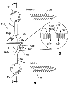
motion ofthe huinan vertebrae. Referring now to Figs. 12a-12d, a dynamic
fixation device 118
in accordance with embodiments of the present invention is shown. The device
includes flexible
rod members 120 and 122, and an anterior-posterior segment 124 aligned
transverse to the spine
and/or substantially in an anterior-posterior orientation relative to the
spine. The dynamic fixation
device 118 can bend relatively easier in one direction (flexion) than the
other (extension).
Additionally, the dynamic fixation device 118 resists motion in the plane of
the segment, which
corresponds to resisting translational movement. Thus, the dynamic fixation
device 118
accommodates at least some rotation of the vertebrae in flexion, while also
resisting translation of
the vertebrae.
The anterior-posterior segment 124 may have an anterior-posterior dimension of
about
20mm and a lateral width of about 10mm; however, dimensions of the anterior-
posterior segment
are anticipated to vary depending upon a number of factors, including the
amount of desired
movement, the size of the patient that is the recipient of the implant, and
the dimensions and
material types used to the construct the device. In accordance with
embodiments of the present
invention, the dynamic fixation device 118 provides on the order of
approximately ten degrees of
rotation in flexion and on the order of approximately negative two degrees of
rotation in the
extension.
Referring now to Fig. 12a, a dynamic fixation device 118 featuring a first
flexible rod
member 120, a second flexible rod member 122 and an anterior-posterior segment
124 is generally
shown. The flexible rod members 120 and 122 allow the dynamic fixation device
118 to rotate
around the effective pivot point 32 when the device 118 is extended in
flexion. The first flexible
rod member 120 is connected to a first rod end 14 which, in turn, is connected
to pedicle screw 20
by means of connector 18a. The second flexible rod member 122 is connected to
a second rod end
16 which, in turn, is connected to pedicle screw 20 by means of connecter 18b.
The first rod
member 120 and the second rod member 122, respectively, attach anteriorly and
posteriorly to the
anterior-posterior segment 124. In accordance with at least one embodiment of
the invention, the
attachments, interconnections or joining portions between the anterior-
posterior segment 124 and
22
CA 02612943 2007-12-19
WO 2007/002409 PCT/US2006/024491
~~:~ t ~h5:~~Kp e~rs,;r. t 1~22 may comprise a flexible connection, such as a
iiving ninge or a
~ :..,., .. ~ . ~ ..~~?. ~..... ~a ...i,.
pinned connection.
At least portions of the dynamic fixation device 118 may be made from one or
more
materials that possess the appropriate strength characteristics necessary to
withstand loading from
the human body when used in medical applications. In addition, the materials
may be chosen to
provide desired flexibility characteristics. In accordance with embodiments of
the present invention,
examples of materials that may be used to make at least portions of the
dynamic fixation device 118
include, but are not necessarily limited to, polyether ether plastics, such as
ketone (PEEK), polyether
ketone ketone (PEKK), ultra high molecular weight polyethylene (UHMWPE), and
polymethylmethacrylate (PMMA); metals, such as titanium and stainless steel;
composites; as well
as other tissue compatible materials.
Still referring to the example of the present embodiment shown in Fig. 12a,
dynamic fixation
device 118 also has a longitudinal axis L-L that is defined by the center of
connectors 18a and 18b.
Rod member 120 generally lies anterior of longitudinal axis L-L, and rod
member 122 generally lies
substantially at or posterior of longitudinal axis L-L. In accordance with at
least one embodiment
of the present invention, the anterior-posterior segment 124 has portions on
both the anterior and
posterior sides of longitudinal axis L-L. Flexible rod members 120 and 122 are
provided with
joints that allow the rod members to bend. Fig. 12a shows joint 126a of rod
member 120, as well
as joint 126b of rod member 122. In order to more clearly explain the function
of the joints, the
following discussion refers to joint 126a of rod member 120. As can be
appreciated, joint 126b of
rod member 122 functions in a similar manner. Joint 126a connects inferior
flexible rod portion
120a and superior flexible rod portion 120b. Joint 126a allows bending of the
flexible rod member
120 througli the angle X, which is defined between the inferior flexible rod
portion 120a and the
anterior-posterior segment 124. Similarly angle defines a range of motion
for joint 126b.
Fig. 12b shows a detailed view of the joint 126a of the flexible rod member
120. In
accordance with at least one embodiment of the present invention, joint 126a
is comprised of
segment 128 axially bordered by two segments 130. The segments 130 comprise a
series of
recessed portions 132. In accordance with at least one embodiment of the
present invention, the
recessed portions 132 are oriented with respect to either the anterior side of
the rod member 120 or
with respect to the posterior side of the of the rod member 120. Thus, the
modified segment 130
comprises a series of recessed portions 132 that alternate between posteriorly
oriented recessed
portions 132a and anteriorly oriented recessed portions 132b. The recessed
portions 132 can be
made using techniques known in the art, such as by use of example, removal of
material, making
23
CA 02612943 2007-12-19
WO 2oo7/ouo2409~p=_ e recessed portions 132 by inj g ection moldin= T~in
aaaiiion9o
~~~ ther
structures for providing flexibility at joints 126a and 126b are within the
scope of the invention,
such as thinned sections, crescent-shaped segments, etc.
As shown in the example illustrated in Figs. 12c and 12d, first rod end 14 is
shown to remain
essentially immobile. Second rod end 16 moves between a neutral or first
position 134, as shown
in Fig. 12c, and a flexed or second position 136, as shown in Fig. 12d. In
moving between first
position 134 and second position 136, dynamic fixation device 118 elongates
and it also rotates
about a physiologic zone of rotation or an effective pivot point 32. The
flexible rod members 120
and 122 with one or more joints 126a and 126b, together with the anterior-
posterior segment 124
provide an effective pivot point 32 that is forward or anterior of the
longitudinal axis L-L. During
movement between first position 134 and second position 136, dynainic fixation
device 118
experiences deformation, whereby it bends and it elongates to accommodate at
least some motion
in flexion of the vertebrae to which it is attached. The effective pivot point
32 is provided by the
geometry of the device 118, including the bending ofj oints 126a and 126b. As
the dynamic fixation
device 118 elongates, joint 126a bends such that the angle X is increased.
Likewise j oint 126b bends
such that the angle is increased. This allows the device to bend as shown in
Fig. 12d. As the
joints 126a and 126b bend, the dynamic fixation 118 device is allowed to
rotate about the effective
pivot point 32. This motion allows the dynamic fixation device 118 to move in
way that closely
approximates the normal motion of the human vertebrae.
Referring now to Figs. 13a-13d, a dynamic fixation device 136 in accordance
with
embodiments of the present invention is shown. The device includes a partially
folded rod segment
138. The partially folded segment 138 can bend relatively easier in one
direction (flexion) than the
other (extension). Additionally, partially folded segment 138 resists motion
in the plane of the
segment, which corresponds to resisting translational movement. Thus, the
dynamic fixation device
136 accommodates at least some rotation of the vertebrae in flexion, while
also resisting translation
of the vertebrae.
The partially folded segment 13 8 may have an anterior-posterior dimension of
about 20mm;
however, dimensions of the partially folded segment 138 are anticipated to
vary depending upon a
number of factors, including the amount of desired movement, the size of the
patient that is the
recipient of the implant, and the dimensions and material types used to the
construct the device. In
accordance with embodiments of the present invention, the dynamic fixation
device 136 provides
on the order of approximately ten degrees of rotation in flexion and on the
order of approximately
negative two degrees of rotation in the extension.
24
CA 02612943 2007-12-19
WO 2007/002409 PCT/US2006/024491
' qg. 13a, a dynamic fixation device 136 featuring a partially folded
.,. .,,
segment 138 is generally shown. The partially folded segment 138 allows
dynamic fixation device
136 to rotate around the effective pivot point 32 when the device 136 is
elongated in flexion. This
folded segment is attached to a first rod end 14 and a second rod end 16. The
first and second rod
ends 14 and 16 are, in turn, connected to pedicle screws 20 by means of
connecters 18a and 18b,
respectively. The dynamic fixation device 136 also has a longitudinal axis L-L
that is defined by
the center of connectors 18a and 18b.
At least portions of the dynamic fixation device 136 may be made from one or
more
materials that possess the appropriate strength characteristics necessary to
withstand loading from
the human body when used in medical applications. In addition, the materials
may be chosen to
provide desired flexibility characteristics. In accordance witll embodiments
of the present invention,
examples of materials that may be used to inake at least portions of the
dynamic fixation device 136
include, but are not necessarily limited to, polyether ether plastics, such as
polyether ether ketone
(PEEK), polyether ketone ketone (PEKK), ultra high molecular weight
polyethylene (UHMWPE),
and polymethylmethacrylate (PMMA); metals, such as titanium and stainless
steel; composites; as
well as other tissue compatible materials.
Still referring to the example of the present embodiment shown in Fig. 13a,
the partially
folded segment 138 comprises a series of substantially planar segments 140.
The partially folded
segment 138 may be made of interconnected elements or, alternatively, machined
out of a single
piece of material. Flexiblejoints, such as living hinges 141, connect adjacent
planar segments 140.
In accordance with at least some embodiments of the present invention, the
planar segments 140
have a quadrilateral shape. Each planar segment has two sides 142a and 142b
oriented substantially
in an anterior-posterior direction and two sides 144a and 144b oriented
substantially in a superior-
inferior direction. In accordance with at least some embodiments of the
present invention, the
length of side 144a, located posterior of longitudinal axis L-L, is longer
than the length of side 144b,
located anterior of longitudinal axis L-L. This difference in length allows
the folded segment 138
to unfold in a manner resembling that of a Japanese fan. Fig. 13b shows close-
up view of the folded
segment 138 viewed from in the posterior to anterior direction.
As shown in the example illustrated in Figs. 13c and 13d, first rod end 14 is
shown to remain
essentially immobile. Second rod end 16 moves between a neutral or first
position 146, as shown
in Fig. 13c, and a flexed or second position 148, as shown in Fig. 13d. In
moving between first
position 146 and second position 148, dynamic fixation device 136 elongates or
accommodates
elongation, and it also rotates about a physiologic zone of rotation or an
effective pivot point 32.
CA 02612943 2007-12-19
WO 2007/002409 PCT/US2006/024491
ept=43 8 thus provides an effective pivot point 32 that is iorwara or anienor
of the longitudinal axis L-L. During movement between first position 146 and
second position 148,
dynamic fixation device 136 experiences deformation, whereby it bends and it
elongates to
accommodate at least some motion in flexion of the vertebrae to which it is
attached.
The folded segment 138 allows the dynamic fixation device 136 to elongate and
rotate about
an effective pivot point 32. As shown in Fig. 13a and 13b, the planar segments
140 are all angled
in a direction towards the spine. In particular, the planar segments 140 are
all oriented on lines that
converge at a point anterior of the dynainic fixation device 136. This point
provides the
approximate location of the effective pivot point of the dynamic fixation
device 136. As the
dynamic fixation device 136 elongates, folded segment 138 unfolds enabling
rotation about the
effective pivot point 32. This allow the device to bend as shown in Fig. 13d.
This motion allows
the dynamic fixation device 118 to move in way that closely approximates the
normal motion of the
human vertebrae in flexion, while also resisting physiologically abnormal
amounts of movement
in translation.
Referring now to Figs. 14a-14d, a dynamic fixation device 150 in accordance
with
embodiments of the present invention is shown. The device includes a partially
folded rod segment
152. The partially folded segment 152 can bend relatively easier in one
direction (flexion) than the
other (extension). Additionally, partially folded segment 152 resists motion
in the plane of the
segment, which corresponds to resisting translational movement. Thus, the
dynamic fixation device
150 accommodates at least some rotation of the vertebrae in flexion, while
also resisting translation
of the vertebrae.
The partially folded segment 152 may have an anterior-posterior dimension of
about 20min;
however, dimensions of the partially folded segment 152 are anticipated to
vary depending upon a
number of factors, including the amount of desired movement, the size of the
patient that is the
recipient of the implant, and the dimensions and material types used to the
construct the device. In
accordance with embodiments of the present invention, the dynamic fixation
device 150 provides
on the order of approximately ten degrees of rotation in flexion and on the
order of approximately
negative two degrees of rotation in the extension.
Referring now to Fig. 14a, a dynamic fixation device 150 featuring a partially
folded
segment 152 is generally shown. The partially folded segment 152 allows
dynamic fixation device
150 to rotate around the effective pivot point 32 wlien the device 150 is
elongated in flexion. The
partially folded segment 152 is attached to a first rod end 14 and a second
rod end 16. The first and
26
CA 02612943 2007-12-19
WO 2007/002409 PCT/US2006/024491
..... .. e)I .. s.. .. . 1 . .
.. .
.oL= . , lk ilw .s)s sA _ u..." ! :k :Y" .. t
fE'j' t'k..u it ~' Ti, f< <.:Yt-ii itii; tr .='' tE: ,.
second rod ends 14 and 16 are, in turn, connected to pedicle screws 20 by
means of connecters 18a
and 18b, respectively.
At least portions of the dynamic fixation device 150 may be made from one or
more
materials that possesses the appropriate strength characteristics necessary to
withstand loading from
the human body when used in medical applications. In addition, the materials
may be chosen to
provide desired flexibility characteristics. In accordance with embodiments
ofthe present invention,
examples of materials that may be used to make at least portions of the
dynamic fixation device 150
include, but are not necessarily limited to, polyether ether plastics, such as
ketone (PEEK), polyether
ketone ketone (PEKK), ultra high molecular weight polyethylene (UHMWPE), and
polymetliylmethacrylate (PMMA); metals, such as titanium and stainless steel;
composites; as well
as other tissue compatible materials.
The partially folded segment 152 comprises a series of planar segments 154.
Flexible j oints,
such as living hinges 156, connect adjacent planar segments 154. The partially
folded segment 152
may be made of interconnected elements or, alternatively, machined out of a
single piece of
material. In accordance witll at least some of the embodiments of the present
invention, the planar
segments 154 have a rectangular shape. Each planar segment 154 has two sides
156a and 156b
oriented substantially in an anterior-posterior direction and two sides 158a
and 158b oriented
substantially in a superior-inferior direction. As shown in Fig. 14a, the
dynamic fixation device 150
also has a longitudinal axis L-L that is defined by the center of connectors
18a and 18b. In
accordance with at least some embodiments of the present invention, the length
of side 158a, located
posterior of longitudinal axis L- L, is same as the length of side 158b,
located anterior of
longitudinal axis L- L. Although the sides of the planar segments 154 are of
similar length, if the
partially folded segment 154 is made of a sufficiently elastic material, it
will accommodate rotation
of the dynamic fixation device 150. Fig. 14b shows a close-up view of the
folded segment 152
viewed from the posterior to anterior direction.
As shown in the example illustrated in Figs. 14c and 14d, first rod end 14 is
shown to remain
essentially immobile. Second rod end 16 moves between a neutral or first
position 160, as shown
in Fig. 14c, and a flexed or second position 162, as shown in Fig. 14d. In
moving between first
position 160 and second position 162, dynamic fixation device 150 elongates or
accommodates
elongation, and it also rotates about a physiologic zone of rotation or an
effective pivot point 32.
The partially folded segment 138 provides an effective pivot point 32 that is
forward or anterior of
the longitudinal axis L-L. During movement between first position 160 and
second position 162,
27
CA 02612943 2007-12-19
WO 2007/002409 PCT/US2006/024491
==i. "5.. ' / :1 51 Y==='.Ap "t II'==' ..' "" y1, ) 11. 15. d1"!x .~e..
dynamic fixation device 160 experiences deformation, whereby it bends and it
elongates to
accommodate at least some motion in flexion of the vertebrae to which it is
attached.
The folded segment 152 allows the dynaniic fixation device 150 to elongate and
rotate about
an effective pivot point 32. As the spine moves from the neutral position
illustrated in Fig. 14c to
the flexed position depicted in Fig. 14d, the dynamic fixation device 150
undergoes an elongation.
This elongation causes the folded segment 150 to unfold. This allow the device
to bend as shown
in Fig. 14d. In accordance with embodiments of the present invention, the
rotation about the
effective pivot point 32 is due to the planar segments and the flexible joint
of the partially folded
segment being made from a material of sufficient strength and flexibility to
allow for the described
movement. Such materials may include, but are not limited to PEEK and PEKK.
This motion
allows the dynamic fixation device 150 to move in way that closely
approximates the normal motion
of the human vertebrae.
Referring now to Figs. 15a-15c, a dynamic fixation device 170 in accordance
with
embodiments of the present invention is shown. The device includes a plurality
of segments 172
connected by hinges 174. The plurality of hinged segments 172 can bend
relatively easier in one
direction (flexion) than the other (extension). Additionally, the plurality of
hinged segments 172
resists motion in the plane of the segment, which corresponds to resisting
translational movement.
Thus the dynainic fixation device 170 accommodates at least some rotation at
the vertebrae in
flexion, while also resisting translation of the vertebrae.
Referring now to Figs. 15a, a dynamic fixation device 170 featuring a
plurality of hinged
segments 172 is shown. In at least one embodiment ofthe present invention, the
hinges 174 contain
pins 176 that are directed forwardly or anteriorly of the dynamic fixation
device 170. This
orientation of the pins 176 of the hinges 174 provides a forwardly or
anteriorly projected pivot point
32 that is similar to the natural pivot point of a first vertebra relative to
the second vertebra when
the spine undergoes flexion. The dynamic fixation device 170 features an
inferior hinged segment
172a, a superior hinged segment 172b, and a interior hinged segment 172c. The
inferior and
superior hinged segments 172a and 172b each have a hinged connection to
connectors 18a and 18b,
respectively. The interior hinged segment 172c is disposed between and has a
hinged connection
to the inferior and superior hinged segments 172a and 172b. Connectors 18a and
18b each attach
to a pedicle screw 20. The hinges 174 that connect both ends of the superior
hinged segment 172b
are shown separated in Fig. 15a in order to illustrate their structure.
28
CA 02612943 2007-12-19
WO 2007/002409 PCT/US2006/024491
it,._
:~'~N
The dynamic fixation device 170 may be made from one or more materials that
possess the
appropriate strength characteristics necessary to withstand loading from the
human body when used
in medical applications. In addition, the materials may be chosen to provide
desired flexibility
characteristics. In accordance with embodiments of the present invention,
examples of materials
that may be used to make at least a portion of the dynamic fixation device 170
include, but are not
limited to: plastics, such as polyether ether ketone (PEEK), polyether ketone
ketone (PEKK), ultra
high molecular weight polyethylene (UHMWPE), polymethylmethacrylate (PMMA);
and more
preferably, metals, such as titanium and stainless steel. In addition, the
device 170 may be made
of a combination of materials, of composites, as well as other tissue
compatible materials.
Still referring to the exainple ofthe present embodiment shown in Fig. 15a,
dynamic fixation
device 170 also has a longitudinal axis L-L that is defined by the center of
connectors 18a and 18b.
In accordance with embodiments of the present invention, inferior hinged
segment 172a, superior
hinged segment 172b, and interior hinged segment 172c generally lie in a plane
perpendicular to the
pedicle screws 20 and at an acute angle with respect to the longitudinal axis
L-L.
The functionality of the dynamic fixation device 170 is illustrated in Figs.
15b and 15c. Fig.
15b illustrates the dynamic fixation device 170 in neutral position 182,
wherein the spine is neither
flexed nor extended. The pedicle screw 20 connected to connecter 18a is
attached to a lower
vertebra. The pedicle screw 20 connected to connector 18b is attached to an
upper vertebra. Fig.
15c illustrates the dynamic fixation device 170 in a flexed position 184. In
moving between first
position 182 and second position 184, dynamic fixation device 170 elongates
and it also rotates
about an effective pivot point 32. During this flexion movement, the plurality
of hinged segments
172 rotate in a more superior-inferior alignment, such that at least the
segments 172 and 172b
become increasingly aligned parallel with respect to longitudinal axis L-L,
thereby providing
lengthening to the dynamic fixation device 170 and allowing the upper vertebra
to rotate forward
relative to the lower vertebra. In addition, the relatively rigid materials
used to form the construct
resist movement in the anterior-posterior direction, thereby resisting
translational motion of the two
interconnected vertebrae. This motion of the hinged segments 172 allows the
dynamic fixation
device 170 to move in a way that closely approximates the normal physiological
motion of the-
human vertebrae. In accordance with embodiments of the present invention, the
dynamic fixation
device 170 provides on the order of approximately ten degrees of rotation in
flexion and on the
order of approximately negative two degrees of rotation in the extension.
29
CA 02612943 2007-12-19
WO 2007/002409 PCT/US2006/024491
4~.' ;jC +f:.
For a dynamic fixation device 170 spanning one joint, it will expand up to
approximately
to 10mm in length, and will rotate forward up to between 5 to 10 degrees to
accommodate flexion
of the spine. Obviously, different size dynamic fixation devices 170 may be
used to acconunodate
the specific needs of each individual patient. More particularly, a relatively
large dynamic fixation
5 device may be needed for a large man, while a relatively small dynamic
fixation device may be
needed for a smaller patient, such as child or a petite woman. However, a
limited number of sizes
may provide adequate coverage for the majority of the patient population. For
any given device,
a potential elongation of the dynamic fixation device consistent with the
desired flexion of the
vetebral motion segment and associated distraction of the plane of the
fixation device is anticipated.
In accordance with embodiments of the present invention, the hinges as
described herein
may not comprise a pin. In particular, the ernbodiments of the present
invention illustrated in Fig.
3a, 4a, 5a, 6a, 7a, 8a, 9a, 11a, 12a, 13a, and 14a may contain flexible
elements such as a living
hinge.
The dynamic fixation devices can be used to flexibly secure a plurality of
vertebra.
Alternatively, the dynamic fixation devices can be located at specific points
where bending of the
spine is desired, while a rigid rod may be used at other locations desired by
the physician. Where
used, rigid rod portions may be curved, thereby influencing the implanted
location of the geometric
shape hinged structures, and thus the effective pivot point.
The structures of the present invention are made from one or more materials
that possesses
the appropriate strength characteristics necessary to withstand loading from
the human body when
used in medical applications. In addition, the materials are compatible with
the human body.
Preferably, materials include ceramics, plastics, metals, or carbon fiber
composites. More
preferably, the materials are made from titanium, a titanium alloy, or
stainless steel.
Examples of plastic materials with in the scope of the invention include
polyether ether
ketone (PEEK), polyether ketone ketone (PEKK), any material chosen from the
polyaryl ether
ketone (PAEK) family, ultra high molecular weight polyethylene (UHMWPE),
CA 02612943 2007-12-19
WO 2007/002409 PCT/US2006/024491
.k== ==q:. '71" :k Y 6'+= ==It =====it '>; t =1 1 +L" .. k
polyinethylmethacrylate (PMMA), polyethlene terephthalate (PET), fluorinated
ethylene propylene
(FEP), polyuretheance (PU), polyimide (Pl), polybutylene terephtlialate (PBT)
polyurethane rubber (PUR). Additionaly, silicon and silicon rubber are
useable, as well as
polysulfone, polyimide , epoxy, and polycyanate.
Elements of the fixation device may be made from a radiolucent polymer,
allowing the
device, once implanted in a patient, to be seen by radiographic methods.
Examples of such
radiolucent materials include polyether ether ketone and polyether ketone
ketone.
Materials chosen for compatibility with the human body should be resistant to
organic and
inorganic chemicals, have desirable strength and rigidity properties, be
resistance to impact over a
wide range of temperatures and be resistant to hydrolysis and corrosion.
In accordance with embodiments of the present invention, elements of the
dynamic fixation
device that are implanted into bone can be made from bone graft material. Such
material can be
allographic meaning grown from an organism of the same species, or
xenographic, meaning grown
from an organism of a different species.
The following patent applications, of which the entire disclosure is herein
incorporated by
reference, contain exemplary uses of biocompatible materials: US Patent
Application No.
2005/0203519, US Patent Application No. 2005/0203517, US Patent Application
No.
2006/0041259, US Patent Application No. 2006/0064090, and US Patent
Application No.
2003/0109880.
The above described alternative configurations offer different bending
characteristics. The
dimensions will vary depending upon the specific design necessary for a
specific patient. More
particularly, the dimensions of geometric shapes and hinged devices will
likely be bigger for a large
heavy man, as opposed to that needed for a small petite woman. Furthermore,
the type of material
used to construct the dynamic fixation devices described herein will also
impact the required
dimensions of the devices. Dynamic fixation devices described herein may be
made of a variety of
materials, preferably metals or materials demonstrating resilient
characteristics, and more
preferably, a titanium alloy or surgical stainless steel. Since different
materials have different
strength and resilient properties, the type of material used will, in part,
dictate the dimensions of the
rod portion required to achieve a certain function in a specific patient.
Devices disclosed herein can also be made of thei7nal memory materials or
materials that
possess different elastic properties at varying temperatures. In this aspect
of the invention, the
subject component(s) may be heated or cooled to a desired teinperature,
implanted, then
31
CA 02612943 2007-12-19
WO 2007/002409 PCT/US2006/024491
ss.: r,,, sa .. x
'_ t . s
?..f 't
i . f
F +~ "::dL A.
subsequently allowed to cool or warm to the temperature of the ambient
conditions that will exist
during the usage period for the subject device, namely, normal body
temperature.
It is to be understood that the present invention may have application to
medical devices
other than spinal implants. For example, the present invention can be used in
external fixator
systems.
Furthermore, it is understood that the present invention has application
outside the medical
field. The dynamic fixation device of the present invention is not limited to
medical implants. The
device could be used in seismic dampening applications. Alternatively, the
present invention could
be used to secure any two objects, such as in linking mechanisms, and has
application to any type
of mechanical device with a moving connection. Other applications, by no means
exhaustive, may
include connecting any articulated device, such as an implement connection to
a tractor. It may also
be used in heretofore static type connection applications, such as attaching
an antenna to a base
structure. One of skill in various of the construction arts will appreciate
how to make and use the
present invention in view of the guidance provided herein (with respect to a
surgical application)
and in view of the figures set forth herein.
The foregoing discussion of the invention has been presented for purposes of
illustration and
description. The foregoing is not intended to limit the invention to the form
or forms disclosed
herein. In the foregoing Detailed Description Of The Invention for example,
various features of the
invention are grouped together in one or more embodiments for the purpose of
streamlining the
disclosure. This method of disclosure is not to be interpreted as reflecting
an intention that the
claimed invention requires more features than are expressly recited in each
claim. Rather, as the
following claims reflect, inventive aspects lie in less than all features of a
single foregoing disclosed
embodiment. Thus, the following claims are hereby incorporated into this
Detailed Description Of
The Invention, with each claim standing on its own as a separate preferred
embodiment of the
invention.
While various embodiments of the present invention have been described in
detail, it is
apparent that modifications and adaptations of those embodiments will occur to
those skilled in the
art. However, it is to be expressly understood that such modifications and
adaptations are within
the spirit and scope of the present invention, as set forth in the following
claims.
32