Note: Descriptions are shown in the official language in which they were submitted.
DEMANDE OU BREVET VOLUMINEUX
LA PRRSENTE PARTIE DE CETTE DEMANDE OU CE BREVET COMPREND
PLUS D'UN TOME.
CECI EST LE TOME 1 DE 2
CONTENANT LES PAGES 1 A 46
NOTE : Pour les tomes additionels, veuillez contacter le Bureau canadien des
brevets
JUMBO APPLICATIONS/PATENTS
THIS SECTION OF THE APPLICATION/PATENT CONTAINS MORE THAN ONE
VOLUME
THIS IS VOLUME 1 OF 2
CONTAINING PAGES 1 TO 46
NOTE: For additional volumes, please contact the Canadian Patent Office
NOM DU FICHIER / FILE NAME:
NOTE POUR LE TOME / VOLUME NOTE:
WO 2007/024653 CA 02613136 2011-01-13 PCT/US2006/032264
1
METHOD AND SUBSTANCES FOR ISOLATING miRNAs
BACKGROUND
MicroRNAs (miRNAs) are small, generally between 18 and 24 residues,
polyribonucleotides derived from longer hairpin noncoding transcripts in
eukaryotes.
miRNAs play a significant role in cellular developmental and differentiation
pathways.
Consequently, there have been considerable efforts made to understand and
characterize the
temporal, spatial and cellular expression levels and patterns of expression of
miRNAs to
ascertain their precise role in cellular development and differentiation in
both normal and
disease states.
miRNAs are currently studied by, first, obtaining the total RNA from a sample.
Next, the total RNA is fractionated into subpopulations by gel electrophoresis
or by
chromatographic fractionation and size selective elution. Then, the
appropriate section of the
gel is cut, and the 18-24 RNAs are eluted from the gel, or the eluted fraction
containing
single stranded RNAs in the size range of 18-24 ribonucleotides is collected.
Next, the
RNAs are isolated by precipitation and the miRNAs are characterized.
Disadvantageously, however, these methods do not work well when the amount of
sample is small, such as samples from tumor tissue or biopsy material.
Further,
characterization of the miRNAs isolated by present methods usually comprises a
several step
amplification procedure followed by detection, quantitation, cloning and
sequencing.
Because of the large number of steps in these processes and the notorious
inefficiencies
associated with the repeated purification, the isolation and identification of
miRNAs using
present methods is time consuming, relatively expensive, requires relatively
large amounts of
material and is not fully representative of the population of miRNAs expressed
within a small
sample, such as within a biopsy of a tumor. Additionally, the present methods
are not
specific to isolating and identifying an miRNA, and therefore, often isolate
and identify
siRNA, tRNA, 5S/5.8SrRNA and degraded RNA from additional cellular RNAs.
CA 02613136 2007-12-20
WO 2007/024653 PCT/US2006/032264
2
Therefore, there is the need for an improved method for isolation and
identification of
miRNAs that is not associated with these disadvantages.
SUMMARY
According to one embodiment of the present invention, there is provided a
capture
probe suitable for use with a method for isolating miRNAs. The capture probe
comprises: a)
a first adapter segment having a first adapter segment sequence, the first
adapter segment
comprising a 3' end and a 5' end; b) a second adapter segment having a second
adapter
segment sequence, the second adapter segment comprising a 3' end and a 5' end;
and c) an
miRNA binding segment having an miRNA binding segment sequence, where the
miRNA
binding segment is substantially complementary to, and capable of hybridizing
to, one or
more than one miRNA of interest by Watson-Crick base pairing, where the 5' end
of the first
adapter segment is connected to the 3' end of the miRNA binding segment, and
where the 3'
end of the second adapter segment is connected to the 5' end of the miRNA
binding segment.
In one embodiment, the capture probe comprises a substance selected from the
group
consisting of one or more than one type of polynucleotide, one or more than
one type of
polynucleotide analog, and a combination of one or more than one type of
polynucleotide and
polynucleotide analog.
According to another embodiment of the present invention, there is provided a
set of
capture probes, where each of the capture probes of the set of capture probes
is a capture
probe according to the present invention, where each of the capture probes
comprises
identical-first adapter segment sequences, where each of the capture probes of
the set of
capture probes comprises identical miRNA binding segment sequences, and where
each of the
capture probes of the set of capture probes comprises identical second adapter
segment
sequences.
According to another embodiment of the present invention, there is provided a
set of
capture probes, where each of the capture probes is a capture probe according
to the present
invention, and where the set comprises at least one capture probe comprising
an miRNA
binding segment that is substantially complementary to, and capable of
hybridizing to, each
miRNA from a single public database.
According to another embodiment of the present invention, there is provided a
set of
capture probes, where each of the capture probes is a capture probe according
to the present
invention, where the set comprises a first capture probe and a second capture
probe, where
CA 02613136 2007-12-20
WO 2007/024653 PCT/US2006/032264
3
the first capture probe and the second capture probe have identical first
adapter segment
sequences, where the first capture probe and the second capture probe have
identical miRNA
binding segment sequences, and where the first capture probe has a second
adapter segment
sequence that is different from the second adapter segment sequence of the
second capture
probe.
According to another embodiment of the present invention, there is provided a
set of
capture probes, where each of the capture probes is a capture probe according
to the present
invention, where the set comprises a first capture probe and a second capture
probe, where
the first capture probe and the second capture probe have identical first
adapter segment
sequences, where the first capture probe and the second capture probe have
identical second
adapter segment sequences, and where the first capture probe has an miRNA
binding segment
sequence that is different from the miRNA binding segment sequence of the
second capture
probe.
According to another embodiment of the present invention, there is provided a
set of
capture probes, where each of the capture probes is a capture probe according
to the present
invention, where the set comprises a first capture probe and a second capture
probe, where
the first capture probe and the second capture probe have identical miRNA
binding segment
sequences, where the first capture probe and the second capture probe have
identical second
adapter segment sequences, and where the first capture probe has a first
adapter segment
sequence that is different from the first adapter segment sequence of the
second capture
probe.
According to another embodiment of the present invention, there is provided a
set of
capture probes, where each of the capture probes is a capture probe according
to the present
invention, where the set comprises a first capture probe and a second capture
probe, where
the first capture probe and the second capture probe have identical first
adapter segment
sequences, where the first capture probe has an miRNA binding segment sequence
that is
different from the miRNA binding segment sequence of the second capture probe,
and where
the first capture probe has a second adapter segment sequence that is
different from the
second adapter segment sequence of the second capture probe.
According to another embodiment of the present invention, there is provided a
set of
capture probes, where each of the capture probes is a capture probe according
to the present
invention, where the set comprises a first capture probe and a second capture
probe, where
CA 02613136 2007-12-20
WO 2007/024653 PCT/US2006/032264
4
the first capture probe and the second capture probe have identical miRNA
binding segment
sequences, where the first capture probe has a first adapter segment sequence
that is different
from the first adapter segment sequence of the second capture probe, and where
the first
capture probe has a second adapter segment sequence that is different from the
second adapter
segment sequence of the second capture probe.
According to another embodiment of the present invention, there is provided a
set of
capture probes, where each of the capture probes is a capture probe according
to the present
invention, where the set comprises a first capture probe and a second capture
probe, where
the first capture probe and the second capture probe have identical second
adapter segment
sequences, where the first capture probe has a first adapter segment sequence
that is different
from the first adapter segment sequence of the second capture probe, and where
the first
capture probe has a miRNA binding segment sequence that is different from the
miRNA
binding segment sequence of the second capture probe.
According to another embodiment of the present invention, there is provided a
set of
capture probes, where each of the capture probes is a capture probe according
to the present
invention, where the set comprises a first capture probe and a second capture
probe, where
the first capture probe has a first adapter segment sequence that is different
from the first
adapter segment sequence of the second capture probe, where the first capture
probe has an
miRNA binding segment sequence that is different from the miRNA binding
segment
sequence of the second capture probe, and where the first capture probe has a
second adapter
segment sequence that is different from the second adapter segment sequence of
the second
capture probe.
According to another embodiment of the present invention, there is provided a
capture
probe according to the present invention, where the first adapter segment, or
the second
adapter segment, or both the first adapter segment and the second adapter
segment are
between 6 and 16 residues.
According to another embodiment of the present invention, there is provided a
capture
probe according to the present invention, where the first adapter segment, or
the second
adapter segment, or both the first adapter segment and the second adapter
segment further
comprise a sequence that is a polynucleotide synthesis promoter motif for a
polynucleotide
polymerase, or that is complementary to a polynucleotide synthesis promoter
motif for a
polynucleotide polymerase. In one embodiment, the polynucleotide synthesis
promoter motif
CA 02613136 2007-12-20
WO 2007/024653 PCT/US2006/032264
is a motif for a polynucleotide synthesis promoter selected from the group
consisting of T7,
SP6, a T3 DNA dependent RNA polymerase, a type 2 RNA polymerase of E. coli and
single
stranded DNA dependent N4 RNA polymerase.
According to another embodiment of the present invention, there is provided a
capture
5 probe according to the present invention, where the first adapter segment,
or the second
adapter segment, or both the first adapter segment and the second adapter
segment further
comprise a restriction site motif. In one embodiment, the restriction site
motif is acted upon
by a restriction enzyme selected from the group consisting of Not I, Xho I,
Xma I and Nhe I.
According to another embodiment of the present invention, there is provided a
capture
probe according to the present invention, where the first adapter segment, or
the second
adapter segment, or both the first adapter segment and the second adapter
segment further
comprise a solid phase binding group to immobilize the capture probe to a
solid phase. In
one embodiment, the solid phase binding group is at or near the 3' end of the
first adapter
segment. In another embodiment, the solid phase binding group is at or near
the 5' end of
the second adapter segment. In another embodiment, the solid phase binding
group
immobilizes the capture probe to the solid phase covalently. In another
embodiment, the
solid phase binding group immobilizes the capture probe to the solid phase non-
covalently.
In another embodiment, the solid phase binding group comprises biotin or an
analog of
biotin.
According to another embodiment of the present invention, there is provided a
capture
probe according to the present invention, where the miRNA binding segment
consists of 18
or 19 or 20 or 21 or 22 or 23 or 24 residues selected from the group
consisting of DNA,
RNA, chimeric DNA/RNA, DNA analogs and RNA analogs. In another embodiment, the
miRNA of interest that the miRNA binding segment is substantially
complementary to, and
capable of hybridizing to, is selected from a public database. In another
embodiment, the
miRNA of interest is a eucaryotic miRNA. In another embodiment, the miRNA of
interest is
a primate miRNA. In another embodiment, the miRNA of interest is a human
miRNA. In
another embodiment, the miRNA binding segment is exactly the complement to the
miRNA
of interest in both length and sequence. In another embodiment, the miRNA
binding segment
is more than 90 % complementary to a segment of the miRNA of interest of the
same length
as the miRNA of interest sequence. In another embodiment, the miRNA binding
segment is
more than 80 % complementary to a segment of the miRNA of interest of the same
length as
CA 02613136 2007-12-20
WO 2007/024653 PCT/US2006/032264
6
the miRNA of interest sequence. In another embodiment, the first adapter
segment has a first
adapter segment sequence according to SEQ ID NO: 1. In another embodiment, the
second
adapter segment has a second adapter segment sequence according to SEQ ID
NO:2.
According to another embodiment of the present invention, there is provided a
method
for isolating an miRNA of interest from a sample comprising the miRNA of
interest. The
method comprises: a) providing a sample comprising the miRNA of interest; b)
providing a
capture probe according to the present invention; c) providing a first linker
and a second
linker; d) combining the sample, the capture probe, the first linker and the
second linker;
e) allowing the first linker to hybridize with the first adapter segment, the
miRNA of interest
to hybridize with the miRNA binding segment, and the second linker to
hybridize with the
second adapter segment; f) ligating the 3' end of the first linker that is
hybridized to the first
adapter segment to the 5' end of the miRNA of interest that is hybridized to
the miRNA
binding segment, and ligating the 3' end of the miRNA of interest that is
hybridized to the
miRNA binding segment to the 5' end of the second linker that is hybridized to
the second
adapter segment, thereby producing a complex defined as a strand of first
linker, miRNA of
interest and second linker that have been ligated together (ligated first
linker-miRNA of
interest-second linker) and that is hybridized to the capture probe; and g)
dehybridizing the
capture probe from the strand of the ligated first linker-miRNA of interest-
second linker,
where the miRNA of interest has an miRNA of interest sequence, and comprises a
3' end and
a 5' end, where the miRNA of interest is substantially complementary to, and
capable of
hybridizing to, the miRNA binding segment of the capture probe by Watson-Crick
base
pairing, where the first linker has a first linker sequence, and comprises a
3' end and a 5'
end, where the first linker is substantially complementary to, and capable of
hybridizing to,
the first adapter segment of the capture probe by Watson-Crick base pairing,
where the
second linker has a second linker sequence, and comprises a 3' end and a 5'
end, and where
the second linker is substantially complementary to, and capable of
hybridizing to, the second
adapter segment of the capture probe by Watson-Crick base pairing. In another
embodiment,
the sample further comprises one or more than one substance that is chemically
related to the
miRNA of interest selected from the group consisting of an RNA other than
miRNA and a
DNA. In another embodiment, the sample is from a eukaryote. In another
embodiment, the
sample is from a primate. In another embodiment, the sample is from a human.
In another
embodiment, the sample comprises a tissue or fluid selected from the group
consisting of
CA 02613136 2007-12-20
WO 2007/024653 PCT/US2006/032264
7
blood, brain, heart, intestine, liver, lung, pancreas, muscle, a leaf, a
flower, a plant root and
a plant stem. In another embodiment, the miRNA of interest consists of 18 or
19 or 20 or 21
or 22 or 23 or 24 RNA residues. In one embodiment, the miRNA of interest is
listed in a
public database. In another embodiment, the sample provided comprises a
plurality of
miRNAs of interest, and each of the plurality of miRNAs of interest have miRNA
of interest
sequences that are identical to one another. In another embodiment, the sample
provided
comprises a plurality of miRNAs of interest comprising a first miRNA of
interest having a
first miRNA of interest sequence, and a second miRNA of interest having a
second miRNA
of interest sequence, and where the first miRNA of interest sequence is
different from the
second miRNA of interest sequence. In another embodiment, the sample provided
comprises
a plurality of miRNAs of interest comprising a first miRNA of interest having
a first miRNA
of interest sequence, a second miRNA of interest having a second miRNA of
interest
sequence, and a third miRNA of interest having a third miRNA of interest
sequence, where
the first miRNA of interest sequence is different from the second miRNA of
interest
sequence, where the first miRNA of interest sequence is different from the
third miRNA of
interest sequence, and where second miRNA of interest sequence is different
from the third
miRNA of interest sequence. In another embodiment, the method further
comprises isolating
the total RNA from the sample after providing the sample.
In one embodiment, the capture probe provided is a set of capture probes,
where each
of the capture probes comprises identical first adapter segment sequences,
where each of the
capture probes of the set of capture probes comprises identical miRNA binding
segment
sequences, and where each of the capture probes of the set of capture probes
comprises
identical second adapter segment sequences. In another embodiment, the capture
probe
provided is a set of capture probes, where the set comprises at least one
capture probe
comprising an miRNA binding segment that is substantially complementary to,
and capable of
hybridizing to, each miRNA listed in a single public database. In another
embodiment, the
capture probe provided is a set of capture probes, where the set comprises a
first capture
probe and a. second capture probe, where the first capture probe and the
second capture probe
have identical first adapter segment sequences, where the first capture probe
and the second
capture probe have identical miRNA binding segment sequences, and where the
first capture
probe has a second adapter segment sequence that is different from the second
adapter
segment sequence of the second capture probe. In another embodiment, the
capture probe
CA 02613136 2007-12-20
WO 2007/024653 PCT/US2006/032264
8
provided is a set of capture probes, where the set comprises a first capture
probe and a
second capture probe, where the first capture probe and the second capture
probe have
identical first adapter segment sequences, where the first capture probe and
the second
capture probe have identical second adapter segment sequences, and where the
first capture
probe has an miRNA binding segment sequence that is different from the miRNA
binding
segment sequence of the second capture probe. In another embodiment, the
capture probe
provided is a set of capture probes, where the set comprises a first capture
probe and a
second capture probe, where the first capture probe and the second capture
probe have
identical miRNA binding segment sequences, where the first capture probe and
the second
capture probe have identical second adapter segment sequences, and where the
first capture
probe has a first adapter segment sequence that is different from the first
adapter segment
sequence of the second capture probe. In another embodiment, the capture probe
provided is
a set of capture probes, where the set comprises a first capture probe and a
second capture
probe, where the first capture probe and the second capture probe have
identical first adapter
segment sequences, where the first capture probe has an miRNA binding segment
sequence
that is different from the miRNA binding segment sequence of the second
capture probe, and
where the first capture probe has'a second adapter segment sequence that is
different from the
second adapter segment sequence of the second capture probe. In another
embodiment, the
capture probe provided is a set of capture probes, where the set comprises a
first capture
probe and a second capture probe, where the first capture probe and the second
capture probe
have identical miRNA binding segment sequences, where the first capture probe
has a first
adapter segment sequence that is different from the first adapter segment
sequence of the
second capture probe, and where the first capture probe has a second adapter
segment
sequence that is different from the second adapter segment sequence of the
second capture
probe. In another embodiment, the capture probe provided is a set of capture
probes, where
the set comprises a first capture probe and a second capture probe, where the
first capture
probe and the second capture probe have identical second adapter segment
sequences, where
the first capture probe has a first adapter segment sequence that is different
from the first
adapter segment sequence of the second capture probe, and where the first
capture probe has
an miRNA binding segment sequence that is different from the miRNA binding
segment
sequence of the second capture probe. In another embodiment, the capture probe
provided is
a set of capture probes, where the set comprises a first capture probe and a
second capture
CA 02613136 2007-12-20
WO 2007/024653 PCT/US2006/032264
9
probe, where the first capture probe has a first adapter segment sequence that
is different
from the first adapter segment sequence of the second capture probe, where the
first capture
probe has an miRNA binding segment sequence that is different from the miRNA
binding
segment sequence of the second capture probe, and where the first capture
probe has an
miRNA binding segment sequence that is different from the miRNA binding
segment
sequence of the second capture probe. In another embodiment, the capture probe
provided is
a set of capture probes, where the set comprises a first capture probe having
a first capture
probe sequence, a second capture probe having a second capture probe sequence,
and a third
capture probe having a third capture probe sequence, where the first capture
probe sequence
is different from the second capture probe sequence, where the first capture
probe sequence is
different from the third capture probe sequence, and where second capture
probe sequence is
different from the third capture probe sequence.
In one embodiment, the first linker segment and the second linker segment
comprise a
substance selected from the group consisting of one or more than one type of
polynucleotide,
one or more than one type of polynucleotide analog, and a combination of one
or more than
one type of polynucleotide and polynucleotide analog. In another embodiment,
the first
linker, or the second linker, or both the first linker and the second linker
are resistant to
nuclease degradation. In another embodiment, the first linker, or the second
linker, or both
the first linker and the second linker comprise nuclease resistant
nucleotides. In another
embodiment, the first linker, or the second linker, or both the first linker
and the second
linker comprise nucleotides with a phosphothioate backbone that render the
first linker, or the
second linker, or both the first linker and the second linker resistant to
nuclease degradation.
In another embodiment, the first linker, or the second linker, or both the
first linker and the
second linker comprise nuclease resistant nucleotides and comprise nucleotides
with a
phosphothioate backbone that render the first linker, or the second linker, or
both the first
linker and the second linker resistant to nuclease degradation. In another
embodiment, the
first linker and the second linker, each comprises between 6 and 50 residues.
In another
embodiment, the first linker comprises at least 10 residues, and at least 10
residues at the 3'
end of the first linker are exactly the complement of the corresponding
residues at or near the
5' end of the first adapter segment. In another embodiment, the second linker
comprises at
least 10 residues, and at least 10 residues at the 5' end of the second linker
are exactly the
complement of the corresponding residues at or near the 3' end of the second
adapter
CA 02613136 2007-12-20
WO 2007/024653 PCT/US2006/032264
segment. In another embodiment, the 5' end of the first linker, or the 3' end
of the second
linker, or both the 5' end of the first linker and the 3' end of the second
linker comprise a
label. In another embodiment, the 5' end of first linker comprises one or more
than one
residue that, extends beyond the 3' end of the first adapter segment after the
first linker
5 hybridizes to the first adapter segment. In one embodiment, the one or more
than one residue
of the 5' end of first linker that extends beyond the 3' end of the first
adapter segment
functions as a primer binding site. In another embodiment, the 3' end of
second linker
comprises one or more than one residue that extends beyond the 5' end of the
second adapter
segment after the second linker hybridizes to the second adapter segment. In
one
10 embodiment, the one or more than one residue of the 3' end of second linker
that extends
beyond the 5' end of the second adapter segment functions as a primer binding
site. In
another embodiment, the sample, the capture probe, the first linker and the
second linker are
combined simultaneously.
In one embodiment, the method further comprises adding one or more than one
RNAse inhibitor to the combination of the sample, the capture probe, the first
linker and the
second linker. In another embodiment, the first adapter segment comprises a
solid phase
binding group, or the second adapter segment comprises a solid phase binding
group, or both
the first adapter segment comprises a solid phase binding group and the second
adapter
segment comprises a solid phase binding group, and the method further
comprises binding the
capture probe to a solid phase before or after combining the sample, the
capture probe, the
first linker and the second linker. In another embodiment, the solid phase is
a plurality of
paramagnetic particles. In another embodiment, the capture probe is bound to a
solid phase
through the first adapter segment or through the second adapter segment or
through both the
first adapter. segment and the second adapter segment, and the method further
comprises
purifying the capture probes with hybridized first linker, miRNA of interest
and second
linker--bound to the solid phase by removing non-hybridized first linkers,
second linkers and
any other substances that are not bound to the solid phase. In another
embodiment, the solid
phase is contained in a vessel comprising a surface and a cap, and purifying
comprises
applying a magnetic field to attract the solid phase to the surface of the
vessel or the cap of
the vessel. In another embodiment, the first linker hybridizes to the first
adapter segment at a
position where the last residue on the 3' end of the first linker hybridizes
to a residue on the
first adapter segment that is between 1 residue and 5 residues from the 3' end
of the miRNA
CA 02613136 2007-12-20
WO 2007/024653 PCT/US2006/032264
11
binding segment. In another embodiment, the first linker hybridizes to the
first adapter
segment at a position where the last residue on the 3' end of the first linker
hybridizes to a
residue on the first adapter segment that is immediately adjacent to the 3'
end of the miRNA
binding segment. In another embodiment, the second linker hybridizes to the
second adapter
segment at a position where the last residue on the 5' end of the second
linker hybridizes to a
residue on the second adapter segment that is between 1 residue and 5 residues
from the 5'
end of the miRNA binding segment. In another embodiment, the second linker
hybridizes to
the second adapter segment at a position where the last residue on the 5' end
of the second
linker hybridizes to a residue on the second adapter segment that is
immediately adjacent to
the 5' end of the miRNA binding segment. In another embodiment, the method
further
comprises purifying the complex. In another embodiment, the complex is bound
to a solid
phase through the first adapter segment or through the second adapter segment
or through
both the first adapter segment and the second adapter segment, and the method
further
comprises purifying the complex by removing non-hybridized first linkers,
second linkers and
any other substances that are not bound to the solid phase. In another
embodiment, the
method further comprises purifying the ligated first linker-miRNA of interest-
second linker
that has been dehybridized from the capture probe.
In one embodiment, the first linker, or the second linker, or both the first
linker and
the second linker comprise nuclease resistant nucleotides, or comprise
nucleotides with a
phosphothioate backbone that render the first linker, or the second linker, or
both the first
linker and the second linker resistant to nuclease degradation, and purifying
the ligated first
linker-miRNA of interest-second linker comprises applying DNAase to a solution
containing
the ligated first linker-miRNA of interest-second linker to destroy any DNA
present in the
solution. In another embodiment, purifying the ligated first linker-miRNA of
interest-second
linker comprises circularizing the ligated first linker-miRNA of interest-
second linker.
According to another embodiment, of the present invention, there is provided a
method for identifying an miRNA of interest. The method comprises: a)
isolating the
miRNAs according to the present invention, and b) sequencing the miRNA of
interest portion
of the strand of the ligated first linker-miRNA of interest-second linker. In
one embodiment,
sequencing comprises subjecting the strand of the ligated first linker-miRNA
of
interest-second linker to reverse transcription to produce a double stranded
product
comprising a first strand of the ligated first linker-miRNA of interest-second
linker and a
CA 02613136 2007-12-20
WO 2007/024653 PCT/US2006/032264
12
second strand that is the complement of the first strand. In another
embodiment, sequencing
comprises amplifying the double stranded product to produce amplification
products. In
another embodiment, sequencing comprises cloning the amplification products
and culturing
the amplification products.
FIGURES
These and other features, aspects and advantages of the present invention will
become
better understood with regard to the following description, appended claims,
and
accompanying figures where:
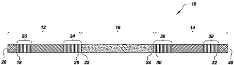
Figure 1 is a schematic diagram of a capture probe according to the present
invention;
Figure 2 through Figure 6 show diagrams of some of the steps in certain
embodiments
of a method for isolating miRNAs and the method for identifying miRNAs,
according to the
present invention; and
Figure 7 shows a sequence trace of the miRNA isolated according to the present
invention compared to a reference sequence of human miRNA.
DESCRIPTION
According to one embodiment of the present invention, there is provided a
method for
isolating microRNAs (miRNAs). According to another embodiment of the present
invention,
there is provided a method for identifying miRNAs. In one embodiment, the
method for
identifying miRNAs comprises, first, isolating the miRNAs according to the
present
invention. According to another embodiment of the present invention, there is
provided one
or more than one capture probe and one or more than one set of capture probes,
suitable for
use with a method for isolating miRNAs. In one embodiment, the method for
isolating
miRNAs is a method according to the present invention. The method and capture
probes will
now be disclosed in detail.
As used in this disclosure, except where the context requires otherwise, the
term
"comprise" and variations of the term, such as "comprising," "comprises" and
"comprised"
are not intended to exclude other additives, components, integers or steps.
As used in this disclosure, the term "miRNAs" means a naturally occurring,
single
stranded polyribonucleotide (polyRNA) of between 18 and 24 RNA residues, which
is
derived from a larger, naturally occurring noncoding eukaryotic precursor RNA
(usually
having a `hairpin' configuration).
As used in this disclosure, the terms "one or more than one miRNAs," "an
miRNA"
CA 02613136 2007-12-20
WO 2007/024653 PCT/US2006/032264
13
and "the miRNA" are intended to be synonymous, that is, are intended to
indicate either one
miRNA of interest or a plurality of miRNA of interest, except where the
context requires
otherwise.
As used in this disclosure, the terms "one or more than one capture probe," "a
capture probe, " the capture probe" and "the capture probes" are intended to
be
synonymous, that is, are intended to indicate either one capture probe or a
plurality of
capture probes, except where the context requires otherwise.
As used in this disclosure, the terms "a first linker," "the first linker" and
"the first
linkers" are intended to be synonymous, that is, are intended to indicate
either one first linker
or a plurality of first linkers, except where the context requires otherwise.
As used in this disclosure, the terms "a second linker," "the second linker"
and "the
second linkers" are intended to be synonymous, that is, are intended to
indicate either one
second linker or a plurality of second linkers, except where the context
requires otherwise.
As used in this disclosure, the term "substantially complementary" and
variations of
the term, such as "substantial complement," means that at least 90 % of all of
the consecutive
residues in a first strand are complementary to a series of consecutive
residues of the same
length of a second strand. As will be understood by those with skill in the
art with reference
to this disclosure, one strand can be shorter than the other strand and still
be substantially
complementary. With respect to the invention disclosed in this disclosure, for
example, the
first adapter segment can be shorter than the first linker and still be
substantially
complementary to the first linker, and the second adapter segment can be
shorter than the
second linker and still be substantially complementary. The miRNA binding
segment can be
the same length or longer than the miRNA of interest.
As used in this disclosure, the term "hybridize" and variations of the term,
such as
"hybridizes" and "hybridized," means a Watson-Crick base pairing of
complementary
nucleic acid single strands or segments of strands to produce an anti-
parallel, double-stranded
nucleic acid, and as used in this disclosure, hybridization should be
understood to be between
substantially complementary strands unless specified otherwise, or where the
context requires
otherwise. As an example, hybridization can be accomplished by combining equal
molar
concentrations of each of the pairs of single strands, such as 100 pmoles, in
the presence of 5
ug yeast tRNA in a total volume of 50 l of aqueous buffer containing 400 mM
MOPS, 80
mM DTT, and 40 mM MgCl2 a pH of 7.3, and then incubating the mixture at 25 C
for
CA 02613136 2007-12-20
WO 2007/024653 PCT/US2006/032264
14
one hour while shaking gently.
As used in this disclosure, the term "near the end" and variations of the
term, means
within 20 % of the residues of the identified end residue. For example, near
the end of a 20
residue strand, means the first four residues of the identified end of the
strand.
According to one embodiment of the present invention, there is provided a
capture
probe suitable for use with a method for isolating miRNAs according to the
present
invention. Referring now to Figure 1, there is shown a schematic diagram of a
capture probe
according to one embodiment of the present invention. The capture probe 10,
and each of
its segments, comprises a substance selected from the group consisting of one
or more than
10 one type of polynucleotide, including ribonucleotides and deoxynucleotides,
one or more than
one type of polynucleotide analog, and a combination of one or more than one
type of
polynucleotide and polynucleotide analog. As can be seen in Figure 1, the
capture probe 10
comprises three segments: a) a first adapter segment 12 having a first adapter
segment
sequence, b) a second adapter segment 14 having a second adapter segment
sequence, and c)
an miRNA binding segment 16 having an miRNA binding segment sequence, where
the
miRNA binding segment 16 is between the first adapter segment 12 and the
second adapter
segment 14.
According to one embodiment of the present invention, there is provided a set
of
capture probes comprising at least one capture probe comprising an miRNA
binding segment
that is substantially complementary to, and capable of hybridizing to, each
miRNA listed in a
single public database.
According to one embodiment of the present invention, there is provided a
plurality of
capture probes, where each capture probe of the plurality of capture probes
comprises
identical' first adapter segment sequences, where each capture probe of the
plurality of capture
probes comprises identical miRNA binding segment sequences, and where each
capture probe
of the plurality of capture probes comprises identical second adapter segment
sequences.
According to one embodiment of the present invention, there is provided a set
of
capture probes comprising a first capture probe and a second capture probe,
where the first
capture probe and the second capture probe have identical first adapter
segment sequences,
where the first capture probe and the second capture probe have identical
miRNA binding
segment sequences, and where the first capture probe has a second adapter
segment sequence
that is different from the second adapter segment sequence of the second
capture probe.
CA 02613136 2007-12-20
WO 2007/024653 PCT/US2006/032264
According to one embodiment of the present invention, there is provided a set
of
capture probes comprising a first capture probe and a second capture probe,
where the first
capture probe and the second capture probe have identical first adapter
segment sequences,
where the first capture probe and the second capture probe have identical
second adapter
5 segment sequences, and where the first capture probe has an miRNA binding-
segment
sequence that is different from the miRNA binding segment sequence of the
second capture
probe.
According to one embodiment of the present invention, there is provided a set
of
capture probes comprising a first capture probe and a second capture probe,
where the first
10 capture probe and the second capture probe have identical miRNA binding
segment
sequences, where the first capture probe and the second capture probe have
identical second
adapter segment sequences, and where the first capture probe has a first
adapter segment
sequence that is different from the first adapter segment sequence of the
second capture
probe.
15 According to one embodiment of the present invention, there is provided a
set of
capture probes comprising a first capture probe and a second capture probe,
where the first
capture probe and the second capture probe have identical first adapter
segment sequences,
and where the first capture probe has an miRNA binding segment sequence that
is different
from the miRNA binding segment sequence of the second capture probe, and where
the first
capture probe has a second adapter segment sequence that is different from the
second adapter
segment sequence of the second capture probe.
According to one embodiment of the present invention, there is provided a set
of
capture probes comprising a first capture probe and a second capture probe,
where the first
capture probe and the second capture probe have identical miRNA binding
segment
sequences, where the first capture probe has a first adapter segment sequence
that is different
from the first adapter segment sequence of the second capture probe, and where
the first
capture probe has a second adapter segment sequence that is different from the
second adapter
segment sequence of the second capture probe.
According to one embodiment of the present invention, there is provided a set
of
capture probes comprising a first capture probe and a second capture probe,
where the first
capture probe and the second capture probe have identical second adapter
segment sequences,
where the first capture probe has a first adapter segment sequence that is
different from the
CA 02613136 2007-12-20
WO 2007/024653 PCT/US2006/032264
16
first adapter segment sequence of the second capture probe, and where the
first capture probe
has an miRNA binding segment sequence that is different from the miRNA binding
segment
sequence of the second capture probe.
According to one embodiment of the present invention, there is provided a set
of
capture probes comprising a first capture probe and a second capture probe,
where the first
capture probe has a first adapter segment sequence that is different from the
first adapter
segment sequence of the second capture probe, where the first capture probe
has an miRNA
binding segment sequence that is different from the miRNA binding segment
sequence of the
second capture probe, and where the first capture probe has a second adapter
segment
sequence that is different from the second adapter segment sequence of the
second capture
probe.
Referring again to Figure 1, the first adapter segment 12 comprises a 3' end
18 and a
5' end 20. As can be seen in Figure 1, the 5' end 20 of the first adapter
segment 12 is
connected to the 3' end 22 of the miRNA binding segment 16, that is the first
adapter
segment 12 is connected upstream of the miRNA binding segment 16. In one
embodiment,
the first adapter segment 12 is substantially complementary to and capable of
hybridizing a
first linker probe designated in this disclosure as a "first linker." When
used in the method
of the present invention, the first adapter segment 12 facilitates the
ligation of the 3' end of
the first linker to the 5' end of the miRNA of interest by aligning the first
linker in position
for ligation to the miRNA of interest.
In one embodiment, the first adapter segment has a number of residues between
5 and
50. In another embodiment, the first adapter segment has a number of residues
between 5
and 20. In another embodiment, the first adapter segment has a number of
residues between
6 and 16.
In one embodiment, the first adapter segment 12 comprises one or more than one
sequence 24 or sequence 26 that is a restriction site motif. In a particularly
preferred
embodiment, the specific restriction site motif, when present, is not present
in the DNA
analog of the miRNA of interest that is being isolated and identified by the
present method.
In one embodiment, the restriction site motif is acted upon by a restriction
enzyme selected
from the group consisting of BamH I, Hind III and EcoR I. In a preferred
embodiment, the
restriction site motif is acted upon by a restriction enzyme selected from the
group consisting
of Not I, Xho I, Xma I and Nhe I, because BamH I, Hind III and EcoR I also act
upon some
CA 02613136 2007-12-20
WO 2007/024653 PCT/US2006/032264
17
sequences of miRNA. As will be understood by those with skill in the art with
reference to
this disclosure, however, other suitable restriction site motifs can also be
used.
In another embodiment, the first adapter segment 12 comprises a sequence 24 or
a
sequence 26 that is a polynucleotide synthesis promoter motif for a
polynucleotide
polymerase, or that is complementary to a polynucleotide synthesis promoter
motif for a
polynucleotide polymerase. In a preferred embodiment, the polynucleotide
synthesis
promoter motif is a motif for a polynucleotide synthesis promoter selected
from the group
consisting of T7, SP6, a T3 DNA dependent RNA polymerase, a type 2 RNA
polymerase of
E. coli and single stranded DNA dependent N4 RNA polymerase. The
polynucleotide
synthesis promoter motif can be a motif for any other suitable polynucleotide
synthesis
promoter, however, as will be understood by those with skill in the art with
reference to this
disclosure.
As will be understood by those with skill in the art with reference to this
disclosure,
the sequence that is a restriction site motif of the first adapter segment 12
can be in either
position 24 or in the position 26 as indicated in Figure 1, and the sequence
that is a
polynucleotide synthesis promoter motif can be in either position 24 or in the
position 26 as
indicated in Figure 1. In a preferred embodiment, there is no other a
restriction site motif
sequence of the first adapter segment 12 other than in the position 24 or in
the position 26 as
shown in Figure 1.
In another embodiment, the first adapter segment 12 comprises a solid phase
binding
group 28 to immobilize the capture probe 10 to a solid phase. In one
embodiment, the solid
phase binding group 28 is at or near the 3' end 18 of the first adapter
segment 12, however,
as will be understood by those with skill in the art with reference to this
disclosure, the solid
phase binding group 28 can be anywhere on the capture probe 10 other than at
or near the 3'
end 18 of the first adapter segment 12. In one embodiment, the solid phase
binding group 28
immobilizes the capture probe 10 to a solid phase covalently. In another
embodiment, the
solid phase binding group 28 immobilizes the capture probe 10 to a solid phase
non-
covalently. In one embodiment, the solid phase binding group 28 immobilizes
the capture
probe 10 to a solid phase reversibly. As used in this context, "reversibly"
means that the
solid phase binding group 28 immobilizes the capture probe 10 to a solid phase
in such a way
that the solid phase binding group 28 can be disassociated from the solid
phase without
destruction of the capture probe 10 and without disruption of hybridization
between the
CA 02613136 2007-12-20
WO 2007/024653 PCT/US2006/032264
18
capture probe 10 and the ligated first linker 48-miRNA of interest 42-second
linker 50 (as
disclosed below). In another embodiment, the solid phase binding group 28
immobilizes the
capture probe 10 to a solid phase non-reversibly. For example, in one
embodiment, the solid
phase binding group 28 immobilizes the capture probe. 10 to a solid phase non-
covalently and
reversibly, where the solid phase binding group 28 comprises biotin or an
analog of biotin
capable of binding with avidin or streptavidin or functional analogs of avidin
or streptavidin
with high affinity, such as with an affinity having an affinity constant of
between about 10e12
and 10e20. Additionally for example, in one embodiment the solid phase binding
group 28 of
the first adapter segment 12 immobilizes the capture probe 10 to a solid phase
covalently and
non-reversibly, where the solid phase binding group 28 comprises a terminal 5'
primary
amino group at the 3' end 18 of the first adapter segment 12 for coupling to a
solid phase
surface having free carboxyl groups using standard carbodiimide chemistry, as
will be
understood by those with skill in the art with reference to this disclosure.
Further, as will be
understood by those with skill in the art with reference to this disclosure,
any solid phase
binding group 28 present in the first adapter segment 12, and any technique
for coupling the
solid phase binding group 28 to a solid phase used in connection with the
present method
should not interfere with the hybridization and capture of the miRNA of
interest to the
miRNA binding segment 16, or with any other step of the present method.
By way of example only, in one embodiment the first adapter segment 12
comprises
DNA and has a first adapter segment, sequence in the 5' to 3' direction of
ATTTAGGTGACACTATAG, SEQ ID NO:1.
The second adapter segment 14 comprises a 3' end 30 and a 5' end 32. As can be
seen in Figure 1, the 3' end 30 of the second adapter segment 14 is connected
to the 5' end 34
of the miRNA binding segment 16, that is the second adapter segment 14 is
connected
downstream of the miRNA binding segment 16. In one embodiment, the second
adapter
segment 14 is substantially complementary to and capable of hybridizing a
second linker
probe designated in this disclosure as a "second linker." When used in the
method of the
present invention, the second adapter segment 14 facilitates the ligation of
the 5' end of the
second linker to the 3' end of the miRNA of interest by aligning the second
linker in position
for ligation to the miRNA of interest.
In one embodiment, the second adapter segment 14 has a number of residues
between
5 and 50. In another embodiment, the second adapter segment 14 has a number of
residues
CA 02613136 2007-12-20
WO 2007/024653 PCT/US2006/032264
19
between 5 and 20. In another embodiment, the second adapter segment 14 has a
number of
residues between 6 and 16.
In another embodiment, the second adapter segment 14 comprises one or more
than
one sequence 36 that is a restriction site motif. In a particularly preferred
embodiment, the
specific restriction site motif, when present, is not present in the DNA
analog of the miRNA
of interest that is being isolated and identified by the present methods. In
one embodiment,
the restriction site motif is acted upon by a restriction enzyme selected from
the group
consisting of BamH I, Hind III and EcoR I. In a preferred embodiment, the
restriction site
motif is acted upon by a restriction enzyme selected from the group consisting
of Not I, Xho
I, Xma I and Nhe I, because BamH I, Hind III and EcoR I also act upon some
sequences of
miRNA. As will be understood by those with skill in the art with reference to
this disclosure,
however,, other suitable restriction site motifs can also be used.
In one embodiment, the one or more than one sequence 24 that is a restriction
site
motif is identical to the one or more than one sequence 36 that is a
restriction site motif. In
another embodiment, the one or more than one sequence 24 that is a restriction
site motif is
different from the one or more than one sequence 36 that is a restriction site
motif.
In one embodiment, the second adapter segment 14 comprises a sequence 38 that
is a
polynucleotide synthesis promoter motif for a polynucleotide polymerase, or
that is
complementary to a polynucleotide synthesis promoter motif for a
polynucleotide polymerase.
In a preferred embodiment, the polynucleotide synthesis promoter motif is a
motif for a
polynucleotide synthesis promoter selected from the group consisting of T7,
SP6, a T3 DNA
dependent RNA polymerase, a type 2 RNA polymerase of E. coli and single
stranded DNA
dependent N4 RNA polymerase. The polynucleotide synthesis promoter motif can
be a motif
for any other suitable polynucleotide synthesis promoter, however, as will be
understood by
those with skill in the art with reference to this disclosure.
As will be understood by those with skill in the art with reference to this
disclosure,
the sequence that is a restriction site motif of the second adapter segment 14
can be in either
position 36 or in the position 38 as indicated in Figure 1, and the sequence
that is a
polynucleotide synthesis promoter motif can be in either position 36 or in the
position 38 as
indicated in Figure 1. In a preferred embodiment, there is no other a
restriction site motif
sequence of the second adapter segment 14 other than in the position 36 or in
the position 38
as shown in Figure 1.
CA 02613136 2007-12-20
WO 2007/024653 PCT/US2006/032264
By way of example only, in one embodiment the second adapter segment 14
comprises
DNA and has a second adapter segment sequence in the 5' to 3' direction of
CCCTATAGTGAGTCGTATTA SEQ ID NO:2.
In another embodiment, the second adapter segment 14 comprises a solid phase
5 binding group 40 to immobilize the capture probe 10 to a solid phase. In one
embodiment,
the solid phase binding group 40 is at or near the 5' end 32 of the second
adapter segment 14,
however, as will be understood by those with skill in the art with reference
to this disclosure,
the solid phase binding group 40 can be anywhere on the capture probe 10 other
than at or
near the 5' end 32 of the second adapter segment 14. In one embodiment, the
solid phase
10 binding group 40 immobilizes the capture probe 10 to a solid phase
covalently. In another
embodiment, the solid phase binding group 40 immobilizes the capture probe 10
to a solid
phase non-covalently. In one embodiment, the solid phase binding group 40
immobilizes the
capture probe 10 to a solid phase reversibly. As used in this context,
"reversibly" means that
the solid phase binding group 40 immobilizes the capture probe 10 to a solid
phase in such a
15 way that the solid phase binding group 40 can be disassociated from the
solid phase without
destruction of the capture probe 10 and without disruption of hybridization
between the
capture probe 10 and the ligated first linker 48-miRNA of interest 42-second
linker 50 (as
disclosed below). In another embodiment, the solid phase binding group 40
immobilizes the
capture probe 10 to a solid phase non-reversibly. For example, in one
embodiment, the solid
20 phase binding group 40 immobilizes the capture probe 10 to a solid phase
non-covalently and
reversibly, where the solid phase binding group 40 comprises biotin or an
analog of biotin
capable of binding with avidin or streptavidin or functional analogs of avidin
or streptavidin
with high affinity, such as with an affinity having an affinity constant of
between about 10e12
and 10e20. Additionally for example, in one embodiment the solid phase binding
group 40 of
the second adapter segment 14 immobilizes the capture probe 10 to a solid
phase covalently
and non-reversibly, where the solid phase binding group 40 comprises a
terminal 3' primary
amino group at the 5' end 32 of the second adapter segment 14 for coupling to
a solid phase
surface having free carboxyl groups using standard carbodiimide chemistry, as
will be
understood by those with skill in the art with reference to this disclosure.
Further, as will be
understood by those with skill in the art with reference to this disclosure,
any solid phase
binding group 40 present in the second adapter segment 14, and any technique
for coupling
the solid phase binding group 40 to a solid phase used in connection with the
present method
CA 02613136 2007-12-20
WO 2007/024653 PCT/US2006/032264
21
should not interfere with the hybridization and capture of the miRNA of
interest to the
miRNA binding segment 16, or with any other step of the present method.
In another embodiment, both the first adapter segment 12 comprises a solid
phase
binding group 28, and the second adapter segment 14 comprises a solid phase
binding group
40. In another embodiment, both the first adapter segment 12 comprises a solid
phase
binding group 28 at or near the 3' end 18 of the first adapter segment 12, and
the second
adapter segment 14 comprises a solid phase binding group 40 at or near the 5'
end 32 of the
second adapter segment 14.
Referring again to Figure 1 and as stated above, the capture probe 10 of the
present
invention further comprises an miRNA binding segment 16. The miRNA binding
segment 16
has an miRNA binding segment sequence comprising a 3' end 22 and a 5' end 34,
and
consists of one or more than one type of polynucleotide, including
ribonucleotides and
deoxynucleotides, or one or more than one type of polynucleotide analog, or a
combination of
one or more than one type of polynucleotide and polynucleotide analog.. The 3'
end 22 of the
miRNA Vinding segment 16 is connected to the 5' end 20 of the first adapter
segment 12 of
the capture probe 10 according to the present invention, that is, the first
adapter segment 12
is connected upstream of the miRNA binding segment 16. The 5' end 34 of the
miRNA
binding segment 16 is connected to the 3' end 30 of the second adapter segment
14 of the
capture probe 10 according to the present invention, that is, the second
adapter segment 14 is
connected downstream of the miRNA binding segment 16.
In one embodiment, the miRNA binding segment consists of between 18 and 24 DNA
residues. In another embodiment, the miRNA binding segment 16 consists of 18
or 19 or 20
or 21 or 22 or 23 or 24 residues selected from the group consisting of DNA,
RNA, chimeric
DNA/RNA, DNA analogs and RNA analogs.
The miRNA binding segment 16 is substantially complementary to, and capable of
hybridizing to, one or more than one miRNA of interest by Watson-Crick base
pairing,
including an miRNA of interest having a predetermined sequence or having a
predetermined
size, from a sample. In one embodiment, the sample comprises substances that
are
chemically related, such as for example, a mixture of messenger RNAs, transfer
RNAs,
ribosomal RNAs and genomic DNA. An miRNA of interest can be selected from any
known
miRNAs from any suitable source, as will be understood by those with skill in
the art with
reference to this disclosure. In one embodiment, the miRNA of interest is
selected from a
WO 2007/024653 CA 02613136 2011-01-13 PCT/US2006/032264
22
public database. In a preferred embodiment, the central repository provided is
the Sanger
Institute to which newly discovered and previously
known miRNA sequences can be submitted for naming and nomenclature assignment,
as well
as placement of the sequences in a database for archiving and for online
retrieval via the
world wide-web. Generally, the data collected on the sequences of miRNAs by
the Sanger
Institute include species, source, corresponding genomic sequences and genomic
location
(usually chromosomal coordinates), as well as full length transcription
products and
sequences for the mature fully processed miRNA (miRNA with a 5' terminal
phosphate
group).
To select the sequence or sequences of the miRNA binding segment 16, an miRNA
of
interest, or set of miRNAs of interest is selected from a suitable source,
such as for example,
the Sanger Institute database or other suitable database, as will be
understood by those with
skill in the art with reference to this disclosure. If a set of miRNAs of
interest is selected
from one or more than one source that contains duplicate entries for one or
more than one
miRNA, in a preferred embodiment, the duplicated entries are first removed so
that the set of
sequences of miRNAs of interest contains only one sequence for each miRNA of
interest. In
one embodiment, the set of miRNAs of interest consists of one of each miRNA
from a single
source or database, including a public source or public database, such as one
of each miRNA
listed in the central repository provided by the Sanger Institute.
In another embodiment the miRNA of interest is a eucaryotic miRNA. In another
embodiment the miRNA of interest is a primate miRNA. In a preferred
embodiment, the
miRNA of interest is a human miRNA. In another embodiment, the miRNAs in the
set of
miRNAs of interest are all eucaryotic miRNAs. In another embodiment, the
miRNAs in the
set of miRNAs of interest are all primate miRNAs. In another embodiment, the
miRNAs in
the set of miRNAs of interest are all human miRNAs.
Next, the miRNA binding segment is selected to be the substantial complement
of the
miRNA of interest sequence. In a preferred embodiment, the miRNA binding
segment is the
exact complement to the miRNA of interest in both length and sequence. In
another
embodiment, the miRNA binding segment is more than 90 % complementary to a
segment of
the miRNA of interest of the same length as the miRNA of interest sequence. In
another
embodiment, the miRNA binding segment is more than 80 % complementary to a
segment of
the miRNA of interest of the same length as the miRNA of interest sequence.
CA 02613136 2007-12-20
WO 2007/024653 PCT/US2006/032264
23
In one embodiment, the miRNA binding segment 16 consists of RNA. In one
embodiment, the miRNA binding segment 16 consists of DNA. In one embodiment,
the
miRNA binding segment 16 consists of polynucleotide analogs. In one
embodiment, the
miRNA binding segment 16 consists of a chimera of more than one polynucleotide
or
polynucleotide analog selected from the group consisting of RNA, DNA,
polynucleotide
analogs of RNA, and polynucleotide analogs of DNA. Once, the miRNA binding
segment
sequence is selected, the miRNA binding segment 16 is synthesized according to
standard
synthesis techniques known to those with skill in the art, as will be
understood by those with
skill in the art with reference to this disclosure.
Table I provides a list of ten sample miRNA binding segments 16 which consist
of
DNA along with the miRNAs that are the exact complement of the miRNA binding
segments.
As will be understood by those with skill in the art with reference to this
disclosure, and as
disclosed in this disclosure, this is a sample list of miRNA binding segments
16, and any
other sequence serving the function of the miRNA binding segments will also be
useful,
including for example miRNA binding segments 16 that are the RNA of the miRNA
binding
segments 16 listed in Table I.
TABLE I
SEQ ID NO: miRNA binding segment sequence 5'-3' iRNA that is complementary
to miRNA binding segment
SEQ ID NO:3 ACTATACAACCTACTACCTCA hsa-let-7a
SEQ ID NO:4 ACCACACAACCTACTACCTCA hsa-let-7b
SEQ ID NO: 5 ACCATACAACCTACTACCTCA hsa-let-7c
SEQ ID NO:6 CTATGCAACCTCCTACCTCT hsa-let-7d
SEQ ID NO:7 CTATACAACCTCCTACCTCA hsa-let-7e-
SEQ ID NO:8 ACTATACAATCTACTACCTCA hsa-let-7f
SEQ ID NO:9 CTGTACAAACTACTACCTCA hsa-let-7g
SEQ ID NO:10 CAGCACAAACTACTACCTCA hsa-let-7i
SEQ ID NO:11 ACAAGTTCGGATCTACGGGTT hsa-miR-100
SEQ ID NO:12 TTCAGTTATCACAGTACTGTA hsa-miR-101
Therefore, by way of example only, a capture probe 10 according to the present
invention for use in a method for isolating miRNA hsa-let-7a, can have the
following
sequence in the 5' to 3' direction:
ATTTAGGTGACACTATAGAAACTATACAACCTACTACCTCACCCTATAGTGAGTCG
TATTA, SEQ ID NO:13.
According to one embodiment of the present invention, there is a set of
capture probes
CA 02613136 2007-12-20
WO 2007/024653 PCT/US2006/032264
24
suitable for use with a method for isolating miRNAs. Referring now to Table
II, in one
embodiment, by way of example, the set consists of at least seven capture
probes 10
according to the present invention, where each capture probe 10 has a first
adapter segment
12 ATTTAGGTGACACTATAG, SEQ ID NO: 1, a second adapter segment 14 of
5 CCCTATAGTGAGTCGTATTA SEQ ID NO:2, and an miRNA binding segment 16 varying
from 18 mer to 24 mer, and having a nucleotide or nucleotide analog (N) (such
as for
example A, G, C, T as ribonucleotides or deoxynucleotides) capable of
hybridizing with a
nucleotide on an miRNA. In a preferred embodiment, as shown, the 5' end 32 of
the second
adapter segment 14 is biotinylated to bind to a solid phase.
10 TABLE II
SEQ ID NO: Capture Probe Sequence 5'-3' Size of miRNA
Captured
5'biotin-
SEQ ID NO:14 ATTTAGGTGACACTATAGNNNNNNNNNNNNN 18 mer
NNNNNCCCTATAGTGAGTCGTATTA
5'biotin-
SEQ ID NO:15 ATTTAGGTGACACTATAGNNNNNNNNNNNNN 19 mer
NNNNNNCCCTATAGTGAGTCGTATTA
5'biotin-
SEQ ID NO:16 ATTTAGGTGACACTATAGNNNNNNNNNNNNN 20 mer
NNNNNNNCCCTATAGTGAGTCGTATTA
5'biotin-
SEQ ID NO:17 ATTTAGGTGACACTATAGNNNNNNNNNNNNN 21 mer
NNNNNNNNCCCTATAGTGAGTCGTATTA
5'biotin-
SEQ ID NO:18 ATTTAGGTGACACTATAGNNNNNNNNNNNNN 22 mer
NNNNNNNNNCCCTATAGTGAGTCGTATTA
5'biotin-
SEQ ID NO:19 ATTTAGGTGACACTATAGNNNNNNNNNNNNN 23 mer
NNNNNNNNNNCCCTATAGTGAGTCGTATTA
5'biotin-
SEQ ID NO:20 ATTTAGGTGACACTATAGNNNNNNNNNNNNN 24 mer
NNNNNNNNNNNCCCTATAGTGAGTCGTATTA
The capture probe 10 of the present invention can be synthesized according to
standard techniques, as will be understood by those with skill in the art with
reference to this
CA 02613136 2007-12-20
WO 2007/024653 PCT/US2006/032264
disclosure. In one embodiment, the capture probe 10 is synthesized as a
contiguous single
sequence for each miRNA of interest to be isolated and detected. In a
preferred embodiment,
there is provided a set of capture probes 10 comprising a first capture probe
and a second
capture probe that are synthesized separately, where the sequence of the first
capture probe
5 has one or more than one difference with the sequence of the second capture
probe, and
where the set of capture probes 10 is produced by mixing the first capture
probe and the
second capture probe after they are synthesized.
In one embodiment, the capture probes 10 are synthesized by combining the
sequence
text strings for the first adapter segment 12, the miRNA binding segment 16,
and the second
10 adapter segment 14 in a database or spreadsheet to generate a capture probe
10 sequence, and
then synthesizing the capture probe 10 according to standard techniques, as
will be
understood by those with skill in the art with reference to this disclosure.
In one
embodiment, the capture probes 10 are designed for use in a method according
to the present
invention, and then purchased from a vendor of polynucleotide or
polynucleotide analog
15 sequences, such as for example, from Integrated DNA Technologies
(Coralville, IA US) or
Invitrogen Corp. (Carlsbad, CA US).
According to another embodiment of the present invention, there is provided a
method
for isolating an miRNA (microRNA) of interest from a sample comprising the
miRNA of
interest. According to another embodiment of the present invention, there is
provided a
20 method for identifying miRNAs. In one embodiment, the method for
identifying miRNAs
comprises, first, isolating the miRNAs according to the present invention.
Referring now to
Figure 2 through Figure 6, there are shown some of the steps in certain
embodiments of the
methods. The steps shown are not intended to be limiting nor are they intended
to indicate
that each step depicted is essential to the method, but instead are exemplary
steps only.
25 As can be seen, the method comprises, first, providing a sample comprising
an
miRNA of interest 42. In one embodiment, the sample further comprises one or
more than
one substance that is chemically related to the miRNA of interest 42, such as
for example, a
substance selected from the group consisting of messenger RNA, transfer RNA,
ribosomal
RNA, siRNA, 5S/5.8SrRNA, genomic DNA and a combination of the preceding. In
one
embodiment, the sample further comprises one or more than one RNA other than
miRNA,
such as for example, a substance selected from the group consisting of
messenger RNA,
transfer RNA, ribosomal RNA, siRNA, 5S/5.8SrRNA and a combination of the
preceding.
CA 02613136 2011-01-13
WO 2007/024653 PCT/US2006/032264
26
All of the RNA in the sample, regardless of the type of RNA, constitutes the
"total RNA" in
the sample.
In one embodiment, the sample is from a eukaryote. In another embodiment, the
sample is from a primate. In a preferred embodiment, the sample is from a
human.
In one embodiment, the sample comprises a tissue or fluid selected from the
group
consisting of blood, brain, heart, intestine, liver, lung, pancreas, muscle, a
leaf, a flower, a
plant root and a plant stem.
The miRNA of interest 42 has an miRNA of interest sequence, and comprises 3'
end
44 and a 5' end 46. In one embodiment, the miRNA of interest consists of
between 18 and
24 RNA residues. In another embodiment, the miRNA of interest consists of 18
or 19 or 20
or 21 or 22'or 23 or 24 RNA residues.
The miRNA of interest 42 is substantially complementary to, and capable of
hybridizing to, an miRNA binding segment 16 of a capture probe 10 according to
the present
invention by Watson-Crick base pairing. In one embodiment, the miRNA of
interest 42 is
listed in a public database. In a preferred embodiment, the public database is
a central
repository provided by the Sanger Institute to which
miRNA sequences are submitted for naming and nomenclature assignment, as well
as
placement of the sequences in a database for archiving and for online
retrieval via the world
wide web. Generally, the data collected on the sequences of miRNAs by the
Sanger Institute
include species, source, corresponding genomic sequences and genomic location
(chromosomal coordinates), as well as full length transcription products and
sequences for the
mature filly processed miRNA (miRNA with a 5' terminal phosphate group).
In one embodiment, the sample provided comprises a plurality of miRNAs of
interest
42, where each of the plurality of miRNAs of interest 42 has miRNA of interest
sequences
that are identical to one another. In one embodiment, the sample provided
comprises a
plurality of miRNAs of interest 42, where at least two of the plurality of
miRNAs of interest
42 have miRNA of interest sequences that are different from one another. In
one
embodiment, the sample provided comprises a plurality of miRNAs of interest 42
comprising
a first miRNA of interest having a first miRNA of interest sequence, and a
second miRNA of
interest having a second miRNA of interest sequence, where the first miRNA of
interest
sequence is different from the second miRNA of interest sequence. In another
embodiment,
the sample provided comprises a plurality of miRNAs of interest 42 comprising
a first
CA 02613136 2007-12-20
WO 2007/024653 PCT/US2006/032264
27
miRNA of interest having a first miRNA of interest sequence, a second miRNA of
interest
having a second miRNA of interest sequence, and a third miRNA of interest
having a third
miRNA of interest sequence, where the first miRNA of interest sequence is
different from the
second miRNA of interest sequence, where the first miRNA of interest sequence
is different
from the third miRNA of interest sequence, and where second miRNA of interest
sequence is
different from the third miRNA of interest sequence.
In one embodiment, the method further comprises isolating the total RNA from
the
sample after providing the sample. In one embodiment, isolating the total RNA
is
accomplished according to techniques well known to those with skill in the
art, such as for
example using a commercially available kit for the isolation of total RNA
available from
Ambion, Inc. (Austin, TX US), Invitrogen Corp. and Qiagen, Inc. (Valencia, CA
US),
among others, as will be understood by those with skill in the art with
reference to this
disclosure. As will be understood by those with skill in the art with
reference to this
disclosure, when the method comprises isolating the total RNA from the sample
after
providing the sample, the term "sample" means the isolated total RNA for the
remaining
steps in the method.
Next, the method further comprises providing a capture probe 10. In one
embodiment, the capture probe 10 provided is a capture probe 10 according to
the present
invention. When the capture probe 10 is a capture probe 10 according to the
present
invention, in all respects, the capture probe 10 provided has the
characteristics and attributes
as disclosed for a capture probe 10 according to the present invention, some
of which will be
repeated hereafter for clarity. The capture probe 10 has a capture probe
sequence, and
comprises three segments: a) a first adapter segment 12 having a first adapter
segment
sequence,' and comprising a 3' end 18 and a 5' end 20; b) a second adapter
segment 14 having
a second adapter segment sequence, and comprising a 3' end 30 and a 5' end 32;
and c) an
miRNA binding segment 16 having an miRNA binding segment sequence, and
comprising a
3' end 22 and a 5' end 34, where the 5' end 20 of the first adapter segment 12
is connected to
the 3' end 22 of the miRNA binding segment 16, and where the 5' end of the
miRNA binding
segment 34 is connected to the 3' end 30 of the second adapter segment 14. The
specificity
of the miRNA binding segment 16 to the miRNA of interest 42 allows the method
to be used
directly on a sample containing substances related to miRNA or on isolated
total RNA
without requiring the specific separation of miRNAs from the sample or from
the total RNA,
CA 02613136 2007-12-20
WO 2007/024653 PCT/US2006/032264
28
such as for example by either gel purification or chromatographic
purification, as necessary
in prior art methods.
In one embodiment, the capture probe 10 provided is a set of capture probes,
where
each of the set of capture probes provided have capture probe sequences that
are identical to
one another. In one embodiment, the capture probe 10 provided is a set of
capture probes,
where at least two capture probes of the set of capture probes have capture
probe sequences
that are different from one another. In another embodiment, the capture probe
10 is a set of
capture probes comprising a first capture probe having a first capture probe
sequence, and a
second capture probe having a second capture probe sequence, where the first
capture probe
sequence is different from the second capture probe sequence. In another
embodiment, the
capture probe 10 provided is a set of capture probes comprising a first
capture probe having a
first capture probe sequence, a second capture probe having a second capture
probe sequence,
and a third capture probe having a third capture probe sequence, where the
first capture probe
sequence is different from the second capture probe sequence, where the first
capture probe
sequence is different from the-third capture probe sequence, and where second
capture probe
sequence is different from the third capture probe sequence.
Then, the method comprises providing a first linker 48 and a second linker 50.
In one
embodiment, the first linker segment and the second linker segment comprise a
substance
selected from the group consisting of one or more than one type of
polynucleotide, including
ribonucleotides and deoxynucleotides, one or more than one type of
polynucleotide analog,
and a combination of one or more than one type of polynucleotide and
polynucleotide analog.
In one embodiment, the first linker 48, or the second linker 50, or both the
first linker 48 and
the second linker 50 are resistant to nuclease degradation. In a preferred
embodiment, the
first linker 48, or the second linker 50, or both the first linker 48 and the
second linker 50
comprise nuclease resistant nucleotides. In another preferred embodiment, the
first linker 48,
or the second linker 50, or both the first linker 48 and the second linker 50
comprise
nucleotides with a phosphothioate backbone that render the first linker 48, or
the second
linker 50, or both the first linker 48 and the second linker 50 resistant to
nuclease
degradation. In another preferred embodiment, the first linker 48, or the
second linker 50, or
both the first linker 48 and the second linker 50 comprise both nuclease
resistant nucleotides
and nucleotides with a phosphothioate backbone that render the first linker
48, or the second
linker 50, or both the first linker 48 and the second linker 50 resistant to
nuclease
CA 02613136 2007-12-20
WO 2007/024653 PCT/US2006/032264
29
degradation.
The first linker 48 has a first linker sequence, and comprises a 3' end 52 and
a 5' end
54. The first linker 48 is substantially complementary to, and capable of
hybridizing to, the
first adapter segment 12 of a capture probe 10 according to the present
invention by Watson-
Crick base pairing. The second linker 50 has a second linker sequence, and
comprises a 3'
end 56 and a 5' end 58. The second linker 50 is substantially complementary
to, and capable
of hybridizing to, the second adapter segment 14 of a capture probe 10
according to the
present invention by Watson-Crick base pairing. The first linker 48 and the
second linker 50
each comprises between 6 and 50 residues.
In a preferred embodiment, the first linker 48 comprises at least 10 residues,
and at
least 10 residues at the 3' end 52 of the first linker 48 are exactly the
complement of the
corresponding residues at or near the 5' end 20 of the first adapter segment
12. In another
embodiment, the second linker 50 comprises at least 10 residues, and at least
10 residues at
the 5' end 58 of the second linker 50 are exactly the complement of the
corresponding
residues at or near the 3' end 30 of the second adapter segment 14.
In a preferred embodiment, the 3' end 52 of the first linker 48 is capable of
being
ligated to the 5' end 46 of the miRNA of interest 42 by a suitable ligase,
such. as for example
T4 polynucleotide ligase, or by another suitable chemical reaction.
In a preferred embodiment, the 5' end 58 of the second linker 50 is a
nucleotide with a
5' pyrophosphate bond between it and its adjacent 5' end nucleotide of the
second adapter
segment 14 to allow ligation of the 5' end 58 of the second linker 50 to the
3' end 44 of the
miRNA of interest 42 by a suitable ligase, such as for example T4
polynucleotide ligase, or
by another suitable chemical reaction. In a- preferred embodiment, the 5' end
58 of the
second linker 50 additionally comprises a 5'pyrophosphate adenosine.
In one embodiment, the 5' end 54 of the first linker 48, or the 3' end 56 of
the second
linker 50, or both the 5' end 54 of the first linker 48 and the 3' end 56 of
the second linker 50
comprise a label, such as for example a fluorescent dye, to facilitate
detection, as will be
understood by those with skill in the art with reference to this disclosure.
Further, the first
linker 48, or the second linker 50, or both the first linker 48 and the second
linker 50 can
comprise a label, such as for example a fluorescent dye, to facilitate
detection at a position
other than at the 5' end 54 of the first linker 48, or the 3' end 56 of the
second linker 50, as
long as the presence of the label does not interfere with other steps of the
present method, as
CA 02613136 2007-12-20
WO 2007/024653 PCT/US2006/032264
will be understood by those with skill in the art with reference to this
disclosure.
In one embodiment, the 5' end 54 of first linker 48 comprises one or more than
one
residue that extends beyond the 3' end 18 of the first adapter segment 12
after the first linker
48 hybridizes to the first adapter segment 1.2. In one embodiment, the one or
more than one
5 residue of the 5' end 54 of first linker 48 that extends beyond the 3' end
18 of the first
adapter segment 12 functions as a primer binding site that allows the first
linker 48 to be
distinguished from the second linker 50 in downstream amplification reactions.
For example,
in one embodiment, the first linker 48 comprises, from the 5' end 54 to the 3'
end 52 of first
linker 48, a T3 promoter sequence, a short spacer sequence and a T7 promoter
sequence,
10 while the nucleotide residues of the first adapter segment 12 consist of
the substantial
complement of the T7 promoter sequence only.
In one embodiment, the 3' end 56 of second linker 50 comprises one or more
than one
residue that extends beyond the 5' end 20 of the second adapter segment 14
after the second
linker 50 hybridizes to the second adapter segment 14. In one embodiment, the
one or more
15 than one residue of the 3' end 56 of second linker 50 that extends beyond
the 5' end 32 of the
second adapter segment 14 functions as a primer binding site that allows the
second linker 50
to be distinguished from the first linker 48 in downstream amplification
reactions. For
example, in one embodiment, the second linker 50 comprises, from the 3' end 56
to the 5'
end 58 of second linker 50, a T3 promoter sequence, a short spacer sequence
and an SP6
20 promoter sequence, while the nucleotide residues of the second adapter
segment 14 consist of
the substantial complement of the SP6 promoter sequence only.
After the first linker sequence of the first linker 48, and the second linker
sequence of
the second linker 50 are designed, the first linker 48 and the second linker
50 can be
synthesized according to standard techniques, as will be understood by those
with skill in the
25 art with reference to this disclosure. Alternately, the first linker 48 and
the second linker 50
can be purchased from a vendor of polynucleotide or polynucleotide analog
sequences, such
as for example, from Integrated DNA Technologies or Invitrogen Corp.
Referring now to Figure 2, the method then -comprises combining the capture
probe
10, the first linker segment 48, the second linker segment 50 and the sample,
represented in
30 Figure 2 by the miRNA of interest 42. In a preferred embodiment, the method
comprises
combining the sample, the capture probe 10, the first linker 48 and the second
linker 50 in a
solution. The capture probe 10, the first linker 48, the second linker 50 and
the sample can
CA 02613136 2007-12-20
WO 2007/024653 PCT/US2006/032264
31
be combined simultaneously, or sequentially in any order, as will be
understood by those with
skill in the art with reference to this disclosure. For example, the capture
probe 10 is
combined with the sample first, and then the capture probe 10 and sample are
combined with
the first linker 48 and second linker 50; or alternately for example, the
capture probe 10, first
linker 48 and second linker 50 are combined first, and then the capture probe
10, first linker
48 and second linker 50 are combined with the sample; or alternately for
example, the first
linker 48 and the second linker 50 are combined with the sample first, and
then the capture
probe 10 is combined with the first linker 48, second linker 50 and the
sample.
In one embodiment, combining the capture probe 10, the first linker 48, the
second
linker 50 and the sample comprises combining approximately equimolar amounts
of the
capture probe 10, the first linker 48 and the second linker 50. In another
embodiment,
combining the capture probe 10, the first linker 48, the second linker 50 and
the sample
comprises combining approximately equimolar amounts of the capture probe 10,
the first
linker 48 and the second linker 50 with an amount of sample expected to
contain
approximately one tenth the molar amount of miRNA of interest 42 as of the
capture probe
10, the first linker 48 and the second linker 50. In another embodiment,
combining the
capture probe 10, the first linker 48, the second linker 50 and the sample
comprises
combining approximately equimolar amounts of the capture probe 10, the first
linker 48 and
the second linker 50 with an amount of sample expected to contain
approximately one half
and one tenths and the molar amount of miRNA of interest 42 as of the capture
probe 10, the
first linker 48 and the second linker 50. In one embodiment, combining the
capture probe
10, the first linker 48, the second linker 50 and the sample comprises
combining the sample
with between 0.1 pmoles and 100 pmoles/ l each of the capture probe 10, the
first linker 48
and the second linker 50 in a suitable buffer to create a solution comprising
the capture probe
10, the first linker 48, the second linker 50 and the sample. In a preferred
embodiment, the
buffer is selected from the group consisting of 1X TE buffer in 0.1-1 M sodium
chloride, and
0.1M MOPS in 1 mM EDTA and 100 mM sodium chloride. As will be understood by
those
with skill in the art with reference to this disclosure, the pH selected for
the buffer will be
one that optimizes the intended reactions. In general, the pH selected will be
between 6 and
8, preferably between 6.4 and 7.4 and more preferably, near 7Ø In a
preferred
embodiment, the method further comprises adding one or more than one RNAse
inhibitor to
the combination of the sample, the capture probe 10, the first linker 48, the
second linker 50,
CA 02613136 2007-12-20
WO 2007/024653 PCT/US2006/032264
32
such as for example an RNAase inhibitor selected from the group consisting of
lithium
dodecylsulfate (LiDS), the ammonium salt of tricarboxylic acid and sodium salt
of aurine
tricarboxylic acid.
Referring now to Figure 3, after combining the capture probe 10, the first
linker 48,
the second linker 50 and the sample, the method comprises allowing the first
linker 48 to
hybridize with the first adapter segment 12, the miRNA of interest 42 to
hybridize with the
miRNA binding segment 16, and the second linker 50 to hybridize with the
second adapter
segment 14, thereby binding the first linker 48, the miRNA of interest 42, and
the second
linker 50 to the capture probe 10. In one embodiment, allowing the first
linker 48 to
hybridize with the first adapter segment 12, the miRNA of interest 42 to
hybridize with the
miRNA binding segment 16, and the second linker 50 to hybridize with the
second adapter
segment 14 comprises incubating the solution comprising first linker 48, the
second linker 50,
the capture probe 10 and the sample for between 1 minute and 60 minutes at
between 25 C
and 50'C until substantially all of the miRNA of interest 42 has hybridized to
the capture
probes 10.
In one embodiment, the first adapter segment 12 comprises a solid phase
binding
group 28, or the second adapter segment 14 comprises a solid phase binding
group 40, or
both the first adapter segment 12 comprises a solid phase binding group 28 and
the second
adapter segment 14 comprises a solid phase binding group 40, as disclosed in
this disclosure,
and the method further comprises binding the capture probe 10 to a solid phase
(not shown)
before or after combining the capture probe 10, the first linker 48, the
second linker 50 and
the sample. In one embodiment, the solid phase is a plurality of beads where
each bead has a
diameter of between 0.01 and 5 , though the solid phase can be any suitable
solid phase as
will be understood by those with skill in the art with reference to this
disclosure. For
example, in one embodiment, the solid phase binding group comprises biotin,
and the solid
phase is paramagnetic particles having a diameter of 1.0 and comprising
streptavidin
immobilized to the surface of the particles.
In one embodiment, the capture probes 10 are bound to a solid phase through
the first
adapter segment 12 or through the second adapter segment 14 or through both
the first
adapter segment 12 and the second adapter segment 14, and the method further
comprises
hybridizing the miRNA of interest 42 to the miRNA binding segment 16 of the
capture
probes 10 bound to the solid phase, and then incubating the capture
probes/miRNA of
CA 02613136 2007-12-20
WO 2007/024653 PCT/US2006/032264
33
interest-bound to the solid phase with the first linker 48 and second linker
50 under
conditions sufficient to hybridize the first linker 48 to the first adapter
segment 12 of the
capture probe 10 and the second linker 50 to the second adapter segment 14 of
the capture
probe 10,
In a preferred embodiment, the first linker 48 hybridizes to the first adapter
segment
12 at a position where the last residue on the 3' end 52 of the first linker
48 hybridizes to a
residue on the first adapter segment 12 that is between 1 residue and 5
residues from the 3'
end 22 of the miRNA binding segment 16. In a particularly preferred
embodiment, the first
linker 48 hybridizes to the first adapter segment 12 at a position where the
last residue on the
3' end 52 of the first linker 48 hybridizes to a residue on the first adapter
segment 12 that is
immediately adjacent to the 3' end 22 of the miRNA binding segment 16.
In a preferred embodiment, the second linker 50 hybridizes to the second
adapter
segment 14 at a position where the last residue on the 5' end 58 of the second
linker 50
hybridizes to a residue on the second adapter segment 14 that is between 1
residue and 5
residues from the 5' end 34 of the miRNA binding segment 16. In a particularly
preferred
embodiment, the second linker 50 hybridizes to the second adapter segment 14
at a position
where the last residue on the 5' end 58 of the second linker 50 hybridizes to
a residue on the
second adapter segment 14 that is immediately adjacent to the 5' end 34 of the
miRNA
binding segment 16.
In one embodiment, the capture probe is bound to a solid phase through the
first
adapter segment 12 or through the second adapter segment 14 or through both
the first
adapter segment 12 and the second adapter segment 14, and the method further
comprises
purifying the capture probe 10 with hybridized first linker 48, miRNA of
interest 42 and
second linker 50-bound to the solid phase by removing non-hybridized first
linkers 48,
second linkers 50 and any other substances such as messenger RNAs, transfer
RNAs,
ribosomal RNAs and genomic DNA that are not bound to the solid phase. In one
embodiment, purifying comprises washing the capture probe 10 with hybridized
first linker
48, miRNA of interest 42 and second linker 50-bound to the solid phase with a
suitable
buffer, such as for example 1X TE buffer in 0.1-1 M sodium chloride (pH 6.4-
7.4,
preferably pH 6.8-7.2). In another embodiment, the solid phase comprises
paramagnetic
particles, the solid phase is contained in a vessel comprising a surface and a
cap, and
purifying comprises applying a magnetic field to attract the solid phase to
the surface of the
CA 02613136 2007-12-20
WO 2007/024653 PCT/US2006/032264
34
vessel or the cap of the vessel. In a preferred embodiment, after applying the
magnetic field
to attract the solid phase to the surface of the vessel or the cap of the
vessel, the method
comprises washing the capture probe 10 with hybridized first linker 48, miRNA
of interest 42
and second linker 50-bound to the solid phase with a suitable buffer, such as
for example
0.1 X TE buffer (pH 7.4).
Next, as show in Figure 4, the method comprises covalently ligating the 3' end
52 of
the first linker 48 that is hybridized to the first adapter segment 12 to the
5' end 46 of the
miRNA of interest 42 that is hybridized to the miRNA binding segment 16, and
then
covalently ligating the 3' end 44 of the miRNA of interest 42 that is
hybridized to the miRNA
binding segment 16 to the 5' end 58 of the second linker 50 that is hybridized
to the second
adapter segment 14. Ligation of the 3' end 52 of the first linker 48 to the 5'
end 46 of the
miRNA of interest 42, and ligation of the 3' end 44 of the miRNA of interest
42 to the 5' end
58 of the second linker 50 can be accomplished in any order, including
simultaneously or
sequentially. In one embodiment, the ligation is accomplished by standard
techniques, as will
be understood by those with skill in the art with reference to this
disclosure. In a preferred
embodiment, the ligation comprises treating the capture probe 10 with the
hybridized first
linker 48, miRNA of interest 42 and second linker 50 with a suitable ligase,
such as for
example T4 polynucleotide ligase in the presence of suitable buffer and
essential cofactors for
a sufficient time for the ligation to proceed to near total completion of
ligation. As will be
understood by those with skill in the art with reference to this disclosure,
the presence of the
first adapter segment 12 and the second adapter segment 14 in the capture
probe 10 facilitate
the ligation of the first linker 48 and the second linker 50 to the miRNA of
interest 42 by
aligning the 3' end 52 of the first linker 48 with the 5' end of the miRNA of
interest 42, and
aligning the 5' end 58 of the second linker 50 with the 3' end 22 of the miRNA
of interest 42.
The ligation step produces a "complex 60" defined as a strand 62 of first
linker 48, miRNA
of interest 42 and second linker 50 that have been ligated together ("ligated
first linker 48-
miRNA of interest 42-second linker 50"), and where the strand 62 is hybridized
to the
capture probe 10.
In one embodiment, the method further comprises purifying the complex 60. In a
preferred embodiment, purifying comprises washing the complex 60 with a
suitable buffer,
such as for example T4 polynucleotide ligase incubation buffer containing ATP
(Promega
Corp., Madison WI US)
CA 02613136 2007-12-20
WO 2007/024653 PCT/US2006/032264
In one embodiment, the complex 60 is bound to a solid phase through the first
adapter
segment 12 or through the second adapter segment 14 or through both the first
adapter
segment 12 and the second adapter segment 14, and the method further comprises
purifying
the complex 60 by removing non-hybridized first linkers 48, non-hybridized
second linkers
5 50 and any other substances such as messenger RNAs, transfer RNAs, ribosomal
RNAs and
genomic DNA that are not bound to the solid phase. In one embodiment,
purifying
comprises washing the complex 60-bound to the solid phase with a suitable
buffer, such as for
example 0.1X TE buffer (pH 7.4). In another embodiment, the solid phase
comprises
paramagnetic particles, and purifying comprises applying a magnetic field to
attract the
10 complex 60-bound to the solid phase to the vessel containing the complex 60-
bound to the
solid phase. In a preferred embodiment, after applying the magnetic field, the
method
comprises washing the complex 60-bound to the solid phase with a suitable
buffer, such as
for example 0.1X TE buffer (pH 7.4).
Next, as shown in Figure 5, the method comprises dehybridizing the strand 62
of the
15 ligated first linker 48-miRNA of interest 42-second linker 50 from the
capture probe 10. In
one embodiment, dehybridization is accomplished by standard techniques, as
will be
understood by those with skill in the art with reference to this disclosure.
In a preferred
embodiment, dehybridizing comprises applying a substance that abolishes or
substantially
reduces the hybridization between the capture probe 10 and the strand 62 of
the ligated first
20 linker 48-miRNA of interest 42-second linker 50. In a preferred embodiment,
the complex is
bound to the solid phase, and dehybridizing comprises applying a low ionic
strength solution
to the bound complex, such as for example a solution of sterile nuclease free
water warmed to
80'C, thereby producing a solution of the ligated first linker 48-miRNA of
interest 42-second
linker 50, and of capture probe 10-bound to the solid phase.
25 In one embodiment, the method further comprises purifying the ligated first
linker 48-
miRNA of interest 42-second linker 50 that has been dehybridized from the
capture probe 10.
In a preferred embodiment, purifying the ligated first linker 48-miRNA of
interest 42-second
linker 50 is accomplished according to standard techniques, as will be
understood by those
with skill in the art with reference to this disclosure. In a preferred
embodiment, the capture
30 probes 10 are bound to a solid phase, and purifying the ligated first
linker 48-miRNA of
interest 42-second linker 50 comprises separating the ligated first linker 48-
miRNA of interest
42-second linker 50 from the capture probes 10-bound to the solid phase by
transferring a
CA 02613136 2007-12-20
WO 2007/024653 PCT/US2006/032264
36
solution containing the ligated first linker 48-miRNA of interest 42-second
linker 50 after
dehybridization to a separate container. In another preferred embodiment, the
first linker 48,
or the second linker 50, or both the first linker 48 and the second linker 50
comprise nuclease
resistant nucleotides, or comprise nucleotides with a phosphothioate backbone
that render the
first linker 48, or the second linker 50, or both the first linker 48 and the
second linker 50
resistant to nuclease degradation, and purifying the ligated first linker 48-
miRNA of interest
42-second linker 50 comprises applying DNAase to a solution containing the
ligated first
linker 48-miRNA of interest 42-second linker 50 to destroy any DNA present in
the solution,
thereby advantageously decreasing false signals during downstream
amplification with PCR.
In another preferred embodiment, purifying the ligated first linker 48-miRNA
of
interest 42-second linker 50 comprises circularizing the ligated first linker
48-miRNA of
interest 42-second linker 50. Circularizing the ligated first linker 48-miRNA
of interest 42-
second linker 50 can be accomplished by standard techniques, as will be
understood by those
with skill in the art with reference to this disclosure. In one embodiment,
circularizing
comprises treating the ligated first linker 48-miRNA of interest 42-second
linker 50 with a
ligase that catalyzes intramolecular ligation (i.e., circularization) of
single-stranded
polynucleotide templates having a 5'-phosphate and a 3'-hydroxyl group, such
as for example
CircLigasel (Epicentre Biotechnologies, Madison, WI US). In another
embodiment,
circularizing comprises treating the ligated first linker 48-miRNA of interest
42-second linker
50 with a polynucleotide kinase, such as for example T4 polynucleotide kinase,
to
phosphorylate the 5' ends of the ligated first linker 48-miRNA of interest 42-
second linker 50
before treating the ligated first linker 48-miRNA of interest 42-second linker
50 with a ligase
that catalyzes intramolecular ligation. In one embodiment, the method
comprises
circularizing the strand 62 of the ligated first linker 48-miRNA of interest
42-second linker
50, and purifying the strand 62 of the ligated first linker 48-miRNA of
interest 42-second
linker 50 comprises treating the solution containing the circularized strand
62 of the ligated
first linker 48-miRNA of interest 42-second linker 50 with one or more than
one exonuclease,
such as for example exonuclease I from E. coli to destroy any polynucleotides
or
polynucleotide analogs present in the solution other than the circularized
strand 62 of the
ligated first linker 48-miRNA of interest 42-second linker 50.
According to another embodiment of the present invention, there is provided a
method
for identifying an miRNA of interest 42. In one embodiment, the method for
identifying an
CA 02613136 2007-12-20
WO 2007/024653 PCT/US2006/032264
37
miRNA of interest 42 comprises, first, isolating the miRNA of interest 42
according to the
present invention. In one embodiment, the method further comprises sequencing
the miRNA
of interest 42 portion of the strand 62 of the ligated first linker 48-miRNA
of interest 42-
second linker 50 by standard techniques, as will be understood by those with
skill in the art
with reference to this disclosure. In a preferred embodiment, as shown in
Figure 6,
sequencing the miRNA of interest 42 of the strand 62 of the ligated first
linker 48-miRNA of
interest 42-second linker 50 comprises subjecting the strand 62 of the ligated
first linker 48-
miRNA of interest 42-second linker 50 to reverse transcription to produce a
double stranded
product 64 comprising a first strand 62 of the ligated first linker 48-miRNA
of interest 42-
second linker 50 and a second strand 66 that is the complement of the first
strand 62, where
the second strand is hybridized to the first strand. In one embodiment, the
second strand 66
is a cDNA of the first strand. In one embodiment, subjecting the strand 62 of
the ligated first
linker 48-miRNA of interest 42-second linker 50 to reverse transcription to
produce a double
stranded product 64 is accomplished by standard techniques, as will be
understood by those
with skill in the art with reference to this disclosure. For example, in one
embodiment,
subjecting the strand 62 of the ligated first linker 48-miRNA of interest 42-
second linker 50
to reverse transcription to produce a double stranded product 64 comprises
hybridizing the
second linker 50 to a substantially complementary primer 68 having a primer
sequence, and
comprising between 16 and 25 residues, and further comprising a 3' end 70
capable of being
extended by the action of a polynucleotide polymerase, such as a reverse
transcriptase, that
can use the strand 62 of the ligated first linker 48-miRNA of interest 42-
second linker 50 as a
template for extension and chain synthesis. In one embodiment, part of the
second linker
sequence is substantially complementary to a SP6 polynucleotide synthesis
promoter motif,
and the primer sequence comprises a SP6 promoter sequence that hybridizes to
the part of the
second linker segment sequence that is substantially complementary to the SP6
polynucleotide
synthesis promoter motif. In this embodiment, after the primer 68 has
hybridized to the
second linker 50, the method comprises contacting the strand 62 of the ligated
first linker 48-
miRNA of interest 42-second linker 50 with the hybridized primer 68 with a
reverse
transcriptase, suitable buffers, cofactors and dNTPs (dA, dG, dC, dT or dU) to
extend the
primer 68, thereby producing the double stranded product 64. As will be
understood by
those with skill in the art with reference to this disclosure, when the strand
62 of the ligated
first linker 48-miRNA of interest 42-second linker 50 subjected to reverse
transcription
CA 02613136 2007-12-20
WO 2007/024653 PCT/US2006/032264
38
comprises both ribonucleotides and polynucleotides other than ribonucleotides,
the
polymerase used to effect reverse transcription must be effective on all of
the types of
polynucleotides present in the strand 62 of the ligated first linker 48-miRNA
of interest 42-
second linker 50.
In one embodiment, sequencing comprises amplifying the double stranded product
64
to produce amplification products. In a preferred embodiment, amplification is
accomplished
by standard techniques, as will be understood by those with skill in the art
with reference to
this disclosure. For example, in one embodiment, amplifying comprises using
PCR,
according to techniques well known to those with skill in the art.
In one embodiment, the strand 62 of the ligated first linker 48-miRNA of
interest 42-
second linker 50 is circularized as disclosed above, the first linker
sequence, or the second
linker sequence, or both the first linker sequence and the second linker
sequence comprise an
N4 RNA polymerase promoter, and the method further amplifying the circularized
ligated
strand of the first linker 48-miRNA of interest 42-second linker 50 with N4
RNA polymerase
(Epicentre Biotechnologies) to produce RNA runoff sequences. The runoff
sequences are
linearly amplified representations of the miRNAs in the sample such that the
amounts of
miRNAs relative to one another in the sample is the same as the amounts of
runoff sequences
produced relative to one another, though, of course, the concentration of the
runoff sequences
are increased with respect to the concentrations of their corresponding miRNAs
present in the
sample.
In one embodiment, sequencing the miRNA of interest 42 of the strand of
ligated first
linker 48-miRNA of interest 42-second linker 50 comprises cloning the
amplification products
and culturing the amplification products as isolated colonies to provide a
library of miRNAs
for further study or for the production of RNAi molecules for each miRNA,
according to
techniques well known to those with skill in the art, as will be understood by
those with skill
in the art with reference to this disclosure.
EXAMPLE I
METHOD FOR ISOLATING miRNAs
According to one embodiment of the present invention, the method for isolating
microRNAs (miRNAs) was performed as follows. First, eight capture probes were
designed,
SEQ ID NO: 13 through SEQ ID NO: 20 as shown in Table III, comprising first
adapter
segment sequences of SEQ ID NO: 1 where the 5'-most residue was biotinylated,
second
CA 02613136 2007-12-20
WO 2007/024653 PCT/US2006/032264
39
adapter segment sequences of SEQ ID NO:2, and miRNA binding segment sequences
that
were the exact complement to eight known human miRNAs from a public database.
The
capture probes were produced by Integrated DNA Technologies according to
embodiments of
the present invention, and were resuspended in 0.1 x TE buffer with 2 %
Acetonitrile (Sigma
Aldrich Corp.; St. Louis, MO US) for a final concentration of each capture
probe of 100
pmol/ l.
TABLE III
CAPTURE PROBE SEQUENCES
miRNA Captured
SEQ ID NO: Capture Probe Sequence 5'-3' by the Capture
Probe
SEQ ID NO:13 TTTAGGTGACACTATAGAAACTATACAACCT
CTACCTCACCCTATAGTGAGTCGTATTA hsa-let-7a
SEQ ID NO:21 TTTAGGTGACACTATAGAACTATACAACCTC
TACCTCACCCTATAGTGAGTCGTATTA hsa-let-7e
SEQ ID NO:22 TTTAGGTGACACTATAGAGCTACCTGCACTG
AAGCACTTTTCCCTATAGTGAGTCGTATTA hsa-miR-106a
SEQ ID NO:23 TTTAGGTGACACTATAGACGCGTACCAAAAG
AATAATGCCCTATAGTGAGTCGTATTA hsa-miR-126*
SEQ ID NO:24 TTTAGGTGACACTATAGATCACATAGGAATA
AAAGCCATACCCTATAGTGAGTCGTATTA hsa-miR-135a
SEQ ID NO:25 TTTAGGTGACACTATAGAGATTCACAACACC
GCTCCCTATAGTGAGTCGTATTA hsa-miR-138
SEQ ID NO:26 TTTAGGTGACACTATAGACGAAGGCAACAC
GATAACCTACCCTATAGTGAGTCGTATTA hsa-miR-154
SEQ ID NO:27 TTTAGGTGACACTATAGAAATAGGTCAACCG
GTATGATTCCCTATAGTGAGTCGTATTA hsa-miR-154*
Next, eight miRNAs having sequences SEQ ID NO:28 through SEQ ID NO:35 that
were the, exact complements of the miRNA binding segment sequences SEQ ID
NO:13, and
SEQ ID NO: 21 through SEQ ID NO:27 of the capture probes were obtained from
Integrated
DNA Technologies as indicated in Table IV. Each of the miRNAs as given in
Table IV were
prepared from ribonucleotides.
CA 02613136 2007-12-20
WO 2007/024653 PCT/US2006/032264
TABLE IV
miRNAs ISOLATED BY THE METHOD
miRNA Sequence 5'-3' miRNA Name
SEQ ID NO:28 UGAGGUAGUAGGUUGUAUAGUU hsa-let-7a
5 SEQ ID NO:29 UGAGGUAGGAGGUUGUAUAGU hsa-let-7e
SEQ ID NO:30 AAAAGUGCUUACAGUGCAGGUAGC hsa-mi-106a
SEQ ID NO:31 CAUUAUUACUUUUGGUACGCG hsa-mi-126*
SEQ ID NO:32 UAUGGCUUUUUAUUCCUAUGUGA hsa-mi-135a
SEQ ID NO:33 UAGGUUAUCCGUGUUGCCUUCG hsa-mi-138
10 SEQ ID NO:34 UAGGUUAUCCGUGUUGCCUUCG hsa-mi-154
SEQ ID NO:35 AAUCAUACACGGUUGACCUAUU hsa-miR-154*
Then, each miRNA, SEQ ID NO:28 through SEQ ID NO:35, was resuspended in a
stabilization buffer containing 1mM Sodium Citrate pH 6.8 (Ambion, Inc.) and
1mM aurine
tricarboxylic acid (Sigma Aldrich Corp.) to a final concentration of 100 pmol/
l for each
15 miRNA. Next, each of the miRNAs, SEQ ID NO:28 through SEQ ID NO:35, was
aliquoted
into 10 .tl working stocks in 0.5 metal laminate 30 tubes (Nalge Nunc
International;
Rochester, NY US) to reduce freeze-thaw effects.
Then, the miRNA (hsa-miR-138), SEQ ID NO:33, was hybridized to the capture
probe SEQ ID NO:25 by placing the following components into 2.0 metal laminate
30
20 polypropylene screw cap tubes (Starstedt, Inc.; Newton, NC US): 10 pmol of
the capture
probe SEQ ID NO:25, 1 pmol of miRNA SEQ ID NO:33, 1 metal laminate 30 of 1X
Lysis
Buffer (5 mM aurine tricarboxcylic acid, 10 mM MOPS, 500 mM lithium chloride,
and 10
mM EDTA and 1 % SDS). Next, the tube was briefly pulsed in a centrifuge to mix
the
components. Then, the tube was left at room temperature for 10 minutes with
occasional
25 inversion of the tube to further mix the components, at which time
hybridization of the
miRNA SEQ ID NO:33 to the capture probe, SEQ ID NO:25, was essentially total,
producing a "capture probe with hybridized miRNA."
Next, the capture probe with hybridized miRNA was coupled to a solid phase As
indicated above, the capture probe portion, SEQ ID NO:25, of the capture probe
with
30 hybridized miRNA was biotinylated at the 5' end. The capture probe with
hybridized
miRNA was coupled to a solid phase of streptavidin-coated, paramagnetic beads
by adding 20
l of Streptavidin MagneSphere paramagnetic particles (Promega Corp.) to the
2.0 metal
laminate 30 tube containing the capture probe with hybridized miRNA. Then, the
tube was
CA 02613136 2007-12-20
WO 2007/024653 PCT/US2006/032264
41
placed on a small tube rotator (Glas-Col, L.L.C.; Terre Haute, IN US) set at
20% for 30
minutes at room temperature, resulting in "capture probe with hybridized miRNA
bound to
the solid phase. "
Next, the capture probe with hybridized miRNA bound to the solid phase was
separated from the remaining components in the tube by adding a magnet
assembly to the cap
of the tube, and inverting the tube with the magnet assembly in order to
collect the capture
probe with hybridized miRNA bound to the solid phase in the cap. The tube with
magnet
assembly was placed upright to permit fluid to drain from the cap, and then,
the magnetic cap
assembly was placed on a new 2.0 metal laminate 30 tube containing 200 l of
Wash Buffer
A (10 mM Tris-HC1 pH 7.5, 500 mM LiCl, 10 mM EDTA pH 8, and 0.1 % LiDS). Next,
the capture probe with hybridized miRNA bound to the solid phase was
resuspended in the
Wash Buffer A by removing the magnet from the cap and gently flicking the
tube. Once
resuspended, the entire volume was transferred to a 0.45 micron Lida filter
spin column
(Nalge Nunc International) that was placed in a 1.5 metal laminate 30
collection tube. Then,
the filter spin column in the collection tube containing the capture probe
with hybridized
miRNA in Wash Buffer A was centrifuged at 1,000 x g for 1 minute, the flow
through
discarded, and the filter spin column placed back in the collection tube.
Next, the capture
probe with hybridized miRNA was washed by adding 100 l of the Wash Buffer B
(10 mM
Tris-HC1 pH 7.5, 500 mM LiCl, and 10 mM EDTA) to the filter spin column in the
collection tube, followed by spinning the filter spin column for 1 minute at
1,000 x g. The
flow through was discarded and the filter spin column containing the capture
probe with
hybridized miRNA was placed back in the collection tube.
To facilitate downstream analysis, a first linker segment having a first
linker segment
sequence of (5'-3') taatacgactcactataggg, SEQ ID NO:36, which comprised a T7
promoter
sequence, and a second linker having a second linker segment sequence of (5'-
3')
tctatagtgtcacctaaat, SEQ ID NO: 37, which comprised a SP6 promoter sequence
which was
phosphorylated at the 5' end, were hybridized, respectively, to the first
adapter segment and
second adapter segment of the capture probe portion of the capture probe with
hybridized
miRNA; and the 3' end of the first linker was ligated to the 5' end of the
miRNA that was
hybridized to the capture probe, and the 5' end of the second linker was
ligated to the 3' end
of the miRNA that was hybridized to the capture probe, producing a "complex"
of a strand of
first linker, miRNA and second linker that were ligated together ("strand of
ligated first
CA 02613136 2007-12-20
WO 2007/024653 PCT/US2006/032264
42
linker-miRNA-second linker"), where the strand is hybridized to the capture
probe.
Hybridization and ligation of the first linker segment and second linker
segment was
performed by adding 20 l of ligation reaction mix 1X rapid ligase buffer
(Promega Corp.), 5
pmol of first linker segment, SEQ ID NO:36, (Integrated DNA Technologies), 5
pmol of
second linker segment, SEQ ID NO:37, (Integrated DNA Technologies), and 1 unit
of T4
polynucleotide ligase (Promega Corp.) and 25 % glycerol) to the filter spin
column in the
collection tub, and allowing the mixture to stand at room temperature for 15
minutes. Then,
the reaction was stopped by adding 200 l of Wash Buffer B and spinning at
1,000 x g for 1
minute. The flow through was discarded, and the filter spin column was placed
back into the
collection tube.
Next, in order to digest and remove excess first linker segments, SEQ ID
NO:36, and
second linker segments, SEQ ID NO:37, 20 l of ExoSAP-IT'S (USB Corp.;
Cleveland, OH
US) digest mix containing 17 l of sterile DI H2O, 1 l of ExoSAP-ITEM and 2
l of lOX PCR
Buffer (Applied Biosciences; Foster City, CA US) was added to each tube and
placed in a
37'C incubator for 30 minutes. The digested product was washed off and removed
by adding
200 d of Wash Buffer B to each tube, and centrifuging the tube at 1,000 x g
for 1 minute.
The flow through was discarded, and the filter spin column was placed back
into the
collection tube.
Then, the capture probe, SEQ ID NO:25, was dehybridized from the strand of
ligated
first linker-miRNA-second linker by adding 20 l of elution buffer (10 mM Tris-
HCl, pH
7.5) that had been pre-heated to 80'C to the filter spin column, and
incubating the filter spin
column for 1 minute at room temperature. Next, the filter spin column was spun
at 1,000 x g
for 1 minute. The flow through containing the strand of ligated first linker-
miRNA-second
linker was removed from the collection tube and placed into a new 1.5 metal
laminate 30
screw cap tube (Starstedt, Inc.), and then stored in a -80 C freezer until
further use,
completing the method for isolating miRNAs.
EXAMPLE II
METHOD FOR IDENTIFYING miRNAs
According to one embodiment of the present invention, the method for
identifying
microRNAs (miRNAs) was performed as follows. First, an miRNA having an miRNA
sequence of SEQ ID NO:33 was isolated as part of a strand of the ligated first
linker-miRNA-second linker, as disclosed in Example I.
CA 02613136 2007-12-20
WO 2007/024653 PCT/US2006/032264
43
Next, a cDNA copy of the ligated first linker-miRNA-second linker was made by
reverse transcription. Reverse transcription was performed by, first,
annealing an SP6
primer, (5'-3') CGATTTAGGTGACACTATAG, SEQ ID NO:38 (Integrated DNA
Technologies) to the ligated first linker-miRNA-second linker, by adding 5 l
of the ligated
first linker-miRNA-second linker to 1 l of 100 pmol of the SP6 primer, 1 1
of dNTP mix
containing 100 mM of each dNTP (Promega Corp.), and 7 l of sterile DI H2O.
These
components were put into a 0.7 metal laminate 30 PCR reaction tube (Applied
Biosystems)
and placed on an MJ Research Thermocycler (Bio-Rad Inc.; Hercules, CA US)
using
calculated control and heated lid at 65 C for 5 minutes, and then immediately
placing the
tube on ice for 1 minute. While on ice, Superscript' III (Invitrogen Corp.)
reaction mix (4
l 5X First Strand Buffer, 1 l 0.1 M DTT, and 1 l of 200 units/ l Superscript'
III Reverse
Transcriptase) was added to the tube, and then briefly pulsed in a centrifuge
to mix the
components. Then, cDNA synthesis was performed by incubating the tube on a
thermocycler
at 50 C for 30 minutes, and heating to 70 C for 15 minutes to terminate the
reaction.
Next, the cDNA that was synthesized was amplified by PCR. PCR was performed
using 5 1 l of the cDNA and a PCR buffer containing 10 pmoles each of a
forward primer
(T7), (5'-3') TAATACGACTCACTATAGGG, SEQ ID NO:39, and a reverse primer (SP6),
(5'-3') CGATTTAGGTGACACTATAG, SEQ ID N0:38, 10% 10X PCR buffer (PE
Biosystems; Foster City, CA US), 2 mM MgC12 (PE Biosystems), 2% Dimethyl
Sulfoxide
(Sigma Aldrich Corp.), 5 mM DTT (Bio-Rad Inc.; Hercules, CA US), 200 uM of
each dNTP
(Promega Corp.), and 0.625 units of TaqGold (PE Biosystems) in a total volume
of 20 l.
These reaction components were assembled in a 96-well multiplate (Bio-Rad
Inc.), and
briefly pulsed in a centrifuge to mix components and placed on a thermocycler
(Bio-Rad
Inc.). Cycling was performed using calculated control and a heated lid with
cycles
comprising 95'C for 12 minutes, followed by 30 cycles comprising 95'C for 30
seconds,
53.5 C for 20 seconds, 72 C for 30 seconds, with a final extension at 72 C for
6 minutes.
Then, electrophoresis was performed to determine the quality of the amplicons
using 2
l of PCR product run on precast Nuseive/GTG 3:1 agarose gels containing
ethidium
bromide (BMA Corp.; Rockland, ME US). The band observed on the gel was
consistent
with size and intensity for the expected amplification polynucleotide product.
Next, in order to digest excess primers, 5 l of ExoSAP-IT'S (USB Corp.)
digest mix
(3.25 l of sterile DI H2O, 1.5 l of ExoSAP-IT'S and 0.25 l of 10OX
Acetylated Bovine
CA 02613136 2007-12-20
WO 2007/024653 PCT/US2006/032264
44
Serum Albumin (Promega Corp.) per 20 l reaction) was then added to each well.
The plates
were then briefly pulsed in a centrifuge to mix components, sealed, and placed
on the
thermocycler. Incubation was performed using block control and a heated lid
with cycles
comprising 37'C for one hour, 65'C for 10 minutes, and 80'C for 10 minutes.
Then, the amplified cDNA was cloned into a vector and allowed to grow. Cloning
of
the amplified cDNA was performed using Zero Blunt TOPO PCR Cloning Kit
(Invitrogen
Corp.). 0.5 In of PCR product was added to 0.8 l pCR -Blunt II-TOPO vector
in a 1.5
metal laminate 30 tube (Starstedt, Inc.), briefly pulsed in a centrifuge to
mix components, and
incubated at room temperature for 5 minutes. Next, 50 l of competent DH5alpha-
T1 cells
was added to the tube and placed on a thermocycler using calculated control
and heated lid,
and a program comprising 4'C for 30 minutes, 42'C for 30 seconds, 15'C for 4
minutes and
4 C for 10 minutes. After the transformation was complete, 750 l of
CircleGrow
(Qbiogene, Carlsbad, CA US) media with 100 ug/metal laminate 30 Ampicillin
(Sigma
Aldrich Corp.) was added to the transformation reaction. Next, the tubes were
placed in a
shaker at 200 x rpm at 37'C for 90 minutes.
Then, the cloned cDNA was amplified by PCR by, first, lysing 1 l of the
transformed DH5alpha culture with 4 l of Sterile DI H2O in 0.2 metal laminate
30 PCR strip
tubes (Fisher Scientific International, Inc.; Hampton, NH US) using the
thermocycler
programmed to 80 C for 5 minutes, 95 C for 5 minutes and 4 C for 5 minutes.
Amplification was conducted using 5 l of the lysed clone culture and a PCR
buffer
containing 10 pmoles each of forward primer M13-20, (5'-3')
GTAAAACGACGGCCAGTG, SEQ ID NO:40, and the reverse primer M13 REV. (5'-3')
GGAAACAGCTATGACCATGA, SEQ ID NO:41, 10% 1 OX PCR buffer (PE Biosystems),
2 MM MgCl2 (PE Biosystems), 2% Dimethyl Sulfoxide (Sigma Aldrich Corp.), 5 mM
DTT
(Bio-Rad Inc.), 200 uM of each dNTP (Promega Corp.), and 0.625 units of
TaqGold (PE
Biosystems) in a total volume of 20 l. The reaction components were assembled
in 96-well
multiplate (Bio-Rad Inc.) and briefly pulsed in a centrifuge to mix the
components. Cycling
was performed using calculated control and a heated lid with cycles comprising
95 C for 12
minutes, followed by 35 cycles comprising 95'C for 30 seconds, 59.2'C for 20
seconds,
72 C for 30 seconds, with a final extension at 72 C for 6 minutes.
Then, electrophoresis was performed to determine the quality of the amplicons
using 2
l of PCR product run on precast Nuseive/GTG 3:1 agarose gels containing
ethidium
CA 02613136 2007-12-20
WO 2007/024653 PCT/US2006/032264
bromide (BMA Corp.). The band observed on the gel was consistent with size and
intensity
for the expected amplifcation polynucleotide product.
Next, in order to digest excess primers, 5 1 of ExoSAP-IT'S' (USB Corp.)
digest mix
(3.25 1 of sterile DI H2O, 1.5 1 of ExoSAP-IT'S and 0.25 1 of 100X Acetylated
Bovine
5 Serum Albumin (Promega Corp.) per 20 1 reaction) was then added to each
well. The plates
were then briefly pulsed in a centrifuge to mix components, sealed, and placed
on the
thermocycler. Incubation was performed using block control and a heated lid
with cycles
comprising 37 C for one hour, 65'C for 10 minutes, and 80'C for 10 minutes.
Then, the amplified cDNA was sequenced using 3 l of each amplicon, 1.4 pmoles
10 each of primer SEQ ID NO:40 and SEQ ID NO:41, and 2 l of BigDye Terminator
Ready
Reactions mix version 3.0 (Applied Biosystems) per 10 l reaction. The
reactions were set
up using each PCR primer in both the forward and reverse orientation. The
reaction
components were assembled in MJ Research 96-well Multiplate and briefly pulsed
in a
centrifuge to mix. Cycling was performed using calculated control and a heated
lid with
15 cycles comprising 95'C for 5 minutes, followed by 35 cycles comprising 95'C
for 30
seconds, 55 C for 20 seconds, and 60 C for 4 minutes.
The finished sequence reaction plate was pulsed in a centrifuge and 1 unit of
shrimp
alkaline phosphatase (USB Corp.) was added to each well. The plate was pulsed
again and
incubated at 37'C for 30 minutes. Next, 10 l of 10% 1-Butanol was added to
each well.
20 The plate was then pulsed to mix and samples were transferred to a Sephadex
(Sigma
Chemical Corp.) matrix for dye removal. The Sephadex matrix was constructed
by filling
the wells of a 45 l Multiscreen Column Loader (Millipore Corp.; Billerica,
MA US)
inverting it into a Multiscreen Plate (Millipore) and filling each well with
300 l DI H2O
followed by placement at 4'C for a minimum of 24 hours prior to use to allow
the gel to
25 completely swell. Before use, excess water was spun out of the plate by
centrifugation at 900
x g for 5 minutes using the S2096 rotor on an Allegra'm' 21 Centrifuge
(Beckman Coulter Inc.;
Fullerton, CA US) After samples were transferred to the Sephadex matrix, a
MicroAmp
Optical 96-well Reaction Plate (Applied Biosystems) was placed under the
Sephadex plate
and the cleaned samples were collected by spinning the two plates again at 900
x g for 5
30 minutes. The plate containing the collected samples, was spun in a SpeedVac
(Telechem
International, Inc.) until completely dried. 10 l of DI Formamide was added
to each well
and the plate was cycled on a thermalcycler at 95'C for 5 minutes, 80'C for 5
minutes, and
CA 02613136 2011-04-20
WO 2007/024653 PCT/US2006/032264
46
4 C for 5 minutes to resuspend a W denature the cDNA. Then, the plate was
placed on an
ABI Prism 3700 DNA Analyzer (Applied Biosystems) using Dye Set "H," mobility
file
"DT3700Pop5(BDv3)vl.mob," cuvette temperature 48 C, injection time 2000
seconds, and
injection temperature 45 C. Sequences were then analyzed using Sequencher 4.5
(Gene
Codes Corp., Ann Arbor, MI US) for basecalling and contig alignment.
Referring now to Figure 7, there is shown a sequence trace of the cDNA,
indicating
that the miRNA (hsa-miR-138), SEQ ID NO:33 was successfully isolated and
identified by
the methods of the present invention.
Using techniques corresponding to the above examples, all of the eight capture
probes
of the capture probes SEQ ID NO: 13, and SEQ ID NO:21 through SEQ ID NO:27
were
evaluated in a variety of different combinations with one another with respect
to their ability
to isolate their corresponding synthesized miRNAs, SEQ ID NO:28 through SEQ ID
NO:35,
and the capture probes were found to be both selective and specific for the
isolation of their
corresponding synthetic miRNAs according to the present invention.
DEMANDE OU BREVET VOLUMINEUX
LA PRRSENTE PARTIE DE CETTE DEMANDE OU CE BREVET COMPREND
PLUS D'UN TOME.
CECI EST LE TOME 1 DE 2
CONTENANT LES PAGES 1 A 46
NOTE : Pour les tomes additionels, veuillez contacter le Bureau canadien des
brevets
JUMBO APPLICATIONS/PATENTS
THIS SECTION OF THE APPLICATION/PATENT CONTAINS MORE THAN ONE
VOLUME
THIS IS VOLUME 1 OF 2
CONTAINING PAGES 1 TO 46
NOTE: For additional volumes, please contact the Canadian Patent Office
NOM DU FICHIER / FILE NAME:
NOTE POUR LE TOME / VOLUME NOTE: