Note: Descriptions are shown in the official language in which they were submitted.
DEMANDES OU BREVETS VOLUMINEUX
LA PRESENTE PARTIE DE CETTE DEMANDE OU CE BREVETS
COMPREND PLUS D'UN TOME.
CECI EST LE TOME DE
NOTE: Pour les tomes additionels, veiltez contacter le Bureau Canadien des
Brevets.
JUMBO APPLICATIONS / PATENTS
THIS SECTION OF THE APPLICATION / PATENT CONTAINS MORE
THAN ONE VOLUME.
THIS IS VOLUME ~l, OF
NOTE: For additional volumes please contact the Canadian Patent Office.
CA 02613174 2007-12-20
WO 2007/002200 PCT/US2006/024148
Mitotic Index Assay
CROSS-REFERENCE TO RELATED APPLICATIONS
This application claims priority from U.S. Provisional Patent Application No.
60/692,927, filed on 6/21/2005, entitled "Mitotic Index Assay," which is
hereby incorporated
by reference in its entirety.
STATEMENT OF GOVERNMENTAL SUPPORT
None
REFERENCE TO SEQUENCE LISTING, COMPUTER PROGRAM, OR COMPACT
DISK
Applicants assert that the paper copy of the Sequence Listing is identical to
the
Sequence Listing in computer readable form found on the accompanying computer
disk.
Applicants incorporate the contents of the sequence listing by reference in
its entirety.
BACKGROUND OF THE INVENTION
FIELD OF THE INVENTION
The present invention relates to the field of assays carried out in cells, and
particularly
to assay for monitoring mitotic index in cultured cell lines.
RELATED ART
Cell cultures find application in a wide variety of ways. In many studies of
cellular
pathways, responses to external stimuli, cell proliferation, and the like, the
cell population is
in different stages of the mitotic cycle. Therefore, the cellular composition
of the cells at the
different stages of the mitotic cycle will be different. Also, the number of
cells will be
varying as to proliferation and cell death. In these studies there is an
interest in knowing over
a period of time, how many cells underwent mitosis as compared to dying or
being dormant.
One area of interest is to know whether cells actively proliferating respond
differently
from cells that are dormant. Depending upon the nature of the cells, the cells
may be of a kind
that actively regenerates in vivo, such as blood cell progenitors, epithelial
cells, endothelial
cells, etc. Other types of cells do not actively regenerate in vivo, such as
brain cells,
pancreatic cells, cardiomyocytes, etc. Whether these cells under the culture
conditions
CA 02613174 2007-12-20
WO 2007/002200 PCT/US2006/024148
proliferate or remain dormant is important in understanding the effects of
external stimuli on
the mitotic cycle.
In determining the effect of drugs on cells in culture, there will frequently
be interest
in knowing the degree of proliferation of the cells during the test. One can
simultaneously
compare a culture comprising a drug and a comparable culture in which the drug
is absent. A
difference in mitotic index (i.e., number of cells in mitosis divided by total
cells) would
indicate that the drug had an effect on proliferation. One may also be
interested in the effect
of a drug on proliferating cells, so that the outcome of the test will depend
to the degree of
proliferation that occurred during the test. There are many other situations
where a simple
method for measuring mitotic index without a significant effect on the purpose
of the
measurement would be of value.
Brief Description of Certain Relevant Literature
The detection of galactosidase and the use of galactosidase as a label is
described in a
large number of patents which describe chromogenic substrates, e.g., U.S.
4,978,613 to
Bieniarz, et al. issued December 18, 1990, entitled "Beta-lactamase assay
employing
chromogenic precipitating substrates;" U.S. 5,338,843 to Quante, et al.,
issued August 16,
1994, entitled "Fluorogenic and chromogenic (3-lactamase substrates," as well
as U.S.
5,583,217, "Fluorogenic and (3lactamase substrates;" U.S. 5,741,657, "
Fluorogenic
substrates for P lactamase and methods of use;" U.S. 5,955,604, "Substrates
for R lactamase
and uses thereof;" U.S. 6,031,094, "Beta-lactam substrates and uses thereof;"
U.S. 6,291,162,
"Cytosolic forms of (3-lactamase and uses thereof;" U.S. 6,472,205 "Cytosolic
forms for (3
lactamase and uses thereof;" U.S. Patent application no. 2003/0003526, "Beta-
lactamase
substrates having phenolic ethers;" European Publication No. 0817785,
"Substrates for Beta-
lactamase and uses thereof;" European Publication No. 0553741, "Fluorogenic
and
chromogenic betalactamase substrates;" and European Publication No. 1081495,
"Quenchers
for fluorescence assays."
The use, generally, of enzyme fragment complementation ("EFC") in other,
unrelated
assays is described, for example, in US PGPUB 2003/0092070 by Zhao, et al.,
published
May 15, 2003, entitled "Genetic construct intracellular monitoring system;" US
PGPUB
2004/0106158 by Naqvi, et al., published June 3, 2004, entitled "IP3 protein
binding assay;"
US PGPUB 2004/0137480 by Eglen, published July 15, 2004, entitled "Monitoring
intracellular proteins;" US PGPUB 2005/0136488 by Horecka, et al., published
June 23,
2005, entitled "Cellular membrane protein assay;" US PGPUB 2006/0019285 to
Horecka et
al., published January 26, 2006 entitled "Analysis of intracellular
modifications," US
2
CA 02613174 2007-12-20
WO 2007/002200 PCT/US2006/024148
5,434,052 to Khanna, issued July 18, 1995, entitled "Complementation assay for
drug
screening;" U.S. 5,037,735 to Khanna, et al., issued August 6, 1991, entitled
"Visual
discrimination qualitative enzyme complementation assay;" and U.S. 5,244,785
to Loor, et
al., issued September 14, 1993, entitled "Determination of high molecular
weight analytes
using a 0-galactosidase complementation assay."
SUMMARY OF THE INVENTION
The following brief summary is not intended to include all features and
aspects of the
present invention, nor does it imply that the invention must include all
features and aspects
discussed in this summary.
The present invention comprises methods employing enzyme fragment
complementation ("EFC") for measuring mitotic index of a cell culture. In EFC,
the members
of the pair are referred to as an enzyme donor ("ED"), which is arbitrarily
the smaller
member, and an enzyme acceptor ("EA"). Cells here will comprise one member of
the pair of
the EFC in the nucleus and the other member of the EFC pair in the cytosol.
Upon
undergoing mitosis, the two members (EA and ED) of the EFC pair come into
complex
formation. In the presence of a substrate that provides a detectable product
the mitotic event
can be determined.
BRIEF DESCRIPTION OF THE DRAWINGS
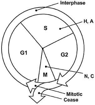
Figure 1 is a schematic diagram of the known mammalian cell cycle, showing
compounds which act at two different stages to arrest/block mitosis;
Figure 2 is set of photographs showing, by immunofluorescence, localization of
EA
(Fig. 2A) and GR-PL (Fig. 2B), where GR is a human glucocorticoid receptor
fragment and
PL is a(3-galactosidase enzyme donor fragment, and wherein the cytoplasm can
be seen to be
stained green and the nuclei stained blue;
Figure 3 is a bar graph showing the results of testing Clone #69 in response
to cell
cycle blocking compounds; and
Figure 4 is a pair of photographs showing immunofluorescence of cell line CHO-
K1+
cyto-EA, with cytoplasm stained green and nuclei stained blue.
The patent or application file contains at least one drawing executed in
color. Copies
of this patent or patent application publication with color drawings will be
provided to the
office upon request and payment of the necessary fee.
3
CA 02613174 2007-12-20
WO 2007/002200 PCT/US2006/024148
DESCRIPTION OF THE SPECIFIC EMBODIMENTS
Simple protocols for the determination of mitosis are provided employing
enzyme
fragment complementation ("EFC"). Cells are engineered to contain an ED and EA
pair for
EFC. The cells comprise one member of the EFC pair in the nucleus and the
other member of
the EFC pair in the cytosol. The members of the pair are referred to as enzyme
donor ("ED"),
which, where the two members are substantially different in size, is
arbitrarily the smaller
member, and enzyme acceptor ("EA"). The ED will generally be in the range of
about 36 to
90, more usually about 40 to 60, amino acids. One of the members of the EFC
pair is joined
to a polypeptide sequence that causes the member to reside in the nucleus. The
member is
preferably the EA. The polypeptide sequence is termed the "NLS/NRS," meaning
either an
NLS (nuclear localization signal), an NRS (nuclear retention signal), or both
an NLS and
NRS. The NLS/NRS member will be directed to the nucleus after translation in
the
cytoplasm.
A number of NLS and NRS sequences are known.
A nuclear localization signal (NLS) is a short stretch of amino acids that
mediates the
transport of nuclear proteins into the nucleus. Such sequences have been
combined in
tandem. Further examples of NLS sequences are given in "Finding nuclear
localization
signals," Murat Cokol, Raj Nair & Burkhard Rost
http://cubic.bioc.columbia.edu/papers/2000_nls/paper.html. One known NLS
sequence is
from SV40. The simian virus 40 large T antigen (SV40 T Ag) NLS seven amino
acid
sequence is the prototype of a classical monopartite NLS, as disclosed, for
example, in
Ilmarinen et al. "The monopartite nuclear localization signal of autoimmune
regulator
mediates its nuclear import and interaction with multiple importin a
molecules," FEB S
Journal 273 (2006) 315-32. As is also disclosed in this publication, some NLS
sequences are
bipartite, and may be brought together, as is discussed below. Further
examples of NLS
sequences are given in Cokol et al., "Finding nuclear localization signals,"
Proc. Nat. Acad.
Sci. Vol. 96, Issue 1, 91-96, January 5, 1999.
An "NRS" is a sequence which promotes protein-protein interactions and directs
subcellular localization and-in certain situations-nucleocytoplasmic shuttling
of individual
proteins, such as the phosphoprotein SR, which contains an RS domain. The RS
domain is
extensively phosphorylated and directs the subeellular localization. Further
details are given
in Cazalla et al. "Nuclear Export and Retention Signals in the RS Domain of SR
Proteins,"
Mol Cell Biol. 2002 October; 22(19): 6871-6882.
4
CA 02613174 2007-12-20
WO 2007/002200 PCT/US2006/024148
A commonly used NRS is the NRS sequence from SC35 (GenBank 600813, 600812),
although other sequences are available. Suitable sequences are given, for
example, in Cazalla
et al., supra, which demonstrates the presence of a dominant nuclear retention
signal in the
RS domain of SC35.
In some cases proteins that do not have a consensus NLS may be used for
directing
the ED or EA member of the EFC pair to the nucleus. The other member will
remain in the
cytosol. Upon mitosis, with the breakdown of the nuclear membrane, the two
members of the
EFC pair are brought together. In the presence of a substrate providing a
detectable product,
the cells may be analyzed by detecting the product. Alternatively, the cells
may be lysed
without lysis of the nucleus and the amount of the EFC complex determined by
use of a
substrate providing a detectable product.
The cell(s) that are employed will be subject to genetic modification for
expressing an
EFC member that is directed and remains in the nucleus and the other EFC
member that
remains in the cytosol. These cells may be subject to prior treatment by being
maintained in
an appropriate medium, washing, exposure to one or more agents that affect the
proteomic
status of the cell, that is, activate and/or inhibit one or more pathways, and
the like. When the
cells are ready to be assayed, the cells are provided in an appropriate
vessel, a controlled
environment provided for the cells and the cells grown for a sufficient period
to provide a
readout of the level of mitosis. The cells are then lysed/permeablized in an
appropriate
medium with enzyme substrate where the dilution of the cell lysate
substantially inhibits
additional complex formation of the EFC members that does not already exist as
a result of
mitosis.
The cells employed are characterized by having two genetic expression
constructs,
one construct comprising a fusion protein of an EFC pair member fused to an
NLS/NRS and
the other construct expressing the other EFC pair meinber. The expression
constructs will
have transcriptional and translational regulatory regions, which may be
inducible or
constitutive.
Usually, the expression constructs will be associated with other functional
genetic
sequences, such as sequences for integration, sequences for maintenance as an
extrachromosomal element, sequences for penetration of the cellular membrane
(i.e. the layer
which separates a cell's interior from its surroundings and controls what
moves in and out),
sequences for selection of cells comprising the expression construct(s), etc.
One may have a
cell with only one of the constructs and add the other construct for transient
expression, have
both constructs integrated into the genome or present as stable or unstable
extrachromosomal
5
CA 02613174 2007-12-20
WO 2007/002200 PCT/US2006/024148
elements, or have both constructs present as transient constructs. Each of
these possibilities
may be exploited in accordance with the purpose of the determination.
Also fused to one or both of the members of the EFC pair may be an epitope
tag, so
that the location of the member of the EFC pair may be determined
independently. Epitope
tags are readily available and a sequence of from about 10 - 30 amino acids
will suffice,
where the sequence is not normally found in the host cell and there is a
convenient binding
member, e.g., antibody for binding to the epitope tag and identifying its
location. For
detection, the antibody may be labeled, two antibodies may be used in sandwich
assays, one
to the tag and the other to the fusion protein, or other convenient assay
protocol can be
employed.
Usually, the cells will have at least about 80% of the total amount of each of
the
members of the EFC pair in a single compartment, preferably there being at
least one, more
preferably both, with at least about 90% of the total amount of the members of
the EFC pair
in a single compartment. The single compartment where a member resides may be
the
nucleus, or the cytoplasm.
A number of proteins associated with mitosis or phase cycle blocking are of
interest.
These proteins include cyclins (e.g., Cyclin A, Cyclin B, Cyclin D, Cyclin E,
Cyclin F,
transcription factors (e.g., p53, Rbl, c-Abl, EF-1), kinases (e.g., p34cdc2,
wee-1, DNA-PK),
phosphatases (e.g., cdc25B, cdc25C) and other accessory proteins (e.g., ATM,
MDM2,
HDAC). These proteins are normally localized to the nucleus, although certain
proteins (e.g.,
MDM2 or ATM) may also be located in the cytoplasm under certain conditions.
See, for
example, Kao et al. "p34(Cdc2) kinase activity is excluded from the nucleus
during the
radiation-induced G(2) arrest in HeLa cells," J Biol Chem. 1999 Dec
3;274(49):34779-84.
By targeting these proteins, where these proteins are fusion proteins and will
maintain
one of the EFC pairs in a particular compartment, while the other member of
the pair is in the
other compartment, one can investigate the effect of such compound on the
protein target and
its effect on mitosis. Using another cell where the cell is negative in the
target protein allows
one to isolate the effect.
In carrying out the determination, the cells in an appropriate culture medium
may be
dispersed, adhering to the surface of a vessel or a combination thereof. A
particular number
of cells will be chosen which may be a single cell, at least ten cells,
usually at least 102 cells
and usually not more than about 105, more usually not more than about 5x10~.
The number of
cells is not critical to this invention and will be selected in accordance
with the purpose of the
determination, the level of signal required, and other pragmatic
considerations. The cells may
6
CA 02613174 2007-12-20
WO 2007/002200 PCT/US2006/024148
be primary cells or cell lines, where the primary cells or cell lines may be
genetically
modified, as appropriate.
The cells may be grown in an appropriate growth medium for a reasonable period
to
stabilize the cells, provide for proliferation of the cells, the cells may be
blocked in a
particular phase, e.g., S-phase, provide for the cells to be in a particular
metabolic or other
status, cell cycle arrested, agonist or antagonist treated, serum starved,
serum stimulated, etc.
The environment may then be changed in accordance with the purpose of the
assay. For
example, if one is interested in the effect of a compound on mitosis, the
compound would be
added to the medium. Temperatures, concentrations, components of the medium,
etc., may be
changed in accordance with the purpose of the assay. Where inducible
transcriptional
regulatory regions have been used, the inducible gene(s) may be turned on or
off, e.g., tet
regulatory region.
After the cells have been subjected to the desired environment for a
sufficient time
period, e.g., incubated, the cells may then be assayed for their mitotic
index.
If the assay is performed intracellularly, the signal from the cells can be
determined in
a variety of ways, e.g., colorimetrically, fluorometrically, such as
fluorescence activated cell
sorter, chemiluminescently, etc. A substrate is introduced into the cells,
where the substrate is
capable of transport across the cell membrane, the membrane is made permeable,
e.g., by
isotonic shock, or the like. Desirably, with a fluorescent product from the
substrate, the
product should have lower permeability than the substrate. Where the
determination is made
extracellularly, the cells are lysed in an appropriate lysing medium and the
signal determined
appropriately. The lysing involves substantial dilution of the cellular
material, usually at least
about 5-fold and may be 10-fold or more, usually not more than about 100-fold.
The rapid
dilution has the effect of substantially inhibiting forming new enzyme
complexes not
previously formed intracellularly. A single determination may be made or a
plurality of
determinations at different time periods from an initial event, e.g.,
termination of exposure to
an environment, lysing, etc.
There are a number of ways in which the assay may be used. The assay may be
used
to determine whether changes in the environment, e.g., candidate agents or
drugs, are able to
affect mitosis. By using the subject assay with modified cells where one or
more genes may
be turned on or off, the effect of compounds on cells having the presence or
absence of
specific proteins can be established. One may also use RNAi, in conjunction
with the subject
assays to determine whether specific transcriptional and translational
products affect mitosis.
In the same way, one can establish pathways involved in mitosis and the
pathway response to
7
CA 02613174 2007-12-20
WO 2007/002200 PCT/US2006/024148
changes in the environment. All of these investigations follow normal testing
procedures,
e.g., high throughput screening, using the subject protocols and components in
analogous
ways. Usually, one will employ a control lacking the candidate agent and
compare the result
in the presence and absence of the candidate agent. A difference indicates
that the candidate
agent modulates mitosis. One may employ high throughput techniques such as
fluorescence
activated cell sorting, since a mitotic signal is either present or not in a
cell, and there is no
need to localize the signal to a particular cellular location.
The subject invention will generally have a fusion protein to maintain the ED
in either
the nuclear or, preferably, in the cytosol compartment and impart stability.
The particular
partner will be primarily arbitrarily chosen as one that does not interfere in
the assay,
maintains the fusion product in the selected compartment and is sufficiently
stable to retain a
sufficient concentration in the cell as to provide a robust signal. The
shorter member of the
EFC will usually be fused to an innocuous protein to enhance its stability. In
view of the low
molecular weight of the shorter member, it appears to be easily degraded, so
as to
substantially diminish its availability. Generally the protein will have a
molecular weight of
at least about SkD, usually at least about lOkD, and generally less than about
50kD. Proteins
that have been used are extensively described in the literature and include
such proteins as
glutathione synthase, green fluorescent protein (GFP), maltose binding protein
(MBP),
annexin proteins, etc.
The first component of the subject invention is the fusion protein described
above
and its expression construct. The ED may be at either the C-terminus, the N-
terminus or
internal to the fusion protein. The particular site of the ED in the fusion
protein will depend
upon convenience, stability and retaining the ability of the fusion protein to
complex with EA
to form an active enzyme.
The ED may be inserted into the coding region in a variety of ways. For a eDNA
gene construct, one may select a suitable restriction site for insertion of
the sequence, where
by using overhangs at the restriction site, the orientation is provided in the
correct direction.
Alternatively, one may use constructs that have homologous sequences with the
target gene
and allow for homologous recombination, where the homologous sequences that
are adjacent
in the target gene are separated by the ED in the construct. By using a
plasmid in yeast
having the cDNA gene, with or without an appropriate transcriptional and
translational
regulatory region, one may readily insert the ED construct into the cDNA gene
at an
appropriate site. Alternatively, one may insert the ED coding region with the
appropriate
splice sites in an intron or in an exon of the gene encoding the protein. In
this way, one can
8
CA 02613174 2007-12-20
WO 2007/002200 PCT/US2006/024148
select for a site of introduction at any position in the protein. In some
instances, it will be
useful to make a number of constructs, where the ED is introduced into an
intron and test the
resulting proteins for ED activity and retention of function of the protein.
Various other
conventional ways for inserting encoding sequences into a gene can be
employed. The
preferred ED and EA are derived from (3 glactosidase. The ED may be prepared
from the N-
terminal region of E. coli (3 galactosidase, Genbank Accession No. AAN78938,
beginning,
e.g., at residue 7, with the addition of an N terminal cysteine and a cysteine
replacement for
arginine near the C terminus. Other regions of the known P galactosidase
sequence may be
adapted for use as the ED.
For expression constructs and descriptions of other conventional manipulative
processes, see, e.g., Sambrook, Fritsch & Maniatis, "Molecular Cloning: A
Laboratory
Manual," Second Edition (1989) Cold Spring Harbor Laboratory Press, Cold
Spring Harbor,
N.Y. (herein "Sambrook et al., 1989"); "DNA Cloning: A Practical Approach,"
Volumes I
and II (D. N. Glover ed. 1985); "Oligonucleotide Synthesis" (M. J. Gait ed.
1984); "Nucleic
Acid Hybridization" [B. D. Hames & S. J. Higgins eds. (1985)]; "Transcription
And
Translation" [B. D. Hames & S. J. Higgins, eds. (1984)]; "Animal Cell Culture"
[R. I.
Freshney, ed. (1986)]; "Iinmobilized Cells And Enzymes" [IRL Press, (1986)];
B. Perbal, "A
Practical Guide To Molecular Cloning" (1984).
The gene encoding the fusion protein will be part of an expression construct.
The
gene is positioned to be under transcriptional and translational regulatory
regions functional
in the cellular host. The regulatory region may include an enhancer, which may
provide such
advantages as limiting the type of cell in which the fusion protein is
expressed, requiring
specific conditions for expression, naturally being expressed with the
protein, and the like. In
many instances, the regulatory regions may be the native regulatory regions of
the gene
encoding the protein, where the fusion protein may replace the native gene,
may be in
addition to the native protein, either integrated in the host cell genome or
non-integrated, e.g.,
on an extrachromosomal element. The protein may be selected in relation to the
desirability
of its regulatory region or an exogenous regulatory region may be used.
It should be understood that the site of integration of the expression
construct will
affect the efficiency of transcription and, therefore, expression of the
fusion protein. One may
optimize the efficiency of expression by selecting for cells having a high
rate of transcription,
one can modify the expression construct by having the expression construct
joined to a gene
that can be amplified and co-amplifies the expression construct, e.g., DHFR in
the presence
of methotrexate, or one may use homologous recombination to ensure that the
site of
9
CA 02613174 2007-12-20
WO 2007/002200 PCT/US2006/024148
integration provides for efficient transcription. By inserting an insertion
element, such as Cre-
Lox at a site of efficient transcription, one can direct the expression
construct to the same site.
In any event, one will usually compare the (3-galactosidase activity from
cells in a
predetermined environment to cells in the environment being evaluated. By
appropriate
choice of transcriptional regulatory region and site of integration, one can
control the level of
the fusion protein in the compartment where it is retained. Similarly, for the
other member of
the EFC pair, one can exploit the same considerations so as to have the
desired level of the
two members in the different compartments. For the most part, the fusion
protein will
comprise the ED or a-fragment of (3-galactosidase.
There are a large number of commercially available transcriptional regulatory
regions that may be used and the particular selection will generally not be
crucial to the
success of the subject invention. Also, the manner in which the fusion gene
construct is,
introduced into the host cell will vary with the purpose for which the fusion
gene is being
used. The transcriptional regulatory region may be constitutive or inducible.
In the former
case, one can have a steady state concentration of the fusion protein and/or
the other member
of the EFC in the cells, while in the latter case one can provide going from
the substantially
total absence (there is the possibility of leakage) to an increasing amount of
the fusion protein
or other member of the EFC until a steady state is reached. With inducible
transcription, one
can cycle the cell from a state where the fusion protein is absent to a state
where the steady
state concentration of the fusion protein is present. f
Copending application PGPUB 2003/0092070 entitled, "Genetic Construct
Intracellular Monitoring System" (referenced in the Background hereof), has a
large section
on vectors for introduction of the constructs, methods for introducing the
vectors, monitoring
the transfection, transcriptional regulatory regions, namely promoters,
strains of host cells
that can find use, and other useful information related to the introduction of
the constructs
into cells, all of which is specifically incorporated herein by reference as
if set forth fully
here.
Briefly, the above-mentioned application refers in part to known vector
systems
such as a defective herpes virus 1(HSV1) vector (Kaplitt et al., 1991, Molec.
Cell. Neurosci.
2:320-330); an attenuated adenovirus vector, such as the vector described by
Stratford-
Perricaudet et al. (1992, J. Clin. Invest. 90:626-630 a defective adeno-
associated virus vector
(Samulski et al., 1987, J. Virol. 61:3096-3101; Samulski et al., 1989, J.
Virol. 63:3822-3828).
Alternatively, "naked DNA" constructs may be used; alternatively a DNA vector
transporter
may be used (see, e.g., Wu et al., 1992, J. Biol. Chem., 267:963-967; Wu and
Wu, 1988, J.
CA 02613174 2007-12-20
WO 2007/002200 PCT/US2006/024148
Biol. Chem. 263:14621-14624; Hartmut et al., Canadian Patent Application No.
2,012,311,
filed March 15, 1990). A number of commercial mammalian vectors are available
with
different capabilities, different promoters, msc's, and selection genes.
pYACneo (Replicon),
pAdvantage, pSI(SV40p), pTarget, pGlneo (Promega), Vitality hrGFP
(Stratagene), pCMS-
EGFP-1, pEGFP-NI (BD Biosciences), pVITROms (Invivogen), pRK-5 GFP (Fujisawa)
and
pCruz 22 (Santa Cruz) (supplier).
For convenience, various components of the subject assays may be provided in
kits. For example, DNA constructs may be provided on the same or different
vectors to
express the components of the EFC assay. Alternatively, cells containing the
constructs may
be provided, where the cells are either genetically modified or unmodified
from the natural
cells or cells strains, e.g., inhibiting or activating a particular gene(s) or
introduction of a
gene(s) that is not expressed by the cell. In addition, buffers may be
included, culture media,
assay substrate to measure EFC activity can be provided, etc.
The following examples are offered by way of illustration and not by way of
limitation.
EXPERIMENTAL
A series of different compounds that block at different stages of the cell
cycle were
tested. Vinblastine, colchicine, nocodazole and paclitaxel (Taxo1TM) all
arrest the cell in the
G2/M phases by acting on microtubule formation and organization. Hydroxyurea
and
aphidicolin block the cell cycle in S-phase by effecting DNA replication
(Figure 1).
Fig. 1 represents a known diagram of a eukaryotic cell cycle showing mitosis.
Mitosis
is nuclear division plus cytokinesis, and produces two identical daughter
cells during
prophase, prometaphase, metaphase, anaphase, and telophase. Interphase, shown
above the
mitotic region, is often included in discussions of mitosis, but interphase is
technically not
part of mitosis, but rather encompasses stages G1, S, and G2 of the cell
cycle. Fig. 1 shows
drugs H and A (hydroxyurea and aphidicolin) acting in S phase, and drugs N,
and C
(nocodazol and colchicine) acting in the "M" phase, which is mitosis. Other
drugs, such as
taxol and viblastine are known to act in different phases of the cell cycle,
depending on the
cell type. For example, taxol acts in M phase in T47D breast cancer cells. As
is shown, cells
may either continue to divide ("Mitotic") or cease division ("Cease").
In all experiments described, 20,000 cells/well were plated in a 96 well
Corning clear
bottom white plate in a total volume of 100 L. The cells were treated for 24
hours with 5 L
of either the appropriate vehicle control or varying concentrations of the six
compounds listed
above. The next day, 100 L of Tropix/ABI Gal screen cell lysis
buffer/substrate mixture
11
CA 02613174 2007-12-20
WO 2007/002200 PCT/US2006/024148
(24:1 ratio of component) was added to the cells/media and the plate was read
on the Victor
II luminescent plate reader at 30, 60 and 120 minutes after the
lysis/substrate addition,
EXAMPLES
ExaMple 1
The initial test of the cell cycle arresting compounds was performed on a
double
stable cell line having both of the constructs expressing the EA-NLS/NRS and
GR-PL. (PL is
(3-galactosidase enzyme donor fragment and EA is the enzyme acceptor fragment
available
from DiscoveRx, Fremont, CA.). The parental line, C2C 12 is derived from mouse
muscle
cells. In the experiments in which EA-NLS/NRS and GR-PL are expressed in the
C2C12
parental cell line, the constructs were generated by subcloning the human GR
sequence into a
MFG-based retroviral vector that had been molecularly altered in a lab at
Stanford. An MFG
vector is described in U.S. 6,544,771. The EA-NLS/NRS fragment was subcloned
into a wzl-
based retroviral vector again molecularly altered in a lab at Stanford. In the
experiments
performed using a CHO-Kl parental cell line background the EA-NLS/NRS was
subcloned
into the Kpn I and Xba I sites of pcDNA3.1 Hygro vector from Invitrogen
(catalog # V870-
20). The plasmid was introduced into the cells via FuGene6 (Roche)
transfection reagent.
Cells were selected in the presence of 250 g/mL of Hygromycin and single cell
clones
isolated that expressed the EA-NLS/NRS. The human GR gene was cloned by PCR
and
subcloned into the Xho I and Bam HI sites of the DiscoveRx vector-pCMV-myc-PL
(C3).
The plasmid was introduced into the selected EA-NLS/NRS expressing clone
isolated above
by FuGene 6 transfection. Another round of screening in the presence of 300-
500 g/mL of
G418 was used to select GR-PL transfected clones. Clonal selection was
performed to finally
identify the clone that was used in these studies. In the studies using the
CHO-Kl+ cyto-EA
and cJUN-PL, the same Invitrogen pcDNA3.1 Hygro vector was used to express EA.
In this
case, the EA fragment was subcloned into the Kpn I/Not I sites of pcDNA3.1
Hygro. The
plasmid DNA was introduced as described above using FuGene6 reagent. Cells
were selected
in the presence of 250 g/mL of hygromycin and clonal selection was performed.
The c-Jun
gene was generated by PCR using an existing template copy of the gene and then
subcloned
into the Xho I/Bam HI sites of pCMV-PL-myc (C3).
In these cells, EA is localized in the nucleus (EA-NLS/NRS), while PL (a 55
mer a-
fragment of (i-galactosidase, SEQ ID NO: 1; fused to the human glucocorticoid
receptor was
retained in the cytoplasm (GR-PL)(>pCMV-PL\C3\Myc\(nuc) SEQ ID NO: 2. An inert
fragment of the glucocorticoid receptor (GR) was chosen from a number of
possible
cytoplasmic proteins, including the hormone receptors, for use in fusing to
the ED to prevent
12
CA 02613174 2007-12-20
WO 2007/002200 PCT/US2006/024148
protease degradation or other instability of the ED. The cells were treated
and assayed as
described above. As seen in TABLE 1, Nocodazole treatment (1-10 g/mL) showed
a-2-
fold increase in EFC activity, whereas, e.g. vinblastine, which does not act
in M phase,
showed no increase in EFC activity.
30 min read/Stanford GR cells
Taxol
Cone (pM) Rl R2 R3 Avg Ratio SD % CV
0 1774 2795 3056 2542 1 678 27
0.03 2624 3115 3543 3094 1 460 15
0.1 2602 4110 4340 3684 1 944 26
0.3 2638 3361 3639 3213 1 517 16
1 3016 3955 4104 3692 1 590 16
Avg%CV=20
Nocodazole
Conc ( g~/mL) Rl R2 R3 Avg Ratio SD % CV
0.0 2849 3739 3274 3287 1 445 14
0.3 4243 4486 4599 4443 1 182 4
1.0 6029 6355 6678 6354 2 325 5
3.3 5669 6175 6360 6068 2 358 6
10.0 4401 7395 5806 5867 2 1498 26
Avg%CV=11
Aphidicolin
Cone (1dV1) Rl R2 R3 Avg Ratio SD % CV
0.0 2782 2985 3178 2982 1 198 7
0.3 3081 3752 3500 3444 1 339 10
1.0 3250 3284 3388 3307 1 72 2
3.3 3171 3121 3105 3132 1 34 1
10.0 2827 3648 3765 3413 1 511 15
Avg%CV=7
13
CA 02613174 2007-12-20
WO 2007/002200 PCT/US2006/024148
Vin,blastine
Conc ( g/mL) Rl R2 R3 Avg Ratio SD %CV
0.0 10819.0 11743 9202 10588 1 1286 12
1.0 10417.0 10773 11042 10744 1 314 3
3.3 10894.0 11873 13468 12078 1 1299 11
10.0 10269.0 10703 11281 10751 1 508 5
30.0 10692.0 10910 11196 10933 1 253 2
Avg%CV=7
Colchicine
Cone ( M) Rl R2 R3 Avg Ratio SD % CV
0.0 11761 9069 9123 9984 1 1539 15
0.03 10442 12449 12456 11782 1 1161 10
0.1 6763 10024 10724 9170 1 2114 23
0.3 11132 10438 12160 11243 1 866 8
1.0 11891 12491 11866 12083 1 354 3
Avg%CV=12
Hydroxyurea
Conc ( g/mL) Rl R2 R3 Avg Ratio SD %CV
0.0 11553 10363 10532 10816 1 644 6
1.0 12080 11562 11424 11689 1 346 3
3.3 12588 10806 11171 11522 1 941 8
10.0 8813 9401 9419 9211 1 345 4
30.0 7272 7226 7165 7221 1 54 1
Avg%CV=4
TABLE 1 above shows the results of a series of experiments determining the
average
readout of luminescence with different drugs with a given coefficient of
variance (% CV)
from testing cell cycle blocking compounds on C2C 12 + EA-NLS/NRS + GR-PL
cells, i.e.,
14
CA 02613174 2007-12-20
WO 2007/002200 PCT/US2006/024148
the mouse muscle cell line C2C 12 engineered with an enzyme acceptor/nuclear
location
signal and the glucocoticoid receptor and enzyme donor fragment PL.
Example 2
In the next experiment, an antibiotic selected pool population of CHO-Kl cells
that
express EA-NLS/NRS and GR-PL were tested with the same six set of cell cycle
blocking
compounds. These cells have been characterized by immunofluorescence using
antibodies
specific to EA-NLS/NRS (Promega monoclonal antibody to beta galactosidase) and
GR
(Abeam polyclonal antibody) and show that greater than 90% of EA-NLS/NRS is
found
localized in the nucleus (see Figure 3a) and greater than 80% of the GR is
found in the
cytoplasm (see Figure 3b). Figures 2a and 2b show the immunofluorescence
localization of
EA and GR-PL in that the blue DAPI nuclear staining can be seen to be
concentrated in the
nucleus, while the green fluorescein stain on the antibody (from Abeam PLC) to
the
glucocorticoid receptor is seen in the cytoplasm. TABLE 2 shows the data from
the testing of
the CHO-Kl + EA-NLS/NRS +GR-PL cells. Again, six tables are presented one for
each of
the six drugs tested.
30 min read/DX M19/GR (pool)
Taxol
Conc (W" Rl R2 R3 Avg Ratio SD %CV
0 5441 6673 7700 6605 1 1131 17
0.03 7314 7582 6605 7167 1 505 7
0.1 10680 9945 9258 9961 2 711 7
0.3 11709 8906 7904 9506 1 1972 21
1 17111 16460 14344 15972 2 1447 9
Avg%CV=12
Nocodazole
Conc ( g/mL) R1 R2 R3 Avg Ratio SD %CV
0.0 9304 10557 8127 9329 1 1215 13
0.3 15003 17995 13585 15528 2 2251 14
1.0 23225 26942 24165 24777 3 1933 8
3.3 24010 24116 23172 23766 3 517 2
10.0 22978 25565 28584 25709 3 2806 11
Avg%CV=10
A Lidicolin
Cone (pM) R1 R2 R3 Avg Ratio SD % CV
0.0 8114 9939 10512 9522 1 1252 13
0.3 5478 5578 5469 5508 1 61 1
CA 02613174 2007-12-20
WO 2007/002200 PCT/US2006/024148
1.0 5656 5666 5491 5604 1 98 2
3.3 4392 4542 4749 4561 0 179 4
10.0 7213 6958 6651 6941 1 281 4
Avg % CV = 5
Vinblast/ne
Conc( g/mL) Rl R2 R3 Avg Ratio SD %CV
0.0 5586 5151 4385 5041 1 608 12
1.0 4205 4523 4277 4335 1 167 4
3.3 4410 4190 4436 4345 1 135 3
10.0 3907 4126 4201 4078 1 153 4
30.0 5161 6014 6583 5919 1 716 12
Avg%CV=7
Colchicine
Conc(}uNI) RI R2 R3 Avg Ratio SD %CV
0.0 3051 2690 2651 2797 1 221 8
0.03 3128 3381 3727 3412 1 301 9
0.1 6507 6048 4886 5814 2 836 14
0.3 7078 7335 7341 7251 3 150 2
1.0 10671 11680 13178 11843 4 1261 11
Avg%CV=9
H drox urea
Conc (pg/mL) R1 R2 R3 Avg Ratio SD %CV
0.0 3303 3851 4203 3786 1 454 12
1.0 4403 4542 4740 4562 1 169 4
3.3 4423 4439 4304 4389 1 74 2
10.0 3865 4116 3897 3959 1 137 3
30.0 3825 3770 3785 3793 1 28 1
Avg % CV = 4
As shown by the increased average fluorescence from the cleavage of the
active,
complemented (3Gal substrate overnight treatment with Taxol, nocodazole and
colchicine
resulted in as great as a 4-fold increase in EFC that was titrated with
increasing
concentrations of each of these compounds. As predicted, both aphidicolin and
hydroxyurea
did not cause an increase in EFC activity. These results suggest that the
compounds that do
not affect the events of nuclear envelope breakdown (i.e., the release of EA
from the nucleus)
but still cause an arrest in cell cycle progression did not result in the
complementation of EA
from the nucleus with the GR-PL that is localized in the cytoplasm to produce
an active
16
CA 02613174 2007-12-20
WO 2007/002200 PCT/US2006/024148
enzyme complex that can turn over the (3-galactosidase chemiluminescent
substrate. This
only occurs with compounds that block the cells in mitosis, allowing EA and
ProLabel to
complement.
Example 3
In the next experiment, a stable clone (clone #69) expressing both EA-NLS/NRS
and
GR-PL was isolated in a CHO-K1 parental background. To demonstrate the
specificity of the
cell cycle blocking compounds, pre-incubation in the presence of RU486 (a
specific
antagonist of GR) was tested. 20,000 cells/well were plated in a 96 well white
corning multi-
well plate and allowed to adhere overnight. The next day, the cells were
washed two times
with serum free F12 media and 100 L of serum free F12 media was added to the
cells. The
cells were then incubated in either vehicle (ethanol-1% final concentration)
or 10 M RU486
for one hour. To the cells, three different concentrations of dexamethasone
(300, 100, 30 M)
(an agonist of GR), RU486 (30, 10, 3.33 M), colchicine (1, 0.3, 0.1 g/mL) or
nocodazole
(10, 3.33, 1.11 g/mL) were added and the incubation went overnight at 37 C
with 5% C02.
The next day, the media was aspirated off and 100 L of Tropix/ABI Gal screen
cell
lysis/substrate reagent was added to the cells. The plate was read on the
Victor II reader at 30,
60 and 120 minutes.
Results are shown in Fig. 3 as ratios of fluorescence to drug concentration
(0, low
medium and high) as well as tables for seven drugs tested. As shown in Figure
3, the cells
showed a very strong response (increased EFC activity) to the dexamethasone
titration that
was blocked by the incubation with RU486. Although RU486 can act as a weak
agonist on its
own, it did not show an increase in EFC activity when titrated. Both
nocodazole and
colchicine showed an increase in EFC activity (-3-4 fold) at each of the
concentrations
tested. This increase in EFC response was not blocked by the incubation with
RU486,
suggesting the response is not related to the nuclear translocation response
of the GR. These
results further support that the increase in EFC activity observed by the
addition of the cell
cycle arresting compounds was due to breakdown of the nuclear envelope and
subsequent
release of the EA to the cytoplasm where it can complement with the GR-PL
present'and turn
over substrate. These results are further presented in TABLE 3 below:
17
CA 02613174 2007-12-20
WO 2007/002200 PCT/US2006/024148
Clone #69
Nocodazole
Cone RI R2 R3 Avg Ratio
0 3346 2737 2219 2767 1
Low 11488 11630 11606 11575 4
Med 11364 11514 11457 11445 4
High 11504 11657 11616 11592 4
Nocodazole/+ R U486
Cone R1 R2 R3 Avg Ratio
0 3179 4433 4438 4017 1
Low 11466 16071 14099 13879 3
Med 15554 15744 12922 14740 4
High 16620 14877 13043 14847 4
Colclzicine
Cone Ri R2 R3 Avg Ratio
0 466 444 371 427 1
Low 1274 1126 1007 1136 3
Med 1345 1304 1073 1241 3
High 1850 1695 1323 1623 4
CoIchicfne/+ RU486
Cone Rl R2 R3 Avg Ratio
0 5578 5606 4110 5098 1
Low 16801 18288 11942 15677 3
Med 18756 19794 12588 17046 3
High 19868 22803 15717 19463 4
Dexamethasone
Conc R1 R2 R3 Avg Ratio
0 394 345 401 380 1
Low 1528 1230 1016 1258 3
Med 2033 1768 1290 1697 4
High 3173 2571 1934 2559 7
DexametGasone/+ R U486
Conc RI R2 R3 Avg Ratio
0 3013 3216 2508 2912 1
Low 2891 3057 2367 2772 1
Med 3020 2663 2370 2684 1
High 2917 2792 2752 2820 1
18
CA 02613174 2007-12-20
WO 2007/002200 PCT/US2006/024148
RU486
Cone R1 R2 R3 Avg Ratio
0 2430 2354 2376 2387 1
Low 2692 2259 2298 2416 1
Med 2578 2379 2249 2402 1
High 3199 2790 2657 2882 1
Example 4
To further test the concept of sequestering of one (3-galactosidase enzyme
fragment in
the nucleus (in this case PL) while localizing the other component in the
cytoplasm (in this
case EA) the following experiment was carried out. A CHO-K1 stable cell line
that expressed
EA (cyto-EA) that was localized in the cytoplasm (greater than 70% as seen in
Figure 4a)
was transfected with cJUN-PL. It has been observed that cJUN-PL when
transiently
transfected into CHO-K1 cells almost exclusively localizes in the nucleus. The
cyto-EA cells
were transiently transfected witli cJUN-PL plasmid DNA. Two days after the
transfection, the
cells were re-plated into a 96 well Corning white clear bottom multiwell plate
at 20,000
cells/well. The cells were allowed to adhere overnight and the next day were
treated with
titrating concentrations of the six different cell cycle blocking compounds.
The incubation
was carried out overnight. The next day the media was removed from the cells
and 100 L of
Tropix/ABI Gal screen cell lysis/substrate reagent was added to the cells. As
seen in Figure
4b, both nocodazole and colchicine addition caused a- 2.1 fold increase in EFC
activity.
Both aphidicolin and hydroxyurea addition resulted in a negligible increase in
EFC activity,
suggesting background activity. These data are further presented in TABLE 4
below:
Nocodazole
Cone (pgJmL) R1 R2 R3 Avg Ratio
0.0 7074 7998 6717 7263 1.0
3.0 9509 10402 9751 9887 1.4
10.0 11991 13821 12371 12728 1.8
30.0 10083 13241 12287 11870 1.6
100.0 13010 15624 16060 14898 2.1
Colclric/ne
Cone OagJ-nL) R1 R2 R3 Avg Ratio
0.0 6799 6653 7659 7037 1.0
0.3 7491 8424 9730 8548 1.2
1.0 11290 11068 9929 10762 1.5
3.0 12822 13938 13005 13255 1.9
19
CA 02613174 2007-12-20
WO 2007/002200 PCT/US2006/024148
....... .. .......
10.0 13772 16082 14302 14719 2.1
Aphidicolin
Conc (pg/mL) R1 R2 R3 Avg Ratio
0.0 9433 8581 8140 8718 1.0
1.5 7218 7840 7576 7545 0.9
4.4 9492 8842 9245 9193 1.1
13.3 6677 6620 7784 7027 0.8
40.0 8645 8931 9818 9131 1.0
H drox urea
Conc (pg/mL) Rl R2 R3 Avg Ratio
0.0 6318 6369 5412 6033 1.0
3.0 7319 8251 6690 7420 1.2
10.0 8237 7967 6699 7634 1.3
30.0 7217 7112 6624 6984 1.2
100.0 7955 7787 7483 7742 1.3
Conclusion
It is evident from the above results that the subject compositions and methods
provide
a rapid and convenient method to identify the effect of changes in
environment, particularly
candidate drugs, on mitosis. The method also allows the identification of
proteins involved in
the phase cycle and how they may affect the cycle going through mitosis. The
method
provides for a robust signal and there is little interfering background.
All publications and patent applications cited in this specification are
herein
incorporated by reference as if each individual publication or patent
application were
specifically and individually indicated to be incorporated by reference.
The above specific description is meant to exemplify and illustrate the
invention and
should not be seen as limiting the scope of the invention, which is defined by
the literal and
equivalent scope of the appended claims. Any patents or publications mentioned
in this
specification are indicative of levels of those skilled in the art to which
the patent pertains and
are intended to convey details of the invention which may not be explicitly
set out but which
would be understood by workers in the field. Such patents or publications are
hereby
incorporated by reference to the same extent as if each was specifically and
individually
incorporated by reference, as needed for the purpose of describing and
enabling the method
or material referred to.
DEMANDES OU BREVETS VOLUMINEUX
LA PRtSENTE PARTIE DE CETTE DEMANDE OU CE BREVETS
COMPREND PLUS D'UN TOME.
CECI EST LE TOME DE _2
NOTE: Pour les tomes additionels, veillez contacter le Bureau Canadien des
Brevets.
JUMBO APPLICATIONS / PATENTS
THIS SECTION OF THE APPLICATION / PATENT CONTAINS MORE
THAN ONE VOLUME.
THIS IS VOLUME OF
NOTE: For additional volumes please contact the Canadian Patent Office.