Note: Descriptions are shown in the official language in which they were submitted.
CA 02613196 2007-12-10
WO 2006/135747
PCT/US2006/022476
PERIPHERAL SEAL FOR A VENTRICULAR PARTITIONING DEVICE
FIELD OF THE INVENTION
[0001] The present invention relates generally to the field of treating
heart disease,
particularly congestive heart failure, and more specifically, to a device and
method for
partitioning a patient's heart chamber and a system for delivering the
treatment device.
BACKGROUND OF THE INVENTION
[0002] Congestive heart failure (CHF) is characterized by a progressive
enlargement of the
heart, particularly the left ventricle and is a major cause of death and
disability in the United
States. Approximately 550,000 new cases occur annually in the U.S. alone. As
the patient's
heart enlarges, it cannot efficiently pump blood forward with each heart beat.
In time, the heart
becomes so enlarged the heart becomes ineffective as a pump and cannot
adequately supply
blood to the body. Even in healthy hearts only a certain percentage of the
blood in a patient's
left ventricle is pumped out or ejected from the chamber during each stroke of
the heart. The
pumped percentage, commonly referred to as the "ejection fraction", is
typically about sixty
percent for a healthy heart. A patient with congestive heart failure can have
an ejection fraction
of less than 40% and sometimes much lower. As a result of the low ejection
fraction, a patient
with congestive heart failure is fatigued, unable to perform even simple tasks
requiring exertion
and experiences pain and discomfort. Further, as the heart enlarges, the
internal heart valves
such as the mitral valve cannot adequately close. An incompetent mitral valve
allows
regurgitation of blood from the left ventricle back into the left atrium,
further reducing the heart's
ability to pump blood forwardly.
[0003] Congestive heart failure can result from a variety of conditions,
including viral
infections, incompetent heart valves (e.g. mitral valve), ischemic conditions
in the heart wall or a
combination of these conditions. Prolonged ischemia and occlusion of coronary
arteries can
result in myocardial tissue in the ventricular wall dying and becoming scar
tissue. Once the
myocardial tissue dies, it is less contractile (sometimes non-contractile) and
no longer
contributes to the pumping action of the heart. It is referred to as
hypokinetic or akinetic. As the
disease progresses, a local area of compromised myocardium may bulge out
during the heart
contractions, further decreasing the heart's ability to pump blood and further
reducing the
ejection fraction. In this instance, the heart wall is referred to as
dyskinetic. The dyskinetic
region of the heart wall may stretch and eventually form an aneurysmic bulge.
[0004] Patients suffering from congestive heart failure are commonly
grouped into four
classes, Classes I, II, III and IV. In the early stages, Classes I and II,
drug therapy is presently
the most common treatment. Drug therapy typically treats the symptoms of the
disease and may
1
CA 02613196 2013-06-11
slow the progression of the disease, but it can not cure the disease.
Presently, the only
permanent treatment for congestive heart disease is heart transplantation, but
heart
transplant procedures are very risky, extremely invasive and expensive and are
performed
on a small percentage of patients. Many patient's do not qualify for heart
transplant for
failure to meet any one of a number of qualifying criteria, and, furthermore,
there are not
enough hearts available for transplant to meet the needs of CHF patients who
do qualify.
[0005] Substantial effort has been made to find alternative treatments for
congestive
heart disease. For example, surgical procedures have been developed to dissect
and
remove weakened portions of the ventricular wall in order to reduce heart
volume. This
procedure is highly invasive, risky and expensive and is commonly only done in
conjunction with other procedures (such as heart valve replacement or coronary
artery
by-pass graft). Additionally, the surgical treatment is usually only offered
to Class III and
IV patients and, accordingly, is not an option for most patients facing
ineffective drug
treatment. Finally, if the procedure fails, emergency heart transplant is the
only presently
available option.
[0006] Mechanical assist devices have been developed as intermediate
procedures for
treating congestive heart disease. Such devices include left ventricular
assist devices and
total artificial hearts. A left ventricular assist device includes a
mechanical pump for
increasing blood flow from the left ventricle into the aorta. Total artificial
heart devices,
such as the Jarvik heart, are usually used only as temporary measures while a
patient
awaits a donor heart for transplant.
[0007] Recently, improvements have been made in treating patient's with CHF by
implanting pacing leads in both sides of the heart in order to coordinate the
contraction of
both ventricles of the heart. This technique has been shown to improve
hemodynamic
performance and can result in increased ejection fraction from the right
ventricle to the
patient's lungs and the ejection fraction from the left ventricle to the
patient's aorta. While
this procedure has been found to be successful in providing some relief from
CHF
symptoms and slowed the progression of the disease, it has not been able to
stop the
disease and is only indicated in patients with ventricular dissynchrony.
[0008] Other efforts to treat CHF include the use of an elastic support, such
as an
artificial elastic sock, placed around the heart to prevent further
deleterious remodeling.
2
CA 02613196 2014-04-10
SUMMARY OF THE INVENTION
[0008a] According to one aspect, the present invention relates to a device for
treating a
patient's heart by partitioning a chamber of the patient's heart into a
primary productive
portion and a secondary non-productive portion, comprising: a reinforced
membrane which
forms a recess when in an expanded deployed configuration; and at least one
outwardly
biased member which is secured to a peripheral portion of the membrane so as
to seal the
peripheral portion of the membrane against a ventricular wall surface defining
in part the
patient's heart chamber.
10008b] Various embodiments of the present invention provide a device for
treating a
patient's heart by partitioning a chamber of the patient's heart into a
primary productive
portion and a secondary non-productive portion, comprising: a) a reinforced
membrane
which forms a recess when in an expanded deployed configuration, wherein the
membrane
comprises a loose and flexible peripheral region configured to seal to a
ventricular wall
surface to partition the ventricle; and b) at least one outwardly biased
member which is
secured to a peripheral portion of the membrane that is radially inward from
the loose
peripheral region of the membrane, wherein the at least one outwardly biased
member is
configured to stiffen at least a portion of the membrane so as to seal the
peripheral portion of
the membrane against the ventricular wall surface defining in part the
patient's heart chamber.
[0008c] According to another aspect, the present invention relates to a device
for treating a
patient with congestive heart failure by partitioning a chamber of the
patient's heart into a
primary productive portion and a secondary non-productive portion, comprising:
a
partitioning component which has an expandable frame formed of a plurality of
ribs having
distal ends secured to a central hub and free outwardly flared proximal ends
and which has a
pressure receiving membrane formed at least in part of flexible material
forming a recess in
an expanded, deployed configuration defining in part the primary productive
portion of the
patient's heart chamber to be partitioned; and an outwardly biased member
which is secured
to the periphery of the membrane so as to seal the periphery of the membrane
against a
ventricular wall surface defining in part the heart chamber.
[0008d] Various embodiments of the present invention provide a device for
treating a patient
with congestive heart failure by partitioning a chamber of the patient's heart
into a primary
2a
CA 02613196 2014-04-10
productive portion and a secondary non-productive portion, comprising: a) a
partitioning
component which has an expandable frame formed of a plurality of ribs having
distal ends
secured to a central hub and free outwardly flared proximal ends and which has
a pressure
receiving membrane secured to the expandable frame, the membrane being formed
at least in
part of flexible material forming a recess in an expanded, deployed
configuration defining in
part the primary productive portion of the patient's heart chamber to be
partitioned, wherein
the membrane comprises a loose and flexible peripheral region configured to
seal to a
ventricular wall surface to partition the ventricle; and b) an outwardly
biased member which
is secured to the periphery of the membrane at a position that is radially
inward from the
loose peripheral region of the membrane so as to seal the periphery of the
membrane against
the ventricular wall surface defining in part the heart chamber.
100091 The present invention is directed to a ventricular partitioning device
and method of
employing the device in the treatment of a patient with heart disease and
particularly
congestive heart failure (CHF). Specifically, the device partitions a chamber
of the patient's
heart into a main productive portion and a secondary non-productive portion.
This
partitioning reduces the total volume of the heart chamber, reduces the stress
applied to
weakened tissue of the patient's
2b
CA 02613196 2007-12-10
WO 2006/135747
PCT/US2006/022476
heart wall and, as a result, improves the ejection fraction thereof. Moreover,
the expansive
nature of the device improves the diastolic function of the patient's heart.
[0010] A partitioning device embodying features of the invention has a
reinforced
partitioning component with a concave, pressure receiving surface which
defines in part the main
productive portion of the partitioned heart chamber when secured within the
patient's heart
chamber. The reinforced partitioning component has a flexible membrane that
forms the pressure
receiving surface. The partitioning component is preferably reinforced by a
radially expandable
frame component formed of a plurality of ribs. The ribs of the expandable
frame have secured
distal ends, which are preferably secured to a central hub, and free proximal
ends. The distal
ends of the ribs are preferably secured to the central hub to facilitate
radial self expansion of the
free proximal ends of the ribs away from a centerline axis. The distal ends of
the ribs may be
pivotally mounted to the hub and biased outwardly or fixed to the hub. The
ribs are preferably
formed of material such as superelastic NiTi alloy which allows for
compressing the free
proximal ends of the ribs toward a centerline axis into a contracted
configuration for delivery and
self expansion when released for deployment to an expanded configuration when
released within
the patient's heart chamber.
[0011] The free proximal ends of the ribs are configured to engage and
preferably penetrate
the tissue lining the heart chamber to be partitioned so as to secure the
peripheral edge of the
partitioning component to the heart wall and fix the partitioning component
within the chamber
so as to partition the chamber in a desired manner. The tissue penetrating
proximal tips are
configured to penetrate the tissue lining at an angle approximately
perpendicular to a center line
axis of the partitioning device. The tissue penetrating proximal tips of the
ribs may be provided
with barbs, hooks and the like which prevent withdrawal from the tips from the
heart wall.
[0012] An expansive member such as one or more strands or swellable pads
extend between
at least one pair of adjacent ribs at or close to the outer edge or periphery
of the membrane to
exert enough pressure to the flexible membrane periphery when the partitioning
device is in an
expanded configuration to ensure an adequate seal between the membrane
periphery and the
lining of the heart wall. In one embodiment, a single strand or strands extend
around essentially
the entire periphery of the membrane so that the flexible periphery of the
membrane between
each pair of ribs is effectively sealed against the heart wall. The expansive
strand or strands are
formed of material which is stiffer than the flexible, unsupported material of
the membrane to
provide an outward expansive force or thrust to prevent formation of inwardly
directed folds or
wrinkles when the ribs of the partitioning device are in at least a partially
contracted
configuration. Suitable strand or strands are formed of material such as
polypropylene suture or
superelastic NiTi alloy wires. Such strands are typically about 0.005 to about
0.03 inch (0.13.-
3
CA 02613196 2007-12-10
WO 2006/135747
PCT/US2006/022476
0.76 mm) in diameter to provide the requisite outward expansive force when
placed in a circular
position such as around the periphery of the membrane in less than completely
expanded
configuration.
[0013] In another embodiment expandable pads are provided between each
adjacent pair of
ribs which are configured to swell upon contact with body fluids to provide an
outward
expansive force or thrust, as above, to prevent formation of inwardly directed
folds or wrinkles
when the ribs of the partitioning device are in at least a partially
contracted configuration.
Preferably the pads are formed of expansive hydrophilic foam. Suitable
swellable materials
includable collagen, gelatin, polylactic acid, polyglycolic acid, copolymers
of polylactic acid and
polyglycolic acid, polycaprolactone, mixtures and copolymers thereof. Other
suitable swellable
bioresorbable polymeric materials may be employed. The expandable pads may be
formed so as
to delivery a variety of therapeutic or diagnostic agents.
[0014] The ribs in their expanded configuration angle outwardly from the hub
and the free
proximal ends curve outwardly so that the membrane secured to the ribs of the
expanded frame
forms a trumpet-shaped, pressure receiving surface.
[0015] The partitioning membrane in the expanded configuration has radial
dimensions from
about 10 to about 160 mm, preferably about 25 to about 50 mm, as measured from
the center line
axis. The membrane is preferably formed of flexible material or fabric such as
expanded
polytetrafiuoroethylene (ePTFE).
[0016] The partitioning device is designed to be oversized with respect to
the chamber in
which it is to be deployed so that the ribs of the device apply an outward
force against the
chamber wall. When the partitioning device is collapsed for delivery, the
outwardly biased
strand or strands ensures that there are no inwardly directed folds or
wrinkles and that none are
formed when the partitioning device is expanded for deployment within the
heart chamber.
[0017] In one partitioning device design embodying features of the
invention, the free ends
of the expansive strand or strands may be secured together or to the
partitioning device.
Alternatively, in another device design, the expansive strand or strands may
be long enough so
that one or both free ends thereof extend out of the patient to facilitate
collapse and retrieval of
the partitioning device. Pulling on the free ends of the strand extending out
of the patient closes
the expanded portion i.e. the ribs and membrane, of the partitioning device to
collapse of the
device and such pulling can pull the collapsed partitioning device into the
inner lumen of a guide
catheter or other collecting device
[0018] The reinforced partitioning component preferably includes a supporting
component or
stem which has a length configured to extend distally to the heart wall
surface to support the
partitioning device within the heart chamber. The supporting component has a
plurality of pods
4
CA 02613196 2007-12-10
WO 2006/135747
PCT/US2006/022476
or feet, preferably at least three, which distribute the force of the
partitioning device about a
region of the ventricular wall surface to avoid immediate or long term damage
to the tissue of the
heart wall, particularly compromised or necrotic tissue such as tissue of a
myocardial infarct
(M1) and the like. Pods of the support component extend radially and
preferably are
interconnected by struts or planes which help distribute the force over an
expanded area of the
ventricular surface.
[0019] The partitioning device may be delivered percutaneously or
intraoperatively. One
particularly suitable delivery catheter has an elongated shaft, a releasable
securing device on the
distal end of the shaft for holding the partitioning device on the distal end
and an expandable
member such as an inflatable balloon on a distal portion of the shaft proximal
to the distal end to
press the interior of the recess formed by the pressure receiving surface to
ensure that the tissue
penetrating tips or elements on the periphery of the partitioning device
penetrate sufficiently into
the heart wall to hold the partitioning device in a desired position to
effectively partition the
heart chamber. A suitable delivery device is described in co-pending
application Serial No.
10/913,608, filed on August 5, 2004, and assigned to the present assignee.
[0020] The partitioning device embodying features of the invention is
relatively easy to
install and is a substantially improved treatment of a diseased heart. A more
normal diastolic
and systolic movement of a patient's diseased heart is achieved.
Concomitantly, an increase in
the ejection fraction of the patient's heart chamber is usually obtained.
These and other
advantages of the invention will become more apparent from the following
detailed description
of the invention and the accompanying exemplary drawings.
BRIEF DESCRIPTION OF THE DRAWINGS
[0021] Figure 1 is an elevational view of a partitioning device
embodying features of the
invention in an expanded configuration.
[0022] Figure 2 is a plan view of the partitioning device shown in Figure 1
illustrating the
upper surface of the device.
[0023] Figure 3 is bottom view of the partitioning device shown in
Figure 1.
[0024] Figure 4 is a perspective view of the non-traumatic tip of the
distally extending stem
of the device shown in Figure 1.
[0025] Figure 5 is a partial cross-sectional view of the hub of the
partitioning device shown
in Figure 2 taken along the lines 5-5.
[0026] Figure 6 is a transverse cross sectional view of the hub shown in
Figure 5 taken along
the lines 6-6.
5
CA 02613196 2007-12-10
WO 2006/135747
PCT/US2006/022476
[0027] Figure 7 is a longitudinal view, partially in section of a
reinforcing rib and membrane
at the periphery of the partitioning device shown in Figure 1.
[0028] Figure 8 is a schematic elevational view, partially in section,
of a delivery system
with the partitioning device shown in Figures 1 and 2 mounted thereon.
[0029] Figure 9 is a transverse cross-sectional view of the delivery system
shown in Figure 8
taken along the lines 9-9.
[0030] Figure 10 is an elevational view, partially in section, of the
hub shown in Figure 5
being secured to the helical coil of the delivery system shown in Figure 8.
[0031] Figures 11A-11E are schematic views of a patient's left
ventricular chamber
.0 illustrating the deployment of the partitioning device shown in Figures
1 and 2 with the delivery
system shown in Figure 8 to partition a patient's heart chamber (left
ventricle) into a primary
productive portion and a secondary, non-productive portion.
[0032] Figure 12 is a schematic plan view of the deployed device shown in
Figure 11E
within a patient's heart chamber.
.5 [0033] Figure 13 is a schematic plan view of the partitioning
device shown in Figure 1
without the expansive strand after deployment within a patient's heart
chamber.
[0034] Figure 14 is a partial schematic view of the partitioning device
shown in Figures 1
and 2 in a contracted configuration resulting from pulling the free ends of
the expansive strand at
the periphery of the reinforced membrane.
.!0 [0035] Figure 15 is a schematic view of the contracted device
shown in Figure 14 being
pulled into an expanded distal end of a receiving catheter to facilitate
withdrawal of the
partitioning device into a receiving catheter.
[0036] Figure 16 is a schematic view of the contracted device shown in
Figure 14 pulled
further into the inner lumen of the receiving catheter.
.!5 [0037] Figure 17 is a plan view of the top of an alternative
partitioning device which has
swellable pads disposed between adjacent ribs to press the membrane between
the ribs against
the heart wall.
[0038] Figure 18 is a cross-sectional view of a swellable pad disposed
between two
membrane layers secured to the ribs of the partitioning device taken on a line
18-18 of Figure 17.
DETAILED DESCRIPTION OF EMBODIMENTS OF THE INVENTION
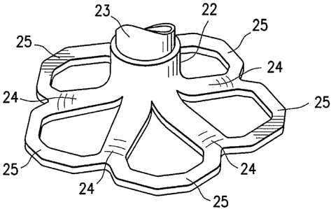
[0039] Figures 1-4 illustrate a partitioning device 10 which embodies
features of the
invention and which includes a partitioning membrane 11, a hub 12, preferably
centrally located
on the partitioning device, and a radially expandable reinforcing frame 13 is
secured to the
proximal or pressure side of the frame 13 as shown in Figure 1. The ribs 14
have distal ends 15
6
CA 02613196 2013-06-11
which are secured to the hub 12 and free proximal ends 16 which are configured
to curve
or flare away from a center line axis. Radial expansion of the free proximal
ends 16
unfurls the membrane 11 secured to the frame 13 so that the membrane presents
a
pressure receiving surface 17 which defines in part the productive portion of
the patient's
partitioned heart chamber. The peripheral edge 18 of the membrane 11 may be
serrated as
shown.
[0040] A continuous expansive strand 19 extends around the periphery of the
membrane 11 on the pressure side thereof to apply pressure to the pressure
side of the
flexible material of the membrane to effectively seal the periphery of the
membrane
against the wall of the ventricular chamber. The ends 20 and 21 of the
expansive
strand 19 are shown extending away from the partitioning device in Figures 2
and 3. The
ends 20 and 21 may be left unattached or may be secured together, e.g. by a
suitable
adhesive or the membrane 11 itself. While not shown in detail, the membrane 11
has a
proximal layer secured to the proximal faces of the ribs 14 and a distal layer
secured to
the distal faces of the ribs in a manner described in U.S. patent application
No. 2006/0030881, filed on August 5, 2004.
[0041] The hub 12 shown in Figures 4 and 5 preferably is connected to a non-
traumatic
support component 22. The support component 22 has a stem 23 a plurality of
pods or
feet 24 extending radially away from the center line axis and the ends of the
feet 24 are
secured to struts 25 which extend between adjacent feet. A plane of material
(not shown)
may extend between adjacent feet 24 in a web-like fashion to provide further
support in
addition to or in lieu of the struts 25. The inner diameter of the stem 23 is
threaded to
secure the partitioning device 10 to a delivery catheter as shown in Figs. 8-
10.
[0042] As shown in Figure 5, the distal ends 15 of the ribs 14 are secured
within the hub
12 and, as shown in Figure 6, a transversely disposed connector bar 26 is
secured within
the hub which is configured to secure the hub 12 to the nontraumatic support
component
22.
[0043] As illustrated in Figures 5 and 6, the connector bar 26 of the hub 12
allows the
partitioning device 10 to be secured to the non-traumatic support component 22
and to be
released from the delivery system within the patient's heart chamber. The
distal ends 15
of the reinforcing ribs 14 are secured within the hub 12 in a suitable manner
or they may
7
CA 02613196 2013-06-11
be secured to the surface defining the inner lumen or they may be disposed
within
channels or bores in the wall of the hub 12. The distal end of the ribs 14 are
preshaped so
that when the ribs are not constrained, other than by the membrane 11 secured
thereto (as
shown in Figures 1 and 2), the free proximal ends 16 thereof expand to a
desired angular
displacement away from the centerline axis which is about 20 to about 90 ,
preferably
about 50 to about 80 . The unconstrained diameter of the partitioning device
10 should
be greater than the diameter of the heart chamber at the deployed location of
the
partitioning device so that an outward force is applied to the wall of
7a
CA 02613196 2007-12-10
WO 2006/135747
PCT/US2006/022476
the heart chamber by the partially expanded ribs 14 during systole and
diastole so that the
resilient frame 13 augments the heart wall movement.
[0044] Figure 7 illustrates the curved free proximal ends 16 of ribs 14
which are provided
with sharp tip elements 27 configured to engage and preferably penetrate into
the wall of the
heart chamber and hold the partitioning device 10 in a deployed position
within the patient's
heart chamber so as to partition the ventricular chamber into a productive
portion and a non-
productive portion.
[0045] Figures 8-10 illustrate a suitable delivery system 30 delivering
the partitioning device
shown in Figures 1 and 2 into a patient's heart chamber and deploying the
partitioning device
10 to partition the heart chamber as shown in Figures 11A ¨ 11E. The
delivery system 30 includes
a guide catheter 31 and a delivery catheter 32.
[0046] The guide catheter 31 has an inner lumen 33 extending between the
proximal end 34
and distal end 35. A hemostatic valve (not shown) may be provided at the
proximal end 34 of
the guide catheter 31 to seal about the outer shaft 37 of the delivery
catheter 32. A flush port 36
on the proximal end 34 of guide catheter 31 is in fluid communication with the
inner lumen 33.
[0047] The delivery catheter 32 has an outer shaft 37 with an adapter 38
on the proximal end
thereof having a proximal injection port 39 which is in fluid communication
with the interior of
the shaft 37. As shown in more detail in Figure 9, the outer shaft 37 has an
inner shaft 41 which
is disposed within the interior thereof and is secured to the inner surface of
the outer shaft 37 by
webs 43 which extend along a substantial length of the inner shaft. The
injection port 39 is in
fluid communication with the passageways 42 between the inner and outer shafts
41 and 37
respectively and defined in part by the webs 43. A torque shaft 44, which is
preferably formed
of hypotubing (e.g. formed of stainless steel or superelastic NiTi), is
disposed within the inner
lumen 45 of the inner shaft 41 and has a proximal end 46 secured within the
adapter 38. Balloon
inflation port 47 is in fluid communication with the inner lumen 48 of the
torque shaft 44.
Torque shaft 44 is rotatably disposed within the inner lumen 45 of the inner
shaft 41 and is
secured to rotating knob 49. A helical coil screw 50 is secured to the distal
end 51 of the torque
shaft 44 and rotation of the torque knob 49 on the proximal end 46 of the
torque shaft 44 rotates
the screw 50 to facilitate deployment of a partitioning device 10. The
proximal end 52 of
inflatable balloon 53 is sealingly secured by adhesive 54 about the torque
shaft 44 proximal to
the distal end 51 of the torque shaft. The balloon 53 has an interior 55 in
fluid communication
with the inner lumen 48 of the torque shaft 44. Inflation fluid may be
delivered to the balloon
interior 55 through port 47 which is in fluid communication with the inner
lumen 48 of the
torque shaft 44. The distal end 56 of the balloon 53 is sealingly secured by
adhesive 57 to the
helical screw 50. The proximal and distal ends 52 and 56 of the balloon 53 are
blocked by the
8
CA 02613196 2007-12-10
WO 2006/135747
PCT/US2006/022476
adhesive masses 54 and 57 to prevent the loss of inflation fluid delivered to
the interior 55 of the
balloon 53. Delivery of inflation fluid through a fluid discharge port 58 in
the distal end 51 of
the torque shaft 44 inflates the balloon 53 which in turn applies pressure to
the proximal surface
of the partitioning device 10 to facilitate securing the partitioning
component 10 to the wall 59 of
heart chamber 60 as shown in Figures 11A-11E discussed below.
[0048] As shown in Figure 11A, the partitioning component 10 is
delivered through a
delivery system 30 which includes a guide catheter 31 and a delivery catheter
32. The
partitioning component 10 is collapsed in a first, delivery configuration
which has small enough
transverse dimensions to be slidably advanced through the inner lumen 33 of
the guide catheter
31. Preferably, the guide catheter 31 has been previously percutaneously
introduced and
advanced through the patient's vasculature, such as the femoral artery, in a
conventional manner
to the desired heart chamber 60. The delivery catheter 32 with the
partitioning component 10
attached is advanced through the inner lumen 33 of the guide catheter 31 until
the partitioning
component 10 is ready for deployment from the distal end of the guide catheter
31 into the
patient's heart chamber 60 to be partitioned.
[0049] As shown in Figure 11B, the partitioning component 10 mounted on the
screw 50 is
urged further out of the inner lumen 33 of the guide catheter 32 until the
support component 22
engages the heart wall 59. The guide catheter 31 is withdrawn while the
delivery catheter 32 is
held in place until the proximal ends 16 of the ribs 14 exit the distal end 35
of the guide catheter.
As shown in Figure 11C, the free proximal ends 16 of ribs 14 expand outwardly
to press the
sharp proximal tips 27 of the ribs 14 against and preferably into the tissue
lining the heart wall
59.
[0050] With the partitioning component 10 deployed within the heart chamber 60
and
preferably partially secured therein, inflation fluid is introduced through
the inflation port 58 in
the distal end 51 torque shaft 44 where it is directed into the balloon
interior 54 to inflate the
balloon 53. The inflated balloon 53 presses against the pressure receiving
surface 17 of the
membrane 11 of the partitioning component 10 to ensure that the sharp proximal
tips 27 are
pressed well into the tissue lining the heart wall 59 as shown in Figure 11D.
[0051] With the partitioning device 10 properly positioned within the
heart chamber 60, the
knob 49 on the torque shaft 44 (as shown in Figure 8) is rotated counter-
clockwise to disengage
the helical coil screw 50 of the delivery catheter 32 from the stem 23 secured
within hub 12. The
counter-clockwise rotation of the torque shaft 44 rotates the helical coil
screw 50 which rides on
the connector bar 26 secured within the hub 12. Once the helical coil screw 50
disengages the
connector bar 26, the delivery system 30, including the guide catheter 31 and
the delivery
catheter 32, may then be removed from the patient.
9
CA 02613196 2013-06-11
[0052] The proximal end 34 of the guide catheter 31 is provided with a flush
port 36 to
inject fluids such as therapeutic, diagnostic or other fluids through the
inner lumen 33
during the procedure. Similarly, the proximal injection port 39 of adapter 38
is in
communication with passageways 42 if the delivery catheter 32 for essentially
the same
purpose.
[0053] The deployment of the partitioning component 10 in the patient's heart
chamber
60 as shown in Figure 11E divides the chamber into a main productive or
operational
portion 61 and a secondary, essentially non-productive portion 62. The
operational
portion 61 is smaller than the original heart chamber 60 and provides for an
improved
ejection fraction and an improvement in blood flow. Over time, the non-
productive
portion 62 fills first with thrombus and subsequently with cellular growth.
Bio-resorbable
fillers such as polylactic acid, polyglycolic acid, polycaprolactone and
copolymers and
blends may be employed to initially fill the nonproductive portion 62. Fillers
may be
suitably supplied in a suitable solvent such as dimethylsulfoxide (DMSO).
Other
materials which accelerate tissue growth or thrombus may be deployed in the
non-
productive portion 62 as well as non-reactive fillers.
[0054] Figure 12 is a top view of the deployed partitioning device shown in
Figure 11E
schematically illustrating the sealed periphery of the membrane 11 against the
ventricular
wall. This is to be compared with the schematic presentation shown in Figure
13 which
illustrates a partitioning device without a strand having folds along the
periphery 18
which do not allow for an effective seal against the wall 59 of the heart
chamber 60.
[0055] The partitioning device 10 may be conveniently formed by the method
described
in U.S. patent application No. 2006/0030881, filed on August 5, 2004.
[0056] While porous ePTFE material is preferred, the membrane 11 may be formed
of
suitable biocompatible polymeric material which includes Nylon, PET
(polyethylene
terephthalate) and polyesters such as Hytrel. The membrane 11 may be
foraminous in
nature to facilitate tissue ingrowth after deployment within the patient's
heart. The
delivery catheter 32 and the guiding catheter 31 may be formed of suitable
high strength
polymeric material such as PEEK (polyetheretherketone), polycarbonate, PET,
Nylon,
and the like. Braided composite shafts may also be employed.
[0057] Figures 14-16 illustrate the collapse and retrieval of a partitioning
device 10 by
CA 02613196 2013-06-11
pulling on the ends 20 and 21 of the expansive strand 19 which extends around
the
periphery of the membrane 11. Typically, the partitioning device 10 would
still be
secured to the delivery catheter 32, but the delivery catheter is not shown to
simplify the
drawings. In Figure 14 the partitioning device 10 is shown in a partially
collapsed
configuration. In Figure 15 the partially collapsed partitioning device 10 is
shown being
withdrawn into the flared distal end 63 of retrieval catheter 64. Figure 16
illustrates the
completely collapsed partitioning device 10 pulled further into the retrieval
catheter 64.
The partitioning device 10 may be withdrawn by pulling the device through the
inner
lumen 65 of the retrieval catheter 64. Optionally, the partitioning device 10
and retrieval
catheter may be withdrawn from the patient together.
[0058] To assist in properly locating the device during advancement and
placement
thereof into a patient's heart chamber, parts, e.g. the distal extremity, of
one or more of
the ribs 14 and/or the hub 12 may be provided with markers at desirable
locations that
provide enhanced visualization by eye, by ultrasound, by X-ray, or other
imaging or
visualization means. Radiopaque markers may be made with, for example,
stainless steel,
platinum, gold, iridium, tantalum, tungsten, silver, rhodium, nickel, bismuth,
other
radiopaque metals, alloys and oxides of these metals.
[0059] Figures 17 and 18 illustrate an alternative design which embodies
features of the
invention in which the partitioning device 10 is provided with swellable
bodies 70,
preferably hydrophilic foam, around the periphery of the membrane 11 between
adjacent
ribs 14. When these bodies contact body fluid, such as blood, upon deployment,
they
swell, thereby sealing the peripheral portion of the membrane 11 against the
patient's
heart wall as previously described. The details of the partitioning device 10
are
essentially the same as in the previous embodiment and elements in this
alternative
embodiment are given the same reference numbers as similar elements in the
previous
embodiments.
[0060] To the extent not otherwise described herein, the various components of
the
partitioning device and delivery system may be formed of conventional
materials and in a
conventional manner as will be appreciated by those skilled in the art.
[0061] While particular forms of the invention have been illustrated and
described herein,
it will be apparent that various modifications and improvements can be made to
the
11
CA 02613196 2013-06-11
invention. Moreover, individual features of embodiments of the invention may
be shown
in some drawings and not in others, but those skilled in the art will
recognize that
individual features of one embodiment of the invention can be combined with
any or all
the features of another embodiment. Accordingly, it is not intended that the
invention be
limited to the specific embodiments illustrated. It is intended that this
invention to be
defined by the scope of the appended claims as broadly as the prior art will
permit.
12