Note: Descriptions are shown in the official language in which they were submitted.
DEMANDE OU BREVET VOLUMINEUX
LA PRESENTE PARTIE DE CETTE DEMANDE OU CE BREVET COMPREND
PLUS D'UN TOME.
CECI EST LE TOME 1 DE 2
CONTENANT LES PAGES 1 A 92
NOTE : Pour les tomes additionels, veuillez contacter le Bureau canadien des
brevets
JUMBO APPLICATIONS/PATENTS
THIS SECTION OF THE APPLICATION/PATENT CONTAINS MORE THAN ONE
VOLUME
THIS IS VOLUME 1 OF 2
CONTAINING PAGES 1 TO 92
NOTE: For additional volumes, please contact the Canadian Patent Office
NOM DU FICHIER / FILE NAME:
NOTE POUR LE TOME / VOLUME NOTE:
CA 02613283 2007-12-20
WO 2007/002007 PCT/US2006/023866
METHODS AND COMPOSITIONS FOR EXPRESSING A HETEROLOGOUS
PROTEASE
1. Field of the Invention
[0001] In one aspect, the present invention provides methods and coinpositions
for
expressing a protease or pro-protease in cells that do not naturally express
the protease or
pro-protease. In other aspects, the invention provides methods of producing
viruses, e.g.,
influenza viruses, in such cells. In other aspects, the invention provides
methods for
increasing the titer of influenza viruses grown in cells that express such a
heterologous
protease or pro-protease. In still other aspects, the invention provides a
heterologous protease
from Sts epton2yces griseus useful in the methods and compositions.
2. Background
[0002] Influenza pandemics are defined by a dramatic global increase in
morbidity and
mortality due to influenza illness. Several factors combine to modulate the
severity and
extent of the pandemic including the low degree of immunity in the population
and the
efficiency with which the virus can transmit among humans. The latter is
generally
influenced not only by the virus itself but the density of the population and
ease of travel into
and out of a region. The virus responsible for the pandemic is generally a
recently emerged
antigenic variant that the majority of the population have not had prior
experience with and,
therefore, have little or no immunity to. In addition, efficient human to
huinan transinission
is a prerequisite for rapid spread and, in the case of zoonotic introduction
of animal viruses
into human populations, the virus must adapt to replication in humans and be
capable of
efficient transmission.
[0003] Pandemic influenza spreads very quickly and can have devastating
impact. The most
severe pandemic of the 20th century, the 1918 pandeinic, killed over 500,000
U.S. citizens
and between 20 to 40 million people worldwide. The pandeinic inay produce
waves of
disease, with peaks of incidence separated by several weeks to inonths. The
relatively rapid
onset and spread of pandemic influenza presents several problems for
responding to a global
attack of this magnitude and imposes overwhelming burdens on einergency
responders and
health care woricers. Rapid identification and response to the emerging
pandeinic is clearly a
necessary element of the solution; several prograins are currently in place
worldwide to
monitor emerging influenza viruses including avian influenza viruses that
infrequently cause
1
CA 02613283 2007-12-20
WO 2007/002007 PCT/US2006/023866
disease in humans. These surveillance data are used in conjunction with
predefined
pandemic alert levels in order to identify the likelihood of the threat and
provide guidance for
an effective response.
[0004] Vaccination is the most iinportant public health measure for preventing
disease
caused by annual epideinics of influenza. The short interval between
identification of a
potential pandemic and the onset of significantly increased disease levels
present significant
challenges for producing sufficient vaccine to protect a large segment of the
population.
Having vaccine technology and manufacturing infrastructure in place prior to
the einergence
of the next pandemic will be critical in aineliorating a significant ainount
of illness and death.
The short response times needed to produce a "pandemic vaccine" will not allow
for
prolonged research or process development to be conducted in order to provide
an effective
response.
[0005] To date, all commercially available influenza vaccines in the United
States have been
propagated in embryonated hen's eggs. Although influenza virus grows well in
hen's eggs,
production of vaccine is dependent on the availability of eggs. Supplies of
eggs must be
organized, and strains for vaccine production selected months in advance of
the next flu
season, limiting the flexibility of this approach, and often resulting in
delays and shortages in
production and distribution. Unfortunately, some influenza vaccine strains,
such as the
prototype A/Fujian/411/02 strain that circulated during the 2003-04 season, do
not replicate
well in embryonated chicken eggs, and have to be isolated by cell culture in a
costly and time
consuming procedure.
[0006] Systeins for producing influenza viruses in cell culture have also been
developed in
recent years (See, e.g., Furminger. Vaccin.e Production, in Nicholson et al.
(eds) Textbook of
Influenza pp. 324-3 32; Merten et al. (1996) Production of influenza virus in
cell cultuNes for
vaccine preparation, in Cohen & Shafferman (eds) Novel Strategies in Design
and
Production of Vaccines pp. 141-151). Typically, these methods involve the
infection of
suitable immortalized host cells with a selected strain of virus. While
eliminating many of
the difficulties related to vaccine production in hen's eggs, not all
pathogenic strains of
influenza grow well and can be produced according to established tissue
culture methods. In
addition, many strains with desirable characteristics, e.g., attenuation,
temperature sensitivity
and cold adaptation, suitable for production of live attenuated vaccines, have
not been
successfully grown in tissue culture using established methods.
2
CA 02613283 2007-12-20
WO 2007/002007 PCT/US2006/023866
[0007] Thus, there is a need for 1) manufacturing facilities and procedures
needed to produce
influenza vaccine from cell culture and 2) development of an effective vaccine
technology
from cell culture to prevent illness caused by seasonal epidemics of
influenza. These
procedures and technologies can be rapidly applied to the production and
distribution of
pandemic vaccine in the event of an iinininent pandeinic.
[0008] One of the many obstacles to be overcome in licensing a cell culture
based influenza
vaccine is the need for proteolytic cleavage of the hemagglutinin (HA) protein
for a newly-
formed virus to productively infect a new cell. During infection of an animal,
the HA protein
is cleaved by a trypsin-like serine protease endogenous to the animal. In
culture,
endopeptidase activity is frequently insufficient to allow robust replication
of the influenza
virus. Accordingly, proteases such as trypsin are fiequently added to the
culture medium
following infection of the cells in culture with an influenza virus of
interest to increase viral
yield. See, e.g., U.S. Patent No. 5,698,433. However, addition of exogenous
proteases to the
cell culture medium introduces additional components to the culture medium,
adding
complexity and the need for additional regulatory review. In addition, the
addition of
exogenous trypin increases the costs associated with malcing vaccine using
cell culture
methds. Thus, new methods and compositions are needed for growing influenza
virus in
culture without the need for addition of exogenous proteases. Further, such
methods and
compositions must overcome the inherent toxicity of expressing active
proteases in cells.
These and other unmet needs are provided by the present invention.
[0009] Citation or discussion of a reference herein shall not be construed as
an admission that
such is prior art to the present invention. In addition, citation of a patent
shall not be
construed as an admission of its validity.
3. Summary
[0010] The present invention provides cells, referred to herein as "cell(s) of
the invention,"
comprising a nucleic acid that encodes a protease or pro-protease, such that
the cell expresses
a higher level of the protease or pro-protease than would be ordinarily
expressed in the cell in
the absence of the nucleic acid. In certain embodiments, the cell does not
normally express
the protease or pro-protease. In certain embodiments, the cell expresses the
protease or pro-
protease at low levels, for example, at levels less than optimal or desired
for a particular
biological activity, e.g., culture of viruses, e.g., influenza viruses. In
certain aspects, the
invention provides a cell comprising a nucleic acid encoding a protease or pro-
protease,
wherein the nucleic acid encoding the protease or pro-protease is stably
integrated into the
3
CA 02613283 2007-12-20
WO 2007/002007 PCT/US2006/023866
cell's genome. In other aspects, the invention provides a cell comprising a
nucleic acid
encoding a protease or pro-protease, wherein the nucleic acid encoding the
protease or pro-
protease is maintained extrachromasomally. In other aspects, the invention
provides a cell
comprising a nucleic acid encoding a protease or pro-protease, wherein the
nucleic acid
encoding the protease or pro-protease is transiently expressed in the cell. In
certain
embodiments, the cell expresses the protease or pro-protease. In certain
embodiments, the
cell constitutively expresses the protease or pro-protease. In certain
embodiments, the cell
inducibly expresses the protease or pro-protease. In certain einbodiments, the
cell secretes
the protease or pro-protease. In certain embodiments, the cell expresses the
protease or pro-
protease in the cytosol of the cell.
[0011] In certain embodiments, the protease is a serine protease. In certain
embodiments, the
serine protease is an S 1 family protease. In certain embodiments, the
protease is trypsin. In
certain embodiments, the serine protease is a bacterial subtilisin. In certain
embodiinents, the
protease is SPRT from Streptofnyces griseus. In certain embodiments, the
protease is a
protease listed in Table 1. In certain embodiments, the protease is a pro-
protease. In certain
embodiments, the pro-protease is trypsinogen. In certain embodiments, the pro-
protease is
processed into an active protease.
[0012] In certain einbodiments, expression of the protease or pro-protease is
under the
control of an inducible promoter. In certain embodiments, the inducible
promoter is induced
by interferon or a downstreain signaling molecule induced by interferon. In
certain
einbodiments, the inducible promoter is induced by a tetracycline-regulated
expression
system. In certain embodiments, expression of the protease or pro-protease is
under the
control of a constitutively active promoter. In certain embodiinents, the
nucleic acid
encoding the protease or pro-protease comprises sequence encoding a secretion
signal that
directs secretion of the protease or pro-protease.
[0013] In certain embodiments, the cell expresses between about 0.1 ng and
about 50 g of
the protease or pro-protease per inl of cell culture. In certain einbodiments,
the cell expresses
between about I ng and about 50 g of the protease or pro-protease per ml of
cell culture. In
certain embodiments, the cell expresses between about 10 ng and about 50 g of
the protease
or pro-protease per ml of cell culture. In certain embodiments, the cell
expresses between
about 100 ng and about 50 g of the protease or pro-protease per ml of cell
culture. In certain
embodiments, the cell expresses between about 1 g and about 50 g of the
protease or pro-
protease per ml of cell culture. In certain einbodiments, the cell expresses
between about 0.1
4
CA 02613283 2007-12-20
WO 2007/002007 PCT/US2006/023866
ng and about 5 g of the protease or pro-protease per ml of cell culture. In
certain
embodiments, the cell expresses between about 0.1 ng and about 100 ng of the
protease or
pro-protease per ml of cell culture. In certain embodiments, the cell
expresses between about
0.1 ng and about 10 ng of the protease or pro-protease per ml of cell culture.
In certain
einbodiments, the cell expresses between about 0.1 ng and about 1 ng of the
protease or pro-
protease per ml of cell culture. In certain einbodiinents, the cell expresses
an amount of
protease or pro-protease sufficient to increase the titer of virus grown in a
culture of the cells
expressing the protease or pro-protease. In certain einbodiments, the
molecular weight of the
protease or pro-protease is calculated based on the pro-protease form of the
enzyine. In
certain embodiments, the molecular weight of the protease or pro-protease is
calculated based
on the mature, active foim of the protease.
[00141 In certain embodiments, the cell expresses at least about 0.1 ng of the
protease or pro-
protease per ml of cell culture. In certain embodiments, the cell expresses at
least about 1 ng
of the protease or pro-protease per ml of cell culture. In certain
embodiments, the cell
expresses at least about 10 ng of the protease or pro-protease per ml of cell
culture. In certain
embodiments, the cell expresses at least about 100 ng of the protease or pro-
protease per ml
of cell culture. In certain embodiments, the cell expresses at least about 1
g of the protease
or pro-protease per ml of cell culture. In certain embodiments, the cell
expresses at least
about 10 g of the protease or pro-protease per ml of cell culture. In certain
einbodiments,
the cell expresses at least about 20 g of the protease or pro-protease per ml
of cell culture.
In certain embodiments, the cell expresses at least about 30 g of the
protease or pro-protease
per ml of cell culture. In certain embodiments, the cell expresses at least
about 40 g of the
protease or pro-protease per ml of cell culture. In certain embodiments, the
cell expresses at
least about 50 g of the protease or pro-protease per ml of cell culture.
[0015j In certain embodiments, the cell is a bacterial cell. In certain
embodiments, the
bacterial cells is an E. coli cell. In certain embodiments, the cell is a
inarnmalian cell. In
certain einbodiments, the mammalian cell is a canine cell. In certain
embodiments, the
canine cell is an MDCK cell. In certain embodiments the MDCK cell is non-
tumorigenic. In
certain embodiments, the mammalian cell is a primate cell. In certain
embodiments, the
primate cell is an African green monkey or human cell. In certain embodiments,
the cell is an
avian cell. In certain eznbodiments, the avian cell is a chicken cell.
[00161 In another aspect, the invention provides a method of producing a
virus, comprising
infecting a cell of the invention with an virus, culturing the cell under
conditions that allow
5
CA 02613283 2007-12-20
WO 2007/002007 PCT/US2006/023866
replication of the virus, and collecting virus from the cell culture. In a
specific einbodiment,
the virus is an influenza virus.
[0017] In another aspect, the invention provides a method of producing a
virus, comprising
transfecting a cell of the invention with nucleic acids comprising a viral
genome, culturing
the cell under conditions that allow replication of the virus, and collecting
virus from the cell
culture. In a specific embodiment, the viral genome is an influenza genome and
the virus is
an influenza virus. In some embodiunents, the influenza virus correspond to an
influenza B
virus. In some embodiments, the influenza virus correspond to an influenza A
virus. In
certain einbodiments, the viruses include an attenuated influenza virus, a
cold adapted
influenza virus, a teinperature sensitive influenza virus, or a virus with any
combination of
these desirable properties. In one embodiment, the influenza virus is an
influenza B/Ann
Arbor/l/66 strain virus, e.g., a cold adapted, temperature sensitive,
attenuated strain of B/Ann
Arbor/l/66. In another embodiment, the influenza virus is an influenza A/Ann
Arbor/6/60
strain virus, e.g., a cold adapted, temperature sensitive, attenuated strain
of A/Ann
Arbor/6/60.
[0018] In certain embodiments, the methods include recovering influenza
viruses and using
the viruses in the preparation of an immunogenic composition, e.g., a vaccine.
In one
einbodiment the virus is capable of eliciting an iirunune response upon
administration, e.g.,
intranasal administration, to a subject. In some embodiments, the viruses used
to prepare a
vaccine are inactivated prior to administration, in other embodiments, live-
attenuated viruses
are used to prepare a vaccine. In certain embodiments, recombinant and
reassortant influenza
A and influenza B viruses are produced according to the methods of the
invention. In one
embodiment, a vaccine is prepared comprising a live, inactivated, or killed
virus derived from
a virus produced by the methods of the invention. In one embodiment, viruses
produced by
the methods of the invention are used to replicate other viruses in cell
culture or eggs. In one
embodiment, a vaccine is provided that comprises iminunogenic polypeptides
derived from a
virus produced by the methods of the invention.
[0019] In certain einbodiments, the cell expresses a pro-protease, and the
metllod further
comprises adding an exogenous protease to the culture medium. In certain
embodiments, the
exogenous protease is added to a maximum concentration of about 0.1 gg/ml. In
certain
einbodiments, the exogenous protease is trypsin.
6
CA 02613283 2007-12-20
WO 2007/002007 PCT/US2006/023866
[0020] In certain embodiments, the virus is a DNA virus. In certain
embodiments, the virus
is an RNA virus. In certain embodiments, the virus is a single-stranded DNA
virus. In
certain embodiments, the virus is a double-stranded DNA virus. In certain
embodiments, the
virus is a positive-sense single-stranded RNA virus. In certain einbodiments,
the virus is a
negative-sense single-stranded RNA virus. In certain embodiments, the virus is
a double-
stranded RNA virus. In certain einbodiinents, the virus is a reverse-
transcribing virus.
[0021] In another aspect, the invention provides a method of replicating an
influenza virus,
coinprising infecting a cell of the invention with an influenza virus,
culturing the cell under
conditions that allow replication of the influenza virus, and collecting
influenza virus from
the cell culture. In certain embodiments, such conditions do not include
exogenously added
proteases or pro-proteases, e.g., trypsin or trypsinogen.
[00221 In still another aspect, the invention provides a metliod of
replicating an influenza
virus, comprising transfecting a cell with nucleic acids encoding an influenza
genome,
culturing the cell under conditions that allow replication of the influenza
virus, and collecting
influenza virus from the cell culture. In certain embodiments, such conditions
do not include
exogenously added proteases or pro-proteases, e.g., trypsin or trypsinogen.
[0023] In certain einbodiments, the cell expresses a pro-protease or
enzymatically active
protease not normally expressed by the cell. In certain embodiments, the cell
is a mammalian
cell. In certain einbodiments, the cell is an avian cell. In certain
embodiments, the cell is a
primate cell, canine cell, hamster cell, mouse cell, or rat cell. In certain
embodiments, the
cell is an MDCK cell. In certain embodiments, the cell is a Vero cell. In
certain
embodiinents, the cell is a chicken cell.
[0024] In yet another aspect, the invention provides a method of increasing
the titer of
influenza virus grown in cell culture, comprising culturing the influenza
virus in a cell
culture, wherein the cells in the cell culture express a protease or pro-
protease that i) is
heterologous to the cell, and ii) cleaves a hemagglutinin of the influenza
virus, thereby
increasing the titer of the influenza virus grown in the cell culture relative
to the titer obtained
by culturing the influenza virus in cells that do not express a heterologous
protease or pro-
protease.
[0025] In certain embodiments, the cells stably express the heterologous
protease. In certain
einbodiments, the cells constitutively express the heterologous protease. In
certain
embodiments, the cells inducibly express the heterologous protease. In certain
embodiments,
7
CA 02613283 2007-12-20
WO 2007/002007 PCT/US2006/023866
the heterologous protease is trypsin. In certain embodiments, the cells
constitutively express
the heterologous pro-protease. In certain embodiments, the cells inducibly
express the
heterologous pro-protease. In certain embodiments, the pro-protease is
trypsinogen. In
certain embodiments, the protease is SPRT protease frozn Streptornyces
griseus. In certain
einbodiments, the pro-protease is prepro-SPRT protease from Sti eptonayces
griseus.
[0026] One indication of the ability of a cell to support viral replication is
the yield of virus
obtained from an infected cell culture. Viral yield can be determined by
numerous methods
known to one skilled in the art. For example, viral yield can be quantified by
determining the
concentration of virus present in a sainple according to a median tissue
culture infectious
dose (TCID50) assay that measures infectious virions. The TCID50 values are
often reported
as the loglo TCID5n/mL. In one embodiment, the cells expressing a heterologous
protease
support the replication of influenza viruses (e.g., ca/ts strains) to a loglo
TCID50/mL of at
least 6.0, or at least 6.2, or at least 6.4, or at least 6.6, or at least 6.8,
or at least 7.0, or at least
7.2, or at least 7.4, or at least 7.6, or at least 7.8, or at least 8.0, or at
least 8.2, or at least 8.4,
or at least 8.6, or at least 8.8, or at least 9.0 , or at least 9.2, or at
least 9.4, or at least 9.6, or at
least 9.8. In a specific embodiment, the cells expressing a heterologous
protease support the
replication of influenza viruses (e.g., ca/ts strains) to commercially
reasonable titers (>107
Log TCID50/mL).
[0027] In certain embodiments, the titer of the influenza virus that is
produced in a cell
culture of cells expressing a heterologous protease is increased by a loglo
TCID50/mL of at
least about 0.1, or at least about 0.2, or at least about 0.3, or at least
about 0.4, or at least
about 0.5, or at least about 0.6, or at least about 0.7, or at least about
0.8, or at least about 0.9,
or at least about 1.0, or at least about 1.2, or at least about 1.4, or at
least about 1.6, or at least
about 1.8, or at least about 2.0, or at least about 2.2, or at least about
2.4, or at least about 2.6,
or at least about 2.6, or at least about 2.8, or at least about 3.0, or at
least about 3.2, or at least
about 3.4, or at least about 3.6, or at least about 3.8, or at least about
4.0, or at least a.bout 4.2,
or at least about 4.4, or at least about 4.6, or at least about 4.8, or at
least about 5.0, relative to
the titer of influenza virus produced in a culture of corresponding cells that
do not express a
heterologous protease and to which no exogenous protease, e.g., trypsin has
been added.
[0028] In certain embodiments, the titer of the influenza virus that is
produced in a cell
culture of cells expressing a heterologous protease is increased by at least
about 10%, relative
to the titer of influenza virus produced in a culture of corresponding cells
that do not express
a heterologous protease and to which no exogenous protease, e.g., trypsin has
been added. In
8
CA 02613283 2007-12-20
WO 2007/002007 PCT/US2006/023866
certain embodiments, the titer of the influenza virus that is produced in a
cell culture of cells
expressing a heterologous protease is increased by at least about 25%. In
certain
embodiments, the titer of the influenza virus that is produced in a cell
culture of cells
expressing a heterologous protease is increased by at least about 50%. In
certain
embodiinents, the titer of the influenza virus that is produced in a cell
culture of cells
expressing a heterologous protease is increased by at least about 100%. In
certain
embodiments, the titer of the influenza virus that is produced in a cell
culture of cells
expressing a heterologous protease is increased by at least about 500%. In
certain
einbodiments, the titer of the influenza virus that is produced in a cell
culture of cells
expressing a heterologous protease is increased by at least about 1000%. In
certain
embodiments, the titer of the influenza virus that is produced in a cell
culture of cells
expressing a heterologous protease is increased by at least about 5000%. In
certain
embodiments, the titer of the influenza virus that is produced in a cell
culture of cells
expressing a heterologous protease is increased by at least about 10,000%. In
certain
embodiments, the titer of the influenza virus that is produced in a cell
culture of cells
expressing a heterologous protease is increased by at least about 30,000%. In
certain
embodiments, the titer of the influenza virus that is produced in a cell
culture of cells
expressing a heterologous protease is increased by at least about 50,000%. In
certain
embodiments, the titer of the influenza virus that is produced in a cell
culture of cells
expressing a heterologous protease is increased by at least about 70,000%. In
certain
embodiments, the titer of the influenza virus that is produced in a cell
culture of cells
expressing a heterologous protease is increased by at least about 100,000%.
[0029] In certain embodiments, the cell culture medium in which a cell of the
invention is
cultured is serum-free. In certain embodiments, the cell culture medium in
which a cell of the
invention is cultured contains serum (e.g., fetal calf serum). In certain
embodiments, the cell
culture medium in which a cell of the invention is cultured contains no
exogenously added
animal proteins, such media is often referred to "animal protein free" or
"APF" media. In
certain einbodiments, the cell culture medium in which a cell of the invention
is cultured
contains exogenous protease (e.g, porcine trypsin).
[0030] In still another aspect, the invention provides a method for producing
a heterologous
protease or pro-protease in a cell, wherein said cell is capable of supporting
influenza
replication, comprising culturing a cell comprising a nucleic acid encoding a
protease or pro-
9
CA 02613283 2007-12-20
WO 2007/002007 PCT/US2006/023866
protease not normally expressed in the cell under conditions that permit
expression of said
protease or pro-protease, thereby producing the protease or pro-protease in
the cell.
[0031] In certain embodiments, the cell expresses a protease. In certain
embodiments, the cell
stably expresses a protease. In certain embodiments, the cell expresses a pro-
protease. In
certain einbodiments, the cell stably expresses a pro-protease. In certain
embodiments, the
cell secretes the protease or pro-protease into the cell culture medium. In
certain
embodiments, the cell expresses between about 0.1 ng and about 50 g of the
protease or pro-
protease per ml of cell culture. In certain embodiments, the cell expresses an
amount of
protease or pro-protease sufficient to increase the titer of virus grown in a
culture of the cells
expressing the protease or pro-protease. In certain embodiments, expression of
the protease
or pro-protease is inducible. In certain embodiments, expression of the
protease or pro-
protease is constitutive.
[0032] In still another aspect, the invention provides a method for inaking a
cell that
expresses a protease or pro-protease not norinally expressed by the cell,
comprising
introducing a nucleic acid encoding a protease or pro-protease operably linked
to regulatory
elements effective to express the protease or pro-protease in the cell (which
may or may not
be secreted or released from the cell), thereby making a cell that expresses a
protease or pro-
protease not nonnally expressed by the cell.
[0033] Any suitable technique and/or vector known by one skilled in the art,
witllout
.20 limitation, can be used to introduce the nucleic acid into the cell. In
certain embodiments, the
nucleic acid is introduced into the cell as a plasmid, a cosmid, a viral
vector, a bacteriophage,
a phagemid, a transposon, or an artificial chromosome. In certain embodiments,
the nucleic
acid is introduced into the cell as a retroviral vector. In certain
embodiments, the nucleic acid
is stably maintained in the cell. In other einbodiments, the nucleic acid is
transiently
maintained.
[0034] In certain embodiments, the nucleic acid encoding the protease or pro-
protease further
comprises a selectable marker. Methods utilizing a selectable marker to select
for those cell
stably expressing the nucleic acid encoding the protease or pro-protease are
known to one
skilled in the art. Any selectable marker known to one skilled in the art to
be effective in the
cell into which the nucleic acid is introduced can be used in such
einbodiments. Thus, in
certain embodiments, the selectable inarker is an antibiotic resistance gene.
In certain
einbodiments, the selectable marker is a gene in an anabolic pathway that
complements a
CA 02613283 2007-12-20
WO 2007/002007 PCT/US2006/023866
deficiency in the cell. For example, the nucleic acid encoding the protease or
pro-protease
can be introduced into a cell deficient in synthesis of, for example, an amino
acid. The
nucleic acid can comprise a gene that complements the deficiency of the cell.
By culturing
the cell in medium that does not coinprise the ainino acid, the presence of
the nucleic acid
coinprising the synthesis gene is selected. Any such gene known to one skilled
in the art,
without limitation, can be used according to the present invention.
[0035] In yet another aspect, the invention provides an isolated nucleic acid
that is at least
about 90% identical to SEQ ID NO.:1. In certain embodiments, the isolated
nucleic acid
coinprises or alternatively consists of SEQ ID NO.:1. In certain embodiments,
the isolated
nucleic acid hybridizes under hybridization conditions to a nucleic acid
encoding SEQ ID
NO.:1 (or the complement thereof). In certain embodiments, the hybridization
conditions are
stringent hybridization conditions. In certain embodiments, the hybridization
conditions are
highly stringent hybridization conditions.
[0036] In yet another aspect, the invention provides an isolated polypeptide
comprising or
alternatively consisiting of the amino acid sequence that is SEQ ID NO.:2.
[0037] In still another aspect, the invention provides an isolated nucleic
acid encoding a
polypeptide comprising or alternatively consisiting of the amino acid sequence
that is SEQ
ID NO.:2.
[0038] In yet another aspect, the invention provides an expression vector
comprising a
nucleic acid of the invention.
[0039] In still another aspect, the invention provides a cell transfected with
an expression
vector of the invention.
4. Brief Description of the Fieures
[0040] Figure 1 presents replication of ca A/Vietnam/1203/2004 (H5N1) in MDCK
cells.
[0041] Figure 2 presents a table showing amounts of luciferase activity
observed in cells
transfected with different retroviral vectors.
[00421 Figure 3 presents a graphical representation comparing luciferase
activity observed in
MDCK cells with the concentration of retroviral particles used to infect the
MDCK cells.
[0043] Figure 4 presents a table showing luciferase expression in 12 different
single MDCK
clones and two different mixtures of MDCK clones.
11
CA 02613283 2007-12-20
WO 2007/002007 PCT/US2006/023866
[0044] Figure 5 presents a table showing a suinmary of MDCK clones obtained
froin viral
particles produced in two different packaging cell lines and transfected with
three different
vectors.
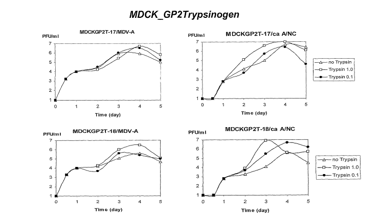
[0045] Figures 6A-6C present graphical representations of influenza viral
titers obtained by
infecting different MDCK clones (panel A control clone, panel B
AinphoTrypsinogen clone,
panel C GP2Typsinogen clone) with MDV-A or ca A/NC influenza strains.
Exogenous
trypsin added: 0.0 g/ml, open triangles; 0.1 g/ml on day 1 at day 1, closed
circles; 1.0
g/ml at days 1-5 open squares.
[0046] Figures 7A-7B present western blots showing expression of 6xHis-labeled
trypsinogen in 12 of 16 MDCK clones.
[0047] Figure 8 presents a table showing inducible luciferase expression in 15
different
MDCK clones.
[0048] Figure 9 presents the nucleotide sequence (SEQ ID NO: 1) of the sprT
gene, encoding
a serine protease from Stfreptomyces griseus.
[0049] Figure 10 presents the amino acid sequence (SEQ ID NO:2) of a serine
protease from
Streptoinyces griseus encoded by the sprT gene.
[0050] Figure 11 presents the nucleotide sequence (SEQ ID NO:3) encoding
trypsinogen.
[0051] Figure 12 presents the Forward and Reverse primers used to clone the
sprT gene
(SEQ ID NOS: 5 and 5, respectively).
[0052] Figure 13 presents the schematic map of the pT-Rex-DEST30/Luciferase
and pT-Rex-
DEST30/Trypsin plasmids transfected into R3/7 clones.
5. Detailed Description of the Invention
5.1 Defmitions
[0053] Unless defined otherwise, all scientific and technical terms are
understood to have the
same meaning as commonly used in the art to which they pertain. For the
purpose of the
present invention the following terms are defined below.
[0054] The terms "nucleic acid," "polynucleotide," "polynucleotide sequence"
and "nucleic
acid sequence" refer to single-stranded or double-stranded deoxyribonucleotide
or
ribonucleotide polymers, or chimeras or analogues thereof. As used herein, the
term
optionally includes polymers of analogs of naturally occurring nucleotides
having the
essential nature of natural nucleotides in that they hybridize to single-
stranded nucleic acids
12
CA 02613283 2007-12-20
WO 2007/002007 PCT/US2006/023866
in a manner similar to naturally occurring nucleotides (e.g., peptide nucleic
acids). Unless
otherwise indicated, a particular nucleic acid sequence of this invention
encoinpasses
complementary sequences, in addition to the sequence explicitly indicated.
[0055] The term "gene" is used broadly to refer to any nucleic acid associated
with a
biological function. Thus, genes include coding sequences and/or the
regulatory sequences
required for their expression. The term "gene" applies to a specific genomic
sequence, as well
as to a cDNA or an mRNA encoded by that genomic sequence.
[0056] Genes also include non-expressed nucleic acid segments that, for
example, form
recognition sequences for other proteins. Non-expressed regulatory sequences
include
"promoters" and "enhancers," to which regulatory proteins such as
transcription factors bind,
resulting in transcription of adjacent or nearby sequences. A "tissue
specific" promoter or
enhancer is one which regulates transcription in a specific tissue type or
cell type, or types.
[0057] The term "vector" refers to plasmids, viral vectors, recombinant
nucleic acids and
cDNA. A vector can also be a naked RNA polynucleotide, a naked DNA
polynucleotide, a
polynucleotide composed of both DNA and RNA within the same strand, a poly-
lysine-
conjugated DNA or RNA, a peptide-conjugated DNA or RNA, a liposome-conjugated
DNA,
or the like, that are not autonomously replicating. Most commonly, the vectors
of the present
invention are plasmids.
[0058] An "expression vector" is a vector, such as a plasmid, which is capable
of promoting
expression, as well as replication of a nucleic acid incorporated therein.
Typically, the
nucleic acid to be expressed is "operably linked" to a promoter and/or
enhancer, and is
subject to transcription regulatory control by the promoter and/or enhancer.
[0059] A "bi-directional expression vector" is typically characterized by two
alternative
promoters oriented in the opposite direction relative to a nucleic acid
situated between the
two promoters, such that expression can be initiated in both orientations
resulting in, e.g.,
transcription of both plus (+) or sense strand, and negative (-) or antisense
strand RNAs.
Alternatively, the bi-directional expression vector can be an ambisense
vector, in which the
viral mRNA and viral genomic RNA (as a cRNA) are expressed from the same
strand.
[0060] In the context of the invention, the term "isolated" refers to a
biological material, such
as a nucleic acid or a protein, which is substantially free from components
that normally
accompany or interact with it in its naturally occurring environment. The
isolated material
optionally comprises material not found with the material in its natural
environment, e.g., a
13
CA 02613283 2007-12-20
WO 2007/002007 PCT/US2006/023866
cell. For example, if the material is in its natural environment, such as a
cell, the material has
been placed at a location in the cell (e.g., genome or genetic element) not
native to a material
found in that environment. For example, a naturally occurring nucleic acid
(e.g., a coding
sequence, a promoter, an enhancer, etc.) becomes isolated if it is introduced
by non-naturally
occurring means to a locus of the genome (e.g., a vector, such as a plasmid or
virus vector, or
amplicon) not native to that nucleic acid. Such nucleic acids are also
referred to as
"heterologous" nucleic acids.
[0061] The term "recombinant" indicates that the material (e.g., a nucleic
acid or protein) has
been artificially or synthetically (non-naturally) altered by human
intervention. The
alteration can be performed on the material within, or removed from, its
natural enviroiunent
or state. Specifically, when referring to a virus, e.g., an influenza virus,
the virus is
recombinant when it is produced by the expression of a recombinant nucleic
acid.
[0062] The term "reassortant," when referring to a virus, indicates that the
virus includes
genetic and/or polypeptide coinponents derived from more than one parental
viral strain or
source. For example, a 7:1 reassortant includes 7 viral genomic seginents (or
gene segments)
derived from a first parental virus, and a single coinplementary viral genomic
segment, e.g.,
encoding hemagglutinin or neuraminidase, from a second parental virus. A 6:2
reassortant
includes 6 genomic segments, inost commonly the 6 internal genes from a first
parental virus,
and two complementary segments, e.g., heinagglutinin and neuraminidase, from a
different
parental virus.
[0063] The term "introduced" when referring to a heterologous or isolated
nucleic acid refers
to the incorporation of a nucleic acid into a eukaryotic or prokaryotic cell
where the nucleic
acid can be incorporated into the genome of the cell (e.g., chromosome,
plasmid, plastid or
mitochondrial DNA), converted into an autonoinous replicon, or transiently
expressed (e.g.,
transfected mRNA). The term includes such methods as "infection,"
"transfection,"
"transforination" and "transduction." In the context of the invention a
variety of methods can
be employed to introduce nucleic acids into prokaryotic cells, including
electroporation,
calciuin phosphate precipitation, lipid mediated transfection (lipofection),
etc.
[0064] The term "host cell" means a cell which contains a heterologous nucleic
acid, such as
a vector, and supports the replication and/or expression of the nucleic acid,
and optionally
production of one or more encoded products including a polypeptide and/or a
virus. Host
cells can be prokaryotic cells such as E. coli, or eukaryotic cells such as
yeast, insect,
14
CA 02613283 2007-12-20
WO 2007/002007 PCT/US2006/023866
amphibian, avian or mammalian cells, including human cells. Exemplary host
cells in the
context of the invention include Vero (African green monkey kidney) cells,
Per.C6 cells
(human embryonic retinal cells), BHK (baby hamster kidney) cells, primary
chick kidney
(PCE-) cells, Madin-Darby Canine Kidney (MDCI,'-) cells, Madin-Darby Bovine
Kidney
(MDBK) cells, 293 cells (e.g., 293T cells), and COS cells (e.g., COS1, COS7
cells). The
term host cell encompasses combinations or mixtures of cells including, e.g.,
mixed cultures
of different cell types or cell lines (e.g., Vero and CEK cells). A co-
cultivation of
electroporated sf vero cells is described for exainple in PCT/USO4/42669 filed
December 22,
2004, which is incorporated by reference in their entirety.
[0065] The expression "artificially engineered" is used herein to indicate
that the virus, viral
nucleic acid or virally encoded product, e.g., a polypeptide, a vaccine,
comprises at least one
mutation introduced by recombinant methods, e.g., site directed inutagenesis,
PCR
mutagenesis, etc. The expression "artificially engineered" when referring to a
virus (or viral
component or product) coinprising one or more nucleotide mutations and/or
ainino acid
substitutions indicates that the viral genome or genome segment encoding the
virus (or viral
coinponent or product) is not derived from naturally occurring sources, such
as a naturally
occurring or previously existing laboratory strain of virus produced by non-
recoinbinant
methods (such as progressive passage at 25 C), e.g., a wild type or cold
adapted A/Ann
Arbor/6/60 or B/Ann Arbor/1/66strain.
[00661 The term "% sequence identity" is used interchangeably herein with the
term
"% identity" and refers to the level of amino acid sequence identity between
two or more
peptide sequences or the level of nucleotide sequence identity between two or
more
nucleotide sequences, when aligned using a sequence alignment program. For
exainple, as
used herein, 80% identity means the same thing as 80% sequence identity
deterinined by a
defined algorithm, and means that a given sequence is at least 80% identical
to another length
of another sequence. Exemplary levels of sequence identity include, but are
not limited to,
60, 70, 80, 85, 90, 95, 98% or more sequence identity to a given sequence.
[00671 The term "% sequence homology" is used interchangeably herein with the
terin
"% homology" and refers to the level of amino acid sequence homology between
two or
more peptide sequences or the level of nucleotide sequence homology between
two or more
nucleotide sequences, when aligned using a sequence alignment program. For
example, as
used herein, 80% homology means the same thing as 80% sequence homology
determined by
a defined algorithm, and accordingly a homologue of a given sequence has
greater than 80%
CA 02613283 2007-12-20
WO 2007/002007 PCT/US2006/023866
sequence homology over a length of the given sequence. Exeinplary levels of
sequence
homology include, but are not limited to, 60, 70, 80, 85, 90, 95, 98% or more
sequence
homology to a given sequence.
[0068] Exemplary computer programs which can be used to determine identity
between two
sequences include, but are not liinited to, the suite of BLAST prograins,
e.g., BLASTN,
BLASTX, and TBLASTX, BLASTP and TBLASTN, publicly available on the Internet at
the
NCBI website. See also Altschul et al., 1990, J. Mol. Biol. 215:403-10 (with
special
reference to the published default setting, i.e., parameters w=4, t= 17) and
Altschul et al.,
1997, Nucleic Acids Res., 25:3389-3402. Sequence searches are typically
carried out using
the BLASTP program when evaluating a given amino acid sequence relative to
amino acid
sequences in the GenBank Protein Sequences and other public databases. The
BLASTX
program is preferred for searching nucleic acid sequences that have been
translated in all
reading frames against amino acid sequences in the GenBaiik Protein Sequences
and other
public databases. Both BLASTP and BLASTX are run using default parameters of
an open
gap penalty of 11.0, and an extended gap penalty of 1.0, and utilize the
BLOSUM-62 matrix.
See' id.
[0069] A preferred aligninent of selected sequences in order to determine "%
identity"
between two or more sequences, is performed using for exainple, the CLUSTAL-W
program
in MacVector version 6.5, operated with default parameters, including an open
gap penalty of
10.0, an extended gap penalty of 0.1, and a BLOSUM 30 similarity matrix.
[0070] "Hybridizing specifically to" or "specific hybridization" or
"selectively hybridize to",
refers to the binding, duplexing, or hybridizing of a nucleic acid molecule
preferentially to a
particular nucleotide sequence under stringent conditions when that sequence
is present in a
complex mixture (e.g., total cellular) DNA or RNA.
[0071] The term "stringent conditions" refers to conditions under which a
probe will
hybridize preferentially to its target subsequence, and to a lesser extent to,
or not at all to,
other sequences. "Stringent hybridization" and "stringent hybridization wash
conditions" in
the context of nucleic acid hybridization experiments such as Southern and
northern
hybridizations are sequence dependent, and are different under different
environinental
parameters. An extensive guide to the hybridization of nucleic acids can be
found in Tijssen,
1993, Labot=atofy Techniques in Biochernistty azid Molecular Biology -
Hybridization with
Nucleic Acid Probes, part I, chapter 2, "Overview of principles of
hybridization and the
16
CA 02613283 2007-12-20
WO 2007/002007 PCT/US2006/023866
strategy of nucleic acid probe assays", Elsevier, NY; Sainbrook et al., 2001,
Molecular
Cloning: A Laboratoiy Manual, Cold Spring Harbor Laboratory, 3'd ed., NY; and
Ausubel et
al., eds., Current Edition, Current Protocols in Molecular Biology, Greene
Publishing
Associates and Wiley Interscience, NY.
[0072] Generally, highly stringent hybridization and wash conditions are
selected to be about
5 C lower than the thermal melting point (Tm) for the specific sequence at a
defined ionic
strength and pH. The Tm is the temperature (under defined ionic strength and
pH) at which
50% of the target sequence hybridizes to a perfectly matched probe. Very
stringent conditions
are selected to be equal to the Tm for a particular probe.
[0073] One example of stringent hybridization conditions for hybridization of
complementary nucleic acids which have more than about 100 complementary
residues on a
filter in a Southern or northern blot is 50% formalin with 1 mg of heparin at
42 C, with the
hybridization being carried out overnight. An example of highly stringent wash
conditions is
0.15 M NaCl at 72 C for about 15 minutes. An example of stringent wash
conditions is a
0.2X SSC wash at 65 C for 15 minutes. See Sainbrook et al. for a description
of SSC buffer.
A high stringency wash)can be preceded by a low stringency wash to reinove
background
probe signal. An exemplary medium stringency wash for a duplex of, e.g., more
than about
100 nucleotides, is lx SSC at 45 C for 15 minutes. An exemplary low
stringency wash for a
duplex of, e.g., more than about 100 nucleotides, is 4-6x SSC at 40 C for 15
minutes. In
general, a signal to noise ratio of 2x (or higher) than that observed for an
unrelated probe in
the particular hybridization assay indicates detection of a specific
hybridization.
[0074] The term "about," as used herein, unless otherwi=se indicated, refers
to a value that is
no more than 10% above or below the value being modified by the term. For
example, the
term "about 5 g/kg" means a range of from 4.5 g/kg to 5.5 g/kg. As another
example,
"about 1 hour" means a range of from 48 minutes to 72 minutes.
[0075] The term "stably integrated," as used herein in reference to a nucleic
acid, refers to a
nucleic acid that has recoinbined with a host cell's genomic nucleic acids and
thus become a
part of a cell's genome. Stably integrated nucleic acids can comprise a
selectable marker to
ensure that the stably integrated nucleic acids reinain a part of the cells
genoine. Stably
integrated nucleic acids need not necessarily remain integrated into the
genome at a single
location; the nucleic acids can integrate at more than one location and can
move from
location to location within the genome.
17
CA 02613283 2007-12-20
WO 2007/002007 PCT/US2006/023866
5.2 Cells Expressing a Heterologous Protease or Pro-protease
[0076] Influenza virus that contains the precursor hemagglutinin molecule
(HAO) on its
surface is not capable of fusing with a cell and initiating infection. The HAO
inust be
proteolytically cleaved, separating the HA1 and HA2 subunits, to achieve its
active form.
Virions with mature HA on the surface actively fuse with a host cell and
initiate infection.
Several cell types in vivo, including cells in the airway, contain proteases
that activate HA;
however, many cell types used in vitro, including MDCK, do not contain active
proteases that
can efficiently cleave HA. For these cells, exogenous trypsin has been added
to the culture at
a concentration that does not negatively impact the cell but allows the HA to
be cleaved and
activated. See, e.g., US Patent Nos. 5,698,433 and 5,756,341.
[0077] Porcine trypsin has been shown to effectively activate HA and is used
routinely, by
several influenza investigators for this purpose. In the examples described
below,
recombinant cells expressing, e.g., porcine trypsin or trypsinogen have been
evaluated and
selected for their ability to support influenza replication in, e.g., MDCK
cells.
[0078] Thus, in certain embodiments, an active protease or pro-protease
expressed from a
recombinant system or from the cell itself is contemplated in connection with
the invention.
Expression of a cloned protease or pro-protease from a recombinant cell line
enables the
reduction or reznoval of animal derived products as well as provides the
protease or pro-
protease in situ enabling the most effective cleavage of the HA molecule.
Alternately, a
protease or pro-protease encoded in the cell's genome that is not normally
expressed by the
cell can be expressed by altering the regulation of expression of the protease
or pro-protease.
For example, an promoter, e.g., an inducible promoter, that directs
transcription and
translation of the protease or pro-protease can be introduced by, e.g.,
homolgous
recombination in the region of the cell's genome that regulates expression of
the protease or
pro-protease. By selecting a promoter (and/or other regulatory sequences) that
is/are active
the selected cell type, the protease or pro-protease can be expressed in a
cell that does not
normally express the protease or or pro-protease.
[0079] Any cell known to be useful for culturing influenza known to one
skilled in the art
without limitation can be used to generate a cell of the invention. For
example, suitable host
cells for the replication of influenza virus include, e.g., Vero cells, Per.C6
cells, BHK cells,
MDCK cells, 293 cells and COS cells, including 293T cells, COS7 cells.
Further, co-cultures
including two or more of the above cell lines, e.g., MDCK cells and either
293T or COS cells
18
CA 02613283 2007-12-20
WO 2007/002007 PCT/US2006/023866
can be employed at a ratio, e.g., of 1:1, to improve replication efficiency.
In such
embodiments, either or both of the co-cultured cells can express the
heterologous protease.
5.2.1 Proteases Expressed by Cells
[0080] Any protease known to one skilled in the art to be useful in cleaving
the HAO
influenza protein can be expressed by a cell according to the present
invention.
[0081] Several proteases can be evaluated for their ability to be produced
from a recoinbinant
system or engineered into a cell, e.g., an MDCK cell, itself. Suitable
proteases and pro-
proteases include, but are not limited to, trypsin, trypsinogen, and SPRT.
Additional
exeinplaiy proteases that can be used are listed in Table 1, below. Further,
active fragments
of the proteases can also be used in the cells and methods of the present
invention.
[0082]_ The skilled artisan can routinely determine whether a particular
protease is suitable
for use in the cells and methods of the invention. Typically, such assays
involve an
assessment of cleavage of a viral protein wherein such cleavage is an
important step in the
virus's life cycle. Cleavage can be assessed directly, e.g, by monitoring
production of two
smaller proteins from a larger protein, or indirectly, e.g., by monitoring
viral titers produced
in the presence of the protease. For example, proteases suitable for cells
and/or methods for
influenza virus culture can be identified by, e.g., assessing cleavage of the
HAO protein or by
monitoring viral titer in cell culture comprising the protease.
Table 1: Proteases
Enzyme Length SwissProt GenBank
S 1 family - SA clan Classificatio (AA) Accession No. Accession
No.
achelase I protease: giant silkworm 3.4.21.- 213 (P23604) /A
oth, satumid moth ACH1_LONAC
achelase II protease: giant silkworm 3.4.21.- 214 (P23605) /A
oth, satumid moth ACH2LONAC
acrosin: goat 3.4.21.10 60 (P10626) /A
ACROCAPHI
acrosin: human 3.4.21.10 421 (P10323) Y00970
ACROHUMAN
acrosin: mouse 3.4.21.10 436 (P23578) S66245
ACROMOUSE
acrosin : pig 3.4.21.10 415 (P08001) J04950
ACROPIG
acrosin : rabbit 3.4.21.10 431 (P48038) U05204
ACRORABIT
acrosin: rat 3.4.21.10 437 (p29293) X59254
ACRO RAT
19
CA 02613283 2007-12-20
WO 2007/002007 PCT/US2006/023866
ancrod: malayan pit viper 3.4.21.74 234 (P26324) /A
AGKRH
ANCl
ancrod: malayan pit viper 3.4.21.74 258 (P47797) L07308
ANC2 AGKRH
ancrod: cantil, tropical inocassin 3.4.21.74 20 (P33588) /A
ANCRAGKBI
ancrod: southern copperhead 3.4.21.74 231 (P09872) /A
ANCRAGKCO
apolipoprotein(A): human 3.4.21.- 4548 (P08519) X06290
AP OAHUMAN
apolipoprotein(A): rhesus macaque 3.4.21.- 1420 (P14417) J04635
APOAMACMU
atroxobin: barba amaril, fer-de-lance 3.4.21.74 255 (P04971) J02684
BATXBOTAT
coinplement C1R component: human 3.4.21.41 705 (P00736) X04701
C1RHUMAN
complement C 1 S component: human 3.4.21.42 688 (P09871) X06596
C1 SHUMAN
roproteinase E (procarboxypeptidaseA /A 253 (P05805) 4/A
complex: bovine CAC3_BOVIN
azurocidin(cathionic antimicrobial 9/A 251 (P20160) M96326
rotein) : human CAP7 HUMAN
azurocidin(cathionic antimicrobial 4/A 219 (P80015) 9/A
rotein CAP37) : pig CAP7_PIG
calcium-dependent serine proteinase : 3 4.21.- 695 (P15156) X16160
golden hamster CASPMESAU
cathepsin G: human 3.4.21.20 255 (P08311) M16117
CATGHUMAN
cathepsin G: mouse 3.4.21.20 261 g3OUSE 2829) M96801 cathepsin G: rat
3.4.21.20 26 (P 17977) /A
CATGRAT
cerastotin: horned desert viper 3.4.21.- 98 (P81038) /A
CERACERCE
cerastobin: sahara sand viper 3.4.21.- 35 (P18692) /A
CERACERVI
cercarial protease: blood fluke 3.4.21.- 264 (P12546) J03946
CERCSCHMA
complement factor B: bovine 3.4.21.47 16 (P81187) /A
CFABBOVIN
coinplement factor B: huinan 3.4.21.47 764 (P00751) X72875
CFABHUMAN
coinplement factor B: mouse 3.4.21.47 761 (P04186) M60646
CFABMOUSE
complement factor B : pig 3.4.21.47 151 (Q03710) M59240
CFAB PIG
complement factor D: human 3.4.21.46 253 (P00746) M84526
CA 02613283 2007-12-20
WO 2007/002007 PCT/US2006/023866
CFAD HUMAN
complement factor D: mouse 3.4.21.46 259 (P03953) Ml 1768
CFADMOUSE
complement factor D : pig 3.4.21.46 259 (P51779) U29948
CFADPIG
compleinent factor D: rat 3.4.21.46 263 (P32038) S73894
CFADRAT
complement factor I: human 3.4.21.45 583 (P05156) y00318
CFAIHUMAN
compleinent factor B-like protease: 3.4.21.- 250 (P81475) /A
chicken CFBL_CHICK
caldecrin: rat 3.4.21.- 268 (P55091) S80379
CLCRRAT
complement C2: human 3.4.21.43 752 g6'MAN 068) M15082 complement C2: mouse
3.4.21.43 760 (P21180) M60579
C02MOUSE
cocoonase: atlantic horseshoe crab 3.4.21.- 14 (P35586) /A
COCOLIMPO
collagenolytic protease 25 KD II/III: 3.4.21.32 20 (P34153) 4/A
crab-beetle COG1CHIOP
collagenolytic protease 28 KD : red 3.4.21.32 20 (P2073 1) 4/A
ing crab COG1_PARCM
collagenolytic protease 35 KD II: crab- 3.4.21.32 20 (P34154) 4/A
beetle COG2CHIOP
collagenolytic protease 36 KD: crab- 3.4.21.32 20 (P34155) 4/A
eetle COG3CHIOP
collagenolytic protease 36 KD A: red 3.4.21.32 20 (P20732) 9/A
ing crab COGA_PARCM
collagenolytic protease36 KD B red 3.4.21.32 20 (P20733) 9/A
ing crab COGB_PARCM
collagenolytic protease36 KD C red 3.4.21.32 20 (P20734) 9/A
ing crab COGCPARCM
collagenase: cattle grub 3.4.21.- 260 (P08897) X74306
COGSH'YPLI
rachyurin: atlantic sand fiddler crab 3.4.21.32 226 (P00771) /A
COGS UCAPU
complement -activating component of 4/A 699 (P48740) D17525
RA-reactive factor : human CRARHUMAN
complement -activating component of 4/A 704 (P98064) D16492
RA-reactive factor : mouse CRARMOUSE
chyinotrypsinl: african malaria 3.4.21.1 259 (Q27289) Z18887
osquito CTR1_ANOGA
chymotrypsin BI: penoied shrimp, 3.4.21.1 271 (Q00871) X66415
european white shrimp CTR1PENVA
chymotrypsin2: african malaria 3,4.21.1 258 (Q17025) Z18888
os uito CTR2 ANOGA
21
CA 02613283 2007-12-20
WO 2007/002007 PCT/US2006/023866
chymotrypsin2: dog 3.4.21.1 263 (P04813) K01173
CTR2CANFA
chymotrypsinBlI: penoied shrimp, 3.4.21.1 271 (P36178) /A
european white shrimp CTR2_PENVA
chymotrypsinII: european hornet 3.4.21.1 218 (P00769) /A
CTR2VESCR
chymotrypsinII : oriental hornet 3.4.21.1 216 (P00768) N/A
CTR2VESOR
chymotrypsinA: bovine 3.4.21.1 245 (P00766) /A
CTRABOVIN
chymotrypsinA: atlantic cod 3.4.21.1 263 (P47796) X78490
CTRAGADMO
chymotrypsinB: bovine 3.4.21.1 245 (P00767) /A
CTRBBOVIN
chymotrypsinB : atlantic cod 3.4.21.1 245 (P80646) /A
CTRBGADMO
chymotrypsinB: human 3.4.21.1 263 (P17538) M24400
CTRBHUMAN
chymotrypsinB: rat 3.4.21.1 263 (P07338) K02298
CTRBRAT
chymotrypsin-like serine proteinase : 3.4.21.- 254 (P35003) X71438
california red abalone CTRL_HALRU
chymotrypsin-like protease CTRL-1: 3.4.21.- 264 (P40313) X71874
uman CTRLHUMAN
chymotrypsin: penoeid shrimp. 3.4.21.1 31 (P35002) /A
CTRPPENMO
duodenase I: bovine 3.4.21.- 226 (P80219) /A
DDN1BOVIN
nite allergen der. F 3: house-dust mite 3.4.21.- 259 (P49275) D63858
DEF3DERFA
ite allergen der. F 6: house-dust mite 3.4.21.- 20 (P49276) /A
DEF6DERFA ,
ite allergen der. P 3: house-dust mite 3.4.21.- 261 (P39675) U11719
DER3DERPT
ite allergen der. P 6 house-dust mite 3.4.21.- 20 (P49277) /A
DER6DERPT
serine protease easter: fruit fly 3.4.21.- 392 (P13582) J03154
EASTDROME
elastase 1: bovine 3.4.21.36 266 (Q28153) M80838
EL1BOVIN
elastase 1: huinan 3.4.21.36 68 (P11423) /A
EL 1 HUMAN
elastase 1: pig 3.4.21.36 266 (P00772) ELl PIG X04036
elastase 1: rat 3.4.21.36 266 (P00773) V01234
ELlRAT
eutrophil elastase 2A : horse 3.4.21.- 85 (P37357) /A
EL2A HORSE
22
CA 02613283 2007-12-20
WO 2007/002007 PCT/US2006/023866
elastase 2A: human 3.4.21.71 269 (08217) M16631
EL2AHUMAN
eutrophil elastase 2B : horse 3.4.21.- 73 (P37358) /A
EL2BHORSE
elastase 2B: human 3.4.21.71 269 (P08218) M16653
EL2BHUMAN
elastase 2: bovine 3.4.21.71 269 (Q29461) X97635
EL2 BOVIN
elastase 2: mouse 3.4.21.71 271 (P05208) X04573
EL2 MOUSE
elastase 2: pig 3.4.21.71 269 (P08419) EL2 PIG M16651
elastase 2: rat 3.4.21.71 271 (p00774) V01233
EL2RAT
elastase IIIA: human 3.4.21.70 270 (P09093) M18700
EL3AHUMAN
elastase IIIB: human 3.4.21.70 270 (P08861) M16630
EL3BHUMAN
elastase : atlantic cod 3.4.21.- 20 (P32197) 'A
ELASGADMO
leukocyteelastase: human 3.4.21.37 267 (P08246) r03545
ELNEHUMAN
enteropeptidase: bovine 3.4.21.9 1035 (P98072) U09859
ENTKBOVIN
enteropeptidase: human 3.4.21.9 1019 (P98073) U09860
ENTKHUMAN
enteropeptidase: mouse 3.4.21.9 1069 (P97435) U73378
ENTKMOUSE
enteropeptidase: pig 3.4.21.9 1034 (P98074) D30799
ENTKPIG
arginine esterase: dog 3.4.21.35 260 (P09582) Y00751
ESTA CANFA
coagulation factor X: bovine 3.4.21.6 492 (P00743) X00673
FAI 0BOVIN
coagulation factor X: chicken 3.4.21.6 475 (P25155) D00844
FA10CHICK
coagulation factor X: human 3.4.21.6 488 (P00742) K03194
FA10HUMAN
coagulation factor X: rabbit 3.4.21.6 490 (Tl 4IaBIT 9AF003200
coagulation factor XI: human 3.4.21.27 625 (P03951) M13142
FA 11 HUMAN
coagulation factor XII: bovine 3.4.21.38 593 (P98140) S70164
FA12BOVIN
coagulation factor XII: guinea pig 3.4.21.38 603 (Q04962) X68615
FA12CAVPO
coagulation factor XII: huinan 3.4.21.38 615 (P00748) M31315
FA12 HUMAN
23
CA 02613283 2007-12-20
WO 2007/002007 PCT/US2006/023866
coagulation factor VII: bovine 3.4.21.21 407 (P22457) /A
FA7BOVIN
coagulation factor VII: human 3.4.21.21 466 (P08709) M13232
FA7HUMAN
coagulation factor VII: mouse 3.4.21.21 446 (P70375) U66079
FA7MOUSE
coagulation factor VII: rabbit 3.4.21.21 444 (P98139) U77477
FA7RABIT
coagulation factor IX: bovine 3.4.21.22 416 (P00741) J00007
FA9BOVIN
coagulation factor IX: dog 3.4.21.22 452 (P 19540) M21757
FA9CANFA
coagulation factor IX : guinea pig 3.4.21.22 285 (P16295) M26237
FA9CAVPO
coagulation factor IX: human 3.4.21.22 461 (P00740) K02402
FA9HUMAN
coagulation factor IX: mouse 3.4.21.22 459 (P 16294) M23109
FA9 MOUSE
coagulation factor IX : pig 3.4.21.22 271 (P 16293) FA9 PIG M26235
coagulation factor IX : rabbit 3.4.21.22 275 (P16292) M26234
FA9RABIT
coagulation factor IX : rat 3.4.21.22 282 (P16296) M26247
FA9RAT
coagulation factor IX : sheep 3.4.21.22 274 (P16291) M26233
FA9SHEEP
flavoxobin: habu 3.4.21.- 260 (P05620) D67078
FLVBTRIFL
gilatoxin: beaded lizard 3.4.21.- 245 (P43685) /A
GILXHELHO
granzyme A: human 3.4.21.78 262 (P12544) M18737
GRAAHUMAN
granzyme A: mouse 3.4.21.78 260 (P11032) X14799
GRAAMOUSE
granzyine B: human 3.4.21.79 247 (P10144) M17016
GRAB HUMAN
granzyme B (G,H): mouse 3.4.21.79 247 g7OUSE 0418) X04072 granzyme C: mouse
3.4.21.- 248 (P08882) M22527
GRACMOUSE
granzyme D: mouse 3.4.21.- 248 (P11033) J03255
GRADMOUSE
granzyme E: mouse 3.4.21.- 248 (P08884) M36901
GRAEMOUSE
granzyme F: mouse 3.4.21.- 248 (P08883) M36902
GRAFMOUSE
granzyine G: mouse 3.4.21.- 248 (P13366) M36900
GRAG MOUSE
24
CA 02613283 2007-12-20
WO 2007/002007 PCT/US2006/023866
granzyme H: human 3.4.21.- 246 (P20718) J02907
GRAHHUMAN
granzyme K: human 3.4.21.- 264 (P49863) U35237
GRAKHUMAN
granzyme K: mouse 3.4.21.- 263 (035205) AF011446
GRAKMOUSE
granzyine K: rat 3.4.21.- 258 (P49864) L19694
GRAKRAT
granzyme M: human 3.4.21.- 257 (P51124) L36936
GRAMHUMAN
granzyme M: rat 3.4.21.- 258 (Q03238) L05175
GRAMRAT
granzyme-like protein I: rat 3.4.21.- 248 (Q06605) X66693
GRLlRAT
granzyme-like protein II: rat 3.4.21.- 248 (Q06606) X68657
GRL2RAT
alistase: gloydius blornhoffii 3.4.21.- 238 (P81176) /A
HAYSAGKHA
serine protease hepsin : human 3.4.21.- 417 (P05981) M18930
HEPSHUMAN
serine protease hepsin : mouse 3.4.21.- 416 (035453) AF030065
HEPSMOUSE
serine protease hepsin : rat 3.4.21.- 416 (Q05511) X70900
HEPSRAT
epatocyte growth factor activator: 3.4.21.- 655 (Q04756) D14012
uman HGFAHUMAN
epatocyte growth factor -like protein: /A 711 (P26927) M74178
uman HGFL_HUMAN
epatocyte growth factor -like protein: /A 716 (P26928) M74180
ouse HGFLMOUSE
epatocyte growth factor (scatter factor /A 728 (P14210) D90334
) : human HGFHUMAN
epatocyte growth factor (scatter factor /A 728 (Q08048) D10212
) : mouse HGFMOUSE
epatocyte growth factor (scatter factor /A 728 (P 17945) D90102
) : rat HGFRAT
ypodermin A: cattle grub 3.4.21.- 256 (P35587) X74303
HYPAHYPLI
ypodermin B: cattle grub 3.4.21.- 256 (P35588) L24915
HYPBHYPLI
plasma kallikrein: human 3.4.21.34 638 (P03952) M13143
KALHUMAN
lasma kallikrein: mouse 3.4.21.34 638 (P26262) M58588
KALMOUSE
lasma kallikrein: rat 3.4.21.34 638 (P14272) M62357
KAL RAT
glandular kallikrein, submandibular : 3.4.21.35 31 (P12322) /A
CA 02613283 2007-12-20
WO 2007/002007 PCT/US2006/023866
guinea pig KLK1_CAVPO
glandular kallikreinl : human 3.4.21.35 262 (P06870) M25629
KLKlHUMAN
glandular kallikreinl: crabeating 3.4.21.35 257 (Q07276) L10039
acaque, cynomolgus monkey KLK1MACFA
glandular kallikrein K1: mouse 3.4.21.35 261 (P15947) M13500
KLKlMOUSE
glandular kallikreinlhainadryas 3.4.21.35 258 (Q28773) L43121
babboon KLK1PAPHA
glandular kallikrein, pancreatic 1: rat 3.4.21.35 261 (P00758) J00758
KLKI RAT
giandular kallikrein, prostatic : guinea 3.4.21.35 239 ~LK2 CAVPO /A
g
glandular kallikrein 2: huinan 3.4.21.35 261 (P20151) M18156
KLK2HUMAN
onin: rat 3.4.21.35 259 (P00759) M11565
KLK2RAT
glandular kallikrein K3: mouse 3.4.21.35 261 (P00756) X01389
KLK3MOUSE
glandular kallikrein 3, submandibular : 3.4.21.35 188 (P15950) M26534
at KLK3RAT
7S nerve growth factor alpha chain: /A 256 (P00757) X01800
ouse KLK4MOUSE
glandular kallikrein K5: mouse 3.4.21.35 261 (P15945) 00500
KLK5MOUSE
glandular kallikrein K6: mouse 3.4.21.35 261 (P00755) V00829
KLK6MOUSE
glandular kallikrein 7, 3.4.21.35 261 (P36373) 19647
submandibular/renal: rat KLK7RAT
glandular kallikrein K8: mouse 3.4.21.35 261 (P07628) X03994
KLK8MOUSE
glandular kallikrein 8, prostatic: rat 3.4.21.35 261 (P36374) M27217
KI,K8RAT
glandular kallikrein K9: mouse 3.4.21.35 261 (P15949) M17962
KLK9MOUSE
glandular kallikrein 9, submandibular: 3.4.21.35 259 (P07647) M11566
at KLK9RAT
glandular kallikrein K11: mouse 3.4.21.35 261 (P15946) X13215
KLKAMOUSE
glandular kallikrein 10: rat 3.4.21.35 244 (P36375) S48142
KLKARAT
glandular kallikrein 12, 3.4.21.35 259 (P36376) M19648
submandibular/renal: rat KLKB RAT
glandular kallikrein K13: mouse 3.4.21.35 261 (P36368) M17982
KLKCMOUSE
E gamma-renin, submandibular gland: (P04071)
ouse 3.4.21.54 261 ~~ MOUSE J03877
26
CA 02613283 2007-12-20
WO 2007/002007 PCT/US2006/023866
glandular kallikrein K22: mouse 3.4.21.35 259 (P15948) M17979
KLKLMOUSE
glandular kallikrein, renal: african soft 3.4.21.35 263 (P32824) X17352
f-urred rat K-LKRPRANA
glandular kallikrein 1,' 26: mouse 3.4.21.35 261 (P36369) K01831
KI.,KZMOUSE
glandular kallikrein: pig 3.4.21.35 232 (P00752) /A
KLKPIG
chyinotrypsin-like serine proteinase : 3.4.21.- 26 (P34168) /A
uman LCLPHUMAN
limulus clotting factor C: japanese 3.4.21.84 1019 (P28175) D90271
lorseshoe crab LFC_TACTR
chymase: dog 3.4.21.39 249 (P21842) J02904
MCT1CANFA
chymase: human 3.4.21.39 247 (P23946) M69137
MCT1HUMAN
chymase: crabeating macaque, (P56435)
cynomolgus monkey 3.4.21.39 247 MCT1_MACFA ~000823
ast cell protease 1: mongolian jird 3.4.21.- 246 (P50340) D45173
MCT1MERUN
ast cell protease 1: mouse 3.4.21.- 246 (P11034) S44609
MCT1MOUSE
chymasehamadryas babboon 3.4.21.39 247 (P52195) U38521
MCT1PAPHA
ast cell protease I: rat 3.4.21.39 260 (P09650) U67915
MCT1RAT
nast cell proteaselA: sheep 3.4.21.- 245 (P80931) Y14654
MCT1SHEEP
ast cell protease 2: mongolian jird 3.4.21.- 247 (P50341) D45174
MCT2MERUN
nast cell protease 2: mouse 3.4.21.- 244 (P 15119) J05177
MCT2MOUSE
ast cell protease II: rat 3.4.21.- 247 (P00770) J02712
MCT2RAT
ast cell protease3 : mouse 3.4.21.- 21 (P21843) /A
MCT3MOUSE
nast cell protease III: rat 3.4.21.- 247 (P50339) D38495
MCT3RAT
ast cell protease 4: mouse 3.4.21.- 246 (P21812) M55617
MCT4MOUE
ast cell protease 5: mouse 3.4.21.- 247 (P21844) X68805
MCT5MOUSE
nast cell protease 6: mouse 3.4.21.- 276 (P21845) M57626
MCT6MOUSE
ast cell protease 7: mouse 3.4.21.- 273 (Q02844) L00654
MCT7 MOUSE
ast cell.protease 7: rat 3.4.21.- 273 (P27435) U67910
27
CA 02613283 2007-12-20
WO 2007/002007 PCT/US2006/023866
MCT7 RAT
ast cell protease 8: mouse 3.4.21.- 247 (P43430) X78545
MCT8MOUSE
ast cell protease 9: mouse 3.4.21.- 246 (035164) AF007119
MCT9MOUSE
nast cell protease-like protein: mouse 3.4.21.- 246 (Q00356) M57401
MCTXMOUSE
serine protease nudel: fruit fly 3.4.21.- 2616 (P98159) U29153
DLDROME
atural killer cell proteasel: rat 3.4.21.- 248 (P18291) M34097
KP 1 RAT
eutrophil proteinase 4: human 3.4.21.- 25 (P I 8078) /A
P4HUMAN
okimaxobin I hime-habu 3.4.21.- 20 (P20005) /A
OKIITRIOK
rocl.otting enzyme: japanese horseshoe 3.4.21.86 375 (P21902) M58366
crab PCETACTR
lasmin: bovine 3.4.21.7 812 (P06868) X79402
PLMNBOVIN
plasmin : dog 3.4.21.7 333 (P80009) /A
PLMNCANFA
lasmin: western european hedgehog 3.4.21.7 810 (Q29485) U33171
PLMNERIEU
lasmin: horse 3.4.21.7 338 (P80010) fA
PLMNHORSE
lasmin: huinan 3.4.21.7 810 (P00747) X05199
PLMNHUMAN
lasmin: rhesus macaque 3.4.21.7 810 (P12545) J04697
PLMNMACMU
lasmin: mouse 3.4.21.7 812 (P20918) J04766
PLMNMOUSE
lasmin: sea lamprey 3.4.21.7 325 (P33574) /A
PLMNPETMA
lasmin : pig 3.4.21.7 790 (P06867) /A
PLMNPIG
lasmin: rat 3.4.21.7 169
77AT M62832
lasmin: sheep 3.4.21.7 343 (P81286) /A
PLMNSHEEP
yeloblastin: human 3.4.21.76 256 (P24158) X56132
PRN3HUMAN
rostate specific antigen: human 3.4.21.77 261 (P07288) X14810
PROSHUMAN
rostate specific antigen: rhesus 3,4.21.35 261 (P33619) X73560
acaque PROSMACMU
itamin-K dependent protein C: bovine 3.4.21.69 456 I(POO745) 1-02435
PRTC BOVIN
28
CA 02613283 2007-12-20
WO 2007/002007 PCT/US2006/023866
itamin-K dependent protein C dog 3.4.21.69 157 (Q28278) D43751
PRTCCANFA
itainin-K dependent protein C goat 3.4.21.69 157 (Q28315) D43752
PRTC CAPHI
itamin-K dependent protein C cat 3.4.21.69 157 (Q28412) D43750
PRTCFELCA
itamin-K dependent protein C horse 3.4.21.69 157 (Q28380) D43753
PRTCHORSE
itamin-K dependent protein C: human 3.4.21.69 461 (P04070) M11228
PRTC_HUMAN
itamin-K dependent protein C: rhesus 3.4.21.69 161 (Q28506) D43754
nacaque PRTC_MACMU
itamin-K dependent protein C: mouse 3.4.21.69 461 (P33587) D10445
PRTCMOUSE
vitamin-K dependent protein C: rabbit 3.4.21.69 458 (Q28661) U49933
PRTCRABIT
vitamin-K dependent protein C: rat 3.4.21.69 461 (P31394) X64336
PRTCRAT
rotease serine-like 1: human 3.4.21.- 276 (043240) AF024605
PSLlHUMAN
rostasin: human 3.4.21.- 343 (Q16651) L41351
PSS8HUMAN
rotease M: human 3.4.21.- 244 (Q92876) U62801
PSS9HUMAN
roteinase RVV-V alpha: russell viper 3.4.21.95 236 (P18964) /A
RVVA DABRU
roteinase RVV-V gamma: russell 3.4.21.95 236 (P18965) 4/A
viper RVVG_DABRU
serine proteaseSP24D: african malaria 3.4.21.- 271 (Q17004) U21917
osquito S24DANOGA
stratum comeum chymotryptic enzyme: 3.4.21.- 253 (P49862) L33404
uman SCCEHUMAN
serine protease S 1 and 2: fruit fly 3.4.21.- 265 (P 17205) M24379
SER1DROME
serine protease3 : fruit fly 3.4.21.- 61 (P17207) M24380
SER3DROME
serine protease snake: fruit fly 3.4.21.- 430 (P05049) X04513
SNAKDROME
serine proteinase stubble : fruit fly 3.4.21.- 786 (Q05319) Ll 1451
STUB DROME
Subtilisin: Bacillus subtilis 3.4.21.- 378 /A M28537
hrombin: bovine 3.4.21.5 625 (P00735) V00135
THRBBOVIN
hrombin: huinan 3.4.21.5 622 (P00734) M17262
THRBHUMAN
thrombin: mouse 3.4.21.5 618 (X52308
THRB MOUSE
29
CA 02613283 2007-12-20
WO 2007/002007 PCT/US2006/023866
(P 18292)
hrombin: rat 3.4.21.5 617 THRBRAT X52835
gyroxin analog: bushmaster 3.4.21.74 228 (P33589) /A
THRLLACMU
transmembrane protease, serine2 : 3.4.21.- 492 (015393) U75329
uman TMS2HUMAN
trypsinl: african inalaria inosquito 3.4.21.4 274 (P35035) Z18889
TRYI ANOGA
rypsin,cationic: bovine 3.4.21.4 243 (P00760) D38507
TRY1BOVIN
rypsin,cationic: dog 3.4.21.4 246 (P06871) M11590
TRYlCANFA
rypsin I-P1 : chicken /A 248 (Q90627) U15155
TRYlCHICK
rypsin 1: atlantic cod 3.4.21.4 241 (P16049) X76886
TRY1GADMO
M22612
rypsin 1: human 3.4.21.4 247 47'UMAN
rypsin I, anionic: rat 3.4.21.4 246 (P00762) V01273
TRYIRAT
rypsin I: atlantic salmon 3.4.21.4 242 (P35031) X70075
TRY1SALSA
iypsin: yellowfever mosquito 3.4.21.4 243 (P19799) X53458
TRY1XENLA
rypsin 2: african malaria mosquito 3.4.21.4 277 (P35036) Z18890
TRY2 ANOGA
trypsin, anionic: bovine 3.4.21.4 247 (Q29463) X54703
TRY2BOVIN
rypsin, anionic: dog 3.4.21.4 247 (P06872) M11589
TRY2CANFA
ypsin I-P38 : chicken /A 248 (Q90628) U15156
TRY2CHICK.
trypsin II: human 3.4.21.4 247 (P07478) M27602
TRY2HUMAN
trypsin,I1, anionic: rat 3.4.21.4 246 (P00763) V01274
TRY2RAT
rypsin II: atlantic salmon 3.4.21.4 231 (P35032) X70073
TRY2 SALSA
rypsin: yellowfever mosquito N/A 244 (P70059) U72330
TRY2XENLA
rypsin 3A1: yellowfever mosquito 3.4.21.4 254 (P29786) X64362
TRY3AEDAE
trypsin3: african malaria mosquito 3.4.21.4 275 (P35037) Z22930
TRY3ANOGA
trypsin II-P29 : chicken /A 248 (090629) U15157
TRY3 CHICK
rypsin III: human 3.4.21.4 247 (P15951) X15505
CA 02613283 2007-12-20
WO 2007/002007 PCT/US2006/023866
TRY3 HUMAN
trypsin alpha-3: green-bottle fly, 3.4.21.4 165 (P35043) L15632
australian sheep blowfly TRY3LUCCU
trypsin III, cationic: rat 3.4.21.4 247 (P08426) M16624
TRY3RAT
trypsin. III: atlantic salmon 3.4.21.4 238 (1'35033) X70074
TRY3SALSA
trypsin 4: african malaria mosquito 3.4.21.4 275 (P35038) Z2293D
TRY4ANOGA
trypsin 4A: human 3.4.21.4 304 (P35030) X72781
RY4HUMAN
rypsin alpha-4: green-bottle fly, 3.4.21.4 (P35044) L15632
australian sheep blowfly 255 TRY4LUCCU
trypsin IV: rat 3.4.21.4 247 (P127$8) X15679
TRY4RAT
iypsin 5G1: yellowfever mosquito 3.4.21.4 238 (P29787) X64363
TRY5 AEDAE
rypsin 5: african malaria mosquito 3.4.21.4 274 (P35039) Z22930
TRY5ANOGA
rypsin 6: african malaria mosquito 3.4.21.4 273 (P35040) Z22930
TRY6ANOGA
rypsin 7: african malaria mosquito 3.4.21.4 267 (P35041) Z22930
TRY7ANOGA
rypsin alpha:fruit fly 3.4.21.4 256 (P54624) U40653
TRYADROER
rypsin alpha: fruit fly 3.4.21.4 256 (P04814) X02989
TRYADROME
alpha-tryyptase: human 3.4.21.59 275 (P15157) M30038
TRYAHUMAN
rypsin, alkaline A: tobacco hawkmoth, N/A 256 (P35045) L16805
orwarm TRYAMANSE
rypsin V-A: rat 3.4.21.4 246 (P32821) X59012
TRYARAT
rypsin beta:fruit fly 3.4.21.4 253 (P54625) U40653
TRYBDROER
rypsin beta: fruit fly 3.4.21.4 253 (P35004) M96372
TRYBDROME
eta-tryptase: human 3.4.21.59 275 (P20231) M37488
TRYB HUMAN
trypsin, alkaline B: tobacco hawkmoth, tA 256 (P35046) L16806
orworm TRYBMANSE
rypsinV-B: rat 3.4.21.4 246 (P32822) X59013
TRYBRAT
trypsin, alkaline C : tobacco hawkmoth, (P35047)
/A 256 RYCMANSE L16807
orworm
rypsin delta/gamma: fruit fly 3.4.21.4 253 (P54626) U40653
TRYD DROER
31
CA 02613283 2007-12-20
WO 2007/002007 PCT/US2006/023866
trypsin delta: fruit fly 3.4.21.4 253 (P42276) U04853
TRYD DROME
rypsin epsilon: fruit fly 3.4.21.4 256 (P54627) U40653
TRYEDROER
trypsin epsilon: fruit fly 3.4.21.4 256 (P35005) M96372
TRYEDROME
trypsin gamma: fruit fly 3.4.21.4 253 (P42277) U04853
TRYGDROME
trypsin iota: fruit fly 3.4.21.4 252 (P52905) U41476
TRYIDROME
rypsin precursor: streptomyces griseus 3.4.21.- 259 (P00775) /A
TRYPSTRGR
ryptase-like protease: rat 3.4.21.59 23 (P27436) /A
TRYLRAT
astocytoma protease: dog 3.4.21.- 269 (P 19236) TRYMCANFA M24665
ast cell tryptase: rat 3.4.21.59 274 (P50343) D38455
TRYMRAT
rypsin I, : broad-fingered crayfish 3.4.21.4 237 (P00765) /A
TRYPASTFL
trypsin CFT-1: spruce budworm 3.4.21.4 256 (P35042) L04749
TRYPCHOFU
rypsin: cat 3.4.21.4 16 (P81071) jA
TRYPFELCA
rypsin: fusarium oxysporum 3.4.21.4 248 (P35049) S63827
TRYPFUS OX
rypsin: mouse 3.4.21.4 246 (P07146) X04574
TRYPMOUSE
rypsin: penoeid shrimp. 3.4.21.4 32 (P35050) fA
TRYPPENMO
rypsin: pig 3.4.21.4 231 (P00761) /A
TRYPPIG
trypsin: plaise 3.4.21.4 250 (P35034) X56744
TRYPPLEPL
trypsin: marbled lungfish 3.4.21.4 21 (P35051) /A
TRYPPROAT
rypsin: streptomyces erythraeus 3.4.21.4 227 (P24664) fA
TRYPSACER
rypsin: grey flesh fly 3.4.21.4 254 (P51588) X94691
TRYPSARBU
trypsin: black fly 3.4.21.4 247 (P35048) L08428
TRYPSIMVI
rypsin: spiny dogfish 3.4.21.4 229 (P00764) /A
TRYP_SQUAC
rypsin-like protease: streptomyces 3.4.21.- 268 (Q54179) U13770
glaucescens TRYP STRGA
, rypsin: streptomyces griseus 3.4.21.4 259 (P00775) M64471
32
CA 02613283 2007-12-20
WO 2007/002007 PCT/US2006/023866
TRYP STRGR
tryptase: dog 3.4.21.59 275 (P15944) M24664
TRYTCANFA
rypsin theta: fruit fly 3.4.21.4 262 (P54628) U40653
TRYTDROER
trypsin theta: fruit fly 3.4.21.4 262 (P42278) U04853
TRYTDROME
nast cell tryptase: mongolian jird 3.4.21.59 270 (P50342) D31789
TRYTMERUN
trypsineta: fruit fly 3.4.21.4 258 (P54629) U40653
TRYUDROER
trypsineta: fruit fly 3.4.21.4 262 (P42279) U04853
TRYUDROME
trYPsinX: atlantic cod 3.4.21.4 241 (Q91041) X76887
TRYXGADMO
rypsin zeta: fruit fly 3.4.21.4 281 (P54630) U40653
TRYZDROER
sin zeta: fruit fly 3.4.21.4 280 (P42280) U04853
TRYZDROME
rokinase-type plasmin activator: 3.4.21.73 433 (Q05589) L03546
ovine UROK BOVIN
rokinase-type plasmin activator: 3.4.21.73 434 (P15120) J05187
chicken UROKCHICK
rokinase-type plasmin activator: 3.4.21.73 431 (P00749) X02419
iuman UROKHUMAN
rokinase-type plasmin activator: 3.4.21.73 433 (P06869) X02389
ouse UROK MOUSE
rokinase-type plasmin activator: 3.4.21.73 433 (P16227) X51935
ellow baboon UROKPAPCY
rokinase-type plasmin activator: pig 3.4.21.73 442 (P04185) X01648
UROKPIG
rokinase-type plasmin activator: rat 3.4.21.73 432 (P29598) X63434
UROKRAT
issue plasmin activator: bovine 3.4.21.68 566 (Q28198) X85800
UROTBOVIN
tissue plasmin activator: human 3.4.21.68 562 (P00750) X07393
UROTHUMAN
issue plasmin activator: mouse 3.4.21.68 559 (P11214) J03520
UROT MOUSE
issue plasmin activator: rat 3.4.21.68 559 (
OT7RAT M23697
LJR
salivary plasmin activator alpha 1: 3.4.21.68 (P98119) M63987
ampire bat 477 URT1DESRO
salivary plasmin activator alpha 2: 3 (P15638) M63988
vampire bat .4.21.68 477 URT2DESRO
salivary plasmin activator beta: vampire 3.4.21.68 431 (P98121) M63989
bat URTB DESRO
33
CA 02613283 2007-12-20
WO 2007/002007 PCT/US2006/023866
saiivary piasmin aciivator gamma: 3.4.21.68 394 (P49150) M63990
ampire bat URTGDESRO
itellin-degradingprotease: silk moth 3.4.21.- 264 (Q07943) D16232
VDP BOMMO
5.2.2 Vectors Encoding a Protease
[0083] Any suitable method known to one skilled in the art without limitation
for expressing
a heterologous gene in a cell can be used to express the heterologous protease
or pro-
protease. Typically, a recoinbinant nucleic acid construct comprising a
nucleic acid encoding
the protease or pro-protease is constructed using conventional molecular
biology techniques,
then the construct is introduced into a host cell line of interest. Cells
comprising the desired
construct are identified, then screened to identify cells that express the
heterologous protein
to the desired concentration. Methods for performing each of these operations
are legion.
[0084] Numerous vectors suitable for this purpose are publicly available,
including, but not
limited to, plasmids, bacteriophage, viral vectors, retroviral vectors,
artificial chromosomes
and episomal vectors. Methods by which a construct can be selected and used
such vectors
are well known to those skilled in the art. Such vectors may be used for
simple cloning and
mutagenesis; however, gene expression vectors should be employed when
introducing a
nucleic acid encoding a protease or pro-protease into a suitable cell. The
vector may be
selected to accommodate a protease or pro-protease coding sequence of any
desired size,
typically from 0.25 kilobases (kb) to 40 kb or more in length.
[0085] Vectors typically contain various functional components, including a
cloning (or
"polylinker") site, an origin of replication and at least one selectable
marker gene. Further, to
express the protease or pro-protease, the vectors typically possess one or
more of the
following: enhancer element, promoter, transcription terinination and signal
sequences, each
positioned in the vicinity of the cloning site, such that they are operatively
linked to the
nucleic acid encoding a protease or pro-protease.
[0086] The expression of a protease or pro-protease protein may be controlled
by any
promoter or enhancer element known in the art. Suitable promoters which may be
used
include, but are not limited to, the SV40 early promoter region (Bemoist and
Chambon, 1981,
Nature 290:304-310), the promoter contained in the 3' long terminal repeat of
Rous sarcoma
virus (Yamamoto, et al., 1980, Ce1122:787-797), the herpes thymidine kinase
promoter
(Wagner et al., 1981, Proc. Natl. Acad. Sci. U.S.A. 78:1441-1445), the
regulatory sequences
34
CA 02613283 2007-12-20
WO 2007/002007 PCT/US2006/023866
of the metallothionein gene (Brinster et al., 1982, Nature 296:39-42), the
tetracycline (Tet)
promoter (Gossen et al., 1995, Proc. Nat. Acad. Sci. USA 89:5547-5551), or a
CMV
promoter (such as the human immediate-early CMV promoter). Further, should
temporal
control of the expression of the protease or pro-protease be desirable, any
inducible promoter
known to one skilled in the art without limitation can be used according to
the present
invention. For example, the regulatory eleinent can be an interferon-
responsive inducible
proinoter or regulatory element. See, e.g., Levy et al., 1988, Gefzes & Devel.
2:383-393,
Reich et al., 1987, P.N.A.S. USA 84:6394-6398, and Pellegrini et al., 1989,
Mol Cell Biol.
9:4605-4612. Vectors may also be inducible because they contain horinone
response
elements, such as the glucocorticoid response element (GRE) and the estrogen
response
element (ERE), which can confer hormone inducibility where vectors are used
for expression
in cells having the respective hormone receptors. To reduce background levels
of expression,
elements responsive to ecdysone, an insect honnone, can be used instead, with
coexpression
of the ecdysone receptor.
[0087] Vectors generally contain nucleic acid sequences that enable the vector
to replicate in
one or more selected host cells. However, when the vector is intended to
integrate into the
host cell's genoine, the vector need not be able to autonoinously replicate,
and, in fact,
desirably does not do so. Typically, this sequence enables the vector to
replicate
independently of the host chromosomal DNA and includes origins of replication
or
autonomously replicating sequences. Such sequences are well known for a
variety of bacteria,
yeast, viruses, and mammalian cells.
[0088] The origin of replication from the plasmid pBR322 is suitable for most
Gram-negative
bacteria, the 2 micron plasmid origin is suitable for yeast, and various viral
origins (e.g. SV
40, adenovirus) are useful for cloning vectors in mammalian cells. Generally,
the origin of
replication is not needed for mainmalian expression vectors unless these are
used in
mammalian cells able to replicate high levels of DNA, such as COS cells.
[0089] Advantageously, the vector may contain a selection gene also referred
to as selectable
marker. This gene encodes a protein necessary for the survival or growth of
transforined host
cells grown in a selective culture mediuin. Host cells not transformed with
the vector
containing the selection gene will therefore not survive in the culture
medium. Typical
selection genes encode proteins that confer resistance to antibiotics and
other toxins, e.g.
ampicillin, neomycin, methotrexate or tetracycline, complement auxotrophic
deficiencies, or
supply critical nutrients not available in the growth media. Since the vectors
encoding
CA 02613283 2007-12-20
WO 2007/002007 PCT/US2006/023866
protease or pro-protease are most cominonly introduced into mammalian cells, a
mammalian
selectable marker, for example, a G418 resistance gene, can advantageously be
einployed.
[0090] A number of selection systems may be used, including but not limited to
the herpes
simplex virus thyinidine kinase (Wigler et al., 1977, Cell 11:223),
hypoxanthine-guanine
phosphoribosyltransferase (Szybalska & Szybalski, 1962, Proc. Natl. Acad. Sci.
USA
48:2026), and adenine phosphoribosyltransferase (Lowy et al., 1980,
Ce1122:817) genes can
be employed in tk-, hgprt- or aprt-cells, respectively. Also, antimetabolite
resistance can be
used as the basis of selection for dhfi, which confers resistance to
methotrexate (Wigler et al.,
1980, Natl. Acad. Sci. USA 77:3567; O'Hare et al., 1981, Proc. Natl. Acad.
Sci. USA
78:1527); gpt, which confers resistance to mycophenolic acid (Mulligan & Berg,
1981, Proc.
Natl. Acad. Sci. USA 78:2072); neo, which confers resistance to the
aminoglycoside G-418
(Colberre-Garapin et al., 1981, J. Mol. Biol. 150:1); and hygro, which confers
resistance to
hygromycin (Santerre et al., 1984, Gene 30:147) genes. Other antibiotic-
resistance genes,
such as those conferring resistance to ampicillin, Claforan, gentamycin, G41
8, hygromycin,
rifampicin, kanamycin, neomycin, spectinomycin, or tetracycline, may also find
use as
selectable markers.
[0091] Expression vectors typically contain a promoter that is recognized by
the host
organism and is operably linked to the coding sequence of interest. Such a
promoter may be
inducible or constitutive. The term "operably linked" refers to a
juxtaposition wherein the
components described are in a relationship pernzitting thein to function in
their intended
manner. A control sequence "operably linked" to a coding sequence is ligated
in such a way
that expression of the coding sequence is achieved under conditions compatible
with the
control sequences.
[0092] Construction of vectors encoding a protease or pro-protease employs
conventional
ligation techniques. Isolated vectors or DNA fragments are cleaved, tailored,
and religated in
the form desired to generate the required vector. If desired, analysis to
confirin that the
correct sequences are present in the constructed vector can be performed in a
known fashion.
Suitable inethods for constructing expression vectors, preparing in vitro
transcripts,
introducing DNA into host cells, and performing analyses for assessing
expression and
function are known to those skilled in the art. The presence of a gene
sequence in a sample is
detected, or its ainplification and/or expression quantified by conventional
methods, such as
Southern or Northern analysis, Western blotting, dot blotting of DNA, RNA or
protein, in situ
hybridization, immunocytochemistry or sequence analysis of nucleic acid or
protein
36
CA 02613283 2007-12-20
WO 2007/002007 PCT/US2006/023866
molecules. Those skilled in the art will readily envisage how these methods
may be modified,
if desired.
[0093] In cases where an adenovirus is used as an expression vector, the
protease or pro-
protease coding sequence of interest may be ligated to an adenovirus
transcription/translation
control complex, e.g., the late promoter and tripartite leader sequence. This
chiineric gene
may then be inserted in the adenovirus genoine by in vitro or in vivo
recombination. Insertion
in a non-essential region of the viral genome (e.g., region El or E3) will
result in a
recombinant virus that is viable and capable of expressing the protease or pro-
protease in
infected hosts (e.g., see Logan & Shenk, 1984, Proc. Natl. Acad. Sci. USA 8
1:355-359).
Alternatively, a retroviral expression vector containing the Harvey murine
sarcoma virus (Ha-
MSV) long terminal repeats (LTRs) flanking the promoter and nucleic acid
encoding the
modified ABC transporter polypeptide may be used. Retroviral vectors may be
replication
competent or replication defective. In the latter case, viral propagation
generally will occur
only in complementing host cells.
[0094] Artificial chromosomes may also be employed to deliver larger fragments
of DNA
than can be contained in and expressed from a plasmid or retroviral vector.
Artificial
chromosomes of about 6 kb to 10 Mb can be constructed and delivered via
conventional
delivery methods (liposomes, polycationic amino polymers, or vesicles) for
therapeutic
purposes. (See, e.g., Harrington, J. J. et al. (1997) Nat. Genet. 15:345-355.)
[0095] Specific initiation signals may also be required for efficient
translation of inserted
protease or pro-protease coding sequences. These signals include the ATG
initiation codon
and adjacent sequences. Furthermore, the initiation codon should be in phase
with the reading
frame of the desired coding sequence to ensure translation of the entire
insert. These
exogenous translational control signals and initiation codons can be of a
variety of origins,
both natural and synthetic. The efficiency of expression may be ei-i.ianced by
the inclusion of
appropriate transcription enhancer elements, transcription terminators, etc.
(see, e.g., Bittner
et al., 1987, Methods in Enzymol. 153:516-544).
[0096] In certain embodiments, cells that stably express a protease or pro-
protease are
provided. To produce such cell lines, rather than using expression vectors
that contain viral
origins of replication, host cells can be transformed with DNA controlled by
appropriate
expression control elements (e.g., promoter, enhancer, sequences,
transcription tei-rninators,
polyadenylation sites, etc.), and a selectable marker. Following the
introduction of the foreign
37
CA 02613283 2007-12-20
WO 2007/002007 PCT/US2006/023866
DNA, engineered cells may be allowed to grow for 1-2 days in an enriched
medium, and then
are switched to a selective medium. The selectable marker in the recombinant
plasmid
confers resistance to the selection and allows cells to stably integrate the
plasinid into their
chromosomes and grow to fonn foci that in turn can be cloned and expanded into
cell lines.
5.2.3 Methods for Making a Cell Expressing a Heterologous Protease
[0097] The methods chosen for introducing the vector encoding a protease or
pro-protease to
a desired cell will typically depend on the vector and the cell.
[0098] Transfonnation and other methods of introducing nucleic acids into a
host cell (e.g.,
conjugation, protoplast transfonnation or fusion, transfection,
electroporation, liposome
delivery, membrane fusion techniques, high velocity DNA-coated pellets, viral
infection and
protoplast fusion) can be accomplished by a variety of methods that are well
known in the art
(See, for instance, Ausubel, infra, and Sambrook et al., infra). Bacterial,
yeast, plant or
mainmal.ian cells can be transformed or transfected with an expression vector,
such as a
plasmid, a cosinid, or the like, wherein the expression vector coinprises the
nucleic acid of
interest, as described above. Alternatively, the cells may be infected by a
viral expression
vector comprising the nucleic acid of interest. Depending upon the host cell,
vector, and
method of transformation used, transient or stable expression of the
polypeptide will be
constitutive or inducible. One having ordinary skill in the art will be able
to decide whether to
express a polypeptide transiently or stably, and whether to express the
protein constitutively
or inducibly, as described above.
[0099] Mammalian cells can be directly infected by packaged viral vectors, or
transfected by
chemical or electrical means. For chemical transfection, DNA can be
coprecipitated with
CaPO4 or introduced using liposomal and nonliposomal lipid-based agents.
Corninercial kits
are available for CaPO4 transfection (CalPhosTM Mammalian Transfection Kit,
Clontech
Laboratories, Palo Alto, Calif., USA), and lipid-mediated transfection can be
practiced using
commercial reagents, such as LIPOFECTAMIN RO 2000, LIPOFECTAMINETM Reagent,
CELLFECTIN RO Reagent, LIPOFECTINO Reagent (Invitrogen, Carlsbad, Calif.,
USA),
DOTAP Liposoinal Transfection Reagent, FuGENE 6, X-tremeGENE Q2, DOSPER,
(Roche
Molecular Biochemicals, Indianapolis, Ind. USA), EffecteneTM, PolyFectO, and
Superfect
(Qiagen, Inc., Valencia, Calif., USA). Protocols for electroporating mammalian
cells can be
found in, for example,; Norton et al. (eds.), Gene Transfer Methods:
Introducing DNA into
Living Cells and Organisms, BioTechniques Books, Eaton Publishing Co. (2000).
Other
transfection techniques include transfection by particle bombardment and
microinj ection.
38
CA 02613283 2007-12-20
WO 2007/002007 PCT/US2006/023866
See, e.g., Cheng et al., Proc. Natl. Acad. Sci. USA 90(10): 4455-9 (1993);
Yang et al, Proc.
Natl. Acad. Sci. USA 87(24): 9568-72 (1990).
5.2.4 Culturing Cells Expressing a Heterologous Protease
j01001 The cells expressing a heterologous protease can be cultured in any
suitable culture
medium known to one skilled in the art without limitation. Typically, cells
are cultured in a
standard cominercial culture medium, such as Dulbecco's modified Eagle's
medium
supplemented with sei-um (e.g., 10% fetal bovine serum), or in serum free
medium, under
controlled humidity and CO2 concentration suitable for maintaining neutral
buffered pH (e.g.,
at pH between 7.0 and 7.2). Optionally, the medium contains antibiotics to
prevent bacterial
growth, e.g., penicillin, streptoznycin, etc., and/or additional nutrients,
such as L-glutainine,
sodiuin pyruvate, non-essential amino acids, additional supplements to promote
favorable
growth characteristics, e.g., trypsin, (3-mercaptoethanol, and the like.
[0101] Procedures for maintaining mammalian cells in culture have been
extensively
reported, and are known to those of skill in the art. General protocols are
provided, e.g., in
Freshney (1983) Culture of Animal Cells: Manual of Basic Technique, Alan R.
Liss, New
York; Paul (1975) Cell and Tissue Culture, 5th ed., Livingston, Edinburgh;
Adams (1980)
Laboratory Techniques in Biochemistry and Molecular Biology-Cell Culture for
Biochemists,
Work and Burdon (eds.) Elsevier, Amsterdam. Additional details regarding
tissue culture
procedures of particular interest in the production of influenza virus in
vitro include, e.g.,
Merten et al. (1996) Production of influenza virus in cell cultures for
vaccine preparation. In
Cohen and Shafferman (eds) Novel Strategies in Design and Production of
Vaccines, which
is incorporated herein in its entirety. Additionally, variations in such
procedures adapted to
the present invention are readily determined through routine experimentation.
[01021 In one embodiment, the cells of the invention are cultivated as
adherent cells on a
surface to which they attach. Adherent surfaces on which tissue culture cells
can be grown
on are well known in the art. Adherent surfaces include but are not limited
to, surface
modified polystyrene plastics, protein coated surfaces (e.g., fibronectin
and/or collagen
coated glass/plastic) as well as a large variety of coznmercially available
microcarriers (e.g.,
DEAE-Dextran microcarrier beads, such as Dormacell, Pfeifer & Langen;
Superbead, Flow
Laboratories; styrene copolymer-tri-methylamine beads, such as Hillex,
SoloHill, Ann
Arbor).
39
CA 02613283 2007-12-20
WO 2007/002007 PCT/US2006/023866
[0103] In one embodiment, the cells of the invention are cultivated as
suspension cells
without a carrier. Methods for adapting cells to for growth in suspension
without a carrier are
known in the art (see, e.g., U.S. Patent Nos. 6,825036 and 6,455,298).
Alternatively, or
optionally, the level of protease or pro-protease expressed by the cells of
the invention may
be regulated so as to result in the growth of the cells in suspension without
any adaptation. In
certain embodiments, the cells of the invention expression sufficient levels
of protease or pro-
protease to grow in suspension without adaptation. In other einbodiments, the
cells of the
invention grown as suspension cells without adaptation are used for the
replication of viru.ses.
In a specific einbodiment, the cells of the invention grown as suspension
cells without
adaptation are used for the replication of influenza viruses.
[0104] In one embodiment, the cells of the invention grow as suspension cells
without
adaptation due to expression of a protease or pro-protease. In certain
embodiments, the
protease or pro-protease is a serine protease. In other embodiments, the
protease or pro-
protease is trypsin or trypsinogen. rn still other embodiments, the protease
or pro-protease is
a mammalian trypsin or trypsinogen. In yet other embodiments, the protease or
pro-protease
is a bacterial trypsin or tiypsinogen.
[0105] In some embodiments, the level of protease or pro-protease expressed by
the cells of
the invention growing in suspension is between about 0.1 ng and about 50 g
per ml of cell
culture. In other embodilnents, the level of protease or pro-protease
expressed by the cells of
the invention growing in suspension is at least 0.1 ng, or at least 0.5 ng, or
at least 1.0 ng, or
at least 5.0 ng, or at least 10 ng, or at least 20 ng, or at least 30 ng, or
at least 40 ng, or at least
50 ng, or at least 60 ng, or at least 70 ng, or at least 80 ng, or at least 90
ng, or at least 1 g, or
at least 2 g, or at least 5 g, or at least 10 g, or at least 20 g, or at
least 30 g, or at least
40 g, or at least 50 g per ml of cell culture.
[0106] Cells for production of influenza virus can be cultured in serum-
containing or seruin
free medium. In some case, e.g., for the preparation of purified viruses, it
is desirable to
grow the host cells in serum free conditions. Appropriate serum fiee media are
described in
U.S. Provisional Application No. 60/638,166, filed Deceinber 23, 2004, U.S.
Provisional
Application No. 60/641,139, filed January 5, 2005 and U.S. Patent Application
No.
11/304,589, filed December 16, 2005, each of which is hereby incorporated by
reference in
its entirety.
CA 02613283 2007-12-20
WO 2007/002007 PCT/US2006/023866
[0107] It will be appreciated by one of skill in the art that the use of serum
or animal extracts
in tissue culture applications may have drawbacks (Lambert, K.J. et al., In:
Animal Cell
Biotechnology, Vol 1, Spier, R.E. et al., Eds., Academic Pres New York, pp. 85-
122 (1985)).
For example, the chemical composition of these supplements may vary between
lots, even
from a single manufacturer. In addition, suppleinents of animal or human
origin may also be
contaminated with adventitious agents (e.g., mycoplasma, viruses, and prions).
These agents
can seriously undermine the health of the cultured cells when these
contaminated
supplements are used in cell culture media formulations. Fur-ther, these
agents may pose a
health risk when substances produced in cultures contaminated with
adventitious agents are
used in cell therapy and other clinical applications. A major fear is the
presence of prions
which cause spongiforin encephalopathies in animals and Creutzfeld-Jakob
disease in
humans. Thus, in certain embodiments, the culture media is completely serum
free.
Advantageously, the culture medium can be completely free of animal products.
Accordingly, in certain embodiments, the culture media is animal protein free
(APF). In
certain embodiments, no exogenous animal-derived protease is added to the
culture medium.
In, certain embodiments, no animal derived product is added to the culture
medium. Specific
media formulations are disclose in, for example, U.S. Patent Application No.
11/304,589,
filed December 16, 2005.
[0108] Cells can be cultured in small scale, e.g., less than 25 ml medium,
culture tubes or
flasks or in large flasks with agitation, in rotator bottles, or on
microcarrier beads (e.g.,
DEAE-Dextran microcarrier beads, such as Donnacell, Pfeifer & Langen;
Superbead, Flow
Laboratories; styrene copolymer-tri-methylamine beads, such as Hillex,
SoloHill, Ann Arbor)
in flasks, bottles or reactor cultures. Microcarrier beads are small spheres
(in the range of
100-200 microns in diameter) that provide a large surface area for adherent
cell growth per
voluine of cell culture. For example a single liter of medium can include more
than 20
million microcarrier beads providing greater than 8000 square centiineters of
growth surface.
For commercial production of viruses, e.g., for vaccine production, it is
often desirable to
culture the cells in a bioreactor or fermenter. Bioreactors are available in
volumes froin
t
under 1 liter to in excess of 1001iters, e.g., Cyto3 Bioreactor (Osmonics,
Minnetonka, MN);
NBS bioreactors (New Brunswick Scientific, Edison, N.J.); laboratory and
commercial scale
bioreactors from B. Braun Biotech International (B. Braun Biotech, Melsungen,
Germany).
[0109] Regardless of the culture volume, in certain embodiments, it is
important that the
cultures be maintained at a temperature less than or equal to 35 C, to insure
efficient
41
CA 02613283 2007-12-20
WO 2007/002007 PCT/US2006/023866
recovery of recombinant and/or reassortant influenza virus that are to some
extent cold-
adapted. For example, in certain embodiments, the cells are cultured at a
temperature
between about 32 C and 35 C, typically at a temperature between about 32 C
and about 34
C, usually at about 33 C.
[0110] Typically, a regulator, e.g., a thermostat, or other device for sensing
and maintaining
the temperature of the cell culture systein can be employed to insure that the
temperature
does not exceed 35 C during the period of virus replication.
5.3 SPRT Protease from StNeptorrzyces griseus
[0111] In addition to the proteases described above, the present invention
provides a novel
bacterial protease, termed SPRT. The gene encoding this protease was cloned
out of
Streptomyces griseus as described in the examples below. The SPRT protease is
suitable for
expressing in cells to be used for culturing viruses, as described above.
[0112] The nucleotide sequence of the sprT gene is presented as Figure 9 (SEQ
ID NO:1),
while the amino acid sequence of the SPRT protease is presented as Figure 10
(SEQ ID
NO:2). In addition to the native nucleic acid encoding the sprT gene, nucleic
acids encoding
sequences homologous to the nucleic acid sequence of the sprT gene are also
conteinplated
according to the present invention. Thus, in certain embodiinents, the
invention provides a
nucleic acid encoding a nucleotide sequence that is about 99%, about 95%,
about 90%, about
85%, about 80%, about 75%, about 70%, about 65%, about 60%, about 55%, or
about 50%
identical to the nucleic acid sequence of Figure 9. In another aspect, the
invention provides, a
nucleic acid that encodes a polypeptide that has a sequence that is about 99%,
about 95%,
about 90%, about 85%, about 80%, about 75%, about 70%, about 65%, about 60%,
about
55%, or about 50% identical to the sequence of the polypeptide encoded by the
nucleic acid
sequence of Figure 9.
[0113] In another embodiment, the invention provides a nucleic acid that
hybridizes to a
nucleic acid comprising the nucleotide sequence presented as Figure 9 (or
encoding a
polypeptide of Table 1). In certain embodiments, the nucleic acid hybridizes
to the nucleic
acid comprising the nucleotide sequence presented as Figure 9 under defined
hybridization
conditions. In certain embodiments, the hybridization conditions are stringent
hybridization
conditions. In certain embodiments, the hybridization conditions are highly
stringent
hybridization conditions.
42
CA 02613283 2007-12-20
WO 2007/002007 PCT/US2006/023866
[0114] Furthermore, the nucleic acids of the invention also encompass
derivative versions of
nucleic acids encoding an SPRT protease. Such derivatives can be made by any
method
known by one of skill in the art without limitation. For example, derivatives
can be made by
site-specific mutagenesis, including substitution, insertion, or deletion of
one, two, three, five,
ten or more nucleotides, of the nucleic acids. Alternatively, derivatives can
be made by
random mutagenesis. One method for randomly mutagenizing a nucleic acid
coinprises
amplifying the nucleic acid in a PCR reaction in the presence of 0.1 mM MnC12
and
unbalanced nucleotide concentrations. These conditions increase the
inisincorporation rate of
the polymerase used in the PCR reaction and result in random mutagenesis of
the amplified
nucleic acid.
[0115] In certain embodiments, the derivative nucleic acids encoding
derivatives of the SPRT
protease have improved properties relative to the wild-type enzyme described
herein. For
example, in some embodiments, the derivative nucleic acids encode a derivative
SPRT
protease that has a greater activity, e.g., greater specific activity than the
wild-type enzyme.
In some embodiments, the derivative SPRT protease can have a derivative
secretion signal
that increases secretion of the SPRT protease relative to the wild-type
protease. In soine
embodiments, the derivative SPRT protease can have a derivative prepropeptide
sequence
that increases cleavage of the prepropeptide from the SPRT protease relative
to the wild-type
protease. In some embodiments, the derivative SPRT protease exhibits an
activity maximum
at a different pH from that of the wild-type protease. In certain embodiments,
the pH is the
pH preferred for culturing viruses, e.g., influenza viruses.
[0116] In other aspects, the invention provides a SPRT polypeptide. In certain
embodiments,
the amino acid sequence of the SPRT polypeptide is at least about 99%, about
95%, about
90%, about 85%, about 80%, about 75%, about 70%, about 65%, about 60%, about
55%, or
about 50% identical to the ainino acid sequence of Figure 10. In other
aspects, the invention
provides a protein having an ainino acid sequence of the polypeptide that is
at least about
99%, about 95%, about 90%, about 85%, about 80%, about 75%, about 70%, about
65%,
about 60%, about 55%, or about 50% identical to the ainino acid sequence of
Table 1.
Further, the invention provides active fragments of a SPRT polypeptide or
protease of the
invention. In certain embodiments, the active fragments comprise at least
about 10, 20, 30,
40, 50, 60, 70, 80, 90, 100, 110, 120, 130, 140, 150, 160, 170, 180, 190, 200,
210, 220, 230,
240, 250, 260, 270, 280, 290, 300, 310, or 320 contiguous amino acids selected
from the
amino acid sequence of Figure 10 (or Table 1) while retaining protease
activity. The skilled
43
CA 02613283 2007-12-20
WO 2007/002007 PCT/US2006/023866
artisan can routinely identify such active fragments by, for example,
expressing the fraginent
and testing for protease activity using conventional techniques.
[0117] Accordingly, in another aspect, the present invention relates to
artificial variants
coinprising a conservative substitution, deletion, and/or insertion of one or
more amino acids
of the mature polypeptide of SEQ ID NO:2 (or Table 1); or a homologous
sequence thereof.
In a specific embodiment, the amino acid changes are of a minor nature, that
is conservative
amino acid substitutions or insertions that do not significantly affect the
folding and/or
activity of the protein; small deletions, typically of one to about 30 amino
acids; small amino-
or carboxyl-terminal extensions, such as an ainino-tenninal methionine
residue; a small linker
peptide of up to about 20-25 residues; or a small extension that facilitates
purification by
changing net charge or another function, such as a poly-histidine tract, an
antigenic epitope or
a binding domain.
[0118] Examples of conservative substitutions are within the group of basic
amino acids
(arginine, lysine and histidine), acidic amino acids (glutamic acid and
aspartic acid), polar
amino acids (glutamine and asparagine), hydrophobic amino acids (leucine,
isoleucine and
valine), aromatic amino acids (phenylalanine, tryptophan and tyrosine), and
small amino
acids (glycine, alanine, serine, threonine and methionine). Amino acid
substitutions which do
not generally alter specific activity are known in the art and are described,
for example, by H.
Neurath and R. L. Hill, 1979, In, The Proteins, Academic Press, New York. The
most
commonly occurring exchanges are Ala/Ser, Val/Ile, Asp/Glu, Thr/Ser, Ala/Gly,
Ala/Thr,
Ser/Asn, Ala/Val, Ser/Gly, Tyr/Phe, Ala/Pro, Lys/Arg, Asp/Asn, Leu/Ile,
Leu/Val, Ala/Glu,
and Asp/Gly.
[0119] In addition to the 20 standard amino acids, non-standard amino acids
(such as 4-
hydroxyproline, 6-M-methyl lysine, 2-aminoisobutyric acid, isovaline, and
alpha-methyl
serine) may be substituted for amino acid residues of a wild-type polypeptide.
A limited
number of non-conseivative amino acids, amino acids that are not encoded by
the genetic
code, and unnatural amino acids may be substituted for amino acid residues.
"Unnatural
amino acids" have been inodified after protein synthesis, and/or have a
chemical structure in
their side chain(s) different from that of the standard amino acids. Unnatural
amino acids can
be chemically synthesized, and are commercially available, and include
pipecolic acid,
thiazolidine carboxylic acid, dehydroproline, 3- and 4-methylproline, and 3,3-
dimethylproline.
44
CA 02613283 2007-12-20
WO 2007/002007 PCT/US2006/023866
[0120] Alternatively, the ainino acid changes are of such a nature that the
physico-cheinical
properties of the polypeptides are altered. For example, amino acid changes
inay improve the
thermal stability of the polypeptide, alter the substrate specificity, change
the pH optimum,
and the like.
[0121] Essential amino acids in the parent polypeptide can be identified
according to
procedures known in the art, such as site-directed inutagenesis or alanine-
scanning
mutagenesis (Cunningham and Wells, 1989, Science 244: 1081-1085). In the
latter technique,
single alanine mutations are introduced at every residue in the molecule, and
the resultant
mutant molecules are tested for biological activity (i.e., lipase activity) to
identify amino acid
residues that are critical to the activity of the molecule. See also, Hilton
et al., 1996, J. Biol.
Chem. 271: 4699-4708. The active site of the enzyme or other biological
interaction can also
be determined by physical analysis of structure, as determined by such
techniques as nuclear
magnetic resonance, crystallography, electron diffraction, or photoaffinity
labeling, in
conjunction with mutation of putative contact site amino acids. See, for
example, de Vos et
al., 1992, Science 255: 306-312; Smith et al., 1992, J. Mol. Biol. 224: 899-
904; Wlodaver et
al., 1992, FEBS Lett. 309: 59-64. The identities of essential amino acids can
also be inferred
from analysis of identities with polypeptides which are related to a
polypeptide according to
the invention.
[0122] Single or multiple amino acid substitutions can be made and tested
using known
methods of inutagenesis, recombination, and/or shuffling, followed by a
relevant screening
procedure, such as those disclosed by Reidhaar-Olson and Sauer, 1988, Science
241: 53-57;
Bowie and Sauer, 1989, Proc. Natl. Acad. Sci. USA 86: 2152-2156; WO 95/17413;
or WO
95/22625. Other methods that can be used include error-prone PCR, phage
display (e.g.,
Lowman et al., 1991, Biochem. 30: 10832-10837; U.S. Pat. No. 5,223,409; WO
92/06204),
and region-directed inutagenesis (Derbyshire et al., 1986, Gene 46: 145; Ner
et al., 1988,
DNA 7: 127).
[0123] Mutagenesis/shuffling methods can be combined with high-throughput,
automated
screening methods to detect activity of cloned, inutagenized polypeptides
expressed by host
cells (Ness et al., 1999, Nature Biotechnology 17: 893-896). Mutagenized DNA
molecules
that encode active polypeptides can be recovered from the host cells and
rapidly sequenced
using standard methods in the art. These inethods allow the rapid
determination of the
iinportance of individual amino acid residues in a polypeptide of interest,
and can be applied
to polypeptides of unknown structure.
CA 02613283 2007-12-20
WO 2007/002007 PCT/US2006/023866
5.4 Expression Vectors
[0124] In still another aspect, the invention provides expression vectors for
expressing the
SPRT protease or another protease of the invention (See, e.g., Table 1).
Generally, expression
vectors are recombinant polynucleotide molecules comprising expression control
sequences
operatively linked to a nucleotide sequence encoding a polypeptide. Expression
vectors can
readily be adapted for function in prokaryotes or eukaryotes by inclusion of
appropriate
promoters, replication sequences, selectable markers, etc. to result in stable
transcription and
translation of mRNA. Techniques for construction of expression vectors and
expression of
genes in cells coinprising the expression vectors are well known in the art.
See, e.g.,
Sambrook et al., 2001, Molecular Cloning -- A Labot=atozry Manual, 3d edition,
Cold Spring
Harbor Laboratory, Cold Spring Harbor, NY, and Ausubel et al., eds., Current
Edition,
Curt-ent Protocols in Molecular Biology, Greene Publishing Associates and
Wiley
Interscience, NY.
[0125] Useful promoters for use in expression vectors include, but are not
limited to, a
metallothionein promoter, a constitutive adenovirus major late promoter, a
dexamethasone-
inducible MMTV promoter, a SV40 promoter, a MRP pol III promoter, a
constitutive MPSV
promoter, a tetracycline-inducible CMV promoter (such as the human immediate-
early CMV
promoter), a constitutive CMV promoter, and an interferon-responsive promoter.
[0126] The expression vectors should contain expression and replication
signals compatible
with the cell in which the SPRT protease is expressed. Suitable expression
vectors include,
but are not limited to, viral vectors such as retroviruses, adenoviruses and
adenoassociated
viruses, plasmid vectors, cosmids, and the like. Viral and plasmid vectors are
preferred for
transfecting the expression vectors into mammalian cells. For example, the
expression vector
pcDNAl (Invitrogen, San Diego, CA), in which the expression control sequence
coinprises
the CMV promoter, provides good rates of transfection and expression into such
cells.
Further examples of expression vectors that can be used are provided in the
Examples, below.
[0127] The expression vectors can be introduced into a cell by any method
known to one of
skill in the art without liinitation. Such methods include, but are not
limited to, e.g., direct
uptake of the molecule by a cell from solution; facilitated uptake through
lipofection using,
e.g., liposomes or immunoliposomes; particle-mediated transfection; etc. See,
e.g., U.S.
Patent No. 5,272,065; Goeddel et al., eds, 1990, Methods in Enzynaology, vol.
185, Academic
Press, Inc., CA; Krieger, 1990, Gene Transfer and Expressiofz -- A Laboratofy
Manual,
Stockton Press, NY; Sambrook et al., 1989, Molecular Cloning -- A Laboratofy
Manual,
46
CA 02613283 2007-12-20
WO 2007/002007 PCT/US2006/023866
Cold Spring Harbor Laboratory, NY; and Ausubel et al., eds., Current Edition,
Curt=ent
Protocols in Molecular Biology, Greene Publishing Associates and Wiley
Interscience, NY.
[0128] The expression vectors can also contain a purification moiety that
simplifies isolation
of the delivery construct. For exainple, a polyhistidine moiety of, e.g., six
histidine residues,
can be incorporated at the amino terminal end of the protein. The
polyhistidine moiety allows
convenient isolation of the protein in a single step by nickel-chelate
chromatography. In
certain embodiments, the purification moiety can be cleaved from the remainder
of the
delivery construct following purification. In other einbodiments, the moiety
does not
interfere witli the function of the functional domains of the delivery
construct and thus need
not be cleaved.
5.5 Additional Methods for Manipulation of Nucleic Acids and Proteins
[0129] In the context of the invention, nucleic acids, including viral nucleic
acids, nucleic
acids encoding a protease or pro-protease, and the like, can be manipulated
according to well
known molecular biology techniques. Detailed protocols for numerous such
procedures,
including amplification, cloning, mutagenesis, transformation, and the like,
are described in,
e.g., in Ausubel et al. Current Protocols in Molecular Biology (supplemented
through 2006)
John Wiley & Sons, New York ("Ausubel"); Sainbrook et al. Molecular Cloning -
A
Laboratory Manual (3rd Ed.), Vol. 1-3, Cold Spring Harbor Laboratory, Cold
Spring Harbor,
New York, 2001 ("Sambrook"), and Berger and K.immel Guide to Molecular Cloning
Techniques, Methods in Enzymolo~y volume 152, Academic Press, Inc., San Diego,
CA
("Berger").
[0130] In addition to the above references, protocols for in vitro
ainplification techniques,
such as the polymerase chain reaction (PCR), the ligase chain reaction (LCR),
Q(3-replicase
amplification, and other RNA polymerase mediated techniques (e.g., NASBA),
useful e.g.,
for amplifying cDNA probes of the invention, are found in Mullis et al. (1987)
U.S. Patent
No. 4,683,202; PCR Protocols A Guide to Methods and Applications (Innis et al.
eds)
Academic Press Inc. San Diego, CA (1990) ("Innis"); Arnheim and Levinson
(1990) C&EN
36; The Journal Of NIH Research (1991) 3:81; Is'-woh et al. (1989) Proc Natt
Acad Sci USA
86, 1173; Guatelli et al. (1990) Proc Natl Acad Sci USA 87:1874; Lomell et al.
(1989) J Clin
Chein 35:1826; Laildegren et al. (1988) Science 241:1077; Van Brunt (1990)
Biotechnology
8:291; Wu and Wallace (1989) Gene 4: 560; Barringer et al. (1990) Gene 89:117,
and
Sooknanan and Malek (1995) Biotechnology 13:563. Additional methods, useful
for cloning
nucleic acids in the context of the present invention, include Wallace et al.
U.S. Pat. No.
47
CA 02613283 2007-12-20
WO 2007/002007 PCT/US2006/023866
5,426,039. hnproved methods of amplifying large nucleic acids by PCR are
suinmarized in
Cheng et al. (1994) Nature 369:684 and the references therein.
[0131] Certain polynucleotides of the invention, e.g., oligonucleotides can be
synthesized
utilizing various solid-phase strategies including mononucleotide- and/or
trinucleotide-based
phosphoramidite coupling chemistry. For example, nucleic acid sequences can be
synthesized by the sequential addition of activated monomers and/or trimers to
an elongating
polynucleotide chain. See e.g., Caruthers, M.H. et al. (1992) Meth Enzpnol
211:3.
[0132] In lieu of synthesizing the desired sequences, essentially any nucleic
acid can be
custom ordered from any of a variety of coinmercial sources, such as The
Midland Certified
Reagent Company, The Great American Gene Company, ExpressGen, Inc., Operon
Technologies, Inc., and many others.
[0133] In addition, substitutions of selected amino acid residues in viral
polypeptides,
proteases, or pro-proteases, can be accomplished by, e.g., site directed
mutagenesis. For
example, viral polypeptides with amino acid substitutions functionally
correlated with
desirable phenotypic characteristic, e.g., an attenuated phenotype, cold
adaptation,
temperature sensitivity, cleavage by a particular protease, etc. can be
produced by introducing
specific mutations into a viral nucleic acid segment encoding the polypeptide.
Methods for
site directed mutagenesis are well known in the art, and described, e.g., in
Ausubel,
Sambrook, and Berger, supra. Numerous kits for performing site directed
mutagenesis are
commercially available, e.g., the Chameleon Site Directed Mutagenesis Kit
(Stratagene, La
Jolla), and can be used according to the manufacturers instructions to
introduce, e.g., one or
more amino acid substitutions into a genome segment encoding a influenza A or
B
polypeptide, respectively, or into a nucleic acid encoding a protease or pro-
protease.
5.6 Use of Cells Expressing a Heterologous Protease to Culture Influenza
[0134] Methods for replicating influenza viruses in cell culture are known to
one of skill in
the art (See, section entitled "Culturing Cells Expressing a Heterologous
Protease," supra).
Typically, these methods involve the infection of suitable host cells with a
selected strain of
virus. Alternatively, recombinant methods are used to introduce the viral
genome into the
host (e.g., plasmid rescue detailed in section entitled "Influenza Genoinic
Vectors in Host
Cells Expressing a Protease or Pro-Protease," itafra). As discussed
extensively above,
exogenous proteases have typically been added to culture medium to efficiently
produce
influenza viruses in cell culture for cells that do not express a protease
that efficiently cleaves
48
CA 02613283 2007-12-20
WO 2007/002007 PCT/US2006/023866
the HAO protein (see, e.g., Appleyard, et al., 1974, J.Gen Virol. 25:351-357;
U.S. Patents
5,824,536; 4,500,513; and Patent Publication WO 96/15232). One of the objects
of the
present invention is to provide a cell that expresses a heterologous protease
to replicate
influenza, and in a specific embodiment, improve the efficiency of influenza
infection to
increase viral titers in cell culture.
[0135] Accordingly, in certain einbodiments, exogenous protease, e.g., porcine
trypsin, is not
added to the cell culture medium in which viruses, e.g., influenza viruses,
are to be replicated.
In certain embodiments, the titer of the influenza virus yielded from a cell
culture expressing
a heterologous protease without exogenous protease is equal or substantially
equal to the titer
that would be achieved from a cell culture not expressing a heterologous
protease with the
addition of exogenous protease. In other embodiments, the titer of the
influenza virus yielded
from a cell culture expressing a heterologous protease without exogenous
protease is greater
than the titer that would be achieved from a cell culture not expressing a
heterologous
protease with the addition of exogenous protease. In still otller embodiments,
a cell culture
expressing a heterologous protease is capable of propagating influenza virus
to a
commercially reasonable titer (>107 Log TCID50/mL).
[0136] In yet other embodiments, exogenous protease, e.g., porcine trypsin or
SPRT
protease, can be added to a cell culture comprising cells of the invention in
which viruses,
e.g., influenza viruses, are to be replicated. Such embodiments are useful,
for example, to
culture viruses in cells of the invention that express a protease at low
levels, e.g., levels
insufficient to propagate the virus to commercially reasonable titer. Such
embodiments are
also useful in methods where the cells of the invention expresses a pro-
protease that is
activated by proteolytic cleavage.
[0137] In one embodiment, methods of generating infectious viral particles of
a negative-
strand RNA virus in cultured cells are provided, wherein no exogenous
protease, e.g., trypsin,
is added to the cell culture medium. In a specific embodiment, methods of
replicating
negative-strand RNA viruses to a titer (logio TCID50/mL) of at least about
6.0, or at least 6.2,
or at least 6.4, or at least 6.6, or at least 6.8, or at least 7.0, or at
least 7.2, or at least 7.4, or at
least 7.6, or at least 7.8, or at least 8.0, or at least 8.2, or at least 8.4,
or at least 8.6, or at least
8.8, or at least 9.0 , or at least 9.2, or at least 9.4, or at least 9.6, or
at least 9.8 in a population
of cells is provided, wherein no exogenous protease is added to the cell
culture medium.
49
CA 02613283 2007-12-20
WO 2007/002007 PCT/US2006/023866
[0138] In certain embodiments, the titer (logio TCID50/mL) of influenza virus
yielded from a
cell culture of cells expressing a heterologous protease without exogenous
protease is at least
6.0, or at least 6.2, or at least 6.4, or at least 6.6, or at least 6.8, or at
least 7.0, or at least 7.2,
or at least 7.4, or at least 7.6, or at least 7.8, or at least 8.0, or at
least 8.2, or at least 8.4, or at
least 8.6, or at least 8.8, or at least 9.0 , or at least 9.2, or at least
9.4, or at least 9.6, or at least
9.8.
[0139] In a specific embodiment, the titer of the influenza virus yielded from
a cell culture of
cells expressing a heterologous protease without exogenous protease has a
logio TCID50/mL
that is at least about 0.1, or at least about 0.2, or at least about 0.3, or
at least about 0.4, or at
least about 0.5, or at least about 0.6, or at least about 0.7, or at least
about 0.8, or at least
about 0.9, or at least about 1.0, or at least about 1.2, or at least about
1.4, or at least about 1.6,
or at least about 1.8, or at least about 2.0, or at least about 2.2, or at
least about 2.4, or at least
about 2.6, or at least about 2.6, or at least about 2.8, or at least about
3.0, or at least about 3.2,
or at least about 3.4, or at least about 3.6, or at least about 3.8, or at
least about 4.0, or at least
about 4.2, or at least about 4.4, or at least about 4.6, or at least about
4.8, or at least about 5.0,
greater then the titer of influenza virus produced in a culture of
corresponding cells that do
not express a heterologous protease and to which no exogenous protease, e.g.,
trypsin has
been added.
[0140] In other einbodiments, less protease than would otherwise be added to
the cell culture
medium can be added to the cell culture medium. Accordingly, in certain
embodiments,
methods of generating infectious viral particles of a negative-strand RNA
virus in cultured
cells are provided, wherein a minimal ainount of exogenous protease, e.g.,
trypsin, is added to
the cell culture medium are provided. In a specific embodiment, methods of
replicating
negative-strand RNA viruses to a titer (loglo TCID50/mL) of at least about
6.0, or at least 6.2,
or at least 6.4, or at least 6.6, or at least 6.8, or at least 7.0, or at
least 7.2, or at least 7.4, or at
least 7.6, or at least 7.8, or at least 8.0, or at least 8.2, or at least 8.4,
or at least 8.6, or at least
8.8, or at least 9.0 , or at least 9.2, or at least 9.4, or at least 9.6, or
at least 9.8 in a population
of cells are provided, wherein about 0.1 ng/nll to about 100 g/ml exogenous
protease is
added to the cell culture medium.
[0141] In another a specific embodiment, methods of replicating negative-
strand RNA
viruses to a titer (loglo TCID50/mL) of at least about 6.0, or at least 6.2,
or a:t least 6.4, or at
least 6.6, or at least 6.8, or at least 7.0, or at least 7.2, or at least 7.4,
or at least 7.6, or at least
CA 02613283 2007-12-20
WO 2007/002007 PCT/US2006/023866
7.8, or at least 8.0, or at least 8.2, or at least 8.4, or at least 8.6, or at
least 8.8, or at least 9.0 ,
or at least 9.2, or at least 9.4, or at least 9.6, or at least 9.8 in a
population of cells are
provided, wherein about 1 mU/ml to about 5000 mU/ml exogenous protease is
added to the
cell culture medium.
[0142] In still another embodiment, the titer (logio TCID50/mL)of influenza
virus yielded
from a cell culture of cells expressing a heterologous protease, to which
about 0.1 ngfml to
about 100 ttg/ml of exogenous protease has been added, is at least 6.0, or at
least 6.2, or at
least 6.4, or at least 6.6, or at least 6.8, or at least 7.0, or at least 7.2,
or at least 7.4, or at least
7.6, or at least 7.8, or at least 8.0, or at least 8.2, or at least 8.4, or at
least 8.6, or at least 8.8,
or at least 9.0 , or at least 9.2, or at least 9.4, or at least 9.6, or at
least 9.8. In a specific
embodiment, the titer (logio TCID50/mL)of influenza virus yielded from a cell
culture of cells
expressing a heterologous protease, to which about 0.1 ng/ml to about 10 g/ml
of exogenous
protease has been added, is at least 7.0, or at least 7.2, or at least 7.4, or
at least 7.6, or at
least 7.8, or at least 8.0, or at least 8.2, or at least 8.4, or at least 8.6,
or at least 8.8, or at least
9.0 , or at least 9.2, or at least 9.4, or at least 9.6, or at least 9.8. In
another specific
embodiment, the titer (loglo TCID50/mL)of influenza virus yielded from a cell
culture of cells
expressing a heterologous protease, to which about 0.1 ng/ml to about 1.0
g/ml of
exogenous protease has been added, is at least 7.0, or at least 7.2, or at
least 7.4, or at least
7.6, or at least 7.8, or at least 8.0, or at least 8.2, or at least 8.4, or at
least 8.6, or at least 8.8,
or at least 9.0 , or at least 9.2, or at least 9.4, or at least 9.6, or at
least 9.8.
[0143] In certain embodiments, the titer (logio TCID50/mL)of influenza virus
yielded from a
cell culture of cells expressing a heterologous protease, to which about 1
mU/ml to about
5000 mU/ml of exogenous protease has been added, is at least 6.0, or at least
6.2, or at least
6.4, or at least 6.6, or at least 6.8, or at least 7.0, or at least 7.2, or at
least 7.4, or at least 7.6,
or at least 7.8, or at least 8.0, or at least 8.2, or at least 8.4, or at
least 8.6, or at least 8.8, or at
least 9.0 , or at least 9.2, or at least 9.4, or at least 9.6, or at least
9.8. In a specific
einbodiment, the titer (loglo TCID50/mL)of influenza virus yielded from a cell
culture of cells
expressing a heterologous protease, to which about 1 mU/ml to about 1000 mU/ml
of
exogenous protease has been added, is at least 7.0, or at least 7.2, or at
least 7.4, or at least
7.6, or at least 7.8, or at least 8.0, or at least 8.2, or at least 8.4, or at
least 8.6, or at least 8.8,
or at least 9.0 , or at least 9.2, or at least 9.4, or at least 9.6, or at
least 9.8. In another specific
embodilnent, the titer (logio TCID50/mL)of influenza virus yielded from a cell
culture of cells
expressing a heterologous protease, to which about 1 mU/ml to about 500 mU/ml
of
51
CA 02613283 2007-12-20
WO 2007/002007 PCT/US2006/023866
exogenous protease has been added, is at least 7.0, or at least 7.2, or at
least 7.4, or at least
7.6, or at least 7.8, or at least 8.0, or at least 8.2, or at least 8.4, or at
least 8.6, or at least 8.8,
or at least 9.0 , or at least 9.2, or at least 9.4, or at least 9.6, or at
least 9.8.
[0144] In certain embodiments, the titer of virus is obtained after incubation
of the cells for
about about 2 days to about 10 days, or optionally about 3 days to about 7
days. In a specific
embodiment, the titer of virus is obtained after incubation of the cells for 2
days, or 3 days, or
4 days, or 5 days, or after 6 days, or 7 days, or 8 days, or 9 days, or 10
days, or 12 days, or 14
days.
[0145] In certain einbodiments, exogenous protease is added to the cell
culture medium to a
final concentration of less than about 0.1 ng/ml. In certain embodiments,
exogenous protease
is added to the cell culture medium to a final concentration of about 0.1
ng/ml. In certain
einbodiments, exogenous protease is added to the cell culture medium to a
final concentration
of about 0.5 ng/ml. In certain embodiments, exogenous protease is added to the
cell culture
medium to a final concentration of about 1 ng/ml. In certain embodiments,
exogenous
protease is added to the cell culture medium to a final concentration of about
5 ng/ml. In
certain embodiments, exogenous protease is added to the cell culture medium to
a final
concentration of about 10 ng/ml. In certain embodiments, exogenous protease is
added to the
cell culture medium to a final concentration of about 50 ng/ml. In certain
embodiments,
exogenous protease is added to the cell culture medium to a final
concentration of about 100
ng/ml. In certain embodiments, exogenous protease is added to the cell culture
medium to a
final concentration of about 250 ng/ml. In certain embodiments, exogenous
protease is added
to the cell culture mediuin to a final concentration of about 500 ng/ml. In
certain
embodiments, exogenous protease is added to the cell culture medium to a final
concentration
of about 750 ng/ml. In certain embodiments, exogenous protease is added to the
cell culture
medium to a final concentration of about 1 g/ml. In certain embodiments,
exogenous
protease is added to the cell culture medium to a final concentration of about
5 g/m1. In
certain embodiments, exogenous protease is added to the cell culture inedium
to a final
concentration of about 10 g/ml. In certain embodiments, exogenous protease is
added to the
cell culture medium to a final concentration of about 25 g/ml. In certain
einbodiments,
exogenous protease is added to the cell culture medium to a final
concentration of about 50
gg/ml. In certain embodiments, exogenous protease is added to the cell culture
mediuin to a
final concentration of about 100 g/ml.
52
CA 02613283 2007-12-20
WO 2007/002007 PCT/US2006/023866
[0146] In certain embodiments, exogenous protease is added to the cell culture
medium to a
final concentration between about 0.01 ng/ml and about 100 g/ml. In certain
embodiments,
exogenous protease is added to the cell culture medium to a final
concentration between
about 1 ng/ml and about 100 g/ml. In certain embodiments, exogenous protease
is added to
the cell culture mediuin to a final concentration between about 10 ng/ml and
about 100
gg/ml. In certain embodiments, exogenous protease is added to the cell culture
medium to a
final concentration between about 100 ng/inl and about 100 gg/ml. In certain
embodiments,
exogenous protease is added to the cell culture medium to a final
concentration between
about 1 g/ml and about 100 g/ml. In certain embodiments, exogenous protease
is added to
the cell culture medium to a final concentration between about 10 gg/ml and
about 100
g/ml.
[0147] In certain embodiments, exogenous protease is added to the cell culture
medium to a
final concentration between about 0.01 ng/ml and about 10 g/mi. In certain
embodiments,
exogenous protease is added to the cell culture medium to a final
concentration between
about 0.01 ng/ml and about 1 g/ml. In certain embodiments, exogenous protease
is added to
the cell culture medium to a fmal concentration between about 0.01 ng/ml and
about 100
ng/ml. In certain embodiments, exogenous protease is added to the cell culture
medium to a
final concentration between about 0.01 ng/ml and about 10 ng/ml. In certain
embodiments,
exogenous protease is added to the cell culture medium to a final
concentration between
about 0.01 ng/ml and about 1 ng/ml. In certain embodiments, exogenous protease
is added to
the cell culture medium to a final concentration between about 0.01 ng/ml and
about 0.1
ng/ml.
[0148] In certain embodiments, exogenous protease is added to the cell culture
medium to a
final concentration between about 1 to 5000 mU/ml, or 5 to 1000 inU/ml, or 100
to 500
mU/ml. In one embodiment, exogenous protease is added to the cell culture
medium to a
final concentration of less than about 1 mU/ml. In one einbodiinent, exogenous
protease is
added to the cell culture medium to a final concentration of less than about 5
inU/ml. In one
embodiment, exogenous protease is added to the cell culture medium to a final
concentration
of less than about 10 mU/ml. In one embodiment, exogenous protease is added to
the cell
culture medium to a:final concentration of less than about 25 mU/ml. In one
einbodiinent,
exogenous protease is added to the cell culture inedium to a final
concentration of less than
abou't 50 mU/ml. In one embodiment, exogenous protease is added to the cell
culture medium
to a final concentration of less than about 100 inU/ml. In one embodiment,
exogenous
53
CA 02613283 2007-12-20
WO 2007/002007 PCT/US2006/023866
protease is added to the cell culture medium to a final concentration of less
than about 250
mU/ml. In one embodiment, exogenous protease is added to the cell culture
medium to a final
concentration of less than about 500 mU/ml. In one embodiment, exogenous
protease is
added to the cell culture medium to a final concentration of less than about
1000 mU/ml.
[0149] These embodiments where less protease than would typically be required
for efficient
production of influenza virus is added to the culture mediuin are particularly
useful when the
cell cultured expresses a pro-protease. Addition of the exogenous protease to
the culture
medium can "prime" the pro-protease by cleaving off the protective pro-
peptide, converting
the pro-protease into its active form. Thereafter, the activated protease can
cleave pro-
proteases subsequently expressed by the cell, thereby converting these newly
produced pro-
proteases to active proteases. Such embodiments thus. allow greatly reduced
usage of
exogenously added proteases, such as porcine trypsin.
[0150] Accordingly, in certain embodiments, exogenous protease is added to the
cell culture
in a single application. In certain einbodiments, the exogenous protease is
added at or near
the same time that the virus is used to infect the cells in the cell culture.
In certain
embodiments, the exogenous protease is added on the same day that the virus is
used to infect
the cells in the cell culture. In other embodiments, the exogenous protease is
added prior to
the addition of the virus used to infect the cells in the cell culture. In
certain embodiments,
the exogenous protease is added at or near the same time that vectors are
introduced into the
cells in the cell culture for production of the virus. In certain embodiments,
the exogenous
protease is added on the same day that vectors are introduced into the cells
in the cell culture
for production of the virus. In other einbodiments, the exogenous protease is
added prior to
the addition of the vectors that are introduced into the cells in the cell
culture for production
of the virus.
[0151] Alternatively, in certain embodiments, the cell can express both a
protease and a pro-
protease. In certain of such embodiments, expression of the protease can be
under the control
of an inducible promoter to permit the transient expression of the protease.
Such
einbodiments allow the protease to be temporarily expressed, thereby
activating the pro-
protease. Thereafter, the activated pro-protease can continue to activate
subsequently
expressed pro-protease. Alternately, in certain embodiinents, the cell can
express two or
more proteases or pro-proteases. In certain embodimetns, the two or lnore pro-
proteases can
be expressed under the saine or different regulatory systems, e.g.,
consitutive or inducible
54
CA 02613283 2007-12-20
WO 2007/002007 PCT/US2006/023866
expression. For inducible expression embodiments, the inducible systemselected
can be the
same or different for each inducibly expressable protease or pro-protease.
[0152] In certain einbodiments, it is useful to temporally control the
expression of the
heterologous protease in the cell culture. For example, it may prove useful to
induce
expression of the protease at a defined time, e.g., about 1, 2, 3, 4, or 5
days following
infection of the cell culture with an influenza virus. In other embodiments,
the expression of
the protease is induced prior to infection of the cell culture with an
influenza virus. In still
other embodiments, the protease is induced at the same time as infection of
the cell culture
with an influenza virus In yet other einbodiinents, the protease is induced
prior to, at the same
time or after the addition of the vectors that are introduced into the cells
in the cell culture for
production of the virus. Thus, in certain embodiments, the heterologous
protease can be
under the control of an inducible promoter or otherwise under the control of a
genetic
regulatory element, as described above.
5.7 Influenza Genomic Vectors in Host Cells Expressing a Protease or Pro-
Protease
[0153] In addition to cell culture-based methods that rely on infecting the
cell culture with
live virus, fully infectious influenza viruses can be produced in cell culture
using recombinant
DNA technology, e.g. plasmid rescue. See, e.g., Neumann et al. (1999) Genes
ation of
influenza A virus entirely f om cloned cDNAs. Proc Natl Acad Sci USA 96:9345-
9350; Fodor
et al. (1999) Rescue of influenza A viy-us ft=om recombinant DNA. J. Virol
73:9679-9682;
Hoffinann et al. (2000) A DNA transfection system fot generation of influenza
A virus from
eight plasmids Proc Natl Acad Sci USA 97:6108-6113; WO 01/83794; Hoffinann and
Webster (2000), Unidirectional RNA polymef ase I-polymerase II transcyiption
svstemfoY the
generation of influenza A virus f om eight plasmids, 81:2843-2847; Hoffinann
et al. (2002),
Rescue of influenza B virfuses frotn 8 plastnids, 99(17): 11411-11416; U.S.
patent nos.
6,649,372 and 6,951,754; U.S. publication nos. 20050003349 and 20050037487,
which are
incorporatated by reference herein.
[0154] In certain embodiments, the methods of the invention comprise
introducing a plurality
of vectors, each of which encodes a portion of an influenza virus genome into
cells
expressing a heterologous protease or pro-protease to obtain influenza
viruses. The cells
expressing a heterologous protease or pro-protease are then cultured under
conditions
permissive for viral growth, and influenza viruses are recovered. In sozne
embodiments, the
CA 02613283 2007-12-20
WO 2007/002007 PCT/US2006/023866
influenza viruses are attenuated viruses, cold adapted viruses and/or
temperature sensitive
viruses. For example, in certain embodiments, the vector-derived recoinbinant
influenza
viruses can be attenuated, cold adapted, teinperature sensitive viruses, such
as are suitable for
adininistration as a live attenuated vaccine, e.g., in a intranasal vaccine
fonnulation. In an
exemplary embodiment, the viruses are produced by introducing a plurality of
vectors
incorporating all or part of an influenza B/Ann Arbor/1/66 virus genome, e.g.,
a ca B/Ann
Arbor/1/66 virus genome.
[0155] In some embodiments, a plurality of vectors coinprising cDNA encoding
at least the 6
internal genome segments of one influenza strain and eDNA encoding one or more
genome
segments (e.g., HA and NA vRNA segments) of a different influenza strain can
be introduced
into cells expressing a heterologous protease or pro-protease to obtain
influenza viruses. For
example, at least the 6 internal genome segments ("the backbone") of an
attenuated, cold
adapted and/or temperature sensitive influenza A or B strain, e.g., a ca, att,
ts strain of B/Ann
Arbor/1/66, can be introduced into cells expressing a heterologous protease or
pro-protease
along with one or more segments encoding iminunogenic antigens derived from
another virus
strain. Typically the immunogenic surface antigens include either or both of
the
hemagglutinin (HA) and/or neuraminidase (NA) antigens. In embodiments where a
single
segment encoding an immunogenic surface antigen is introduced, the 7
complementary
segments of the selected virus are also introduced into the host cells.
[0156] In some embodiments, a plurality of vectors comprising cDNA encoding 1-
7 internal
genome seginents of one influenza strain and cDNA encoding 1-7 genome segments
(e.g.,
HA and NA vRNA segments) of a different influenza strain can be introduced
into cells
expressing a heterologous protease or pro-protease to obtain influenza
viruses.
[0157] In certain embodiments, expression vectors encoding influenza vRNA are
cotransfected into the cells expressing a heterologous protease or pro-
protease by
electroporation. In certain embodiments, the expression vectors are introduced
into cells
expressing a heterologous protease or pro-protease by transfection into cells
in the presence
of a liposoinal transfection reagent or by means of calcium phosphate
precipitation. In
certain einbodiments, the expression vectors are plasmids. In certain
embodiments, the
expression vectors comprise a separate expression vector for expression of
each genomic
RNA segment of said virus or the corresponding coding RNAs.
56
CA 02613283 2007-12-20
WO 2007/002007 PCT/US2006/023866
[0158] In certain embodiments, a plurality of plasmid vectors encoding each
influenza virus
vRNA are introduced into a population of host cells. For example, in certain
embodiment, 8
plasmids, each of which encodes a different vRNA segment can be utilized to
introduce a
complete influenza genome into the host cells. In one einbodiment, plasmid
vectors also
encode mRNA of at least one influenza polypeptide. Alternatively, a greater
number of
plasmids, incorporating smaller genomic subsequences can be einployed.
[0159] In accordance with the present invention, viral genomic RNA
corresponding to each
of the eight genomic segments of influenza can be inserted into a recoinbinant
expression
vector for manipulation and production of influenza viruses. A variety of
vectors, including
viral vectors, plasmids, cosmids, phage, and artificial chromosomes, can be
employed in the
context of the invention. Typically, for ease of manipulation, the viral
genomic segments are
inserted into a plasmid vector, providing one or more origins of replication
functional in
bacterial and eukaryotic cells, and, optionally, a marker convenient for
screening or selecting
cells incorporating the plasmid sequence. These vectors can then be introduced
into cells
expressing a heterologous protease or pro-protease to obtain influenza
viruses.
[0160] In another embodiment, the invention provides a method for producing a
recombinant
influenza virus, comprising introducing into cells of the invention a
plurality of expression
vectors comprising an RNA pol I promoter operably linked to one or more cI?NAs
encoding
each influenza genomic RNA and one or more expression vectors that express
viral mRNA
that encodes one or more influenza polypeptides: PB2, PB1, PA, HA, NP, NA, M1,
M2, and
NS2; and isolating, i.e., harvesting said recombinant influenza virus from the
cells.
[0161] In one einbodiment, the present invention provides for methods of
generating
infectious recombinant influenza virus in host cells of the invention using
expression vectors
to express the vRNA seginents or corresponding cRNAs and influenza virus
proteins, in
particular PB1, PB2, PA and NA. In accordance with this embodiment, helper
virus may or
may not be included to generate the infectious recombinant influenza viruses.
[0162] The present invention provides a method for generating in cultured
cells of the
invention infectious viral particles of a negative-strand RNA virus, said
method comprising:
(a) introducing into a population of said cells a set of expression vectors
capable of both
expressing in said cells genoinic vRNA to provide the coinplete genomic vRNA
of said virus
and capable of expressing mRNA encoding one or more polypeptides of said
virus; (b)
culturing said cells whereby said viral particles are produced. In certain
embodiments, the
57
CA 02613283 2007-12-20
WO 2007/002007 PCT/US2006/023866
cells are canine cells. In certain embodiments, the cells are MDCK cells. In
certain
einbodiments, the virus is influenza B virus. In certain einbodiinents, the
set of expression
vectors is contained in 1-17 plasmids. In certain embodiments, the set of
expression vectors
is contained in 1-8 plasmid. In certain embodiments, the set of expression
vectors is
contained in 1-3 plasmids. In certain embodiments, the sets of expression
vectors are
introduced by electroporation. In certain embodiments, the set of expression
vectors encode
each vRNA segment of an influenza virus. In certain embodiments, the set of
expression
vectors encode the mRNA of one or more influenza polypeptide. In certain
einbodiiuents, the
set of expression vectors encode each vRNA seginent of an influenza virus and
the mRNA of
one or more influenza polypeptide. In certain einbodiments, the set of
expression vectors
encode a vRNA or mRNA of a second virus. For instance, the set of vectors
comprises one
or more vectors encoding the HA and/or NA mRNA and/or vRNA of a second
influenza
virus. In one embodiment, helper virus is used in the method. In one
embodiment, the
cultured cells used in the method are canine cells.
[0163] The present invention further provides a method for generating in
cultured cells of the
invention infectious recombinant viral particles of a negative-strand RNA
virus, said method
comprising: (a) introducing into a population of said cells a first set of
expression vectors
capable of expressing in said cells genomic vRNA to provide the complete
genomic vRNA of
said virus; (b) introducing into said cells a second set of expression vectors
capable of
expressing mRNA encoding one or more polypeptides of said virus; and (c)
culturing said
cells whereby said viral particles are produced. In certain embodiments, the
cells are canine
cells. In certain embodiments, the cells are MDCK cells. In certain
embodiments, the virus
is influenza B virus. In certain einbodiments, the first set of expression
vectors is contained
in 1-8 plasmids. In certain embodiinents, the first set of expression vectors
is contained in
one plasmid. In certain embodiments, the second set of expression vectors is
contained in 1-8
plasmids. In certain embodiments, the second set of expression vectors is
contained in one
plasmid. In certain embodiments, the first, second, or both sets of expression
vectors are
introduced by electroporation. In certain einbodiments, the first set of
expression vectors
encode each vRNA segment of an influenza virus. In certain embodiments, the
second set of
expression vectors encode the mRNA of one or more influenza polypeptide. In
certain
embodiments, the first set or second set of expression vectors (or both sets)
coinprise a
nucleic acid of the invention. In one embodiment, helper virus is used in the
method.
58
CA 02613283 2007-12-20
WO 2007/002007 PCT/US2006/023866
[0164] The present invention also provides a method for generating in cultured
cells of the
invention infectious recoinbinant viral particles of a seginented negative-
strand RNA virus
having greater than 3 genomic vRNA segments, for example an influenza virus
such as an
influenza A virus, said method comprising: (a) introducing into a population
of said cells a
first set of expression vectors capable of expressing in said cells genomic
vRNA segments to
provide the complete genomic vRNA seginents of said virus; (b) introducing
into said cells a
second set of expression vectors capable of expressing mRNA encoding one or
more
polypeptides of said virus; and (c) culturing said cells whereby said viral
particles are
produced. In certain embodiments, the cells are canine cells. In certain
einbodiments, the
cells are MDCK cells. In certain embodiments, the recombinant virus is
influenza A or B
virus. hi certain embodiments, the first set of expression vectors is
contained in 1-8 plasinids.
In certain einbodiments, the first set of expression vectors is contained in
one plasinid. In
certain embodiments, the second set of expression vectors is contained in 1-8
plasmids. In
certain embodiments, the second set of expression vectors is contained in one
plasmid. In
certain embodiments, the first, second, or both sets of expression vectors are
introduced by
electroporation. In certain einbodiments, the first set of expression vectors
encode each
vRNA segment of an influenza virus. In certain embodiments, the second set of
expression
vectors encode the mRNA of one or more or all influenza polypeptides. In
certain
embodiments, the first set or second set of expression vectors (or both sets)
encode a vRNA
or mRNA of a second virus. For instance, a set of vectors comprises one or
more vectors
encoding the HA and/or NA mRNA and/or vRNA of a second influenza virus. In one
embodiment, helper virus is used in the method.
[0165) The present invention further provides a method for generating in
cultured cells of the
invention infectious recombinant viral particles of a segxnented negative-
strand RNA virus
having greater than 3 genomic vRNA seginents, for exainple an influenza virus
such as an
influenza A virus, said method comprising: (a) introducing into a population
of said cells a
set of expression vectors capable of both expressing in said cells genomic
vRNA segments to
provide the complete genomic vRNA segments of said virus and capable of
expressing
mRNA encoding one or more polypeptides of said virus; (b) culturing said cells
whereby said
viral particles are produced. In certain embodiments, the cells are canine
cells. In certain
embodiments, the cells are MDCK cells. In certain embodiments, the virus is
influenza A or
B virus. In certain embodiments, the set of expression vectors is contained in
1-17 plasmids.
In certain embodiments, the set of expression vectors is contained in 1-8
plasmid. In certain
59
CA 02613283 2007-12-20
WO 2007/002007 PCT/US2006/023866
embodiments, the set of expression vectors is contained in 1-3 plasmids. In
certain
embodiments, the set of expression vectors is contained in one plasmid. In
certain
embodiments, the sets of expression vectors are introduced by electroporation.
In certain
embodiments, the set of expression vectors encode each vRNA segment of an
influenza virus.
In certain einbodiments, the set of expression vectors encode the mRNA of one
or more
influenza polypeptide. In certain einbodiments, the set of expression vectors
encode each
vRNA segment of an influenza virus and the mRNA of one or more influenza
polypeptide.
In certain embodiments, the set of expression vectors comprise a nucleic acid
of the
invention. In certain embodiments, the set of expression vectors encode a vRNA
or inRNA of
a second virus. For instance, the set of vectors coinprises one or more
vectors encoding the
HA and/or NA mRNA and/or vRNA of a second influenza virus. In certain
einbodiments,
the first set or second set of expression vectors (or both sets) encode a vRNA
or mRNA of a
second virus. For instance, a set of vectors comprises one or more vectors
encoding the HA
and/or NA mRNA and/or vRNA of a second influenza virus. In one embodiment,
helper virus
is used in the method.
[0166] The plasmid expression vectors may be bi-directional expression vectors
capable of
initiating transcription of the inserted viral genomic segment in either
direction, that is, giving
rise to both (+) strand and (~ ) strand viral RNA molecules. To effect bi-
directional
transcription, each of the viral genomic seginents is inserted into a vector
having at least two
independent promoters, such that copies of viral genomic RNA are transcribed
by a first
RNA polymerase promoter (e.g., a RNA pol I promoter), from one strand, and
viral mRNAs
are synthesized from a second RNA polymerase promoter (e.g., a RNA Pol II
promoter or
other promoter that can initiate transcription by RNA pol II in cells).
Accordingly, the two
proinoters can be arranged in opposite orientations flanking at least one
cloning site (i.e., a
restriction enzyme recognition sequence) preferably a unique cloning site,
suitable for
insertion of viral genomic RNA segments. Alternatively, an "ambisense" vector
can be
employed in which the (+) strand inRNA and the (-) strand viral RNA (as a
cRNA) are
transcribed from the same strand of the vector.
[0167] To ensure the correct 3' end of each expressed vRNA or cRNA, each vRNA
or cRNA
expression vector can incorporate a ribozyme sequence or appropriate
terminator sequence
downstream of the RNA coding sequence. This may be, for example, the hepatitis
delta virus
genomic ribozyme sequence or a functional derivative thereof, or the murine
rDNA
tenninator sequence (Genbank Accession Number M12074). Alternatively, for
example, a
CA 02613283 2007-12-20
WO 2007/002007 PCT/US2006/023866
Pol I terminator may be employed (Neumann et al., 1994, Virology 202:477-479).
The RNA
expression vectors may be constructed in the saine manner as the vRNA
expression vectors
described in Pleschka et al., 1996, J. Virol. 70:4188-4192; Hoffinann and
Webster, 2000, J.
Gen Virol. 81:2843-2847; Hoffinann et al., 2002, Vaccine 20:3165-3170; Fodor
et al., 1999,
J. Virol. 73:9679-9682; Neumann et al., 1999, P.N.A.S.USA 96:9345-9350; and
Hoffinann et
al., 2000, Virology 267:310-317; U.S. patent nos. 6,649,372 and 6,951,754;
U.S. publication
nos. 20050003349 and 20050037487, each of which is hereby incorporated by
reference in its
entirety.
[0168] In other systeins, the viral sequences transcribed by the pol I and pol
II promoters can
be transcribed from different vectors. In these embodiments, vectors encoding
each of the
viral genomic segments under the control of a pol I promoter and vectors
encoding at least
PA, PB 1, PB2, and NP under the control of a pol II promoter can be used.
[0169] In either case, with regard to the pol II promoter, the influenza virus
genome segment
to be expressed can be operably linked to an appropriate transcription control
sequence
(promoter) to direct mRNA synthesis. A variety of promoters are suitable for
use in
expression vectors for regulating transcription of influenza virus genome
seginents. In
certain embodiments, the cytomegalovirus (CMV) DNA dependent RNA Polymerase II
(Pol
II) promoter is utilized. If desired, e.g., for regulating conditional
expression, other
promoters can be substituted which induce RNA transcription under the
specified conditions,
or in the specified tissues or cells. Numerous viral and mammalian, e.g.,
huinan promoters
are available, or can be isolated according to the specific application
contemplated. For
example, alternative promoters obtained from the genomes of animal and huinan
viruses
include such promoters as the adenovirus (such as Adenovirus 2), papilloma
virus, hepatitis-
B virus, and polyoma virus, and various retroviral promoters. Mammalian
promoters include,
among many others, the actin promoter, immunoglobulin promoters, heat-
shock,promoters,
and the like. In a specific embodiment, the regulatory sequence comprises the
adenovirus 2
major late promoter linked to the spliced tripartite leader sequence of human
adenovirus 2, as
described by Berg et al., Bio Techniques 14:972-978. In addition,
bacteriophage promoters
can be employed in conjunction with the cognate RNA polyinerase, e.g., the T7
promoter.
[0170] Expression vectors used to express viral proteins, in particular viral
proteins for RNP
complex formation, will preferably express viral proteins hoinologous to the
desired virus.
The expression of viral proteins by these expression vectors inay be regulated
by any
61
CA 02613283 2007-12-20
WO 2007/002007 PCT/US2006/023866
regulatory sequence known to those of skill in the art. The regulatory
sequence may be a
constitutive promoter, an inducible promoter or a tissue-specific promoter.
[0171] Further examples of promoters which may be used to control the
expression of viral
proteins in protein expression vectors include, but are not limited to, the
SV40 early promoter
region (Bernoist and Chambon, 1981, Nature 290:304-310), the promoter
contained in the 3'
long terminal repeat of Rous sarcoma virus (Yamamoto, et al., 1980, Cell
22:787-797), the
herpes thyinidine kinase promoter (Wagner et al., 1981, Proc. Natl. Acad. Sci.
USA 78:1441-
1445), the regulatory sequences of the metallothionein gene (Brinster et al.,
1982, Nature
296:39-42); prokaryotic expression vectors such as the (3-lactamase promoter
(Villa-
Kainaroff et al., 1978, Proc. Natl. Acad. Sci. USA 75:3727-3731), or the tac
promoter
(DeBoer et al., 1983, Proc. Natl. Acad. Sci. USA 80:21-25); see also "Useful
proteins from
recombinant bacteria" in Scientific American, 1980, 242:74-94; plant
expression vectors
comprising the nopaline synthetase promoter region (Herrera-Estrella et al.,
Nature 303:209-
213) or the cauliflower mosaic virus 35S RNA promoter (Gardner et al., 1981,
Nucl. Acids
Res. 9:2871), and the promoter of the photosynthetic enzyme ribulose
biphosphate
carboxylase (Herrera-Estrella et al., 1984, Nature 310:115-120); promoter
elements from
yeast or other fungi such as the Gal 4 promoter, the ADC (alcohol
dehydrogenase) promoter,
PGK (phosphoglycerol kinase) promoter, alkaline phosphatase promoter, and the
following
animal transcriptional control regions, which exhibit tissue specificity and
have been utilized
in transgenic animals: elastase I gene control region which is active in
pancreatic acinar cells
(Swift et al., 1984, Cel138:639-646; Omitz et al., 1986, Cold Spring Harbor
Symp. Quant.
Biol. 50:399-409; MacDonald, 1987, Hepatology 7:425-515); insulin gene control
region
which is active in pancreatic beta cells (Hanahan, 1985, Nature 315:115-122),
iinmunoglobulin gene control region which is active in lymphoid cells
(Grosschedl et al.,
1984, Cell 38:647-658; Adames et al., 1985, Nature 318:533-538; Alexander et
al., 1987,
Mol. Cell. Biol. 7:1436-1444), mouse maminary tumor virus control region which
is active in
testicular, breast, lymphoid and mast cells (Leder et al., 1986, Ce1145:485-
495), albumin
gene control region which is active in liver (Pinkert et al., 1987, Genes and
Devel. 1:268-
276), alpha-fetoprotein gene control region which is active in liver
(Kruinlauf et al., 1985,
Mol. Cell. Biol. 5:1639-1648; Hammer et al., 1987, Science 235:53-58; alpha 1-
antitrypsin
gene control region which is active in the liver (Kelsey et al., 1987, Genes
and Devel. 1:161-
171), beta-globin gene control region which is active in myeloid cells (Mogram
et al., 1985,
Nature 315:338-340; Kollias et al., 1986, Ce1146:89-94; myelin basic protein
gene control
62
CA 02613283 2007-12-20
WO 2007/002007 PCT/US2006/023866
region which is active in oligodendrocyte cells in the brain (Readhead et al.,
1987, Cell
48:703-712), myosin light chain-2 gene control region which is active in
skeletal muscle
(Sani, 1985, Nature 314:283-286), and gonadotropic releasing hormone gene
control region
which is active in the hypothalainus (Mason et al., 1986, Science 234:1372-
1378).
[0172] In a specific embodiinent, the protein expression vector comprises a
promoter
operably linked to a nucleic acid sequence, one or more origins of
replication, and,
optionally, one or more selectable markers (e.g., an antibiotic resistance
gene). In another
embodiment, a protein expression vector that is capable of producing
bicistronic mRNA may
be produced by inserting bicistronic mRNA sequence. Certain internal ribosome
entry site
(IRES) sequences may be utilized. Preferred IRES elements include, but are not
liinited to the
mammalian BiP IRES and the hepatitis C virus IRES.
[0173] Expression vectors containing gene inserts can be identified, e.g., by
three general
approaches: (a) nucleic acid hybridization; (b) presence or absence of
"marker" gene
functions; and (c) expression of inserted sequences. In the first approach,
the presence of the
viral gene inserted in an expression vector(s) can be detected by nucleic acid
hybridization
using probes comprising sequences that are homologous to the inserted gene(s).
In the second
approach, the recombinant vector/host system can be identified and selected
based upon the
presence or absence of certain "marker" gene functions (e.g., resistance to
antibiotics or
transformation phenotype) caused by the insertion of the gene(s) in the
vector(s). In the third
approach, expression vectors can be identified by assaying the gene product
expressed. Such
assays can be based, for example, on the physical or functional properties of
the viral protein
in in vits'o assay systeins, e.g., binding of viral proteins to antibodies.
[0174] In a specific embodiment, one or more protein expression vectors encode
and express
the viral proteins necessary for the fonnation of RNP coinplexes. In another
embodiment, one
or more protein expression vectors encode and express the viral proteins
necessary to form
viral particles. In yet another embodiment, one or more protein expression
vectors encode
and express the all of the viral proteins of a particular negative-strand RNA
virus.
[0175] Transcription can optionally be increased by including an enhancer
sequence.
Enhancers are typically short, e.g., 10-500 bp, cis-acting DNA elements that
act in concert
with a promoter to increase transcription. Many enhancer sequences have been
isolated from
mammalian genes (hemoglobin, elastase, albuinin, alpha.-fetoprotein, and
insulin), and
eukaryotic cell viruses. The enhancer can be spliced into the vector at a
position 5' or 3' to
63
CA 02613283 2007-12-20
WO 2007/002007 PCT/US2006/023866
the heterologous coding sequence, but is typically inserted at a site 5' to
the promoter.
Typically, the promoter, and if desired, additional transcription enhancing
sequences are
chosen to optimize expression in the host cell type into which the
heterologous DNA is to be
introduced (Scharf et al. (1994) Heat stress promoters and transcription
factors Results Probl
Cell Differ 20:125-62; Kriegler et al. (1990) Assenably of enhancef s,
promoters, and splice
signals to control expression of transfef-red genes Methods in Enzymol 185:
512-27).
Optionally, the ainplicon can also contain a ribosome binding site or an
internal ribosoine
entry site (IRES) for translation initiation.
[0176] The vectors can also include sequences for the terinination of
transcription and for
stabilizing the mRNA, such as a polyadenylation site or a terminator sequence.
Such
sequences are commonly available from the 5' and, occasionally 3',
untranslated regions of
eukaryotic or viral DNAs or cDNAs. In some embodiments, the SV40
polyadenylation
sequences provide a polyadenylation signal.
[0177] In addition, as described above, the expression vectors optionally
include one or more
selectable marker genes to provide a phenotypic trait for selection of
transformed host cells,
in addition to genes previously listed, markers such as dihydrofolate
reductase or neomycin
resistance are suitable for selection in eukaryotic cell culture.
[0178] The vector containing the appropriate DNA sequence as described above,
as well as
an appropriate proinoter or control sequence, can be employed to transform a
host cell
permitting expression of the protein. While the vectors can be replicated in
bacterial cells,
most frequently it will be desirable to introduce them into mammalian cells,
e.g., Vero cells,
BHK cells, MDCK cell, 293 cells, COS cells, for the purpose of expression. In
a specific
embodiment, MDCK cells are used for the purpose of expression.
[0179] The expression vectors can be used to directing the expressing of
genomic vRNA(s)
or corresponding cRNA(s) which have one or more inutations. These mutations
may result in
the attenuation of the virus. For example, the vRNA segments may be the vRNA
segments of
an influenza A virus having an attenuated base pair substitution in a pan-
handle duplex
promoter region, in particular, for example, the known attenuating base pair
substitution of A
for C and U for G at position 11-12' in the duplex region of the NA-specific
vRNA (Fodor et
al., 1998, J. Virol. 6923-6290). By using the methods to produce recoinbinant
negative-strand
RNA viras, new attenuating mutations may be identified.
64
CA 02613283 2007-12-20
WO 2007/002007 PCT/US2006/023866
[01801 Further, any of the expression vectors described in U.S. Patent Nos.
6,951,754,
6,887,699, 6,649,372, 6,544,785, 6,001,634, 5,854,037, 5,824,536, 5,840,520,
5,820,871,
5,786,199, and 5,166,057 and U.S. Patent Application Publication Nos.
20060019350,
20050158342,20050037487,20050266026,20050186563,20050221489,20050032043,
20040142003, 20030035814, and 20020164770 can be used in accordance with the
present
invention.
[0181] Still further, the expression vectors can also be used to make chimeric
viruses that
express sequences heterologous to a viral genome. Expression vectors directing
the
expression of vRNA(s) or corresponding cRNA(s) and introduced into host cells
along with
expression vectors direct the expression of viral proteins to generate novel
infectious
recoinbinant negative-strand RNA viruses or chimeric viruses. Heterologous
sequences
which may be engineered into these viruses include antisense nucleic acids and
nucleic acid
such as a ribozyme. Alternatively, heterologous sequences which express a
peptide or
polypeptide may be engineered into these viruses. Heterologous sequences
encoding the
following peptides or polypeptides may be engineered into these viruses
include: 1) antigens
that are characteristic of a pathogen; 2) antigens that are characteristic of
autoiminune
disease; 3) antigens that are characteristic of an allergen; and 4) antigens
that are
characteristic of a tumor. For example, heterologous gene sequences that can
be engineered
into the chimeric viruses of the invention include, but are not limited to,
epitopes of human
immunodeficiency virus (HIV) such as gp160; hepatitis B virus surface antigen
(HBsAg); the
glycoproteins of herpes virus (e.g., gD, gE); VP 1 of poliovirus; and
antigenic deterniinants of
nonviral pathogens such as bacteria and parasites to name but a few.
[0182] Yet further, the methods of the present invention may be modified to
incorporate
aspects of methods known to those skilled in the art, in order to improve
efficiency of rescue
of infectious viral particles. For example, the reverse genetics technique
involves the
preparation of synthetic recoinbinant viral RNAs that contain the non-coding
regions of the
negative strand virus RNA which are essential for the recognition by viral
polymerases and
for packaging signals necessary to generate a mature virion. The recombinant
RNAs are
synthesized from a recombinant DNA template and reconstituted in vitro with
purified viral
polymerase complex to form recombinant ribonucleoprotein (RNPs) which can be
used to
transect cells. A more efficient transfection is achieved if the viral
polymerase proteins are
present during transcription of the synthetic RNAs either in vitro or in vivo.
The synthetic
recombinant RNPs can be rescued into infectious virus particles. The foregoing
techniques
CA 02613283 2007-12-20
WO 2007/002007 PCT/US2006/023866
are described in U.S. Pat. No. 5,166,057 issued Nov. 24, 1992; in U.S. Pat.
No. 5,854,037
issued Dec. 29, 1998; in U.S. Pat. No. 5,789,229 issued Aug. 4, 1998; in
European Patent
Publication EP 0702085A1, published Feb. 20, 1996; in U.S. Pat. application
Ser. No.
09/152,845; in International Patent Publications PCR W097/12032 published Apr.
3, 1997;
W096/34625 published Nov. 7, 1996; in European Patent Publication EP-A780475;
W099/02657 published Jan. 21, 1999; W098/53078 published Nov. 26, 1998;
W098/02530
published Jan. 22, 1998; W099/15672 published Apr. 1, 1999; W098/13501
published Apr.
2, 1998; W097/06720 published Feb. 20, 1997; and EPO 780 47SA1 published Jun.
25,
1997, each of which is incorporated by reference herein in its entirety.
[0183] Vectors comprising influenza genome segments can be introduced (e.g.,
transfected)
into host cells of the invention according to methods well known in the art
for introducing
heterologous nucleic acids into eukaryotic cells, including, e.g., calcium
phosphate co-
precipitation, electroporation, microinjection, lipofection, and transfection
employing
polyaiuine transfection reagents. For example, vectors, e.g., plasmids, can be
transfected into
host cells, such as COS cells, 293T cells, MDCK cells, or combinations of COS
or 293T cells
and MDCK cells, using the polyamine transfection reagent TransIT-LT1 (Mirus)
according to
the manufacturer's instructions. Approximately 1 g of each vector to be
introduced into the
population of host cells with approximately 2 l of TransIT-LT1 diluted in 160
gl medium,
(e.g., serum-free medium), in a total vol. of 200 l. The DNA:transfection
reagent mixtures
are incubated at room teinperature for 45 min followed by addition of 800 gl
of medium. The
transfection mixture is added to the host cells, and the cells are cultured as
described above.
Accordingly, for the production of recoiubinant or reassortant viruses in cell
culture, vectors
incorporating each of the 8 genoiue segments, (PB2, PB 1, PA, NP, M, NS, HA
and NA) are
mixed with approximately 20 l TranslT-LT1 and transfected into host cells.
Optionally,
sei-u.m-containing medium is replaced prior to transfection with serum-free
medium, e.g.,
Opti-MEM I, and incubated for 4-6 hours.
[0184] Alternatively, electroporation can be employed to introduce vectors
incorporating
influenza genome seginents into host cells of the invention. For exainple,
plasmid vectors
incorporating an influenza A or influenza B virus are favorably introduced
into Vero cells
using electroporation accordingto the following procedure. In brief, 5 x 10⁶
Vero cells, e.g.,
grown in Modified Eagle's Mediuin (MEM) supplemented with 10% Fetal Bovine
Serum
(FBS) are resuspended in 0.4 ml OptiMEM and placed in an electroporation
cuvette. Twenty
micrograms of DNA in a volume of up to 25 l is added to the cells in the
cuvette, which is
66
CA 02613283 2007-12-20
WO 2007/002007 PCT/US2006/023866
then mixed gently by tapping. Electroporation is performed according to the
manufacturer's
instructions (e.g., BioRad Gene Pulser II with Capacitance Extender Plus
connected) at 300
volts, 950 microFarads with a time constant of between 28-33 msec. The cells
are remixed
by gently tapping and approximately 1-2 minutes following electroporation 0.7
ml MEM
with 10% FBS is added directly to the cuvette. The cells are then transferred
to two wells of
a standard 6 well tissue culture dish containing 2 ml MEM, 10% FBS or OPTI-MEM
without
seruin. The cuvette is washed to recover any remaining cells and the wash
suspension is
divided between the two wells. Final volume is approximately 3.5 mls. The
cells are then
incubated under conditions pennissive for viral growth, e.g., at
approxiinately 33 C for cold
adapted strains.
5.8 Recovery of Viruses from Cell Culture
[0185] Viruses can be typically recovered from the culture mediuin, in which
infected
(transfected) cells have been grown. Typically crude medium is clarified prior
to
concentration of influenza viruses. Common metllods include filtration,
ultrafiltration,
adsorption on barium sulfate and elution, and centrifugation. For example,
crude mediuin
from infected cultures can first be clarified by centrifugation at, e.g., 1000-
2000 x g for a time
sufficient to remove cell debris and other large particulate matter, e.g.,
between 10 and 30
minutes. Alternatively, the mediuin is filtered through a 0.8 m cellulose
acetate filter to
remove intact cells and other large particulate matter. Optionally, the
clarified medium
supernatant is then centrifuged to pellet the influenza viruses, e.g., at
15,000 x g, for
approximately 3-5 hours. Following resuspension of the virus pellet in an
appropriate buffer,
such as STE (0.01 M Tris-HCI; 0.15 M NaCI; 0.0001 M EDTA) or phosphate
buffered saline
(PBS) at pH 7.4, the virus is concentrated by density gradient centrifugation
on sucrose
(60%-12%) or potassiuin tartrate (50%-10%). Either continuous or step
gradients, e.g., a
sucrose gradient between 12% and 60% in four 12% steps, are suitable. The
gradients are
centrifuged at a speed, and for a time, sufficient for the viruses to
concentrate into a visible
band for recovery. Alternatively, and for most large scale commercial
applications, virus is
elutriated from density gradients using a zonal-centrifuge rotor operating in
continuous mode.
Additional details sufficient to guide one of skill through the preparation of
influenza viruses
from tissue culture are provided, e.g., in Furminger. Vaccine Production, in
Nicholson et al.
(eds) Textbook of Influenza pp. 324-332; Merten et al. (1996) Production of
influenza virus
in cell cultures for vacciiae preparation, in Cohen & Shafferman (eds) Novel
Strategies in
Design and Production of Vaccines pp. 141-151, and United States patents no.
5,690,937. If
67
CA 02613283 2007-12-20
WO 2007/002007 PCT/US2006/023866
desired, the recovered viruses can be stored at -80 C in the presence of
sucrose-phosphate-
glutainate (SPG) as a stabilizer.
[0186] In one embodiment, the invention provides coinpositions comprising
viruses (or
portions thereof) replicated in the cells of the invention that have been
treated with an agent
such as benzonase; to eliminate potential oncogenes. Accordingly, an oncogene-
free vaccine
coinposition is specifically included within the embodiinents of the
invention.
5.9 Other Viruses
[0187] In addition to the influenza viruses described above, the cells and
methods of the
invention can also be used to culture other viruses. In certain einbodiments,
one or more
polypeptides of the virus undergo proteolytic cleavage by a host cell protease
at some point
during the life-cycle of the virus. In specific embodiments, use of a cell or
method of the
invention will increase the yield of the virus, the infectivity of the virus,
or improve another
desirable biological property of the virus known to one skilled in the art.
[0188] Accordingly, in cei-tain embodiments, the cultured virus is a DNA
virus. In certain
embodiments, the virus is an RNA virus. In certain embodiments, the virus is a
single-
stranded DNA virus. In certain embodiments, the virus is a double-stranded DNA
virus. In
certain embodiments, the virus is a positive-sense single-stranded RNA virus.
In certain
einbodiments, the virus is a negative-sense single-stranded RNA virus. In
certain
embodiments, the virus is a double-stranded RNA virus. In certain embodiments,
the virus is
a reverse-transcribing virus.
[0189] In certain einbodiments, the virus is a,member of an order that is
selected from the
group consisting of Caudovirales, Mononegavirales, and Nidovirales.
[0190] In certain embodiments, the virus is a member of a family or subfainily
that is
selected from the group consisting of Myoviridae, Siphoviridae, Podoviridae,
Rudiviridae,
Tectiviridae, Corticoviridae, Lipothrixviridae, Plasmaviridae, Fuselloviridae,
Phycodnaviridae, Guttaviridae, Poxviridae, Chordopoxvirinae, Entomopoxvirinae,
Iridoviridae, Polydnaviridae, Herpesviridae, Alphaherpesvirinae,
Betaherpesvirinae,
Gammaherpesvirinae, Polyomaviridae, Papillomaviridae, Adenoviridae,
Ascoviridae,
Baculoviridae, Nimaviridae, and Asfarviridae.
[0191] In certain einbodiments, the virus is a member of a genus that is
selected from the
group consisting of T4-like viruses, P 1-like viruses, P2-like viruses, Mu-
like viruses, SPO1-
like viruses, (DH-like viruses, ?,-like viruses, T1-like viruses, T5-like
viruses, c2-like viruses,
68
CA 02613283 2007-12-20
WO 2007/002007 PCT/US2006/023866
L5-like viruses, WM1-like viruses, T7-like viruses, cp29-like viruses, P22-
like viruses,
Rudivirus, Tectivirus, Corticovirus, Alphalipothrixvirus, Betalipotluixvirus,
Gainmalipothrixvirus, Plasmavirus, Fusellovirus, Chlorovirus, Prasinovirus,
Prymnesiovirus,
Phaeovirus, Raphidovirus, Coccolithovirus, Guttavirus, Orthopoxvirus,
Parapoxvirus,
Avipoxvirus, Capripoxvirus, Leporipoxvirus, Suipoxvirus, Molluscipoxvirus,
Yatapoxvirus,
Alphaentoinopoxvirus, Betaentomopoxvirus, Gammaentomopoxvirus, Iridovirus,
Chloriridovirus, Ranavirus, Lyinphocystivirus, Ichnovirus, Bracovirus,
Ictalurivirus,
Simplexvirus, Varicellovirus, Mardivirus, Iltovirus, Cytomegalovirus,
Muromegalovirus,
Roseolovirus, Lymphocryptovirus, Rhadinovirus, Polyomavirus, Papillomavirus,
Mastadenovirus, Aviadenovirus, Atadenovirus, Siadenovirus, Ascovirus,
Mimivirus,
Nucleopolyhedrovirus, Granulovirus, Whispovirus, Asfivirus, and Rhizidiovirus.
[0192] In certain embodiments, the virus is selected from the group consisting
of
Enterobacteria phage T4, Enterobacteria phage P 1, Enterobacteria phage P2,
Enterobacteria
phage Mu, Bacillus phage SP01, Halobacterium virus (DH, Enterobacteria phage
k,
Enterobacteria phage T1, Enterobacteria phage T5, Lactococcus phage c2,
Mycobacterium
phage L5, Methanobacterium VM1, Enterobacteria phage T7, Bacillus phage cp29,
Enterobacteria phage P22, Sulfolobus virus SIRV1, Enterobacteria phage PRD1,
Alteromonas phage PM2, Thermoproteus virus 1, Sulfolobus islandicus
filamentous virus,
Acidianus filainentous virus 1, Acholeplasma phage L2, Sulfolobus virus SSV1,
Paramecium
bursaria Chlorella virus 1, Micromonas pusilla vii-us SP'l, Chrysochroinulina
brevifilum virus
PW1, Ectocarpus siliculosis virus 1, Heterosigma akashiwo virus 01, Einiliania
huxleyi virus
86, Sulfolobus neozealandicus droplet-shaped virus, Vaccinia virus, Orf virus,
Fowlpox
virus, Sheeppox virus, Myxoma virus, Swinepox virus, Molluscum contagiosum
virus, Yaba
monkey tuinor virus, Melolontha melolontha entoinopoxvirus, Amsacta inoorei
entomopoxvirus, Chironomus luridus entomopoxvirus, Invertebrate iridescent
virus 6,
Invertebrate iridescent virus 3, Frog virus 3, Lymphocystis disease virus 1,
Campoletis
sonorensis ichnovirus, Cotesia melanoscela bracovirus, Ictalurid herpesvirus
1, Human
herpesvirus 1, Huinan herpesvirus 3, Gallid herpesvirus 2, Gallid herpesvirus
1, Human
herpesvirus 5, Murid herpesvirusl, Human herpesvirus 6, Human herpesvirus 4,
Saimiriine
herpesvirus 2, Simian virus 40, Cottontail rabbit papillomavirus, Human
adenovirus C, Fowl
adenovirus A, Ovine adenovirus D, Turkey adenovirus B, Spodoptera frugiperda
ascovirus,
Acanthamoeba polyphaga mimivirus, Autographa califomica nucleopolyhedrovirus,
Cydia
69
CA 02613283 2007-12-20
WO 2007/002007 PCT/US2006/023866
pomonella granulovirus, White spot syndrome virus 1, African swine fever
virus, and
Rhizidiomyces virus.
[0193] In certain embodiments, the virus is a member of a family or subfamily
that is
selected from the group consisting of Inoviridae, Microviridae,
Geininiviridae, Circoviridae,
Nanoviridae, Parvoviridae, Parvovirinae, and Densovirinae.
[0194] In certain embodiments, the virus is a member of a genus that is
selected from the
group consisting of Inovirus, Plectrovirus, Microvirus, Spiromicrovirus,
Bdellomicrovirus,
Chlamydiamicrovirus, Mastrevirus, Curtovirus, Begomovirus, Circovirus,
Gyrovirus,
Nanovirus, Babuvirus, Parvovirus, Erythrovirus, Dependovirus, Densovirus,
Iteravirus, and
Brevidensovirus.
[0195] In certain embodiments, the virus is selected from the group consisting
of
Enterobacteria phage M13, Acholeplasma phage MV-L51, Enterobacteria phage
cpX174,
Spiroplasma phage 4, Bdellovibrio phage MACI, Chlamydia phage 1, Maize streak
virus,
Beet curly top virus, Bean golden mosaic viius - Puerto Rico, Porcine
circovirus, Chicken
anaemia virus, Subterranean clover stunt virus, Babana bunchy top virus,
Minute virus of
mice, B 19 virus, Adeno-associated virus 2, Junonia coenia densovirus, Bombyx
mori
densovirus, and Aedes aegypti densovirus.
[0196] In certain embodiments, the virus is a member of a family or subfamily
that is
selected from the group consisting of Hepadnaviridae and Caulimoviridae.
[0197] In certain embodiments, the virus is a member of a genus that is
selected from the
group consisting of Orthohepadnavirus, Avihepadnavirus, Badnavirus,
Caulimovirus,
Tungrovirus, Soymovirus, Cavemovirus, and Petuvirus.
[0198] In certain embodiments, the virus is selected from the group consisting
of Hepatitis B
virus, Duck hepatitis B virus, Commelina yellow mottle virus, Cauliflower
mosaic virus, Rice
tungro bacilliform virus, Soybean chlorotic mottle virus, Cassava vein mosaic
virus, and
Petunia vein clearing virus.
[0199] In certain embodiments, the virus is a member of a fainily or subfamily
that is
selected from the group consisting of Pseudoviridae, Metaviridae, and
Retroviridae.
[0200] In certain embodiments, the virus is a member of a genus that is
selected from the
group consisting of Pseudovirus, Hemivirus, Metavirus, Errantivirus,
Alpharetrovirus,
CA 02613283 2007-12-20
WO 2007/002007 PCT/US2006/023866
Betaretrovirus, Gammaretrovirus, Deltaretrovirus, Epsilonretrovirus,
Lentivirus, and
Spumavirus.
[0201] In certain embodiments, the virus is selected from the group consisting
of
Saccharomyces cerevisiae Tyl virus, Drosophila melanogaster copia virus,
Saccharomyces
cerevisiae Ty3 virus, Drosophila melanogaster gypsy virus, Avian leucosis
virus, Mouse
mammary tumour virus, Murine leukeamia virus, Bovine leukaemia virus, Walleye
dermal
sarcoma virus, Human immunodeficiency virus 1, and Chimpanzee foamy virus.
[0202] In certain embodiments, the virus is a member of a fainily or subfamily
that is
selected from the group consisting of Cystoviridae, Reoviridae, Bimaviridae,
Totiviridae,
Chrysoviridae, Partitiviridae, and Hypoviridae.
[0203] In certain embodiments, the virus is a meinber of a genus that is
selected from the
group consisting of Cystovirus, Orthoreovirus, Orbivirus, Rotavirus,
Coltivirus,
Aquareovirus, Cypovirus, Fijivirus, Phytoreovirus, Oryzavirus, Aquabirnavirus,
Avibimavirus, Entomobimavirus, Totivirus, Giardiavirus, Leishmaniavirus,
Chrysovirus,
Partitivirus, Alphacryptovirus, Betacryptovirus, Hypovirus, and Varicosavirus.
[0204] In certain embodiments, the virus is selected from the group consisting
of
Pseudomonas phage W6, Mammalian orthoreovirus, Bluetongue virus, Rotavirus A,
Colorado
tick fever virus, Aquareovirus A, Cypovirus 1, Fiji disease virus, Rice dwarf
virus, Rice
ragged stunt virus, Infectious pancreatic necrosis virus, Infectious bursal
disease virus,
Drosophila X virus, Saccharomyces cerevisiae virus L-A, Giardia lamblia virus,
Leishmania
RNA virus 1-1, Penicillium chrysogenum virus, Atkinsonella hypoxylon virus,
White clover
cryptic virus 1, White clover cryptic virus 2, Cryphonectria hypovirus 1-
EP713, and Lettuce
big-vein associated virus.
[0205] In certain embodiments, the viral RNA encodes genomic viral RNA of a
virus from
the order. In certain embodiments, the virus is a member of a fainily or
subfamily that is
selected from the group consisting of Paramyxoviridae, Pneumovirinae,
Rhabdoviridae,
Filoviridae, Bornaviridae, Orthomyxoviridae, Bunyaviridae, and Arenaviridae.
[0206] In certain embodiments, the virus is a inember of a genus that is
selected from the
group consisting of Respirovirus, Morbillivirus, Rubulavirus, Henipavirus,
Avulavirus,
Pneumovirus, Metapneumovirus, Vesiculovirus, Lyssavirus, Ephemerovirus,
Cytorhabdovirus, Nucleorhabdovirus, Novirhabdovirus, Marburgvirus, Ebolavirus,
Bornavirus, Influenzavirus A, Influenzavirus B, Influenzavirus C,
Thogotovirus, Isavirus,
71
CA 02613283 2007-12-20
WO 2007/002007 PCT/US2006/023866
Orthobunyavirus, Hantavirus, Nairovirus, Phlebovirus, Tospovirus, Arenavirus,
Ophiovirus,
Tenuivirus, or Deltavirus.
[0207] In certain embodiments, the virus is selected from the group consisting
of Sendai
virus, Measles virus, Mumps virus, Hendra virus, Newcastle disease virus,
Human respiratory
syncytial virus, Avian pneumovirus, Vesicular stomatitis Indiana virus, Rabies
virus, Bovine
ephemeral fever virus, Lettuce necrotic yellows virus, Potato yellow dwarf
virus, Infectious
hematopoietic necrosis virus, Lake Victoria marburgvirus, Zaire ebolavirus,
Boma disease
virus, Influenza A virus, Influenza B virus, Influenza C virus, Thogoto virus,
Infectious
salmon anemia virus, Bunyamwera virus, Hantaan virus, Dugbe virus, Rift Valley
fever
virus, Tomato spotted wilt virus, Lymphocytic chorioineningitis virus, Citrus
psorosis virus,
Rice stripe virus, and Hepatitis delta virus. In a specific embodiment, the
virus is an
Influenza A virus or an Influenza B virus. In another specific embodiment, the
virus is an
attenuated influenza virus, a cold adapted influenza virus, a temperature
sensitive influenza
virus, or a virus with any combination of these desirable properties. In one
embodiment, the
influenza virus incorporates an influenza B/Ann Arbor/l/66 strain virus, e.g.,
a cold adapted,
temperature sensitive, attenuated strain of B/Ann Arbor/1/66. In another
einbodiment, the
influenza virus incorporates an influenza A/Ann Arbor/6/60 strain virus, e.g.,
a cold adapted,
temperature sensitive, attenuated strain of A/Ann Arbor/6/60.
[0208] In certain embodiinents, the virus is a member of a family or subfamily
that is
selected from the group consisting of Leviviridae, Dicistroviridae,
Picomaviridae,
Sequiviridae, Comoviridae, Potyviridae, Caliciviridae, Astroviridae,
Nodaviridae,
Tetraviridae, Tombusviridae, Coronaviridae, Arteriviridae, Roniviridae,
Togaviridae,
Flaviviridae, Bromoviridae, Closteroviridae, Bamaviridae, Luteoviridae,
Tymoviridae, and
Flexiviridae.
[0209] In certain embodiments, the virus is a meinber of a genus that is
selected from the
group consisting of Levivirus, Allolevivirus, Cripavirus, Iflavirus,
Enterovirus, Rhinovirus,
Hepatovirus, Cardiovirus, Aphthovirus, Parechovirus, Sequivirus, Waikavirus,
Comovirus,
Fabavirus, Nepovirus, Potyvirus, Rymovirus, Bymovirus, Macluravirus,
Ipomovirus,
Tritimovirus, Vesivirus, Lagovirus, Norovirus, Sapovirus, Hepevirus,
Mamastrovirus,
Avastrovirus, Alphanodavirus, Betanodavirus, Betatetravirus, Omegatetravirus,
Tombusvirus, Carmovirus, Necrovirus, Dianthovirus, Machlomovirus, Avenavirus,
Aureusvirus, Panicovirus, Coronavirus, Torovirus, Arterivirus, Okavirus,
Alphavirus,
Rubivirus, Flavivirus, Pestivirus, Hepacivirus, Alfamovirus, Ilarvirus,
Broinovirus,
72
CA 02613283 2007-12-20
WO 2007/002007 PCT/US2006/023866
Cucumovirus, Oleavirus, Closterovirus, Crinivirus, Ampelovirus, Barnavirus,
Luteovirus,
Polerovirus, Enamovirus, Tobamovirus, Tobravirus, Hordeivirus, Furovirus,
Pomovirus,
Pecluvirus, Benyvirus, Idaeovirus, Sobemovirus, Umbravirus, Tymovirus,
Marafivirus,
Maculavirus, Aliexivirus, Manadrivirus, Carlavirus, Capillovirus, Foveavirus,
Potexvirus,
Trichovirus, Vitivirus, and Ourmiavirus.
[0210] In certain embodiments, the virus is selected from the group consisting
of
Enterobacteria phage MS2, Enterobacteria phage Q(3, Cricket paralysis virus,
Infectious
flacherie virus, Poliovirus, Human rhinovirus A, Hepatitis A virus,
Encephalomyocarditis
viius, Foot-and-mouth disease virus, Human parechovirus, Parsnip yellow fleck
virus, Rice
tungro spherical virus, Cowpea mosaic virus, Broad bean wilt virus 1, Tobacco
ringspot
virus, Potato virus Y, Ryegrass mosaic virus, Barley yellow mosaic virus,
Maclura mosaic
virus, Sweet potato mild mottle virus, Wheat streak mosaic virus, Swine
vesicular exanthema
virus, Rabbit hemorrhagic disease virus, Norwalk virus, Sapporo virus,
Hepatitis E virus,
Human astrovirus, Turkey astrovirus, Nodamura virus, Striped jack nervous
necrosis virus,
Nudaurelia capensis (3 virus, Nudaurelia capensis co virus, Tomato bushy stunt
virus,
Carnation mottle virus, Tobacco necrosis virus A, Carnation ringspot virus,
Maize chlorotic
mottle virus, Oat chlorotic stunt virus, Pothos latent virus, Panicum mosaic
virus, Infectious
bronchitis virus, Equine torovirus, Equine arteritis virus, Gill-associated
virus, Sindbis virus,
Rubella virus, Yellow fever virus, Bovine viral diarrhea virus, Hepatitis C
virus, Alfalfa
mosaic virus, Tobacco streak virus, Brome mosaic virus, Cucumber mosaic virus,
Olive
latent virus 2, Beet ellows virus, Lettuce infectious yellows virus, Grapevine
leafroll-
associated virus 3, Mushrooin bacilliform virus, Barley yellow dwarf virus-
PAV, Potato
leafroll virus, Pea enation mosaic virus-1, Tobacco mosaic virus, Tobacco
rattle virus, Barley
stripe mosaic virus, Soil-borne wheat mosaic virus, Potato mop-top virus,
Peanut clump
virus, Beet necrotic yellow vein virus, Raspberry bushy dwarf virus, Southern
bean mosaic
virus, Carrot mottle virus, Turnip yellow mosaic virus, Maize rayado fino
virus, Grapevine
fleck virus, Shallot virus X, Indian citrus ringspot virus, Carnation latent
virus, Apple stein
grooving virus, Apple stem pitting virus, Potato virus X, Apple chlorotic leaf
spot virus,
Grape vine virus A, and Ourmia melon virus.
[0211] In certain einbodiments, the virus is a member of the family
Namaviridae.
[0212] In certain einbodiments, the virus is a member of a genus that is
selected from the
group consisting of Narnavirus and Mitovirus.
73
CA 02613283 2007-12-20
WO 2007/002007 PCT/US2006/023866
[0213] In certain embodiments, the virus is selected from the group consisting
of
[0214] In certain embodiments, the virus is a member of a family or subfamily
that is
selected from the group consisting of Saccharomyces cerevisiae 20SRNA
nariiavirus and
Cryphonectria parasitica mitovirus- 1 NB63 1.
6. Specific Embodiments
[0215] 1. A host cell comprising a nucleic acid encoding a protease or pro-
protease, wherein
the nucleic acid encoding the protease or pro-protease is integrated into the
host cell's
genome, and wherein the protease or pro-protease is not normally expressed in
the cell.
[0216] 2. The host cell of embodiment 1, wherein the cell expresses a
protease.
[0217] 3. The host cell of embodiment 2, wherein the protease is a serine
protease.
[0218] 4. The host cell of embodiinent 3, wherein the serine protease is an S
1 family
protease.
[0219] 5. The host cell of einbodiment 4, wherein the protease is trypsin.
[0220] 6. The host cell of embodiment 2, wherein the serine protease is a
bacterial
subtilisin.
[0221] 7. The host cell of embodiment 6, wherein the protease is SPRT from
Stfrepto3nyCes
griseus.
[0222] 8. The host cell of embodiment 2, wherein the protease is a protease
listed in Table
1.
[0223] 9. The host cell of embodiment 1, wherein the protease is a pro-
protease.
[0224] 10. The host cell of embodiment 9, wherein the pro-protease is
trypsinogen or
prepro-SPRT protease from Streptoinyces griseus.
[0225] 11. The host cell of any of the preceding einbodiments, wherein
expression of the
protease or pro-protease is under the control of an inducible promoter.
[0226] 12. The host cell of embodiment 11, wherein the inducible promoter is
induced by
interferon or a downstreain signaling molecule induced by interferon.
[0227] 13. The host cell of einbodiment 11, wherein the inducible promoter is
induced by a
tetracycline-regulated expression system.
74
CA 02613283 2007-12-20
WO 2007/002007 PCT/US2006/023866
[0228] 14. The host cell of any one of embodiments 1-10, wherein expression of
the
protease or pro-protease is under the control of a constitutively active
promoter.
[0229] 15. The host cell of any of the preceding embodiments, wherein the
nucleic acid
comprises a secretion signal that directs secretion of the protease or pro-
protease.
[0230] 16. The host cell of any of the preceding einbodiments, wherein the
cell is a
mammalian cell.
[0231] 17. The host cell of embodiment 16, wherein the cell is a canine cell.
[0232] 18. The host cell of embodiment 17, wherein the cell is an MDCK cell.
[0233] 19. The host cell of embodiment 16, wherein the cell is a priinate
cell.
[0234] 20. The host cell of embodiment 19, wherein the cell is an African
green monkey or
huinan cell.
[0235] 21. The host cell of any of embodiments 1-15, wherein the cell is an
avian cell.
[02361 22. The host cell of embodiment 21, wherein the cell is a chicken cell.
[0237] 23. The host cell of any of the preceding embodiments, wherein the cell
grows in
suspension without being adapted.
[0238] 24. A method of producing an influenza virus comprising infecting the
host cell of
any of the preceding embodiments with an influenza virus, culturing the cell
under conditions
that allow replication of the influenza virus, and collecting influenza virus
from the cell
culture.
[0239] 25. A method of producing an influenza virus, comprising transfecting
the host cell
of any one of embodiments 1 to 23 with nucleic acids encoding an influenza
genome,
culturing the cell under conditions that allow replication of the influenza
virus, and collecting
influenza virus from the cell culture.
[0240] 26. The method of embodiment 24 or 25, wherein the host cell expresses
a pro-
protease, and wherein the method further coinprises adding an exogenous
protease to the
culture medium.
[0241] 27. The method of embodiment 26, wherein the exogenous protease is
added to a
maximum concentration of about 0.1 gg/hnl.
[0242] 28. The method of embodiment 26, wherein the exogenous protease is
trypsin.
CA 02613283 2007-12-20
WO 2007/002007 PCT/US2006/023866
[0243] 29. A method of replicating an influenza virus in the absence of
exogenously added
trypsin comprising infecting a host cell with an influenza virus, culturing
the cell under
conditions that allow replication of the influenza virus, wherein said
conditions do not
include exogenously added trypsin, and collecting influenza virus from the
cell culture.
[0244] 30. A method of replicating an influenza virus in the absence of
exogenously added
trypsin comprising transfecting a host cell with nucleic acids encoding an
influenza genome,
culturing the cell under conditions that allow replication of the influenza
virus, wherein said
conditions do not include exogenously added trypsin, and collecting influenza
virus from the
cell culture.
[0245] 31. The method of embodiment 29 or 30, wherein the host cell expresses
an
enzymatically active protease or pro-protease not norinally expressed by the
cell.
[0246] 32. The method of embodiment 29 or 30, wherein the host cell is a
mammalian cell.
[0247] 33. The method of enzbodiment 29 or 30, wherein the host cell is an
avian cell.
[0248] 34. The method of embodiment 32, wherein the host cell is a primate
cell, canine
cell, hamster cell, mouse cell, or rat cell.
[0249] 35. The method of embodiment 32, wherein the host cell is an MDCK cell.
[0250] 36. The method of embodiment 34, wherein the host cell is a Vero cell.
[02511 37. The method of embodiment 33, wherein the host cell is a chicken
cell.
[0252] 38. A method of increasing the titer of influenza virus grown in cell
culture,
comprising culturing an influenza virus in a cell culture, wherein the cells
in the cell culture
stably expresses a protease or pro-protease that i) is heterologous to the
cell, and ii) cleaves a
heinagglutinin of the influenza virus, thereby increasing the titer of the
influenza virus grown
in the cell culture relative to the titer obtained by culturing the influenza
virus in cells that do
not express a heterologous protease or pro-protease.
[0253] 39. The method of embodiment 38, wherein the cells constitutively
express the
protease or pro-protease.
[0254] 40. The method of embodiinent 38, wherein the cells inducibly express
the protease
or pro-protease.
[0255] 41. The method of embodiment 38, 39 or 40, wherein the cell expresses a
protease.
76
CA 02613283 2007-12-20
WO 2007/002007 PCT/US2006/023866
[0256] 42. The method of embodiment 38, 39 or 40, wherein the cell expresses a
pro-
protease.
[0257] 43. The method of embodiment 41 wherein the protease is trypsin.
[0258] 44. The method of embodiment 41, wherein the protease is SPRT protease
froin
Streptoinyces gf=iseus.
[0259] 45. The method of embodiment 42, wherein the pro-protease is
trypsinogen or
prepro-SPRT protease from Streptonzyces griseus.
[0260] 46. A method for producing a heterologous protease or pro-protease in a
host cell
capable of supporting influenza replication, comprising culturing a cell
comprising a nucleic
acid encoding a protease or pro-protease not normally expressed in the cell
under conditions
that permit expression of said protease or pro-protease, thereby producing the
protease or pro-
protease in the cell.
[0261] 47. The method of embodiment 46, wherein the host cell stably expresses
a protease.
[0262] 48. The inethod of embodiment 46, wherein the ho'st cell stably
expresses a pro-
protease.
[0263] 49. The method of embodiment 46, 47 or 48, wherein the host cell
secretes the
protease or pro-protease into the cell culture medium.
[0264] 50. The method of embodiment 46, 47, 48 or 49, wherein the host cell
expresses
between about 0.1 ng and about 10 g of the protease or pro-protease per ml of
host cell
culture.
[0265] 51. The method of embodiment 46, 47, 48, 49 or 50, wherein the cell
expresses an
amount of protease or pro-protease sufficient to increase the titer of virus
grown in a culture
of the host cells expressing the protease or pro-protease.
[0266] 52. The method of einbodiment 46, 47, 48, 49, 50 or 51, wherein
expression of the
protease or pro-protease is inducible.
[0267] 53. The method of embodiment 46, 47, 48, 49, 50 or 51, wherein
expression of the
protease or pro-protease is constitutive.
[0268] 54. An isolated nucleic acid encoding a nucleotide sequence that is at
least about
90% identical to SEQ ID NO.:1.
77
CA 02613283 2007-12-20
WO 2007/002007 PCT/US2006/023866
[0269] 55. The nucleic acid of embodiment 54, wherein the nucleic acid encodes
the
nucleotide sequence of SEQ ID NO.:1.
[0270] 56. An isolated polypeptide comprising the amino acid of SEQ ID NO.:2.
[0271] 57. An isolated nucleic acid encoding the polypeptide of embodiinent
56.
[0272] 58. An expression vector comprising the nucleic acid of embodiment 54,
55 or 57.
[0273] 59. An isolated cell comprising the expression vector of einbodiment
58.
[0274] 60. A method of inaking a host cell that stably expresses a protease or
pro-protease,
comprising introducing a nucleic acid encoding a protease or pro-protease into
a host cell,
wherein the protease or pro-protease is not normally expressed in the host
cell, and isolating
the host cell that stably expresses the protease or pro-protease.
[0275] 61. A method of growing an adherent host cell in suspension comprising
introducing
a nucleic acid encoding a protease or pro-protease, wherein the nucleic acid
encoding the
protease or pro-protease is integrated into the host cell's genome, and
wherein the protease or
pro-protease is not normally expressed in the cell.
[0276] 62. The method of embodiment 60 or 61, wherein the host cell stably
expresses a
protease.
[0277] 63. The method of embodiment 60 or 61, wherein the host cell stably
expresses a
pro-protease.
[0278] 64. The method of embodiment 60, 61, 62 or 63, wherein the host cell
secretes the
protease or pro-protease into the cell culture medium.
[0279] 65. The method of embodiment 60, 61, 62, 63 or 64, wherein the host
cell expresses
between about 0.1 ng and about 50 g of the protease or pro-protease per ml of
host cell
culture.
[0280] 66. The method of embodiment 60, 61, 62, 63, 64 or 65, wherein the cell
expresses
an amount of protease or pro-protease sufficient to increase the titer of
virus grown in a
culture of the host cells expressing the protease or pro-protease.
[0281] 67. The method of embodiment 60, 61, 62, 63, 64, 65 or 66, wherein
expression of
the protease or pro-protease is inducible.
[0282] 68. The method of embodiment 60, 61, 62, 63, 64, 65 or 66, wherein
expression of
the protease or pro-protease is constitutive.
78
CA 02613283 2007-12-20
WO 2007/002007 PCT/US2006/023866
[0283] 69. The method of embodiment 60, 61, 62, 63, 64, 65, 66, 67 or 68,
wherein the host
cell is a mammalian cell.
[0284] 70. The method of einbodiment 60, 61, 62, 63, 64, 65, 66, 67 or 68,
wherein the host
cell is an avian cell.
[0285] 71. The method of embodiment 69, wherein the host cell is a primate
cell, canine
cell, hamster cell, mouse cell, or rat cell.
[0286] 72. The method of embodiment 71, wherein the host cell is an MDCK cell.
[0287] 73. The method of embodiment 71, wherein the host cell is a Vero
[0288] 74. The method of embodiment 70, wherein the host cell is a chicken
cell.
[0289] 75. The method of embodiment 60, wherein the host cell is a bacterial
cell.
[0290] 76. The method of any one of embodiments 60-62 or 64-75, wherein the
protease is
trypsin.
[0291] 77. The method of any one of embodiments 60-62 or 64-75, wherein the
protease is
SPRT protease from Streptomyces gf iseus.
[0292] 78. The method of any one of embodiments 60-61 or 63-75, wherein the
pro-
protease is trypsinogen.
7. Examples
[0293] The following examples serve merely to illustrate the invention and are
not intended
to limit the invention in any way.
7.1 Example 1: Growth of Influenza Strains in MDCK Cells
[0294] This example describes characterization of several cell lines for
culturing influenza.
Several different cell lines and primary cells were evaluated for the
production of genetic
reassortants derived from two laboratory adapted influenza strains, type A and
type B,
including MRC-5, WI-38, FRhL-2, PerC6, 293, NIH 3T3, CEF, CEK, DF-1, Vero, and
MDCK. While many of the cell types supported the replication of some cold-
adapted
influenza strains to a limited extent, only MDCK consistently produced high
titers of both
type A and type B viruses. The MDCK cells were also tested for their ability
to support
replication of a potential pandemic vaccine, ca A/Vietnam/1203/2004. MDCK
cells were
infected at a low multiplicity of infection with ca A/Vietnam/1203/2004 and
virus in the
supematant was quantified at various times post infection. By 48 hours post
infection, the
79
CA 02613283 2007-12-20
WO 2007/002007 PCT/US2006/023866
titers of ca A/Vietnam/1203/2004 reached approximately 8 loglo TCID50/mL and
remained
stable for the next 3 to 4 days. See Figure 1.
[0295] In the experiments, MDCK cells obtained from the ATCC (Accession No.
CCL-34)
were expanded a limited number of times in either media containing 10% fetal
bovine serum
sourced from the United States or in an appropriate seruin free media (e.g.,
SFMV 100) to
produce pre-master cell stocks for initial characterization studies.
Appropriate serum free
media are described in U.S. Provisional Application No. 60/638,166, filed
December 23,
2004, and in U.S. Provisional Application No. 60/641,139, filed January 5,
2005. Cells were
readily grown in both types of media and both stocks of cells supported the
replication of
cold-adapted vaccine strains and pandemic strains as shown in Table 2, below,
and in Figure
1, respectively.
Table 2
Coinparison of productivity of cold-adapted influenza
strains in serum and serum free grown MDCK cells.
TCIDso/mL (logio)
Virus strain (6:2 reassortant) MDCK with serum MDCK w/out serum
A/New Caledonia/20/99 (H1N1) 8.1 7.8
A/Panama/20/99 (H3N2) 6.8 6.4
A/S dne /05/97 (H3N2) 7.0 6.5
B/Brisbane/32/2002 7.2 7.5
B/Hong Kona/330/2001 7.2 7.4
B/Victoria/504/2000 6.9 7.5
7.2 Example 2: Tumorigenicity of MDCK Cell Lines
[0296] The potential tumorigenicity of the two pre-master cell stocks of MDCK
cells, one
grown in media containing serum and the other in serum free media, were
evaluated in the
athymic nude mouse model at a stage that would represent 5 cell passages after
that expected
to be used for vaccine production. To evaluate tumorigenicity, 107 cells were
injected
subcutaneously into groups of 10 mice and after 84 days the animals were
sacrificed and
examined. Neoplasias were observed in six of the 10 animals inoculated with
the cells
passaged in serum free media. In contrast, there was no evidence of neoplasia
in any of the
animals inoculated with cells passaged in inedia supplemented with 10% fetal
bovine serum;
although some fibrosarcomas were observed at the site of inoculation, cells
passaged in
seruin were not tumorigenic as shown in Table 3.
CA 02613283 2007-12-20
WO 2007/002007 PCT/US2006/023866
Table 3
Tumorigenicity and Karyology of MDCK cells passed in two different media
Serum free 10% Serum
Passage 4 Passage 20 Passage 4 Passage 20
Neoplasias No neoplasias.
Tumorigenicity ND noted ND Fibrosarcomas
at injection site
Estimated TPsoy 7
(no animals with ~'10 Not estimable
tumors / total ND ND (>1()7)
animals) (6/10)
(0/10)
78; Large 78; Large 78; Few cells
distribution of distribution of with 78; Few cells
Karyology cells with cells with anomalous with anomalous
Median number; chromosome
comments chromosome chromosome chromosome number (70 to
number of52 nuinber of 52- number (70 to 82)
to 82 82 82)
*TP50: Number of cells required to induce tuinors in 50% of animals
ND: Not done
[0297] As shown in Table 3, karyotype analyses were also performed on these
two premaster
cell stocks at both the fourth and twentieth passage in their respective
media. The non-
tumorigenic cells passaged in 10% FCS had a median number of 78 metaphase
chromosomes
with relatively limited distribution of cells with other chromosome numbers
(70 to 82).
While the cells passaged in serum free media also had a median nuinber of 78
metaphase
chromosomes, significantly more cells were observed witll an aneuoploid
chromosome
number ranging from 52 to 82 metaphase chromosomes. In both cases, the
karyology did
not change following passage.
7.3 Example 3: Adapting MDCK Cells to Grow in Serum Free Media
[0298] MDCK cells from the ATCC are passaged in media containing gamma
irradiated
FBS. These cells are then passaged a limited number of times in a serum free
media
formulation chosen to support cell bank production. Seruin free media are
described in U.S.
Provisional Application Nos. 60/638,166 and 60/641,139. These additional
passages may be
performed at either 37 C or 33 C. Passage of MDCK cells in three media
containing plant-
derived supplements rather than serum yielded cells with karyotypes similar to
that of MDCK
cells passaged in FCS containing media (data not shown).
81
CA 02613283 2007-12-20
WO 2007/002007 PCT/US2006/023866
7.4 Example 4: Cloning of MDCK Cells
[0299] Cells were biologically cloned through limiting dilution in order to
ensure that the
production cells are derived from a unique genetic constellation. Clones were
screened for
various phenotypic properties including doubling time and relative
tuinorigenicity, as well as
viral production. In an initial proof of concept experiment, fifty-four MDCK
clones were
obtained in media containing FCS. These clones were passaged and each was
infected with a
low multiplicity of infection of ca A/New Caledonia/20/99. Several days after
infection, the
supematant was removed and the quantity of virus in the supernatant was
measured by
TCID50. A minority of the clones produced relatively high titers of virus,
greater than was
produced in the noncloned parental cells. Clones with superior biological and
physiological
properties are used to establish a Master Cell Bank (MCB) as described below.
7.5 Example 5: Testing and Characterization of a Master Cell Bank
[0300] The MCB is extensively tested to ensure that there is no evidence of
adventitious
agents. For example, one or more of several PCR and/or antibody-specific tests
for available
viral agents are conducted, as shown in Table 4, below.
Table 4
Testing regimen for the MCB
General tests PCR* / Ab specific
Sterility AAV Types 1 &2
Mycoplasma HCMV
Adventitious agents in vitro (multiple cell lines) EBV
Adventitious agents in vivo HSV
PERT Hepatitis B, C & E
Co-cultivation HHV 6, 7& 8
Karyology HIV 1 &2
Electron microscopy HPV
Tumorigenicity intact cells (TP50) HTLV I & II
Oncogenicity of cellular DNA Polyoma (BK and JC viruses)
Oncogenicity of cellular lysate Circovirus
Bovine viruses per 9CFR Canine Parvovirus
Porcine viruses per 9CFR Canine distein er
Adenovirus
SV40
7.6 Example 6: Preclinical Characterization
of Cell Culture-Derived Influenza Virus
[0301] This exainple describes characterization of influenza strains produced
fiom cell
culture as well as from eggs and compares the viruses produced from the
systems. Generally,
the influenza viruses are suitable for use as vaccines in huinans, and have
biological
82
CA 02613283 2007-12-20
WO 2007/002007 PCT/US2006/023866
properties that make the viruses suitable for such use. In this exainple, the
influenza viruses
are cold-adapted (ca; have the ability to replicate efficiently at lower
temperatures),
temperature sensitive (ts; have restricted replication in vitro at higher
temperatures), and
attenuated (att; no detectable replication in lung tissues of ferrets), and
are referred to herein
as ca ts att strains. The comparison includes: biochemical, antigenic, and
genetic evaluation
(sequencing) of viral product; biological and biocheinical characterization of
the virus
following replication in human cells; replication in a permissive animal
model; and
immunogencity in a permissive animal model.
7.6.1 Genetic, biochemical and antigenic comparability
[0302] Ca ts att strains of type A/H1N1, A/H5N1, A/H3N2 and B replicated to
relatively
high titers in MDCK cells. In addition, passaging these ca ts att strains in
MDCK cells did
not alter their genomic sequence. Three ca ts att strains, ca A/Sydney/05/97,
ca
A/Beijing/262/95, and ca B/Ann Arbor/l/94 were passaged once or twice in MDCK
cells and
the entire coding regions of all 6 internal genes were sequenced and compared
to the starting
material. No nucleotide changes were observed, demonstrating that this
passaging through
this substrate did not change the genetic composition of these strains.
Further sequence
characterizations is performed on different vaccine strains produced in MDCK
cells under
conditions that are expected to mimic the production process including media
composition,
input dose (moi), temperature of incubation and time of harvest. Based on the
preliminary
data, it is expected that there will be no changes in the genomic sequence of
MDCK-
produced virus.
[0303] Because the genome was genetically stable following passage in MDCK
cell, the
biological traits of the vaccine produced in eggs or MDCK cells are expected
to be
indistinguishable. However, the primary viral product from cell culture may
have some
subtle differences coinpared to the egg based product, particularly with
respect to post-
translational modification of viral proteins including HA and NA, or
composition of lipids in
the viral membrane; both of which could potentially change the overall
physical properties of
the virion. Preliminary preclinical data on the antigenicity of cell culture
produced and egg
produced vaccine deinonstrated that there were no detectable differences in
this important
parameter. Egg stocks of several vaccine strains were passaged through MDCK
cells and the
antigenicity of both products was determined by measuring the HAI titers using
reference
antisera. As show in Table 5, all the HAI titers were within 2-fold of one
another, indicating
83
CA 02613283 2007-12-20
WO 2007/002007 PCT/US2006/023866
that replication of the vaccine in cells did not change the antigenicity of
the vaccine
compared to egg derived material.
Table 5
HAI Titers of strains produced in eggs and MDCK cells
HAI Titer
Strain Egg derived MDCK
derived
A/Panama/20/99 256 256
A/Wuhan/359/95 1024 2048
A/W oniin /03/2003 512 1024
B/Jilin/20/2003 64 32
B/Hong Kong/330/01 64 64
B/Jiangsu/10/2003 128 128
7.7 Example 7: Infection of Human Epithelial Cells in Culture
[0304] In certain embodiments, to evaluate the biochemical, biological, and
structural
similarities following replication of the MDCK and egg produced vaccines in
cells of human
origin, vaccines may be passaged once in relevant diploid huinan cells, such
as norinal human
bronchial epithelial cells (NHBE). This passage will serve to mimic a single
infection event
in the human airway and then enable comparison of the progeny virus, the virus
that is
ultimately responsible for eliciting an effective immune response. Studies of
the vaccines'
hemagglutinin (binding and fusion) and neuraminidase activities are measured
on these
materials as well as other biochemical and structural studies including
electron microscopy,
infectious to total particle ratios, and viral genome equivalents can be
evaluated. Overall,
these comparisons serve to demonstrate the comparability of the cell-derived
vaccine to the
effective and safe egg produced vaccine. A suminary of analytical studies is
suinmarized in
Table 6.
Table 6
Preclinical studies to compare cell and egg produced vaccines
In vivo (ferrets) In vitro*
Attenuation / Replication Virus binding
Extent of replication in upper airway Hemagglutination titer
Kinetics of replication in upper airway Binding of different sialic acids
Iminunogenicity Physical properties
Cross-reactivity Morphology by EM
Kinetics Infectious : Total particles (genomes)
Infectivity Fusion activity
Dose required for detectable replication pH optimum
Dose required for antibody response temperature optimum
Genomic sequence
84
CA 02613283 2007-12-20
WO 2007/002007 PCT/US2006/023866
Neuraininidase activity
*Compare primary products and after one passage in human cells
7.8 Example 8: Preclinical Animal Models
[0305] The ferret is a robust animal model used to evaluate the attenuation
and
immunogenicity of attenuated influenza vaccines and coinponent vaccine
strains. The
perfonnance of cell-derived influenza strains produced from the MCB are
compared to the
same strains produced in eggs. Head to head coinparison of these materials in
controlled
studies enables a high level of assurance of the comparability of these viral
products.
[0306] In order to evaluate the ability of the two vaccines to infect or
achieve a "take" in the
ferret, animals are lightly anesthetized and inoculated intranasally with
either the cell or egg
produced viral preparations. Nasal wash material is collected at several time
points following
inoculation and the quantity of virus is evaluated by one of several available
methods in order
to evaluate the kinetics and extent of viral replication in the animals' upper
respiratory tract.
Experiments are performed with a range of doses and include multiple strains
and different
trivalent mixtures to generalize the relative infectivity of cell culture
grown strains to egg
produced strains. These same studies are also used to evaluate the
immunogenicity of the
influenza strains, a property that is inherently linked to the ability of the
virus to initiate
infection. Aliimals are bled and nasal washes are harvested at various points
(weeks) post
inoculation; these specimens are used to assess the serum antibody and nasal
IgA responses
to infection. The culmination of these data, infectivity, seruin antibody and
inucosal antibody
responses, will be used to compare and evaluate the relative infectivity of
the cell-produced
vaccine to the egg produced vaccine. The most likely outcome is predicted to
be that the cell
and egg produced vaccine strains have similar infectivity and immunogenicity.
If the cell
derived vaccine appeared to be more infective or more iminunogenic than the
egg-derived
product, further studies evaluating the possibility of lower dosage are
performed.
[0307] A number of immunogenicity and replication studies are perforined in
the ferret
model to evaluate the cell culture-derived vaccines with a single unit human
dose. Infection
with catsatt strains generally elicits strong and rapid antibody responses in
ferrets. In
addition, individual catsatt strains are routinely tested and shown to express
the attenuated
(att) phenotype by replicating to relatively high titers in the nasopharynx
but to undetectable
levels in the lung of these animals. The impact of cell culture growth on
these biological
traits is also assessed. However, it is unlikely that any differences will be
seen, since the att
phenotype is an integral part of the genetic composition of these strains. The
growth kinetics
CA 02613283 2007-12-20
WO 2007/002007 PCT/US2006/023866
and crossreactivity of these strains is evaluated following administration of
a single human
dose in these animals. The selicits serum antibodies that cross-react with
inultiple strains
within a genetic lineage; and it is expected that a cell-derived vaccine will
have the same
capability.
[0308] These comparability evaluations should provide significant insight into
potential
biochemical and/or biophysical differences of the primary virus product and
demonstrate the
impact of these epigenetic differences on the performance of the catsatt
strains measured by
first passaging the virus in human cells or animal studies. Based on the
sequence information
to date, there is no expected impact on the catsatt strains iminunogenic
performance resulting
from production on MDCK cells.
[0309] Ferrets are a well docuinent animal model for influenza and are used
routinely to
evaluate the attenuation phenotype and immunogenicity of catsatt strains. In
general, 8 - 10
week old animals are used to assess attenuation; typically study designs
evaluate n=3-5
animals per test or control group. Immunogenicity studies are evaluated in
animals fiom 8
weeks to 6 months of age and generally require n=3-5 animals per test article
or control
group. These numbers provide sufficient information to obtain statistically
valid or
observationally important comparisons between groups. During most studies
Influenza-like
signs may be noticed, but are not likely. Ferrets do not display signs of
decrease in appetite
or weight, nasal or ocular discharge; observing signs of influenza-like
illness is a necessary
part of the study and interventions such as analgesics are not warranted.
Other signs of
discomfort, such as open sores or significant weight loss, would result in
appropriate
disposition of the animal following discussion with the attending
veterinarian.
7.9 Example 9: Master virus seed (MVS) development
[0310] Currently influenza vaccine strains are generated by co-infecting avian
cells with a
wild type virus and either the type A or type B MDV and isolating and
screening the progeny
for the desired 6:2 genetic constellation. This process requires several
passages of the virus
through avian cell cultures and/or SPF eggs. Recently, plasmid rescue has been
introduced
for producing influenza viral preparation. In this process, Vero (African
green monkey) cells
from an extensively tested and characterized cell bank are electroporated with
8 DNA
plasmids, each containing a eDNA copy of one of the 8 influenza RNA segments.
Several
days after electroporation the supernatant of these electroporated cells
contains influenza
virus. The supematants are then inoculated into SPF eggs to amplify and
biologically clone
the vaccine strain. Both of these procedures result in a vaccine strain that
is inoculated into
86
CA 02613283 2007-12-20
WO 2007/002007 PCT/US2006/023866
SPF eggs to produce the MVS. While plasmid rescue has several advantages
including more
reliable timing, more genetically accurate gene segments and less potential
containination
with adventitious agents from the wild type isolate, individual MVS's
generated by these two
methods are indistinguishable from one another and can be used to initiate
bulk vaccine
production.
[0311] Final anlplification of the vaccine strains is conducted in cells
derived from the
MDCK cell banks. This final amplification can be achievable with small-scale
cultures
(<20L) of MDCK cells. The supematant from these cells is collected,
concentrated and
characterized/tested to produce the MVS.
7.10 Example 10: Proteolytic Activation of Infectious Virus with MDCK Cells
Expressing a Heterologous Pro-Protease
[0312] The following example describes construction of MDCK cell lines
constitutively
expressing porcine trypsinogen.
[0313] First, the gene encoding porcine trypsinogen was cloned into the
retroviral vector
pLNHX (Clonetech Inc., Mountain View, CA). To do so, a recombinant nucleotide
sequence
encoding trypsinogen, the sequence of which is presented as Figure 11 (SEQ ID
NO.:3) was
synthesized and cloned into a shuttle vector. The trypsinogen gene was then
digested out of
the shuttle vector with Bgl 11 and cloned into the Bgl II site of the pLNHX
polylinker. The
vector thus produced is referred to herein as pTGEN.
[0314] Next, pTGEN was transfected into two different packaging cell lines,
Ampho 293 and
GP2, using conventional techniques. Retrovirus containing pTGEN (referred to
herein as
vTGEN) or a luciferase expression vector (referred to herein as vLLRN) for use
as a positive
control were isolated 48 hours post-infection by lysing the packaging cells
using conventional
techniques. Infection of MDCK cells with vLLRN indicated that vLLRN produced
in GP2
cells could 1) introduce the luciferasae gene into the MDCK genome, as
luciferase activity
could be detected in MDCK cells at 48 hours post infection (Figure 2) and 2)
the amount of
luciferase expressed by the MDCK cells at 48 hours post infection was
proportionate to the
concentration of vLLRN used to infect the MDCK cells (Figure 3). Thus, the
positive
controls demonstrate that the pLNHX systein can be used to transfect MDCK
cells with
heterologous genes, and that such genes are expressed by the MDCK cells.
[0315] To identify clones stably transfected with pTGEN, the following
procedure was used.
48 hours post-infection with vTGEN, vLLRN, or vLNHX (vector-only negative
control),
87
CA 02613283 2007-12-20
WO 2007/002007 PCT/US2006/023866
G418 was added to the growth medium to select for resistant MDCK clones.
Approximately
3 weeks later, individual MDCK clones were isolated and expanded as isogenic
cultures.
[0316] Expression of luciferase from stably-transformed MDCK clones is shown
in Figure 4.
As seen in Figure 4, 12 different individual clones and two different mixtures
of 3 clones
expressing luciferase at high levels were obtained by this procedure. The 12
individual
clones produced between about 10 ng to 300 ng active luciferase per 1000
cells. Thus, these
data deinonstrate that stably-transformed MDCK clones expressing trans-genes
at high levels
can be obtained from pLNHX vector systems.
[0317] A suininary of the individual clones and clone mixes obtained from the
transfection
experiments is presented as Figure 5. As shown in Figure 5, 18 MDCK clones
expressing
trypsinogen each were isolated fiom retroviral particles produced in Ampho 293
cells and in
GP2 cells.
7.11 Example 11: Influenza Infection and Growth in MDCK Cells Expressing
Trypsinogen in Culture
[0318] This example describes infection and growth of two different influenza
strains in
MDCK cells that express trypsinogen in the presence and absence of exogenous
trypsin.
[0319] The ability of two MDCK clones expressing trypsinogen (one produced by
transfecting MDCK cells with particles produced in Ampho 293 cells, the other
with particles
in produced in GP2 cells) to serve as a viral host was assessed as follows.
Cells from the
appropriate clone were infected with either strain ca A/Ann Arbor/6/60 MDV-A
or ca A/New
Caladonia/20/90 at a multiplicity of infection of 0.01 (i.e., 1 influenza
virus per 100 cells) at
time 0. Cells transfected with pLLRN or pLNHX were used as controls. Viral
titer in the
cultures was assessed at 12 hours, 24, hours, 48 hours, 72 hours, 96 hours,
and 120 hours
following infection.
[0320] In one group of experiments, exogenous porcine trypsin was added to the
cell culture
to a final concentration of 0.1 gg/ml at 24 hours following infection (closed
circles). In
another group of experiments, the concentration of exogenous porcine trypsin
in the culture
was maintained at 1 ghnl from day 1 (24 hours following infection) until day
5 (120 hours
following infection). In the last group of experiments, no exogenous trypsin
was added.
[0321] The results of the experiments are shown as Figures 6A-6C. Figure 6A
present the
results of influenza infection of mock-infected MDCK cells with MDV-A (top
left) or ca
A/NC (top right) and of luciferase-expressing MDCK cells with MDV-A (bottom
left) or ca
88
CA 02613283 2007-12-20
WO 2007/002007 PCT/US2006/023866
A/NC (bottom right). As expected, overall viral yield was substantially
greater for all
experiments when exogenous trypsin was added to I g/ml from day 1 to day 5
(open
squares) and yields were lowest when no exogenous trypsin was added (open
triangles).
Addition of exogenous trypsin at 0.1 ghnl on day I did not significantly
increase viral yield
(closed circles).
[0322] Figures 6B-6C present the results of influenza infection of two
different trypsinogen-
expressing MDCK clones with MDV-A (left panels) or ca A/NC (right panels). The
MDCK
clones used in the experiment shown in Figures 6B were made by transfection
with viral
particles produced in Ainpho 293 cells, while the MDCK clones used in these
experiment
shown in Figures 6C were made by transfection with viral particles produced in
GP2 cells
[0323] As shown in Figures 6B-6C, viral titers were significantly increased
when the infected
cells expressed trypsinogen. In all cases, viral titers obtained in the
absence of exogenous
trypsin (open triangles) were less than those obtained in the presence of 1
g/ml trypsin (open
squares), though greater than the viral titers observed for the controls.
However, in several
cases (e.g., Fig. 6B top left, top right and bottom right; Fig. 6C top left
and bottom right),
addition of a small amount of trypsin (0.1 g/ml) at day 1(closed circles)
increased resulting
viral titers to be comparable to those observed in the presence of exogenous
trypsin. Without
intending to be bound to any particular theory or mechanism of action, it is
believed that the
addition of exogenous trypsin was sufficient to cleave the pro-peptide of
trypsinogen, thereby
activating the proenzyrne to its fully active form. Thus, "priming" the
culture with a small
amount of peptidase can activate the proenzyme produced by the cells in the
culture,
increasing overall viral yields.
7.12 Example 12: Expression of Tagged Trypsinogen in MDCK Cells
[0324] This exainple describes production and detection of trypsinogen tagged
with six
histidine residues in MDCK cells. 1
[0325] To identify clones expressing trypsinogen at high levels, another set
of clones were
made by transfecting a gene encoding a 6xHis-labeled trypsinogen fusion
protein into MDCK
cells. Briefly, the recombinant nucleotide sequence encoding porcine
trypsinogen presented
as Figure 11 was cloned into the polylinker of pDESTTM26 (Invitrogen Inc.,
Carlsbad, CA)
and the resulting vector transfected into MDCK cells. The pDESTTM26 plasmid
encodes the
6x His tag adjacent to the polylinker, such that expression of the artificial
trypsinogen gene
from pDESTTM26 yields 6x His-tagged porcine trypsinogen. The transfected cells
were then
89
CA 02613283 2007-12-20
WO 2007/002007 PCT/US2006/023866
cultured for 48 hours, then transfectants were selected by culturing for about
3 weeks in the
presence of G418. 16 G418-resistant clones were then isolated and expanded as
single
clones.
[0326] Expression of trypsinogen by the 16 clones was then assessed by western
blot.
Briefly, each clone was cultured for 1-2 days, then protein was isolated from
the cells by
collecting the cells by centrifugation, washing the cells once with lx
phosphate buffered
saline, then lysing the cells in 200 l of 1x protein lysis buffer (Promega
Corp.; Madison,
WI). Next, 10 [t1 of each protein lysate was loaded on a 12% denaturing
polyacrylimide gel
for Western blot analysis. 6xHis-labeled trypsinogen was detected with a
rabbit anti-trypsin
polyclonal antibody (Chemicon AB 1823; Chemicon International; Temecula, CA)
specific
for trypsin diluted at 1:10,000. Goat anti-rabbit HRP conjugated secondary
antibody diluted
at 1:3,000 was added, washed twice, and protein visualized by addition of ECL
plus substrate
(GE Healthcare Bio-Sciences Corp.; Piscataway, NJ). The results of the western
blots are
presented as Figures 7A and 7B. As shown in these figures, 12 of 16 G418
resistant MDCK
clones produced 6xHis-labeled trypsinogen in detectable amounts.
7.13 Example 13: Expression of Trypsin in MDCK Cells Under the Control of an
Inducible Promoter
[0327] This example describes the construction of MDCK cell lines that express
trypsin
under the control of an inducible promoter.
[0328] To test another way to provide trypsin to cell cultures at the
appropriate time
following viral infection, MDCK cells that express trypsin under the control
of an inducible
promoter were made as follows. First, MDCK cell lines were transfected with
pcDNATM6/TR
(Invitrogen, Inc., Carlsbad, CA) and transfectants were selected by culturing
in the presence
of blasticidin. Antibiotic-resistant clones were then isolated and expanded
from single
clones.
[0329] Next, expression of the tetracycline (tet) repressor protein from the
integrated vector
in the individual clones was tested with the positive control vector
pDEST30Luc.
(Invitrogen, Inc., Carlsbad, CA). The goal of this experiment was to identify
MDCK clones
that express sufficient tet repressor protein to have very low luciferase
expression in the
absence of doxycycline (dox) and high expression in the presence of dox. To do
so,
approximately 20,000 cells from each clone to be screened were placed in each
well of a 6-
well microtiter plate. Each sample was transiently transfected with pDEST30Luc
using
conventional techniques, then dox was added to 3 of the wells at one day
following
CA 02613283 2007-12-20
WO 2007/002007 PCT/US2006/023866
transfection. The cells were harvested at two days following transfection, and
20 l
supematant was used for each luciferase assay.
[0330] Results of the luciferase assays for 12 clones are presented as Figure
8. As shown in
Figure 8, clone 3 exhibited the greatest difference between activities
observed in the presence
and absence of doxycycline. 36 MDCK-Trex clones were screened with the
transient
Luciferase assay. Two clones MDCK/R3 and MDCIQR7 were selected for making
inducible
Trypsin expression MDCK cells (fold induction: +DC/-DC=13.1 and 14.0,
respectively).
MDCK/R3 or R7 were transfected with pT-Rex-DEST30/Luciferase (Figure 13) as a
control.
29 antibiotics resistant clones R3/Luc and R7/Luc were isolated and expanded
for Luciferase
assay with or without Doxycycline (DC) induction. There is up to a 70-fold
difference for R3
and up to a 56-fold difference for R7 after the chemical induction (+DC/-DC=
70 and 56).
Our data indicate that R3/Luc and R7/Luc clones stably carried Luciferase gene
and that
Luciferase expression is inducible by DC.
[0331] To make inducible trypsin expression MDCK cells, MDCK/R3 or R7 were
transfected with pT-Rex-DEST30/Trypsin (Figure 13) and antibiotics resistant
clones were
selected with MDCK growth medium+Blasticidin/G418 for 2-3 weeks. 70 clones
expressing
trypsin were isolated and expanded for protease activity assays. Genomic DNAs
from these
clones were isolated and trypsin gene was detected by PCR. No DNA nucleoside
inutations
were found by DNA sequence analysis.
[0332] Next, the clones exhibiting the greatest difference between activities
observed in the
presence and absence of doxycycline are evaluated by growth curve studies such
as those
described in Exainple 11 to assess growth and titers of influenza viruses
cultured on cells
inducibly expressing trypsinogen. Approximately 15 clones had high trypsin
with DC
induction and low trypsin expression without DC. For example, one clone R3/6U7
has
baseline Trypsin expression of -10 ng/ml in the absence of DC which increases
to -2.5 g/ml
(-250 fold induction) when induced with 3 g/ml DC. For the replication of
most influenza
vaccine strains trypsin is generally used at a concentration of -1.0 ghnl.
The expression
level of trypsin in these cells is induced and controlled with different DC
concentrations for
flu vaccine production.
[0333] Many inducible trypsin expression MDCK clones are perinissive for
infection and
replication of influenza strains useful for the production of vaccines. The
parental MDCK
cells generally grow as adherent cells with less then 10% of the cells in
suspension.
91
CA 02613283 2007-12-20
WO 2007/002007 PCT/US2006/023866
However; the inducible trypsin MDCK clones are less adherent with about 30% of
the cells
growing in suspension cells. The majority of the cells in suspension (>90%)
are viable by
trypan blue staining analysis. By adjusting the endogenous trypsin expression,
the percent of
cells in suspension may be increased. The use of suspension cells for flu
vaccine production
can reduce the cost of goods by eliininating the need for carriers.
7.14 Example 14: Cloning of a Bacterial Serine Protease from Streptofizyces
griseus
[0334] This example describes the cloning of a serine protease from the
bacterium
Stfreptonzyces griseus.
[0335] To do so, a priiner pair (Spr T forward and reverse) was used to
ainplify the coding
sequence of the SPRT protease from genomic DNA using conventional techniques.
The
nucleotide sequences of the primer pair are presented as Figure 12. A
Streptomyces gt iseus
strain deposited with the ATCC (Accession No. 23915) was used as the source
for the
genomic DNA. The sprT gene was then cloned into pDESTTM14 using conventional
techniques, and the nucleotide sequence of the sprT gene determined using
conventional
techniques.
[0336] The nucleotide sequence of the sprT gene is presented as Figure 9.
[0337] While the foregoing invention has been described in some detail for
purposes of
clarity and understanding, it will be clear to one skilled in the art from a
reading of this
disclosure that various changes in form and detail can be made without
departing fiom the
true scope of the invention. For exainple, all the techniques and apparatus
described above
may be used in various combinations. All publications, patents, patent
applications, or other
documents cited in this application are incorporated by reference in their
entirety for all
purposes to the same extent as if each individual publication, patent, patent
application, or
other document were individually indicated to be incorporated by reference for
all puiposes.
In addition, U.S. Provisional Patent Application Nos.: U.S. 60/ 793,522, filed
April 19, 2006;
U.S. 60/ 793,525, filed April 19, 2006; U.S. 60/702,006, filed July 22, 2005;
U.S.
60/699,556, filed July 15, 2005; U.S. 60/699,555, filed July 15, 2005; U.S.
60/692,965 filed
June 21, 2005; and U.S. 60/692,978 filed June 21, 2005, are incorporated by
reference in
their entirety for all purposes.
92
DEMANDE OU BREVET VOLUMINEUX
LA PRESENTE PARTIE DE CETTE DEMANDE OU CE BREVET COMPREND
PLUS D'UN TOME.
CECI EST LE TOME 1 DE 2
CONTENANT LES PAGES 1 A 92
NOTE : Pour les tomes additionels, veuillez contacter le Bureau canadien des
brevets
JUMBO APPLICATIONS/PATENTS
THIS SECTION OF THE APPLICATION/PATENT CONTAINS MORE THAN ONE
VOLUME
THIS IS VOLUME 1 OF 2
CONTAINING PAGES 1 TO 92
NOTE: For additional volumes, please contact the Canadian Patent Office
NOM DU FICHIER / FILE NAME:
NOTE POUR LE TOME / VOLUME NOTE: