Note: Descriptions are shown in the official language in which they were submitted.
CA 02613991 2008-01-02
WO 2007/007129
PCT/GB2006/050207
1
DIAGNOSTIC METHOD FOR BRAIN DAMAGE-RELATED DISORDERS
BACKGROUND OF THE INVENTION
Field of the invention
This invention relates to a diagnostic method for brain damage-related
disorders.
No biological marker is currently available for the routine diagnosis of brain
damage-related disorders including cerebrovascular, dementia and
neurodegenerative diseases. This invention relates to the use of cerebrospinal
fluid
from deceased patients as a model for the discovery of brain damage-related
disorder markers, and to the use of such markers in diagnosis of human and
animal
brain damage-related disorders.
Description of the related art
Over the last two decades, a number of biological markers (biomarkers) have
been
studied in the cerebrospinal fluid (CSF) and serum of patients with brain
damage-
related disorders, including creatine kinase-BB [1], lactate dehydrogenase
[2],
myelin basic protein [3], S100 protein [4], neuron-specific enolase (NSE) [5],
glial fibrillary acidic protein [6] and tau [7]. Most of them have not proved
useful
indicators of the extent of brain damage and accurate predictors of clinical
status
and functional outcome. In fact, the diagnostic value of biomarkers for brain
damage-related disorders has been hampered by their late appearance and a
delayed peak after the damage event, their poor sensitivity and specificity,
and the
limited understanding of the mechanisms governing the release of these
molecules
into the CSF and ultimately in the blood. As a result of these limitations,
the use
of brain damage-related disorder biomarkers is currently limited to research
settings and none has been recommended for routine assessment [8].
WO 01/42793 relates to a diagnostic assay for stroke in which the
concentration
of heart or brain fatty acid binding protein (II-FABP or B-FABP) is determined
in
a sample of body fluid.
CA 02613991 2008-01-02
WO 2007/007129
PCT/GB2006/050207
2
SUMMARY OF THE INVENTION
Ideally, a biomarker for the diagnosis, monitoring and prognosis of brain
damage-
related disorders should include at least the following characteristics: (1)
it should
be brain-specific; (2) because of obvious difficulties to obtain CSF samples
in
patients, detection in more readily available body fluids such as blood,
serum,
plasma, urine, saliva or tears is highly desirable; (3) it should appear very
early;
(4) its peak level, alternatively the area under the curve of sequential
concentrations, should reflect the extent of brain damage; fmally (5) it
should be
indicative of functional outcome. We demonstrate here new brain damage-related
disorder biomarkers.
We describe how proteins have been identified as new diagnostic biomarkers for
brain damage-related disorders using a proteomics-based analysis of CSF from
deceased patients as a model of massive brain damage. Diagnostic assays for
stroke based on such markers using FABP's have been described in WO 01/42793
and using RNA-BP, UFD1 and NDKA have been described in W02005/029088.
Diagnostic assays for Huntington's disease using clusterin have been described
in
WO 2006/061610. Diagnostic assays for Alzheimer's disease using
Apolipoprotein A-IV, complement factor H, complement factor 3a and alpha-2-
macroglobulin have been described in WO 2006/035237. Diagnostic assays for
Creutzfeld-Jakob disease (CJD) and its variant form vCJD using FABP's have
been described in WO 01/67108, and similar assays based on haemoglobin
isoforms and cystatin C have been described in WO 2004/040316. A further
diagnostic assay for CJD and vCJD based on haemoglobin beta has been
described in WO 2006/061609. Methods and compositions relating to
Alzheimer's disease are disclosed in WO 2006/021810. Use of the polypeptides
according to the present invention can be validated in a similar way.
According to a first object of the invention, compositions are provided which
comprise polypeptides for which the level was found either increased or
decreased
in the cerebrospinal fluid from deceased patients compared to cerebrospinal
fluid
CA 02613991 2008-01-02
WO 2007/007129
PCT/GB2006/050207
3
from healthy donors. According to this same object, compositions are disclosed
which comprise antibodies which are derived from the above polypeptides
According to a second object of the invention, methods are provided which
utilize
the inventive compositions in the diagnosis and prognosis of brain damage-
related
disorders including cerebrovascular, dementia and neurodegenerative diseases.
Such methods may be carried out in vitro.
The present invention provides the following:
1. A method of diagnosis of a brain damage-related disorder or the
possibility
thereof in a subject suspected of suffering therefrom, which comprises
detecting at
least one polypeptide, or a variant, mutant or isoform thereof, in a sample of
body
fluid taken from the subject, wherein the polypeptide is one for which the
level is
either increased or decreased in cerebrospinal fluid from deceased patients
compared to cerebrospinal fluid from healthy donors.
2. A method of diagnosis of a brain damage-related disorder or the
possibility
thereof in a subject suspected of suffering therefrom, which comprises
detecting at
least one polypeptide, or a variant, mutant or isoform thereof, selected from
Table
1 below in a sample of body fluid taken from the subject.
3. A method of diagnosis of a brain damage-related disorder or the
possibility
thereof in a subject suspected of suffering therefrom, which comprises
detecting at
least one polypeptide, or a variant, mutant or isoform thereof, selected from
Table
2 herein in a sample of body fluid taken from the subject.
4. A method of diagnosis of a brain damage-related disorder or the
possibility
thereof in a subject suspected of suffering therefrom, which comprises
detecting at
least one polypeptide, or a variant, mutant or isoform thereof, selected from
Table
3 herein in a sample of body fluid taken from the subject.
5. A method of diagnosis of a brain damage-related disorder or the
possibility
thereof in a subject suspected of suffering therefrom, which comprises
detecting at
CA 02613991 2008-01-02
WO 2007/007129
PCT/GB2006/050207
4
least one polypeptide, or a variant, mutant or isoform thereof, selected from
Table
4 herein in a sample of body fluid taken from the subject.
6. A method of following the progression of a brain damage-related disorder
in a subject previously diagnosed as suffering therefrom, which comprises
measuring the levels of at least one polypeptide, or a variant, mutant or iso
form
thereof, selected from Table 1, 2, 3 or 4 herein in multiple samples of body
fluid
taken from the subject at different times and determining the change in levels
of
the at least one polypeptide in the most recently tested sample compared to
levels
in previously tested samples and correlating such change to the progression,
regression or stabilization of said brain damage-related disorder.
7. A method according to any of 1 to 6, in which the at least one
polypeptide
is differentially contained in the body fluid of brain damage-related disorder-
affected subjects and non-brain damage-related disorder-affected subjects
(control
subjects), and the method includes determining whether the concentration of
polypeptide in the sample is consistent with that found in patients with a
brain
damage-related disorder, thereby providing diagnosis of a brain damage-related
disorder.
8. A method according to any of 1 to 7, in which an antibody to the at
least
one polypeptide is used in the detection or the determination of the
concentration.
9. A method according to any of 1 to 8, in which the body fluid is
cerebrospinal fluid, plasma, serum, blood, tears, urine or saliva.
10. A method according to any of 1 to 9, in which the at least one
polypeptide
is present in the body fluid of brain damage-related disorder-affected
subjects and
not present in the body fluid of non-brain damage-related disorder-affected
subjects, whereby the presence of the at least one polypeptide in a body fluid
sample is indicative of a brain damage-related disorder.
11. A method according to any of 1 to 9, in which the at least one
polypeptide
is not present in the body fluid of brain damage-related disorder-affected
subjects
CA 02613991 2008-01-02
WO 2007/007129
PCT/GB2006/050207
and present in the body fluid of non-brain damage-related disorder-affected
subjects, whereby the non-presence of the at least one polypeptide in a body
fluid
sample is indicative of brain damage-related disorder.
5 12. A method according to any of 1 to 11, in which the presence,
absence
and/or amount of a plurality of peptides is determined in the sample.
13. A method according to any of 1 to 12, in which one or more specific
iso forms of the at least one polypeptide are determined.
14. A method according to 13, in which diagnosis is made on the basis of
differing levels of specific iso forms of the at least one polypeptide.
15. A method according to any of 1 to 14, in which the at least one
polypeptide
is differentially subject to post-translational modification in the body fluid
of brain
damage-related disorder-affected subjects and non-brain damage-related
disorder-
affected subjects, and the method includes detecting the post-translational
modification of the polypeptide in the sample and determining whether this is
consistent with that found in patients with a brain damage-related disorder,
thereby
providing diagnosis of a brain damage-related disorder.
16. A method according to 15, in which the post-translational modification
comprises N-glycosylation.
17. A method according to any of 1 to 16, in which the at least one
polypeptide
is detected by determination of at least one autoantibody thereto.
18. A method according to any of 1 to 17, in which two or more markers
selected from antibodies to the at least one polypeptide are used in a single
well of
an ELISA microtiter plate.
19. A method according to any of 1 to 18, in which two or more of the
polypeptides are separately assayed, and a predictive algorithm is used for
diagnosis.
CA 02613991 2008-01-02
WO 2007/007129
PCT/GB2006/050207
6
20. Use of a polypeptide, or a variant or mutant thereof, wherein the
polypeptide is one for which the level is either increased or decreased in
cerebrospinal fluid from deceased patients compared to cerebrospinal fluid
from
healthy donors, or wherein the polypeptide is selected from Table 1, 2, 3 or
4, or a
combination of such polypeptides, for diagnostic, prognostic and therapeutic
applications relating to brain damage-related disorders, or in the manufacture
of a
medicament for treatment of a brain damage-related disorder.
21. Use according to 20, in which the or each polypeptide is differentially
contained in a body fluid of brain damage-related disorder-affected subjects
and
subjects not affected by a brain damage-related disorder.
22. Use according to 20 or 21, in which a vaccine directed against a
polypeptide, or a variant or mutant thereof, or an antigenic determinant
thereof, is
administered to a subject, wherein the polypeptide is one for which the level
is
either increased or decreased in cerebrospinal fluid from deceased patients
compared to cerebrospinal fluid from healthy donors, or wherein the
polypeptide is
selected from Table 1, 2, 3 or 4.
23. Use for diagnostic, prognostic and therapeutic applications, relating
to
brain damage-related disorders, or in the manufacture of a medicament for
treatment of a brain damage-related disorder, of a material which recognises,
binds
to or has affinity for a polypeptide, or a variant or mutant thereof, wherein
the
polypeptide is one for which the level is either increased or decreased in
cerebrospinal fluid from deceased patients compared to cerebrospinal fluid
from
healthy donors, or wherein the polypeptide is selected from Table 1, 2, 3 or
4.
24. Use according to 23 of a combination of materials, each of which
respectively recognises, binds to or has affmity for a polypeptide, or a
variant or
mutant thereof, wherein the polypeptide is one for which the level is either
increased or decreased in cerebrospinal fluid from deceased patients compared
to
cerebrospinal fluid from healthy donors, or wherein the polypeptide is
selected
from Table 1, 2, 3 or 4.
CA 02613991 2008-01-02
WO 2007/007129
PCT/GB2006/050207
7
25. Use according to 23 or 24, in which the or each material is an
antibody or
antibody chip.
26. Use according to 25, in which the material is an antibody with
specificity
for any polypeptide for which the level is either increased or decreased in
cerebrospinal fluid from deceased patients compared to cerebrospinal fluid
from
healthy donors, or listed in Table 1, 2, 3 or 4, or a variant or mutant
thereof.
27. An assay device for use in the diagnosis of brain damage-related
disorders,
which comprises a solid substrate having a location containing a material,
which
recognizes, binds to or has affmity for a polypeptide, or a variant or mutant
thereof, or an autoantibody thereof, wherein the polypeptide is one for which
the
level is either increased or decreased in cerebrospinal fluid from deceased
patients
compared to cerebrospinal fluid from healthy donors, or wherein the
polypeptide is
selected from Table 1, 2, 3 or 4.
28. An assay device according to 27, in which the solid substrate has a
plurality
of locations each respectively containing a material which recognizes, binds
to or
has affinity for a polypeptide, or a variant or mutant thereof, or an
autoantibody
thereof, wherein the polypeptide is one for which the level is either
increased or
decreased in cerebrospinal fluid from deceased patients compared to
cerebrospinal
fluid from healthy donors, or wherein the polypeptide is selected from Table
1, 2,
3 or 4.
29. An assay device according to 27 or 28, in which the material is an
antibody
or antibody chip.
30. An assay device according to 29, which has a unique addressable
location
for each of a plurality of antibodies to said polypeptides, thereby to permit
an
assay readout for each individual polypeptide or for any combination of
polypeptides.
CA 02613991 2008-01-02
WO 2007/007129
PCT/GB2006/050207
8
31. An assay device according to 27 or 28, which has a unique addressable
location for each of a plurality of said polypeptides, thereby to permit an
assay
readout for each individual autoantibody of a polypeptide or for any
combination
of autoantibodies of said polypeptides.
32. An assay device according to any of 27 to 31, including an antibody to
any
polypeptide for which the level is either increased or decreased in
cerebrospinal
fluid from deceased patients compared to cerebrospinal fluid from healthy
donors,
or listed in Table 1, 2, 3 or 4, or a variant or mutant thereof.
33. An assay device according to any of 27 to 32, further having a location
containing a material which recognizes, binds to or has affinity for
glutathione S
transferase P.
34. An assay device according to 33, in which the material is an antibody
or
antibody chip.
35. A kit for use in the diagnosis of brain damage-related disorders,
comprising an assay device according to any of 27 to 34, and means for
detecting
the amount of one or more of the polypeptides in a sample of body fluid taken
from a subject.
The polypeptides (also referred to as proteins) useful in the present
invention are
those for which the level was found either increased or decreased in the
cerebrospinal fluid from deceased patients compared to cerebrospinal fluid
from
healthy donors. In this context, the term "increased" means that the
polypeptide
occurs exclusively in deceased CSF as opposed to healthy CSF, or that it
occurs in
deceased CSF at a higher level than in healthy CSF, such as at least 1.2 fold
higher, preferably at least 1.5 fold higher, or even at least 8-10 fold
higher. The
term "decreased" means that the polypeptide is absent in deceased CSF as
opposed to healthy CSF, or that it occurs in deceased CSF at a lower level
than in
healthy CSF, such as lower by a factor of 0.8 or less, preferably 0.7 or less.
CA 02613991 2013-01-23
9
It is a reasonable prediction that all such polypeptides will be useful as
markers
for brain damage-related disorders. This has been validated for certain
polypeptides, as described in the Examples below. The use of other
polypeptides
has been validated by data in WO 01/42793 ; WO 01/67108 ; W02004/040316 ;
WO 2005/029088 ; WO 2006/035237 ; WO 2006/061609; and WO
2006/061610
The polypeptides (also referred to as proteins) useful in the present
invention are
not restricted to the sequences corresponding to the accession numbers in
Tables
1, 2, 3 and 4, and include variants, mutants and isoforms thereof. A variant
is
defined as a naturally occurring variation in the sequence of a polypeptide
which
has a high degree of homology with the given sequence, and which has
substantially the same functional and immunological properties. A mutant is
defined as an artificially created variant. A high degree of homology is
defined as
at least 90%, preferably at least 95% and most preferably at least 99%
homology.
Variants may occur within a single species or between different species. An
isoform of a polypeptide has the same function as the polypeptide but is
encoded
by a different gene and may have small differences in its sequence. The above
proteins are of human origin, but the invention encompasses use of the
corresponding polypeptides from other mammalian species, e.g. bovine animals.
Brain damage-related disorders in the context of the present invention include
the
following: head trauma, ischemic stroke, hemorrhagic stroke, subarachnoid
hemorrhage, intra cranial hemorrhage, transient ischemic attack, vascular
dementia corticobasal ganglionic degeneration, encephalitis, epilepsy, Landau-
Kleffner syndrome, hydrocephalus, pseudotumor cerebri, thalamic diseases,
meningitis, myelitis, movement disorders, essential tremor, spinal cord
diseases,
syringomyelia, Alzheimer's disease (early onset), Alzheimer's disease (late
onset), multi-infarct dementia, Pick's disease, Huntingdon's disease,
Parkinson,
Parkinson syndromes, frontotemporal dementia, corticobasal degeneration,
multiple system atrophy, progressive supranuclear palsy, Lowy body disease,
arnyotrophic lateral sclerosis, Creutzfeldt-Jakob disease, Dandy-Walker
syndrome, Friedreich ataxia, Machado-Joseph disease, migraine, schizophrenia,
mood disorders and depression. Corresponding disorders in non-human mammals
CA 02613991 2008-01-02
WO 2007/007129
PCT/GB2006/050207
are also included, such as transmissible spongiform encephalopathies (TSEs),
e.g.
bovine spongiform encephalopathy (BSE) in cattle or scrapie in sheep. The term
"patient" accordingly encompasses both humans and non-human mammals.
5 In one embodiment the brain damage-related disorder is stroke and the
polypeptide is a homolog of one of the proteins listed in Table 1, 2, 3 or 4.
The term "diagnosis", as used herein, includes determining whether a brain
damage-related disorder is present or absent, and may also include determining
10 the stage to which it has progressed. The diagnosis can serve as the
basis of a
prognosis as to the future outcome for the patient and for monitoring efficacy
of
treatment.
The term "control" refers to a normal subject (human or non-human mammal),
i.e.
one not suffering from a brain damage-related disorder (also called a "healthy
donor"), and also to a sample taken from the same subject that provided the
diagnostic sample, but at an earlier time.
References to an increased or decreased concentration compared with a sample
of
a control do not imply that a step of comparing is actually undertaken, since
in
many cases it will be obvious to the skilled practitioner that the
concentration is
abnormally high or low. Further, when the stages of a brain damage-related
disorder are being monitored progressively, the comparison made can be with
the
concentration previously seen in the same subject in earlier progression of
the
disorder.
BRIEF DESCRIPTION OF THE DRAWINGS
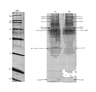
Figures 1-4 show portions of 1-DE maps after Off-gel electrophoresis for ante-
and post-mortem CSF, with arrows indicating bands corresponding to proteins
listed in Table 1. 5-10 1.tg of protein was loaded on a SDS PAGE slab gel
(12.5%T / 2.6% C). The gel was silver stained.
CA 02613991 2008-01-02
WO 2007/007129
PCT/GB2006/050207
11
Figures 5-7 show results of an assay for UFD1 for two groups of patients: a
control group and a group with acute stroke.
Figure 8 shows Western blots of four proteins that were identified only in
postmortem fractions of CSF.
Figure 9 shows results of an assay for GSTP-1 for groups of stroke patients
and
controls, as described in Example 5.
Figure 10 shows Western blot validation of Apolipoprotein A-IV in Alzheimer's
disease, as described in Example 6.
Figure 11 is a scatter plot of values for Complement Factor 3a in plasma from
Alzheimer's disease patients and controls, as described in Example 8.
Figure 12 shows a correlation of complement factor II levels determined by
western blot with Global Dementia Scale in patients with presumed Alzheimer's
disease.
Figure 13 is a Receiver Operating Curve (ROC) for complement factor II and
alpha-2-macroglobulin as candidate plasma biomarkers of Alzheimer's disease.
DESCRIPTION OF PREFERRED EMBODIMENTS
The invention presented here is directed towards compositions and methods for
detecting increasing or reducing polypeptides levels in body fluids including
blood components (e.g. plasma or serum) or cerebrospinal fluid from subjects
affected by a brain damage-related disorder including cerebrovascular,
dementia
and neurodegenerative diseases, as compared with control (non-affected)
subjects.
For this purpose, use can be made of antibodies or any specific polypeptide
detection method.
The invention also includes embodiments where the polypeptides, in particular
those of Table 1, 2, 3 or 4, are determined indirectly. For example, at least
one
CA 02613991 2008-01-02
WO 2007/007129
PCT/GB2006/050207
12
autoantibody to one or more of the polypeptides, in particular those of Table
1, 2,
3 or 4, may be determined.
Antibodies against brain damage protein markers, in particular their protein-
binding domains, are suitable as detection tools. Molecular biological and
biotechnological methods can be used to alter and optimize the antibody
properties of the said molecules in a specific manner. In addition to this,
the
antibodies can be modified chemically, for example by means of acetylation,
carbamoylation, formylation, biotinylation, acylation, or derivatization with
polyethylene glycol or hydrophilic polymers, in order to increase their
stability.
A specific polypeptide marker selected from any of the proteins listed in
Table 1,
2, 3 or 4 is determined in a body fluid sample, for example by using an
antibody
thereto. The marker may simply be detected and/or its concentration may be
measured. The marker is preferably measured by an immunoassay, using a
specific antibody to the polypeptide and measuring the extent of the antigen
(polypeptide)/antibody interaction. The antibody may be a monoclonal antibody
or an engineered (chimeric) antibody. Antibodies to the polypeptides are known
and are commercially available. Also, the usual Kohler-Milstein method may be
used to raise antibodies. Less preferably, the antibody may be polyclonal. In
the
context of the present invention, the term "antibodies" includes binding
fragments
of antibodies, such as single chain or Fab fragments.
Any known method of immunoassay may be used. In a sandwich assay an
antibody (e.g. polyclonal) to the polypeptide is bound to the solid phase such
as a
well of a plastics microtitre plate, and incubated with the sample and with a
labelled second antibody specific to the polypeptide to be detected.
Alternatively,
an antibody capture assay (also called "indirect immunoassay") can be used.
Here, the test sample is allowed to bind to a solid phase, and the anti-
polypeptide
antibody (polyclonal or monoclonal) is then added and allowed to bind. If a
polyclonal antibody is used in this context, it should desirably be one which
exhibits a low cross-reactivity with other forms of polypeptide. After washing
away unbound material, the amount of antibody bound to the solid phase is
determined using a labeled second antibody, anti- to the first.
CA 02613991 2008-01-02
WO 2007/007129
PCT/GB2006/050207
13
A direct assay can be performed by using a labelled anti-polypeptide antibody.
The test sample is allowed to bind to the solid phase and the anti-polypeptide
antibody is added. After washing away unbound material, the amount of antibody
bound to the solid phase is determined. The antibody can be labeled directly
rather than via a second antibody.
In another embodiment, a competition assay can be performed between the sample
and a labeled polypeptide or a peptide derived therefrom, these two antigens
being
in competition for a limited amount of anti-polypeptide antibody bound to a
solid
support. The labeled polypeptide or peptide can be pre-incubated with the
antibody
on the solid phase, whereby the polypeptide in the sample displaces part of
the
polypeptide or peptide thereof bound to the antibody.
In yet another embodiment, the two antigens are allowed to compete in a single
co-
incubation with the antibody. After removal of unbound antigen from the
support
by washing, the amount of label attached to the support is determined and the
amount of protein in the sample is measured by reference to standard titration
curves established previously.
Throughout, the label is preferably an enzyme. The substrate for the enzyme
may
be color-forming, fluorescent, chemiluminescent or electrochemical, and can be
soluble or precipitating. Alternatively, the label may be a radioisotope or
fluorescent, e.g. using conjugated fluorescein.
The enzyme may, for example, be alkaline phosphatase or horseradish peroxidase
and can conveniently be used colorimetrically, e.g. using p-nitrophenyl
phosphate
as a yellow-forming substrate with alkaline phosphatase.
For a chemiluminescent assay, the antibody can be labeled with an acridinium
ester or horseradish peroxidase. The latter is used in enhanced
chemiluminescent
(ECL) assay. Here, the antibody, labeled with horseradish peroxidase,
participates
in a chemiluminescent reaction with luminol, a peroxide substrate and a
CA 02613991 2008-01-02
WO 2007/007129
PCT/GB2006/050207
14
compound, which enhances the intensity and duration of the emitted light,
typically, 4-iodophenol or 4-hydroxycinnamic acid.
An amplified immunoassay such as immuno-PCR can be used. In this technique,
the antibody is covalently linked to a molecule of arbitrary DNA comprising
PCR
primers, whereby the DNA with the antibody attached to it is amplified by the
polymerase chain reaction. See E. R. Hendrickson et al., Nucleic Acids
Research
1995; 23, 522-529 (1995) or T. Sano et al., in "Molecular Biology and
Biotechnology" ed. Robert A. Meyers, VCH Publishers, Inc. (1995), pages 458 -
460. The signal is read out as before.
In one procedure, an enzyme-linked immunosorbent assay (ELISA) can be used to
detect the polypeptide.
The full automation in a widely used clinical chemistry analyser such as the
COBASTM MIRA Plus system from Hoffinann-La Roche, described by M.Robers
et al. Clin Chem. 1998 Jul;44(7):1564-7 or the AxSYMTm system from Abbott
Laboratories, is possible and can be applied for routine clinical diagnosis of
brain
damage-related disorders.
The polypeptide concentrations can be measured by other means than
immunoassay. For example, the sample can be subjected to 2D-gel
electrophoresis
and the amount of the polypeptide estimated by densitometric scanning of the
gel
or of a blot therefrom. However, it is desirable to carry out the assay in a
rapid
manner, so that the patient can be treated promptly.
In principle, any body fluid can be used to provide a sample for diagnosis,
but
preferably the body fluid is cerebrospinal fluid (CSF), plasma, serum, blood,
urine,
tears or saliva.
According to the invention, a diagnosis of brain damage-related disorders may
be
made from determination of a single polypeptide or any combination of two or
more of the polypeptides.
CA 02613991 2008-01-02
WO 2007/007129
PCT/GB2006/050207
The invention also relates to the use of one or more of the specified
polypeptides
which is differentially contained in a body fluid of brain damage-affected
subjects
and non-brain damage-affected subjects, for diagnostic, prognostic and
therapeutic
applications, including for the manufacture of a medicament for treatment of a
5 brain damage-related disorder. This may involve the preparation and/or
use of a
material which recognizes, binds to or has some affinity to the above-
mentioned
polypeptide. Examples of such materials are antibodies and antibody chips. The
term "antibody" as used herein includes polyclonal antiserum, monoclonal
antibodies, fragments of antibodies such as Fab, and genetically engineered
10 antibodies. The antibodies may be chimeric or of a single species. The
above
reference to "prognostic" applications includes making a determination of the
likely course of a brain damage-related disorder by, for example, measuring
the
amount of the above-mentioned polypeptide in a sample of body fluid. The above
reference to "therapeutic follow-up" applications includes making a
determination
15 of the likely course of a brain damage-related disorder by, for example,
measuring
the amount of the above-mentioned polypeptide in a sample of body fluid (and
evaluating its level as a function of the treatment, the disability recovery
or not, the
size of the lesions etc.). The above reference to "therapeutic" applications
includes, for example, preparing materials which recognize, bind to or have
affinity to the above-mentioned polypeptides, and using such materials in
therapy.
The materials may in this case be modified, for example by combining an
antibody
with a drug, thereby to target the drug to a specific region of the patient.
In a
further embodiment, a vaccine directed against a polypeptide, or a variant or
mutant thereof, selected from Table 1, 2, 3 or 4, or an antigenic determinant
(epitope) thereof, is administered to a subject.
The above reference to "presence" or "absence" of a polypeptide, and the
equivalent expressions "present" and "not present", should be understood to
mean
simply that there is a significant difference in the amount of a polypeptide
which
is detected in the affected and non-affected (or control) sample. Thus, the
"absence" of a polypeptide in a test sample may include the possibility that
the
polypeptide is actually present, but in a significantly lower amount than in a
comparative test sample. According to the invention, a diagnosis can be made
on
the basis of the presence or absence of a polypeptide, and this includes the
CA 02613991 2008-01-02
WO 2007/007129
PCT/GB2006/050207
16
presence of a polypeptide in a significantly lower or significantly higher
amount
with reference to a comparative (or control) test sample.
The above references to "detecting" a polypeptide should be understood to
include a reference to compositions and methods for detecting post-
translational
modifications of the polypeptides in addition to quantitative variations.
The invention therefore encompasses the detection of post-translational
modifications in general, and determining whether such modifications of a
polypeptide are consistent with a diagnosis of a brain damage-related
disorder.
One example of such post-translational modification is N-glycosylation.
Kits and assay devices for use in diagnosis of brain damage-related disorders
are
also within the scope of the invention. These may include one or more
antibodies
to a polypeptide selected from any of the proteins listed in Table 1, 2, 3 or
4. The
antibodies will bind to the appropriate polypeptides in a fluid sample taken
from a
patient. The antibodies may be immobilised on a solid support. Preferably,
each
antibody is placed in a unique addressable location, thereby to permit
separated
assay readout for each individual polypeptide in the sample, as well as
readouts for
any selected combination of polypeptides. Such kits and assay devices may also
include antibodies to other marker polypeptides in addition to one or more of
those
in Table 1, 2, 3 or 4. Such other marker polypeptides include those described
in
W001/42793 and W02005/029088. In one particular embodiment, the other
marker polypeptide is glutathione S transferase P.
An assay device according to the invention may comprise a solid substrate
having
one or more locations containing a material which recognizes, binds to or has
affinity for a polypeptide, or a variant or mutant thereof, as defined above,
in
particular selected from Table 1, 2, 3 or 4. Preferred polypeptides which may
be
detected by such a device are fatty acid binding proteins, glutathione S
transferase
P, RNA-BP, UFD1, NDKA, clusterin, Apolipoprotein A-IV, complement factor
II, complement factor 3a, alpha-2-macroglobulin, haemogobin iso forms,
cystatin
C, haemoglobin beta, Apolipoprotein E, Glutathione 5-transferase
Mu 1, Tubulin beta-4 chain, Ubiquitin carboxyl-terminal hydrolase isozyme Li,
Transgelin 3, Neuronal protein Np25, Rab GDP dissociation inhibitor 1,
CA 02613991 2008-01-02
WO 2007/007129
PCT/GB2006/050207
17
Dihydropyrimidinase-like 2 (DRP-2), Aspartate aminotransferase cytoplasmic,
Fructose-bisphosphate aldolase C, and Proteasome subunit alpha type 6. The
assay device may include antibodies to two or more of these polypeptides, to
three
or more, four or more, five or more, or in some cases ten or twenty or more.
The following Examples illustrate the invention.
Abbreviations
CSF: cerebrospinal fluid; II-FABP: heart fatty acid-binding protein; NDKA:
nucleoside diphosphate kinase A; CJD: Creutzfeldt-Jakob disease; OGE: off-gel
electrophoresis; UFD1: ubiquitin fusion degradation protein 1; GST-P:
glutathione S-transferase P; SBPs: spectrin breakdown products.
EXAMPLE 1
Using one-dimensional gel electrophoresis (1-DE) separation of cerebrospinal
fluid (CSF) proteins and mass spectrometry techniques, 58 polypeptides named
in
Table 1 were found elevated or decreased in the CSF of deceased patients, used
as
a model of massive brain damage.
Study population and sample handling
Twenty CSF samples were used for the proteomics-based approach aiming at
discovering brain damage-related disorder markers. Five of these samples were
obtained at autopsy from deceased patients 6 hours after death with no
pathology
of the central nervous system. Fifteen others were collected by lumbar
puncture
from living patients who had a neurological workup for benign conditions
unrelated to brain damage (atypical headache and idiopathic peripheral facial
nerve palsy). CSF samples were centrifuged immediately after collection,
aliquoted, frozen at -80 C and stored until analysis.
CSF depletion fractionation
Immunodepletion of human serum albumin, transferrin, haptoglobin, IgG, IgA
and antitrypsin was performed using a Multiple Affinity Removal System
(Agilent Technologies, Wilmington, USA). 3 ml of CSF was concentrated to
CA 02613991 2008-01-02
WO 2007/007129
PCT/GB2006/050207
18
approximately 300 tl using ultrafiltration (10 kDa MWCO, Vivascience). The
CSF was divided into 200 tl aliquots for immunodepletion according to the
manufacturer's instructions. Combined fractions following depletion were
concentrated using ultrafiltration. Final CSF protein concentrations of
between
600 and 900 iug/ 1 were measured using a Bradford assay. All reagents and
apparatus for off-gel electrophoresis (OGE) have been described in detail
elsewhere (Ros, A., et al., Protein purification by Off-Gel electrophoresis.
Proteomics, 2002. 2(2): p. 151-6). 750 1 of the immunodepleted CSF samples
were loaded on the strip for OGE using all-well loading (50 1 per well). The
samples were focused for a total of 31.6 kVhrs (1 hr at 100 V, 1 hr at 500 V,
1 hr
at 1000 V, 15 hrs at 2000 V). The current was limited to 50 A and the
temperature was controlled at 20 C. Fractions (20-100 1) were collected from
each well and stored at -20 C prior to SDS-PAGE.
1-DE of OGE fractionated CSF proteins
Fractions from OGE were mixed with a 5X concentrated solution of Laemmli's
buffer (0.125 M Tris¨I1C1, 4% SDS, 40% glycerol, 0.1% bromophenol blue, p11
6.8) up to 70 1 and heated at 95 C for 5 min. Samples were centrifuged at
14000g and supernatant loaded on the 12.5% SDS-polyacrylamide gel. Migration
was performed in a Tris-Glycine-SDS p11 8.3 buffer. The gel was then stained
using MS compatible silver staining derived from Blum [Blum, 11., Beier, 11.
and
Gross, 11. J., Electrophoresis 1987, 8, 93-99]. The gel was first fixed for a
minimum of 30 min 50% (v/v) methanol 10% (v/v) acetic acid and then 15 min in
5% (v/v) methanol. The gel was then washed 3 times 5 min in milli-Q 1120 and
incubated 2 min in 0.2 g/L (w/v) fresh sodium thiosulfate (Na25203, 5 1120).
The
gel was further washed 3 times 30 sec in milli-Q 1120, and incubated in the
staining solution, i.e. 25 min in 2 g/L silver nitrate (AgNO3) solution. The
gel
was washed 3 times 1 min in milli-Q 1120, and incubated in the developing
solution (sodium carbonate Na2CO3 30g/L (w/v), 0.05% of 37% IICOH (v/v), 2%
(v/v) of a fresh 0.2 g/L (w/v) sodium thiosulfate (Na25203, 5 1-120)) for 10
min
maximum. The gel development was stopped using a 14 g/1 (w/v) Na2-EDTA
solution for 10 min before washing in milli-Q 1-120. The apparent molecular
masses were determined by running 2 jug of broad range molecular weight
CA 02613991 2013-01-23
19
standards (Bio-Rad, Hercules, CA, USA). The gel was scanned on a Arcus 11
Agfa scanner, with Agfa Fotolook version 3.6 software. Bands to be identified
were cut, placed in an Eppendorf tubes and destained. Each gel piece was
incubated in 30pldestaining solution (30 mM K3FeCN6, 100 mM Na2S203) with
occasional vortexing until the gels were completely destained (5-10 mM). Gel
pieces were then washed twice for 10 min with a minimum of 100 1 milli-Q-1120
for 10 and then stored at 4 C in 10% ethanol (v/v).
Identification of the proteins by nanoLC-ESI-MS/MS
Gel pieces were washed with 200 1 of 50 mM ammonium bicarbonate, for 10
mM. Gel pieces were then dehydrated with 100 1of 100% CH3GN and dried in a
vacuum centrifuge (HETO, Allerod, Denmark). Trypsin digestion was performed
as described previously [Scherl, A., Coute, Y., Deon, C., Calle, A.,
Kindbeiter, K.,
et al., Mol Biol Cell 2002, 13, 4100-9]. NanoLC-ESI-MS/MS was performed on a
LCQ DecaXP ion trap (Therrnofmnigan, San Jose, CA) coupled to a LC-PAL
autosarnpler (CTC Analytics, Zwingen, Switzerland) and a Rheos 2000 Micro
HPLC Pump (Flux Instruments, Basel, Switzerland). For each experiment, 5111 of
sample in 5 % CH3CN, 0.1 % formic acid was injected on a C18 reverse phase
column (75 p.m inner diameter) packed in house with 5p.m Zorbax 300Extend-
C18 ( Agilent Technologies, Wilmington USA). Peptides were eluted from the
column using a CH3CN gradient in the presence of 0.1 % formic acid. For
peptide elution, the acetonitrile concentration was increased from 8 to 47 %
in 15
inM. A flow splitter was used to decrease the flow rate from 40 1/min to
approximately 0.2411/min. A 1.8 kV potential was applied on the nano-
electrospray capillary (New Objective, Woburn, MA). Helium was used as
collision gas. The collision energy was set at 35 % to the maximum. MS/MS
spectra were acquired by automatic switching between MS and MS/MS mode:
The two highest peaks from each MS scan were chosen for MS/MS. Dynamic
exclusion was applied with a repeat count of 2 and a repeat duration of 0.5
mins.
Following these two MSMS acquisitions on the same precursor, the precursor was
excluded from MSMS analysis for 1.0 mM. Spectra were converted to DTA files,
regrouped using in house software and the database search was performed with
MASCOT 1.8. A tolerance of 2.0 Da was
CA 02613991 2013-01-23
chosen for the precursor and 1.0 Da for fragments. ESI-TRAP was selected as
the
instrument. The UniProt Swiss-Prot database was searched without species
restriction. In these conditions, the threshold of significance was given by a
score
of 42 or higher by Mascot. The data was also searched against the UniProt
5 SwissProt database using the Phenyx program.
Protein hits with less than three peptides above the threshold were manually
validated. The data was further searched against the Trembl database,
resulting in
the identification of a further 22 proteins. The results are shown in Table 1.
CA 02613991 2008-01-02
WO 2007/007129
PCT/GB2006/050207
21
Table 1:
Post-mortem CSF
Accession number Protein name
000241 Signal-regulatory protein beta-1
043396 Thioredoxin-like protein 1
043488 Aflatoxin B1 aldehyde reductase member 2
043707 Alpha-actinin 4
075223 Protein C7orf24
095336 6-phosphogluconolactonase
095861 3'(2'),5'-bisphosphate nucleotidase 1
P00352 Retinal dehydrogenase 1
P00390 Glutathione reductase, mitochondrial
P00491 Purine nucleoside phosphorylase
P00915 Carbonic anhydrase I
P01859 Ig gamma-2 chain C region*
P01876, P01877 Ig alpha-1 or -2 chain C region
P02024 Hemoglobin beta chain
P02545 Lamin A/C (70 kDa lamin)
P02741 C-reactive protein
P02760 AMBP protein
P04642 L-Lactate dehydrogenase A chain
P04746, P04745, P19961 Alpha-amylase (pancreatic, salivary or 2B)
P05089 Arginase 1
P05209, Q9BQE3 Tubulin alpha-1 or alpha-6 chain
P05413 Fatty acid-binding protein, heart (II-FABP)
P05976 or P06741 myosin light chain 1 or 3, skeletal muscle
isoform
P06576 ATP synthase beta chain, mitochondria'
P06753 Tropomyosin alpha 3 chain
P07148 Fatty acid-binding protein, liver (L-FABP)
P07203 Glutathione peroxidase 1
P07225 Vitamin K-dependent protein S
P07226 Tropomyosin alpha 4 chain
P07237 Protein disulphide-isomerase
P07357 Complement C8 alpha chain
P07738 Bisphosphoglycerate mutase
P07900 Heat shock protein ITSP 90-alpha (ITSP 86)
P07996 Thrombospondin 1
P08059 Glucose-6-phosphate isomerase
P08133 Annexin A6
P08758 Annexin A5
P09417 Dihydropteridine reductase
P09488 Glutathione S-transferase Mu 1
P09493 or P06753 Tropomyosin 1 alpha chain or alpha 3 chain
CA 02613991 2008-01-02
WO 2007/007129
PCT/GB2006/050207
22
P09525 Annexin A4
P09668 Cathepsin II
P10586 Receptor-type tyrosine-protein phosphatase F
P10599 Thioredoxin
P10768 Esterase D
P11021 78 kDa glucose-regulated protein
P12833 Myosin heavy chain, cardiac muscle beta
isoform
P12882 Myosin heavy chain, skeletal muscle, adult 1
P13489 Placental ribonuclease inhibitor
P13535 Myosin heavy chain, skeletal muscle, perinatal
P13611 Versican core protein
P13693 Translationally controlled tumor protein
(TCTP)
P13716 Delta-amino levulinic acid dehydratase
P13929 Beta enolase
P14136 Glial fibrillary acidic protein, astrocyte (GFAP)
P14550 Alcohol dehydrogenase [NADP+]
P14923 Junction plakoglobin
P15103 Glutamine synthetase
P15121 Aldose reductase
P15259 Phosphoglycerate mutase 2
P15289 Arylsulfatase A
P15924 Desmoplakin
P16930 Fumarylacetoacetase
P17066 Ileat shock 70 kDa protein 6
P18206 Vinculin
P21266 Glutathione S-transferase Mu 3
P21333 Filamin A
P21695 Glycerol-3-phosphate dehydrogenase [NAD+],
cytoplasmic
P22061 Protein-L-isoaspartate (D-aspartate) 0-
methyltransferase
P22314 Ubiquitin-activating enzyme El
P23141 Liver carboxylesterase 1
P24534 Elongation factor 1-beta
P25788 Proteasome subunit alpha type 3
P26038 Moesin
P26641 Elongation factor 1-gamma
P27169 Serum paraoxonase/arylesterase 1
P27348 14-3-3 protein tau
P28072 Proteasome subunit beta type 6
CA 02613991 2008-01-02
WO 2007/007129
PCT/GB2006/050207
23
P28161 Glutathione S-transferase Mu 2
P28827 Receptor-type protein-tyrosine phosphatase mu
P29218 Inositol-1 [or 4] -monophosphate
P29401 Transketolase
P30040 Endoplasmic reticulum protein ERp29
P30041 Peroxiredoxin 6
P30101 Protein disulfide-isomerase A3
P30626 Sorcin (22 kDa protein)
P31946 14-3-3 protein beta/alpha
P31948 Stress-induced-phosphoprotein 1
P34932 Heat shock 70 kDa protein 4
P35080 Profilin-2
P35237 Placental thrombin inhibitor
P36980 Complement factor II-related protein 2
P37837 Transaldolase
P40121 Macrophage capping protein
P42126 3,2-trans-enoyl-CoA isomerase, mitochondria'
P42655 14-3-3 protein epsilon
P45381 Aspartoacylase
P46940 Ras GTPase-activating-like protein IQGAP1
P47756 F-actin capping protein beta subunit
P48637 Glutathione synthetase
P49419 Aldehyde dehydrogenase family 7 member Al
P50135 Histamine N-methyltransferase
P50395 Rab GDP dissociation inhibitor beta
P52565 Rho GDP-dissociation inhibitor 1
P52566 Rho GDP-dissociation inhibitor 2
P52907 F-actin capping protein alpha-1 subunit
P54289 Dihydropyridine-sensitive L-type, calcium
channel alpha-2/delta subunits
P54652 Heat shock-related 70 kDa protein 2
P54922 ADP-ribosylarginine hydrolase
P55287 Cadherin-11
P55854, P61956 Ubiquitin-like protein SMT 3A or 3B
P57087 Junctional adhesion molecule 2
P60900 Proteasome subunit alpha type 6
P61088 Ubiquitin-conjugating enzyme E2 N
P62258 14-3-3 protein epsilon
P62993 Growth factor receptor-bound protein 2
P63104 14-3-3 protein zeta/delta
P68133 Actin, alpha skeletal muscle
CA 02613991 2008-01-02
WO 2007/007129
PCT/GB2006/050207
24
Q00169 Phosphatidylinositol transfer protein alpha
isoform
Q01082 Spectrin beta chain, brain 1
Q01995 Transgelin
Q04917 14-3-3 protein eta
Q06033 Inter-alpha-trypsin inhibitor heavy chain 113
Q12765 Secemin 1
Q13332 Receptor-type tyrosine-protein phosphatase S
Q13509 Tubulin beta-4
Q13740 CD166 antigen
Q13813 Spectrin alpha chain, brain
Q13938 Calcyphosine
Q14126 Desmoglein 2
Q15149 Plectin 1
Q15181 Inorganic pyrophosphatase
Q16620 BDNF/NT-3 growth factors receptor
Q16881 Thioredoxin reductase 1, cytoplasmic
Q86UP2 Kinectin
Q86YZ3 Hornerin
Q8N0Y7 Putative phosphoglycerate mutase 3
Q8TAG5 Immunoglobulin-like domain protein
MGC33530
Q8TD26 Chromodomain-helicase-DNA-binding protein
6
Q92598 Heat shock protein 105 kDa
Q92890 Ubiquitin fusion degradation protein 1 homolog
Q969118 Protein C19 or F10 precursor
Q96IU4 CCG1-interacting factor B
Q9BX68 Histidine triad nucleotide-binding protein 2
Q911477 Ribokinase
Q9NVS9 Pyridoxine-5'-phosphate oxidase
Q9NZT1 Calmodulin-like protein 5
Q9POLO Vesicle-associated membrane protein-
associated protein A
Q9P121 Neurotrimin
Q9UBQ7 Glyoxylate reductase/hydroxypyruvate
reductase
Q9UKK9 ADP-sugar pyrophosphatase
Q9UKX2 Myosin heavy chain, skeletal muscle, adult 2
CA 02613991 2008-01-02
WO 2007/007129
PCT/GB2006/050207
Q9UN36 NDRG2 protein
Q9Y617 Phosphoserine aminotransferase
Q9Y623 Myosin heavy chain, skeletal muscle, fetal
Ante-mortem CSF
P00748 Coagulation factor XII
P01833 polymeric-immunoglobulin receptor
P04083 Annexin Al
P04121 Macrophage capping protein
P05109 Calgranulin A (MRP-8)
P12109 Collagen alpha 1(VI) chain
P22352 Plasma glutathione peroxidase
P35247 Pulmonary surfactant-associated protein D
P43121 Cell surface glycoprotein MUC18
P58876 + others Histone 112B (different forms)
P78509 Reelin
Trembl accession no. Description
095784 IgG Fc binding protein (Fragment)
Q07898, Q07899, M130 antigen; M130 antigen cytoplasmic
Q07900, Q07901, variant 1; variant 2; M130 antigen extracellular
Q86VB7 variant; Similar to CD163 antigen
Hypothetical protein DKFZp779N0926
Q7Z664 (Fragment)
Q7Z623 hypothetical protein
Hepatocellular carcinoma associated protein
Q8IZY7 TB6
Q8N240 Hypothetical protein FLJ34957
Hypothetical protein with 1 extra peptide over
Q8N466 SP entry (Contactin Q12860)
Q8NCW5 ApoA-I binding protein precursor
Q8NFZ8 or Q9Y4A4 TSLC1-like 2 or F22162_1 (Fragment)
Q969J9 Hypothetical protein (Similar to dystroglycan 1)
Q96AC3, Q96FV2, Hypothetical protein, Ses2 protein, Similar to
Q9BUO4 KIAA0193 gene product (Fragment)
Q96B89, Q9H3J8, Hypothetical protein, My027 protein,
Q9HC37, Q9HC38, Hypothetical protein, Hypothetical protein,
Q9Y3E8 CGI-150 protein
Q96B89, Q9H3J8,
Q9HC37, Q9HC38,
Q9Y3E8 various names
Q96B89, Q9H3J8, Hypothetical protein, My027 protein,
Q9HC38, Q9Y3E8 Hypothetical protein, CGI-150 protein
Q96EI3, Q9HOW9 Hypothetical protein
Q96NV4, Q9HOR4 Hypothetical protein FLJ30028, Hypothetical
CA 02613991 2008-01-02
WO 2007/007129
PCT/GB2006/050207
26
protein
Phospholysine phosphohistidine inorganic
Q9H008 pyrophosphate phosphatase
Inositol 1-phosphate synthase, Myo-inositol 1-
Q9H2Y2, Q9NPH2, phosphate synthase Al, Hypothetical protein
Q9NVW7 FLJ10463
Q9NQ56, Q9NQ48 Leucine zipper transcription factor-like 1
DJ665N4.2 (Similar to hypothetical protein
Q9NX46 FLJ20446) (ADP-ribosyl-hydrolase precursor)
Heme-binding protein, Heme-binding protein
Q9Y5Z5, Q9NRV9 (Hypothetical protein)
Q9Y6R7 Human Fc gamma BP (Fragment)
EXAMPLE 2
Introduction
One of the proteins identified as being upregulated in deceased CSF was
evaluated as a potential biomarker of cerebrovascular disease, an example of a
brain damage-related disorder. A survey of stroke patients was carried out and
the
results are shown in Figures 5 to 7. An ELISA intensity signal was obtained
for
Ubiquitin fusion degradation protein 1 homolog (UFD1) in plasma samples of the
patients and of negative control patients. Plasma samples were taken from
patients between 0-24 hours and/or after 72 hours of arrival at emergency
hospital, and were matched for age/sex with samples from control patients.
ELISA was performed using 96-well Reacti-BindTM NeutrAvidinTM coated Black
Plates (Pierce, Rockford, IL). Plates were first rinsed in Borate Buffer
Saline pH
8.4 (BBS) (100 mM H3B03, 25 mM Na2B407 (Sigma, St Louis, MO, USA), 75
mM NaC1 (Merck, Darmastadt, Germany)) on a NOVAPATHTm washer (Bio-
Rad, Hercules, CA). Then, 50 1 of biotin-conjugated antibody (2 iug/m1)
prepared
in the dilution buffer A at pH 7 (DB, Polyvinyl Alcohol, 80% hydrolyzed, Mol.
Wt. 9000-10,000 (Aldrich, Milwaukee, WI, USA), MOPS (3[N-Morpholino]
propane sulfonic acid) (Sigma), NaC1, MgC12 (Sigma), ZnC12 (Aldrich), pH6.90,
BSA 30% Solution, Manufacturing Grade (Serological Proteins Inc., Kankakee,
IL)), was added and incubated for one hour at 37 C. Plates were then washed 3
times in BBS in the plate washer. 50 tl of antigen was then added and
incubated
for one hour at 37 C. Recombinant proteins were diluted at 100, 50, 25, 12.5,
CA 02613991 2008-01-02
WO 2007/007129
PCT/GB2006/050207
27
6.25 ng/ml in the dilution buffer A to establish a calibration curve. Plasma
samples were diluted to the appropriate concentration in the dilution buffer
A.
After the washing step, 50 1 of alkaline phosphatase-conjugated antibody was
added at the appropriate dilution in buffer A and incubated for one hour at 37
C.
The 96-well plate was then washed 3 times with BBS in the plate washer and 50
iLt1 of Attophos AP Fluorescent substrate (Promega, Madison, WI) was added.
Plates were read immediately on a SpectraMax GEMINI-XS fluorometer
microtiter plate reader, (Molecular Devices Corporation, Sunnyvale, CA,
U.S.A.)
(kexcitation=444 nm and kemission=555 nm). Results are expressed in RFU and
can be
obtained in endpoint mode (only one reading) or in kinetic mode for 10
minutes.
In kinetic mode, the plate reader was set to record using 6 flashes (per well)
which were then integrated into an average. In this manner each well was
analysed 6 times using a minimal interval time between each reading. This
translated to a 2 minutes delay between readings. The slope was calculated and
used to determine the final value for each well. The best cut-off value to
discriminate between the control and the stroke (Ischemic plus hemorrhagic or
Ischemic vs. Hemorrhagic) groups was determined using ROC curves generated
in GraphPad Prism 4 software.
Conclusion
It is clear from Figure 5 that UFD1 is overexpressed in the plasma of stroke
patients compared to control patients. Statistical analysis was performed and
ROC
curves (GraphPad Prism 4 software) indicating sensitivity of the test as a
function
of 1-specificity (Figures 6) were drawn. Best cutoff values to distinguish
between
stroke and control patients were deduced from these ROC curves. A sensitivity
and specificity of 94.4% and 77.8%, respectively, was obtained using the best
cutoff values. A non-parametric Mann-Whitney test was performed to compare
stroke and control groups. Very low p values (<0.0001) were obtained,
indicating
that the difference between stroke and controls was highly significant.
This result demonstrates that Ubiquitin fusion degradation protein 1 homo log
(UFD1) is a useful plasmatic marker for early diagnosis of stroke, alone, or
in
combination with other biomarkers.
CA 02613991 2008-01-02
WO 2007/007129
PCT/GB2006/050207
28
As UFD1 has been found in deceased CSF, it is a reasonable prediction that
other
polypeptides and proteins differentially expressed in deceased CSF will also
be
useful as markers for brain damage-related disorders.
EXAMPLE 3
This Example provides additional data showing plasma levels of UFDP1 in stroke
and control patients. Additional data has been obtained from two cohorts of
patients and controls, the smaller from Geneva, and a more comprehensive panel
from the US.
ELISA was performed using 96-well Reacti-BindTM NeutrAvidinTM coated
Black Plates (Pierce, Rockford, IL). Plates were first rinsed in Borate Buffer
Saline pH 8.4 (BBS) (100 mM II3B03, 25 mM Na2B407 (Sigma, St Louis,
MO, USA), 75 mM NaC1 (Merck, Darmastadt, Germany)) on a
NOVAPATHTm washer (Bio-Rad, Hercules, CA). Then, 50 1 of relevant
biomarker specific biotin-conjugated antibody (2 iug/mL) prepared in the
dilution buffer A at p1-17 was added and incubated for one hour at 37 C.
Plates were then washed 3 times in BBS in the plate washer. 50 tl of antigen
or plasma was then added and incubated for one hour at 37 C. Recombinant
protein antigens were diluted at 100, 50, 25, 12.5, 6.25, 3.125, 1.56 ng/ml in
dilution buffer A to generate a calibration curve. Plasma samples were diluted
to the appropriate concentration in dilution buffer A. After a further washing
step, 50 1 of relevant biomarker specific alkaline phosphatase-conjugated
antibodies was added at the appropriate concentration in dilution buffer A and
incubated for one hour at 37 C. The 96-well plate was then washed 3 times
with BBS in the plate washer and 50 tl of Attophos AP Fluorescent
substrate (Promega, Madison, WI) was added. Plates were read immediately
on a SpectraMax GEMINI-XS fluorometer microtiter plate reader (Molecular
Devices Corporation, Sunnyvale, CA, U.S.A.) (X
excitation - -excitation = 444 MT1 and kemission
= 555 nm).
CA 02613991 2008-01-02
WO 2007/007129
PCT/GB2006/050207
29
Results are expressed in RFU and can be obtained in endpoint mode (only one
reading) or in kinetic mode for 10 minutes. In kinetic mode, for each well 6
flashes were averaged and each well was analysed 6 times using a minimal
interval time between each reading (2 minutes). The slope was calculated and
used to determine the fmal value for each well. The best cut-off value to
discriminate between the control and the stroke (Ischemic plus hemorrhagic or
Ischemic vs. Hemorrhagic) groups was determined using ROC curves generated
in GraphPad Prism 4 software.
The results are shown in Figure 7. This result further demonstrates that
Ubiquitin
fusion degradation protein 1 homolog (UFD1) is a useful marker for early
diagnosis of stroke, alone, or in combination with other biomarkers.
As UFD1 has been found in deceased CSF, it is a reasonable prediction that
other
polypeptides and proteins differentially expressed in deceased CSF will also
be
useful as markers for brain damage-related disorders.
EXAMPLE 4
In the current work, we have used an alternative method to 2-DE in order to
further characterize the human postmortem CSF proteome. A pool of postmortem
CSF samples (n = 5) was analyzed using a four step protocol: (i)
immunodepletion of abundant CSF proteins (albumin, IgG, IgA, transferrin,
antitrypsin, and haptoglobin), (ii) fractionation of CSF proteins according to
their
p/ using off-gel electrophoresis (OGE) (24), (iii) analysis of fractions from
OGE
by SDS-PAGE, (iv) protein identification by LC-MS/MS. Selected proteins that
were identified in postmortem CSF were validated using Western blots of
individual postmortem and ante-mortem CSF samples. The potential interest of
proteins identified in postmortem CSF as biomarkers of brain damage will be
discussed.
CA 02613991 2008-01-02
WO 2007/007129
PCT/GB2006/050207
Experimental Procedures
Materials:
All chemicals, unless otherwise stated, were purchased from Sigma Aldrich (St.
Louis, MI, USA) and were of the highest purity available. CH3CN was purchased
5 from Biosolve (Westford, MA, USA).
CSF collection:
Postmortem CSF samples from five different patients were collected by
ventricular puncture at autopsy, 6 hours after death on average. Deceased
patients
10 had no history, symptoms or signs of any psychiatric or neurological
condition.
Cause of death was unrelated to any dysfunction of the central or peripheral
nervous system and neuropathological data of the brain were consistent with
age-
related changes with no relevant pathology. Control ante-mortem CSF samples
were used for Western blot validation. They were collected by diagnostic
lumbar
15 puncture from five living patients who had a neurological workup for
benign
conditions unrelated to brain damage (atypical headache and idiopathic
peripheral
facial nerve palsy). Each patient or patient's relatives gave informed consent
prior
to enrolment. Atraumatic CSF samples were centrifuged immediately after
collecting, aliquoted, frozen at ¨ 80 C, and stored until analysis.
Blood sample collection:
Plasma samples obtained from the Geneva University Hospital were used for the
assessment of the level of GST-P1. The local institutional ethical committee
board
approved the clinical protocol. Seven consecutive stroke and control patients
admitted to the Geneva University Hospital emergency unit were enrolled in
this
study. Of the 7 consecutive patients enrolled, 3 were diagnosed with non-
neurological conditions and classified as control samples (2 men and 1 women,
average age of 70.26 years) and 4 were diagnosed with stroke (3 men and 1
women, average age of 71.81 years) including 2 ischemic and 1 intra-cerebral
hemorrhagic strokes. The diagnosis of stroke was established by a trained
neurologist and was based on the sudden appearance of a focal neurological
deficit and the subsequent delineation of a lesion consistent with the
symptoms on
brain CT or MRI images. The control group included patients with cancer (n=2)
and a gastro-intestinal disorder (n=1). For each patient, a blood sample was
CA 02613991 2008-01-02
WO 2007/007129
PCT/GB2006/050207
31
collected at the time of admission in dry heparin-containing tubes within the
three
hours window after onset of symptoms. After centrifugation at 1500g for 15 min
at 4 C, samples were aliquoted and stored at ¨80 C until analysis. Analyses
were
performed on frozen samples.
Depletion of abundant proteins:
Pooled postmortem CSF samples were concentrated to 300 1 using 10 kDa
MWCO ultrafiltration devices (Vivaspin UF 4, Vivascience, Germany). The
protein load was approximately 1.6 mg. The sample was then diluted 1:5 in
MARS buffer A (Agilent, Palo Alto, CA, USA) and passed through a 0.22 pun
filter. Aliquots of 200 I were injected on a 4.6 x 100 mm MARS column
(Agilent). The flow-through fractions were collected, pooled and concentrated
to
approximately 1 ml using ultrafiltration. These concentrated fractions were
washed twice with 10 mM NH4HCO3. A protein concentration assay was
performed using the Bradford method (Bio-Rad, Hercules, CA, USA).
Off-gel electrophoresis:
The OGE fractionation was performed as in Heller, M., Michel, P.E., Morier,
P.,
Crettaz, D., Wenz, C., Tissot, J.D., Reymond, F., and Rossier, J.S. (2005) Two-
stage Off-Gel isoelectric focusing: protein followed by peptide fractionation
and
application to proteome analysis of human plasma. Electrophoresis 26, 1174-
1188. The depleted CSF was prepared for OGE by adding urea, thiourea and
DTT to final concentrations of 7M, 2M and 65 mM, respectively. IPG strips (13
cm, pH 4.0 ¨ 7.0) were rehydrated in a solution containing 7 M urea, 2 M
thiourea, 65 mM DTT, 0.5% (v/v) ampholytes (pH 4.0 ¨ 7.0) and 5% glycerol. A
15 well device was then placed on the rehydrated IPG and 50 1 of sample was
loaded in each well across the whole strip. Several multiwell devices were
used in
parallel to allow fractionation of the whole sample in a single experiment.
The
voltage was started at 100 V (1 hour) then increased to 500 V (for 1 hour),
1000 V
(for 1 hour) and finally to 2000 V where it was maintained for 15 hours. The
focusing was performed at 20 C with a current limit of 50 mA. Fractions were
recovered from each of the wells.
CA 02613991 2008-01-02
WO 2007/007129
PCT/GB2006/050207
32
SDS-PAGE and in-gel digestion:
Proteins from OGE fractions were separated by SDS-PAGE on home-made 12%
T Tris-Glycine gels (8 x 5 x 0.15 cm). Approximately 60 IA of each fraction
was
loaded on the gel. After the migration, gels were stained with an MS-
compatible
silver stain (Blum, II., Beier, II., and Gross, 1-1.J. (1987) Improved silver
staining
of plant proteins, RNA and DNA in polyacrylamide gels. Electrophoresis 8, 93-
99). Bands cut from the silver-stained gels were destained with 15 mM
K3Fe(CN6), 50 mM Na25203, and washed with MilliQ water (Millipore, Billerica,
MA, USA) (26). The gel pieces were then dehydrated in 100% CI-13CN and dried
in a vacuum centrifuge. The proteins were in-gel digested using standard
protocols (Scherl, A., Coute, Y., Deon, C., Calle, A., Kindbeiter, K.,
Sanchez,
J.C., Greco, A., Hochstrasser, D., and Diaz, J.J. (2002) Functional proteomic
analysis of human nucleolus. MoL Biol. Cell 13, 4100-4109). Peptides were
extracted with 1% TFA followed by 50% CI-13CN, 0.1% TFA. The combined
extracts were concentrated by vacuum centrifugation.
LC-MS/MS:
Peptides extracted following in-gel digestion were dissolved in 9 15% CI-
13CN,
0.1% formic acid and 5 I was loaded for LC-MS/MS analysis. A precolumn (100
m inner diameter, 2 - 3.5 cm long) was connected directly to an analytical
column (75 m inner diameter, 9 - 10 cm long). Both columns were packed in-
house with 5 m, 3A Zorbax Extend C-18 (Agilent). A gradient from 4 to 56%
solvent B in solvent A (Solvent A: 5% CI-13CN, 0.1% formic acid, Solvent B:
80%
CI-13CN, 0.1% formic acid) was developed over 15 minutes at a flow rate of
approximately 300 nl/min. The concentration of solvent B was increased to 95%
before returning to start conditions for re-equilibration of the column. The
eluate
was sprayed directly into the nano-ESI source of an LCQ DecaXP ion trap mass
spectrometer (Thermo Finnigan, San Jose, CA) with a spray voltage of 1.8 - 2.2
kV. Data dependent acquisition was used to automatically select 2 precursors
for
MS/MS from each MS spectrum (m/z range 400-1600). MS/MS spectra were
acquired with a normalized collision energy of 35%, an activation Q of 0.25
and
an isolation width of 4 m/z. The activation time was 30 milliseconds. Dynamic
exclusion was applied with a repeat count of 2, an exclusion time of 30
seconds,
and an exclusion peak width of 1,5 Da. Wideband activation was also applied.
CA 02613991 2008-01-02
WO 2007/007129
PCT/GB2006/050207
33
Maximum injection times of 50 milliseconds and 200 milliseconds were used for
MS and MS/MS acquisitions, respectively, and the corresponding automatic gain
control targets were set to 108.
Data extraction and database interrogation:
Peak lists were generated using Bioworks 3.1 software (Thermo Finnigan, San
Jose, CA). The resulting data files from each analysis were automatically
combined into a single text file. The resulting peak lists were searched
against the
UniProt/Swiss-Prot database without species restriction using Mascot operating
on a local server (version 1.8, Matrix Sciences, U.K.) and Phenyx Virtual
Desktop
(Gene Bio, Switzerland). Mascot was used with average mass selected, a
precursor mass error of 2.0 Da and a peptide mass error of 1.0 Da. Trypsin was
selected as the enzyme, with a single potential missed cleavage. ESI ion trap
was
selected as the instrument type and oxidized methionine as a variable
modification. For Phenyx, ion trap was selected for the instrument type and
LCQ
for the algorithm. Two search rounds were used, both with trypsin selected as
the
enzyme and oxidized methionine as a variable modification. In the first round
1
missed cleavage was allowed and the normal cleavage mode was used. This round
was selected in 'turbo' search mode. In the second round 2 missed cleavages
were allowed and the cleavage mode was set to half-cleaved. The minimum
peptide length allowed was 6 amino acids and the parent ion tolerance was 2.0
Da
in both search rounds. The acceptance criteria were slightly lowered in the
second
round search (round 1: AC score 7.0, peptide Z-score 7.0, peptide p-value 1 E-
6;
round 2: AC score 7.0, peptide Z-score 6.0, peptide p-value 1 E-5).
Proteins that were identified as human proteins with 3 or more high-scoring
peptides from both Mascot and Phenyx were accepted to be true matches. 'High
scoring peptides' corresponded to peptides that were above the threshold in
Mascot searches (5% probability of false match for each peptide above this
score)
and above a peptide score of 8.5 for Phenyx searches using the LCQ scoring
algorithm. Matches with fewer than 3 peptides were manually validated. Single
peptide matches were only included if they were high scoring peptides in the
results from both programs and if the data was considered to match the peptide
sequence well.
CA 02613991 2008-01-02
WO 2007/007129
PCT/GB2006/050207
34
The peak lists were also searched against the UniProt combined Swiss-Prot and
TrEMBL database restricted to human entries using Phenyx Virtual Desktop
(Gene Bio, Switzerland). The acceptance criteria were more stringent than for
the
search of the Swiss Prot database alone (round 1: AC score 16.0, peptide Z-
score
8.0, peptide p-value 1 E-7; round 2: AC score 10.0, peptide Z-score 7.0,
peptide p-
value 1 E-6).
Two-dimensional gel electrophoresis:
A volume of 30 1.t1 of crude or depleted CSF was mixed with 120 1.t1 of a
rehydration solution. The final solution contained 8M urea, 4% (w/v) CHAPS, 65
mM DTT, 2% (v/v) Resolytes 3.5-10 and a trace of bromophenol blue. The whole
sample corresponding to approximately 6 1.tg of proteins was used for
rehydration
of a commercial 7 cm non-linear pH 3-10 IPG strip (GE Healthcare, Uppsala,
Sweden). IEF was carried out. The second dimensional separation was performed
on in-house manufactured SDS-PAGE gels (9 x 8 x 0.15 cm, 12% T, 2.6% C).
Gels were then stained with ammoniacal silver.
Immunoblot analyses of ante- and postmortem CSF samples:
Postmortem and ante-mortem CSF samples (20 1) were loaded on home-made
12% T Tris-Glycine gel (8 x 7 x 0.1 cm). The following positive controls were
used: 100 ng of recombinant calcyphosine (Scientific Proteins, Switzerland),
100
ng of recombinant ubiquitin fusion degradation protein 1 (UFD1) (Biosite, San
Diego, CA, USA), 1 IA of U373 cell line extract for 14-3-3 protein isoform
beta,
and 5 IA of HeLa cell line extract for glutathione S-transferase P (GST-P).
Proteins separated by SDS-PAGE were electroblotted onto a PVDF membrane as
described by Towbin et al. (Towbin, H., Staehelin, T., and Gordon, J. (1979)
Electrophoretic transfer of proteins from polyacrylamide gels to
nitrocellulose
sheets: procedure and some applications. Proc. Natl. Acad. Sci. USA 76, 4350-
4354). Membranes were stained with Amido-Black, destained with water and
dried. Immunodetection was performed using specific antibodies and BM
Chemiluminescence Western Blotting Kit (Roche, Basel, Switzerland). The
following antibodies were used: anti-human calcyphosine rabbit polyclonal
antibody (Scientific Proteins, Witterswil, Switzerland) diluted 1/1000, anti-
human
UFD1 mouse Omniclonal antibody (Biosite, San Diego, CA, USA) diluted
CA 02613991 2008-01-02
WO 2007/007129
PCT/GB2006/050207
1/1000, anti-human 14-3-3 B rabbit polyclonal antibody (Santa Cruz
Biotechnology, Santa Cruz, CA, USA) diluted 1/500, anti-human GST-P mouse
monoclonal antibody (Transduction Laboratories, Lexington, KY, USA) diluted
1/1000.
5
Immunoblot detection of 14-3-3 protein in OGE fractions:
Five IA of OGE fractions obtained from postmortem and ante-mortem CSF pools
were loaded on home-made 12% T Tris-Glycine gels (8 x 7 x 0.1 cm). Five IA of
crude postmortem and ante-mortem CSF pools were used as positive and negative
10 controls, respectively. Proteins separated by 1-DE were electroblotted
onto a
PVDF membrane as described by Towbin et al. (30). Membranes were stained
with Amido-Black, destained with water and dried. Immunodetection was
performed using anti-human 14-3-3 rabbit polyclonal antibody (Santa Cruz
Biotechnology, Santa Cruz, CA, USA) diluted 1/500 and BM Chemiluminescence
15 Western Blotting Kit (Roche, Basel, Switzerland).
Sandwich ELISA detection of GST-P 1 :
As no commercial kit was available for the detection of GST-P1, a homemade
ELISA test was developed. A trained laboratory technician carried out the
assays
20 (in an un-blind manner) with less than 15% coefficient variation.
Sandwich
ELISA was performed using 96-well Reacti-BindTM NeutrAvidinTM coated Black
Plates (Pierce, Rockford, IL). Plates were first rinsed in Borate Buffer
Saline pH
8.4 (BBS) (100 mM II3B03, 25 mM Na2B407 (Sigma, St Louis, MO, USA) 75
mM NaC1 (Merck, Darmastadt, Germany)) on a NOVAPATHTm washer (Bio-
25 Rad, Hercules, CA). Then, 50 L of GST-P1 monoclonal antibody-biotin
conjugated (2 iug/mL) prepared in the dilution buffer A at p1-17 (DB,
Polyvinyl
Alcohol, 80% hydrolyzed, Mol. Wt. 9000-10,000 (Aldrich, Milwaukee, WI,
USA), MOPS (Sigma), NaC1, MgC12 (Sigma), ZnC12 (Aldrich), pI16.90, BSA
30% Solution, Manufacturing Grade (Serological Proteins Inc., Kankakee, IL)),
30 were added and incubated for one hour at 37 C. Plates were then washed 3
times
in BBS in the plate washer. Fifty tL of blood or CSF samples were used diluted
twice and incubated for one hour at 37 C. Each sample was assayed in duplicate
and distributed randomly on the plate. Recombinant GST-P1 protein
(Invitrogen,)
CA 02613991 2008-01-02
WO 2007/007129
PCT/GB2006/050207
36
was diluted at 100 ng/mL in the dilution buffer A. The calibration curve was
performed in the same plate at a concentrations of 100, 50, 25, 12.5, 6.25,
3.125,
1.56 and 0 g/L. After the washing step, 50 L of alkaline phosphatase
conjugated
GST-P1 monoclonal antibodies were added at the appropriate dilution in the
dilution buffer A and incubated for one hour at 37 C. The 96-well plate was
then
washed 3 times with BBS in the plate washer and 50 tL of fluorescence
Attophos AP Fluorescent substrate (Promega, Madison, WI) were added. Plates
were read immediately on a SpectraMax GEMINI-XS, (Molecular Devices
Corporation, Sunnyvale, CA, U.S.A.) fluorometer microtiter plate reader using
the
endpoint mode relative fluorescence units (RFU)
-excitation=444 nm and
kemissiori555 nm). A calibration curve was performed using a linear regression
in
the linear range of the curve. Protein levels were initially expressed in
relative
fluorescence units (RFU) and the concentrations were calculated via the
calibration curve.
Results:
Abundant protein depletion:
The analysis of body fluids, such as CSF, poses a challenge in terms of the
high
dynamic range of protein concentrations. The dominance of particular proteins
such as albumin and immunoglobulins results in many proteins of lower
abundance remaining undetected by conventional techniques such as 2-DE and
mass spectrometry. Therefore immunodepletion of some of the most abundant
CSF proteins (albumin, serotransferrin, IgG, IgA, haptoglobin, and -1-
antitrypsin) was performed in order to improve the coverage of low abundance
proteins. To access the results from depletion of abundant proteins, 2-DE of
the
CSF samples was performed before and after immunoaffinity subtraction. The
gels show major similarities before and after depletion and confirm that the
removal of some abundant proteins enabled the detection of spots of lower
abundance. This result obtained for the postmortem CSF sample reproduces
perfectly those presented by Maccarrone et al. (Maccarrone, G., Milfay, D.,
Birg,
I., Rosenhagen, M., Holsboer, F., Grimm, R., Bailey, J., Zolotarjova, N., and
Turck, C.W. (2004) Mining the human cerebrospinal fluid proteome by
immunodepletion and shotgun mass spectrometry. Electrophoresis 25, 2402-12)
CA 02613991 2008-01-02
WO 2007/007129
PCT/GB2006/050207
37
for ante-mortem CSF with similar depletion reproducibility from run to run
(data
not shown).
Off-gel electrophoresis:
Following depletion of abundant proteins, the postmortem CSF sample was
fractionated by OGE according to their p/. OGE was performed using a p1I
gradient ranging from 4.0 to 7Ø The fractions obtained from OGE were then
separated by SDS-PAGE. Figure 2 shows a silver-stained SDS-PAGE gel of
postmortem CSF sample. As a result of the OGE fractionation, with some bands
were represented in multiple fractions and others concentrated in one or two
fractions. Western blots were also used to verify the quality of the OGE
fractionation. A sample of the pooled postmortem CSF was separated by SDS-
PAGE along with each of the fractions from OGE of the sample. For example, 14-
3-3 protein gamma was apparent in the un-fractionated postmortem CSF sample
and in a single fraction following OGE of the postmortem CSF sample (fraction
3). These results corresponded with the identifications obtained by MS and
database searching. The 14-3-3 protein gamma was identified in one band of
fraction 3 from the postmortem CSF fractionation (see Table 2). The ante-
mortem
CSF sample did not show any band for the gamma 14-3-3 protein.
Identification by mass spectrometry:
Proteins were identified from bands cut from the gels. Bands were cut from
equivalent regions of both the postmortem CSF gels and the ante-mortem CSF
gels. The only parts of the lanes that were not excised were those where
neither
the post- nor the ante-mortem samples showed bands. A total of 316 proteins
were
identified in this study and these results are listed in Tables 2 and 3, in
which
Table 2 contains proteins from the UniProt/Swiss-Prot database (searched with
all
species) and Table 3 contains proteins identified from the UniProt TrEMBL
database (searched with the taxonomy restricted to human) (see Supplementary
data). Of all the proteins identified, 294 were identified from the Swiss Prot
database and a further 22 from the human TrEMBL searches. Of the 299 proteins
that were identified from the postmortem CSF fractions, 201 were uniquely
identified in postmortem CSF. A total of 115 proteins were identified in ante-
mortem CSF fractions and 17 of these proteins were unique to these fractions.
Of
CA 02613991 2008-01-02
WO 2007/007129
PCT/GB2006/050207
38
all the proteins identified, 98 were present in both the postmortem and ante-
mortem CSF fractions.
CA 02613991 2008-01-02
WO 2007/007129
PCT/GB2006/050207
39
Table 2
000241 Signal-regulatory protein beta-1
000584 Ribonuclease T2
014745 Ezrin-radixin-moesin binding phosphoprotein 50
015394 Neural cell adhesion molecule 2
043396 Thioredoxin-like protein
043488 Aflatoxin B1 aldehyde reductase member 2
043505 N-acetyllactosaminide beta-1,3-N-
acetylglucosaminyltransferase
043707 Alpha-actinin 4
075223 Protein C7orf24
094760 NG,NG-dimethylarginine dimethylaminohydrolase 1
094919 Probable Exonuclease KIAA0830
094985 Calsyntenin-1
095336 6-phosphogluconolactonase
095502 Neuronal pentraxin receptor
095861 3'(2'),5'-bisphosphate nucleotidase 1
095865 NG,NG-dimethylarginine dimethylaminohydrolase 2
P00352 Retinal dehydrogenase 1
P00390 Glutathione reductase, mitochondria'
P00441 Superoxide dismutase [Cu-Zn]
P00450 Ceruloplasmin
P00491 Purine nucleoside phosphorylase
P00734 Prothrombin
P00738 Haptoglobin
P00751 Complement factor B
P00915 Carbonic anhydrase I
P00918 Carbonic anhydrase 2
P01008 Antithrombin-III 130
P01009 Alpha-l-antitrypsin
P01011 Alpha-1 -antichymotrypsin
P01019 Angiotensinogen
P01023 Alpha-2-macroglobulin 1
P01024 Complement C3
P01028 Complement C4
P01034 Cystatin C
P01042 Kininogen
P01834 Ig kappa chain C region
P01857 Ig gamma-1 chain C region
P01859 Ig gamma-2 chain C
P01876, P01877 Ig alpha-1 or -2 chain C region
P02545 Lamin A/C (70Kda lamin)
P02647 Apolipoprotein A-I
P02649 Apolipoprotein E
P02675 Fibrinogen beta chain
CA 02613991 2008-01-02
WO 2007/007129
PCT/GB2006/050207
P02679 Fibrinogen gamma chain
P02741 C-reactive protein
P02743 Serum amyloid P-component
P02748 Complement component C9
P02751 Beta-2-glycoprotein I
P02751 Fibronectin
P02753 Plasma retinol-binding protein
P02760 AMBP protein
P02765 Alpha-2-ITS-glycoprotein
P02766 Transthyretin
P02768 Serum albumin
P02774 Vitamin D-binding protein
P02787 Serotransferrin
P02790 Hemopexin
P02792 Ferritin light chain
P02794 Ferritin heavy chain
P04217 Alpha-1B-glycoprotein
P04406 Glyceraldehyde 3-phosphate dehydrogenase
P04746, P04745, P19961 Alpha-amylase (Pancreatic, salivary or 2B)
P05089 Arginase-1
P05090 Apolipoprotein D
P05156 Complement factor I
P05216 Tubulin alpha-6 chain
P05413 Fatty acid-binding protein (II-FABP)
P05452 Tetranectin
P05543 Thyroxine-binding globulin
P05976 Myosin light chain 1
P06396 Gelsolin
P06576 ATP synthase beta chain
P06702 Calgranulin B (MRP-14)
P06727 Apolipoprotein A-IV
P06733 Alpha enolase
P06753 Tropomyosin alpha 3 chain
P07148 Fatty acid-binding protein
P07195 L-lactate dehydrogenase B chain
P07203 Glutathione peroxidase 1
P07225 Vitamin K-dependent protein S
P07237 Protein disulfide-isomerase
P07339 Cathepsin D
P07357 Complement component C8 alpha chain
P07738 Bisphosphoglycerate mutase
P07900 Heat shock protein ITSP 90-alpha (1-ISP 86)
P07996 Thrombospondin-1
P08107 Heat shock 70 kDa protein 1
P08133 Annexin A6
P08238 Heat shock protein ITSP 90-beta (1-ISP 84)
P08294 Extracellular superoxide dismutase [Cu-Zn]
P08571 Monocyte differentiation antigen CD14
CA 02613991 2008-01-02
WO 2007/007129
PCT/GB2006/050207
41
P08603 Complement factor II
P08670 Vimentin
P08758 Annexin A5
P09211 Glutathione S-transferase P
P09417 Dihydropteridine reductase
P09486 SPARC
P09488 Glutathione S-transferase Mu 1
P09493, P06753Tropomyosin 1 alpha chain or alpha 3 chain
P09525 Annexin A4
P09668 Cathepsin II
P09871 Complement Cis component
P09936 Ubiquitin carboxyl-terminal hydrolase isozyme Li
P09972 Fructose-bisphosphate aldolase C
P10451 Osteopontin
P10586 Receptor-type tyrosine-protein phosphatase F
P10599 Thioredoxin
P10643 Complement component C7
P10768 Esterase D
P10909 Clusterin
P11021 78 kDa glucose-regulated protein
P11142 Heat shock cognate 71 kDa protein
P12277 Creatine kinase, B chain
P12882 Myosin heavy chain, skeletal muscle, adult 1
P12883 Myosin heavy chain, cardiac muscle beta isoform
P13489 Placental ribonuclease inhibitor
P13535 Myosin heavy chain, skeletal muscle, perinatal
P13592 Neural cell adhesion molecule 1, 120 kDa isoform
P13611 Versican core protein
P13693 Translationally controlled tumor protein (TCTP)
P13716 Delta-aminolevulinic acid dehydratase
P13929 Beta enolase
P14136 Glial fibrillary acidic protein, astrocyte (GFAP)
550 Alcohol dehydrogenase [NADP+]
P14618 Pyruvate kinase, M1 isozyme
P14923 Junction plakoglobin
P15090 Fatty acid-binding protein, adipocyte (AFABP)
P15121 Aldose reductase
P15259 Phosphoglycerate mutase 2
P15289 Arylsulfatase A
P15311 Ezrin
P15924 Desmoplakin
P16035 Metalloproteinase inhibitor 2
P16083 NRH dehydrogenase [quinone] 2
P16870 Carboxypeptidase FT
P16930 Fumarylacetoacetase
04967 Heat shock 70 kDa protein 6
P17174 Aspartate aminotransferase, cytoplasmic
P18206 Vinculin (Metavinculin)
CA 02613991 2008-01-02
WO 2007/007129
PCT/GB2006/050207
42
P18669 Phosphoglycerate mutase 1
P19022 Neural-cadherin
P21266 Glutathione S-transferase Mu 3
P21333 Filamin A
P21695 Glycerol-3-phosphate dehydrogenase [NAD+]
P22061 Protein-L-isoaspartate(D-aspartate) 0-methyltransferase
P22314 Ubiquitin-activating enzyme El
P23141 Liver carboxylesterase 1
P23142 Fibulin-1
P23528 Cofilin, non-muscle isoform
P24534 Elongation factor 1-beta
P24592 Insulin-like growth factor binding protein 6
P25786 Proteasome subunit alpha type 1
P25788 Proteasome subunit alpha type 3
P26041 Moesin
P26641 Elongation factor 1-gamma
P27169 Serum paraoxonase/arylesterase 1
P27348 14-3-3 protein tau
P28072 Proteasome subunit beta type 6
P28161 Glutathione S-transferase Mu 2
P28827 Receptor-type protein-tyrosine phosphatase mu
P29218 Inosito1-1(or 4)-monophosphatase
P29401 Transketolase
P30040 Endoplasmic reticulum protein ERp29
P30041 Peroxiredoxin 6
P30044 Peroxiredoxin 5, mitochondrial
P30086 Phosphatidylethanolamine-binding protein (PEBP)
P30101 Protein disulfide-isomerase A3
P30626 Sorcin
P30740 Leukocyte elastase inhibitor (LEI)
P31150 Rab GDP dissociation inhibitor alpha
P31947 14-3-3 protein beta/alpha
P31948 Stress-induced-phosphoprotein 1
P32119 Peroxiredoxin 2
P34932 Heat shock 70 kDa protein 4
P35080 Profilin-2
P35237 Placental thrombin inhibitor
P36955 Pigment epithelium-derived factor
P36980 Complement factor II-related protein 2
P37837 Transaldolase
P40121 Macrophage capping protein
P40925 Malate dehydrogenase, cytoplasmic
P41222 Prostaglandin-II2 D-isomerase
P42126 3,2-trans-enoyl-CoA isomerase, mitochondrial
P43652 Afamin
P45381 Aspartoacylase
P46940 Ras GTPase-activating-like protein IQGAP1
P47756 F-actin capping protein beta subunit
CA 02613991 2008-01-02
WO 2007/007129
PCT/GB2006/050207
43
P48637 Glutathione synthetase
P49419 Aldehyde dehydrogenase family 7 member Al
P50135 Histamine N-methyltransferase
P50395 Rab GDP dissociation inhibitor beta
P51693 Amyloid-like protein 1
P51884 Lumican
P52565 Rho GDP-dissociation inhibitor 1
P52566 Rho GDP-dissociation inhibitor 2
P52907 F-actin capping protein alpha-1 subunit
P54289 Dihydropyridine-sensitive L-type, calcium channel alpha-2/delta
subunits
P54652 Heat shock-related 70 kDa protein 2
P54764 Ephrin type-A receptor 4
P54922 ADP-ribosylarginine hydrolase
P55287 Cadherin-11
P55855 Ubiquitin-like protein SMT 3A or 3B
P57087 Junctional adhesion molecule 2
P60174 Triosephosphate isomerase
P60709 Actin, cytoplasmic 1
P60900 Proteasome subunit alpha type 6
P61088 Ubiquitin-conjugating enzyme E2 N
P61917 Epididymal secretory protein El
P61981 14-3-3 protein gamma
P62258 14-3-3 protein epsilon
P62941 Peptidyl-prolyl cis-trans isomerase A
P62988 Ubiquitin
P87379 Growth factor receptor-bound protein 2
P63103 14-3-3 protein zeta/delta
P63261 Actin, cytoplasmic 2
P67936 Tropomyosin alpha 4 chain
P68136 Actin, alpha skeletal muscle
P68224 Hemoglobin beta chain
P78324 Tyrosine-protein phosphatase non-receptor type substrate 1
P78417 Glutathione transferase omega 1
P81605 Dermcidin
P98160 Basement membrane-specific heparan sulfate proteoglycan core
protein
Q00169 Phosphatidylinositol transfer protein alpha isoform
Q01082 Spectrin beta chain, brain 1
Q01469 Fatty acid-binding protein, epidermal (E-FABP)
Q01995 Transgelin
Q03591 Complement factor H-related protein 1
Q02246 Contactin-2
Q04917 14-3-3 protein eta
Q06033 Inter-alpha-trypsin inhibitor heavy chain 113
Q06830 Peroxiredoxin 1
Q12765 Secernin 1
Q12860 Contactin 1
CA 02613991 2008-01-02
WO 2007/007129
PCT/GB2006/050207
44
Q13228 Selenium-binding protein 1
Q13332 Receptor-type tyrosine-protein phosphatase S
Q13449 Limbic system-associated membrane protein
Q13509 Tubulin beta-4
Q13740 CD166 antigen
Q13813 Spectrin alpha chain, brain
Q14118 Dystroglycan
Q14126 Desmoglein-2
Q14515 SPARC-like protein 1
Q14624 Inter-alpha-trypsin inhibitor heavy chain 114
Q15818 Neuronal pentraxin I
Q15149 Plectin 1
Q15181 Inorganic pyrophosphatase
Q16270 Insulin-like growth factor binding protein 7
Q16555 Dihydropyrimidinase related protein-2
Q16620 BDNF/NT-3 growth factors receptor
Q16881 Thioredoxin reductase 1, cytoplasmic
Q86UP2 Kinectin
Q86YZ3 Hornerin
Q8N0Y7 Putative phosphoglycerate mutase 3
Q8TAG5 Immunoglobulin-like domain protein MGC33530
Q8TD26 Chromodomain-helicase-DNA-binding protein 6
Q92520 Protein FAM3C
Q92598 Heat-shock protein 105 kDa
Q92823 Neuronal cell adhesion molecule
Q92876 Kallikrein-6
Q92890 Ubiquitin fusion degradation protein 1 homolog
Q969118 Protein C19 or fl 0
Q96IU4 CCG1-interacting factor B
Q96KN2 Glutamate carboxypepticlase-like protein 2
Q96NY7 Chloride intracellular channel 6
Q99497 DJ-1 protein
Q9BX68 Histidine triad nucleotide-binding protein 2
Q911477 Ribokinase
Q9NVS9 Pyridoxine-5'-phosphate oxidase
Q9NZT1 Calmodulin-like protein 5
Q9POLO Vesicle-associated membrane protein-associated protein A
Q9P121 Neurotrimin
Q9P1W8 Signal-regulatory protein beta-2
Q9P2S2 Neurexin 2-alpha
Q9UBP4 Dickkopf related protein-3
Q9UBQ7 Glyoxylate reductase/hydroxypyruvate reductase
Q9UKK9 ADP-sugar pyrophosphatase
Q9UKX2 Myosin heavy chain, skeletal muscle, adult 2
Q9UN36 NDRG2 protein
Q9Y617 Phosphoserine aminotransferase
Q9Y623 Myosin heavy chain, skeletal muscle, fetal
TrEMBL entries
CA 02613991 2008-01-02
WO 2007/007129
PCT/GB2006/050207
000533 Neural cell adhesion molecule
043598 RCL (Similar to putative C-MYC-responsive)
095784 IgG Fc binding protein (Fragment)
Q07898,
Q07899, M130 antigen; M130 antigen
Q07900, cytoplasmic variant 1; variant 2;
Q07901, M130 antigen extracellular variant;
Q86VB7 Similar to CD163 antigen
Q7Z664 Hypothetical protein DKFZp779N0926 (Fragment)
Q7Z7P9 Hypothetical protein
Q8IZY7 Hepatocellular carcinoma associated protein TB6
Q8N240 Hypothetical protein FLJ34957
Q8NCW5 ApoA-I binding protein
Q8NFZ8 TSLC1-like 2
Q96AC3, Hypothetical protein, Ses2 protein,
Q96FV2, Similar to KIAA0193 gene product
Q9BUO4 (Fragment)
Q96B89,
Q9H3j8 Hypothetical protein, My027
Q9Hc37, protein, Hypothetical protein,
Q9HC38 Hypothetical protein, CGI-150
,
Q9y3E8 protein
Q96EI3, Q9HOW9 Hypothetical protein
Q96NV4, Q9HOR4 Hypothetical protein
FLJ30028
Q9H008 Phospholysine phosphohistidine
inorganic pyrophosphate phosphatase
Inositol 1-phosphate synthase;
Q9H2Y2, Myo-inositol 1-phosphate synthase
Q9NPH2, Al; Hypothetical protein
Q9NVW7 FLJ10463
Q9NQ56, Q9NQ48 Leucine zipper transcription factor-like 1
Q9NX46 DJ665N4.2 (Similar to hypothetical protein FLJ20446) (ADP-
ribosyl-hydrolase precursor)
Q9Y5Z5, Q9NRV9 Heme-binding protein (Hypothetical protein)
Q9Y6R7 Human Fc gamma BP (Fragment)
Entries in regular letters indicates Phenyx software results
These proteins were identified both with Phenyx and
MASCOT softwares
Entries in italic indicates Phenyx software results
These proteins were identified only with Phenyx
software
Entries in italic and bold indicates MASCOT
software results
These proteins were identified only with
MASCOT software
CA 02613991 2008-01-02
WO 2007/007129
PCT/GB2006/050207
46
Table 3
Previously
Identified in . . .
UniProt
identified in
the following
Accessio Protein name CSF
in the
post-mortem
n number
fractions a
following
references
000241 Signal-regulatory protein beta-1 14
000533 Neural cell adhesion molecule 4 5,7,9 14,
13, 15
000584 Ribonuclease T2 5 15
014745 Ezrin-radixin-moesin binding phosphoprotein 5,6,7,8
13
015394 Neural cell adhesion molecule 2 8
043396 Thioredoxin-like protein 1 5
043488 Aflatoxin B1 aldehyde reductase member 2 12,14,15
043505 N-acetyllactosaminide beta-1,3-N- 3,5,6,7,8,10 7,
9, 14, 13, 15
acetylglucosaminyltransferase
043598 RCL (Similar to putative C-MYC-responsive) 5
043707 Alpha-actinin 4 3,5,7,8
075223 Protein C7orf24 5
094760 NG,NG-dimethylarginine 6,7,8,9 13
dimethylaminohydrolase 1
094919 Probable Exonuclease KIAA0830 3
094985 Calsyntenin-1 3 15
095336 6-phosphogluconolactonase 6,8,10,11
095502 Neuronal pentraxin receptor 6, 14,
13, 15
095784 IgG Fc binding protein (Fragment) 7
095861 3'(2'),5'-bisphosphate nucleotidase 1 8
095865 NG,NG-dimethylarginine 10 13
dimethylaminohydrolase 2
P00352 Retinal dehydrogenase 1 14,15
P00390 Glutathione reductase, mitochondrial 3
P00441 Superoxide dismutase [Cu-Zn] 8,9,10,12 7, 9,
14
P00450 Ceruloplasmin 5,6,7,8,9 4,
7, 9, 14, 13,
P00491 Purine nucleoside phosphorylase 14,15
P00734 Prothrombin 5 9,
14, 13, 5, 15
P00738 Haptoglobin 8 7,
9, 13, 15
P00751 Complement factor B 12,14 9, 15
P00915 Carbonic anhydrase I 14,15 10
P00918 Carbonic anhydrase II 11,12,13,14, 13
P01008 Antithrombin-III 4,5,6 ,7 9, 13,
15
P01009 Alpha-l-antitrypsin 8 4,
7, 9, 14, 13
P01011 Alpha-l-antichymotrypsin 1,2,3,4,5 4, 7,
9, 14
P01019 Angiotensinogen 5,6,7,8,9,10 9,
14, 13, 15
P01023 Alpha-2-macroglobulin 2,3,4,5,7,8,9, 4,
7, 14, 15
11,12,13,14
P01024 Complement C3 1,2,3,4,5,6,7, 7,
9, 14, 13, 15
CA 02613991 2008-01-02
WO 2007/007129
PCT/GB2006/050207
47
8,9,10,11,12,
13,14, 15
P01028 Complement C4 2,3,4,5,6,7,8, 7, 9, 14,
13, 5,
9,12,13, 15
14,15
P01034 Cystatin C 12,15 4, 7, 9, 13,5,
15
P01042 Kininogen 3 9, 15
P01834 Ig kappa chain C region 5,8,11 7, 5,
15
P01857 Ig gamma-1 chain C region 8,11 4, 7,
9, 5, 15
P01859 Ig gamma-2 chain C region* 8 7
P01876, Ig alpha-1 or -2 chain C region 5,8,11
P01877
P02545 Lamin A/C (70 kDa lamin) 14
P02647 Apolipoprotein A-I 5,6,7,8 4, 7, 9, 14, 13,
P02649 Apolipoprotein E 1,2,3,5,6,7,8, 4, 7, 9, 13,
5, 15
9,10,12, 14
P02675 Fibrinogen beta chain 8,10,11,12,13 7, 9,
15
,14
P02679 Fibrinogen gamma chain 6,7,8,9,10,11, 7, 9,
14, 15
12
P02741 C-reactive protein 6
P02743 Serum amyloid P-component 9 9
P02748 Complement component C9 5,6
P02749 Beta-2-glycoprotein I (Apolipoprotein II) 12,14,15 9, 15
P02751 Fibronectin 5,7,8,9 9,
14, 13, 15
P02753 Plasma retinol-binding protein 6,7 7, 9,
13, 15
P02760 AMBP protein 3,5 4, 9, 5
P02765 Alpha-2-ITS-glycoprotein 2,3 9, 14, 15
P02766 Transthyretin 4,5,6,7,8,9,10 4, 7, 9,
14, 13,
,11 5, 15
P02768 Serum albumin 5,8,9 4, 7,
9, 5, 15
P02774 Vitamin D-binding protein 5,6,7 7, 9, 14, 13, 5,
P02787 Serotransferrin 8 4, 7, 9, 13, 15
P02790 Hemopexin 3,5,6,7,8,9,10 4, 9, 14, 13,
15
,11,14
P02792 Ferritin light chain 5,6,7,8,9 13
P02794 Ferritin heavy chain 5,6,7 13
P04217 Alpha-1B-glycoprotein 5,6,7 9, 14, 15
P04406 Glyceraldehyde-3-phosphate dehydrogenase, 9 7, 13, 10
liver
P04746, Alpha-amylase (pancreatic, salivary or 2B) 5
P04745,
P19961
P05089 Arginase 1 15
P05090 Apolipoprotein D 3 4, 7,
15
P05156 Complement factor I 5,8,9 7, 15
P05209, Tubulin alpha-1 or alpha-6 chain 5,6
CA 02613991 2008-01-02
WO 2007/007129
PCT/GB2006/050207
48
Q9BQE3
P05413 Fatty acid-binding protein, heart (11-FABP) 12
P05452 Tetranectin 6,8,9 7, 9, 15
P05543 Thyroxine-binding globulin 5,6
P05976 myosin light chain 1 or 3, skeletal muscle 9
or isoform
P06741
P06396 Gelsolin 2,3,5,6,8,9,10 4, 7, 9,
14, 13,
,11,12, 14,15 5, 15
P06576 ATP synthase beta chain, mitochondria' 5
P06702 Calgranulin B (MRP-14) 9
P06727 Apolipoprotein A-IV 5,6 4, 7, 9, 13, 5,
15
P06733 Alpha enolase 10,11,12,13, 13, 10
14,15
P06753 Tropomyosin alpha 3 chain 3
P07148 Fatty acid-binding protein, liver (L-FABP) 12
P07195 L-lactate dehydrogenase B chain 1,2,3,5,7,8,9, 7, 13
10, 11,12
P07203 Glutathione peroxidase 1 10,12
P07225 Vitamin K-dependent protein S 5
P07237 Protein disulphide-isomerase 3
P07339 Cathepsin D 6,7,8,9,10 9, 13
P07357 Complement C8 alpha chain 10
P07738 Bisphosphoglycerate mutase 10
P07900 Heat shock protein IISP 90-alpha (IISP 86) 3,4,5,6
P07996 Thrombospondin 1 14
P08107 Heat shock 70 kDa protein 1 7,8,9,10 13
P08133 Annexin A6 8,9
P08238 Heat shock protein IISP 90-beta (IISP 84) 5 13
(IISP 90)
P08294 Extracellular superoxide dismutase [Cu-Zn] 5,6,9,10,11,1 7, 13,
15
2,13,14, 15
P08571 Monocyte differentiation antigen CD14 5,6,7,8 9, 13, 15
P08603 Complement factor 11 8,9,12 9, 15
P08670 Vimentin 3 13
P08758 Annexin A5 5
P09211 Glutathione S-transferase P 5,6,7,8,9,11 13
P09417 Dihydropteridine reductase 14
P09486 SPARC 3 13, 15
P09488 Glutathione S-transferase Mu 1 13,14
P09493 Tropomyo sin 1 alpha chain or alpha 3 chain 9
Or
P06753
P09525 Annexin A4 10
P09668 Cathepsin 11 9,11
P09871 Complement Cis subcomponent 3
P09936 Ubiquitin carboxyl-terminal hydrolase isozyme 6,7,8 15
Li
P09972 Fructose-bisphosphate aldolase C 12,13,14,15 13
CA 02613991 2008-01-02
WO 2007/007129
PCT/GB2006/050207
49
P10451 Osteopontin 5 14,
13, 15
P10586 Receptor-type tyrosine-protein phosphatase F 8
P10599 Thioredoxin 5
P10643 Complement component C7 12 15
P10768 Esterase D 14
P10909 Clusterin 3,4,5,6,7,8,9, 4,
7, 9, 13, 15
10,11,12
P11021 78 kDa glucose-regulated protein 5
P11142 Heat shock cognate 71 kDa protein 3,6,7,8,9,14 15
P12277 Creatine kinase, B chain 6,7,8,9 13
P12882 Myosin heavy chain, skeletal muscle, adult 1 9 7
P12883 Myosin heavy chain, cardiac muscle beta 8
isoform
P13489 Placental ribonuclease inhibitor 3
P13535 Myosin heavy chain, skeletal muscle, perinatal 9
P13591, Neural cell adhesion molecule 1, 140kDa 2,3
P13592 isoform or 120kDa isoform
P13611 Versican core protein 3
P13693 Translationally controlled tumor protein 4
(TCTP)
P13716 Delta-amino levulinic acid dehydratase 14,15
P13929 Beta enolase 14
P14136 Glial fibrillary acidic protein, astrocyte 3,4,5 4
(GFAP)
P14550 Alcohol dehydrogenase [NADP+] 14,15
P14618 Pyruvate kinase, isozymes M1/M2 5 13, 15
P14923 Junction plakoglobin 15
P15090 Fatty acid-binding protein, adipocyte (AFABP) 12
P15121 Aldose reductase 14,15 10
P15259 Phosphoglycerate mutase 2 10
P15289 Arylsulfatase A 9
P15311 Ezrin 3,5,12 13
P15924 Desmoplakin 15
P16035 Metalloproteinase inhibitor 2 14 15
P16083 NRH dehydrogenase [quinone] 2 11 13
P16870 Carboxypeptidase E 3,5,6 13, 15
P16930 Fumarylacetoacetase 14
P17066 Heat shock 70 kDa protein 6 5,8,9,10,13
P17174 Aspartate aminotransferase, cytoplasmic 14,15 13, 10
P18206 Vinculin 8
P18669 Phosphoglycerate mutase 1 10,11,12,13,1 13
4,15
P19022 Neural-cadherin 2,5 13, 15
P21266 Glutathione S-transferase Mu 3 6,7 9
P21333 Filamin A 6
P21695 Glycerol-3-phosphate dehydrogenase [NAD+], 11
cytoplasmic
P22061 Protein-L-isoaspartate (D-aspartate) 0- 13,14
methyltransferase
CA 02613991 2008-01-02
WO 2007/007129 PCT/GB2006/050207
P22314 Ubiquitin-activating enzyme El 8,9
P23141 Liver carboxylesterase 1 14,15
P23142 Fibulin-1 5 14, 13,
15
P23528 Cofilin, non-muscle isoform 12,13,14 13
P24534 Elongation factor 1-beta 3
P24592 Insulin-like growth factor binding protein 6 14 14,
13, 15
P25786 Proteasome subunit alpha type 1 14 13
P25788 Proteasome subunit alpha type 3 6
P26038 Moesin 3,5,7,12,13,1
4
P26641 Elongation factor 1-gamma 11
P27169 Serum paraoxonase/arylesterase 1 3
P27348 14-3-3 protein tau 3,6
P28072 Proteasome subunit beta type 6 4
P28161 Glutathione S-transferase Mu 2 6,11,13,14
P28827 Receptor-type protein-tyrosine phosphatase mu 5
P29218 Inositol-1 [or 4] -monophosphate 5,6
P29401 Transketolase 7
P30040 Endoplasmic reticulum protein ERp29 10
P30041 Peroxiredwrin 6 5,7,8,9,10,11,
12,13,14
P30044 Peroxiredoxin 5, mitochondria' 14 13
P30086 Phosphatidylethanolamine-binding protein 13,14,15 7, 14, 13
(PEBP)
P30101 Protein disulfide-isomerase A3 10
P30626 Sorcin (22 kDa protein) 6
P30740 Leukocyte elastase inhibitor (LEI) 8,10,11,12
P31150 Rab GDP dissociation inhibitor alpha 5, 7
P31946 14-3-3 protein beta/alpha 3,8
P31947 14-3-3 protein sigma 3
P31948 Stress-induced-phosphoprotein 1 11,12,14,15
P32119 Peroxiredoxin 2 5,6,7,8,9,10 9, 13
P34932 Heat shock 70 kDa protein 4 3
P35080 Profilin-2 11
P35237 Placental thrombin inhibitor 3,4,5,7
P36955 Pigment epithelium-derived factor 5,6,7,8,9,10, 7, 9,
13, 5, 15
11,12
P36980 Complement factor II-related protein 2 14
P37837 Transaldolase 7,8,9,11,12,
14
P40121 Macrophage capping protein 10,12,13,14
P40925 Malate dehydrogenase, cytoplasmic 1,2,3,7,8,11,1 13, 10
2, 13,14,15
P41222 Prostaglandin-II2 D-isomerase 2,3,4,5,6,7,8, 4, 7,
9, 14, 13,
9,10,11,12,13 15
, 14,15
P42126 3,2-trans-enoyl-CoA isomerase, mitochondrial 13
P43652 Afamin 5 15
P45381 Aspartoacylase 14
CA 02613991 2008-01-02
WO 2007/007129 PCT/GB2006/050207
51
P46940 Ras GTPase-activating-like protein IQGAP1 5
P47756 F-actin capping protein beta subunit 6,7
P48637 Glutathione synthetase 9,10
P49419 Aldehyde dehydrogenase family 7 member Al 14
P50135 Histamine N-methyltransferase 6
P50395 Rab GDP dissociation inhibitor beta 5,10,11,12,13
, 14
P51693 Amyloid-like protein 1 3,5 4,
14, 13, 15
P51884 Lumican 3,5,7 13, 15
P52565 Rho GDP-dissociation inhibitor 1 5,9,10,14,15
P52566 Rho GDP-dissociation inhibitor 2 6
P52907 F-actin capping protein alpha-1 subunit 7,8
P54289 Dihydropyridine-sensitive L-type, calcium 5
channel alpha-2/delta subunits
P54652 Heat shock-related 70 kDa protein 2 5
P54764 Ephrin type-A receptor 4 7
P54922 ADP-ribosylarginine hydrolase 12
P55287 Cadherin-11 3
P55854, Ubiquitin-like protein SMT 3A or 3B 5
P61956
P57087 Junctional adhesion molecule 2 14
P60174 Triosephosphate isomerase 10,11,12,13,1 13
4, 15
P60709 Actin, cytoplasmic 1 5,6,7,8 7, 9
P60900 Proteasome subunit alpha type 6 12,14
P61088 Ubiquitin-conjugating enzyme E2 N 11
P61916 Epididymal secretory protein El 8 7, 14,
15
P61981 14-3-3 protein gamma 3,8 13
P62258 14-3-3 protein epsilon 2,3
P62937 Peptidyl-prolyl cis-trans isomerase A 12,14,15 13, 15
P62988 Ubiquitin 14 4,9, 13
P62993 Growth factor receptor-bound protein 2 11,14
P63104 14-3-3 protein zeta/delta 3,6,8
P63261 Actin, cytoplasmic 2 (Gamma-actin) 7 13
P67936 Tropomyosin alpha 4 chain 3
P68133 Actin, alpha skeletal muscle 9
P68871 Hemoglobin beta chain 12,13,14 7, 9,
13, 5
P78324 Tyrosine-protein phosphatase non-receptor 12,14 14,
13, 15
type substrate 1
P78417 Glutathione transferase omega 1 10,12 13
P81605 Dermcidin 5 7, 15
P98160 Basement membrane-specific heparan sulfate 8,10 9, 13,
15
proteoglycan core protein
Q00169 Phosphatidylinositol transfer protein alpha 14
isoform
Q01082 Spectrin beta chain, brain 1 5
Q01469 Fatty acid-binding protein, epidermal (E- 12,13 13
FABP)
Q01995 Transgelin 12,14
CA 02613991 2008-01-02
WO 2007/007129
PCT/GB2006/050207
52
Q03591 Complement factor II-related protein 1 9,12
Q02246 Contactin 2 14 10, 15
Q04917 14-3-3 protein eta 3
Q06033 Inter-alpha-trypsin inhibitor heavy chain 113 5
Q06830 Peroxiredoxin 1 12,13,14,15 13
Q07898 M130 antigen, CD163 4,5 15
Q12765 Secernin 1 3
Q12860 Contactin 1 5,6,7,8,9,10 9, 13
Q13228 Selenium-binding protein 1 5,10,12 13
Q13332 Receptor-type tyrosine-protein phosphatase S 10
Q13449 Limbic system-associated membrane protein 3 13, 15
Q13509 Tubulin beta-4 5
Q13740 CD166 antigen 5
Q13813 Spectrin alpha chain, brain 5
Q13938 Calcyphosine 3
Q14118 Dystroglycan 5 13, 15
Q14126 Desmoglein 2 3
Q14515 SPARC-like protein 1 3,12 14, 13,
15
Q14624 Inter-alpha-trypsin inhibitor heavy chain 114
3,4,5,6,7,8 14, 13
Q15149 Plectin 1 8
Q15181 Inorganic pyrophosphatase 8
Q15818 Neuronal pentraxin-1 12 15
Q16270 Insulin-like growth factor binding protein 7 11,14,15
13, 15
Q16555 Dihydropyrimidinase related protein-2 8,9 13
Q16620 BDNF/NT-3 growth factors receptor 3
Q16881 Thioredoxin reductase 1, cytoplasmic 12
Hypothetical protein DKFZp779N0926
Q7Z664 8
(Fragment)
Q7Z7P9 PHYHD1 protein 11
Q86UP2 Kinectin 7
Q8N0Y7 Putative phosphoglycerate mutase 3 14
Q8N240 Hypothetical protein FLJ34957 12
Q8NCW
ApoA-I binding protein precursor 7
Q8NFZ8 TSLC1-like 2 3,5,6,7,8,9
Q8TAG5 Immunoglobulin-like domain protein 8
MGC33530
Q8TD26 Chromodomain-helicase-DNA-binding protein 5,6
6
Q92520 Protein FAM3C 7,14 7,13, 15
Q92598 Heat shock protein 105 kDa 3
Q92823 Neuronal cell adhesion molecule 4,5,7,8,10,12, 15
14
Q92876 Kallikrein 6 14,15 7,
9, 13, 5, 15
Q92890 Ubiquitin fusion degradation protein 1 5
homo log
Q96AC3 Secernin 2 9
Q96EI3 PTD012 protein 14
Q969H8 Protein C19 or F10 precursor 15
CA 02613991 2008-01-02
WO 2007/007129
PCT/GB2006/050207
53
Q96IU4 CCG1-interacting factor B 11
Q96KN2 Glutamate carboxypeptidase-like protein 2 5
14, 13, 15
Q96NV4 Hypothetical protein FLJ30028 12
Q96NY7 Chloride intracellular channel 6 3,5 13
Q99497 DJ-1 protein 8,9,11,13 13,
15
Q9BX68 Histidine triad nucleotide-binding protein 2 14
Phospholysine phosphohistidine inorganic
Q9H008 15
pyrophosphate phosphatase
Q9H2Y2 Inositol 1-phosphate synthase 7,8,9 13
Q9H3J8 My027 protein 7,8
Q911477 Ribokinase 4
Q9NQ56 Leucine zipper transcription factor-like 1 7
Q9NVS9 Pyridoxine-5'-phosphate oxidase 11,12
Q9NX46 Hypothetical protein FLJ20446 5
Q9POLO Vesicle-associated membrane protein- 14
associated protein A
Q9P121 Neurotrimin 3
Q9P1W8 Signal-regulatory protein beta-2 14
Q9P2S2 Neurexin 2-alpha 12 13
Q9UBP4 Dickkopf related protein-3 1 13,
15
Q9UBQ7 Glyoxylate reductase/hydroxypyruvate 14
reductase
Q9UKK9 ADP-sugar pyrophosphatase 3
Q9UKX2 Myosin heavy chain, skeletal muscle, adult 2 9
Q9UN36 NDRG2 protein 5,6
Q9Y5Z5 Heme-binding protein 8
Q9Y617 Phosphoserine aminotransferase 12,14,15
Q9Y623 Myosin heavy chain, skeletal muscle, fetal 9
Q9Y6R7 Human Fc gamma BP (Fragment) 14
a Bold numbers indicate fractions in which the protein was identified from a
single
peptide
Identification validation by immunoblot :
Proteins of specific interest, such as proteins that were only identified in
postmortem CSF fractions or those known to be associated with brain disorders,
were further investigated using immunoblots. Figure 8 shows Western blots of
four proteins that were identified only in postmortem fractions. As described
in
the methods section, unfractionated CSF samples were separated on an SDS-
PAGE gel and then electroblotted on to a PVDF membrane. The membrane was
then probed for proteins of interest using specific antibodies. The results
for the
14-3-3 protein beta, calcyphosine, GST-P, and UFD1 are shown in Figure 8. For
CA 02613991 2008-01-02
WO 2007/007129
PCT/GB2006/050207
54
the first three proteins, evidence for their increased concentration in
postmortem
CSF compared to ante-mortem CSF is clear from the strong signal apparent in
each of the postmortem CSF samples but not in the ante-mortem samples. The
result is less clear for UFD1, but an increased concentration of this protein
in the
postmortem CSF samples is still apparent. Other isoforms of the 14-3-3 protein
were also tested (epsilon, gamma, teta, zeta) and gave results identical to
isoform
beta (data not shown).
Localization and functional classification:
Bibliographic searches of the proteins identified from the Swiss-Prot database
enabled their classification by their putative localization and function.
Classical
circulating proteins (51%) and secreted proteins (9%) together represented the
majority of the proteins identified in the ante-mortem CSF fractions. In
contrast,
most of the proteins identified in the postmortem CSF sample had a putative
intracellular localization (57.5%) and there was a lower proportion of
classical
circulating proteins (21%) and secreted proteins (3%). Considering proteins
identified only in the postmortem CSF fractions, more than 75% were found to
have a putative intracellular localization. These data strongly suggest that
most of
these proteins arose in postmortem CSF by tissue leakage. Differences were
also
noted in the functions represented by the proteins identified in ante-mortem
CSF
compared to postmortem CSF. In ante-mortem CSF, numerous proteins were
found to be involved in protein binding and transport, coagulation, immunity
or
inflammation. In postmortem CSF, the proportion of these functional classes
was
much lower whilst the proportion of functional classes such as enzymes,
structural
proteins, and signal transduction proteins was higher. The majority of the
proteins
identified uniquely in the postmortem CSF pool were associated with
intracellular
functions including metabolic enzymes, structural proteins, and proteins
involved
in signal transduction pathways and protein metabolism.
Discussion
A previous 2-DE study identified several proteins with increased levels in
postmortem CSF compared to ante-mortem CSF. Further validation studies
showed the potential interest of some of these proteins as biochemical markers
of
various neurological disorders. The goal of the present study was to further
CA 02613991 2008-01-02
WO 2007/007129
PCT/GB2006/050207
characterize the postmortem CSF proteome in order to identify new potential
markers of brain damage.
We performed a parallel analysis of pooled ante-mortem and postmortem CSF
samples using a protocol combining several steps of protein fractionation
prior to
5 protein identification by MS. A total of 115 proteins was identified in
the ante-
mortem pool and 299 in the postmortem pool, resulting in a total of 316
distinct
protein identifications. Comparison between the ante-mortem and postmortem
protein lists indicated that 201 proteins were uniquely identified in the
postmortem CSF fractions. In order to reduce the risk of introducing
differences
10 between the samples due to technical bias, each step of the analysis was
carefully
controlled. For protein depletion, we used a highly specific method based on
immunoaffinity subtraction chromatography. This system minimizes the risk of
non-specific protein removal. CSF proteins were further fractionated according
to
their p/ using OGE. The OGE technique has been shown to reliably separate
15 proteins with a resolution up to 0.15 pH units. Immunodetection of the
gamma
isoform of the 14-3-3 protein in only a single fraction of the postmortem
fractions
following OGE confirmed the resolving power of the technique. SDS-PAGE and
2-DE gel analysis of replicate fractionations of ante-mortem and postmortem
CSF
samples also confirmed the high reproducibility of OGE (data not shown). In
the
20 current study, OGE fractionation of ante-mortem and postmortem samples
was
performed in the same run using a multiwell device in order to avoid inter-
assay
variations. The fractions obtained from OGE were separated by SDS-PAGE.
Corresponding ante- and postmortem protein fractions were always loaded on the
same gel. After silver staining, the gel lanes were sliced using an identical
pattern
25 for corresponding ante- and postmortem fractions. In-gel protein
digestion and
peptide extraction were performed in parallel for corresponding ante- and
postmortem fractions. In the final step of the protocol, proteins were
identified by
LC-ESI-MS/MS analysis using an ion-trap mass spectrometer. Data-dependant
LC-ESI-MS/MS analysis is often considered to be poorly reproducible between
30 replicate data acquisitions. This is generally the case for large-scale
proteome
studies investigating very complex protein samples. In the study presented
here,
LC-ESI-MS/MS analysis was performed on peptides extracted from small SDS-
PAGE gel bands. This approach reduced the complexity of the peptide mixture
analyzed and lowered the risk of missed protein identifications. Immunoblot
CA 02613991 2008-01-02
WO 2007/007129
PCT/GB2006/050207
56
experiments were performed in order to check that differences between the ante-
and postmortem protein lists really corresponded to differences in protein
concentration. Results obtained from both unfractionated CSF samples and OGE
fractions confirmed results of the LC-ESI-MS/MS analysis.
The use of postmortem CSF as a source of potential protein markers of brain
damage was based on the assumption that the global brain necrosis following
death results in protein leakage from damaged tissues into CSF, thereby
mimicking events associated with brain tissue lesions in various neurological
disorders. Accordingly, 75% of the 201 proteins identified uniquely in the
postmortem CSF sample had a putative intracellular location, most likely due
to
their leakage from damaged brain cells. In addition, most of the proteins
identified
from postmortem CSF were found to be associated with intracellular functions
(metabolic enzymes, structural proteins, signal transduction proteins and
proteins
involved in synthesis and degradation). Further support for the argument that
most
of the proteins specifically identified in postmortem CSF arose from tissue
leakage came from the comparison of our results with previous studies of CSF
from healthy subjects. Approximately 70% of the proteins identified in the
ante-
mortem CSF pool have already been described in at least one of these studies.
In
contrast, only 15% of the proteins detected uniquely in the postmortem CSF
pool
were reported in these previous studies. Since the ante- and postmortem
samples
were analyzed under identical conditions, this discrepancy suggests that most
of
the proteins identified uniquely in the postmortem sample are either absent in
healthy ante-mortem CSF or present at very low levels. Their detection was
presumably facilitated in postmortem CSF following their release from damaged
cells.
Bibliographic searches of the 201 proteins specifically identified in
postmortem
CSF also revealed that a number of them had previously been described as
potential markers of brain disorders. For example, II-FABP and DJ-1, which
were
previously identified in the postmortem CSF 2-DE study, have been validated as
potential early plasmatic markers of stroke. II-FABP was also shown to be a
potential marker of CJD and other neurodegenerative dementias. Glial
fibrillary
acidic protein and creatine kinase BB have been described as potential markers
of
various brain damage-related disorders, although their clinical utility has
been
questioned. We also identified several isoforms of the 14-3-3 protein, which
is a
CA 02613991 2008-01-02
WO 2007/007129
PCT/GB2006/050207
57
known CSF marker of CJD. Another interesting finding was the identification in
postmortem CSF of a fragment of the brain spectrin alpha-chain. Spectrin
fragments, called spectrin breakdown products (SBPs), are produced in a
variety
of neurodegenerative conditions by caspase-3 and calpain-mediated proteolysis.
They are particularly stable and were proposed as potential CSF markers of
traumatic brain injury. The fragment identified in this study had a molecular
weight of approximately 120 kDa, corresponding to a specific SBP produced by
caspase-3 proteolysis.
Many additional protein identifications from this study are of interest as
potential
markers of brain disorders owing to their elevated levels in postmortem CSF
compared to ante-mortem CSF. From the list of 201 proteins uniquely identified
in postmortem CSF, several have been highlighted since they have been reported
to be brain specific, have high expression levels in the brain and/or have
been
associated with nervous system injury or pathology. A total of 22 proteins
have
been selected using these criteria (Table 4).
CA 02613991 2008-01-02
WO 2007/007129
PCT/GB2006/050207
58
Table 4
Brain Highly Potential Other
spec- expressed link with
ific in brain neurological
disorders
Glutathione S-transferase
25 613/6.02 1
Mu 2
Glutathione S-transferase
26 428/5.37 1
Mu 3
Aflatoxin B1 aldehyde
39 589/6.70
red uctase member 2
Aspartoacylase 35 735/6.06
Fructose-bisphosphate
39 325/6.46 *
aldolase C
NG,NG-dimethylarginine
30 991/5.53 2
dimethylaminohydrolase 1
Phosphoserine
40 423/7.56
aminotransferase
Pyruvate kinase, isozymes 57 931/7.59
M1/M2
Cadherin-11 81 986/4.50 3
CD166 antigen 62 293/5.71 3
Contactin 2 107 467/7.26 * 3
Neurotrimin 31 738/5.80 * 3
SPARC-like protein 1 73 577/4.66 3
BDNF/NT-3 growth factors
88 319/5.86 4
receptor
Dihydropyridine-sensitive L-
104 304/5.30 5
type, calcium channel alpha- _
16 466/4.47
2/delta subunits
Inosito1-1 [or 4] -
30 189/5.16
monophosphate
Receptor-type protein-
3
161 704/6.13
tyrosine phosphatase mu
Receptor-type tyrosine-
210 283/5.94 6
protein phosphatase F
Spectrin alpha chain, brain 284 527/5.22
Spectrin beta chain, brain 1 274 631/5.41
Alcohol dehydrogenase
36 442/6.35
[NADP+]
CA 02613991 2008-01-02
WO 2007/007129
PCT/GB2006/050207
59
Chromodomain-helicase-
305 153/5.89
DNA-binding protein 6
Dihydropyrimidinase related
62 294/5.95 *
protein-2 7
Histidine triad nucleotide-
17 162/9.20
binding protein 2
Immunoglobulin-like domain
24 453/7.77
protein MGC33530
NDRG2 protein 40 798/5.08
Neurexin 2-alpha 182 042/5.55 8
Neuronal pentraxin-1 45 393/5.84 * 8
1 Antioxidant protein
2 Endothelial dysfunction
3 Axonal growth
4 Neural development and survival
5 Synaptic function
6 Neural development
7 Neuronal polarity
8 Synaptogenesis
These proteins all have a putative intracellular or membrane location and,
with the
exception of two proteins, were identified from SDS-PAGE gel bands with Mr
corresponding to the theoretical MW of the full-length protein. Receptor-type
protein-phosphate F and Mu were detected in gel bands with Mr of approximately
120 kDa whereas the theoretical MWs of the full-length proteins are 210 282
kDa
and 161 704 kDa, respectively. Several of the proteins shown in Table 4 have
also
been detected in previous studies of ante-mortem CSF (see Table 2). This is
unsurprising since tissue leakage products are also released at low levels
from
healthy tissues into body fluids (1). As methods for the identification of
proteins
from complex mixtures continue to attain lower limits of detection, it is
anticipated that additional tissue leakage products will be found in ante-
mortem
CSF. In the current study, however, these proteins were identified uniquely in
the
postmortem fractions suggesting that their concentration in CSF was increased
in
the model of massive brain injury.
Taken together these data strongly suggest that the 22 selected proteins
represent
highly interesting potential markers of brain damage. According to our model,
they were released from damaged cells into CSF following brain tissue
necrosis.
CA 02613991 2008-01-02
WO 2007/007129
PCT/GB2006/050207
In addition, they have been reported to be brain specific or have high
expression
levels in the brain, thereby increasing the chance of being specific markers
of
brain injury. Furthermore, altered expression levels of several of these
proteins
have been found in neurological disorders or following nervous system injury.
5 Validation studies using both serum and CSF samples from patients will
determine the utility of these proteins as markers of brain damage.
EXAMPLE 5
10 ELISA validation was performed to evaluate blood concentration of
glutathione S
transferase P (GSTP-1) on two independent cohorts of patients encompassing
stroke with different sub-types (ischemia, hemorrhage and transient ischemic
attack (TIA)) and control patients. Details of the cohorts are as follows:
15 Swiss population
Plasma samples corresponded to ten controls and ten stroke patients age (birth
from 1911 to 1935) and gender (7 women and 3 men) matched collected and
tested in Geneva. Stroke and control patients were admitted in the Geneva
university Hospital emergency unit and enrolled in this study from August 1996
to
20 January 1997. For each patient, a blood sample was collected in dry-
heparin
containing tube at the time of admission. After centrifugation at 1500g for 15
min
at 4 C, plasma samples were aliquoted and stored at ¨20 C until analysis.
The control group (7 women and 3 men; mean age: 78,3 years; range: 66-89
years) is composed of patients suffering from various medical or surgical
25 conditions, including cancer, gastrointestinal disorders, orthopedic and
ophthalmologic pathologies. None of them has a past or recent history of
cerebrovascular event.
The stroke group is composed of patients diagnosed with stroke (7 women and 3
30 men; mean age: 74,1 years; range: 62-85 years) including 9 ischemic and
1
hemorrhagic strokes. The time interval between the neurological event and the
first blood draw was ranging from inferior to 12 hours (n= 6) and up to 2 days
(n=2 for 24 hours and n=2 for 2 days). The diagnosis of stroke was established
by
a trained neurologist and was based on a sudden appearance of a focal
neurologic
CA 02613991 2008-01-02
WO 2007/007129
PCT/GB2006/050207
61
deficit and the subsequent delineation of a lesion consistent with the symptom
on
brain CT or MRI images. The stroke group was separated according to the type
of
stroke (ischemia or hemorrhage), location of the lesion (brainstem or
hemisphere)
and clinical evolution over time (TIA when complete recovery occurred within
24
hours or established stroke when the neurological event was still present
after 24
hours).
Spanish cohort
Twenty-nine control and 39 stroke patients were enrolled in this study (Table
5).
Tests were performed on sera samples. The stroke subgroup included 10
hemorrhagic and 29 ischemic patients. The ischemic population was divided into
(i) cardioembolic among them partial (n=5) and total (n=4) anterior
circulation
infarct, (ii) atherothrombotic among them partial (n=5) and total (n=5)
anterior
circulation infarct and (iii) lacunar infarct (n=5) and TIA (n=5). The 39
stroke
patients were recruited within 24 hours after onset of symptoms, and exact
time
was obtained for 18 patients. The average time interval between the
neurological
event and the first blood draw for these patients was 10.0 hours (range 30 min
to
6.25 days).
Table 5
Spanish cohort Stroke Control
39 29
Age mean SD (min-max) 70.2 12.1 (44-95) 69.3 9.5 (54-87)
Female n (%)/Male n (%) 17 (43.6) / 22 (56.4) 14 (48.3) / 15
(51.7)
Time onset of symptoms (hrs)
mean SD (min-max) 10.03 29.96 (0.5-150)
median (25-75 percentiles) 3.05 (1.92-7.27)
Hemorrhagic n (%) 10 (25.6)
Ischemic n (%) 29 (74.4)
Cardioembolic PACT n (%) 5 (12.8)
Cardioembolic TACT n (%) 4 (10.4)
Lacunar n (%) 5 (12.8)
Atherothrombotic PACT n (%) 5 (12.8)
Atherothrombotic TACT n (%) 5 (12.8)
TIA n (%) 5 (12.8)
CA 02613991 2008-01-02
WO 2007/007129
PCT/GB2006/050207
62
The results are shown in Figure 9. The level of GS'TP-1 was significantly
higher
in the blood of stroke patients in the Swiss and Spanish cohorts (p<0.0001,
Mann-
Whitney tests) with 100% of sensitivity and specificity in the Swiss cohort
and
with 72% sensitivity and 93% specificity in the Spanish cohort.
This result demonstrates that GSTP-1 is a useful marker for early diagnosis of
stroke, alone, or in combination with other biomarkers.
As GSTP-1 has been found over-expressed in deceased CSF, it is a reasonable
prediction that other polypeptides and proteins differentially expressed in
deceased
CSF will also be useful as markers for brain damage-related disorders.
EXAMPLE 6
Validation of APO-AIV fragments as diagnostic markers of Alzheimer's
disease using Western blotting
Apolipoprotein A-IV (ApoA-IV) was first identified in the proteomic analysis
of
CSF described in Example 4 above. To evaluate its utility in diagnosis of
brain
damage-related disorders its presence in the plasma of patients with
Alzheimer's
disease (AD) was studied using Western blotting.
Plasma samples were diluted 1:10 with double distilled water and assayed using
a
Bradford dye-binding method (diluted samples permit handling of suitably sized
aliquot volumes).
SDS-PAGE was carried out using 20 1.tg sample per lane (2 mg if sample is a
denatured primary or secondary antibody) on 16 % acrylamide gels, 1.5 mm
thick,
10 wells (NOVEX) for 1 hr 80 V; 1 1/2 hrs 125 V. This was followed by Western
Blotting onto nitrocellulose membrane at 50 V for 1 1/2 hrs. The blots were
probed
with the following antibodies:
Anti-ApoA-IV (N-terminal specific), Santa Cruz Biotechnology, Inc.
Anti-ApoA-IV (C-terminal specific), Santa Cruz Biotechnology, Inc.
CA 02613991 2008-01-02
WO 2007/007129
PCT/GB2006/050207
63
Both antibodies are affinity purified goat polyclonals raised against a
peptide
mapping near the amino (N-terminal) or carboxy (C-terminal) terminus of ApoA-
IV of human origin. These antibodies were chosen since probing for the N- and
C-terminals should increase the chance of detection of the ApoA-IV protein
and/or fragments. The results of this analysis are shown in Figure 10.
Several bands were found that appear to be ApoA-IV specific and also
discriminatory for AD. These bands do not appear in the secondary antibody-
only
control blot for control or AD samples.
Bands 3-6 which are observed in the 10-16 kDa region are discriminatory for
AD,
but also appear to align with bands in the denatured ApoA-IV antibody lanes.
It
has also been observed that bands 3 ¨6 are much stronger on blots where the N-
terminal specific anti-ApoA-IV antibody has been used.
Two other key bands are observed. Band 1 is observed at approximately 45kDa
and appears to correspond to the full length mature APO-AIV protein. Band 2 is
observed at approximately 28kDa and appears to be an N-terminal fragment of
APO-AIV.
EXAMPLE 7
Validation of Complement Factor H as diagnostic markers of Alzheimer's
disease using Western blotting
Complement Factor II (CFH) was first identified in the proteomic analysis of
CSF
described in Example 4 above. To evaluate its utility in diagnosis of brain
damage-related disorders its presence in the plasma of patients with
Alzheimer's
disease (AD) was studied using Western blotting.
Plasma samples were diluted to 1 in 8 in Phosphate buffered saline (PBS). An
equal volume of Laemmli 2x sample buffer was added and then boiled for 10min
until use.
CA 02613991 2008-01-02
WO 2007/007129
PCT/GB2006/050207
64
Western blot
SDS gel electrophoresis was performed using the Fisher Scientific 36 well,
1.5mm gels (all solutions were purchased from National Diagnostics). Samples
were separated on a 10% resolving gel with a 4% stacking gel (all solutions
were
purchased from National Diagnostics). Samples (20 1) were separated initially
for
30min at 110V and then for 60min at 150V until the dye front just began to
enter
the running buffer.
The gel was transferred to PVDF (Amersham Biosciences) using a Semi-dry
transblot (Bio-Rad) for 45min at 15V. The membrane was then blocked in 5%
milk made in PBS-Tween and probed with Complement factor II primary
antibody (Abcam, UK) overnight at 4 C. The bands were detected with a
chemiluminescence Western detection kit (ECL+, Amersham Biosciences) and
the membranes were scanned using Storm fluorescence scanner (Amersham
Biosciences).
An immunoreactive band was observed at 139kDa (CfH) and the optical density
was quantified using the Image Quant (Amersham Biosciences) software.
Analysis was by non-parametric Mann-Whitney using the SPSS package.
Results
Western blot data was acquired from plasma from 128 people with NINCDS-
ADRDA probable AD and 78 normal healthy elderly controls. Cases with AD had
a 32% increase in CFH (Mann-Whitney; Table 6).
Table 6
Diagnosis Number Mean CFH SD SEM
Controls 128 65.6 65.5 5.8
Probable AD 78 96.0 96.8 11.0
There was a gender difference with a relatively higher CFH value in females
overall relatives to males (p=0.05). However CFH was higher in cases with AD
relative to controls even when considering genders separately (p<0.01; Table
7).
CA 02613991 2008-01-02
WO 2007/007129
PCT/GB2006/050207
Table 7
Mean
Females only Number CFH SEM
Controls 78 73.0 8.9
Probable AD 64 102.7 13.0
Total 142 86.4 7.7
A receiver operator curve (ROC) analysis showed that CFH performs better than
chance as a diagnostic test.
5
To further evaluate the performance of CFH as a diagnostic plasma marker for
AD levels of CFH were determine using the same Western blot methodology in a
number of clinically similar dementias. It was shown that CFH levels were only
significantly elevated relative to controls in the AD cohort and not in any
other
10 dementias.
EXAMPLE 8
Validation of Complement Factor 3a as diagnostic marker of Alzheimer's
disease
1. Summary:
In this study, it was possible to show for plasma samples that the
concentration of the C3a peptide of individuals with Alzheimer's disease (AD)
is changed.
2. Introduction:
C3 is a glycoprotein of 180 kDa that acts as a component of the complement
system. It activates the complement system, being processed by the removal
of four arginin residues to form two chains, a and 13, linked by a disulphide
bond. In a proteolytic event the 77 amino acid residue long C3a peptide
(anaphylatoxin) of 4 kDa is subsequently released from the a chain. C3a has
been shown to be a pro-inflammatory and an anti-inflammatory mediator that
binds to C3aR, a G-protein coupled receptor.
CA 02613991 2008-01-02
WO 2007/007129
PCT/GB2006/050207
66
The aim of this study was the quantitative determination of the C3a peptide in
human plasma samples from a control and a case (AD) group.
3. Experimental procedure:
In these experiments, we analysed human plasma samples using a commercial
C3a ELISA assay (BD OptEIA Cat. No. 550499) from BD Biosciences (San
Diego, CA 92121 (USA)). The plates were washed in the instrument
Powerwasher384 from Tecan GmbH (Crailsheim, Germany) and,
subsequently, measured in a GeniosPro absorbance reader from Tecan GmbH
(Crailsheim, Germany) at 450/620 nm with 10 reads per well. All procedures
were carried out according to the manufactures' instructions. The human
plasma samples were diluted 1:500 prior to analysis.
In this method, C3a standards or patient samples from either a case (AD) or a
control group are first added to wells that were coated before with C3a-desArg
monoclonal antibodies. After washing of the wells a mixture of biotinylated
polyclonal anti-human C3a antibody and streptavidin-horseradish peroxidase
is added, producing an antibody-antigen-antibody sandwich. The activity of
the enzyme present on the surface of the well is being quantitated by reaction
with a suitable substrate (TMB) to produce colour. As controls the assay
includes C3a standard solutions with a concentration range from 0 to 5 ng/ml.
Two experiments were performed: experiment 1 with 20 patient samples per
group and experiment 2 with 30 patient samples per group (the experiment 2
was a repeat of experiment 1 with another 10 patient samples per group). Each
patient and control sample was analysed in double.
In order for the assay results to be considered valid the concentrations of
the
controls must meet certain criteria as given by the manufacturer. For the
standard curves of experiment 1 and 2 coefficients of determination of 0,995
and 0,998 have been determined, respectively. Furthermore, the measured
absorbance values were statistically analysed by a two-tailed t-test
(statistigL
program package 1.5).
CA 02613991 2008-01-02
WO 2007/007129
PCT/GB2006/050207
67
4. Results and Discussion:
Among the individual absorbance values a significant biological variation was
observed for both the control and the case group (coefficient of variation in
experiment VL050802: 26 and 27%; coefficient of variation in experiment
VL051012: 37 and 30%). The scatter plot in Figure 11 show the measured values
for the first ELISA experiments. In both experiments the difference between
the
two groups was found to be statistically significant as was indicated by the
probability values of 0,005 and 0,003. The calculated ratios (Control/AD) for
the
abundance of C3a were 0,77 and 0,76 in the two ELISA experiments (see Table
8).
These ratios indicate a weak modulation of the C3a expression in plasma
samples
of AD patients.
Table 8: C3a modulations from the ELISA experiments
Data source Case group, Control group, Control/Case
Medium Medium
Abs. 450 nm Abs. 450 nm
lst ELISA 2,428 0,626 1,881 0,515 0,77
Experiment
(VL050802)
2nd ELISA 2,310 0,687 1,769 0,654 0,76
Experiment
(VL051012)
This result demonstrates that Complement Factor 3a is a useful marker for
Alzheimer's disease, alone, or in combination with other biomarkers.
As Complement Factor 3a has been found over-expressed in deceased CSF, it is a
reasonable prediction that other polypeptides and proteins differentially
expressed
in deceased CSF will also be useful as markers for brain damage-related
disorders.
EXAMPLE 9
A list of proteins observed in post-mortem CSF was supplied based on Examples
1 and 4 above. The list was examined to find those proteins, which had been
previously noted to change in expression in other experimental paradigms such
as
CA 02613991 2008-01-02
WO 2007/007129
PCT/GB2006/050207
68
transgenic mice studied in the context of Alzheimer's Disease (PRO-TAMAD
project).
Results
Table 9 indicates the subset of proteins observed in human post-mortem CSF
which also show differential expression in the hippocampus material isolated
from
transgenic mice studied within the PRO-TAMAD project. In this respect,
reference is made to WO 2006/021810.
CA 02613991 2008-01-02
WO 2007/007129
PCT/GB2006/050207
69
Table 9 The overlap of Candidate Tissue Biomarkers of AD (mice) with
proteins observed in the analysis of human postmortem CSF
Protein Name Mouse Human Function Behaviour in PRO-
TAMAD study
Apolipoprotein E P08226 P02649 Risk factor for AD Up-regulated in
all
and is implicated in transgenic conditions
other neurological (ROH)
conditions. Involved
in the processing of
lipoprotein particles
and is secreted in
plasma.
Glutathione S-transferase P10649 P09488
Conjugation of 2DE spot absent from
Mu 1 reduced glutathione all
transgenic
*** to many substrates conditions
Tubulin beta-4 chain Q9D6F9 Q13509 Tubulin is the major 2DE spot
absent in
*** constituent of double transgenic
microtubules.
It binds two moles of
GTP
Ubiquitin carboxyl-terminal Q9ROP9 P09936 Ubiquitin-protein
Down-regulated in
hydrolase isozyme Li hydrolase involved in both single
transgenic
the processing of conditions
ubiquitin precursors
and of ubiquinated
proteins.
Transgelin 3 Q9R1Q8 Q01995 Actin binding Down-regulated in
Neuronal protein Np25 transgenic
conditions
***
Rab GDP dissociation P50396 P31150 Regulates the Up-regulated
in
inhibitor 1 GDP/GTP exchange transgenic
conditions
*** reaction.
Highly expressed in
the brain
Dihydropyrimidinase-like 2 P47942 Q16555 Involved in the Observed to
change
(DRP-2) formation of neurons in numerous
studies
and is generally
considered to be a
post-mortem artefact
Aspartate aminotransferase P05201 P17174 Catalytic activity:
Several 2DE spots
cytoplasmic L-aspartate +2- down-regulated in
oxoglutarate = multiple transgenic
oxaloacetate + L- conditions
glutamate
Fructose-bisphosphate P05063 P09972 Brain-type aldolase Down-
regulated in
aldolase C Glycolysis; sixth step the single
transgenic
conditions
Proteasome subunit alpha Q9QUM9 P60900 Protease involved in Exhibits a 2
fold
type 6 non-lysosomal decrease in the
*** proteolytic pathway hippocampus
of
double transgenic
mice
Notes:
= *** Denotes those proteins which have not been previously cited to be
present in human CSF
= ROH = Rest of Hemisphere
CA 02613991 2008-01-02
WO 2007/007129
PCT/GB2006/050207
Conclusions
The knowledge of proteins circulating in CSF as a consequence of brain damage,
in this instance post-mortem, is an extremely useful resource. The task of
linking
5 various subsets of these proteins to other neurological conditions can be
easily
undertaken and this Example demonstrates how a simple review of legacy data
can provide further evidence to support key candidate proteins as biomarkers
of a
particular disease.
10 In considering the historical PRO-TAMAD data, there appears to be
considerable
overlap with the proteins observed to change in the hippocampus and rest of
hemisphere (ROII) tissue of the transgenic mice model of Alzheimer's disease.
Ten of the original seventeen proteins reported in the PRO-TAMAD study have
now been shown to be present in post-mortem human CSF and notably five of
15 these have never been previously cited in CSF. Taken together these
findings
suggest further importance of these proteins in neurological diseases,
Alzheimer's
disease particularly. Not only have we shown changes that correlate with
disease
response within brain tissue, albeit in the mouse, but we have now also
observed
the appearance of these proteins in the CSF as a consequence of tissue damage.
Comparison of protein changes across different experimental paradigms is
therefore useful and represents a valuable exercise which can be used to
establish
the utility of particular protein entities as biomarkers that bridge between
species,
tissues and body fluids. Consequently such an exercise should be a routine
consideration when biomarker discovery experiments produce new candidates.
EXAMPLE 10
A case-control study was performed using two dimensional gel electrophoresis
analysis of plasma followed by mass spectrometry to identify the proteins
differing between an Alzheimer's disease group and a control group. These were
then validated by western blotting. For proteomics analysis 50 people with AD
were recruited through secondary services and 50 normal elderly controls
through
CA 02613991 2008-01-02
WO 2007/007129
PCT/GB2006/050207
71
primary care. For validation purposes a total of 511 subjects with AD and
other
neurodegenerative disease and normal elderly controls were examined.
Image analysis of the protein distribution of the gels alone identifies cases
with
AD with 56% sensitivity and 80% specificity. Mass spectrometric analysis of
the
changes observed in two dimensional electrophoresis identified a number of
proteins previously implicated in AD pathology, including complement factor II
(CFH) precursor and a-2-Macroglobulin (a-2M). The elevation of CFH and a-2M
was validated by Western blotting and CFH was shown to be specific for AD and
to correlate with disease severity.
Results are shown in Figures 12 and 13. Figure 12 shows a correlation of
complement factor II levels determined by western blot with Global Dementia
Scale in patients with presumed Alzheimer's disease. Figure 13 is a Receiver
Operating Curve (ROC) for complement factor II and alpha-2-macroglobulin as
candidate plasma biomarkers of Alzheimer's disease.
References
[1] Vaagenes P, Urdal P, Melvoll R, Valnes K: Enzyme level changes in the
cerebrospinal fluid of patients with acute stroke. Arch Neurol 1986;43:357-
362.
[2] Lampl Y, Paniri Y, Eshel Y, Sarova-Pinhas I: Cerebrospinal fluid lactate
dehydrogenase levels in early stroke and transient ischemic attacks. Stroke
1990;21:854-857.
[3] Matias-Guiu J, Martinez-Vazquez J, Ruibal A, Colomer R, Boada M, Codina
A: Myelin basic protein and creatine kinase BB isoenzyme as CSF markers of
intracranial tumors and stroke. Acta Neurol Scand 1986;73:461-465.
[4] Persson L, Hardemark HG, Gustafsson J, Rundstrom G, Mendel-Hartvig I,
Esscher T, Pahlman S: S-100 protein and neuron-specific enolase in
cerebrospinal
CA 02613991 2008-01-02
WO 2007/007129
PCT/GB2006/050207
72
fluid and serum: markers of cell damage in human central nervous system.
Stroke
1987;18:911-918.
[5] Cunningham RT, Young IS, Winder J, O'Kane MJ, McKinstry S, Johnston CF,
Dolan OM, Hawkins SA, Buchanan KD: Serum neurone specific enolase (NSE)
levels as an indicator of neuronal damage in patients with cerebral
infarction. Eur
J Clin Invest 1991;21:497-500.
[6] Herrmann M, Vos P, Wunderlich MT, de Bruijn CII, Lamers KJ: Release of
glial tissue-specific proteins after acute stroke: A comparative analysis of
serum
concentrations of protein 5-100B and glial fibrillary acidic protein. Stroke
2000;31:2670-2677.
[7] Bitsch A, Horn C, Kemmling Y, Seipelt M, Hellenbrand U, Stiefel M,
Ciesielczyk B, Cepek L, Balm E, Ratzka P, Prange H, Otto M: Serum tau protein
level as a marker of axonal damage in acute ischemic stroke. Eur Neurol
2002;47:45-51.
[8] Watson MAScott MG: Clinical utility of biochemical analysis of
cerebrospinal
fluid. Chin Chem 1995;41:343-360.
[9] Hochstrasser DF, Frutiger S, Paquet N, Bairoch A, Ravier F, Pasquali C,
Sanchez JC, Tissot JD, Bjellqvist B, Vargas R, et al.: Human liver protein
map: a
reference database established by microsequencing and gel comparison.
Electrophoresis 1992;13:992-1001.
[10] Sanchez J-C, Chiappe D, Converset V, Hoogland C, Binz P-A, Paesano S,
Appel RD, Wang S, Sermitt M, Nolan A, Cawthorne MA, Hochstrasser DF: The
mouse SWISS-2D PAGE database: a tool for proteomics study of diabetes and
obesity. Proteomics 2001;1:136-163.
[11] Hochstrasser DFMerril CR: 'Catalysts' for polyacrylamide gel
polymerization
and detection of proteins by silver staining. Appl Theor Electrophor 1988;1:35-
40.
CA 02613991 2008-01-02
WO 2007/007129
PCT/GB2006/050207
73
[12] Appel RD, Palagi PM, Walther D, Vargas JR, Sanchez JC, Ravier F, Pasquali
C, Hochstrasser DF: Melanie II--a third-generation software package for
analysis
of two- dimensional electrophoresis images: I. Features and user interface.
Electrophoresis 1997;18:2724-2734.