Note: Descriptions are shown in the official language in which they were submitted.
CA 02614235 2007-12-13
BIOPSY SYSTEM WITH VACUUM CONTROL MODULE
BACKGROUND
[001] Some embodiments of the present invention relate in general to biopsy
devices
for obtaining tissue samples from within the body, and more particularly to a
biopsy system including a lightweight, portable biopsy control module.
[002] When a suspicious tissue mass is discovered in a patient's breast or
aother area
through examination, ultrasound, MRI, X-ray imaging or the like, it may be
necessary to perform a biopsy procedure to remove one or more samples of that
tissue in order to determine whether the mass contains cancerous cells. A
biopsy
may be performed using an open or percutaneous method. Medical devices for
obtaining tissue samples for subsequent sampling and/or testing are known in
the
biopsy art. For instance, a biopsy instrument now marketed under the tradename
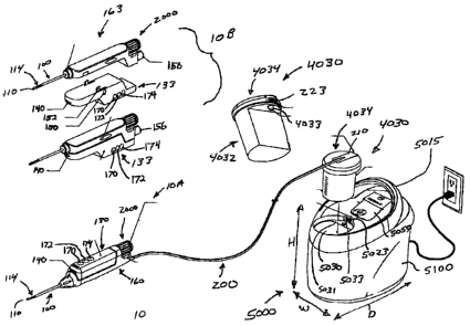
MAMMOTOME is commercially available from Ethicon Endo-Surgery, Inc. for
use in obtaining breast biopsy samples. This device generally retrieves
multiple
core biopsy samples from one insertion into breast tissue with vacuum
assistance.
[003] The following patent documents disclose various biopsy devices and are
incorporated herein by reference in their entirety: US 6,273,862 issued Aug.
14,
2001; US 6,231,522 issued May 15, 2001; US 6,228,055 issued May 8, 2001; US
6,120,462 issued September 19, 2000; US 6,086,544 issued July 11, 2000; US
6,077,230 issued June 20, 2000; US 6,017,316 issued Jan. 25, 2000; US
6,007,497 issued Dec. 28, 1999; US 5,980,469 issued Nov. 9, 1999; US
5,964,716 issued Oct. 12, 1999; US 5,928,164 issued July 27, 1999; US
5,775,333 issued July 7, 1998; US 5,769,086 issued June 23, 1998; US 5,649,547
issued July 22, 1997; US 5,526,822 issued June 18, 1996; and US Patent
Application 2003/0199753 published Oct. 23, 2003 to Hibner et al.
-1-
CA 02614235 2007-12-13
[004] Some vacuum-assisted biopsy devices may employ a re-usable control
module
that includes a vacuum pump and other control apparatus. Such vacuum control
modules may be relatively large and heavy, and may be mounted on wheels or on
a wheeled platform so that they can be moved from room to room in a surgical
area.
[005] While a variety of biopsy systems have been made and used, it is
believed that no
one prior to the inventors has made or used a biopsy system as described in
the
appended claims.
BRIEF DESCRIPTION OF THE DRAWINGS
[006] While the specification concludes with claims particularly pointing out
and
distinctly claiming the present invention, it is believed the same will be
better
understood by reference to the following description, taken in conjunction
with
the accompanying drawings in which:
[007] Figure 1 is a schematic illustration of a biopsy system according to one
embodiment of the present invention;
[008] Figure IA is a schematic illustration of a distal portion of a tissue
piercing
cannula having a vacuum lumen and a cutter lumen, with the distal portion of
the
cutter shown in the cutter lumen.
[009] Figure 1B is a schematic illustration of the vacuum level provided in a
vacuum
lumen as a cutter is advanced and retracted in a cutter lumen relative to a
tissue
receiving aperture.
[0010] Figure 2 is a schematic exploded view illustration of certain
components of the
vacuum control module depicted in Figure 1.
-2-
CA 02614235 2007-12-13
[0011] Figure 3 is a schematic illustration of a vacuum canister that can be
used with the
biopsy system of Figure 1.
[0012] Figure 4 is a schematic illustration of the interface of a multilumen
cable and a
vacuum canister cover.
[0013] Figure 5 is a schematic illustration of a control module including a
handle and a
hinged lid.
[0014] Figure 5A illustrates a USB port positioned on the back surface of the
control
module of Figure 5.
[0015] Figure 6 illustrates various steps that can be performed with respect
to a USB
device inserted into a USB port on a control module.
[0016] Figure 7 illustrates a pneumatic circuit that may be used with a biopsy
system.
[0017] Figure 8 illustrates multiple control states that can be employed in
controlling a
biopsy device in a biopsy system.
DETAILED DESCRIPTION
[0018] Figure 1 illustrates a biopsy system according to one embodiment of the
present
invention. The biopsy system of the present example includes a biopsy device
10
having a translating cutter 120 for severing tissue samples, a vacuum control
module 5000, and an umbilicus 200 extending from the biopsy device 10 to the
control module 5000. The umbilicus 200 can be in the form of a cable having
multiple lumens for providing one or more of electrical power, vacuum,
pneumatics, hydraulics, or saline to the biopsy device 10. The biopsy device
10
can be a hand held device 10A such as is suitable for use with ultrasound
imaging, or alternatively, a stereotactic device 10B configured to be mounted
on
a stereotactic or X-ray table. The vacuum control module 5000 can be a
-3-
CA 02614235 2007-12-13
relatively lightweight, portable unit having a smoothly shaped outer cover
5100
and a carrying handle 5015. The lightweight control module 5000 of this
example can be lifted and moved easily by a single person, with one hand. The
multilumen umbilicus 200 of this example provides a single connection between
the biopsy device 10 and the control module 5000, eliminating the complexity
of
having multiple electrical, saline, pneumatic, hydraulic, and/or vacuum lines
extending from the biopsy device 10.
[0019] As noted above and as shown in Figure 1, biopsy device 10 can be a
handheld
biopsy device l0A suitable for use with ultrasound imaging. The biopsy device
l0A can include a reusable holster 130 and a disposable probe unit 160 that is
detachable from holster 130. Together, the holster 130 and the probe 160 form
a
handpiece that can be comfortably held in and operated with a single hand.
Biopsy device lOB can include a disposable probe unit 163, and a reusable
stereotactic holster 133 having a firing mechanism for firing a tissue
piercing
portion of the biopsy device into tissue. The firing mechanism may be power
driven (e.g., motorized), and may include a button 150 that may be actuated to
activate the firing mechanism; as well as firing members 152 that are
configured
to engage probe 160 to fire at least a portion of probe 160 into tissue. Any
suitable configuration for the firing mechanism may be used, to the extent
that a
firing mechanism is included at all. The stereotactic holster 133 can be
configured for operable mounting onto a stereotactic X-ray table. Of course,
biopsy device l0A and biopsy device lOB may alternatively be used in a variety
of other settings or configurations.
[0020] The probe units 160 and 163 of the present example include a distally
extending
tissue piercing portion, such as a cannula 100 extending distally from probe
160.
The cannula 100 can include a distal tissue piercing tip 110 and a tissue
receiving
aperture 114 spaced proximally of the tip 110. The cannula 100 can also
include
a cutter lumen 104 and a vacuum lumen 108, with passageways 107 and 109
providing flow communication between the lumen 104 and lumen 108 in the
-4-
CA 02614235 2007-12-13
distal portion of cannula 100 (Figure 1 A). The cannula 100 can be inserted
into
or adjacent to a tissue mass to be sampled, and the biopsy device 10 is
operable
to obtain a plurality of severed tissue samples with a single insertion of
cannula
100 into tissue.
[0021] With cannula 100 being inserted into tissue, tissue drawn into the
aperture 114
can then be severed by sharpened distal end 122 (Figure 1A) of a tubular
cutter
120 translating within the cannula 100. Vacuum can be applied axially through
the cutter 120 and also in vacuum lumen 108 to assist in drawing tissue into
aperture 114. Figure 1B illustrates graphically a variable vacuum level that
can
be provided in vacuum lumen 108 as the cutter 120 is translated relative to
the
aperture 114. The probe units 160 and 163 can also include a tissue storage
assembly 2000, which can be disposed at a proximal end of the probe unit 160,
163 or elsewhere. The tissue storage assembly 2000 can be employed to store
multiple tissue samples severed by the cutter translating within the cannula
100
and transferred proximally through the hollow cutter 120 to the tissue storage
assembly 2000.
[0022] The probe units 160 and 163 of the present example each also include a
light 140
positioned near cannula 100. By way of example only, light 140 may comprise
an LED or other source of light, and may be configured to at least partially
illuminate a site into which cannula 100 is to be inserted. Probe 163 also has
a
remote thumbwheel 156, which may be rotated to rotate the cannula 100 of probe
163 relative to the remainder of probe 163. Suitable mechanisms for causing
such rotation will be apparent to those of ordinary skill in the art in view
of the
teachings herein. Of course, either probe 160, 163, as well as holsters 130,
133,
may be subject to numerous variations and modifications as desired.
[0023] Vacuum control module 5000 of the present example can be portable by a
single
hand, and may have a weight of no more than about 40 pounds, and in one
embodiment a weight of less than about 25 pounds. Alternatively, vacuum
control module 5000 may be of any other suitable weight. The control module
-5-
CA 02614235 2007-12-13
5000 handle 5015 is shown extending upward from an upper portion of the unit,
with the handle 5015 being the upper most component of the control module
5000, as shown in Figure 1. The vacuum control module 5000 of the present
example can have maximum outer dimensions of width W, depth D, and height
H, each of which is less than about 1.5 feet. Alternatively, vacuum control
module 5000 may have any other suitable dimensions. Vacuum control module
5000 of the present examples further includes a standard power cord for
receiving electrical power from a standard electrical outlet.
[0024] Figure 2 illustrates various components of the control module 5000 of
the present
example. The control module 5000 can include an internal aluminum (or other
suitable metal) chassis 5110, which can directly or indirectly support a
vacuum
pump 4010, an AC/DC power supply 5073, and a microprocessor control board
5040. A suitable power supply 5073 may include a 250-Watt power supply
model GPFC250 Commercial/GPFM250 Medical 250 manufactured by Condor
D.C. Power Supplies of Oxnard, California. Alternatively, any other suitable
power supply 5073 may be used. A suitable vacuum pump 4010 may include a
2-headed diaphragm pump having a maximum flow rate of less than about 18
liters per minute, and providing a maximum vacuum of about 25.1 inches of
Mercury (Inch Hg.). Alternatively, vacuum pump 4010 may have any other
suitable components and properties. By way of example only, one suitable
vacuum pump 4010 may be a model 7006ZVDP-2,3E Diaphragm pump
available from Reitschle Thomas, Thomas Products Division of Sheboygan,
Wisconsin.
[0025] The control module 5000 can also include an LCD display 5050, or other
type of
display, supported on an outside surface of the control module 5000, or
elsewhere (e.g., external to control module 5000, etc.). By way of example
only,
display 5050 may include a backlit, color LCD display. Alternatively, any
other
type of display 5050 may be used, or no display 5050 at all. A keypad 5080 may
also be provided, near display 5050, on control module 5000. Keypad 5080 may
-6-
CA 02614235 2007-12-13
comprise capacitive switches or other input devices, and may be used to enter
commands to or otherwise interact with control module 5000. Display 5050 may
display operating conditions, menus, or other information, such that display
5050
and keypad 5080 collectively provide a user interface. Of course, a user
interface
may alternatively be provided using any other suitable components in any other
suitable fashion.
[0026] In the present example, control module also has an attachment assembly
5200
that is configured to receive an off-the-shelf saline bag 5300. Attachment
assembly 5200 is coupled with a flex circuit 5202, and is configured to sense
the
weight of a saline bag 5300. In particular, control module 5000 may be
configured such that it will prevent operation of biopsy device 10 when no
weight or insufficient weight is sensed by attachment assembly 5200, which may
indicate that no saline bag 5300 is present or that the saline bag 5300
contains an
insufficient amount of saline. In addition or in the alternative, control
module
5000 may be configured to inform the user, such as via display 5050, that the
a
saline bag 5300 is not coupled with attachment assembly 5200 or that the
saline
bag 5300 contains an insufficient amount of saline. Alternatively, data from
attachment assembly 5200 may be used in any other suitable way, or attachment
assembly 5200 may be omitted altogether.
[0027] The vacuum control module 5000 of the present example can have a vacuum
canister 4030 that is releasably received within an opening 5030 disposed in a
generally upward facing outer surface of the control unit 5000. The vacuum
canister 4030 may serve as a vacuum "capacitor" for the biopsy vacuum circuit,
and can have a volume of less than about 300 cubic centimeters. More
particularly, the vacuum canister 4030 can have a volume of about 200 to about
250 cc. Alternatively, vacuum canister 4030 may have any other suitable
capacity or properties.
[0028] Referring to Figure 3, the umbilicus 200 of the present example can
comprise
multiple lumens, and can comprise a first lumen 202 for conveying vacuum from
-7-
CA 02614235 2007-12-13
the vacuum pump 4010 to the biopsy device 10, a second lumen 204 for
conveying one or more electrical conductors for conveying power and control
signals from the control module 5000 to the biopsy device 10, and a third
lumen
206 for conveying saline. Alternatively, umbilicus 200 may have any other
suitable number or type of lumens. As yet another variation, one or more
lumens
202, 204, 206 are provided in separate conduits or cables, instead of being
integrated into a single umbilicus 200. The proximal end of the umbilicus 200
can include a connection terminator 210. The distal end of the umbilicus 200
can
be received in the disposable portion of the biopsy device 10, such as the
probe
160 or the probe 163, so that electrical power and control signals are
directed
through the disposable probe 160, and then to holster 130. One or more
controls
(e.g., control buttons 170, 172, 174, etc.) can be located on the reusable
holster
130. Alternatively, the distal end of the umbilicus 200, or a portion thereof,
may
be received in holster 130 or elsewhere. Similarly, controls may be located on
probe 160, 163 in addition to or in lieu of controls on holster 130.
[0029] The vacuum canister 4030 of the present example can include a cup or
container
shaped body portion 4032 and a lid 4034. The vacuum canister 4030 is
configured to be inserted into an opening 5030 in an upwardly facing outer
surface of the control module 5000. The canister 4030 can be supported on a
lip
5031 that extends at least partially around the opening 5030. Of course, there
are
a variety of alternative ways in which vacuum canister 4030 may be configured;
as well as alternative ways in which vacuum canister 4030 may engage with
control module 5000.
[0030] In the present example, the body portion 4032 of the canister 4030
includes a
male vacuum port 4033 communicating with the interior of the canister 4030.
The port 4033 can sealingly engage a female vacuum port 5033 disposed in an
opening in the lip 5031, when the canister 4030 is inserted into the opening
5030.
In other variations, the lip 5031 or other portion of the control module 5000
may
include a male vacuum port 4033; with the canister 4030 having a
-8-
CA 02614235 2007-12-13
complimentary female vacuum port 5033. In the present example, the vacuum
port 5033 can be connected with a flexible hose or tube or other conduit that
communicates with the outlet of the vacuum pump 4010 disposed within the
control module 5000. Accordingly, when the canister 4030 is inserted into the
opening 5030 in the present example, a vacuum connection is established from
the vacuum pump 4010 to the interior of the canister 4030.
[0031] The lid 4034 of the present example is adapted to receive the
connection
terminator 210 disposed at the proximal end of the umbilicus 200. The lid 4034
can include a upper, first lid portion 4036 and a lower, second lid portion
4038.
The connection terminator 210 can be captured between the first and second lid
portions 4036, 4038, and the two lid portions 4036, 4038 joined together (such
as
by a snap fit, by adhesive, or by any other suitable means). The position of
the
terminator 210 between the lid portions 4036, 4038 can be established by guide
pins 212 on the lower lid portion which mate with corresponding guide holes
214
disposed around the perimeter of the terminator 210. Alternatively, any other
suitable structures or features may be used to establish the position of the
terminator 210 between the lid portions 4036, 4038, if any are used at all.
Indeed, lid 4034 may instead be formed of a single piece instead of two lid
portions 4036, 4038, and umbilicus 200 may be secured relative thereto in any
other suitable fashion. The multilumen umbilicus 200 and the vacuum canister
4030 can be provided as separate disposable items or provided together as a
unitary disposable item.
[0032] As shown in Figures 1, 3, and 4, the connection terminator 210 of the
present
example provides a multi-contact electrical connection 223, which faces
outward
from the lid 4034 when the terminator 210 is positioned on the lid 4034. The
control module 5000 includes a mating multi-contact electrical connection
5023.
The multi-contact connection 5023 is disposed adjacent the opening 5030 of the
control module 5000, such that when the vacuum canister 4030 is positioned in
the opening 5030, the electric contact is established between the control
module
-9-
CA 02614235 2007-12-13
5000 and the multilumen umbilicus 200 via the contacts 223 and 5023. It will
be
appreciated, however, that electrical contact may be provided between control
module 5000 and umbilicus 200 using a variety of alternative structures and
techniques.
[0033] Referring to Figures 3 and 4, the connection terminator 210 can further
include a
downward facing male vacuum port 227 extending from a bottom surface of the
connection terminator 210. The vacuum port 227 communicates with the
vacuum lumen 202 in umbilicus 200. When terminator 210 is disposed on lid
4034 between lid portions 4036 and 4038, the vacuum port 227 extends through
an opening 4037 in the lower lid portion 4038 to communicate with the interior
of the vacuum canister 4030. Alternatively, vacuum lumen 202 may
communicate with the interior of the vacuum canister 4030 using any other
suitable structures or techniques.
[0034] Accordingly, in the present example, the vacuum canister 4030,
terminator 210,
and control module 5000 are configured such that positioning the canister 4030
in the opening 5030 of the control module 5000 provides an electrical
connection
and vacuum communication between the biopsy device 10 and the vacuum
control module 5000. In other words, fluid and electrical connections between
biopsy device 10 and the vacuum control module 5000 are established merely by
inserting the canister 4030 into opening 5030, such that additional tube
connections or cable connections (e.g., connection of a tube with the canister
4030), etc. need not be established by the user before or after canister 4030
is
inserted into opening 5030. As used herein, the term "fluid" should be read to
include a vacuum, pressurized air, atmospheric air, liquids (e.g., saline,
blood,
etc.), and the like, regardless of whether solid materials (e.g., tissue
samples or
particles, etc.) are conveyed therewith.
[0035] Still referring to Figures 3 and 4, a float 4063 can be positioned in
the canister
4030. The float 4063 is operable to close vacuum port 4033 in the event fluid
accumulates in the canister above an acceptable level. A filter assembly 5035
-10-
CA 02614235 2007-12-13
including a filter pad 5037 and filter pad cover 5038 can be provided at the
vacuum port 5033 if desired. Of course, like other components described
herein,
float 4063, filter assembly 5035, filter pad 5037, and filter pad cover 5038
are all
merely optional, and may be modified, substituted, supplemented, or omitted as
desired.
[0036] Figure 5 provides a schematic illustration of an embodiment of the
control
module 5000 that includes a hinged cover 5600. Cover 5600 covers a storage
cavity 5610, similar to tissue storage compartment 5620 of the control module
shown in Figure 2. The storage cavity 5610 can be sized to store one or more
biopsy device holsters 130. If desired, a plurality of LJV light sources 5612
(only
one shown) can be positioned near or within cavity 5610. Light sources 5612
can be configured to be "on" when the cover 5600 is closed (and "off' when the
cover 5600 is opened), and can be employed to disinfect or sterilize the
item(s)
stored in the cavity 5610. In other embodiments, other components, features,
or
devices are included to disinfect or sterilize one or more items stored in the
cavity 5610, in addition to or in lieu of having light sources 5612. In still
other
embodiments, no features are included for disinfecting or sterilizing items.
Similarly, some variations of control module 5000 lack a cavity 5610
altogether.
[0037] Referring to Figure 5A, one or more Universal Serial Bus (USB) ports
5700 can
be provided on an outer surface of the control module 5000. Alternatively, any
other type of port (e.g., an SD card slot, an ethernet port, a serial
connection port,
a proprietary connection port, etc.) may be provided on control module 5000.
Figure 6 illustrates a flow chart describing a sequence of operational steps
that
can be employed when a USB memory device (not shown) is coupled with
control module 5000 via port 5700. Based on the contents of the USB memory
device, a course of action can be determined. For instance, based on the
contents
of the USB memory device, the control module microprocessor control may
write vacuum control module data to the USB memory device (e.g., usage time,
operational status, first use or first in service date, and the like.) The
control
- 11 -
CA 02614235 2007-12-13
microprocessor control may also write biopsy device data to the USB memory
device (e.g., usage time, operational status, first use, or first in service
date).
Alternatively, the vacuum control module 5000 may read new calibration
information, software, or software updates, etc., from the USB memory device,
reprogram operation of the vacuum module, and/or reprogram operation of the
biopsy device 10. Alternatively, a computer (e.g., desktop or laptop PC), or
network (e.g., internet) connection may be made with control module 5000 via
port 5700 or in some other fashion.
[0038] Figure 7 is a schematic illustration of a pneumatic configuration that
can be used
with the biopsy device 10 and vacuum control module 5000 of the present
example. Figure 8 illustrates multiple control states that can be employed in
controlling the biopsy device 10.
[0039] As shown in Figure 7, the control module 5000 of the present example
comprises
the vacuum pump 4010, a regulator 4020, and a the vacuum canister 4030
disposed in a pneumatic circuit. A master control valve 4060 and a
saline/vacuum valve 4080 are shown schematically as being positioned in the
disposable probe 160.
[0040] The vacuum provided by vacuum pump 4010 can be directed through the
vacuum lumen 202 of umbilicus 200 to the disposable probe 160, to provide
vacuum to the cutter 120 and cutter lumen 104. This vacuum can be provided
without valving, so that the vacuum provided to the interior of cutter 120 and
to
cutter lumen 104 of cannula 100 is always "on" when vacuum pump 4010 is
operating. Alternatively, one or more valves or similar components may be
provided in probe 160, canister 4030, control module 5000, and/or elsewhere.
The vacuum provided by umbilicus 200 can also provide a vacuum to the
vacuum lumen 108 via the two valves 4060 and 4080. Valve 4080 can be a 3-
port/2-position valve, with two input ports. One input port can be connected
to
the vacuum source and the other input port can be connect to a source of
saline
4016 (or alternatively vented to atmospheric pressure through filter 4018).
The
-12-
CA 02614235 2007-12-13
output port of valve 4080 communicates with an import port of master valve
4060. Other suitable configurations and couplings for the ports will be
apparent
to those of ordinary skill in the art in view of the teachings herein.
[0041] The operational position of the valve 4080 can be configured to
correspond to the
position of the cutter 120, such as by having cutter 120 extend through the
valve
4080 so that the position of the cutter 120 within the valve body determines
the
operational status (opened or closed) of the valve ports. When the cutter 120
is
retracted proximally (e.g., such that tissue aperture 114 is open), the valve
4080
communicates vacuum to the master control valve 4060. When the cutter 120 is
advanced distally (e.g., such that tissue receiving aperture 114 is closed),
the
valve 4080 communicates saline to the master control valve 4060.
Alternatively,
if saline is not available, or not desired, then valve 4080 communicates
atmospheric air via filter 4018 to the master control valve 4060. The valve
4060
can be actuated by a microprocessor controlled motor, by a mechanical link to
the cutter 120, or otherwise. The valve 4080 can be spring loaded in one
position, and movement of the cutter 120 (such as movement of the cutter 120
to
the distal position) can be employed to change the valve operational position.
Alternatively, the operational position of the valve 4080 can be configured to
correspond to the position of the cutter 120 in any other suitable way.
Furthermore, valve 4080 may be configured such that its operational position
does not necessarily correspond with the position of the cutter 120.
[0042] The master control valve 4060 can be a 3-port/3-position valve. One
input port
can be connected to the output port of the valve 4080. The second input port
can
be vented to filtered atmospheric air. The output port of the valve 4060 can
be
connected to the proximal end of vacuum lumen 108 of cannula 100. The valve
position of valve 4060 can be controlled by the operator of the biopsy device
10
using one or more user control interfaces, such as the control buttons 170,
172,
174 listed in Figure 8 or any other interface. The control buttons 170, 172,
174
can be located at any convenient position on the body of the biopsy device 10,
-13-
CA 02614235 2007-12-13
including for instance on handpiece 130, or elsewhere. The valve 4060 can be
actuated by a solenoid, motor, via a link to the cutter 120, or otherwise.
[0043] With reference to Figure 8, the "Ready State" of biopsy device 10
corresponds to
the cutter 120 being advanced to its distal most position and tissue aperture
114
being closed. In the Ready State, the valve 4080 communicates saline to the
master control valve 4060 and the master control valve is positioned to seal
off
(close) its other ports, including the output port communicating with vacuum
lumen 108. By closing the port to the lateral lumen 108 while in the Ready
State,
airflow through the device may be minimized, which may allow the pump 4010
to more easily maintain the desired vacuum level at the vacuum canister 4030.
[0044] When the operator depresses the "Sample" button 170 in the present
example, the
cutter motor is activated to cause the cutter 120 to retract proximally. As
the
cutter retracts, the valve 4080 changes position to communicate vacuum to the
master control valve 4060. At the same time, the master control valve changes
position to communicate a vacuum to the vacuum lumen 108. With the tissue
aperture 114 open, vacuum from vacuum pump 4010 is applied to the cutter 120
(such as via the tissue storage assembly 2000) and cutter lumen 104 (via the
cutter 120), as well as to the vacuum lumen 108 (via the valves 4080 and
4060).
Vacuum applied to both cutter lumen 104 and vacuum lumen 108 assists in
prolapsing tissue into aperture 114 of cannula 100.
[0045] After maintaining this vacuum state for a second or more to ensure
tissue has
prolapsed into aperture 114, the cutter 120 is advanced distally (and
simultaneously rotated) to close the aperture 114 and severe a tissue sample
in
the distal portion of the hollow cutter 120. As the cutter 120 advances
distally,
the cutter 120 can contact or otherwise actuate the valve 4080 to change the
valve position to communicate saline to the master control valve 4060. Also,
as
the cutter 120 advances, a microprocessor can be employed to change the master
control valve 4060 position to communicate filtered atmospheric air to vacuum
lumen 108, which in turn is communicated via passageways 107, 109 to the
distal
-14-
CA 02614235 2007-12-13
face of the severed tissue sample positioned in the distal portion of hollow
cutter
120. The atmospheric air on the distal face of the tissue sample provides a
proximal pushing force on the tissue sample, while the vacuum provided in
cutter
120 (via the tissue storage assembly 2000) provides a proximally directed
pulling
force on the severed tissue sample. The resulting proximally directed force on
the tissue sample conveys the tissue sample through the hollow cutter 120 into
tissue storage assembly 2000. Of course, any other suitable structures or
techniques may be used to capture a tissue sample and communicate it to a
tissue
storage assembly 2000.
[0046] In an alternative embodiment, the microprocessor can be employed to
change the
position of master control valve 4060 to first communicate saline to vacuum
lumen 108 for a predetennined period of time, and then change the valve's
position to communicate atmospheric air to the lumen 108. Accordingly, a fixed
volume of saline can be delivered via passageways 107,109 to the distal end of
hollow cutter 120, thereby assisting in moving the severed tissue sample
proximally through hollow cutter 120 to tissue storage assembly 2000. The
control system can be programmed to return to the Ready State after a
predetermined period of time (e.g., one or more seconds).
[0047] The biopsy device operator can depress the "Clear Probe" button 172
while in the
Ready State (e.g., after having operated the "Sample" button 170 to sever
tissue)
in order to direct a microprocessor control to cause the cutter 120 to
reciprocate
slightly to open and close aperture 114 a fraction of an inch (e.g. 0.2
inches), or
to any suitable degree. This reciprocation of cutter 120 can be effective to
dislodge the tissue sainple or otherwise free the sample so that the sample
can
travel freely through hollow cutter 120. While the cutter 120 is
reciprocating, the
vacuum control valve 4060 can be repositioned to communicate saline to the
vacuum lumen 108 and through passageways 107, 109 to provide a pushing force
on the distal face of the tissue sample. After a predetermined period of time,
the
microprocessor can return the pneumatic system to the Ready State.
-15-
CA 02614235 2007-12-13
[0048] The operator can depress and release the "Aspirate/Insert" button 174
when the
device is in the Ready State to insert medication or a tissue marker into the
tissue
being sampled or into the site from which a tissue sample has been or will be
taken. When the button 174 is depressed in this example, the cutter 120 moves
proximally to open aperture 114. The position of the master control valve 4060
is changed to communicate atmospheric air to the vacuum lumen 108.
Depressing the "Aspirate/Insert" button 174 also turns off the vacuum (such as
by either turning off the pump 4010 or opening regulator 4020 to vent pump
4010 outlet to atmosphere, etc.). The tissue marker applier (or medication)
can
be fed into the proximal end of the cannula 100 through hollow cutter 120,
such
as via the tissue storage assembly 2000 or otherwise, with the marker (or
medication) being then deployed through the open aperture 114 in cannula 100.
After the marker or medication has been deployed, the user may press any
button, which may advance the cutter 120 to return to the Ready State with the
master control valve 4060 positioned up.
[0049] The operator can depress and hold the "Aspirate/Insert" button 174 to
aspirate
fluid in the vicinity of aperture 114. When the operator depresses the button
174
in this example, the cutter 120 moves proximally to open aperture 114. With
the
cutter positioned proximally, the valve 4080 communicates vacuum to the master
control valve 4060, and the master control valve 4060 is positioned to
communicate vacuum to the vacuum lumen 108. Accordingly, vacuum is
applied to both the lumen 108 and the cutter lumen 104 (because vacuum is
provided continuously through cutter 120 to lumen 104 while the pump 4010
operates in this example). The vacuum provided to lumen 104 and lumen 108
aspirates any liquid near the aperture 114. When the Aspirate/Insert button
174
is released, the pneumatic system is controlled to return the Ready State. The
cutter 120 is advanced to close aperture 114. As the cutter 120 is advanced in
this example, the master control valve 4060 is positioned to communicate
filtered
atmospheric air to the vacuum lumen 108. Once the aperture 114 is closed, the
master control valve 4060 is positioned to close all its ports to attain the
Ready
-16-
CA 02614235 2007-12-13
State. As with other operational sequences described herein, the foregoing
operational sequence is merely illustrative, and any other suitable
operational
sequences may be used in addition to or in lieu of those explicitly described
herein.
[0050] The length of the umbilicus 200 from the control module 5000 housing
the
vacuum pump 4010 to the biopsy device 10 can be relatively long (as much as 20
feet or more in some cases) in order to accommodate movement of the biopsy
device 10 in the operating room, or due to limitations of the position of the
control module 5000 in magnetic resonance imaging environments. Of course,
umbilicus 200 may be of any desired length. In the present example, the vacuum
line in the umbilicus 200 can account for a considerable portion of the flow
volume that needs to be supplied or maintained by the vacuum pump 4010 and
vacuum canister 4030 when the tissue aperture 114 is open. Placing the saline
vacuum valve 4080 and the master control valve 4080 at the distal end of the
vacuum line (the end associated with the biopsy device 10) instead of in the
biopsy vacuum control unit 5000 may mean that a smaller vacuum pump 4010
and a smaller vacuum canister 4030 can be used. In some conventional biopsy
devices, valving may be placed in the control unit that includes the vacuum
pump, and the control unit is may be mounted on wheels due to its weight and
size. In Figures 2-4, the valves 4060 and 4080 are shown disposed in the
biopsy
device 10. The valves can be disposed in a disposable probe portion 160, 163
that includes the cannula 100 and cutter 120 andlor in a non-disposable (e.g.,
in
the holster 130) portion of the biopsy device 10. Such a valve placement may
allow a relatively low weight diaphragm vacuum pump 4010 having a flow rate
of about 18 liters per minute to be used, as compared to a conventional pump
and
valve arrangement requiring more than 80 liters per minute. Of course, any
desired vacuum pump having any desired properties may be used.
(0051] Similarly, the vacuum canister 4030 can be relatively small, with a
volume of
less than about 300 cubic centimeters, as compared to a conventional vacuum
-17-
CA 02614235 2007-12-13
canister having a volume storage capacity of 1200 cc's or more. As a result, a
relatively lightweight, hand-portable vacuum control module 5000 can be
employed. The vacuum control module 5000 (Figures 1, 2, and 5) can weigh less
than 25 pounds, can be carried by one hand, and can have height, width, and
length dimensions each less than about 1.5 feet. Alternatively, vacuum
canister
4030 and control module 5000 may have any other suitable capacity, size,
weight, or other properties.
[0052] If desired, a foot pedal (not shown) or remote keypad (not shown) can
be
employed to provide control input or instructions to the biopsy device 10
directly
and/or to the vacuum control module 5000, such as via a connector 180. The
foot pedal and remote keypad can be tethered (e.g., with one or more wires
extending from the food pedal/ keypad to the vacuum control module 5000,
etc.).
Alternatively, "wireless" communication between the foot pedal/keypad and the
control module 5000 and/or the biopsy device 10 can be employed. For instance,
wireless "Bluetooth" communication and associated hardware and software can
be employed to provide wireless control signals to the vacuum control module
5000 andJor the biopsy device 10 without requiring a "line of sight" for
signal
transmission and reception. AlteYnatively, an infrared transmitter and
receiver
can be employed to send and receive control signals. Other ways in which
communication may be provided between components of a biopsy system (e.g.,
between a pedal/keypad and control module 5000), whether wired, wireless, or
otherwise, will be apparent to those of ordinary skill in the art in view of
the
teachings herein.
[0053] While preferred embodiments of the present invention have been shown
and
described herein, it will be obvious to those skilled in the art that such
embodiments are provided by way of example only. Numerous variations,
changes, and substitutions will now occur to those skilled in the art without
departing from the spirit and scope of the appended claims. Additionally, each
-18-
CA 02614235 2007-12-13
element described in relation to the invention can be alternatively described
as a
means for perforrning that element's function.
-19-