Note: Descriptions are shown in the official language in which they were submitted.
CA 02615073 2007-12-17
Docket 3060 US
SINGLE LIGHT SOURCE UNIFORM PARALLEL LIGHT CURTAIN
CROSS-REFERENCE TO RELATED APPLICATIONS
This application claims priority under 35 U.S.C. 119 to U.S. Provisional
Patent
Application No. 60/871,640 filed December 22, 2006, the entire contents of
which are
incorporated herein by reference.
TECHNICAL FIELD OF THE INVENTION
The present invention relates generally to surgical systems and methods. More
particularly, the present invention relates to a system and method for sensing
a fluid level
in a surgical cassette in an ophthalmic surgical system.
1
CA 02615073 2007-12-17
Docket 3060 US
BACKGROUND OF THE INVENTION
The human eye in simplest terms functions to provide vision by transmitting
light
through a clear outer portion called the cornea, and focusing the image by way
of a lens
onto the retina. The quality of the focused image depends on many factors
including the
size and shape of the eye, and the transparency of the cornea and lens.
When age or disease causes the lens to become less transparent, vision
deteriorates because of the diminished light which can be transmitted to the
retina. This
deficiency in the lens of the eye is medically known as a cataract. An
accepted treatment
for this condition is surgical removal of the lens and replacement of the lens
function by
an artificial intraocular lens (IOL).
In the United States, the majority of cataractous lenses are removed by a
surgical
technique called phacoemulsification. During this procedure, a thin
phacoemulsification
cutting tip is inserted into the diseased lens and vibrated ultrasonically.
The vibrating
cutting tip liquefies or emulsifies the lens so that the lens may be aspirated
out of the eye.
The diseased lens, once removed, is replaced by an artificial lens.
A typical ultrasonic surgical device suitable for ophthalmic procedures
consists of
an ultrasonically driven hand piece, an attached cutting tip, an irrigating
sleeve, and an
electronic control console. The hand piece assembly is attached to the control
console by
an electric cable and flexible tubing. Through the electric cable, the console
varies the
power level transmitted by the hand piece to the attached cutting tip and the
flexible
tubing supply irrigation fluid to, and draw aspiration fluid from, the eye
through the hand
piece assembly.
In use, the ends of the cutting tip and irrigating sleeve are inserted into a
small
incision of predetermined width in the cornea, sclera, or other location. The
cutting tip is
ultrasonically vibrated along its longitudinal axis within the irrigating
sleeve by the
2
CA 02615073 2007-12-17
Docket 3060 US
crystal-driven ultrasonic hom, thereby emulsifying the selected tissue in
situ. The hollow
bore of the cutting tip communicates with the bore in the horn that in turn
communicates
with the aspiration line from the hand piece to the console. A reduced
pressure or vacuum
source in the console draws or aspirates the emulsified tissue from the eye
through the
open end of the cutting tip, the cutting tip and horn bores, and the
aspiration line and into
a collection device. The aspiration of emulsified tissue is aided by a saline
flushing
solution or irrigant that is injected into the surgical site through the small
annular gap
between the inside surface of the irrigating sleeve and the cutting tip.
Recently, a new cataract removal technique has been developed that involves
the
injection of hot (approximately 45 C. to 105 C.) water or saline to liquefy
or gellate the
hard lens nucleus, thereby making it possible to aspirate the liquefied lens
from the eye.
Aspiration is conducted concurrently with the injection of the heated solution
and the
injection of a relatively cool solution, thereby quickly cooling and removing
the heated
solution.
In the liquefracture technique of cataract removal, the cataractous lens is
liquefied
or emulsified by repetitive pulses of a surgical fluid that are discharged
from the hand
piece. The liquefied lens may then be aspirated from the eye. Since the
surgical fluid is
actually used to liquefy the cataractous lens, a consistent, pressurized
source of surgical
fluid is important to the success of the liquefracture technique. In addition,
different
surgical fluids may be advantageous for the removal of different hardness of
cataracts or
for various patient conditions.
Conventional ophthalmic surgical instrument systems use vacuum to aspirate the
surgical site and positive pressure to irrigate the site. Typically, a
cassette is serially
connected between the means used to generate pressure and the surgical
instrument. The
use of cassettes with surgical instruments to help manage irrigation and
aspiration flows
at a surgical site is well known. U.S. Pat Nos. 4,493,695 and 4,627,833
(Cook),
4,395,258 (Wang, et al.), 4,713,051 (Steppe, et al.), 4,798,580 (DeMeo, et
al.), 4,758,238,
3
CA 02615073 2007-12-17
Docket 3060 US
4,790,816 (Sundblom, et al.), and 5,267,956, 5,364,342 (Beuchat) and 5,747,824
(Jung, et
al.) all disclose ophthalmic surgical cassettes with or without tubes, and
they are
incorporated in their entirety by this reference. Aspiration fluid flow rate,
pump speed,
vacuum level, irrigation fluid pressure, and irrigation fluid pressure, and
irrigation fluid
flow rate are some of the parameters that require precise control during
ophthalmic
surgery.
For aspiration instruments, the air pressure in the cassette is below
atmospheric
pressure, and fluid within the cassette has been removed from the surgical
site. For
irrigation instruments, the air pressure in the cassette is higher than the
atmospheric
pressure, and the fluid will be transported to the surgical site. In both
types of
instruments, the cassette acts as a reservoir for the fluid that buffers
variations caused by
the pressure generation means.
For the cassette to act as an effective reservoir, the level of fluid (and
thus the
empty volume) within the cassette must be controlled so that the cassette is
neither
completely filled nor emptied. If fluid fills the cassette in an aspiration
system, fluid may
be drawn into the means for generating vacuum (typically a venturi), which
would
unacceptably interfere with the vacuum level of the surgical instrument. An
empty
cassette in an aspiration system will result in air being pumped into the
drain bag, which
would waste valuable reservoir space within the bag. Moreover, constant volume
within
the cassette in an aspiration system enables more precise control level of
vacuum within
the surgical instrument. Control of the fluid level within cassettes of
irrigation systems is
similarly desirable.
Additionally, the size of the reservoir within the cassette affect the
response time
of the cassette. A larger reservoir provides more storage capacity but slows
the response
time of the system. A smaller reservoir increases the response time of the
system, but
may not have adequate storage capacity. This dilemma has been addressed by
cassettes
having two internal reservoirs. Such a cassette is illustrated in U.S. Patent
No. 4,758,238
4
CA 02615073 2007-12-17
Docket 3060 US
(Sundblom, et al.) (the "Sundblom Cassette"). The smaller reservoir is in
direct fluid
communication with the surgical handpiece while a larger reservoir is
positioned between
the smaller reservoir and the source of vacuum. This allows for a faster
response time
and larger storage capacity. The small reservoir, however, must be
periodically emptied
into the larger reservoir prior to the smaller reservoir filling up. This
requires that the
smaller reservoir contain a fluid level sensor that notifies the control
console to empty the
smaller reservoir at the appropriate time. The Sundblom Cassette uses two
electrical
probes 76 (see FIG. 8) that form an open electrical alarm circuit. When the
surgical fluid
(which is electrically conductive) fills small reservoir 30, both probes 76
are submersed
in the fluid, thereby closing the circuit and triggering the alarm that
reservoir 30 is full.
The fluid level sensor used in the Sundblom cassette has the limitation of
being a simple
"On/Off' switch. The sensor has no other function other than to trigger a
'reservoir full"
alarm and provides no other information to the user about the amount of fluid
in the small
reservoir.
Other pressure sensors, such as the one disclosed in U.S. Patent No. 5,747,824
(Jung, et al.) use an optical device for continuous fluid level sensing by
reading the
location of the air/fluid interface. These optical devices require relatively
expensive
phototransmitters and receivers and are subject to inaccuracies due to foaming
of the fluid
within the reservoir. In addition, the accuracy of optical pressure sensors
can be affected
by ambient light levels.
Acoustic pressure sensors have been used in the past to monitor the fluid
elvel in
water tanks. The ultrasound transducers are mounted within the tank at the top
of the
tank and an ultrasound signal is sent downward toward the top of the water
contained
within the tank. This arrangement, however, is not suitable for use with
surgical
equipment where sterility is important and the transducer cannot be allowed to
come into
contact with the fluid. In addition, as surgical devices generally are
disposable, locating
the transducer within the chamber is undesirable.
5
CA 02615073 2007-12-17
Docket 3060 US
Accordingly, a need continues to exist for a simple reliable and accurate
fluid
level sensor.
6
CA 02615073 2007-12-17
Docket 3060 US
SUMMARY OF THE INVENTION
Embodiments of the present invention provide a system and method operable for
determining fluid level in an ophthalmic surgical device, and for notifying a
user of a
liquefracture hand piece of certain fluid level conditions.
More specifically, a continuous high resolution fluid-level monitoring system
is
provided by embodiments of the present invention. One embodiment of the
continuous
high resolution fluid level monitoring system includes a unique fluid level
sensor having
a point light source, parabolic reflector, sensor array, and a detection,
processing and
control system. The point light source illumines a parabolic reflector wherein
the point
light source is located at the focus of the parabolic reflector. The parabolic
reflector
reflects light from the point light source to produce a parallel light
curtain. The parallel
light curtain is parallel to an axis of symmetry of the parabolic reflector.
The parallel
light curtain illumines a chamber, such as a chamber in an ophthalmic surgical
device,
used to contain surgical fluid. The sensor array coupled to the chamber
detects the
parallel light curtain illuminating the chamber. The sensor array provides an
output to a
detection/processing/control system in order to determine the fluid level
within the
chamber. This optical method of determining the surgical fluid levels may be
advantageous in that it prevents physical contamination of the surgical
fluids.
Yet another embodiment provides a method of determining surgical fluid levels
within a chamber of an ophthalmic surgical device.
7
CA 02615073 2007-12-17
Docket 3060 US
BRIEF DESCRIPTION OF THE DRAWINGS
For a more complete understanding of the present invention and the advantages
thereof, reference is now made to the following description taken in
conjunction with the
accompanying drawings in which like reference numerals indicate like features
and
wherein:
FIG. 1 depicts a prior art solution where a linear light source is used to
determine
a fluid level within a chamber;
FIG. 2 depicts a linear light source in accordance with an embodiment of the
present invention;
FIG. 3 depicts a fluid-level sensor system in accordance with embodiments of
the
present invention; and
FIG. 4 provides a logic flow diagram of a method of determining the fluid
level
within a chamber in accordance with embodiments of the present invention.
8
CA 02615073 2007-12-17
Docket 3060 US
DESCRIPTION OF THE INVENTION
Preferred embodiments of the present invention are illustrated in the FIGs.,
like
numerals being used to refer to like and corresponding parts of the various
drawings.
It is often important to monitor liquid or fluid levels within medical
devices. It is
important that the fluid levels be monitored and at the same time not be
contaminated by
the monitoring devices. One such means of doing so is ultrasonic technology.
Another
way is to use an optical fluid level sensor.
FIG. 1 depicts a prior art solution where a linear light source is used to
determine
a fluid level within a chamber. Optical level sensing system 10 includes
chamber 12,
linear light source 16, a linear sensor array 18 and a processing module 20.
Linear light
source 16 is typically a laser line generator or a linear LED light emitting
diode bar.
However, these choices for the linear light source typically do not meet the
requirements
for a continuous high resolution optical level sensing system. This is due to
the large
viewing angle subtended by the linear light source chosen to illuminate fluid
14 within
chamber 12. Additionally, because a number of LEDs may be used within linear
light
source 16, uniformity problems may exist with the intensity of the light
generated by the
linear light source 16.
In order to achieve uniform intensity the linear light source 16 provided in
FIG. 1
typically uses a highly diffused array of LED's. As a result, the light source
16 has a very
large viewing angle wherein the rays or beams of light are not parallel. To
address this
issue, an embodiment of the present invention provides a light curtain wherein
the light is
substantially uniform and parallel.
FIG. 2 depicts a linear light source 200 used to generate a uniform parallel
light
curtain in accordance with embodiments of the present invention. This linear
light source
200 includes a point light source 202 and a parabolic reflector 204. Point
light source
9
CA 02615073 2007-12-17
Docket 3060 US
202 is located at the focus of a parabola used to define parabolic reflector
204. Point light
source 202 illuminates an arc 206 wherein the rays of light 208 within the arc
are not
parallel. However, parabolic reflector 204 reflects rays 208 to yield parallel
light curtain
210. Parallel light curtain has a curtain 210 height 212. Curtain height 212
may be
defined by the requirements of the fluid chamber for which the linear light
source 200
may be used to determine the fluid level within. Curtain height 212 may be
used to then
determine the parabolic arc of the parabolic reflector 204 defined by the
parabola.
Similarly, once this parabolic arc has been defined, the arc 206 illuminated
by the point
light source 202 is also defined. The location of the focus of the parabolic
reflector 204
again may also be driven by the curtain height 212 requirements associated
with the
parabolic reflector 204.
The light curtain 210 provided has parallel light rays 214 with a uniform
intensity.
The uniform intensity results from the illuminating rays originating from a
single point
light source 202. Additionally, light curtain 210 does not have a large
viewing angle
because the light rays 214 are in parallel. This is essential to a high-
resolution continuous
optical level sensing system. The reflector approach offers a smaller size
when compared
to an optical lens approach, especially when the height of the light curtain
210 is
relatively large.
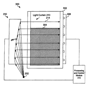
FIG. 3 depicts a fluid-level sensor system 300 in accordance with embodiments
of
the present invention. Fluid-level sensor System 300 includes a fluid-level
sensor using
the parabolic linear light source 200 discussed with reference to FIG. 2.
Additionally
fluid-level sensing System 300 includes a fluid chamber 302 containing fluid
304 and a
linear sensor array 308 as well as detection/processing/control module 310. A
single-
point light source 202 is used to illuminate a parabolic reflector 204 and
creates a parallel
light curtain 210 having parallels light rays 214. Linear light source 200 may
be optically
coupled to illuminate fluid chamber 302. The light curtain 210 illuminates the
fluid
chamber 302 and the light rays from light curtain 210 are detected by sensor
array 308.
Some of the sensors at sensor array 208 will detect low density light
(indicating light that
CA 02615073 2007-12-17
Docket 3060 US
has traveled through fluid 304 and other sensors will detect higher intensity
light that has
not traveled through fluid 304. Sensor array 308 provides an output to
detection/processing/control module 310 representative of the different light
intensities
received at its sensors. Processing and control module 310 is then able to
provide a high
resolution continuous measure of the level of the fluid within Chamber 302
based on the
signal from sensor array 308 indicating at what height the array detected a
substantial
change in the intensity of light received, in a manner that will be familiar
to those skilled
in the art. The fluid level measure (signal) may be used within an ophthalmic
surgical
device wherein it is important to know when surgical fluid levels are below a
certain
level, as previously discussed above.
The detection/processing/control module 310 system may be a single processing
device or a plurality of processing devices. Such a processing device may be a
microprocessor, micro-controller, digital signal processor, microcomputer,
central
processing unit, field programmable gate array, programmable logic device,
state
machine, logic circuitry, analog circuitry, digital circuitry, and/or any
device that
manipulates signals (analog and/or digital) based on operational instructions
stored in
memory. The memory may be a single memory device or a plurality of memory
devices.
Such a memory device may be a read-only memory, random access memory, volatile
memory, non-volatile memory, static memory, dynamic memory, flash memory,
cache
memory, and/or any device that stores digital information. Note that when the
system
controller implements one or more of its functions via a state machine, analog
circuitry,
digital circuitry, and/or logic circuitry, the memory storing the
corresponding operational
instructions may be embedded within, or external to, the circuitry comprising
the state
machine, analog circuitry, digital circuitry, and/or logic circuitry. The
memory stores,
and the system controller executes, operational instructions corresponding to
at least
some of the steps and/or functions illustrated in FIG. 4 associated with
embodiments of
the present invention.
FIG. 4 provides a logic flow diagram of a method of determining the fluid
level
within a chamber in accordance with embodiments of the present invention.
Operations
11
CA 02615073 2007-12-17
Docket 3060 US
400 begin with Step 402 where a point light source is placed at the focus of a
parabolic
reflector. The parabolic reflector is illuminated by the point light source in
Step 404.
Light is reflected from the parabolic reflector to produce a parallel light
curtain parallel to
an axis of symmetry of the parabolic reflector in Step 406. This allows a
substantially
uniform curtain of parallel light to be generated from a single light source.
Additionally
the parallel light curtain has a relatively small viewing angle when compared
to other
light curtains generated using, for example, a linear array of LED's. In Step
408, a fluidic
chamber is illuminated, wherein the fluidic chamber may contain a surgical
fluid for use
within an ophthalmic surgical procedure. A linear sensor array or other sensor
array also
coupled to the fluidic chamber may then sense/determine a fluid level within
the
chamber. This allows continuous high resolution determination of the fluid
levels within
the chamber. The position and height of the parabolic arc may be defined by
the required
height of the parallel light curtain.
In summary, embodiments of the present invention provide a continuous high
resolution fluid level monitoring system and method. Embodiments of the
continuous
high resolution fluid level monitoring system can include a unique fluid level
sensor
having a point light source, a parabolic reflector, a sensor array, and a
detection,
processing and control system. The point light source illumines a parabolic
reflector,
wherein the point light source is located at the focus of the parabolic
reflector. The
parabolic reflector reflects light from the point light source to produce a
parallel light
curtain. The parallel light curtain is parallel to an axis of symmetry of the
parabolic
reflector. The parallel light curtain illumines a chamber, such as a chamber
in an
ophthalmic surgical device, used to contain surgical fluid. The ophthalmic
surgical
device can be, for example, a surgical cassette for use in a
phacoemulsification system or
vitriol-retinal system as known to those having slcill in the art. The sensor
array coupled
to the chamber detects the parallel light curtain illuminating the chamber.
The sensor
array provides an output to a detection/processing/control system in order to
determine
the fluid level within the chamber. This optical method of determining the
surgical fluid
12
CA 02615073 2007-12-17
Docket 3060 US
levels may be advantageous in that it prevents physical contamination of the
surgical
fluids.
As one of average skill in the art will appreciate, the term "substantially"
or
"approximately", as may be used herein, provides an industry-accepted
tolerance to its
corresponding term. Such an industry-accepted tolerance ranges from less than
one
percent to twenty percent and corresponds to, but is not limited to, component
values,
integrated circuit process variations, temperature variations, rise and fall
times, and/or
thermal noise. As one of average skill in the art will further appreciate, the
term
"operably coupled", as may be used herein, includes direct coupling and
indirect coupling
via another component, element, circuit, or module where, for indirect
coupling, the
intervening component, element, circuit, or module does not modify the
information of a
signal but may adjust its current level, voltage level, and/or power level. As
one of
average skill in the art will also appreciate, inferred coupling (i.e., where
one element is
coupled to another element by inference) includes direct and indirect coupling
between
two elements in the same manner as "operably coupled". As one of average skill
in the
art will further appreciate, the term "compares favorably", as may be used
herein,
indicates that a comparison between two or more eleinents, items, signals,
etc., provides a
desired relationship. For example, when the desired relationship is that
signal 1 has a
greater magnitude than signal 2, a favorable comparison may be achieved when
the
magnitude of signal 1 is greater than that of signal 2 or when the magnitude
of signal 2 is
less than that of signal 1.
Although the present invention is described in detail, it should be understood
that
various changes, substitutions and alterations can be made hereto without
departing from
the spirit and scope of the invention as described.
13