Note: Descriptions are shown in the official language in which they were submitted.
CA 02615130 2008-01-10
WO 2007/016083 PCT/US2006/028828
-1-
METHODS AND SYSTEMS FOR TREATMENT OF PROLAPSE
Priority Claim
The present non-provisional patent Application claims priority under 35
USC 119(e) from United States Provisional Patent Applications having serial
number 60/702,704, filed on July 26, 2005, by James E. Cox and titled
CONNECTORLESS IMPLANT SYSTEM; 60/702,705, filed on July 26, 2005, by
Guillermo Wiley Davila et al. and titled TRANSVAGINAL SYSTEM FOR
APICAL SUPPORT; and 60/702,700, filed on July 26, 2005, by James E. Cox et al.
and titled METHODS AND SYSTEMS FOR TRANSVAGTNAL TREATMENT
OF PROLAPSE, wherein the entirety of these provisional patent applications are
incorporated herein by reference.
Field of the Invention
Described herein are features of surgical articles, surgical methods, and
surgical tools, for use in the field of pelvic surgery, e.g., to install
support devices
for use in treating vaginal prolapse.
Background
Medical conditions involving pelvic prolapse are conditions of great
importance. An aging population can be prone to such conditions. Pelvic
prolapse
develops when intra-abdominal pressure, muscle failure, a surgical procedure
such
as a hysterectomy, or other factors, allow or cause a tissue of a pelvic organ
such as
the vagina to become displaced. Within the general category of pelvic organ
prolapse, specific types include vault prolapse (apical); cystocele
(anterior);
rectocele and enterocele (posterior); and combinations of these.
CA 02615130 2008-01-10
WO 2007/016083 PCT/US2006/028828
-2-
Various techniques have been designed to correct or ameliorate prolapse and
prolapse symptoms, with varying degrees of success. Nonsurgical treatments
involve measures to improve the factors associated with prolapse, including
treating
chronic cough, obesity, and constipation. Other nonsurgical treatments niay
include
pelvic muscle exercises or supplementation with estrogen.
A variety of surgical procedures have also been atteinpted for the treatment
of prolapse. See for example United States patent application serial number
10/834,943, entitled "Method and Apparatus for Treating Pelvic Organ
Prolapse,"
filed Apri130, 2004, and serial number 10/306,179, entitled "Transobturator
Surgical Articles and Methods," filed November 27, 2002, the entireties of
each of
these two patent applications being incorporated herein by reference. Such
patent
applications describe articles and methods for treating pelvic organ prolapse
by use
of a support member for supporting specific tissue. Application serial number
10/834,943, for example, discusses a support member that includes a central
tissue
support portion and two end (extension) portions, and related methods for
implantation. The central tissue support portion can be attached at tissue of
a
vaginal vault. The end portions of the support member are then positioned
through
respective tissue pathways extending to an external incision at the perirectal
region,
to place the support member in a therapeutic position.
Methods of supporting vaginal tissue to treat vaginal prolapse can be
differentiated in ternis of the location of implanted materials or anatomical
tissue
used to support the vaginal tissue. One current method of treating posterior
vaginal
tissue prolapse involves the use of an intravaginal slingplasty ("IVS")
tunneler
device. Methods of treating prolapse using an IVS tunneler involve supporting
CA 02615130 2008-01-10
WO 2007/016083 PCT/US2006/028828
-3 -
vaginal tissue by attaching a portion of a surgical iinplant to vaginal tissue
and
passing another portion of the implant through the iliococcygeus muscle below
the
white line, for support. A different technique, known as sacrospinous ligament
fixation, involves supporting vaginal tissue by attachment to the sacrospinous
ligaments. Both of these metllods have drawbacks, such as not providing
completely correct anatomical support for the vagina. Attaching vaginal tissue
to
the sacrospinous ligaments can pull the vagina down toward the pelvic floor.
The
use of an IVS tunneler to pass an implant through the iliococcygeous muscle
allows
for support from a location higher up in the anatomy, but still not an
anatomically
correct location.
Summary
The invention relates to transvaginal methods of treating posterior vaginal
prolapse, and surgical devices. Embodiments relate to methods, tools, and
surgical
systems useful for transvaginal placement of an implant (e.g., a synthetic
mesh
in-iplant or an implant that contains a combination of synthetic and biologic
inaterials) in a position to support posterior tissue of the vagina, wherein
the implant
passes bi-laterally through opposing tissue paths that each include passage at
a
region of the arcus tendineus, e.g., near the ischial spine.
Some embodiments of inethods may involve an external incision at a
perirectal region, to place an extension portion of an implant, while
alternate
embodiments to not require and can avoid an external incision. The implant can
be
introduced to the pelvic region transvaginally; transvaginally attached to
tissue of
the vaginal vault for support; and then distal ends of the implant can be
passed bi-
CA 02615130 2008-01-10
WO 2007/016083 PCT/US2006/028828
-4-
laterally near a region of each arcus tendineus (e.g., near both of the
patient's ischial
spines), as described, using a transvaginal procedure.
The tissue path through a region of the arcus tendineus can result in benefits
including proper anatomical positioning of the supported vaginal tissue, and
fixation
of the implant in tissue near the arcus tendineus due to tissue ingrowth.
Additionally, exemplary transvaginal methods can allow for the location of
extension portions to be adjusted by movement of the extension portion
extending
through the tissue path, e.g., through a tissue path that surrounds the arcus
tendineus.
According to certain embodiments an implant can be used to treat vaginal
vault prolapse. The support member can include a tissue support portion that
can be
attached to tissue of the vaginal vault and two end portions attached to the
tissue
support portion. The implant can be used to place vaginal tissue in a
therapeutic
position for treatment of vaginal vault prolapse by attaching the tissue
support
portion to tissue of the vaginal vault, and attaching the end portions to
separate
locations for positioning or supporting the prolapsed tissue,
Exemplary implants, methods, tools, and systenis provide anatomical support
to treat vaginal prolapse (e.g., vaginal vault prolapse, enterocele, and
rectocele) by
using a supportive implant attached to vaginal tissue, that passes from
posterior
vaginal tissue to a location in a region of the arcus tendineus ("white
line"), e.g.,
near the ischial spine. The iniplant can pass from the point of attachment at
the
vaginal tissue, through a tissue path that includes passage through tissue at
the
inunediately anterior edge of the ischial spine and at the level of the
ischial spine
near the connection of the ischial spine to the arcus tendineus, and above or
below
the arcus tendineus.
CA 02615130 2008-01-10
WO 2007/016083 PCT/US2006/028828
-5-
As used herein, the terminology that refers to positions "above" the white
line refers to anatomy that includes the obturator internus muscle, and
refereiices to
positions "below" the white line refer to anatomy that includes the
iliococcygeus
muscle. Stated differently, einbodiments of the invention involve a tissue
path that
is defined to include a curved-rectangular-shaped area above and below the
curved
arcus tendineus. The region includes a specific region having a height
extending
from 2 inches above to 2 inches below the arcus tendineus, and a length
starting at
the ischial spine and extending 3 centimeters to the anterior of the ischial
spine.
In one aspect, the invention relates to a method of supporting posterior
vaginal tissue. The method includes: providing an implant comprising a tissue
support portion and an extension portion extending from the tissue support
portion,
creating a vaginal incision, transvaginally contacting the support portion
with
posterior vaginal tissue, transvaginally producing a tissue path between the
position
of the tissue support portion and a region of the arcus tendineus, and
transvaginally
extending the extension portion through the tissue path.
In another aspect, the invention relates to a pelvic implant assembly that
includes: an implant comprising supportive portions comprising a tissue
support
portion, and elongate extension portions extending from of the tissue support
portion; and an insertion tool at a distal end of an extension portion, the
insertion
tool comprising a curved portion sized and shaped to be used in a transvaginal
procedure to define a tissue path that exits the pelvic floor region in a
region of the
arcus tendineus, partially extends around an arcus tendineus, and re-enters
the pelvic
floor.
CA 02615130 2008-01-10
WO 2007/016083 PCT/US2006/028828
-6-
Brief Description of the Drawings
Figure 1 illustrates anatomy relevant to the invention, including specific
pelvic anatomy.
Figure 2 illustrates anatomy relevant to the invention, including specific
pelvic anatomy.
Figure 3 illustrates anatomy relevant to the invention, including specific
pelvic anatomy, and also exemplifies an implant according to the invention.
Figure 4 illustrates an exemplary system of the invention including an
exemplary tool and an exemplary implant with attached dilators.
Figure 4A is an end view of an exemplary tool.
Figure 4B is an end view of an exemplary tool with attached dilator of an
implant.
Figure 5 illustrates an exemplary implant according to the invention.
Figure 6 illustrates an exenlplary implant assembly according to the
invention, including an implant and attached insertion tools.
Figure 7 illustrates an exemplary implanted pelvic implant according to the
invention.
Figure 7A illustrates an exemplary implanted pelvic implant according to the
invention.
Figure 8 illustrates an exemplary implanted pelvic implant according to the
invention.
Figure 9 illustrates an exemplary implanted pelvic implant according to the
invention.
CA 02615130 2008-01-10
WO 2007/016083 PCT/US2006/028828
-7-
Figure 9A illustrates an exemplary implanted pelvic implant according to the
invention.
Figure 10 illustrates an exemplary kit or system according to the invention,
including a Deschampes Needle, an implant, and a tunneler device.
Detailed Descri tp ion
According the invention, surgical implants can be used to treat conditions of
vaginal prolapse. Also contemplated herein are various features of surgical
implants, surgical tools, systems that include implant and tool, and surgical
methods.
The implants and tools are useful for treating conditions of vaginal prolapse
including vaginal vault prolapse, but will also be appreciated to be useful
for treating
other conditions of pelvic tissue prolapse.
In general, the invention relates to methods, tools, and systems useful for
attaching one portion of a surgical implant to pelvic tissue such as vaginal
tissue and
passing another portion of the implant through a tissue path that includes
tissue in
the region of the arcus tendineus or "white line," preferably near the ischial
spine.
The location of this tissue path passing through a region of the arcus
tendineus or "white line" can result in improved anatomical correctness of the
position of supported vaginal tissue. The location in the region of the white
line can
provide a proper axis for supported vaginal tissue, higher than support
provided by
aiternate methods of treating vaginal prolapse such as those that involve
sacrospinous ligament fixation or use of the IVS tunneler, which alternate
methods
would typicaRy produce a tissue path more directly through the buttocks. A
location
in the region of the arcus tendineus, e.g., above the arcus tendineus, does
not cause
vaginal tissue to be pulled down toward the pelvic floor as with attachment to
the
CA 02615130 2008-01-10
WO 2007/016083 PCT/US2006/028828
-8-
sacrospinous ligaments. Also, a better vaginal length can result compared to
the use
of an IVS tunneler, because the support is located closer to the ischial
spine.
Also advantageously, a tissue path can be one that wraps around the outside
portion (relative to the region of the pelvic floor) of the arcus tendineus,
meaning
that an extension portion of an implant exits the pelvic region near the arcus
tendineus (either above or below the arcus tendineus), continues along a path
that
wraps or bends around the white line, then re-enters the pelvic region on the
other
side of the white line; i.e., below or above the arcus tendineus, whichever is
opposite
of the direction of entry. The tissue path can include a relatively sharp
turning
radius to place the extension portion near the arcus tendineus. By extending
around
the white line, the extension portion contacts tissue that surrounds the white
line and
can become ingrown into that tissue. This ingrowth can provide fixation of the
extension portion into the tissue. During the procedure the placement of the
extension portion and implant can be adjusted by manipulating the extension
portion
from the pelvic region side, after passing the extension portion around the
arcus
tendineus. Specifically, the extension portion will include two portions
within the
pelvic region, and those two portions can be manipulated to adjust the
position of
one or more of the extension portion, a central portion of an implant, and
tissue
attached to a central portion of the implant.
Figure 1 schematically illustrates a top view of anatomy of vagina and
nearby tissue. Vagina 2 and cervix 4 are schematically illustrated in relation
to arcus
tendineus or "white line" 6, ischial spine 8, and uterosacral ligaments 10. An
exemplary region of a tissue path according to the invention, a region of the
arcus
tendineus, 6, is illustrated as an approximately-rectangular regions 12 (shown
by
CA 02615130 2008-01-10
WO 2007/016083 PCT/US2006/028828
-9-
dashed lines -- one on each side of the pelvic region). Each region 12
includes area
above and below arcus tendineus, 6, starting at ischial spine 8 and extending
in an
anterior direction along the length of the arcus tendineus. Although obturator
internus and iliococcygeus muscles are not shown in figure 1, region 12 is
located to
include or be adjacent to the obturator intemus muscle (above the white line)
and the
iliococcygeus muscle (below the white line); a tissue path that passes through
region
12 also passes through obturator internus muscle or iliococcygeous muscle.
A preferred example of a region of the arcus tendineus, e.g., as illustrated
as
region 12, can be defined as a curved-rectangular-shaped area defined to
include a
region that extends 2 centimeters above and 2 centimeters below (e.g., 1
centimeter
above and 1 centimeter below) the arcus tendineus and that has a length
starting at
the ischial spine and extending in an anterior direction along the arcus
tendineus,
e.g., a distance of up to about 3 centimeters anterior of the ischial spine
(e.g., up to
about 1 centimeter anterior to the ischial spine). A particularly preferred
tissue path
can be very near or as close as possible to the ischial spine and either above
or below
the arcus tendineus, such as through tissue at the immediately anterior edge
of the
ischial spine and at the level of the ischial spine near the connection of the
ischial
spine to the arcus tendineus; dimensions can be 0.5 or 1 centimeter above or
below
the arcus tendineus, and 0.5 or 1 centimeter anterior to the ischial spine
along the
arcus tendineus.
Figure 2, a top view of pelvic features, schematically illustrates
sacrospinous
ligament 11, arcus tendineus 6, ischial spine 8, sacrum 16, and exemplary
tissue path
14 illustrated as circular line 14 passing around an outside of arcus
tendineus 6, i.e.,
wrapping around arcus tendineus 6. This wrapped configuration places an
implant
CA 02615130 2008-01-10
WO 2007/016083 PCT/US2006/028828
-10-
material near the arcus tendineus, e.g., in surrounding tissue such as
surrounding
muscle tissue, which allows for ingrowth of tissue into the implant material
located
at path 14, and fixation of that implant material at that location. Figure 2
illustrates
an example of a circular tissue path, 14, that passes above arcus tendineus 6
near.
(e.g., within 2 centimeters, such as within I or 0.5 centimeters) ischial
spine 8. Path
14 includes an arrow showing a direction of a needle (not showii) following
path 14,
starting below arcus tendineus 6, exiting the pelvic region by passing below
the
arcus tendineus and through the iliococcygeus muscle (levator ani) (not
shown),
passing around arcus tendineus 6, and then passing back into the pelvic region
by
passing above the arcus tendineus 6 through the obturator internus muscle (not
shown).
Figure 2 illustrates what can be referred to as a "bottom-up" technique for
passage of an end portion or extension portion of implant material through a
region
of the arcus tendineus. The bottom-up method inserts a distal end of an
extension
portion of an implant below the arcus tendineus and through the iliococcygeus
muscle; the imptant extension portion distal end is then passed around the
back or
outside of the arcus tendineus to a location above the arcus tendineus, and
then
through the tissue above the arcus tendineus (i.e., through the obturator
internus
muscle) where the implant end then re-enters the region of the pelvic floor.
Alternately, a tissue path as illustrated in figure 2 may be produced using a
"top-down" method. As opposed to the bottom-up method specifically illustrated
at
figure 2, a top-down method inserts the implant extension portion distal end
above
the arcus tendineus through the obturator internus muscle, wraps the extension
portion around the arcus tendineus, then the extension portion re-enters the
region of
CA 02615130 2008-01-10
WO 2007/016083 PCT/US2006/028828
-11-
the pelvic floor below the arcus tendineus by passing through the
iliococcygeus
muscle. A top-down method may be a preferred method because top-down methods
allow a needle tip to extend away from the bladder when passing through the
obturator intemus muscle. In a top-down method, the implant extension distal
end,
after wrapping around the arcus tendineus and re-entering the pelvic floor
region
(i.e., through the iliococcygeus muscle), may be led through one of various
tissue
path options. For exainple, the end extension may re-enter the pelvic floor
region
and then extend to and terminate at a location internal to the pelvic region.
The
distal end of the extension portion could be sutured, e.g., using a "stay
suture," to
maintain placement within the pelvic floor. This method advantageously does
not
require an external incision for manipulating the extension portion. Another
variation of the method is to use a tissue path as mentioned, but that
continues
through the pelvic region and then to and through an external incision such as
through the buttock and then through an external incision, wherein the
extension
portion can be adjusted and cut off to a desired length.
As illustrated in figure 3, embodiments of the invention involve the use of a
single mesh bridge (e.g., a uniform mesh strip) implant between tissue paths
described herein, above the white line and optionally and preferably near the
ischial
spines, to repair vaginal vault prolapse. Optionally, if desired, a mesh strip
implant
may use a modified middle section with denser construction (e.g., use of a
biologic
material for a middle section, 22) to hold sutures better than standard mesh.
A
vaginal vault repair results in an anatomically correct "pear" shape to the
distal
vagina, with the vaginal vault correctly positioned between the ischial
spines.
CA 02615130 2008-01-10
WO 2007/016083 PCT/US2006/028828
-12-
Referring to figure 3, this figure illustrates a top view of the anatomy of
figure 1 including a mesh support implant 20 installed according to an
exemplaxy
tissue path described herein. Implant 20 includes central (tissue support)
portion 22
sutured to the apex of vagina 2 by sutures 18 (designated as "x"s). Implant 20
also
includes two end portions, 24 and 26, extending bilaterally from tissue
support
portion 22. Each of end portions 24 and 26 is illustrated to pass from apex of
vagina
2, laterally toward arcus tendineus 6. Each end portion 24 and 26 passes above
arcus tendineus 6, near ischial spine 8 -- i.e., through tissue at the
immediately
anterior edge of ischial spine 8 and at the level of the ischial spine near
the
connection of the ischial spine to the arcus tendineus -- through the
obturator
internus muscle (not shown), around arcus tendineus 6. In figure 3, extension
portions 24 and 26 are shown to continue through tissue below the arcus
tendineus
(i.e., through the iliocoecygeus muscle, not shown) and back into the pelvic
floor
region.
Referring still to figure 3, sections 21 and 23 of end portions 24 and 26 are
located internal to the pelvic floor region. During a surgical implantation
procedure,
these portions can be manipulated by grasping manually or with a surgical
instrument such as a forceps, needle-passer, or pliers, to adjust the position
of end
portions 24 and 26, as well as support portion 22 and overall implant 20.
The invention also relates to implants, tools, and kits, that may be used
according to methods described herein, and that may also be useful for
treating
conditions other than vaginal prolapse, e.g., other types of pelvic tissue
prolapse. In
general, implants that may be useful according to methods described herein can
include those types of implants kn.own for use to treat vaginal prolapse, and
similar
CA 02615130 2008-01-10
WO 2007/016083 PCT/US2006/028828
- 13 - -
implants. Exemplary implants can be in the form of a biocompatible mesh
material
such as a mesh strip made of a single uniform length of mesh, or, alternately,
can be
a multi-portion implant that includes a support portion for attachment to
pelvic (e.g.,
vaginal) tissue connected to end portions or extensions. Embodiments include a
lengtli of mesh strip of generally uniform thickness and width, as well as
implants
having distinct or discernible sections of different sizes, materials, or
mechanical
properties. Other exemplary embodiments include a tissue support portion of a
biologic material and extension portions of synthetic mesh material.
Exemplary implants may be a mesh strip such as mesh strips and multi-
component implants illustrated in the accompanying figures. As illustrated,
exemplary implants may consist of a strip of uniform thickness and width, as
well as
implants that include portions of different sizes, shapes, and materials, for
connecting to tissue and for supporting tissue. A tissue support portion may
be of a
biologic material or a synthetic (e.g., mesh) material. Attached to a tissue
support
portion can be one, two, or more, extensions (or "extension portions" or "end
portions") shaped and sized to extend from the point of attachment with the
support
portion of the implant to another location of the anatomy. Each extension may
be an
elongate material that is biologic or synthetic, e.g., an elongate synthetic
mesh
attached directly to the support portion.
Various implant products are available commercially for treating prolapse
conditions, e.g., from American Medical Systems Inc., of Minnetonka Minnesota.
Examples of such products include: the line of PERIGEETM products for
treatment
of cystocele, from American Medical Systems, Inc.; the APOGEETM product for
treating enterocele, rectocele, and vaginal vault prolapse, also available
from
CA 02615130 2008-01-10
WO 2007/016083 PCT/US2006/028828
-14-
American Medical Systems Inc.; as well as products for CAPS procedures
(combined-prolapse-repair-with sling) for treating cystocele and stress
urinary
incontinence.
Examples of implants that can be used or modified for use according to the
present description are described, e.g., in US application number
2004/0039453,
"Pelvic Health Implants and Methods," (describing implants useful for treating
multiple pelvic disorders) having serial number 10/423,662, and filed on April
25,
2003; US application nuinber 2005/0245787, "Method and Apparatus for Treating
Pelvic Organ Prolapse," having serial number 10/834,943, and filed on April
30,
2004; United States patent application serial number 11/347,063, filed
February 3,
2006, entitled "Pelvic Implants and Related Methods; United States patent
application serial number 11/398,368, filed April 5, 2006, entitled "Articles,
Devices, and Methods for Pelvic Surgery"; and United States patent application
serial number 11/243,802, filed October 5, 2005, entitled "Method for
Supporting
Vaginal Cuff' the entireties of each of these being incorporated herein by
reference.
Exemplary implants can include a tissue support portion for placing in
contact with tissue to be supported, and one or more "extension" portions (or
"end
portions"), the tissue support portion being useful to support pelvic tissue
such as
vaginal tissue (anterior, posterior, apical, etc.). The tissue support portion
can be
sized and shaped to contact the desired tissue when installed. A tissue
support
portion that is located between two or more extension or end portions is
sometimes
referred to herein as a "central support portion."
Dimensions of an implant or a portion of an implant can be as desired and
useful for any particular installation procedure, treatment, or combination of
CA 02615130 2008-01-10
WO 2007/016083 PCT/US2006/028828
-15-
treatments, and to support a specific tissue, type of tissue, or multiple
tissues (e.g.,
bladder, vagina, urethra, etc.). Exemplary dimensions can be sufficient to
allow the
tissue support portion to contact tissue to be supported and to allow one or
more
extension portion to extend from the tissue support portion to a desired
anatomical
location, e.g., through a tissue path through a region of the arcus tendineus,
as
described.
A tissue support portion can be sized and shaped to an overall area for
contacting tissue being supported, and can depend on the condition being
treated,
e.g., vault prolapse, enterocele, rectocele, or a combination of these. The
tissue
support portion is of sufficient size and shape to at least partially surround
or
otherwise be in contact to support prolapsed tissue. A tissue support portion
can
optionally be of a width that is greater than a width of an extension portion.
An
increased width of a tissue support portion may take the form of one or two
lateral
extensions that extend the width of the tissue support portion in at least one
direction, beyond the width of an extension portion. The shape of the tissue
support
portion can also be varied, depending on the intended application and treated
condition, and may be square, rounded, angled, rectangular, etc. Exemplary
widths
of a tissue support portion, measured laterally (i.e., perpendicular to
lengths of
extension portions), can be in the range from 1 to 8 centimeters, such as from
2 to 6
centimeters. Generally, exemplary lengths of a tissue support portion can be
up to 8
centimeters, such up to about 4 centimeters.
Extension portions are elongate pieces of material that extend from the tissue
support portion and are integral with or connected to the tissue support
portion.
Extension portions are useful to attach to other anatomical features and
thereby
CA 02615130 2008-01-10
WO 2007/016083 PCT/US2006/028828
-16-
provide support for the tissue support portion and the supported tissue. One
or
multiple (e.g., one, two, or four) extension portions can extend from the
tissue
support portion as elongate "ends," "arms," or "extensions," that are used to
attach
to other anatomy. Extension portions extending from a tissue support portion
in
contact with posterior vaginal tissue, can be extended through a tissue path
as
described herein, passing through a region of the arcus tendineus such as
above the
arcus tendineus.
A width of an extension portion can be a width useful for implanting the
implant and for providing desired strength and fixation properties during and
after
implantation and optional tensioning of the sling. Typical widths of an
extension
portion can be in the range from 0.5 to 3 centimeters, e.g., from 0.8 to 2
centimeters,
such as from 0.8 to 1.5 centimeters. Extension portions can typically have a
uniform
or substantially uniform width along the length, normally not varying by more
than
about 25 percent of the average width along the length of the installed
portion of the
extension portion. A length of an extension portion can be as desired to
extend from
a tissue support portion installed at a desired pelvic tissue location,
through a tissue
path of a desired length, e.g., from a tissue support portion installed at a
vaginal
tissue, to a region above the arcus tendineus, optionally back into the pelvic
cavity,
and optionally further passing through the buttock to an exterior incision
external to
the buttock.
Al.l example of a particular type of pelvic implant is the type that includes
supportive portions including or consisting of a central support portion and
two
elongate extension portions extending from the central support portion. The
term
"supportive portions" refers to portions of an implant that function to
support tissue
CA 02615130 2008-01-10
WO 2007/016083 PCT/US2006/028828
-17-
after the implant has been inlplanted, and specifically includes extension
portions
and a tissue support portion, and does not include optional or appurtenant
features of
an implant such as a sheath, dilator, attached or engaged insertion tool, ox
other
connected tools or implantation aids. Figures 4, 5, and 6, for example,
illustrate
iinplants having supportive portions consisting of a tissue support portion
and two
elongate extension portions.
According to certain embodiments of implants, various features can be
incorporated into a useful implant to facilitate installation of a device
during a
surgical procedure. For instance, a suture may be attached to an implant,
along a
length of an extension portion, for use in adding tension or in positioning
the
implant or a portion (e.g., extension) of the iinplant. Alternately or in
addition an
exemplary implant may include a removable sheath such as a flexible plastic,
transparent elongate tube that can cover extension portions of an implant to
allow a
surgeon to apply tension or pressure on the sheath to indirectly apply
pressure or
tension to the extension portion, for placing or adjusting the location of the
implant.
An implant can be installed according to the present description by use of
standard surgical instruments, or by use of instruments that are designed or
particularly useful for placing an extension portion through a tissue path as
described, e.g., in a region of the arcus tendineus, such as above the arcus
tendineus.
Generally, an insertion tool may include a portion for creating a tissue path,
that
portion being curved or straight, with exemplary embodiments including a
curved
portion of a sized and shaped (e.g., length and curvature) that will be useful
to form
a tissue path as described herein, in a region of the arcus tendineus and
preferably
wrapping around the arcus tendineus, to lead a distal end of an extension
portion at
CA 02615130 2008-01-10
WO 2007/016083 PCT/US2006/028828
-18-
least partially through a tissue path that wraps around the arcus tendineus.
An
exen3plary length (measured as the circumferential arclength of the curved
portion)
of a curved portion can be from 3 to 5 centimeters, and an exemplary radius of
curvature of a curved portion may be, e.g., in the range from 0.5 to 1.5
centimeters.
Exemplary geometric forms of a curved portion can be a form of a partial
circle, such as a half circle. The partial circle is arranged to be in a plane
that does
not include a line defined by a longitudinal axis of a shaft. With this
configuration,
the curved portion can be located to define a partial circle having the shaft
of the
tool (and a longitudinal axis of the shaft) as a center of the circle. The
curved
portion can then engage tissue and be rotated around the shaft and
longitudinal axis
by rotation of the handle abotit the longitudinal axis, to cause the curved
portion to
define a circular tissue path. The partial circle can have a relatively
uniform radius
of curvature, such in the range from 0.5 to 1.5 centimeters (e.g., from 0.7 to
1.2
centimeters), extending over an arclength that traverses from 90 to 270
degrees, e.g.,
from 170 to 190 degrees, about 180 degrees. This arclength, when measured as
the
circumference of the partial circle, can be, e.g., from 3 to 5 centimeters.
An example of a useful insertion tool is a small curved needle attached at a
distal end of an extension portion, which can be manipulated using a surgical
instrument such as a forceps or pliers. The small curved needle can consist of
a
single length of curved needle material, e.g., metal or plastic, attached at a
distal end
of the extension portion. The needle may be considered to be a two-dimensional
form, in that it the curvature of the needle can define a two-dimensional
plane. The
small curved needle may be manipulated transvaginally and passed through
tissue in
a region of the arcus tendineus, preferably near the ischial spine. The needle
is
CA 02615130 2008-01-10
WO 2007/016083 PCT/US2006/028828
- 19-
curved to exit the pelvic region, wrap around the ischial spine, and then lead
the
extension portion back into the pelvic region to cause the extension portion
to follow
a tissue path that wraps around the arcus tendineus. The needle may then be
removed by cutting the extension portion and the position of the extension
portion
may be adjusted by manipulation of portions of the extension portions that are
located within the region of the pelvic floor.
Another example of a tool useful for placing an end portion of an implant
transvaginally is a small three-dimensional looped needle such as a Deschamps
needle or a similar needle that can be introduced transvaginally and that can
then be
used to pass an end portion of an implant through a tissue path in a region of
the
arcus tendineus, e.g., at the level of the ischial spine. The tool can be
considered to
include a shaft and curved end portion that exist in three dimensions, with
the shaft
defining a longitudinal axis and the curved end portion originating from that
axis
and extending in two additional directions. In use, an end portion of an
implant may
be attached to a tip of a curved distal portion of the tool and passed through
tissue of
a region of the arcus tendineus using the curved tip, transvaginally. For
example,
the curved distal end portion can be inserted transvaginally, and the handle
can be
rotated to rotate the curved distal end portion to define a circular tissue
path. The
needle is curved to exit the pelvic region, wrap around the arcus tendineus,
and then
lead the extension portion back into the pelvic region to cause the extension
portion
to follow a tissue path that wraps around the arcus tendineus. Optionally,
according
to certain exemplary methods, an additional surgical tool such as a tunneler
(e.g., the
IVS Tuzuleler device available commercially from Tyco) can be inserted through
an
external incision (e.g., in a perirectal region) into the pelvic region,
attached to a
CA 02615130 2008-01-10
WO 2007/016083 PCT/US2006/028828
-20-
distal end of the extension portion, and then removed to lead the extension
portion of
the implant from the pelvic region to an external location.
An insertion tool or tools (e.g., Deschamps needle, or a small needle to be
manipulated by a standard operating room tool such as a needle-driver) may
engage
a distal encl of an extension portion of an implant by any useful engagement
configuration. The engagement may be a permanent attachment or removable
engagement, as desired.
According to certain embodiments of the invention, an insertion tool may be
attached to the implant at a distal end of an extension portion. The term
"attach,"
when used with regard to an end portion attached to an insertion tool, will
refer to
engagement configurations that are not easily removable, such as in an a
manner
designed to be used during a surgical implantation method and that would
normally
be removed only by cutting the extension portion of the implant, as opposed to
releasing (e.g., un-threading) the implant from the insertion tool. Examples
of types
of attachment mechanisms include attachment by adhesive such as a pressure
sensitive adhesive, a structural adhesive, a two-part reactive adhesive such
as an
epoxy adhesive, etc.; a tight knot using thread or suture material that would
not be
easily untied during a surgical installation procedure; a non-removable
mechanical
interaction between the insertion tool and the implant portion such as by
permanently threading the implant portion through an eye, eyelet, slot, or
hole in the
tool so the end portion is not easily removed; a nietal or plastic mechanical
attachment such as a metal crimp at the end of the insertion tool; a polymeric
attachment such as the use of a heat-shrinkable polynmeric sheath; attachment
by a
molding manufacturing process such as by injection molding or insert molding a
CA 02615130 2008-01-10
WO 2007/016083 PCT/US2006/028828
-21-
plastic dilator or plastic needle to a distal end of an extension portion; or
any other
mechanical or adhesive type of permanent or semi-permanent attaching
mechanism.
In contrast to an attachment, examples of types of removable engagement
mechanisms include use of a loose lcnot that is easily untied during a
surgical
implantation procedure; an threaded dilator that removably engages a threaded
needle tip for easy engagement and dis-engagement by threading and un-
threading;
and threading a distal end of an extension portion through an aperture (e.g.,
eye or
eyelet) of an insertion tool such as a needle or a curved distal portion of a
tool.
Attaching an insertion tool (e.g., dilator, needle, etc.) to a distal end of
an
extension portion of an implant eliminates the need for a surgeon to make that
connection, which reduces preparation of the implant before or during surgery.
The
term "pre-attached" refers to an implant that includes an insertion tool
attached to
the implant as the implant is commercially supplied to a surgeon. The pre-
attached
device can be manufactured for distribution and sale in a condition where only
minimal preparation (if any at all) needs to be performed by the surgeon prior
to
surgical implantation. Minimal preparation may include modification to size or
shape of a portion of an implant (e.g., by trimming), or removing loose
material or
loose pieces, but does not include a step of creating a direct attachment
between a
portion of the implant and an insertion tool that will allow the portion of
the implant
to be placed or led through a tissue path.
Thus, embodiments of optional features of implants include an insertion tool
that is attached e.g., pre-attached, or alternately is removably engaged, at a
far (i.e.,
distal) end of an extension portion of an implant, the tool being a structure
that
facilitates installation of the extension portion and implant by being of a
size, shape,
CA 02615130 2008-01-10
WO 2007/016083 PCT/US2006/028828
22 -
rigidity, and overall design, to be capable of being used transvaginally to
create a
tissue path that passes through tissue in a region of the arcus tendineus,
optionally
and preferably wrapping around the arcus tendineus. Examples of insertion
tools
that may be either attached or removably engaged to a distal end of an
extension
portion include: a tip (distal end) of an extension portion that includes an
attached
rigid tip or "dilator," optionally designed to cooperate and removably engage
an end
of another insertion tool for use together to create a tissue path during
installation of
an iinplant; a distal end of an extension portion that is permanently attached
to an
insertion tool in the form of a curved distal needle portion and a shaft and
proximal
handle portion, or another tool that can be inserted transvaginally to
manipulate an
extension portion of an implant during installation; and a distal end of an
extension
portion that is permanently attached to an insertion tool such as a small
needle that
can be manipulated by a grasping tool such as a needle driver, a forceps, or a
pliers,
etc., to manipulate an extension portion of an implant during transvaginal
installation.
As one exemplary design, an implant may include a rigid (e.g., plastic),
pushable dilator attached at a distal end of an extension portion, the dilator
including
a sharp tip at a first end and an opening at an opposing end, the opening
designed
and adapted to fit and removably engage an end of an insertion tool such as a
needle.
The pushable dilator can be designed to fit the leading edge of an insertion
tool such
as a long needle having a handle and a distal portion with a tip adapted to
fit and
removably engage the pushable dilator. The dilator can engage the end of the
insertion tool and may be pushed or pulled by the insertion tool through
tissue to
either follow or produce a path in the tissue, such as by rotating the handle
to cause
CA 02615130 2008-01-10
WO 2007/016083 PCT/US2006/028828
- 23 -
the distal portion to produce a curved tissue path. To produce a path in the
tissue by
pushing the dilator through the tissue, the pushable dilator can be
sufficiently sharp
and rigid to pass through tissue when pushed using the needle (e.g., by
rotating the
needle).
A dilator (whetlier or not sufficiently sharp and rigid to be "pushable") may
be straight, or, according to certain specific embodiments of the invention,
may be
ciuved in a manner that will improve manipulation of the dilator during a
surgical
procedure, e.g., in a manner that will facilitate pushing the dilator through
tissue to
either produce or follow a particular path of tissue. Optionally for use with
methods
described herein with a tissue path in the region of the arcus tendineus,
e.g., that
v, raps around the arcus tendineus, the external size and shape of a dilator
may be
suited to produce a tissue path that curves around the arcus tendineus, e.g.,
to exit
the pelvic region by passing through the obturator internus above the arcus
tendineus, pass behind the arcus tendineus, and re-enter the pelvic region by
passing
through the levator ani at a location below the arcus tendineus. The curved
dilator
may be considered to be a two-dimensional form, in that it the curvature of
the
dilator can define a two-dimensional plane.
As exemplary dimensions, a curved dilator may include a curved portion that
has a radius of curvature in the range from 0.5 to 1.5 centimeters and a
length
(measured as the arclength of the curved portion) of from 3 to 5 centimeters.
Also
optionally, a curved shape or radius of a curved dilator can approximate or
match a
curved shape or radius of an insertion tool (e.g., a curved distal portion of
an
insertion tool such as a curved distal needle), and the curves of both a
dilator and a
curved distal portion of an insertion tool (e.g., needle) may be shaped to
match a
CA 02615130 2008-01-10
WO 2007/016083 PCT/US2006/028828
-24-
tissue path that exits the pelvic area at a location near the ischial spine
and above the
arcus tendineus by passing through the obturator internus muscle, continues
around
the arcus tendineus, and re-enters the pelvic region below the arcus tendineus
by
passing through the levator ani muscle. Thus, according to certain specific
embodiments of the use of a curved dilator, a curved dilator may be used with
a
curved needle (or other insertion tool) designed to fit within an internal
space at a
hollow interior of the dilator, with the curved insertion tool and the curved
dilator
having a size and shape to define a tissue path passing around the arcus
tendineus as
described.
Further design features can relate to dilators and insertion tools that
include
anti-rotation or alignment features, in particular with the use of a curved
needle
insertion tool and a curved dilator. An anti-rotation or alignment feature may
be in
the form of opposing and coordinated structural features of the dilator and a
tip of an
insertion tool (e.g., needle) that together can: interconnect the dilator and
tip of
insertion tool to produce a desired alignment; prevent relative movement of
the two
pieces such as to prevent rotation of the dilator relative to the tool; or
both. The
alignment feature causes the dilator to be placed on the needle in a specific
alignment, which if the needle and dilator are both curved as discussed above,
causes the curve of the needle to be aligned with the curve of the dilator. An
example of an alignment and anti-rotation feature is a keyed structure, as
will be
understood, that includes one or more inter-connecting surfaces and structures
between the dilator and the needle to allow the dilator to removably connect
to the
needle when the two are properly aligned, and then to also prevent rotation
between
the two when the two are connected. Other mechanical structures will also
allow the
CA 02615130 2008-01-10
WO 2007/016083 PCT/US2006/028828
- - 25 -
dilator to be attached at an end of a needle in a manner to produce a desired
alignment and to prevent rotation of the dilator relative to the needle.
An example of an implant having a curved (two dimensional) dilator adapted
to removably engage a three dimensional insertion tool that includes a curved
(two
dimensionally curved) distal end, is illustrated at figure 4. Figure 4
schematically
illustrates a prolapse support device (i.e., implant) 110 for treating vaginal
prolapse,
and insertion tool 122. Implant 110 includes end (i.e., "extension") portions
114 and
116 connected to central support portion 120. Sutures (optional) 118 extend
along
the lengths of each of extensions 114 and 116 and are connected to central
support
portion 118 and extensions 114 and 116. Rigid, curved dilators (in two
dimensions)
112 are attached at each distal end of extension portions 114 and 116.
Still referring to figure 4, insertion tool 122 (not to scale) includes handle
124, shaft 126, and curved distal portion or "needle" 128. (Needle 128
includes a
two-dimensional semi-circular form, and needle 128 along with shaft 126
together
are in three dimensions.) Needle 128 is designed to engage dilators 112 by
fitting
inside of a dilator 112, e.g., inside of internal space 113 of each dilator,
so that
needle 128, by use of handle 124 of tool 122, can be used to push dilator 112
through tissue by rotating handle 124; rotation of handle 124 about axis 121,
defined
by handle 124 and shaft 126, rotates curved needle 128. Curved needle 128,
shaft
126, and handle 124 are in three dimensions. Curved needle 128 is
substantially a
partial circle (e.g., half circle) that rotates to define a curved tissue path
when handle
124 is rotated about longitudinal axis 121. Not shown is an optional
protective
flexible cover or sheaths that could extend over and contain extension
portions 114
and 116.
CA 02615130 2008-01-10
WO 2007/016083 PCT/US2006/028828
-26-
Figure 4A shows an end view of tool 122, illustrating the half-circle shape of
curved end 128, in view of handle 124. Curved end 128 includes an arclength of
approximately 180 degrees, starting at a trailing end 129 that connects to
shaft 126,
and extending to needle tip 127. Figure 4B illustrates these features of tool
122
when connected to dilator 112, which is also attached to extension portion 116
of
inlplant 110.
According to other embodiments of implants, distal ends of extension
portions can be attached or engaged with an insertion tool in the form of a
needle or
in the form a tool comprising a distal curved portion (e.g., needle) with
attached
shaft and handle, the former needle being capable of being manipulated using a
separate tool such as a needle-driver, forceps, or surgical pliers, the latter
type of
needle being capable of being manipulated using the handle, which will be
external
to the patient during a transvaginal surgical procedure. Thus, exemplary
insertion
tools include a small two-dimensional needle that can be manipulated during a
transvaginal installation by another surgical tool; a tool having a distal
portion
comprising a curved needle (including a needle tip), shaft, and handle, that
can be
used directly to install the implant; a dilator; or another type of tool that
can be used
to allow ends of an extension portion of an implant to be transvaginally
installed in
tissue as desired.
A specific example of an attached insertion tool can be a small needle that
can be securely attached at distal end of an extension portion of an implant.
The
needle can be straight or curved (e.g., in two dimensions), and may preferably
be
sized to be manipulated using a standard surgical grasping instrument such as
a
pliers, forceps, or needle-passer, and can be can be shaped and sized as
desired, such
CA 02615130 2008-01-10
WO 2007/016083 PCT/US2006/028828
-27-
as with a curve and length that facilitate passing the needle through a tissue
path that
wraps around an arcus tendineus. For example, pre-attached small, two-
dimensional
curved needle may include a curved portion that has a radius of curvature in
the
range from 0.5 to 1.5 centimeters and a length (measured as the arclength of
the
curved portion) of from 3 to 5 centimeters. A distal end of an extension
portion of
an implant may be attached at a trailing end of the needle. (Attachment at
trailing
end of a needle or at a leading end of a needle refers to attachment that is
within a
distance of an end of a needle of 25 percent of the total needle length.) The
attached
insertion tool (e.g., needle) can optionally include useful features that
allow
manipulation and placement of the needle as desired to place the implant in a
useful
position. For example, a needle may include flat portions that allow easy
grasping
and manipulation with a standard needle-driver, forceps, or pliers. The needle
may
be plastic or metal. The curve and sizing of the needle can be shaped to match
a
tissue path that exits the pelvic area at a region of the arcus tendineus,
wraps around
an arcus tendineus, and re-enters the pelvic region.
Figure 5 schematically illustrates a prolapse support device for treating
vaginal prolapse, the device including attached insertion tools in the form of
small
curved two-dimensional needles having length L and radius of curvature R.
Implant
130 includes end portions 134 and 136 connected to central support portion
140.
Sutures 138 extend along the lengths of each of extensions 134 and 136 and are
connected to central support portion 140 and extensions 134 and 136. Attached
insertion tools 132, in the form of small curved two-dimensional needles, are
attached to distal ends of extensions 134 and 136. Length L (the arclength of
each
needle) can be as desired, e.g., from 3 to 5 centimeters. Curvature of radius
R can
CA 02615130 2008-01-10
WO 2007/016083 PCT/US2006/028828
-28-
be as desired, e.g., from 0.5 to 1.5 centimeters. Insertion tools 132 are
designed to
be transvaginally manipulated by a tool such as a needle driver, surgical
pliers,
surgical forceps, etc., during installation of implant 130, e.g., according to
methods
described herein.
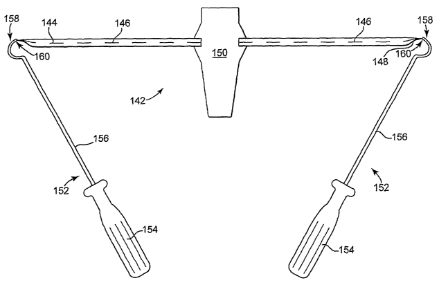
Figure 6 schematically illustrates a prolapse support device that includes an
implant having two pre-attached insertion tools. Implant 142 includes end
portions
144 and 148 connected to central support portion 150. Sutures 146 extend along
the
lengths of each of extension 144 and 148 and are connected to central support
portion 150 and extensions 144 and 148. Insertion tools 152 include shaft 156,
handle 154, and curved distal portion, needles 158, each needle being attached
at a
distal end of extension portions 144 and 148.
Each tool 152 includes handle 154, shaft 152, curved needle end 158 at a
distal end of shaft 152, and needle tip 160. Tips 160 are leading edges of
curved
needles 158, and each is attached to a distal end of an extension portion 144
or 148.
With this design, a surgeon receives the implant product 142 with tools 152
attached; the surgeon installs implant 142, transvaginally, with support
portion 150
being attached to vaginal tissue. The surgeon uses each of tools 152 to place
ends of
extension portions 146 and 148 bilaterally through tissue in the region of the
arcus
tendineus, e.g., near the ischial spine. Curved distal end 158 of each tool
152 allows
the surgeon to lead a distal end of each extension portion 144 and 148 around
the
outside of the arcus tendineus and back into the pelvic region at a location
below the
arcus tendineus, e.g., through the iliococcygeous muscle. This placement can
be
performed by movement or each tool 152, the movement including rotation of
handle 154 about an axis that includes shaft 156, to rotate curved distal end
158, also
CA 02615130 2008-01-10
WO 2007/016083 PCT/US2006/028828
-29-
about the axis of shaft 156. This can pass tip 160 and a distal end of an
extension
portion around an arcus tendineus, with the distal end exiting and re-entering
the
pelvic region. The surgeon can then disconnect the end of each extension
portion
from each tip 160 by cutting the extension portions 144 and 148 near tip 160
of each
tool 152, After cutting the end of each extension portion 144 and 148, the
needle
and tool can be removed from the pelvic region and each extension portion 144
and
148 can be manipulated to adjust and position the extension portion and
implant 142
as desired to support vaginal tissue connected to support portion 150. This
can
optionally include producing another tissue path to an external location and
leading
the end of each extension portion 144 and 148 through that tissue path to the
external location in the perirectal region such as is illustrated in figures 7
and 9. Or,
each extension portion 144 and 148 can be severed and severed ends can be left
at
locations within the pelvic floor region without leading extension portions
144 and
148 to an external incision.
According to particular embodiments, a kit according to the invention can
include an implant and a tool or multiple tools for installation, e.g., with
the
insertion tool being either removably enjoyable or attached (e.g.,
permanerztly) at a
distal end of an extension portion of the implant. Referring to figure 10, a
kit is
illustrated (not to scale) to include a mesh tape implant (220) for supporting
vaginal
tissue, insertion tool 200 coinprising a distal end having a curved needle,
and
tunneler device 230. Figure 4 includes an illustration of an exemplary
insertion tool
useful for installing a mesh to pass above the arcus tendineus. Tool 200
(Deschamps
Needle) of figure 10 is shown as a side view. Tunneler device 230 (e.g., IVS
tunneler) includes hollow trocar 232 and flexible plastic plug 234, as are
known.
CA 02615130 2008-01-10
WO 2007/016083 PCT/US2006/028828
-30-
Tool 200 includes handle 202, shaft 204 having a proximal end connected to
handle
202, shaft 204 connected at a distal end to curved end (e.g., "needle") 208.
The
diameter of the curved needle portion 208 is shown to be 1 inch, meaning that
the
radius of curvature is about 0.5 inch; other sizes of the curve may also be
useful.
Curved end 208 defines a two-dimensional curve, and the combination of curved
end 208 with shaft 204 together are in three dimensions.
Still referring to figure 10, tip 210 of curved end 208 can be adapted to
allow
an end of a mesh implant to be held (removably engaged) by tip 210. Tip 210
can
also be sharpened to allow the tip to be passed through tissue. Tool 200 may
be a
Deschamps Needle or similar structure having a handle, shaft, and a curved
portion
at the end of the shaft. A distal end (222) of mesh implant 220 can be
attached to
the end (tip) of curved portion of curved needle 208 of tool 200. During use,
a
surgeon can make a vaginal incision to allow transvaginal insertion of curved
end
208 of tool 200, with a distal end 222 of implant 220 attached at the end
(tip) 210 of
curved portion 208. Center portion 224 of implant 220 is attached to the
vaginal
apex to provide support, and each of ends 222 can be extended bi-laterally
through
tissue paths that pass through a region of the arcus tendineus, such as
through the
obturator internus muscle just above the arcus tendineus at the level of the
ischial
spine. Tunneler device 230 can be used to separately retrieve each of ends 222
of
mesh implant 220 and draw ends 222 through a tissue path to an external
incision,
e.g., an external incision in the perirectal region.
The invention contemplates placement of an implant, as described, to treat a
condition of vaginal tissue prolapse, by use of transvaginal surgical methods.
The
present description identifies certain combinations of implants, tools, and
CA 02615130 2008-01-10
WO 2007/016083 PCT/US2006/028828
-31 -
procedures, but as will be understood based on the present description, many
different variations on the present methods, tools, and procedures will be
useful, as
will combination of implant, tool, and procedure.
According to one embodiment of the invention, using a curved Deschamps
or similar needle, an extension portion of an implant can be inserted through
a
vaginal incision and a central portion of the implant can be attached to
vaginal tissue
that is to be supported. Two opposing tissue paths are created from the
vaginal
tissue to regions of the arcus tendineus, e.g., above the arcus tendineus at a
level of
the ischial spine, one on each side of the pelvic region. Each end
("extension")
portion of the implant can then be extended through a tissue path and above or
below the arcus tendineus, such as through the obturator internus muscle or
iliococcygeus, optionally also as close to the ischial spine as possible,
using the
curved needle. The end portion of the implant is passed around the outside of
the
white line and passed back into the pelvic region either below or above the
white
line, either through obturator intemus or the iliococcygeus muscle. This
location of
the inesh end is illustrated in figures 7, 7A, 8, 9, and 9A. The end of the
mesh thus
passes back into the pelvic floor and can be subsequently passed through a
tissue
passage that is created below that pelvic floor location, to an incision made
external
to the rectal region at each buttock (e.g., 2 to 3 centimeters lateral and 2
to 3
centimeters posterior to the anus).
Methods of the invention can involve placement of an implant and extension
portion using a system as sllown in figure 4. Distal curved portion 128 of
tool 122 is
engaged with interior space 113 of dilator 112, and tool 122 can be inserted
transvaginally and rotated to produce a tissue path in a region of the arcus
tendineus.
CA 02615130 2008-01-10
WO 2007/016083 PCT/US2006/028828
-32-
Tool 122 then engages the second dilator to make a second opposite tissue
path,
transvaginally. Each extension portion can be adjusted as desired, and tissue
support
portion 120 can be attached to vaginal tissue and adjusted. In alternate
embodiments
that also use tool 122 and implant 110, a tissue path can be produced
transvaginally
using tool 122, as a first step, then dilator 112 can be engaged with distal
end 128
after formation of the tissue path. After engaging dilator 112 and distal end
128,
tool 122 can be counter-rotated to cause the dilator to be pulled back through
the
tissue path. The extension portion and tissue support portion 120 can then be
adjusted.
Methods of the invention include variations that advantageously do not
require any external incision such as two incisions perirectal incisions.
Again
entering the pelvic region transvaginally, e.g., using a curved Deschamps
Needle (or
alternately a small curved needle as shown in figure 5), an extension portion
of an
implant can be inserted through a vaginal incision and a central portion of
the
iinplant attached to vaginal tissue that is to be supported. Opposing tissue
paths are
created starting at the vaginal tissue and extending to opposite locations at
regions of
the arcus tendineus, e.g., above the arcus tendineus at the level of the
ischial spine.
Each end portion of the iinplant can then be extended through one of the
opposing
tissue paths and through obturator internus muscle, as close to the ischial
spine as
possible. The end portion of the implant is passed (wrapped) around the
outside of
the white line and passed back into the pelvic region below the white line,
through
the iliococcygeus muscle. The end of the mesh thus passes back into the pelvic
floor. Instead of passing the end portion through a tissue passage that is
created
between that pelvic floor location and an external incision (e.g., in the
rectal region
CA 02615130 2008-01-10
WO 2007/016083 PCT/US2006/028828
-33-
at each buttocks), however, the end portion can be cut off, e.g., within the
pelvic
region, and the severed end left in that internal position of the anatomy. The
end
portion may optionally be secured using a suture. The severed end portions of
the
implant will remain in place within the defined tissue path and will the
iinplant will
support the vaginal tissue.
Figure 7 illustrates an installed mesh tape implant 220 (e.g., as shown in
figure 10) that includes a central support portion (224) secured to tissue of
the
vaginal apex. Each of two distal ends 222 of extension portions 221 of implant
220
extends bilaterally to pass above each arcus tendineus (6), near each ischial
spine
(8). Each of the two distal ends 222 of extension portions 221 of implant 220
then
passes thorough the iliococcygeus muscle (not shown), below the white line
(6),
back to the pelvic floor region and then through two tissue paths, each path
extending to an external incision (70) in the perirectal region.
Figure 7A illustrates an alternate embodiment of an installed mesh tape
implant 220 (e.g., as shown in figure 10) that includes a central support
portion 224
secured to tissue of the vaginal apex. Each of two extension portions 221 of
implant
220 extends bilaterally to pass above each arcus tendineus (6) near each
ischial spine
(8). Each of the two extension portions 221 of iniplant 220 then passes
thorough the
iliococcygeus muscle (not shown) below the white line (6) and back to the
pelvic
floor region. Each of the two extension portions 221 of implant 220 is severed
to
create an end of the extension portion located within the pelvic floor and
extension
portions 221 are not extended through tissue paths to any external incision.
Figure 8 illustrates another view of an installed mesh tape implant, such as
implant 220 shown in figure 10. Figure 8 looks in a direction down into the
pelvic
CA 02615130 2008-01-10
WO 2007/016083 PCT/US2006/028828
-34-
cavity, such as from a position in the abdomen. For reference are shown pubic
bone
80, bladder 82, obturator internus muscle 84, and iliococcygeous muscle 86. In
figure 8, a central support portion 224 of a mesh implant 220 is secured using
sutures (not shown) to tissue of the apex of vagina 2. Each of the two distal
ends
222 of extension portions 221 of implant 220 are extended bilaterally and
passed
through opposing tissue paths above each arcus tendineus (6), near each
ischial spine
(8), e.g., through obturator internus muscle 84. The far portions and distal
ends 222
of the extension portions 221 of implant 220 are not shown in figure 8. These
far
portions of extension portions 221 may extend through alternate tissue paths,
such as
back into the pelvic region by passing through iliococcygeous muscle 86 at a
location below arcus tendineus 6, then either severed at a location within the
pelvic
floor region or passed to an external incision.
Figure 9 illustrates still another view of an exemplary installed mesh tape
implant 220 that includes central support portion 224 secured to tissue of the
vaginal
apex. Figure 9 is a view from exterior of the patient, facing the vagina and
anus
(90). Each of two distal ends 221 of the implant 220 extends bilaterally to
pass
above each arcus tendineus 6 near each ischial spine $, around the arcus
tendineus 6,
and then thorough the iliococcygeus muscle (not shown) below the white line 6
and
back to the pelvic floor region. From there each extension portion 221 passes
through a tissue path extending to an external incision (70) in the perirectal
region.
Figure 9A illustrates an alternate embodiment of an installed mesh tape
implant that includes a central support portion 224 secured to tissue of the
vaginal
apex. Figure 9 is a view from exterior of the patient, facing the vagina and
anus
(90). Each of two extension portions 221 of implant 220 extends bilaterally to
pass
CA 02615130 2008-01-10
WO 2007/016083 PCT/US2006/028828
-35-
above each arcus tendineus 6 near each ischial spine 8. Each of the two
extension
portions 221 of the implaa3t 220 then passes around arcus tendineus 6 and then
through the iliococcygeus muscle (not shown) below the white line 6 back to
the
pelvic floor region. Each of the two ends of the implant is severed to create
an end
that remairzs internally within the pelvic floor, and the end portions are not
extended
through tissue paths to external incisions in the perirectal region.