Note: Descriptions are shown in the official language in which they were submitted.
CA 02615391 2008-01-14
WO 2007/011797 PCT/US2006/027515
THE IMMUNOPHENOTYPE AND IMMUNOGENICITY OF HUMAN ADIPOSE
DERIVED CELLS
BACKGROUND OF THE INVENTION
The emerging field of regenerative medicine seeks to combine
biomaterials, growth factors, and cells as novel therapeutics to repair
damaged tissues and
organs. As this specialty grows, there is a demand for a reliable, safe, and
effective
source of human adult stem cells to serve in tissue engineering applications.
For
regulatory purposes, these cells must be defined by quantifiable measures of
purity. For
practical purposes at the clinical level, these cells should be available as
an "off the shelf'
product immediately available upon demand at the point of care. From a
commercial
standpoint, the ability to use allogeneic, as opposed to autologous, adult
stem cells for
transplantation would have a significant positive impact on product
development. Under
these circumstances, a single lot of cells derived from one donor could be
transplanted to
multiple patients, reducing the costs of both quality control and quality
assurance.
Stem cells also exist in tissues of the adult organism. The best
characterized example of an adult stem cell is the hematopoietic progenitor
cell isolated
from the bone marrow and peripheral blood. In the absence of treatment,
lethally
irradiated mice died because they failed to replenish their circulating blood
cells;
however, transplantation of bone marrow cells from syngeneic donor animals
rescued the
host animal. The donor cells were responsible for repopulating the circulating
blood
cells. Studies have since been conducted to demonstrate that undifferentiated
hematopoietic stem cells are capable of regenerating the different blood cell
lineages in a
host animal. These studies have provided the basis for bone marrow
transplantation, a
widely accepted therapeutic modality for cancer and inborn errors of
metabolism.
Until recently, hematopoietic stem cells (HSC) of bone marrow origin
were the only accepted "adult" stem cell capable of multipotent
differentiation and self
renewal. Now, evidence is accumulating to support the existence of stem cells
in
multiple tissue sites. These include multipotent adult progenitor cells (MAPC)
mesenchymal stem cells (MSC) from the bone marrow, dermal stem cells, ear
MSCs,
PHIP\436219\2
CA 02615391 2008-01-14
WO 2007/011797 PCT/US2006/027515
neural stem cells from the central nervous system, hepatic and pancreatic stem
cells, and
stem cells from skeletal muscle. Adipose-derived stem cells (ASCs) exhibit
several
advantageous features. Adult stem cells derived from white adipose tissues can
differentiate along the adipocyte, chondrocyte, endothelial, hematopoietic
support,
hepatocyte, neuronal, myogenic, and osteoblast lineage pathways in vitro
(Gimble et al.
2003 Curr. Top. Dev. Biol. 58:137-60; Halvorsen et al. 2001 Metabolism 50:407-
13;
Halvorsen et al. 2001 Tissue Eng. 7:729-41; Hicok et al. 2004 Tissue Eng.
10:371-80;
Erickson et al. 2002 Biochem. Biophys. Res. Commun. 290:763-9; Safford et al.
2004
Exp. Neurol. 187:319-28; Safford et al. 2002 Biochem. Biophys. Res. Comniun.
294:371-
9; Zuk et al. 2001 Tissue Eng. 7:211-28; Zuk et al. 2002 Mol. Biol. Cell.
13:4279-95;
Mizuno et al. 2003 J. Nippon Med. Sch. 70:300-6; Seo et al. 2005 Biochem.
Biophys.
Res. Commun. 328:258-64). Adipose tissue is accessible, abundant, and
replenishable,
thereby providing a potential adult stem cell reservoir for each individual.
These findings
represent the work of many groups working independently. However, the cell
preparations in different laboratories are not identical. It is believed that
these
independent groups begin their cell isolation procedures by subjecting the
minced
adipose tissue to a collagenase digestion followed by a centrifugation step.
The initial
cell pellet is identified as the "stromal vascular fraction" (SVF). Some
groups have
focused their attention exclusively on this minimally processed cell
population. Others
expand the plastic adherent subpopulation of the SVF cells for multiple
passages; these
are the cells that have been identified as ASCs.
The mammalian immune system plays a central role in protecting
individuals from infectious agents and preventing tumor growth. However, the
same
immune system can produce undesirable effects such as the rejection of cell,
tissue and
organ transplants from unrelated donors. The immune system does not
distinguish
beneficial intruders, such as a transplanted tissue, from those that are
harmful, and thus
the immune system rejects transplanted tissues or organs. Rejection of
transplanted
organs is generally mediated by alloreactive T cells present in the host which
recognize
donor alloantigens or xenoantigens.
The transplantation of cells, tissues, and organs between genetically
disparate individuals invariably results in the risk of graft rejection.
Nearly all cells
PHIP\436219\2 2
CA 02615391 2008-01-14
WO 2007/011797 PCT/US2006/027515
express products of the major histocompatibility complex, MHC class I
molecules.
Further, many cell types can be induced to express MHC class II molecules when
exposed to inflammatory cytokines. Additional immunogenic molecules include
those
derived from minor histocompatibility antigens such as Y chromosome antigens
recognized by female recipients. Rejection of allografts is mediated primarily
by T cells
of both the CD4 and CD8 subclasses (Rosenberg et al., 1992 Annu. Rev. Immunol.
10:333). Alloreactive CD4+ T cells produce cytokines that exacerbate the
cytolytic CD8
response to alloantigen. Within these subclasses, competing subpopulations of
cells
develop after antigen stimulation that are characterized by the cytokines they
produce.
Th1 cells, which produce IL-2 and IFN-y, are primarily involved in allograft
rejection
(Mossmann et al., 1989 Annu. Rev. Immunol. 7:145). Th2 cells, which produce IL-
4 and
IL-10, can down-regulate Th1 responses through IL-10 (Fiorentino et., 1989 J.
Exp. Med.
170:208 1). Indeed, much effort has been expended to divert undesirable Thl
responses
toward the Th2 pathway. Undesirable alloreactive T cell responses in patients
(allograft
rejection, graft versus host disease) are typically treated with
immunosuppressive drugs
such as prednisone, azathioprine, and cyclosporine A. Unfortunately, these
drugs
generally need to be maintained for the life of the patient and they have a
multitude of
dangerous side effects including generalized immunosuppression. A much better
approach than pan immunosuppression is to induce specific or localized
suppression to
donor cell alloantigens, leaving the remaining immune system intact.
It is believed that there are numerous ways to induce immunologic
tolerance to alloantigens that would allow transplantation of allogeneic stem
cells.
Unfortunately, many of the approaches that have worked well in rodent animal
models
have not been successful when applied to nonhuman primates or humans.
Similarly, the
use of nuclear transfer to create clones of embryonic stem cells genetically
identical to
the recipient has been problematic for higher species, although limited
success was
recently reported for humans (Hwang et al., 2004, Science 303:1669). It is not
clear how
this technology could be applied to engineering other types of stem cells, and
whether the
time required for manipulation and expansion would obviate their usefulness.
Stem cells were reported to exhibit a low degree of immunogenicity,
possibly due to their immature state of differentiation and immunoregulatory
properties.
PHIP\436219\2 3
CA 02615391 2008-01-14
WO 2007/011797 PCT/US2006/027515
Rat embryonic stem cell-like lines express low levels of MHC class I antigens
and they
are negative for expression of MHC class II molecules and CD80(B7-1)/86(B7-2)
costimulatory molecules (Fandrich et al., 2002 Nat. Med. 8:17 1). These cells
engrafted
in the liver of immunocoinpetent allogeneic recipient rats when injected into
the portal
vein. Engraftment was attributed to lack of costimulatory molecules and the
expression
of FasL by the stem cell lines. Activated T cells express the Fas receptor,
thus rendering
them susceptible to apoptosis by the stem cell lines. Whether these properties
are shared
by other embryonic stem cell lines is currently unknown as transplanted fetal
and
embryonic stem cell-derived tissues are frequently rejected by the recipient's
immune
system (Bradley et al., 2002 Nat. Rev. 2:859; Kaufinan et al., 2000 E-biomed
1:11).
Neural stem cells derived from rodents express low or negligible levels of MHC
class I or
class II antigens (McLaren et al., 2001 J. Neuroimmunol 112:35), but these
cells are
usually rejected after implantation into allogeneic recipients unless
immunosuppressive
drugs are used (Mason et al., 1986 Neuroscience 19:685; Sloan et al., 1991
Trends
Neurosci. 14:341; Wood et al., 1996 Neuroscience 70:775). Rejection may be
initiated
after MHC molecules are up-regulated on cell membranes after exposure to
inflammatory
cytokines of the IFN family (McLaren et al., 2001 J. Neuroimmunol 112:35).
A major goal in organ transplantation is the permanent engraftment of the
donor organ without inducing a graft rejection immune response generated by
the
recipient, while preserving the immunocompetence of the recipient against
other foreign
antigens. Typically, in order to prevent host rejection responses, nonspecific
immunosuppressive agents such as cyclosporine, methotrexate, steroids and
FK506 are
used. These agents must be administered on a daily basis and if administration
is
stopped, graft rejection usually results. However, a major problem in using
nonspecific
immunosuppressive agents is that they function by suppressing all aspects of
the immune
response, thereby greatly increasing a recipient's susceptibility to infection
and other
diseases, including cancer. Furthermore, despite the use of immunosuppressive
agents,
graft rejection still remains a major source of morbidity and mortality in
human organ
transplantation. Most human transplants fail within 10 years without permanent
graft
acceptance. Only 50% of heart transplants survive 5 years and 20% of kidney
transplants
survive 10 years. (Opelz et al., 1981 Lancet 1:1223).
Pxrn\4362i 9\2 4
CA 02615391 2008-01-14
WO 2007/011797 PCT/US2006/027515
It is currently believed that a successful transplantation is dependent on
the prevention and/or reduction of an unwanted immune response by the host to
a
transplant mediated by immune effector cells to avert host rejection of donor
tissue. Also
advantageous for a successful transplantation is a method to eliminate or
reduce an
unwanted immune response by the donor tissue against a recipient tissue known
as graft
versus host disease. Thus, there is long-felt need for methods to suppress or
otherwise
prevent an unwanted immune response associated with transplantation of cells,
tissues,
and organs between genetically disparate individuals. The present invention
meets this
need.
BRIEF SUMMARY OF THE INVENTION
The present invention includes an isolated adipose tissue-derived adult
stromal (ADAS) cell exhibiting a non-immunogenic characteristic, wherein the
cell has
been passaged up to at least the second passage, further wherein the cell
expresses a stem
cell associated characteristic selected from the group consisting of human
multidrug
transporter (ABCG2) and aldehyde dehydrogenase (ALDH).
In one aspect of the invention, the ADAS cell has been passaged up to at
least the sixteenth passage.
In another aspect, exogenous genetic material has been introduced into the
ADAS cell.
In yet another aspect, the ADAS cell is derived from a human.
In another aspect, the ADAS cell allogeneic to a recipient thereof. In yet
another aspect, the ADAS cell is xenogeneic to a recipient thereof.
The invention also includes a method of treating a transplant recipient to
reduce in the recipient an immune response of effector cells against an
alloantigen,
comprising administering to a transplant recipient, an ADAS cell exhibiting a
non-
immunogenic characteristic, wherein the ADAS cell has been passaged up to at
least the
second passage, further wherein the ADAS cell expresses a stem cell associated
characteristic selected from the group consisting of human multidrug
transporter
(ABCG2) and aldehyde dehydrogenase (ALDH), in an amount effective to reduce an
PHIP\436219\2 5
CA 02615391 2008-01-14
WO 2007/011797 PCT/US2006/027515
immune response of effector cells against an alloantigen, whereby in the
transplant
recipient, the effector cells have a reduced immune response against the
alloantigen.
In one aspect, the effector cell is a T cell. In another aspect, the T cell is
from a donor and the alloantigen is from a recipient. In yet another aspect,
the T cell is
from a recipient and the alloantigen is from a donor.
In another aspect, the T cell is present in the transplant.
In yet another aspect, the effector cell is a T cell activated prior to
administration of the ADAS cell to a recipient, and further wherein the immune
response
is the reactivation of the T cell from the donor.
In a further aspect, the ADAS cell is administered to the transplant
recipient to treat rejection of the transplant by the recipient.
In another aspect, the ADAS cell is derived from a mammal. Preferably,
the mammal is a human.
In a furtlier aspect, an immunosuppressive agent is administering to the
recipient in combination with an ADAS cell.
In one aspect, the ADAS cell is administered to the recipient prior to the
transplant. In another aspect, the ADAS cell is administered to the recipient
concurrently
with the transplant. In yet another aspect, the ADAS cell is administered as
part of the
transplant. In another aspect, the ADAS cell is administered to the recipient
subsequent
to the transplantation of the transplant.
In one aspect, the ADAS cell is administered intravenously to the
recipient.
In another aspect, the effector cell is a cell of the recipient of the donor
transplant.
In yet another aspect, the ADAS cell is genetically modified.
The invention also includes a method of reducing an immune response by
an effector cell against an alloantigen, comprising contacting an effector
cell with an
ADAS cell exhibiting a non-immunogenic characteristic, wherein the ADAS cell
has
been passaged up to at least the second passage, further wherein the ADAS cell
expresses
a stem cell associated characteristic selected from the group consisting of
human
multidrug transporter (ABCG2) and aldehyde dehydrogenase (ALDH), in an amount
PHIP\436219\2 6
CA 02615391 2008-01-14
WO 2007/011797 PCT/US2006/027515
effective to reduce an immune response by the effector cell against the
alloantigen.
Preferably, the effector cell is a T cell.
The invention also includes a method of isolating an ADAS cell from a
population of cells derived from adipose tissue, the method coinprising
providing an
antibody specific for ABCG2; contacting the population of adipose-derived
cells with the
antibody under conditions suitable for formation of an antibody-adipose tissue-
derived
stromal cell complex; and substantially separating the antibody-adipose tissue-
derived
stromal cell complex from the population of adipose-derived cells; thereby
isolating the
adipose tissue-derived stromal cell.
In one aspect, the antibody is conjugated to a physical support.
In another aspect, the physical support is selected from the group
consisting of a microbead, a magnetic bead, a panning surface, a dense
particle for
density centrifugation, an adsorption column and an adsorption membrane.
In yet another aspect, the physical support is selected from the group
consisting of a streptavidin bead and a biotin bead.
In one aspect, the antibody-adipose tissue-derived stromal cell complex is
substantially separated from the population of adipose-derived cells using a
method
selected from the group consisting of fluorescence activated cell sorting
(FACS) and
magnetic activated cell sorting (MACS).
The invention also includes a method of enriching adipose tissue-derived
stromal cells from a population of adipose-derived cells, the method
comprising
providing an antibody specific for ABCG2; contacting the population of adipose-
derived
cells with the antibody under conditions suitable for formation of an antibody-
adipose
tissue-derived stromal cell complex; and substantially separating the antibody-
adipose
tissue-derived stromal cell complex from the population of adipose-derived
cells; thereby
isolating the adipose tissue-derived stromal cell.
The invention also includes a method of identifying an ADAS cell positive
for ALDH from a population of cells derived from adipose tissue, the method
comprising
providing a cleavable substrate specific for ALDH to the population of cells,
wherein the
substrate when so present in an ALDH+ cell is cleaved, further wherein the
cleaved
substrate emits a fluorescence thereby identifying an ALDH+ ADAS cell.
PHIP\436219\2 7
CA 02615391 2008-01-14
WO 2007/011797 PCT/US2006/027515
BRIEF DESCRIPTION OF THE DRAWINGS
For the purpose of illustrating the invention, there are depicted in the
drawings certain embodiments of the invention. However, the invention is not
limited to
the precise arrangements and instrumentalities of the embodiments depicted in
the
drawings.
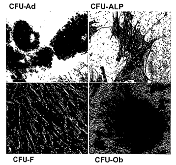
Figure 1, comprising Figures lA through 1D, is a series of images
depicting Colony Forming Unit Assays (CFU) of cells derived from adipose
tissue. The
images depict staining profiles representative of the following colonies:
(Figure 1A)
Toluidine blue" CFU-F; (Figure IB) Alkaline phosphatase"- CFU-ALP; (Figure 1C)
Oil
Red O+ CFU-Ad; and (Figure ID) Alizarin Red+ CFU-Ob.
Figure 2 is a graph depicting a flow cytometry histogram of adipose
derived cells. The flow cytometry histograms for selected hematopoietic, stem
cell, and
stromal cell markers from a representative donor are displayed at the stromal
vascular
fraction (SVF) and passage 2 (P2) stages. The percentage of cells staining
positive is
depicted in the upper right corner of each panel. The blue line indicates the
positive
staining cells while the red line indicates the isotype matched monoclonal
antibody
control.
Figure 3, comprising Figures 3A and 3B is a series of charts
demonstrating the relative change in the immunophenotype of adipose derived
cells as a
function of purification and passage. The percentage of positive staining
cells is
displayed relative to the isolation stage and passage number. Figure 3A
depicts the
stromal cell associated markers CD 166, CD73, CD44, and CD29. Figure 3B
depicts the
stem cell associated markers human multidrug transporter (ABCG2) and CD34 (the
order
of the passage numbers is reversed in Figure 3A relative to Figures 3B).
Figure 4 is a chart depicting the aldehyde dehydrogenase staining of
adipose derived cells as a function of purification and passage,
Figure 5 is a graph depicting a flow cytometry histogram of adipose
derived cells. The flow cytometry histograms for selected hematopoietic
markers from a
representative donor are displayed at the stromal vascular fraction (SVF) and
passage 2
PHIP\436219\2 $
CA 02615391 2008-01-14
WO 2007/011797 PCT/US2006/027515
(P2) stages. The percentage of cells staining positive is depicted in the
upper right corner
of each panel. The blue line indicates the positive staining cells while the
red line
indicates the isotype matched monoclonal antibody control.
Figure 6 is a graph depicting the immunogenicity of adipose derived cells
as evaluated by mixed lymphocyte reaction (MLR) of adipose derived cells as a
function
of purification and passage. Figure 5 depicts a representative MLR from a
single donor.
The proliferation of T cells was determined in the absence of stimulator
cells, in the
presence of autologous irradiated PBMCs (negative control), in the presence of
allogeneic irradiated PBMCs (positive control), and in the presence of adipose
derived
cells (SVF, PO-P4). The stimulator cells were present at densities of 5,000,
10,000, or
20,000 per well.
i Figure 7 is a chart demonstrating the immunosuppressive effects of human
adipose derived cells, including human SVF cells and ADAS cells, in a two-way
mixed
lymphocyte reaction.
Figure 8 is a chart comparing the immunosuppressive effects between
bone marrow stromal cells (BMSCs) and ADAS cells as measured by MLR. The
difference between the ADAS and BMSC groups was not significant (p>0.05,
Student's
t-test).
DETAILED DESCRIPTION
The present invention relates to the discovery that adipose tissue-derived
adult stromal (ADAS) cells possess novel immunophenotypical and immunological
characteristics. The novel characteristics of ADAS cells provide methods for
isolating,
culturing and using these cells in cell and/or gene therapy. The present
invention
includes compositions and methods for isolating and culturing ADAS cells as
well as
transplanting ADAS cells to a recipient where the likelihood of immune
rejection by
either the host or the graft is reduced.
The present invention is useful in transplantation of a transplant, for
example a biocompatible lattice or a donor tissue, organ or cell, by reducing
and/or
eliminating an immune response against the transplant by the recipient's own
immune
PHIP\436219\2 9
CA 02615391 2008-01-14
WO 2007/011797 PCT/US2006/027515
system. As described more fully below, ADAS cells play a role in inhibiting
and/or
preventing allograft rejection of a transplant.
In addition, the disclosure provided herein demonstrates that ADAS cells
are useful for the irihibition and/or prevention of an unwanted immune
response by a
donor transplant, for example, a biocompatible lattice or a donor tissue,
organ or cell,
against a recipient tissue known as graft versus host disease.
Accordingly, the present invention encompasses methods and
compositions for reducing and/or eliminating an immune response to a
transplant in a
recipient by treating the recipient with an amount of ADAS cells effective to
reduce or
inhibit host rejection of the transplant. Also encompassed are methods and
compositions
for reducing and/or eliminating an immune response in a host by the foreign
transplant
against the host, i.e., graft versus host disease, by treating the donor
transplant and/or
recipient of the transplant ADAS cells in order to inhibit or reduce an
adverse response
by the donor transplant against the recipient.
Definitions
As used herein, each of the following terms has the meaning associated
with it in this section.
The articles "a" and "an" are used herein to refer to one or to more than
one (i.e. to at least one) of the grammatical object of the article. By way of
example, "an
element" means one element or more than one element.
The term "about" will be understood by persons of ordinary skill in the art
and will vary to some extent on the context in which it is used.
The term "adipose tissue-derived cell" refers to a cell that originates from
adipose tissue. The initial cell population isolated from adipose tissue is a
heterogenous
cell population including, but not limited to stromal vascular fraction (SVF)
cells.
As used herein, the term "adipose derived stromal cells," "adipose tissue-
derived stromal cells," "adipose tissue-derived adult stromal (ADAS) cells,"
or "adipose-
derived stem cells (ASCs)" are used interchangeably and refer to stromal cells
that
originate from adipose tissue which can serve as stem cell-like precursors to
a variety of
different cell types such as but not limited to adipocytes, osteocytes,
chondrocytes,
PHIP\436219\2 10
CA 02615391 2008-01-14
WO 2007/011797 PCT/US2006/027515
Inuscle and neuronal/glial cell lineages. Based on the present disclosure,
ADAS cells
encompass a substantially homogenous population of stem cell-like cells that
possess
novel iminunophenotypic characteristics including but not limited to the
expression of
ABCG2 and ALDH. Further, the ADAS cells of the present invention are not
immunogenic with respect to the elicitation of T cell proliferation. ADAS
cells make up
a subset population derived from adipose tissue which can be separated from
other
components of the adipose tissue using standard culturing procedures or
otherwise
methods disclosed herein. In addition, ADAS cells can be isolated from a
mixture of
cells using the cell surface marlcers disclosed herein.
As used herein, the term "late passaged adipose tissue-derived stromal
cell," refers to a cell exhibiting a less immunogenic characteristic when
compared to an
earlier passaged cell. The immunogenicity of an adipose tissue-derived stromal
cell
corresponds to the number of passages. Preferably, the cell has been passaged
up to at
least the second passage, more preferably, the cell has been passaged up to at
least the
third passage, and most preferably, the cell has been passaged up to at least
the fourth
passage.
"Adipose" refers to any fat tissue. The adipose tissue may be brown or
white adipose tissue. Preferably, the adipose tissue is subcutaneous white
adipose tissue.
Such cells may comprise a primary cell culture or an immortalized cell line.
The adipose
tissue may be from any organism having fat tissue. Preferably the adipose
tissue is
mammalian, most preferably the adipose tissue is human. A convenient source of
human
adipose tissue is that derived from liposuction surgery. However, the source
of adipose
tissue or the method of isolation of adipose tissue is not critical to the
invention.
"Allogeneic" refers to a graft derived from a different animal of the same
species.
As defined herein, an "allogeneic adipose derived adult stromal cell" is
obtained from a different individual of the same species as the recipient.
"Alloantigen" is an antigen that differs from an antigen expressed by the
recipient.
PHIP\436219\2 11
CA 02615391 2008-01-14
WO 2007/011797 PCT/US2006/027515
As used herein, the term "autologous" is meant to refer to any material
derived from the same individual to which it is later to be re-introduced into
the
individual.
"Xenogeneic" refers to a graft derived from an animal of a different
species.
As used herein, the term "biocompatible lattice," is meant to refer to a
substrate that can facilitate formation into three-dimensional structures
conducive for
tissue development. Thus, for example, cells can be cultured or seeded onto
such a
biocompatible lattice, such as one that includes extracellular matrix
material, synthetic
polymers, cytokines, growth factors, etc. The lattice can be molded into
desired shapes
for facilitating the development of tissue types. Also, at least at an early
stage during
culturing of the cells, the medium and/or substrate is supplemented with
factors (e.g.,
growth factors, cytokines, extracellular matrix material, etc.) that
facilitate the
development of appropriate tissue types and structures.
"Donor antigen" refers to an antigen expressed by the donor tissue to be
transplanted into the recipient.
"Differentiation medium" is used herein to refer to a cell growth medium
comprising an additive or a lack of an additive such that a stem cell, adipose
derived
adult stromal cell or other such progenitor cell, that is not fully
differentiated when
incubated in the medium, develops into a cell with some or all of the
characteristics of a
differentiated cell.
As used herein, an "effector cell" refers to a cell which mediates an
immune response against an antigen. In the situation where a transplant is
introduced
into a recipient, the effector cells can be the recipient's own cells that
elicit an immune
response against an antigen present in the donor transplant. In another
situation, the
effector cell can be part of the transplant, whereby the introduction of the
transplant into a
recipient results in the effector cells present in the transplant eliciting an
immune
response against the recipient of the transplant.
"Expandability" is used herein to refer to the capacity of a cell to
proliferate, for example, to expand in number or in the case of a cell
population to
undergo population doublings.
PHIP\436219\2 12
CA 02615391 2008-01-14
WO 2007/011797 PCT/US2006/027515
"Graft" refers to a cell, tissue, organ or otherwise any biological
compatible lattice for transplantation.
By "growth factors" is intended the following specific factors iticluding,
but not limited to, growth hormone, erythropoietin, thrombopoietin,
interleukin 3,
interleukin 6, interleukin 7, macrophage colony stimulating factor, c-kit
ligand/stem cell
factor, osteoprotegerin ligand, insulin, insulin like growth factors,
epidermal growth
factor (EGF), fibroblast growth factor (FGF), nerve growth factor, ciliary
neurotrophic
factor, platelet derived growth factor (PDGF), and bone morphogenetic protein
at
concentrations of between picogram/ml to milligram/mi levels.
As used herein, the term "growth medium" is meant to refer to a culture
medium that promotes growth of cells. A growth medium will generally contain
animal
serum. In some instances, the growth medium may not contain animal serum.
"Immunophenotype" of a cell is used herein to refer to the phenotype of a
cell in terms of the surface protein profile of a cell.
An "isolated cell" refers to a cell which has been separated from other
components and/or cells which naturally accompany the isolated cell in a
tissue or
mammal.
As used herein, the term "multipotential" or "multipotentiality" is meant
to refer to the capability of a stem cell of the central nervous system to
differentiate into
more than one type of cell.
As used herein, the term "modulate" is meant to refer to any change in
biological state, i.e. increasing, decreasing, and the like.
As used herein, the term "non-immunogenic" is meant to refer to the
discovery that ADAS cells do not induce proliferation of T cells in an MLR.
However,
non-immunogenic should not be limited to T cell proliferation in an MLR, but
rather
should also apply to ADAS cells not inducing T cell proliferation in vivo.
"Proliferation" is used herein to refer to the reproduction or multiplication
of similar forms, especially of cells. That is, proliferation encompasses
production of a
greater number of cells, and can be measured by, among other things, simply
counting
the numbers of cells, measuring incorporation of 3H-thymidine into the cell,
and the like.
PHIP\436219\2 13
CA 02615391 2008-01-14
WO 2007/011797 PCT/US2006/027515
"Progression of or through the cell cycle" is used herein to refer to the
process by which a cell prepares for and/or enters mitosis and/or meiosis.
Progression
through the cell cycle includes progression through the Gl phase, the S phase,
the G2
phase, and the M-phase.
The terms "precursor cell," "progenitor cell," and "stem cell" are used
interchangeably in the art and herein and refer either to a pluripotent, or
lineage-
uncommitted, progenitor cell, which is potentially capable of an unlimited
number of
mitotic divisions to either renew itself or to produce progeny cells which
will
differentiate into the desired cell type. Unlike pluripotent stem cells,
lineage-committed
progenitor cells are generally considered to be incapable of giving rise to
numerous cell
types that phenotypically differ from each other. Instead, progenitor cells
give rise to one
or possibly two lineage-committed cell types.
The term "stromal cell medium" as used herein, refers to a medium useful
for culturing ADAS cells. An example of a stromal cell medium is a medium
comprising
DMEM/F 12 Ham's, 10% fetal bovine serum, 100 U penicillin/100 g
streptomycin/0.25
g Fungizone. Typically, the stromal cell medium comprises a base medium, serum
and
an antibiotic/antimycotic. However, ADAS cells can be cultured with stromal
cell
medium without an antibiotic/antimycotic and supplemented with at least one
growth
factor. Preferably the growth factor is human epidermal growth factor (hEGF).
The
preferred concentration of hEGF is about 1-50 ng/ml, more preferably the
concentration
is about 5 ng/ml. The preferred base medium is DMEM/F12 (1:1). The preferred
serum
is fetal bovine serum (FBS) but other sera may be used including horse serum
or human
serum. Preferably up to 20% FBS will be added to the above media in order to
support
the growth of stromal cells. However, a defined medium could be used if the
necessary
growth factors, cytokines, and hormones in FBS for stromal cell growth are
identified
and provided at appropriate concentrations in the growth medium. It is further
recognized that additional components may be added to the culture medium. Such
components include but are not limited to antibiotics, antimycotics, albumin,
growth
factors, amino acids, and other components known to the art for the culture of
cells.
Antibiotics which can be added into the medium include, but are not limited
to, penicillin
and streptomycin. The concentration of penicillin in the culture medium is
about 10 to
PHIP\436219\2 14
CA 02615391 2008-01-14
WO 2007/011797 PCT/US2006/027515
about 200 units per ml. The concentration of streptomycin in the culture
medium is about
to about 200 g/ml. However, the invention should in no way be construed to be
limited to any one medium for culturing stromal cells. Rather, any media
capable of
supporting stromal cells in tissue culture may be used.
5 As used herein, a "substantially purified" cell is a cell that is
essentially
free of other cell types. Thus, a substantially purified cell refers to a cell
which has been
purified from other cell types with which it is normally associated in its
naturally
occurring state.
"Transplant" refers to a biocompatible lattice or a donor tissue, organ or
10 cell, to be transplanted. An example of a transplant may include but is not
limited to a
tissue, a stem cell, a neural stem cell, a skin cell, bone marrow, and solid
organs such as
heart, pancreas, kidney, lung and liver.
As used herein, a "therapeutically effective amount" is the amount of
ADAS cells sufficient to provide a beneficial effect to the subject to which
the cells are
administered.
By the term "treating a transplant recipient to reduce in the recipient an
immune response of effector cells against an alloantigen to the effector
cells," as the
phrase is used herein, is meant decreasing the endogenous immune response
against the
alloantigen in a recipient by any method, for example administering ADAS cells
to a
recipient, compared with the endogenous immune response in an otherwise
identical
animal which was not treated with ADAS cells. The decrease in endogenous
immune
response can be assessed using the methods disclosed herein or any other
method for
assessing endogenous immune response in an animal.
As used herein "endogenous" refers to any material from or produced
inside an organism, cell or system.
"Exogenous" refers to any material introduced from or produced outside
an organism, cell, or system.
"Encoding" refers to the inherent property of specific sequences of
nucleotides in a polynucleotide, such as a gene, a cDNA, or an mRNA, to serve
as
templates for synthesis of other polymers and macromolecules in biological
processes
having either a defined sequence of nucleotides (i.e., rRNA, tRNA and mRNA) or
a
PHTP\436219\2 15
CA 02615391 2008-01-14
WO 2007/011797 PCT/US2006/027515
defined sequence of amino acids and the biological properties resulting
therefrom. Thus,
a gene encodes a protein if transcription and translation of mRNA
corresponding to that
gene produces the protein in a cell or other biological system. Both the
coding strand, the
nucleotide sequence of which is identical to the mRNA sequence and is usually
provided
in sequence listings, and the non-coding strand, used as the template for
transcription of a
gene or cDNA, can be referred to as encoding the protein or other product of
that gene or
cDNA.
Unless otherwise specified, a "nucleotide sequence encoding an amino
acid sequence" includes all nucleotide sequences that are degenerate versions
of each
other and that encode the same amino acid sequence. Nucleotide sequences that
encode
proteins and RNA may include introns.
An "isolated nucleic acid" refers to a nucleic acid segment or fragment
which has been separated from sequences which flank it in a naturally
occurring state,
i.e., a DNA fragment which has been removed from the sequences which are
normally
adjacent to the fragment, i.e., the sequences adjacent to the fragment in a
genome in
which it naturally occurs. The term also applies to nucleic acids which have
been
substantially purified from other components which naturally accompany the
nucleic
acid, i.e., RNA or DNA or proteins, which naturally accompany it in the cell.
The term
therefore includes, for example, a recombinant DNA which is incorporated into
a vector,
into an autonomously replicating plasmid or virus, or into the genomic DNA of
a
prokaryote or eukaryote, or which exists as a separate molecule (i.e., as a
cDNA or a
genomic or cDNA fragment produced by PCR or restriction enzyme digestion)
independent of other sequences. It also includes a recombinant DNA which is
part of a
hybrid gene encoding additional polypeptide sequence.
In the context of the present invention, the following abbreviations for the
commonly occurring nucleic acid bases are used. "A" refers to adenosine, "C"
refers to
cytosine, "G" refers to guanosine, "T" refers to thymidine, and "U" refers to
uridine.
The phrase "under transcriptional control" or "operatively linked" as used
herein means that the promoter is in the correct location and orientation in
relation to the
polynucleotides to control RNA polymerase initiation and expression of the
polynucleotides.
PH1P\436219\2 16
CA 02615391 2008-01-14
WO 2007/011797 PCT/US2006/027515
As used herein, the term "promoter/regulatory sequence" means a nucleic
acid sequence which is required for expression of a gene product operably
linked to the
promoter/regulatory sequence. In some instances, this sequence may be the core
promoter sequence and in other instances, this sequence may also include an
enhancer
sequence and other regulatory elements which are required for expression of
the gene
product. The promoter/regulatory sequence may, for example, be one which
expresses
the gene product in a tissue specific manner.
A "constitutive" promoter is a nucleotide sequence which, when operably
linked with a polynucleotide which encodes or specifies a gene product, causes
the gene
product to be produced in a cell under most or all physiological conditions of
the cell.
An "inducible" promoter is a nucleotide sequence which, when operably
linked with a polynucleotide which encodes or specifies a gene product, causes
the gene
product to be produced in a cell substantially only when an inducer which
corresponds to
the promoter is present in the cell.
A "tissue-specific" promoter is a nucleotide sequence which, when
operably linked with a polynucleotide which encodes or specifies a gene
product, causes
the gene product to be produced in a cell substantially only if the cell is a
cell of the
tissue type corresponding to the promoter.
A "vector" is a composition of matter which comprises an isolated nucleic
acid and which can be used to deliver the isolated nucleic acid to the
interior of a cell.
Numerous vectors are knnown in the art including, but not limited to, linear
polynucleotides, polynucleotides associated with ionic or amphiphilic
compounds,
plasmids, and viruses. Thus, the term "vector" includes an autonomously
replicating
plasmid or a virus. The term should also be construed to include non-plasmid
and non-
viral compounds which facilitate transfer of nucleic acid into cells, such as,
for example,
polylysine compounds, liposomes, and the like. Examples of viral vectors
include, but
are not limited to, adenoviral vectors, adeno-associated virus vectors,
retroviral vectors,
and the like.
"Expression vector" refers to a vector comprising a recombinant
polynucleotide comprising expression control sequences operatively linked to a
nucleotide sequence to be expressed. An expression vector comprises sufficient
cis-
PHIP\436219\2 17
CA 02615391 2008-01-14
WO 2007/011797 PCT/US2006/027515
acting elements for expression; other elements for expression can be supplied
by the host
cell or in an in vitro expression system. Expression vectors include all those
lcnown in
the art, such as cosmids, plasmids (i.e., naked or contained in liposomes) and
viruses that
incorporate the recombinant polynucleotide.
Description
The present invention relates to the discovery that when an adipose tissue-
derived adult stromal (ADAS) cell is contacted with a T cell obtained from a
different
individual (allogeneic T cells), the allogeneic T cell does not proliferate.
Prior art dogma
suggests that when T cells are mixed with any other cell type, T cell
proliferation ensues.
The mixed lymphocyte reaction (MLR) is a standard assay used to evaluate
immunogenicity (i.e., the ability for a cell to induce T cells to proliferate
as measured by
MLR). The data disclosed herein demonstrate that a T cell derived from one
individual is
not responsive to an ADAS cell obtained from a different individual.
Therefore, based
upon the disclosure provided herein, an ADAS cell is not immunogenic to the
immune
system with respect to manifesting a T cell response.
In an embodiment of the invention, the immunophenotype and
immunogenicity of an ADAS cell corresponds to the number of passages. Based on
the
disclosure provided herein, the later passaged cell is less immunogenic when
compared to
the earlier passaged cell. Preferably, the cell has been passaged for at least
two passages.
Preferably, the cell has been passaged for at least three passages. More
preferably, the
cell has been passaged for at least four passages.
In another embodiment of the invention, the cells can be cultured
following isolation and, if appropriate, assayed for their immunogenicity and
immunophenotype prior to therapeutic use. Preferably, the cells are cultured
without
differentiation using the standard cell culture media disclosed herein.
Preferably, the
cells can be passaged to at least five passages, and more preferably, the
cells can be
passaged to at least 10 passages or more. For example, the cells can be
passaged to at
least 15 passages, preferably at least 16 passages, more preferably at least
17 passages,
yet more preferably at least 18 passages, preferably at least 19 passages or
even at least
20 passages without losing their multipotentiality. Based on the disclosure
presented
PHIP143621912 18
CA 02615391 2008-01-14
WO 2007/011797 PCT/US2006/027515
herein, one skilled in the art would appreciate that the cells are not
immunogenic and
therefore are advantageous for transplantation into a mammal.
In addition to the non-immunogenic phenotype of the ADAS cell of the
present invention with respect to T lymphocytes in a different individual,
based on the
disclosure provided herein, one skilled in the art would appreciate that an
ADAS cell can
suppress an MLR between allogeneic cells, for example between a T cell from
one
individual and a peripheral blood mononuclear cell (PBMC) from another
individual. In
one aspect, an ADAS cell can actively reduce the allogeneic T cell response in
MLRs
between a T cell and a PBMC, each obtained from different individuals.
Moreover, as discussed in more detail elsewhere herein, the
immunophenotype of an ADAS cell relates to the method used in culturing the
cell. For
example, the immunophenotype of ADAS cells is defined as a function of, but
not limited
to, their stage of isolation, their passage number, whether the cells were
cultured as an
adherent population, and the length of time in culture. Based on the present
disclosure,
an ADAS cell can be successfully used in cell and/or gene therapy. That is,
the cells of
the present invention have a reduced likelihood of immune rejection by either
the host of
the graft when the cells are transplanted into an individual. In addition, an
ADAS cell
can be used as a therapeutic to inhibit host rejection of a transplant, and as
a therapeutic
to prevent or otherwise inhibit graft versus host disease following
transplantation. As
such, the present invention comprises compositions and methods for generating
an ADAS
cell useful for experimental/therapeutic purposes.
1. Isolation and culturing of ADAS
The ADAS cells useful in the methods of the present invention may be
isolated by a variety of methods known to those skilled in the art. For
example, such
methods are described in U.S. Pat. No. 6,153,432, which is incorporated herein
in its
entirety. In a preferred method, an ADAS cell is isolated from a mammalian
subject,
preferably a human subject.
The immunophenotype of adipose derived cells change progressively
depending on culturing procedures (i.e. passage number). The adherence to
plastic and
subsequent expansion of human adipose-derived cells selects for a relatively
PHIP\436219\2 19
CA 02615391 2008-01-14
WO 2007/011797 PCT/US2006/027515
homogeneous cell population, enriching for cells expressing a "stromal"
immunophenotype, as compared to the heterogeneity of the crude stromal
vascular
fraction. ADAS cells also express stem cell associated markers including, but
not limited
to human multidrug transporter (ABCG2) and aldehyde dehydrogenase (ALDH).
Based on the present disclosure, the immunophentype of adipose derived
cells can be exploited to serve as unique identifiers for ADAS cells. That is,
the unique
cell surface markers on the cells of the present invention can be used to
isolate a specific
sub-population of cells from a mixed population of cells derived from adipose
tissue.
One skilled in the art would appreciate that an antibody specific for a cell
surface marker
can be conjugated to a physical support (i.e. a streptavidin bead) and
therefore provide
the opportunity to isolate cell surface specific adipose derived cells. The
isolated cell can
then be cultured and expanded in vitro using methods disclosed herein or
conventional
methods.
A further embodiment of the present invention encompasses a method of
depleting or separating a subpopulation of cells derived from adipose tissue.
The
invention relates to the discovery that the imcnunophenotype of cells derived
from
adipose tissue is a function of passage number. As such, a specific cell
population such
as ADAS cells can be depleted from such a mixed population of cells derived
from
adipose tissue by incubating an antibody that specifically binds to an ADAS
cell within
the mixed population of cells followed by a separation step including but not
limited to
magnetic separation. An example of an antibody that specifically binds to an
ADAS cell
includes, but is not limited to anti-ABCG2 antibody. The process of magnetic
separation
is accomplished by using magnetic beads, including but not limited to
Dynabeads"
(Dynal Biotech, Brown Deer, WI). Further to the use ofDynabeads ,1VIA.CS
separation
reagents (Miltenyi Biotec, Auburn, CA) can be used to deplete ADAS cells from
a mixed
population of cells. As a result of the separation step, a population of
enriched ADAS
cells can be obtained. Preferably, the population of ADAS cells is a purified
cell
population.
The immunophenotype of the cells of the invention offers a method to sort
specific adipose derived cells using a flow cytometry-based cell sorter.
Preferably,
ADAS cells are isolated using the methods disclosed herein. The isolated ADAS
cell can
PHIP\436219\2 20
CA 02615391 2008-01-14
WO 2007/011797 PCT/US2006/027515
then be cultured in vitro to generate a desirable number of cells useful for
experimental or
therapeutic purposes.
Any medium capable of supporting fibroblasts in cell culture may be used
to culture ADAS. Media forinulations that support the growth of fibroblasts
include, but
are not limited to, Minimum Essential Medium Eagle, ADC-1, LPM (bovine serum
albumin-free), F10 (HAM), F12 (HAM), DCCM1, DCCM2, RPMI 1640, BGJ Medium
(with and without Fitton-Jackson Modification), Basal Medium Eagle (BME-with
the
addition of Earle's salt base), Dulbecco's Modified Eagle Medium (DMEM-without
serum), Yamane, IMEM-20, Glasgow Modification Eagle Medium (GMEM), Leibovitz
L-15 Medium, McCoy's 5A Medium, Medium M199 (M199E-with Earle's salt base),
Medium M199 (M199H-with Hank's salt base), Minimum Essential Medium Eagle
(MEM-E-with Earle's salt base), Minimum Essential Medium Eagle (MEM-H-with
Hank's salt base) and Minimum Essential Medium Eagle (MEM-NAA with non-
essential
amino acids), and the like. A preferred medium for culturing ADAS is DMEM,
more
preferably DMEM/F 12 (1:1).
Additional non-limiting examples of media useful in the methods of the
invention can contain fetal serum of bovine or other species at a
concentration at least 1%
to about 30%, preferably at least about 5% to 15%, most preferably about 10%.
Embryonic extract of chicken or other species can be present at a
concentration of about
1% to 30%, preferably at least about 5% to 15%, most preferably about 10%.
Following isolation, ADAS cells are incubated in stromal cell medium in a
culture apparatus for a period of time or until the cells reach confluency
before passing
the cells to another culture apparatus. Following the initial plating, the
cells can be
maintained in culture for a period of about 6 days to yield the Passage 0(P0)
population.
The cells can be passaged for an indefinite number of times, each passage
comprising
culturing the cells for about 6-7 days, during which the cell doubling times
can range
between 3-5 days. The culturing apparatus can be of any culture apparatus
commonly
used in culturing cells in vitro. A preferred culture apparatus is a culture
flask with a
more preferred culture apparatus being a T-225 culture flask.
ADAS cells can be cultured in stromal cell medium supplemented with
hEGF in the absence of an antibiotic/antimycotic for a period of time or until
the cells
PHIP\436219\2 21
CA 02615391 2008-01-14
WO 2007/011797 PCT/US2006/027515
~
reach a certain level of confluence. Preferably, the level of confluence is
greater than
70%. More preferably, the level of confluence is greater than 90%. A period of
time can
be any time suitable for the culture of cells in vitro. Stromal cell medium
may be
replaced during the culture of the ADAS cells at any time. Preferably, the
stromal cell
medium is replaced every 3 to 4 days. ADAS cells are then harvested from the
culture
apparatus whereupon the ADAS cells can be used immediately or cryopreserved to
be
stored for use at a later time. ADAS cells may be harvested by trypsinization,
EDTA
treatment, or any other procedure used to harvest cells from a culture
apparatus.
ADAS cells described herein may be cryopreserved according to routine
procedures. Preferably, about one to ten million cells are cryopreserved in
stromal cell
medium containing 10% DMSO in vapor phase of Liquid N2. Frozen cells can be
thawed
by swirling in a 37 C bath, resuspended in fresh growth medium, and grown as
usual.
The present invention also relates to the discovery that the
immunophenotype of an ADAS cell is a function of the passage number. The
immunophenotype and immunogenic properties of ADAS cells are defined as a
function
of culturing procedures (i.e. adherence property, passage number, length of
time in
culture). The present disclosure demonstrates that freshly isolated stromal
vascular
fraction (SVF) cells and early passaged ADAS cells stimulated PBMCs, whereas
later
passaged ADAS cells were not immunogenic.
It was observed that human SVF cells and early passaged adherent cells
derived from adipose tissue elicited a dose-dependent MLR response comparable
to that
of allogeneic PMBCs. With progressive passaging, the ADAS cells elicited a
decreased
MLR response that fell to levels comparable to those observed with autologous
PBMCs
by Passage 1(P1). The cells can be passaged for an indefinite number of times.
In fact,
the later passaged ADAS cells are not immunogenic. For example, the cells are
passaged
at least to P2; more preferably, the cells are passaged at least to P3; yet
more preferably,
the cells are passaged at least to P4. The observed lack of immunogenic
characteristics of
a late passaged ADAS cell is an indication that there is a reduced likelihood
of an
immune rejection by either the host or the graft with respect to administering
an ADAS
cell to a mammal for cell/gene therapy.
PHIP\436219\2 22
CA 02615391 2008-01-14
WO 2007/011797 PCT/US2006/027515
Based on the present disclosure, it is also believed that later passaged cells
may express immunosuppressive factors inhibiting the proliferative response of
PBMCs
to known stimulator cells. Therefore, the cells of the present invention can
be used to
induce an immunosuppressive effect in the mammal into which they are
introduced. For
example, when added to MLRs in the presence of allogeneic PBMCs as stimulatory
cells,
the later passaged cells can suppress the proliferative response.
As encompassed in the present invention, ADAS cells are typically
isolated from liposuction material from a human. If the cell of the present
invention is to
be transplanted into a human subject, it is preferable that the ADAS cell be
isolated from
that same subject so as to provide for an autologous transplant. However,
allogeneic
transplants are also contemplated by the present invention.
Thus, in another aspect of the invention, the administered ADAS cell may
be allogeneic with respect to the recipient. An allogeneic ADAS cell can be
isolated
from a donor that is a different individual of the same species as the
recipient. Following
isolation, the cell is cultured using the methods disclosed herein to produce
an allogeneic
product. The invention also encompasses an ADAS cell that is xenogeneic with
respect
to the recipient.
II. Therapv to inhibit host rejection of a transplant
The present invention includes a method of using an ADAS cell as a
therapy to inhibit host rejection of a transplant. The invention is based on
the discovery
that ADAS cells do not stimulate allogeneic T cell proliferation. As such, the
invention
encompasses using ADAS cells to suppress T cell proliferation in response to
transplant
of exogenous organs, tissues or cells. The invention also includes a method of
administering an ADAS cell to a mammal in an amount effective to reduce an
immune
response with respect to T cell proliferation.
One skilled in the art would appreciate, based upon the disclosure
provided herein, that ADAS cells can be exploited to include suppression of T
cell
proliferation in response to any type of organ, tissue or cell transplanted
into a mammal
and obtained from a different individual. For example, the T cell
proliferation in
response to a cell including, but not limited to a neural stem cell (NSC), a
liver cell, a
PHIP\436219\2 23
CA 02615391 2008-01-14
WO 2007/011797 PCT/US2006/027515
cardiac cell, a chondrocyte, a kidney cell, an adipose cell, and the like, can
be suppressed
using ADAS cells.
The present invention encompasses a method of reducing and/or
eliminating an immune response to a transplant in a recipient by administering
to the
recipient of the transplant an alnount of ADAS cells effective to reduce or
inhibit host
rejection of the transplant. Without wishing to be bound to any particular
theory, the
ADAS cells that are administered to the recipient of the transplant inhibit
the activation
and proliferation of the recipient's T cells.
The transplant includes a biocompatible lattice or a donor tissue, organ or
cell, to be transplanted. An example of a transplant may include, but is not
limited to
stem cells, skin cells or tissue, bone marrow, and solid organs such as heart,
pancreas,
kidney, lung and liver. Preferably, the transplant is a human NSC.
Based upon the disclosure provided herein, an ADAS cell can be obtained
from any source, for example, from the tissue donor, the transplant recipient
or an
otherwise unrelated source (a different individual or species altogether). The
ADAS cell
may be autologous with respect to the T cells (obtained from the same host) or
allogeneic
with respect to the T cells. In the case where the ADAS cell is allogeneic,
the ADAS cell
may be autologous with respect to the transplant to which the T cells are
responding to,
or the ADAS cell may be obtained from an individual that is allogeneic with
respect to
both the source of the T cells and the source of the transplant to which the T
cells are
responding to. In addition, the ADAS cells may be xenogeneic to the T cells
(obtained
from an animal of a different species), for example rat ADAS cells may be used
to
suppress activation and proliferation of human T cells.
In a further embodiment, the ADAS cell used in the present invention can
be isolated, from adipose tissue of any species of mammal, including but not
limited to,
human, mouse, rat, ape, gibbon, bovine. Preferably, the ADAS cell is isolated
from a
human, a mouse, or a rat. More preferably, the ADAS cell is isolated from a
human.
Another embodiment of the present invention encompasses the route of
administering ADAS cells to the recipient of the transplant. An ADAS cell can
be
administered by a route which is suitable for the placement of the transplant,
i.e. a
biocompatible lattice or a donor tissue, organ or cell, to be transplanted. An
ADAS cell
PHIP\436219\2 24
CA 02615391 2008-01-14
WO 2007/011797 PCT/US2006/027515
can be administered systemically, i.e., parenterally, by intravenous injection
or can be
targeted to a particular tissue or organ. An ADAS cell can be administered via
a
subcutaneous implantation or by injection of the cell into a connective
tissue, for
example, muscle.
ADAS cells can be suspended in an appropriate diluent, at a concentration
of from about 0.01 to about 5 X 10⁶ cells/ml. Suitable excipients for
injection solutions
are those that are biologically and physiologically compatible witli the ADAS
cells and
with the recipient, such as buffered saline solution or other suitable
excipients. The
composition for administration can be formulated, produced and stored
according to
standard methods complying with proper sterility and stability.
The dosage of the ADAS cells varies within wide limits and may be
adjusted to the individual requirements in each particular case. The number of
cells used
depends on the weight and condition of the recipient, the number and/or
frequency of
administrations, and other variables known to those of skill in the art.
Between about 105 and about 1013 ADAS cells per 100 kg body weight can
be administered to the individual. Tn some embodiments, between about 1.5 x
106 and
about 1.5 x 10¹² cells are administered per 100 kg body weight. In some
embodiments,
between about 1 x 10⁹ and about 5 x 10¹¹ cells are administered per 100 kg
body weight.
In other embodiments, between about 4 x 10⁹ and about 2 x 10¹¹ cells are
administered
per 100 kg body weight. In yet other embodiments, between about 5 x 10⁸ cells
and
about 1 x 101Q cells are administered per 100 kg body weight.
In another embodiment of the present invention, ADAS cells are
administered to the recipient prior to, or contemporaneously with a transplant
to reduce
and/or eliminate host rejection of the transplant. While not wishing to be
bound to any
particular theory, ADAS cells can be used to condition a recipient's immune
system to the
transplant by administering ADAS cells to the recipient, prior to, or at the
same time as
transplantation of the transplant, in an amount effective to reduce, inhibit
or eliminate an
immune response against the transplant by the recipient's T cells. The ADAS
cells affect
the T cells of the recipient such that the T cell response is reduced,
inhibited or
eliminated when presented with the transplant. Thus, host rejection of the
transplant may
PHIP\436219\2 25
CA 02615391 2008-01-14
WO 2007/011797 PCT/US2006/027515
be avoided, or the severity thereof reduced, by administering ADAS cells to
the recipient,
prior to, or at the same time as transplantation.
In yet another embodiment, ADAS cells can be administered to the
recipient of the transplant after the administration of the transplant.
Further, the present
invention comprises a method of treating a patient who is undergoing an
adverse immune
response to a transplant by administering ADAS cells to the patient in an
amount
effective to reduce, inhibit or eliminate the immune response to the
transplant, also
known as host rejection of the transplant.
TII. Therapy to inhibit graft versus host disease following transplantation
The present invention includes a method of using an ADAS cell as a
therapy to inhibit graft versus host disease following transplantation. The
invention is
based on the discovery that ADAS cells do not stimulate allogeneic T cell
proliferation.
It is envisioned that ADAS cells can suppress T cell proliferation in an MLR
reaction.
The invention also includes a method of administering an ADAS cell to a mammal
in an
amount effective to reduce an immune response with respect to T cell
proliferation.
The present invention also provides a metliod of reducing and/or
eliminating an immune response by a donor transplant against a recipient
thereof (i.e.
graft versus host reaction). Accordingly, the present invention encompasses a
method of
contacting a donor transplant, for example a biocompatible lattice or a donor
tissue, organ
or cell, preferably a neural stem cell, with ADAS cells prior to
transplantation of the
transplant into a recipient. The ADAS cells serve to ameliorate, inhibit or
reduce an
adverse response by the donor transplant against the recipient.
As discussed elsewhere herein, ADAS cells can be obtained from any
source, for example, from the tissue donor, the transplant recipient or an
otherwise
unrelated source (a different individual or species altogether) for the use of
eliminating or
reducing an unwanted immune response by a transplant against a recipient of
the
transplant. Accordingly, ADAS cells can be autologous, allogeneic or
xenogeneic to the
tissue donor, the transplant recipient or an otherwise unrelated source.
In an embodiment of the present invention, the transplant is exposed to
ADAS cells prior to transplantation of the transplant into the recipient. In
this situation,
PHIP\436219\2 26
CA 02615391 2008-01-14
WO 2007/011797 PCT/US2006/027515
an immune response against the transplant caused by any alloreactive recipient
cell is
suppressed by the ADAS cells present in the transplant. The ADAS cells are
allogeneic
with respect to the recipient and may be derived from the donor or from a
source other
than the donor or recipient. In some cases, ADAS cells autologous to the
recipient may
be used to suppress an immune response against the transplant. In another
case, the
ADAS cells may be xenogeneic with respect to the recipient, for example mouse
or rat
ADAS cells can be used to suppress an immune response in a human. However, it
is
preferable to use human ADAS cells in the present invention.
In addition to treating the transplant with ADAS cells prior to
transplantation of the transplant into the recipient, the donor transplant can
be
"preconditioned" or "pretreated" with cells or a tissue from the recipient
prior to
transplantation in order to activate T cells that may be associated with the
transplant.
Following the treatment of the transplant with cells or a tissue from the
recipient, the cells
or tissue may be removed from the transplant. The treated transplant is then
further
contacted with ADAS cells in order to reduce, inhibit or eliminate the
activity of the T
cells that were activated by the treatment of the cells or tissue from the
recipient.
Following this treatment of the transplant with ADAS cells, the ADAS cells may
be
removed from the transplant prior to transplantation into the recipient.
However, some
ADAS cells may adhere to the transplant, and therefore, may be introduced to
the
recipient with the transplant. In this situation, the ADAS cells introduced
into the
recipient can suppress an immune response against the recipient caused by any
cell
associated with the transplant. Without wishing to be bound to any particular
theory, the
treatment of the transplant with ADAS cells prior to transplantation of the
transplant into
the recipient serves to reduce, inhibit or eliminate the activity of the
activated T cells,
thereby preventing restimulation, or inducing hyporesponsiveness of the T
cells to
subsequent antigenic stimulation from a tissue and/or cells from the
recipient. One
skilled in the art would understand based upon the present disclosure, that
preconditioning or pretreatment of the transplant prior to transplantation may
reduce or
eliminate the graft versus host response.
For example, in the context of bone marrow or peripheral blood stem cell
(hematopoietic stem cell) transplantation, attack of the host by the graft can
be reduced,
PHIP\436219\2 27
CA 02615391 2008-01-14
WO 2007/011797 PCT/US2006/027515
inhibited or eliminated by preconditioning the donor marrow by using the
pretreatment
methods disclosed herein in order to reduce the immunogenicity of the graft
against the
recipient. As described elsewhere herein, a donor marrow can be pretreated
with ADAS
cells from any source, preferably with recipient ADAS cells in vitro prior to
the
transplantation of the donor marrow into the recipient. In a preferred
embodiment, the
donor marrow is first exposed to recipient tissue or cells and then treated
with ADAS
cells. Although not wishing to be bound to any particular theory, it is
believed that the
initial contact of the donor marrow with recipient tissue or cells function to
activate the T
cells in the donor marrow. Treatment of the donor marrow with the ADAS cells
induces
hyporesponsiveness or prevents restimulation of T cells to subsequent
antigenic
stimulation, thereby reducing, inhibiting or eliminating an adverse affect
induced by the
donor marrow on the recipient.
In an embodiment of the present invention, a transplant recipient suffering
from graft versus host disease may be treated by administering ADAS cells to
the
recipient to reduce, inhibit or eliminate the severity thereof from the graft
versus host
disease where the ADAS cells are administered in an amount effective to reduce
or
eliminate graft versus host disease.
In this embodiment of the invention, preferably, the recipient's ADAS
cells may be obtained from the recipient prior to the transplantation and may
be stored
and/or expanded in culture to provide a reserve of ADAS cells in sufficient
amounts for
treating an ongoing graft versus host reaction. However, as discussed
elsewhere herein,
ADAS cells can be obtained from any source, for example, from the tissue
donor, the
transplant recipient or an otherwise unrelated source (a different individual
or species
altogether).
IV. Advantages of usiniz ADAS cells
Based upon the disclosure provided herein, it is envisioned that the ADAS
cells of the present invention can be used in conjunction with current modes,
for example
the use of immunosuppressive drug therapy, for the treatment of host rejection
to the
donor tissue or graft versus host disease. An advantage of using ADAS cells in
conjunction with immunosuppressive drugs in transplantation is that by using
the
PHIP\436219\2 28
CA 02615391 2008-01-14
WO 2007/011797 PCT/US2006/027515
methods of the present invention to ameliorate the severity of the immune
response in a
transplant recipient, the amount of immunosuppressive drug therapy used and/or
the
frequency of administration of immunosuppressive drug therapy can be reduced.
A
benefit of reducing the use of immunosuppressive drug therapy is the
alleviation of
general immune suppression and unwanted side effects associated with
immunosuppressive drug therapy. In one embodiment, the cells of the invention
is used
without the requirement of immunosuppressive drug therapy.
It is also contemplated that the cells of the present invention may be
administered into a recipient as a "one-time" therapy for the treatment of
host rejection of
donor tissue or graft versus host disease. A one-time administration of ADAS
cells into
the recipient of the transplant eliminates the need for chronic
immunosuppressive drug
therapy. However, if desired, multiple administrations of ADAS cells may also
be
employed.
The invention described herein also encompasses a method of preventing
or treating transplant rejection and/or graft versus host disease by
administering ADAS
cells in a prophylactic or therapeutically effective amount for the
prevention, treatment or
amelioration of host rejection of the transplant and/or graft versus host
disease. Based
upon the present disclosure, a "therapeutic effective amount" of ADAS cells is
an amount
of cells that inhibit or decrease the number of activated T cells, when
compared with the
number of activated T cells in the absence of the administration of ADAS
cells. In the
situation of host rejection of the transplant, an effective amount of ADAS
cells is an
amount that inhibits or decreases the number of activated T cells in the
recipient of the
transplant when compared with the number of activated T cells in the recipient
prior to
administration of the ADAS cells. In the case of graft versus host disease, an
effective
amount of ADAS cells is an amount that inhibits or decreases the number of
activated T
cells present in the transplant.
An effective amount of ADAS cells can be determined by comparing the
number of activated T cells in a recipient or in a transplant prior to the
administration of
ADAS cells thereto, with the number of activated T cells present in the
recipient or
transplant following the administration of ADAS cells thereto. A decrease, or
the
absence of an increase, in the number of activated T cells in the recipient of
the transplant
PHIP\436219\2 29
CA 02615391 2008-01-14
WO 2007/011797 PCT/US2006/027515
or in the transplant itself that is associated with the administration of ADAS
cells thereto,
indicates that the number of ADAS cells administered is a therapeutic
effective amount
of ADAS cells.
Genetic modification
The cells of the present invention can also be used to express a foreign
protein or molecule for a therapeutic purpose or in a method of tracking their
assimilation
and/or differentiation in the recipient. Thus, the invention encompasses
expression
vectors and methods for the introduction of exogenous DNA into ADAS cells with
concomitant expression of the exogenous DNA in the ADAS cells. Methods for
introducing and expressing DNA in a cell are well known to the skilled artisan
and
include those described, for example, in Sambrook et al. (2001, Molecular
Cloning: A
Laboratory Manual, Cold Spring Harbor Laboratory, New York), and in Ausubel et
al.
(1997, Current Protocols in Molecular Biology, John Wiley & Sons, New York).
The isolated nucleic acid can encode a molecule used to track the
migration, assimilation, and survival of ADAS cells once they are introduced
in the
recipient. Proteins useful for tracking a cell include, but are not limited
to, green
fluorescent protein (GFP), any of the other fluorescent proteins (e.g.,
enhanced green,
cyan, yellow, blue and red fluorescent proteins; Clontech, Palo Alto, CA), or
other tag
proteins (e.g., LacZ, FLAG-tag, Myc, His6, and the like).
Tracking the migration, assimilation and/or differentiation of an ADAS
cell of the present invention is not limited to the use of detectable
molecules expressed by
a vector or virus. The migration, assimilation, and/or differentiation of a
cell can also be
assessed using a series of probes that facilitate localization of transplanted
ADAS cells
within a mammal. Tracking an ADAS cell transplant may further be accomplished
using
antibodies or nucleic acid probes for cell-specific markers detailed elsewhere
herein, such
as, but not limited to, ABCG2, ALDH, and the like.
The term "genetic modification" as used herein refers to the stable or
transient alteration of the genotype of an ADAS cell by intentional
introduction of
exogenous DNA. DNA may be synthetic, or naturally derived, and may contain
genes,
portions of genes, or other useful DNA sequences. The term "genetic
modification" as
PHIP\436219\2 30
CA 02615391 2008-01-14
WO 2007/011797 PCT/US2006/027515
used herein is not meant to include naturally occurring alterations such as
that which
occurs through natural viral activity, natural genetic recombination, or the
like.
Exogenous DNA may be introduced to an ADAS cell using viral vectors
(retrovirus, modified herpes viral, herpes-viral, adenovirus, adeno-associated
virus,
lentiviral, and the like) or by direct DNA transfection (lipofection, calcium
phosphate
transfection, DEAE-dextran, electroporation, and the like).
When the purpose of genetic modification of the cell is for the production
of a biologically active substance, the substance will generally be one that
is useful for
the treatment of a given disorder. For example, it may be desired to
genetically modify
cells so that they secrete a certain growth factor product.
The cells of the present invention can be genetically modified by having
exogenous genetic material introduced into the cells, to produce a molecule
such as a
trophic factor, a growth factor, a cytokine, and the like, which is beneficial
to culturing
the cells. In addition, by having the cells genetically modified to produce
such a
molecule, the cell can provide an additional therapeutic effect to the patient
when
transplanted into a patient in need thereof.
As used herein, the term "growth factor product" refers to a protein,
peptide, mitogen, or other molecule having a growth, proliferative,
differentiative, or
trophic effect on a cell. For example, growth factor products useful in the
treatment of
CNS disorders include, but are not limited to, nerve growth factor (NGF),
brain-derived
neurotrophic factor (BDNF), the neurotrophins (NT-3, NT-4/NT-5), ciliary
neurotrophic
factor (CNTF), amphiregulin, FGF-1, FGF-2, EGF, TGFa, TGF(3s, PDGF, IGFs, and
the
interleukins; IL-2, IL-12, IL-13.
Cells can also be modified to express a certain growth factor receptor (r)
including, but not limited to, p75 low affinity NGFr, CNTFr, the trk family of
neurotrophin receptors (trk, trkB, trkC), EGFr, FGFr, and amphiregulin
receptors. Cells
can be engineered to produce various neurotransmitters or their receptors such
as
serotonin, L-dopa, dopamine, norepinephrine, epinephrine, tachykinin,
substance-P,
endorphin, enkephalin, histamine, N-methyl D-aspartate, glycine, glutamate,
GABA,
ACh, and the like. Useful neurotransmitter-synthesizing genes include TH, dopa-
decarboxylase (DDC), DBH, PNMT, GAD, tryptophan hydroxylase, ChAT, and
histidine
PHIP\436219\2 31
CA 02615391 2008-01-14
WO 2007/011797 PCT/US2006/027515
decarboxylase. Genes that encode various neuropeptides which may prove useful
in the
treatment of CNS disorders, include substance-P , neuropeptide-Y, enkephalin,
vasopressin, VIP, glucagon, bombesin, cholecystokinin (CCK), somatostatin,
calcitonin
gene-related peptide, and the lilce.
The cells of the present invention can also be modified to express a
cytokine. The cytokine is preferably, but not exclusively selected from the
group
consisting of IL-12, TNFa, IFNa, IFNP, IFNy, IL-7, IL-2, IL-6, IL-15, IL-21,
and IL-23.
According to the present invention, gene constructs which comprise
nucleotide sequences that encode heterologous proteins are introduced into the
ADAS
cells. That is, the cells are genetically altered to introduce a gene whose
expression has
therapeutic effect in the individual. According to some aspects of the
invention, ADAS
cells from the individual to be treated or from another individual, or from a
non-human
animal, may be genetically altered to replace a defective gene and/or to
introduce a gene
whose expression has therapeutic effect in the individual being treated.
In all cases in which a gene construct is transfected into a cell, the
heterologous gene is operably linked to regulatory sequences required to
achieve
expression of the gene in the cell. Such regulatory sequences typically
include a
promoter and a polyadenylation signal.
The gene construct is preferably provided as an expression vector that
includes the coding sequence for a heterologous protein operably linked to
essential
regulatory sequences such that when the vector is transfected into the cell,
the coding
sequence will be expressed by the cell. The coding sequence is operably linked
to the
regulatory elements necessary for expression of that sequence in the cells.
The
nucleotide sequence that encodes the protein may be cDNA, genomic DNA,
synthesized
DNA or a hybrid thereof or an RNA molecule such as mRNA.
The gene construct includes the nucleotide sequence encoding the
beneficial protein operably linked to the regulatory elements and may remain
present in
the cell as a functioning cytoplasmic molecule, a functioning episomal
molecule or it may
integrate into the cell's chromosomal DNA. Exogenous genetic material may be
introduced into cells where it remains as separate genetic material in the
form of a
plasmid. Alternatively, linear DNA which can integrate into the chromosome may
be
PHIP\436219\2 32
CA 02615391 2008-01-14
WO 2007/011797 PCT/US2006/027515
introduced into the cell. When introducing DNA into the cell, reagents which
promote
DNA integration into chromosomes may be added. DNA sequences which are useful
to
promote integration may also be included in the DNA molecule. Alternatively,
RNA
niay be introduced into the cell.
The regulatory elements for gene expression include: a promoter, an
initiation codon, a stop codon, and a polyadenylation signal. It is preferred
that these
elements be operable in the cells of the present invention. Moreover, it is
preferred that
these elements be operably linked to the nucleotide sequence that encodes the
protein
such that the nucleotide sequence can be expressed in the cells and thus the
protein can be
produced. Initiation codons and stop codons are generally considered to be
part of a
nucleotide sequence that encodes the protein. However, it is preferred that
these
elements are functional in the cells. Similarly, promoters and polyadenylation
signals
used must be functional within the cells of the present invention. Examples of
promoters
useful to practice the present invention include but are not limited to
promoters that are
active in many cells such as the cytomegalovirus promoter, SV40 promoters and
retroviral promoters. Other exanlples of promoters useful to practice the
present
invention include but are not limited to tissue-specific promoters, i.e.
promoters that
function in some tissues but not in others; also, promoters of genes normally
expressed in
the cells with or without specific or general enhancer sequences. In some
embodiments,
promoters are used which constitutively express genes in the cells with or
without
enhancer sequences. Enhancer sequences are provided in such embodiments when
appropriate or desirable.
The cells of the present invention can be transfected using well known
techniques readily available to those having ordinary skill in the art.
Exogenous genes
may be introduced into the cells using standard methods where the cell
expresses the
protein encoded by the gene. In some embodiments, cells are transfected by
calcium
phosphate precipitation transfection, DEAE dextran transfection,
electroporation,
microinjection, liposome-mediated transfer, chemical-mediated transfer, ligand
mediated
transfer or recombinant viral vector transfer.
In some embodiments, recombinant adenovirus vectors are used to
introduce DNA with desired sequences into the cell. In some embodiments,
recombinant
PHIP\436219\2 33
CA 02615391 2008-01-14
WO 2007/011797 PCT/US2006/027515
retrovirus vectors are used to introduce DNA with desired sequences into the
cells. In
some embodiments, standard CaPO4a DEAE dextran or lipid carrier mediated
transfection
techniques are einployed to incorporate desired DNA into dividing cells.
Standard
antibiotic resistance selection techniques can be used to identify and select
transfected
cells. In some embodiments, DNA is introduced directly into cells by
microinjection.
Similarly, well-known electroporation or particle bombardment techniques can
be used to
introduce foreign DNA into the cells. A second gene is usually co-transfected
or linked
to the therapeutic gene. The second gene is frequently a selectable antibiotic-
resistance
gene. Transfected cells can be selected by growing the cells in an antibiotic
that will kill
cells that do not take up the selectable gene. In most cases where the two
genes are
unlinked and co-transfected, the cells that survive the antibiotic treatment
have both
genes in them and express both of them.
It should be understood that the methods described herein may be carried
out in a number of ways and with various modifications and permutations
thereof that are
well known in the art. It may also be appreciated that any theories set forth
as to modes
of action or interactions between cell types should not be construed as
limiting this
invention in any manner, but are presented such that the methods of the
invention can be
more fully understood.
V. Transplantation
The present invention encompasses methods for administering an ADAS
cell to an animal, including a human, in order to treat a disease where the
introduction of
new, undamaged cells will provide some form of therapeutic relief.
The skilled artisan will readily understand that ADAS cells can be
transplanted into a recipient whereby upon receiving signals and cues from the
surrounding milieu, the cells can further differentiate into mature cells in
vivo dictated by
the neighboring cellular milieu. Alternatively, the ADAS cells can be
differentiated in
vitro into a desired cell type and the differentiated cell can be administered
to an animal
in need thereof.
The invention also encompasses grafting ADAS cells in combination with
other therapeutic procedures to treat disease or trauma in the body, including
the CNS,
PHIP1436219\2 34
CA 02615391 2008-01-14
WO 2007/011797 PCT/US2006/027515
skin, liver, kidney, heart, pancreas, and the like. Thus, ADAS cells can be co-
grafted
with other cells, both genetically modified and non-genetically modified cells
wliich exert
beneficial effects on the patient. Therefore the methods disclosed herein can
be
combined with other therapeutic procedures as would be understood by one
skilled in the
art once armed with the teachings provided herein.
The ADAS cells of this invention can be transplanted into a patient using
techniques known in the art such as i.e., those described in U.S. Pat. Nos.
5,082,670 and
5,618,531, each incorporated herein by reference, or into any other suitable
site in the
body.
Transplantation of the cells of the present invention can be accomplished
using techniques well known in the art as well as those described herein or as
developed
in the future. The present invention comprises a method for transplanting,
grafting,
infusing, or otherwise introducing the cells into a mammal, preferably, a
human.
Exemplified herein are methods for transplanting the cells into cardiovascular
tissue of
various mammals, but the present invention is not limited to such anatomical
sites or to
those mammals. Also, methods that relate to bone transplants are well known in
the art
and are described for example, in U.S. Patent No. 4,678,470, pancreatic cell
transplants
are described in U.S. Patent No. 6, 342,479, and U.S. Patent No. 5,571,083,
teaches
methods for transplanting cells to any anatomical location in the body.
The cells may also be encapsulated and used to deliver biologically active
molecules, according to known encapsulation technologies, including
microencapsulation
(see, e.g., U.S. Pat Nos. 4,352,883; 4,353,888; and 5,084,350, herein
incorporated by
reference), or macroencapsulation (see, e.g., U.S. Pat. Nos. 5,284,761;
5,158,881;
4,976,859; and 4,968,733; and International Publication Nos. WO 92/19195; WO
95/05452, all of which are incorporated herein by reference). For
macroencapsulation,
cell number in the devices can be varied; preferably, each device contains
between 103-
109 cells, most preferably, about 105 to 107 cells. Several macroencapsulation
devices
may be implanted in the patient. Methods for the macroencapsulation and
implantation
of cells are well known in the art and are described in, for example, U.S.
Patent
6,498,018.
PHIP\436219\2 35
CA 02615391 2008-01-14
WO 2007/011797 PCT/US2006/027515
The dosage of the ADAS cells varies within wide limits and may be
adjusted to the individual requirements in each particular case. The number of
cells used
depends on the weight and condition of the recipient, the number and/or
frequency of
administration, and other variables known to those of skill in the art.
The number of ADAS cells administered to a patient may be related to, for
example, the cell yield after adipose tissue processing. A portion of the
total number of
cells may be retained for later use or cyropreserved. In addition, the dose
delivered
depends on the route of delivery of the cells to the patient. In one
embodiment of the
invention, a number of cells to be delivered to the patient is expected to be
about 5.5 x
104 cells. However, this number can be adjusted by orders of magnitude to
achieve the
desired therapeutic effect.
The mode of administration of the cells of the invention to the patient may
vary depending on several factors including the type of disease being treated,
the age of
the mammal, whether the cells are differentiated or not, whether the cells
have
heterologous DNA introduced therein, and the like. The cells may be introduced
to the
desired site by direct injection, or by any other means used in the art for
the introduction
of compounds administered to a patient suffering from a particular disease or
disorder.
The ADAS cells can be administered into a host in a wide variety of ways.
Preferred modes of administration are intravascular, intracerebral,
parenteral,
intraperitoneal, intravenous, epidural, intraspinal, intrastemal, intra-
articular, intra-
synovial, intrathecal, intra-arterial, intracardiac, or intramuscular.
The ADAS cells may also be applied with additives to enhance, control, or
otherwise direct the intended therapeutic effect. For example, in one
embodiment, the
cells may be further purified by use of antibody-mediated positive and/or
negative cell
selection to enrich the cell population to increase efficacy, reduce
morbidity, or to
facilitate ease of the procedure. Similarly, cells may be applied with a
biocompatible
matrix which facilitates in vivo tissue engineering by supporting and/or
directing the fate
of the implanted cells.
Prior to the administration of the ADAS cells into a patient, the cells may
be stably or transiently transfected or transduced with a nucleic acid of
interest using a
plasmid, viral or alternative vector strategy. The cells may be administered
following
PHIP\4362I9\2 36
CA 02615391 2008-01-14
WO 2007/011797 PCT/US2006/027515
genetic manipulation such that they express gene products that intended to
promote the
therapeutic response(s) provided by the cells.
The use of ADAS cells for the treatment of a disease, disorder, or a
condition provides an additional advantage in that the ADAS cells can be
introduced into
a recipient without the requirement of an immunosuppressive agent. Successful
transplantation of a cell is believed to require the permanent engraftment of
the donor cell
without inducing a graft rejection immune response generated by the recipient.
Typically, in order to prevent a host rejection response, nonspecific
immunosuppressive
agents such as cyclosporine, methotrexate, steroids and FK506 are used. These
agents
are administered on a daily basis and if administration is stopped, graft
rejection usually
results. However, an undesirable consequence in using nonspecific
immunosuppressive
agents is that they function by suppressing all aspects of the immune response
(general
immune suppression), thereby greatly increasing a recipient's susceptibility
to infection
and other diseases.
The present invention provides a method of treating a disease, disorder, or
a condition by introducing ADAS cells or differentiated ADAS cells into the
recipient
without the requirement of immunosuppressive agents. The present invention
includes
the administration of an allogeneic or a xenogeneic ADAS cell, or otherwise an
ADAS
cell that is genetically disparate from the recipient, into a recipient to
provide a benefit to
the recipient. The present invention provides a method of using ADAS cells or
differentiated ADAS cells to treat a disease, disorder or condition without
the
requirement of using imniunosuppressive agents when administering the cells to
a
recipient. There is therefore a reduced susceptibility for the recipient of
the transplanted
ADAS cell or differentiated ADAS cell to incur infection and other diseases,
including
cancer relating conditions that is associated with immunosuppression therapy.
The following examples further illustrate aspects of the present invention.
However, they are in no way a limitation of the teachings or disclosure of the
present
invention as set forth herein.
PHIP\436219\2 37
CA 02615391 2008-01-14
WO 2007/011797 PCT/US2006/027515
EXAMPLES
The invention is now described with reference to the following Examples.
These Examples are provided for the purpose of illustration only, and the
invention is not
limited to these Examples, but rather encompasses all variations which are
evident as a
result of the teachings provided herein.
The following experiments were performed to define the
immunophenotype of human adipose derived cells, including human SVF cells and
ADAS cells, at various stages of isolation, purification and expansion, using
a flow
cytometric based assay. In addition, the immunogenicity of the human adipose
derived
cells, including human SVF cells and ADAS cells, was examined in an in vitro
mixed
lymphocyte reaction. The results disclosed herein demonstrate that allogeneic
transplantation of ADAS is feasible as a means for cell and/or gene therapy.
The results disclosed herein indicate that the isolation and expansion of
ADAS cells selects for a relatively homogeneous cell population relative to
the initial
SVFs. The in vitro MLR assay demonstrates that it would be feasible to
transplant
allogeneic ADAS cells into a host and provides support for the clinical use of
adult stem
cells as an "off the shelf' product available to the physician and patient at
the point of
care.
Example 1: ImmunophenotXpe of human adipose derived cells: Temporal chan eg s
in
stromal- and stem cell-associated markers
Adipose tissue represents an abundant and accessible source of
multipotent adult stem cells for tissue engineering applications. However, not
all
laboratories use cells at equivalent stages of isolation and passage. In view
of the fact
that some investigators use freshly isolated stromal vascular fraction (SVF)
cells for
tissue engineering purposes, the experiments provided herein were performed to
compare
the immunophenotype of human adipose derived cells, including human SVF cells
and
ADAS cells, as a function of adherence and passage. The immunophenotype of
freshly
isolated human adipose tissue-derived stromal vascular fraction cells (SVFs)
was
compared with serial passaged ADAS cells. The initial SVFs contained colony
forming
unit-fibroblasts (CFU-F) at a frequency of 1:30. Colony forming unit-
adipocytes (CFU-
PHIP\436219\2 38
CA 02615391 2008-01-14
WO 2007/011797 PCT/US2006/027515
Ad) and -osteoblasts (CFU-Ob) were present in the SVF at comparable
frequencies (1:40
and 1:12, respectively). The immunophenotype of the ADAS cells based on flow
cytometry changed progressively with adherence and passage. For exatnple,
stromal cell
associated markers (CD13, CD29, CD44, CD63, CD73, CD90, CD166) were initially
low on SVFs and increased significantly with successive passages. The stem
cell
associated marker CD34 was at peak levels in the SVFs and/or early passage
ADAS cells
and remained present, although at reduced levels, throughout the culture
period.
Aldehyde dehydrogenase (ALDH) and the multidrug resistance transport protein
(ABCG2), both of which have been used to identify and characterize
hematopoietic stem
cell, were observed to be expressed by SVFs and ADAS cells at detectable
levels.
Endothelial cell associated markers (CD31, CD144 or VE-cadherin, VEGF receptor
2,
von Willebrand factor) were expressed on SVFs and did not change significantly
with
serial passage. Thus, the adherence to plastic and subsequent expansion of
human ADAS
cells in fetal bovine serum supplemented medium selects for a relatively
homogeneous
cell population, enriching for cells expressing a "stromal" immunophenotype,
as
compared to the heterogeneity of the crude stromal vascular fraction.
The materials and methods employed in the experiments disclosed herein
are now described.
ADAS cell isolation and expansion
Liposuction aspirates from subcutaneous adipose tissue sites were
obtained from male and female subjects undergoing elective procedures in local
plastic
surgical offices. Tissues were washed 3-4 times with phosphate buffered saline
(PBS)
and suspended in an equal volume of PBS supplemented with 1% bovine serum and
0.1%
collagenase type I prewarmed to 37 C. The tissue was placed in a shaking water
bath at
37 C with continuous agitation for 60 minutes and centrifuged for 5 minutes at
300-500
X g at room temperature. The supernatant, containing mature adipocytes, was
aspirated.
The pellet was identified as the stromal vascular fraction (SVF). Portions of
the SVF
were resuspended in cryopreservation medium (10% dimethylsulfoxide, 10% DMEMJF
12 Ham's, 80% fetal bovine serum), frozen at -80 C in an ethanol jacketed
closed
container and subsequently stored in liquid nitrogen. Portions of the SVF were
used in
PHIP\436219\2 39
CA 02615391 2008-01-14
WO 2007/011797 PCT/US2006/027515
colony forming unit assays as disclosed herein. The remaining cells of the SVF
were
suspended and plated immediately in T225 flasks in stromal medium (DMEM/F 12
Ham's, 10% fetal bovine serum (Hyclone, Logan, UT), 100 U penicillin/100 g
streptomycin/0.25 g Fungizone) at a density of 0.156 ml of tissue
digest/square cm of
surface area for expansion and culture. This initial passage of the primary
cell culture
was referred to as "Passage 0" (P0). Following the first 48 hours of
incubation at 37 C
at 5% C02, the cultures were washed with PBS and maintained in stromal media
until
they achieved 75-90% confluence (approximately 6 days in culture). The cells
were
passaged by trypsin (0.05%) digestion and plated at a density of 5,000
cells/cm2
("Passage 1"). Cell viability and numbers at the time of passage were
determined by
trypan blue exclusion and hemacytometer cell counts. Cells were passaged
repeatedly
after achievirig a density of 75-90% (approximately 6 days in culture) until
Passage 4.
Adipogenesis
Confluent cultures of primary ADAS cells were induced to undergo
adipogenesis by replacing the stromal media with adipocyte induction medium
comprising DMEM/F-12 with 3% FBS, 33 M biotin, 17 gM pantothenate, 1 M
bovine
insulin, 1 gM dexamethasone, 0.25 mM isobutylmethylxanthine (IBMX), 5 M
rosiglitazone, and 100 U penicillin/100 g streptomycin/0.25 g Fungizone.
After three
days, media was changed to adipocyte maintenance medium that was identical to
induction media except for the deletion of both IBMX and rosiglitazone. Cells
were
maintained in culture for up to nine days, with 90% of the maintenance media
replaced
every three days. Cultures were rinsed with PBS, fixed in formalin solution,
and
adipocyte differentiation was determined by staining of neutral lipids with
Oil Red O.
Osteogenesis
Confluent cultures of primary ADAS cells were induced to undergo
osteogenesis by replacing the stromal medium with osteogenic induction medium
comprising DMEM/F-12 Ham's with 10% FBS, 10 mM (3-glycerophosphate, 50 gg/ml
sodium ascorbate2-phosphate, 100 U penicillin/100 g streptomycin/0.25 g
Fungizone.
Cultures were fed with fresh osteogenic induction medium every 3-4 days for a
period of
PHIP\436219\2 40
CA 02615391 2008-01-14
WO 2007/011797 PCT/US2006/027515
up to 3 weeks. Cultures were rinsed in 0.9% NaCI, fixed in 70% ethanol, and
osteogenic
differentiation was determined by staining for calcium phosphate with Alizarin
Red.
Colony FormingLUnit (CFU) Assays
The frequency of colony forming units was determined by limiting
dilution assay with the assumption that the number of progenitor cells follows
a Poisson
distribution (Bellows et al. 1989 Dev. Biol. 133:8-13). A portion of the SVF
equivalent
to 25 ml of liposuction tissue aspirate was committed to limiting dilution
assays to
determine the frequency of CFUs. The SVF pellet was suspended in 20 ml of PBS
supplemented with 1% BSA and filtered through an autoclaved metal screen to
remove
large tissue fragments. A 400 l portion of the cell suspension was removed to
a 2 ml
centrifuge tube, centrifuged for 3 minutes at 3,000 rpm at room temperature,
and the
pellet was then resuspended in 400 l of Red Cell Lysis Buffer (Sigma, St.
Louis, MO).
After a 5 minute lysis period, a 20 l volume of the lysate was mixed with an
equal
volume of trypan blue and the number of nucleated cells was determined by
hemacytometer count. The remaining cells of the SVF were centrifuged at 300 X
g for 5
minutes at room temperature and the resulting pellet was resuspended in
stromal medium
at a final concentration of 2 X 10⁵ cells per ml.
Four 96 well plates were prepared with 100 l of stromal medium per
well. The SVF cell suspension was serially diluted two-fold across the twelve
columns
of each plate, resulting in columns containing from about 104 to 4 cells per
well. The 96
well plates were incubated at 37 C, 5% C02, for nine days. At that time, one
of the four
plates was committed to a CFU-Fibroblast (CFU-F) assay. The plate was rinsed
with
PBS, fixed in formalin, stained for 20 minutes with 0.1% toluidine blue in
formalin,
rinsed with water, and the number of negative wells (i.e., those that did not
contain
colonies of >20 toluidine blue+ cells) was determined for each cell
concentration. This
data was used to determine the number of CFU-F according to the equations F =
e-" and
u= -In F , where F is the fraction of wells without colonies and u is the
average number
of precursors per well. Thus, when the fraction of wells without colonies is
"0.37", the
average number of precursor cells per well is "1".
PHIP\436219\2 41
CA 02615391 2008-01-14
WO 2007/011797 PCT/US2006/027515
The second plate was committed to a CFU-Alkaline Phosphatase (CFU-
ALP) assay. The plate was rinsed with PBS, fixed in 100% ethanol, incubated
for 1 hour
in the presence of a solution comprising 36 mM sodium metaborate, 0.46 mM 5-
bromo-
4-chloro-3-indoxyl phosphate, 1.2 mM nitroblue tetrazolium, and 8.3 mM
magnesium
sulfate (pH 9.3), rinsed with water, and the number of wells that did not
contain colonies
of greater than 20 ALP+ cells was determined for each cell concentration. This
data was
used to determine the number of CFU-ALP according to the above formula.
The remaining two 96 well plates were induced to undergo adipogenesis
and osteogenesis, respectively, as described herein. The CFU-Adipocyte (CFU-
Ad) was
determined by Oil Red 0 staining 9 days following induction. The CFU-
Osteoblast
(CFU-O) was determined by Alizarin Red staining >14 days following induction.
Flow Cytometry
Flow cytometry was performed on cells from the SVF as well as from
cultured cells from passages 0 to 4. Cells were analyzed for phenotypic
markers falling
within three general categories (hematopoietic, stromal and stem cell) as well
as aldehyde
dehydrogenase (ALDH) activity (Stem Cell Technologies, Seattle, WA). The cells
were
analyzed using both conjugated and unconjugated mouse monoclonals. Briefly,
approximately 4-8x10⁶ were acquired from each population. Ix106 cells were
removed
for ALDH analysis and 1-2x.106 cells were removed for staining with the
unconjugated
monoclonals. 10,000 events were acquired per antibody set and a minimum of
25,000
events was acquired for the ALDH assay on a Becton Dickinson FACSCaliber flow
cytometer using CELLQuest acquisition software (Becton Dickinson). Data
analysis was
performed using Flow Jo analysis software (Tree Star).
Conjugated Monoclonal Antibodies
The cells were washed once in flow wash buffer (lX DPBS, 0.5% BSA
and 0.1% sodium azide), resuspended in blocking buffer (wash buffer with 25
pg/ml
mouse IgG) and incubated for 10 minutes on ice. 100 pl of cell suspension
(approximately 5 x 10⁵ cells) was aliquoted per tube and appropriately labeled
mAbs
were added for tri-color analysis (FITC, PE and APC). Appropriate isotype
control
PI-IIP\436219\2 42
CA 02615391 2008-01-14
WO 2007/011797 PCT/US2006/027515
combinations were performed to reflect the monoclonal isotype combinations.
Antibodies
directed against the following antigens (catalog #) were purchased from BD-
Pharmingen
unless otherwise indicated and used at the vendor recommended quantities: CD13
PE
(#555394), CD29 FITC (Caltag #CD2901), CD31 FITC (Caltag #MHCD3101), CD34
PE (#348057), CD44 FITC (Cell Sciences #852.601.010), CD49a PE (#559596), CD63
FITC (#557288), CD73 PE (#550257), CD90 FITC (#555595), CD105 PE (Caltag
#MHCD 10504), CD 144 (Chemicon # MAB 1989), CD 146 PE (#550315), CD 166 PE
(#559263), ABCG2 FITC (Chemicon #MAB4155F), VEGFr2 (Chemicon #MAB1667),
and von Willebrand Factor (Chemicon MAB3442). All tubes were incubated on ice
and
protected from light for 30 minutes. The cells were washed once in wash buffer
and
fixed in 200 l of 1% paraformaldehyde.
Unconjugated Monoclonal Antibodies
The cells were washed as stated above, blocked in wash buffer containing
5% goat serum, incubated for 10 minutes and distributed into 100 ul aliquots.
The
primary antibodies (CD144, anti-VEGFR2 [KDR] and anti-Von Willebrand's Factor)
were added (10 Rg/ml) and the cells were incubated for 30 minutes on ice. The
cells
were washed once in wash buffer and resuspended in wash buffer without serum.
Goat
anti-mouse PE-conjugated secondary antibody was added (5 p.g/ml) to the
suspensions
containing primary ailtibody as well as a "secondary only" control. The cells
were
incubated on ice and protected from light for 15 minutes. The cells were then
washed in
flow wash buffer and fixed with 1% paraformaldehyde.
The results of these experiments are now described.
Cell Yield
Subcutaneous adipose tissue lipoaspirates obtained from a total of 44
donors were processed by collagenase digestion and differential
centrifugation. The age
(mean S.D; 41 + 10 with a range of 18-64) and BMI (mean S.D; 26.1 4.8
with a
range of 19.9 to 39.2), as well as the gender distribution (84% female: 16%
male) in the
44 donors were comparable to those reported in previous studies (Aust et aI.
2004
Cytotherapy 6:7-14; Sen et al. 2001 J. Cell. Biochem. 81:312-9). To assess the
frequency
PHIP\436219\2 43
CA 02615391 2008-01-14
WO 2007/011797 PCT/US2006/027515
of progenitor cells in the adipose tissue, the mean number of nucleated cell
number
present in the stromal vascular fraction was determined as 308,849 per ml of
lipoaspirate
tissue (Table 1A). Based on these calculations, CFU assays were established in
96 well
plates by limiting dilution assays to determine the CFU frequency for specific
lineage
phenotypes based on histochemical staining characteristics (Table 2). After 9
days in the
culture, the number of wells containing cells staining positive for toluidine
blue or
alkaline phosphatase was used to determine the frequency of CFU-F and CFU-ALP,
respectively (Figure 1). At that time, identical plates were induced to
undergo
adipogenesis or osteogenesis. The number of wells staining positive for
neutral lipids by
Oil Red 0 or for calcium phosphate by Alizarin Red were determined after an
additional
9 days or > 14 days, respectively. The resulting mean CFU frequencies were as
follows:
CFU-F, 1:30; CFU-ALP, 1:285; CFU-Ad, 1:40; and; CFU-Ob, 1:12 (Table 2).
Table IA: Cell Yields per ml of Lipoaspirate Tissue
Parameter Mean S.D. (n) Mean Days in Culture
Nucleated SVF Cells 308,849 140,354 (14)
P0 Cells per CM2 247,401 136,514 (42) 6.0 2.4
Table 2: Frequency of Colony Forming Units Within the Nucleated SVF Cell
Population
CFU Assay Frequency (n) Range
CFU-F 1:32 48 (12) 1:5 to 1:164
CFU-ALP 1:328 531 (12) 1:11 to 1:1828
CFU-Ad 1:28 =L 49 (10) 1:3 to 1:160
CFU-Ob 1:16 22 (7) 1:4 to 1:65
Following the initial plating, cells were maintained in culture for a mean
period of 6 days (Table 1B) to yield the Passage 0(P0) population. Upon
harvest by
trypsin digestion, a mean of 247,401 adherent P0 cells (Table 1B) were
obtained per ml
of original lipoaspirate tissue. These values are comparable to previous
studies (Aust et
al. 2004 Cytotherapy 6:7-14). Cells were passaged through an additional four
successive
PHIP\436219\2 44
CA 02615391 2008-01-14
WO 2007/011797 PCT/US2006/027515
passages of 6 to 7 days each. During each passage, the cell doubling times
ranged
between 3.6 to 4.7 days (Table 1B).
Table 1 B: Mean Cell Doubling Times and Passage Lengths
Passage Mean Doubling Mean Days in N
Time (Days) ~ Passage S.D.
S.D.
P1 4.2~2.6 6.3 2.1 21
P2 4.7~2.5 7.0 2.4 18
P3 3.6+-0.7 6.111.2 14
P4 4.4 2.3 6.6 2.0 7
Immunophenotype
Flow cytometric analysis was performed on cells cryopreserved after each
stage of purification and passage (Table 3); representative flow histograms
are shown in
Figure 2. The initial SVF cells contained a subset of cells that were positive
for a panel of
endothelial cell-associated markers, including CD3 1, CD 144 (VE-cadherin),
the VEGF-
receptor 2, and von Willebrand factor (Table 3 and Figure 2). The levels of
these markers
did not change significantly through passage 4 (P4).
Table 3: Phenotypic Characterization of Human Adipose-derived Cells at
Progressive
Stages of Isolation and Passage 1
Antigen SVF (n=7) P0 (n=7) P1 (n=7) P2 (n=7) P3 (n=7) P4 (n=5)
CD13 37.0 0.2 79.5 93.0:~ 95.5J= 95.9 96.8 2.3
9.7** 4.1*** 2.3*** 2.6***
CD29 47.7 13.3 71.1 77.1 :L 82.1 87=4 94.7 2.05
30.3* 23.6** 21.2** 18.8***
CD31 21.8 10.8 24.4:L 17.4 7.9 6.0 7.2 5.4 20.8 14.5 21.0 19.9
CD34 60.0 + 11.5 59.2 25.4 21.5 5.42=0 1.7 +1.0
15.1*** 6.3*** 2.0***
CD44 63.8 14.5 84.1 8.2* 93.4 95.7 :~ 96.9 :~ 98.1 1.0
2.1** 1.8*** 3.2***
CD49a 35.6 18.6 28.3 58.8 ~ 64.0 ~ 53.4~: 29.4 56.4 29.3
50.2 ~ 29.5* 29.1**
CD63 42.0 ~ 7.8 66.1 21.1 73.6 68.5 79.0 + 66.1 25.1
10.6** 21.1* 21.9**
PHIP\436219\2 45
CA 02615391 2008-01-14
WO 2007/011797 PCT/US2006/027515
CD73 25.0+-6.2 74.7 85.3:h 89.3 93.9 94.2 4.2
10.2*** 37.2*** 10.9*** 5.5***
CD90 54.8 10,9 76.6 9.6* 90.4 f 94.8 96.2 :L 97.2 1.0
3.0*** 1.8*** 1.9***
CD105 4.9:L 3.5 42=6 52.8 61.6 68.9f .70.5 12.1
17.7*** 27.4*** 16.6*** 16.1***
CD1442 3.5 1.9 2.7-+1.7 7.9:L 11.6 4.6:L 5.1 2.3 0.7 1.8 0.3
CD1461 21.4+-9.3 29'4 19.8::L. 15.9 10.8~4.5 5.1 1.5 4.8 2.8*
10.8*
CD166 0.8 0.8 21.7 48.5 :L 62.8 64.1 -+ 69.2 - 17.4
18.6* 23.5** 21.4** 30.1**
ABCG2 31.1~15.7 21.5:~ 13.0 35.5+7.6 19.1:L 2.8 22.1 12.3 13.9 5.4
ALDH3 14.3~3 71.6 15.6 79.8-+5.1 74.2 3.3 84.6 4.6 71.6 4.8
VEGFr-22 2.0~ 1.6 2.8 3.3 10.2 13.6 8.9:L 5.2 2.4 1.9 1.4+-0.2
von
Willebrand 5.8 ~ 1.5 4.6 1.8 6.8 6.2 6.3 :L 6.2 2.5 1.3 2.0 :E 0.4
2
1Data is presented as the mean zL standard deviation obtained from the number
of donors
indicated in parentheses.
2Data represents the mean of n=4 donors.
'5 3Data represents the mean of n= 3 donors.
* P value < 0.05 relative to SVF cells by Student t-test; ** P value < -0.01
relative to
SVF by Student t-test: * * * P value <-0.001 relative to SVF by Student t-
test.
Only a subset of the initial SVF cell population expressed stromal cell-
associated markers (Table 3 and Figure 3). Less than 1% of the SVFs expressed
the
Activated Lymphocyte Common Adhesion Molecule (ALCAM, CD166) while 63% of
the SVFs expressed the hyaluronate receptor (CD44); the levels of CD29, CD73,
CD90,
and CD 105 were intermediate to these values. With successive passages, the
percentage
of cells staining positive for each of these markers increased, rising to
between 69%
(CD166) and 98% (CD44) by passage 4(P4).
The initial SVF contained a subpopulation of cells positive for stem cell
associated markers. A mean of 60% of the SVFs expressed the hematopoietic stem
cell-
associated marker CD34, a sialomucin and L-selectin ligand (Shailubhai et al.,
1997
Glycobiology 7:305-14). The CD34 levels remained comparable in the P0
population
and then declined significantly in successive passages (Figure 3). The size of
the CD34}
population consistently exceeded that of the hematopoietic cell population in
each
passage based on expression of the pan-hematopoietic marker, CD45. A mean of
31% of
PHIP\436219\2 46
CA 02615391 2008-01-14
WO 2007/011797 PCT/US2006/027515
the SVFs displayed ABCG2, the multidrug resistance transporter responsible for
the
efflux of the Hoescht dye and used in the identification of the side scatter
population of
hematopoietic stem cells (Goodell et al., 1996 J. Exp. Med. 183:1797-806).
While these
levels increased during passages P0 and P 1 and decreased in subsequent
passages, the
changes were not statistically significant relative to the SVFs.
High levels of the enzyme aldehyde dehydrogenase (ALDHbr) has proven
to be a novel marker for the identification and isolation of hematopoietic
stem cells
(Storms et al., 1999 Proc. Natl. Acad. Sci. U.S.A. 96:9118-23; Fallon et al.,
2003 Br. J.
Haematol. 122:99-108; Storms et al., 2005 Blood). Based on flow cytometric
analysis
using a fluorescent substrate, the adipose derived cells contained an ALDHbr
subpopulation (Table 3, Figure 4). While the ALDH levels were low in the SVF
cells,
the percentage of ALDHbr reached >70% between passages P0 to P4 with mean
fluorescent intensities of 114 to 306. The percentage of ALDHbr ADAS cells
fell to 10%
when the cells were maintained in culture up to P9.
The results disclosed herein and from other groups demonstrate the
immunophenotype of plastic adherent ADAS cells at passage 2 or later (Gronthos
et al.
2001 J. Cell. Physiol. 189:54-63; Aust et al. 2004 Cytotherapy 6:7-14; Zuk et
al. 2002
Mol. Biol. Cell. 13:4279-95). The ADAS cells displayed a surface protein
profile that
resembles that of bone marrow derived stromal cells or MSCs (Pittenger et al.
1999
Science 284:143-7) and the ADAS cells can differentiate along multiple lineage
pathways (Gimble et al. 2003 Curr. Top. Dev. Biol. 58:137-60). Indeed, the
ring cloning
analyses of human ADAS cells have demonstrated that >50% of the clones
expanded
through passage 4 are capable of differentiation along two or more lineage
specific
pathways (Gimble et al. 2003 Curr. Top. Dev. Biol. 58:137-60). Consequently,
adipose
tissue presents an accessible, abundant, and alterna.tive source of adult stem
cells for
potential regenerative medical applications. Studies using bone marrow MSCs
isolated
from 51 adult human subjects determined that the frequency of CFU-F was
approximately 1:10,000 STRO-1+ cells (Stenderup et al., 2001 J. Bone Miner.
Res.
16:1120-9). Since these authors employed an enrichment step with the STRO-1
antibody, these values are at least 3 orders of magnitude less than those
currently reported
PHIP\436219\2 47
CA 02615391 2008-01-14
WO 2007/011797 PCT/US2006/027515
for human adipose tissue. Thus, the abundance of CFU-F in adipose tissue is
substantially greater than that of bone marrow.
The frequencies of CFU-Ad and CFU-Ob in adipose tissue were
comparable to that of the CFU-F; however, the incidence of CFU-ALP was
approximately one order of magnitude less frequent. Alkaline phosphatase
enzyme
activity has been used as a defining characteristic of bone marrow osteoblast
progenitors
and Westin-Bainton stromal cells (Friedenstein, 1968 Clin. Orthop. Relat. Res.
59:21-37;
Westen et al., 1979 J. Exp. Med. 150:919-37). The current study measured
alkaline
phosphatase activity after 9 days in culture while alizarin red staining was
performed
after an additional 14 to 21 days. Since robust alkaline phosphatase staining
was
associated with multi-tiered cell layers (Figure 1), it is believed that the
frequency of
CFU-ALP would have been closer to that of CFU-F and CFU-Ob if it had been
assessed
after an extended culture period.
Multiple groups have begun to isolate adipose derived cells for both in
vitro and in vivo applications; however, the degree of consistency between
laboratories
with respect to the isolation and characterization of the cell population
under
investigation remains unclear. Recent studies have focused on adipose tissue
derived
cells at earlier stages of isolation, focusing on the SVF or adherent cells at
early passage
number. These cells displayed markers for the VEGF receptor, Flk-1, CD31, VE-
cadherin, von Willebrand's factor, and other markers associated with the
endothelial cell
lineage. Adipose-derived SVF cells have been used to reconstitute the bone
marrow of
lethally irradiated mice. The SVF population has been reported to contain
progenitors for
macrophages and, potentially, other hematopoietic lineages. Likewise, the
present
disclosure indicated that the SVF cell population includes hematopoietic
lineage cells
based on their expression of CD11, CD14, CD45, and other markers. However,
their
expression is lost with progressive passage, suggesting that they do not
account for the
adherent cell population.
The levels of "stem cell" associated markers (CD34, ABCG2, ALDHbr)
reach their peak levels in the earliest stages of culture (passages 0/1). The
results
presented herein demonstrate the presence of mitochondrial ALDH by tandem mass
spectroscopy proteomic analysis of undifferentiated and adipocyte
differentiated human
PHIP\436219\2 48
CA 02615391 2008-01-14
WO 2007/011797 PCT/US2006/027515
ADAS cells. The percentage of ADAS cells that are ALDHbr greatly exceeds the
percentage of ALDHbr cells detected in unfractionated bone marrow, which falls
at or
below 1% of the total cell population (Storms et al., 1996 Proc. Natl. Acad.
Sci. U.S.A.
96:9118-23; Fallon et al., 2003 Br. J. Haematol. 122:99-108. Other groups have
used
several of these same "stem cell" associated markers (CD 34 and ABCG2) in
combination with CD31 to characterize and define endothelial progenitor cells
in adipose
derived cell populations (Miranville et al., 2004 Circulation 110:349-55). It
remains to
be determined if a subset of antigens or enzyme markers within this panel can
be used
exclusively to define stem cells derived from adipose tissue in a manner
similar to that
now used to characterize and isolate hematopoietic stem cells from bone
marrow.
In the earliest stages of isolation, the cells of the stromal vascular
fraction
(SVF) exhibit low levels of "stromal" associated markers (CD13, CD29, CD44,
CD73,
CD90, CD105, CD166). By the later stages of culture (passages 314), the cells
assume a
more homogeneous profile with consistently high levels of "stromal" markers.
Overall,
this temporal expression pattern resembles that reported for human bone marrow-
derived
MSCs. Bone marrow MSCs progressively increased their surface expression of the
markers identified as SH2 and SH3, corresponding to endoglin (CD105) and 5'-
ecto
nucleotidase (CD73) respectively, over 14 days of culture in vitro. By passage
4, five of
the "stromal markers" (CD 13, CD29, CD44, CD73, CD90) are consistently present
on
>90% of the ADAS cell population. Additional "stromal markers", such as CD 10,
may
also be of value in demonstrating the homogeneity of this population. These
findings are
consistent with the current immunophenotypic characterization of the adipose
derived
cells at various stages of isolation and expansion.
The experiments in this Example were designed to examine cells derived
from human adipose tissue based on adherence characteristics and
immunophenotype. It
was observed that the initially isolated stromal vascular fraction cells were
heterogeneous. However, only about 1 out of 30 cells actually adhered and
accounted for
the subsequent expansion of those cells termed adipose-derived stem cells. The
frequency of adipocyte and osteoblast progenitors in the stromal vascular
fraction was
comparable to that of the adherent cell population. This close correlation
between CFU-
F, CFU-Ad, and CFU-Ob data is consistent with others demonstrating the
presence of bi-
PHIP\436219\2 49
CA 02615391 2008-01-14
WO 2007/011797 PCT/US2006/027515
potent and tri-potent clonal cells in human adipose tissue (Zuk et al., 2002
13:4279-95).
Classical "stromal" cell markers (CD13, CD29, CD44, CD73, CD90, CD 105, CD166)
were observed to be present on only 0.8% to 54% of the initial stromal
vascular fraction
cells. By late passage, stromal markers were present on up to 98% of the
adipose-derived
stem cell population. These temporal changes in expression resemble those
reported for
human bone marrow MSCs. The human ADAS cells also express stem cell associated
markers such as CD34, ABCG2 and aldehyde dehydrogenase. Thus, the results
presented herein demonstrate that significant changes occur in the adipose-
derived cell
population as a function of their isolation and culture, and have implications
concerning
the potential utility of human adipose tissue as a source of adult stem cells
for
regenerative medical therapies.
Example 2: The immunogenicity of human adipose derived cells
Regenerative medical techniques require an abundant source of human
adult stem cells that can be readily available at the point of care. Without
wishing to be
bound by any particular theory, it is believed that allogeneic stem cells can
achieve this
goal. Since adipose tissue represents an untapped reservoir of human cells,
the following
experiments were designed to compared the immunogenic properties of freshly
isolated
human adipose tissue-deriyed stromal vascular fraction cells (SVFs) relative
to passaged
ADAS cells. The results presented herein demonstrate that the expression of
hematopoietic associated markers (CD 11 a, CD 14, CD45, CD86, HLA-DR) on
adipose-
derived cells decreased with passage.
In addition, it was observed that in mixed lymphocyte reactions (MLRs),
SVFs and early passage ADAS cells stimulated proliferation by allogeneic
responder T
cells. In contrast, the ADAS cells that were passaged beyond passage P 1
failed to elicit a
response from T cells. Further, it was observed that late passaged ADAS cells
suppressed the MLR response. Thus, the adherence to plastic and subsequent
expansion
of human adipose-derived cells selects for a relatively homogeneous cell
population
based on immunophenotype and immunogenicity. These results support the
feasibility of
the use of allogeneic human ADAS cell in transplantation.
PHIP\436219\2 50
CA 02615391 2008-01-14
WO 2007/011797 PCT/US2006/027515
The materials and methods employed in the experiments disclosed herein
are now described.
BMSC cell isolation and expansion
Bone marrow stromal (BMSC) cells were used in the following
experiments as a control with respect to the results observed from adipose
tissue-derived
cells, including but not limited to SVFs and ADAS cells. Briefly, human bone
marrow
was purchased from Cambrex Bioscience (Walkersville, MD) or AllCells, LLC
(Berkeley, CA). Bone marrow aspirates were collected with heparin and
fractionated
over a 1.073g/ml density gradient (Lymphocyte Separation Medium [LSM], Cambrex
Bio Sciences, Walkersville, MD) and mononuclear cells collected at the
interface were
plated in Dulbecco's Modified Eagles Medium - Low Glucose (HyQ DME/Low
Glucose,
HyClone, Logan, UT) containing 10% FBS (JRH Biosciences, Lenexa, KS) that was
selected based on its ability to support BMSC expansion. Nucleated cells were
plated at
a density of 30 x 107 cells per T185-cm2 flask. Cells were grown in primary
cultures (P0)
for 12 to 17 days with media changes every 3 or 4 days. When the cells became
confluent, the culture was passaged using 0.05% trypsin (GIBCO, Grand Island,
NY) to
remove adherent cells and replated as P1 cells at 1 x 10⁶ cells per T185-cm2
flask. From
this point on, the BMSCs were passaged every 7 days, with one media change
every 3 to
4 days. At final harvest, BMSC were cryopreserved using a freeze solution
containing
10% DMSO (Edwards Life Sciences, Irvine, CA) and 5% human serum albumin (JRH
Biosciences) in plasmalyte (Baxter Health Care, Deerfield, IL). Expanded BMSCs
(P2-
P4) represented a homogenous population that was fibroblastic in appearance
and
negative for hematopoietic markers (CD45, CD14, CD3, MHC class II antigens)
and
positive for stromal markers (CD13, CD29, CD44, CD90, CD105). BMSCs were
multipotent at P2 and P4 as shown by their ability to differentiate along the
osteogenic
and adipogenic lineages.
Flow C. ometrX
Flow cytometry was performed as described elsewhere herein. Antibodies
directed against the following antigens (catalog #) were purchased from BD-
Pharmingen
PHIP\436219\2 51
CA 02615391 2008-01-14
WO 2007/011797 PCT/US2006/027515
unless otherwise indicated and used at the vendor recommended quantities:
CD11a APC
(#550852), CD14 APC (#555394), CD40 APC (#555591), CD45 FITC (#555482), CD54
APC (#559771), CD80 FITC (Caltag #MHCD8001), CD86 PE (Caltag #MHCD8601),
HLA-ABC APC (#555555), HLA-DR APC (#559868).
Mixed Lymphocyte Reaction (MLR)
Human lymphocyte populations
Peripheral blood mononuclear cells (PBMCs) were prepared by
centrifugation of leukopheresed peripheral blood cells (AllCells, LLC) over an
LSM
density gradient. T cells were purified from a portion of the PBMCs by
negative
selection using magnetic beads. Briefly, PBMCs were treated with a cocktail of
monoclonal antibodies (mAbs, all from Serotec, Inc., Raleigh, NC) chosen to
bind to
monocytes (anti-CD14; clone UCHM1), B cells (anti-CD19; clone LT19), natural
killer
cells (anti-CD56; clone ERIC-1) and cells expressing MHC class II antigens
(anti-MHC
class II DR; clone HL-39). PBMCs were mixed with magnetic beads coated with
antimouse IgG antibody (Dynal Corp, Lake Success, NY). Bead-bound cells were
removed using a magnet, leaving a population of purified T cells (>90% T cells
by flow
cytometry using anti-CD3 mAb). Both PBMCs and purified T cells were aliquoted
and
cryopreserved in liquid nitrogen.
Immunogenicity assay
The one-way MLR assay was used to determine the immunogenicity of
fat-derived cell populations. The MLR was performed in 96 well microtiter
plates using
Iscove's Modified Dulbecco's Medium (IMDM) supplemented with sodium pyruvate,
non-essential amino acids, antibiotics/antimycoties, 2-mercaptoethanol (all
reagents from
GIBCO, Grand Island, NY) and 5% human AB serum (Pel-Freez Biologicals, Rogers,
AK). Purified T cells derived from 2 different donors were plated at 2 x 10⁵
cells/donor/well. Different donors were used to maximize the chance that at
least one of
the T cell populations was a major mismatch to the fat-derived test cells.
Stimulator cells
used in the assay included autologous PBMCs (baseline response), allogeneic
PBMCs
(positive control response), and the test fat-derived cell populations.
Stimulator cells
were irradiated with 5000 rads gamma radiation delivered by a cesium
irradiator prior to
PHIP\436219\2 52
CA 02615391 2008-01-14
WO 2007/011797 PCT/US2006/027515
being added to the culture wells at various numbers, typically ranging from
5000-20,000
cells per well. Additional control cultures consisted of T cells plated in
medium alone
(no stimulator cells). Triplicate cultures were performed for each treatment.
The cultures
were incubated at 37 C in 5% CO2 for 6 days, pulsed with 3H-thymidine (1
Ci/well,
Amersham Biosciences, Piscataway, NJ) for 16 hours, and the cells were
harvested on to
glass fiber filter mats using a Skatron 96 well cell harvester (Molecular
Devices Corp.,
Sunnyvale, NY). Radioactivity incorporated into the dividing T cells deposited
on the
filters was determined using a scintillation counter (Microbeta Trilux
Scintillation and
Luminescence Counter, Wallac Inc., Gaithersburg, MD).
Three criteria were used in assessing the immunogenicity of cell
populations. These were: 1) a statistically significant difference in the T
cell proliferative
response (CPM) relative to that induced by autologous PBMCs (p<0.05, Student's
t-test);
2) a difference of at least 750 CPM from the response induced to autologous
PBMCs; and
3) a stimulation index (CPM induced by the test population divided by CPM
induced by
autologous PBMCs) of at least 3Ø Test populations that passed all 3 criteria
were
considered immunogenic.
Suppression assay
The two-way MLR assay was used to evaluate suppression by adipose-
derived cell populations. Briefly, PBMCs from two different donors were mixed
in
complete culture medium at 2 x 10⁵ cells/donor/well in 96 well microtiter
plates. Fat-
derived cells were added to the MLRs at 5,000, 10,000 and 20,000 cells/well.
Control
MLR cultures had no fat-derived cells added, or human splenic fibroblasts (CRL-
7433,
American Type Culture Collection, Manassas, VA) were added at the numbers used
for
ADAS cells. Splenic fibroblasts were found to be the least suppressive
fibroblastic cell
type analyzed and were used in these experiments to define cell doses in the
assay that
were appropriate for calculating suppression by ADAS cells; i.e., the highest
dose of
splenic fibroblasts that did not mediate more than 10% suppression of the
control MLR.
Suppression was calculated by the following formula: Percent Suppression =(1-
[Test
Cell + MLR cpm = MLR cpm]) x 100. Statistical significance between control and
test
cultures was evaluated using the Student's t-test.
The results of the experiments are now described.
PHIP\436219\2 53
CA 02615391 2008-01-14
WO 2007/011797 PCT/US2006/027515
Immunophenotype
Flow cytometric analysis was performed on cells cryopreserved after each
stage of purification and passage (Table 1, Figure 5). The initial SVF and P0
cells
contained a subset of cells that appeared to be monocytes since they were
positive for a
panel of hematopoietic marlcers, including the common leukocyte antigen CD45,
the
monocyte/macrophage markers CD 11 a and CD 14, the MHC class II DR
histocompatibility antigen and the costimulatory molecule, CD86. This
population
disappeared by P1 according to decreased expression for most of the
aforementioned
markers. The presence of monocytes in the population is significant as these
cells are
immunogenic and can induce a rejection response. Other hematopoietic
associated
markers displayed trends similar to "stromal cell" associated markers. The
surface levels
of CD40, CD54 (ICAM-1), and MHC class I ABC histocompatibility antigen
increased
significantly between the SVFs and the P3 ADAS cell populations (Table 4). The
range
of change varied between 1.3% to 66% for CD40 to 67% to 92% for HLA-ABC. The
high level of class I antigen expression coupled with intermediate to high
levels of
molecules associated with costimulatory activity (CD40, CD54, CD80) would
suggest
that these cells could function as antigen presenting cells in the mixed
lymphocyte
reaction. This was investigated as described below.
Table 4: Phenotypic Characterization of Human Adipose-derived Cells at
Progressive
Stages of Isolation and Passagel
Antigen SVF (n=7) P0 (n=7) P1 (n=7) P2 (n=7) P3 (n=7) P4 (n=5)
CD11a 8.1 t 3.8 2.2 1.6** 3.2 3.0** 1.8 t 2.4* 1.5 1.9** 3.1 * 3.9
CD14 10.1 t5.6 2.3t1.7 0.4t0.5** 0.5~1.1** 1.0~1.4* 0.2 0.2
CD40 1.3 t 0.7 14.6 11.2* 8.2 8.9 18.6 t 11.7* 39.6 t 25.2** 65.7 17.7
CD45 17.6 ~ 7.7 3.4 t 2.0*** 1.1 0.9** 0.7 0.8** 0.8 0.7** 0.9 0.7
CD54 59.9 15.3 73.1 12.9 76.2t12.1 77.4t8.6* 72.1 t19.3 81.9t14.1
CD80 6.0t3.9 6.8t6.0 12.8 9.3 11.9 6.1 9.6t6.4 6.2t3.0
CD86 10.2~9.7 2.9t2.6 0.5t0.5 0.3 0.3 0.4t0.4* 0.6t0.4
HLA-ABC 66.5~19.2 90.0t7.3** 94.0t4.2** 91.2t8.7** 90.0t10.3*'' 92.4 6.3
HLA-DR 13.2 6.8 4.0t3.0** 1.3f0.6** 1.9t1.0** 2.3t1.4** 2.2 2.5
'Data is presented as the mean standard deviation obtained from the number
of donors
indicated in parentheses.
PHIP\436219\2 54
CA 02615391 2008-01-14
WO 2007/011797 PCT/US2006/027515
2Data represents the mean of n=4 donors.
*P value < 0.05 relative to SVF cells by Student's t-test; **P value < 0.01
relative to SVF
by Student's t-test: ***P value < 0.001 relative to SVF by Student's t-test.
Immunogenicit~
One-way MLR assays were performed to assess the immunogenicity of
human adipose derived cells, including human SVF cells and ADAS cells. The
proliferation of T cells was measured based on 3H-thymidine incorporation in
the
presence of increasing doses of irradiated stimulator cells. Autologous and
allogeneic
PBMCs served as negative and positive stimulator cell controls, respectively.
It was
observed that human SVF cells elicited a dose-dependent MLR response
comparable to
that of allogeneic PBMCs (Figure 6). With progressive passage, the ADAS cells
elicited
a decreased response that fell to levels comparable to those observed with
autologous
PBMCs by P 1. Immunogenicity of adipose derived cell populations, including
human
SVF cells and ADAS cells, from multiple donors is shown in Table 5. Positive
and
negative designations for immunogenicity are based on criteria described in
elsewhere
herein and are shown for the highest cell dose in each experiment which ranged
from
20,000 cells/well (donors 902-917) to 30,000 cells/well (donors 407-611).
Based on
positive responses for either or both T cell populations, the following
populations were
immunogenic: SVF cells (4/7 donors), P0 cells (7/9 donors) and P1 cells (4/7
donors).
The remaining passaged cell populations (P2-P4) did not induce T cell
proliferation in
MLR assays with the exception of P2 cells from one donor.
30
PHIP\436219\2 55
CA 02615391 2008-01-14
WO 2007/011797 PCT/US2006/027515
Table 5: Immunogenicity of Adipose Derived Cell Populations Assessed in the
MLR
Assay Against T Cells Derived from Two Different Donors.
ADAS T Cell Adipose Derived Cell Po ulation 20-30K Cells/Well
Donor Donor SVF P0 P1 P2 P3 P4
L040407 4 ND + ND ND ND ND
5 ND + ND ND ND ND
L040513 4 ND + ND ND ND ND
5 ND + ND ND ND ND
L040519 4 - + + - - -
5 - + - - - -
L040608 4 + - + - - ND
5 + - + - - ND
L040611 4 + - - - - ND
5 + - - - - ND
L040902 4 - + - - - -
5 - + + + - -
L040910 4 + + - - - -
5 + + - - - -
L040914 4 + + + - - -
5 + + - - - -
L040917 4 - + - - - -
5 - - - - - -
+ = Immunogenic (all 3 criteria described in Methods were satisfied)
-= Nonimmunogenic (>1 of the 3 criteria described in Methods were not
satisfied)
ND = Not Done
Immunosuppression:
Without wishing to be bound by any particular theory, it is believed that
the inability of passaged ADAS cells to stimulate a T cell response may be due
to
inherent low immunogenicity, to active immunosuppressive mechanisms mediated
by the
ADAS cells or to a combination of both properties. To determine whether the
fat-derived
cells were immunosuppressive, they were added to MLR cultures at 5000, 10,000
or
20,000 cells/well. Control MLR cultures either had no cells added or
nonsuppressive
human splenic fibroblasts were added at the numbers described elsewhere herein
to
control for suppression due to cell crowding. As shown in Figure 7, splenic
fibroblasts
suppressed the MLR cultures only at the highest dose (20,000 cells/well).
Using the
lower two doses as being valid (no artifactual suppression), significant
suppression was
PHIP\436219\2 56
CA 02615391 2008-01-14
WO 2007/011797 PCT/US2006/027515
mediated by all ADAS cells passages except the SVF population. Percent
suppression of
the control MLR response (no cells added) mediated by P0-P4 ADAS cells ranged
from
33-63%. The results from 4 donors are summarized in Table 6. Percent
suppression was
determined at the lowest dose of cells (5000 cells/well) since there was no
suppression of
the MLR at this dose of splenic fibroblasts in any of these experiinents. Mean
suppression by the SVF population was minimal (10%) whereas suppression by P0-
P4
cells averaged 32.0 + 3.2%. This degree of suppression is significant in view
of the low
percentage of ADAS cells in these cultures (1.3%). It was of interest to
compare the
suppressive properties of ADAS cells to BMSCs since BMSCs have similar
phenotypic
characteristics and differentiation potential as ADAS cells (Gimble et al.,
2003 Curr Top
Dev Biol 58:137-60). Both cell types suppressed the MLR when added at doses of
3300-
10,000 cells/well (Figure 8). The magnitude of suppression by ADAS cells
exceeded that
of BMSCs by up to 13%.
Table 6: Percent suppression of MLR cultures by adipose derived cell
populations from
four different donors.
ADAS Adipose Derived Cell Population
Donor SVF P0 P9 P2 P3 P4
L040902 6.5 8.2 53.2 11.5 -6.3* 7.1
L040910 13.6 22.8 14.1 38.4 38.7 42.2
L040914 2.8* 42.7 28.3 -21.7* 12.3 36.2
L040917 18.8* 38.6 47.2 53.6 44.6 33.7
Mean 10 28.1 35.7 34.5 31.9 29.8
Std Dev 5 15.8 17.9 21.3 17.2 15.5
*Values not included in means due to poor viability (<50%).
Temporal changes:
The results presented herein demonstrate that freshly isolated SVF cells
can elicit a T cell proliferative response equivalent to that of allogeneic
peripheral blood
mononuclear cells in a mixed lymphocyte reaction. This immunogenic response
declined
for early passage (P0, P1) ADAS cells and essentially disappeared for later
passage
ADAS cells (P2-P4). The immunogenicity of a cell population in the context of
alloreactivity is determined primarily by the presence of antigen presenting
cells (APCs)
within the population. The classic APC is a hematopoietic cell, typically a
dendritic cell
PHIP\436219\2 57
CA 02615391 2008-01-14
WO 2007/011797 PCT/US2006/027515
or macrophage, that expresses MHC class I and class II molecules in addition
to
costimulatory molecules such as CD80 and CD86. It is noteworthy that the SVF
and P0
populations of adipose derived cells, which were found to be immunogenic,
contain an
APC subpopulation of cells that are most likely monocytes (positive for CD45,
CD11a,
CD14, CD86 and MHC class II antigens) whereas P1-P4 populations, which did not
contain monocytes, were generally not immunogenic. Without wishing to be bound
by
any particular theory, it is believed that the ADAS cells may alternatively
behave as
APCs themselves since they express alloantigen (MHC class I antigens) and a
number of
cell surface molecules which can exhibit costimulatory activity including
CD54, CD40,
CD80 and CD86. Interestingly, ADAS cells express most of these molecules
through at
least P4 suggesting that these proteins are not sufficient to endow ADAS cells
with APC
function or that other mechanisms, such as active immunosuppression, may
override
immunogenicity. In this study, it has been shown that ADAS cells significantly
suppressed T cell proliferation in the MLR. This property was pronounced in P0-
P4 cells
(mean 32% suppression), but not in the SVF population (mean 10% suppression).
To
avoid artifactual interpretation of results, i.e., suppression due to cell
crowding,
suppression experiments were performed at very high ratios of responding cells
in the
MLR to the test cells (80:1). Control splenic fibroblasts were not suppressive
at this
ratio. Suppression by ADAS cells was compared to BMSCs since both cell types
have
similar phenotypic and functional characteristics and BMSCs have been shown to
be
immunosuppressive by their ability to inhibit T cell proliferation in MLR
assays as well
as to mitogenic stimulation. Indeed, it was observed that ADAS cells and BMSCs
exhibited similar magnitude of suppression. The results presented herein
confirm and
extend those recently reported by Puissant et al., (2005 Br. J. Haematol.
129:118-29).
BMSCs have been reported to elaborate suppressive molecules, including
hepatocyte growth factor and transforming growth factor beta, prostaglandins
and
indoleamine 2,3-dioxygenase. Several different mechanisms have been proposed
to
account for BMSC-mediated suppression of lymphocyte proliferation. These
include
division arrest of activated T cells and B cells by inhibition of cyclin D2
expression,
induction of regulatory T cells or APCs, and interference with dendritic cell
and cytotoxic
T cell maturation. Without wishing to be bound by any particular theory, it is
believed
PHIP\436219\2 58
CA 02615391 2008-01-14
WO 2007/011797 PCT/US2006/027515
that ADAS cells mediate suppression may have similar mechanisms to that of
BMSCs.
The immunological data presented herein demonstrate that culture-expanded
adipose
derived cells do not stimulate, but actively suppress alloreactive T cell
proliferation
demonstrating that these cells can be transplanted across classical
histocompatibility
barriers. BMSCs have been reported to survive in immunocompetent allogeneic
and
xenogeneic recipients for longer than expected periods of time. Due to the
immunogenic
nature of the SVF population, it is likely that transplantation of SVF cells
will be limited
solely to autologous applications, although manipulation of the graft to
remove
monocytes may diminish immunogenicity of this population. However, the use of
allogeneic ADAS cells as a source of cells for tissue repair or replacement
has important
implications with respect to the ready availability of adult stem cells for
clinical practice
and to the practical and commercial aspects of their manufacture and quality
assurance.
The results presented herein demonstrate that the characteristics of cells
derived from human adipose tissue change as a function of adhesion and
expansion in
vitro. The stromal vascular fraction cells, isolated by collagenase digestion
and
differential centrifugation, were heterogeneous with respect to expression of
classical
hematopoietic markers. Between 8.1% to 17.6% of these initial cells expressed
the
monocyte/macrophage and pan-hematopoietic antigens CD 11 a, CD 14, CD45, CD86,
and
HLA-DR. After four successive passages, less than 1% of the adherent adipose
derived
stem cells expressed CD14, CD45, or CD86 while only 3% or fewer of the cells
expressed either CDl la or HLA-DR. These changes in immunophenotype correlated
with the level of immunogenicity displayed by the human adipose derived cells
in mixed
lymphocyte reactions. While the stromal vascular fraction cells and early
passage
adipose derived stem cells (P0/P1) elicited a proliferative response from
allogeneic T-
cells, later passage cells failed to do so. Indeed, the addition of adipose
derived stem
cells to mixed lymphocyte reactions suppressed the proliferative response of T
cells to
allogeneic stimulator cells. The results presented herein indicates that it is
possible to
transplant adipose derived stem cells across traditional histocompatibility
barriers.
Example 3: Selection of ADAS cells
PHIP\436219\2 59
CA 02615391 2008-01-14
WO 2007/011797 PCT/US2006/027515
The present disclosure demonstrates that ADAS cells express stem cell
associated markers including, but not limited to human multidrug transporter
(ABCG2)
and aldehyde dehydrogenase (ALDH). With respect to ALDH, ALDH is an
intracellular
enzyme that can be used to select for ADAS cells. Without wishing to be bound
by any
particular theory, it is believed that a cleavable substrate can be provided
to ADAS cells,
wherein the substrate when so present in an ALDH+ ADAS cells is cleaved
causing the
cleaved substrate to signal for the presence of ADLH+ ADAS cells. Such a
signal can be
in a form of a fluorescence which can be used to sort ALDH+ ADAS cells.
The disclosures of each and every patent, patent application, and
publication cited herein are hereby incorporated herein by reference in their
entirety.
While this invention has been disclosed with reference to specific
embodiments, it is apparent that other embodiments and variations of this
invention may
be devised by others skilled in the art without departing from the true spirit
and scope of
the invention. The appended claims are intended to be construed to include all
such
embodiments and equivalent variations.
PHIP\436219\2 60