Note: Descriptions are shown in the official language in which they were submitted.
CA 02616140 2008-01-22
WO 2007/019367 PCT/US2006/030561
PERIODONTAL SURGERY OPERATION METHODS AND
INSTRUMENTS
CROSS-REFERENCE TO RELATED PATENT APPLICATIONS
[0001] This application claims priority under 35 USC 119(e) from U.S.
Provisional
Application Serial No. 60/706,247, filed August 4, 2005, entitled "Chao Single
Invisible
Incision Trans-mucosal Flap with Papillae Elevation and Augmentation
Approach," the
entirety of which is incorporated herein by reference.
BACKGROUND OF THE INVENTION
FIELD OF THE INVENTION
[0002] The present invention relates generally to methods of performing
periodontal
surgeries, and instruments for performing said surgeries.
RELATED ART
[0003] Gum diseases, such as periodontitis and gingivitis, can cause damages
to the
gum near the root of a tooth. In some cases, the gum line near a tooth can
recede,
exposing the root of the tooth in a condition known gingival recession. The
receded gum
line is called a gingival defect. The gingival defect of a receding gum near
the root of a
tooth is unsightly, can cause discomfort, and can lead to severe damages to
the gum and
tooth.
[0004] When a gingival defect becomes severe, it is sometimes necessary to use
periodontal surgeries to correct this defect. There are several conventional
methods of
-1-
CA 02616140 2008-01-22
WO 2007/019367 PCT/US2006/030561
performing gingival defect correction surgeries (also known as gingival
augmentation
surgeries).
[0005] A common approach (for root coverage) involves making large incisions
and
grafting tissues to the gum to cover the gingival defect. First, a horizontal
incision is
made along the gum line where the gum comes into contact with the teeth (also
known as
the gingival margin). This horizontal incision is made around the effected
tooth or group
of teeth and the immediately adjacent teeth. Next, two vertical incisions,
along the length
direction of the teeth, are made at the two ends of the horizontal incision.
The vertical
incisions are made from the horizontal incision to the muco-gingival junction.
[0006] Next, a flap is created by peeling open the region of gum defined by
the
horizontal and vertical incisions, either in the upward direction for
surgeries on the an
upper tooth, or in the downward direction for a lower tooth, thus exposing the
underlying
bone. Then new tissues are grafted under the flap onto existing tissues. The
new grafted
tissues can come from the patient's own palate tissues, or from donor or
animal tissues.
[0007] After the grafting of new tissues, the flap is closed down onto the
grafted
tissues, and the incisions are closed using multiple sutures.
[0008] While this surgery technique is capable of repairing gingival defects
for one
tooth or a group of adjacent teeth, it is a complicated surgery with a
relatively long
recovery time and significant morbidity. The outcome of the surgery is
technique
sensitive - the surgery is subject to failure from errors made by even well-
trained
surgeons or operators. Also, the incision area is large, which increases the
recovery time
and increases the possibility of an infection. Lastly, due to the large
incisions made
during the surgery and the grafting of new tissues, it is likely that the
patient will have
visible permanent scars on the gum tissues resulting from the surgery. In
addition, the
grafted tissue often does not match with the patient's own tissues in color
and
appearance, which may further create an unaesthetic appearance for the
patient.
[0009] A second and less invasive surgery technique is also available to
correct minor
gingival defects. First, a small incision is made approximately 2-3 mm away
from the
-2-
CA 02616140 2008-01-22
WO 2007/019367 PCT/US2006/030561
receded gum line. Another incision is made at the gum line. A split-thickness
dissection
(operation to delicately "fillet" the inner side of the flap) is then
performed. If not
properly done, this dissection procedure can lead to a loss of blood supply
and necrosis of
the flap. When dissection is completed, this thinned out flap of gingival
tissue is
collapsed into the defect and held for a few minutes. Suturing is generally
not necessary.
[0010] While this surgery technique is less invasive compared to the previous
technique, there are several disadvantages. First, because part of the gum is
moved to
cover the gingival defect, this leaves a gap at the point of incision. This
gap can expose
part of the root of the tooth and may lead to other complications. Secondly,
this
technique allows at most a movement of the gum line for up to 3 millimeters
(mm), and is
not available for more severe gingival defects where the gum line recedes
more. Further,
this technique can be used to repair gingival defect for only one tooth at a
time, not a
group of teeth. Hence, each tooth with a gingival defect requires a separate
incision.
Like the previous technique, this technique can also leave unsightly permanent
scars on
the gum of the patient. Lastly, this technique is not recommended for
operations on
lower teeth.
[0011] The above-described surgical methods are typically executed using
conventional
instruments. The design of these instruments, in terms of size, blade design,
angulations
of connectors and other characteristics, require extensive incisions and
intricate suturing
techniques. These instruments are not designed for minimally invasive gingival
or
papillae augmentation surgeries. For example, a "Goldman Knife" is an angled
dental
surgery instrument, with a shaft, a curved connector section connecting to the
shaft, and a
protruding blade section, wherein the blade has a cutting surface
perpendicular to the
length direction of the connector section immediately connecting to it.
Because this
instrument and other conventional instruments are not designed in particular
to be used
for gingival or papillae augmentation surgeries, the use of these instruments
require large
incisions and awkward operating angles for the surgeon, increasing the
recovery time for
the patient and decreasing the success rate of the surgeries.
-3-
CA 02616140 2008-01-22
WO 2007/019367 PCT/US2006/030561
[0012] Therefore, because of the disadvantages and limitations of the
conventional
surgical methods described above, it is highly desirable to have a surgical
method which
enables the efficient correction of severe gingival defects of varying degrees
with one
minimally invasive incision. A minimally invasive technique can minimize
bleeding,
swelling, and other post operative symptoms. Furthermore, a technique that
does not
interrupt the blood supply from gingival and mucosal tissues promotes rapid
healing and
minimize chances of infection. In addition, a technique that requires no
suturing of soft
tissues saves the surgeon operating time and minimizes tissue trauma and
patient
discomfort. Also, a method that is not "technique sensitive", requiring no
complex flap
design and intricate suturing techniques, increases the success rate of the
operation. It is
also highly desirable to have a method that is effective in all four quadrants
of the mouth,
and applicable to large gingival defects, such as defects with recession of 7
mm or more
in Miller I and II situations. Lastly, it is highly desirable to have a method
that is
cosmetically ideal and requires no tissue matching. In addition, it is also
highly desirable
to have instruments designed especially for performing gingival defect
correction
surgeries (gingival or papillae augmentation surgeries) with the
characteristics described
above to minimize the incision size and increase the surgeon's or operator's
efficiency
and success rate.
SUMMARY OF THE DISCLOSURE
[0013] Embodiments of the present invention relate generally to surgical
methods of
and surgical instruments for periodontal surgeries, such as a gingival or
papillae
augmentation operations, according to the Chao Trans-Mucosal and Papillae
Elevation
(TMPE) approach. Further, the embodiments of the surgical instruments have
designs
that minimize the size of the incision size and maximizes the efficiency of
the operation.
[0014] A method of performing periodontal surgery to correct a gingival defect
of a
patient according to a general embodiment of the present invention comprises
the steps of
making an incision at or near a fornix of the patient near the gingival
defect, inserting an
-4-
CA 02616140 2008-01-22
WO 2007/019367 PCT/US2006/030561
instrument into the incision to detach a flap, extending the flap horizontally
and vertically
without enlarging the incision, elevating papillae within the flap, advancing
the flap to
cover the gingival defect, and pressing against the flap to promote fibrin
formation.
[0015] In various embodiments of the method of performing periodontal surgery
to
correct a gingival defect, the incision is between 3-5 mm in length. In
general
embodiments of this method, no suturing is required. In various embodiments,
the step
of extending the flap horizontally further comprises extending the flap to
cover a tooth
immediately distal to the incision or a tooth immediately mesial to the
incision. In
various embodiments, the method of performing periodontal surgery further
comprises
the steps of determining whether the flap is stable, and performing papillary
augmentation upon a determination that the flap is unstable.
[0016] An instrument for performing periodontal surgery according to one
general
embodiment comprises a handle, a first shank connecting to and extending from
the
handle, a connector section, and a blade section. In this embodiment, the
connector
section comprises a first end and a second end, wherein the first end connects
to the first
shank at a first angle, and the second end connects to the blade section at a
second angle.
Further, in this embodiment, the blade section comprises a cutting surface
lying on a
plane substantially parallel to a length direction of the second end of the
connector
section.
[0017] In various embodiments, each of the first angle and the second angle is
approximately 90 degrees, and the connector section is substantially straight.
In some
embodiments, the first angle is approximately 90 degrees in the counter-
clockwise
direction, and the second angle is approximately 90 degrees in the clockwise
direction.
In some embodiments, the first angle is approximately 90 degrees in the
clockwise
direction, and the second angle is approximately 90 degrees in the counter-
clockwise
direction.
[0018] In various embodiments, the blade section is one of either a half-moon
shape or
a spear shape. In some embodiments, the blade section is 1-3 mm wide and has a
sharp
-5-
CA 02616140 2008-01-22
WO 2007/019367 PCT/US2006/030561
point and cutting edges on two sides. In some embodiments, the blade section
is thin and
needle-like. Further, in some embodiments, a tip of the blade section curves
10-30
degrees.
[0019] In various embodiments, the instrument is composed of one of either
surgical-
grade stainless steel, titanium, or titanium nitride. In some embodiments, the
instrument
further comprises a protectant coating. In some embodiments, the protectant
coating is
composed of Titanium-Nitride (TiN).
[0020] In various embodiments, the connector section is approximately 11 mm
long,
and the blade section is approximately 13 mm long. In some embodiments, the
connector
section is within a range of 4-18 mm long, and the blade section is within a
range of 4-21
mm long.
[0021] In various embodiments, the connector section further comprises a
second shank
comprising the first end of the connector section connected to the first shank
at the first
angle, and a third shank comprising the second end of the connector section
connected to
the blade section, at the second angle, wherein the second and third shanks
adjoins and
forms a third angle.
[0022] In some embodiments, the first angle in approximately 60 degrees, the
second
angle is approximately 90 degrees, and the third angle is approximately 60
degrees. In
some embodiments, the first angle is approximately 100 degrees, the second
angle is
approximately 90 degrees, and the third angle is approximately 110 degrees. In
some
embodiments, the first angle is within a range of 10-90 degrees, the second
angle is
approximately 90 degrees, and third angle is within a range of 10-90 degrees.
In some
embodiments, the first angle is within a range of 90-135 degrees, the second
angle is
approximately 90 degrees, and the third angle is within a range of 90-135
degrees.
[0023] In some embodiments, the second shank is approximately 11 mm long, and
the
third shank is approximately 8 mm long. In some embodiments, the second shank
is
approximately 6 mm long, and the third shank is approximately 11 mm long. In
some
-6-
CA 02616140 2008-01-22
WO 2007/019367 PCT/US2006/030561
embodiments, each of the second shank and the third shank is within a range of
4-18 mm
long.
BRIEF DESCRIPTION OF THE DRAWINGS
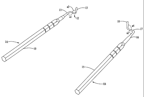
[0024] FIG. 1 illustrates a top view of Chao TMPE (Trans-mucosal and Papillae
Elevator) instruments 1A and 1B, according to one preferred embodiment;
[0025] FIG. 2 illustrates a perspective view of the instruments 1A and 1B
shown in
FIG. 1;
[0026] FIG. 3 illustrates a top view of Chao TMPE instruments 2A and 2B,
according
to another embodiment;
[0027] FIG. 4 illustrates a perspective view of the instruments 2A and 2B
shown in
FIG. 3)-
[00281 FIG. 5 illustrates a top view of Chao TMPE instruments 3A and 3B,
according
to yet another embodiment;
[0029] FIG. 6 illustrates a perspective view of the instruments 3A and 3B
shown in
FIG. 5;
[0030] FIG. 7 illustrates a top view of Chao Papillae Elevator instruments 4A
and 4B,
according to yet another embodiment;
[0031] FIG. 8 illustrates a perspective view of the instruments 4A and 4B
shown in
FIG. 7;
[0032] FIG. 9 illustrates a top view of Chao Papillae Elevator instruments 5A
and 5B,
according to yet another embodiment;
[0033] FIG. 10 illustrates a perspective view of the instruments 5A and 5B
shown in
FIG. 9;
[0034] FIG. 11 illustrates a top view of Chao Papillae Elevator instruments 6A
and 6B,
according to yet another embodiment;
[0035] FIG. 12 illustrates a perspective view of the instruments 6A and 6B
shown in
FIG. 10;
-7-
CA 02616140 2008-01-22
WO 2007/019367 PCT/US2006/030561
[0036] FIG. 13 illustrates a top view of the blade designs of instruments 1A
and 4A,
according to some varying embodiments;
[0037] FIG. 14 illustrates a side view of the blade designs of instruments 1A
and 4A,
according to some varying embodiments;
[0038] FIG. 15 is a flow-chart illustrating the steps of performing a gingival
or papillae
augmentation surgery according to one preferred embodiment; and,
[0039] FIG. 16 is a block diagram illustrating the step of extending a flap
horizontally
without enlarging the incision, according to one preferred embodiment.
DETAILED DESCRIPTION OF PREFERRED EMBODIMENTS
[0040] (1) SURGICAL TECHNIQUES FOR GINGIVAL AUGMENTATION
[0041] The Chao Trans-Mucosal and Papillae Elevation Approach (Chao TMPE
approach) is a technique requiring a minimal incision but capable of
performing gingival
or papillae augmentation at multiple sites. The Chao TMPE approach is a method
capable of repairing gingival defects in all four quadrants of the mouth, and
is predictably
effective for defects up to 7 mm and defects larger than 7 mm in Miller I, II
classification
of root defects.
[0042] Fig. 15 is a flow-chart illustrating the steps of performing a gingival
or papillae
augmentation surgery in accordance with the Chao TMPE approach, according to
one
preferred embodiment. First, as shown by step SO1 of Fig. 15, the Chao TMPE
approach
involves making a horizontal minimal incision of approximately 3-5 millimeters
(mm) at
or near the fornix (depth of the bucco-muccal fold). This incision can be made
using a
standard dental surgical instrument such as, but not limited to, a scalpel.
The incision is
practically invisible because of its small size and its location far away from
the visible
gum line. TMPE instruments, which are discussed in detail later, are designed
so that
generally only one incision of 3-5 mm is needed for up to three gingival
defects. If more
than three gingival defects are being treated, another 3-5 mm incision can be
made two or
more teeth away from the first incision point. A more experienced operator
will be able
-8-
CA 02616140 2008-01-22
WO 2007/019367 PCT/US2006/030561
to routinely utilize an incision of 3 mm, whereas a less experienced operator
may require
up to 5 mm for each incision. Where there are two adjacent defects, the
incision should
be made at the fornix between the buccal roots of the two teeth. Where there
are three
adjacent defects, the incision should be made near the root of the tooth in
the center.
Where there are four or more adjacent defects, two incisions should be made.
When
making two adjacent incisions to correct four adjacent defects, the mesial
incision should
be made between the two most mesial roots and the second incision should be
made
between the two most distal roots. When making the incision, the angle of the
blade of
the TMPE instruments, which are discussed in detail later, should be
approximately at 90
degrees to the underlying bone. It is noted that while the minimum incision
required for
this procedure is between 3-5 mm in length, this procedure is applicable for
incisions
longer than 5 mm.
[0043] Next, as shown by step S02, the appropriate surgical instrument is
inserted to
detach a flap and extend the flap vertically. Next, the flap is extended
horizontally (Fig.
15, step S03). Then, the papillae surrounding the effected tooth is elevated
(Fig. 15, step
S04). Making the incision at the fornix allows maximum "give" or room to move
the
aperture mesially (closer to the middle of the front of the jaw) and distally
(away from the
middle of the front of the jaw) for the release of the flap and papillae (the
gum tissue
between two neighboring teeth).
[0044] To detach a flap (Fig. 15, step S02), the appropriate TMPE instruments,
as
discussed in detail later, is introduced through the incision pointing in the
coronal
direction (towards the crown of the tooth). The blade of the TMPE instrument
is placed
at an acute angle onto the bony surface. With a slicing action the blade is
pressed
coronally to separate the mucosal and gingival tissue from their bony
attachments. Care
should be taken not to perforate the flap. This is accomplished by keep the
blade angled
towards and pressed against the bone.. By increments the flap is released from
the
underlying bone. A connective tissue nodule is often encountered between the
upper
anterior teeth. This nodule is generally located between the roots near the
muco-gingival
-9-
CA 02616140 2008-01-22
WO 2007/019367 PCT/US2006/030561
junction. Since this nodule has not been described in the literature, the
nodule is called
the Chao Nodule. Special care should be taken to undermine the Chao Nodule
with a
slicing motion of the blade without perforating the flap.
[0045] In extending the flap horizontally (Fig. 15, step S03), the flap should
extend
distally well beyond the distal papillae of the tooth immediately distal to
the tooth or
group of teeth being treated, and mesially well beyond the mesial papillae of
the tooth
immediately mesial to the tooth or group of teeth being treated. Vertically
this flap
should extend from the incision to the gingival margin of the treated tooth
and the teeth
adjacent to it. By using the appropriate TMPE instruments, the flap can be
extended
horizontally without increasing the size of the initial 3-5 mm incision.
Careful use of the
appropriate instrument can avoid causing perforations to the flap or injuries
to the root or
implant surfaces while at the same time negating the need for any additional
gingival
incisions. Before proceeding further, the operator should ascertain that all
ligaments or
tissues are complete detached from the flap. The completely loosened flap
allows
convenient access to all papillae.
[0046] Fig. 16 is a block diagram further illustrating the detaching and
horizontal
extension of the flap according to steps S02-S03 of Fig. 15. It is noted that
Fig. 16 is a
block diagram illustrating the concept of the operation only; therefore the
shapes and
geometries are not drawn to scale. In Fig. 16(a), an incision 95 of 3-5 mm is
first made
on the gum 91 above a group of teeth 93. Next, the blade section 97 of an
instrument is
inserted through the incision 95. The instrument comprises at least a blade
section 97 and
a connector section 98. The remainder of the instrument 99 is at an angle with
respect to
the plane of the drawing of Fig. 16, and is not illustrated. In Fig. 16, the
portion of the
instrument submerged beneath the surface of the gum tissue is illustrated by
dashed lines.
In Fig. 16(a), as the blade section 97 extends downwards, the flap is extended
vertically.
Fig. 16(b) illustrates the position of the instrument after a horizontal
extension of the flap,
when the lateral component (connector section 98) is extended through the
incision 95 in
a leftward direction with respect to its position in Fig. 16(a), without
enlarging the size of
-10-
CA 02616140 2008-01-22
WO 2007/019367 PCT/US2006/030561
the incision 95. Depending on the length of the connector section 98 of the
instrument,
the horizontal extension of the flap can reach at least the tooth adjacent to
the incision
and possibly further. However, in order to achieve this horizontal extension,
the plane
the cutting surface of the blade section 97 lies on must be substantially
parallel to the
connector section 98 immediately connecting to the blade section 97.
Currently,
instruments that satisfy this criteria cannot be found on the market. For
example, the
"Goldman Knife" has a blade section with a cutting surface perpendicular to
the
connector section, which prevents the horizontal extension of the flap through
the same
incision. The Chap TMPE instruments, which are discussed in detail later, are
specially
designed to be used in this method.
[0047] Next, the appropriate Chao TMPE papillae elevators, as discussed in
detail later,
can be used to carefully elevate and detach the papillae (Fig. 15, step S04)
from the roots
(hereafter roots refer to natural roots as well as implants or their
abutments) and the
underlying bone. Papillae elevation should extend as lingually as possible,
without
cutting or unnecessarily traumatizing the papillae. Any cutting of the
papillae may result
in the inability to augment the flap properly. This can also lead to
subsequent shrinkage
of the papillae. When an atraumatic elevation of the papillae is accomplished,
the result
is that the entire gingival flap is mobile and can be easily moved coronally.
The Chao
TMPE papillae elevator instruments, as discussed in detail later, are also
designed to
reach the papillae distal to or mesial to the incision without enlarging the
incision.
[0048] Next, the flap is advanced coronally with gentle digital pressure until
the
gingival defect is completely covered (Fig. 15, step S05). The operator may
further
"stretch" the flap by using one finger to press the flap against the root
while another
finger pushes and stretches the bucc-mucal fold, then release digital
pressure. The flap
tissue over the gingival defect is then pressed gently for up to five minutes
to promote
fibrin formation (Fig. 15, step S06).
[0049] Next, the operator can determine if the flap is stable (Fig. 15, step
S07). The
determination whether the flap is stable (Fig. 15, S07) can be accomplished by
checking
-11-
CA 02616140 2008-01-22
WO 2007/019367 PCT/US2006/030561
if the defect remains covered by the flap when the mucosa is pulled apically
or
horizontally. If the defect remains covered, then the flap is stable (Fig. 15,
S07:YES),
and the surgical aspect of the procedure is finished. No further steps, such
as suturing, is
necessary. The patient can be dismissed with proper instructions. Typically,
the 3-5 mm
initial incision heals within the span of less than one week.
[0050] In some cases where the flap is unstable (Fig. 15, S07:NO), papillary
augmentation (Fig. 15, step S08) with resorbable collagen membraneous graft
material
may be used. The graft material (Biogide is recommended) may be rolled or cut
into a
triangular shaped pieces, or any other convenient shape, that fits into the
triangular space
under the papilla. This material is then inserted via the incision with gentle
pressure
under the papilla with un-serrated cotton pliers. Then the appropriate
papillae elevator is
used to "tug" the graft snugly under the papilla. This is repeated for all the
papillae
needing augmentation. Tugging the graft material underneath the papillae will
generally
stabilize both the graft material and the flap. Tugging the mucosa, cheek or
lip should
not result in any movements of the flap. If movement is observed, the operator
should re-
examine the flap to see whether all ligamentous and tissue attachments have
been severed
from the flap. When thoroughly released from the roots and bone, the flap will
likely
remain immobile. Pressing the flap over the root defect for up to five minutes
will assure
fibrin formation and end the procedure.
[0051] In summary, the result of the method illustrated in Fig. 15 is the
coverage of the
gingival defect with a technique that requires only one 3 mm-5 mm practically
invisible
incision, with no suturing of the patient's oral tissue required. Asa result
of the minimal
incision size and lack of suturing, the bleeding and swelling of the gum is
kept to a
minimum and thus recovery of the patient is accelerated. Often, the patient
will see that
the gums appears to have grown over the defect in just one appointment. Due to
the
vascularity and collagenous nature of mucosal tissue, healing of the incision
is rapid and
generally the incision becomes virtually undetectable within a week. Post-
operative
-12-
CA 02616140 2008-01-22
WO 2007/019367 PCT/US2006/030561
symptoms are minimal and generally require no more than one or two over-the-
counter
NSAIDS (Non-Steroidal Anti-Inflammatory Drugs).
[0052] It is recommended, whenever feasible, to perform dentinoplasty as part
of root
preparation. The performance of dentioplasty appears to allow easier
advancement of the
flap, as well as allow better long term stability of the re-attachment.
[0053] (2) OSSEOUS SURGERY USING THE CHAO TMPE APPROACH
[0054] The incision technique described above, as well as the surgical
instruments
discussed in detail later, can also be used to perform osseous surgery. For
purposes of
osseous surgery, the Chao TMPE approach to flap and papillae elevation as
described
above allows access to the bony defect through the mucosal incision. The
incision may
need to be extended to allow sufficient reflection of the flap to allow direct
visualization
of the bony defect from the labial or buccal aspect of the bony defect. This
approach will
minimize gingival recession, which is associated with techniques that call for
labial or
buccal gingival marginal incision. Furthermore, grafting is simplified because
graft
materials can be inserted underneath the papillae and flap, often without the
need for
suturing. The clinician does not have to be concerned with the complete
enclosure of the
graft material by the flap. Since blood supply to the grafted area remains
relatively
uncompromised, healing is accelerated compared to techniques that employ
gingival
incisions. Specially designed ultrasonic instruments can access mesial and
distal bony
pockets for root planning purposes. Placing bone graft and membranes can be
accomplished with the Chao TMPE elevator instruments, which are discussed in
detail
below.
[0055] For buccal furcation of mandibular or maxillary molars the Chao TMPE
approach allows for the buccal gingival margin to be so slackened such that
gentle
retraction of the flap from the buccal wall allows full access to the
furcation from the
buccal perspective. Advancing the flap coronally to give full coverage to the
furcation
area is not particularly technically challenging, even where gingival
recession has
exposed the furcation. Shrinkage and resorption of the flap over the buccal
furcation is
-13-
CA 02616140 2008-01-22
WO 2007/019367 PCT/US2006/030561
minimal because blood supply is not compromised by gingival incisions. This
flap
conveniently gives coverage to the graft materials and membranes which often
does not
need to be sutured. The ends of the membrane that covers the buccal furcation
can be
tugged under the mesial and distal papillae that stabilization is obtained
without suturing.
If suturing is desired, a simple sling suture engaging the graft membrane
under the mesial
and distal papillae of the tooth will stabilize the graft.
[0056] (3) PAPILLAE REGENERATION OR AUGMENTATION USING THE
CHAO TMPE APPROACH
[0057] The incision technique described above, as well as the surgical
instruments
discussed in detail later, can also be used in papillae regeneration or
augmentation.
Papillae regeneration or augmentation may be accomplished by the Chao TMPE
approach as described above. The use of an autogenous graft or membranous
allograft
(Alloderm) is recommended. By using Alloderm, or alternatively an autogenous
graft,
and stabilization using the Chao suturing technique, there is a higher
likelihood of a
predictably successful outcome compared to conventional methods. The reason
for this
higher success rate is that this approach leaves blood supply for the papillae
and the
gingival apparatus virtually unimpaired. Until now, other conventional papilla
regeneration techniques have not been deemed predictably successful because of
the need
to make multiple incisions in the gingival region and the inability of current
techniques to
augment and elevate the papillae without restricting blood supply.
[0058] One alternative method for papillae augmentation (Fig. 15, step S08) is
to cut a
section of an acelluar dermal matrix allograft (Alloderm) into a U-shaped
configuration.
With a unique suturing technique described below, each end of this membrane is
then
tugged snugly under the mesial and distal papillae, with the middle segment
overlying the
gingival defect. The Chao TMPE allograph or autogenous graft (from the
patient)
suturing technique, with three variations, is described in the following
paragraphs.
[0059] In a first variation of the Chao TMPE suturing technique, a U-shaped
section
approximately half the circumference of the root at the cemento-enamel
junction (neck of
-14-
CA 02616140 2008-01-22
WO 2007/019367 PCT/US2006/030561
the tooth) and at least 3 mm in width is cut from the Alloderm specimen.
(Alternatively,
connective tissue from another part of the mouth can be used.) The needle of a
resorbable suture (Dexon or Chromic Catgut) with 1/2 round point (26.19 mm) is
first
threaded from the lingual through the mesial interproximal space, without
piercing the
papilla. Then the needle is threaded under the gingival margin, through the
flap to appear
at the horizontal incision. Next, the needle is made to engage one end of the
U-shaped
graft. It is suggested that the graft be placed against the convex surface of
the end of a
periosteal elevator or tissue retractor held firmly at the other end by a
dental assistant
while threading the needle through the graft, with the needle pointing away
from the
fingers of the operator. This will enhance the ease of accomplishing this
procedure as
well as protect against an exposure (puncture) incident.
[0060] Then the needle is threaded under the flap to appear at the gingival
margin (gum
line) without engaging the flap in any way. Next, the needle is threaded
through the
menial interproximal space to the lingual of the tooth. From the lingual the
needle is
passed through the distal interproximal space without engaging the papillae,
to appear on
the facial side. The needle is next threaded under the flap to appear at the
horizontal
incision. Then the needle is made to puncture and thread through the other end
of the U-
shaped graft with the use of the convex surface of an instrument. With the
specimen thus
engaged by the suture (as if pulled by a sling from each end), the needle is
thread under
the flap, then passed through the distal interproximal space to reappear at
the lingual.
Then as both ends of the suture are pulled, the graft is pulled under the flap
to rest against
the root, with each end tugged under a papilla.
[0061] Next, The flap should be elevated by digital pressure to cover the
defect before
the suture is tightened. The graft material should remain under the flap, and
not be
squeezed over the gingival margin. Careful effort must be made to push any
excess graft
material under the flap. The knot can be tied at the lingual. This suturing
will tightly
attach the graft against the root defect while elevating and stabilizing the
entire gingival
apparatus (the flap and the papillae) at the desired position. At the
operator's choice, the
-15-
CA 02616140 2008-01-22
WO 2007/019367 PCT/US2006/030561
ends of the suture can be threaded under the opposite interproximal space and
the knot
then can be tied at the labial. A lingual knot makes the suture invisible,
while presenting
the patient with a possible annoyance to the tongue. A facial or labial knot
may be
visible to the eye and possibly displace the gingival margin of the flap.
[0062] When the distal and mesial papillae of the treated tooth and the
papillae of the
adjacent teeth have both been augmented by the graft material, the flap should
now be
relatively immovably fixated over the defect. Pulling or tugging the cheek,
lip or
mucosal tissue should not be result in any observable movement in the flap. It
should be
noted that in some circumstances the specimen may be "tugged" under the
papillae
without suturing. However, dislodgement may still be possible.
[0063] A second variation of the Chao TMPE graft suturing technique is
described as
follows. In this variation, the operator may use a double ended needle. As
above, a U-
shape configuration of the graft is cut that approximates the facial
circumference of the
neck of the tooth. Each end is threaded from the lingual to the facial, passed
under the
flap to appear at the horizontal incision, without engaging tissue. Then each
needle is
made to engage each end of the graft, and then passed through to the lingual
without
engaging any part of the flap. The knot is then tied at the lingual, or
wrapped around the
lingual and tied at the labial. Although this method appear easier to
visualize, the
surgeon will, with experience, probably find that using a single needle suture
as described
under the first variation of the suturing method to be simpler and easier.
[0064] In a third variation of the suturing technique, the U-shaped graft
material is
tugged underneath each papilla. With finger pressing against the graft just
apical to the
mesial papilla, a needle is made to pass from the outer surface of the flap,
through the
graft under the papilla, to appear at the lingual side. Then, the needle is
wrapped around
the root to the distal, and threaded through the distal interproximal space to
appear on the
facial side, without engaging any tissue. Next, with the finger pressing the
graft against
the distal papilla, the needle is made to engage the graft underneath the
papilla from the
facial aspect of the flap to emerge on the lingual side of the tooth. Next,
the suture is
-16-
CA 02616140 2008-01-22
WO 2007/019367 PCT/US2006/030561
passed through the mesial interproximal space to appear on the facial side of
the tooth.
The knot is then tightened and tied at the facial side.
[00651 According to the Chao TMPE approach, the papillae is loosened by
disengaging
the flap and the papillae from the root and the bone with only one incision
that is remote
from the papillae. Then, this technique calls for the stabilization of the
elevated papillae
by the insertion of an autogenous graft or Alloderm, without restricting blood
supply
from surrounding tissue. Current conventional methods call for the external
suturing of
the papillae after elevation and thus limits blood supply, resulting in less
predictable
results. The unique Chao papillae regeneration technique assures a high,
predictable
success rate, when performed as described above.
[0066] (4) THE SURGICAL INSTRUMENTS
[00671 The Chao TMPE (Trans-mucosal and Papillae Elevator) instruments are
instruments designed to be used in the performance of the surgeries and
operations
discussed above. The TMPE instruments are specially designed to allow the
operator to
elevate, without direct vision, a full-thickness flap through the access
provided by a
minimal incision of 3-5 mm. This reflection of the flap is done in a unique
way, i.e., the
operator cannot directly see the tip of the elevator at any time during the
procedure. The
operator must deduce the location of the blade by the inflated shape and
movement the
elevator traces underneath the mucosa or gingiva. The design of the
instruments
facilitates the unique techniques discussed above. Depending on which quadrant
of the
patient's mouth the surgery is being performed, different TMPE instruments can
be used.
[0068] Figs. 1-2 illustrates a first set of embodiments of the Chao TMPE
(Trans-
mucosal and Papillae Elevator) instruments that can be used to perform the
surgeries and
operations discussed above. Fig. 1 shows a top view of a set of instruments 1A
and 1B,
and Fig. 2 shows a perspective view of the same set of instruments. These two
instruments IA and 1B maybe also called the Chao TMPE Universal instruments.
[0069] As shown in Figs. 1-2, TMPE instrument 1A is an instrument with a
handle 10,
a first shank 11 linearly extending from the handle 10, a second shank 12
connected to
-17-
CA 02616140 2008-01-22
WO 2007/019367 PCT/US2006/030561
the first shank 11 at one end and rotated at an angle al from the first shank
11 in the
counter-clockwise direction, and a blade section 13 rotated at an angle a3
from the
second shank 12 in the clockwise direction and connected to a second end of
the second
shank 12.
[0070] Also shown in Figs. 1-2, TMPE instrument 1B is an instrument with a
handle
15, a first shank 16 linearly extending from the handle 15, a second shank 17
connected
to the first shank 16 at one end and rotated at an angle a2 from the first
shank 15 in the
clockwise direction, and a blade section 18 rotated at an angle a4 from the
second shank
17 in the counter-clockwise direction and connected to a second end of the
second shank
17.
[0071] For TMPE instrument 1A and 1B, the second shanks (12 and 17) act as
connector sections that connect the blade sections (13 and 18) to the handles
(10 and 15)
and first shanks (11 and 16).
[0072] As illustrated in Figs. 1-2, TMPE instruments 1A and 1B have generally
the
same shape and design, but differ in the rotational direction of the second
shanks (12 and
17) with respect to the first shanks (11 and 16), and the rotational direction
of the blade
sections (13 and 18) with respect to the second shanks (12 and 17). TMPE
instruments
1A and 1B are essentially "mirror images" of each other, when viewed along a
line
parallel to the length direction of the handles 10 and 15.
[0073] As illustrated on Figs. 1-2, the angles al, a2, a3, and a4 are all
approximately 90
degrees. Further, for TMPE IA, the blade section 13 is orthogonal to a plane
formed by
the length direction of the first shank 11 and the length direction of the
second shank 12.
In other words, the length direction of the blade section 13 is perpendicular
to the length
directions of both the first 11 and the second 12 shanks. Likewise, for TMPE
1B, the
blade section 18 is orthogonal to a plane formed by the length direction of
the first shank
16 and the length direction of the second shank 17.
[0074] While the embodiments illustrated in Figs. 1-2 have the angles al, a2,
a3, and a4
at approximately 90 degrees, it is not necessary for these angles to be
exactly 90 degrees.
-18-
CA 02616140 2008-01-22
WO 2007/019367 PCT/US2006/030561
Instruments according to other embodiments have angles substantially close to
but not
exactly 90 degrees, up to a variation of +l- 10 degrees from 90 degrees, that
accomplish
the same results.
[0075] In one embodiment, the second shanks (12 and 17) for TMPE 1A and 1B are
each 11 mm long. However, other embodiments could have the second shank
varying in
length. In various embodiments, the lengths of the second shank (12 or 17)
could vary
from 4 mm to 18 mm for instruments 1A and lB.
[0076] In one preferred embodiment, each of the blade section (13 and 18) for
TMPE
1A and 1B is approximately 13 mm long. The length of the blade section (13 or
18)
could vary depending on the tooth and gum dimensions of the patient. Some
embodiments have a shorter blade section of 4 mm, some embodiments have a
longer
blade section of 21 mm, and some embodiments have blade sections varying
between 4-
21 mm. The width of the blade section could also vary from 1 mm to 3 mm. The
width
of the blade section limits the incision of the Chao TMPE Approach to about 3-
5 mm.
Further, as illustrated in Fig. 2, the width directions of the blade sections
13 and 18 are
substantially perpendicular to the length direction of the handles 10 and 15,
respectively.
In other words, the planes of the cutting surfaces of the blade sections 13
and 18 are each
substantially parallel. to the second shanks 12 and 17. As discussed above,
this allows the
blade section to expand the flap horizontally without increasing the incision
size.
Because the second shanks (12 and 17) are parallel to the planes of the
cutting surfaces,
they can be submerged under the flap through the incision during the
horizontal extension
of the flap as illustrated in Fig. 16. This cannot be accomplished with any
convention
instruments on the market today.
[0077] In addition, each of the blade sections (13 and 18) could also vary in
shape. For
example, the blade could be basically round with a cutting edge formed by
flattening of
one surface (half-moon shaped), or a double-edged blade (spear-shaped).
[0078] Using TMPE instruments IA or 1B, the operator or surgeon can insert the
sharp
tip of the blade section 13 or 18 into an incision of 3-5 mm (Fig. 15, S0l).
Pressure is
-19-
CA 02616140 2008-01-22
WO 2007/019367 PCT/US2006/030561
applied, according to the techniques detailed above in section (1), such that
the blade
section (13 or 18) of TMPE lA or 1B slices under the mucosa or gingival,
detaching a
flap (Fig. 15, S02). Next, the blade section can be moved laterally to extend
the flap
horizontally to the teeth adjacent to the incision (Fig. 15, S03). As
illustrated in Figs.
16(a) and 16(b), during the horizontal extension of the flap, the lateral
component of
instruments 1 A or 1B, the second shanks 12 or 17, becomes submerged under the
gum
through the incision point. Hence, the "reach" of instruments 1A or 1B is
determined by
the length of the second shanks 12 or 17.
[0079] Using the techniques described above and in earlier sections, TMPE IA
can be
used to reflect the flap distally in the in the upper right and lower left
quadrants of the
patient's mouth. TMPE 1A can also be used to release the flap mesially in the
upper left
and lower right quadrants. TMPE 1B is an instrument that can used to release
the flap
mesially in the upper right and lower left quadrants of a patient's mouth.
TMPE 1B can
also be used to release the flap distally in the lower right and upper left
quadrants.
[0080] When using TMPE instrument IA or 1B, the operator may find that when
treating posterior teeth, the movement of the instrument is impeded by the
handle
impinging the cheek or the corner of the mouth. As shown in Figs. 3-6, TMPE
instruments 2A, 2B, 3A, and 3B are instruments designed in a manner that the
handle is
angled mesially away from the corner of the mouth. The angled design allows
the blade
section of the instrument to reach under the flap and access mesial and distal
aspects of
the flap without hindrance from facial structures. Hence, instruments 2A, 2B,
3A, and
3B can reach posterial surgical sites, e.g., buccal to second bicuspids or
first molars,
without impinging the patient's facial features. Further, "bends" in the
design of the
shanks allow the surgeon to apply a controlled force to elevate the attached
gingiva.
[0081] As illustrated in Figs. 3-4, 2A and 2B are a second set of embodiments
of TMPE
instruments. TAPE instrument 2A comprises a shaft 20, a first shank 21
extending
linearly from the shaft 20, a second shank 22 connected to the first shank 21
on one end
and rotated from the first shank 21 at an angle b1 in the counter-clockwise
direction, a
-20-
CA 02616140 2008-01-22
WO 2007/019367 PCT/US2006/030561
third shank 23 connected to a second end of the second shank 22 and rotated
from the
second shank 22 at an angle b2 in the clockwise direction, and a blade section
24
connected to the third shank 23 rotated at an angle b3 from the third shank 23
in the
clockwise direction.
[0082] Also illustrated in Figs. 3-4, TMPE instrument 2B comprises a shaft 25,
a first
shank 26 extending linearly from the shaft 25, a second shank 27 connected to
the first
shank 26 on one end and rotated from the first shank 26 at an angle b4 in the
counter-
clockwise direction, a third shank 28 connected to a second end of the second
shank 27
and rotated from the second shank 27 at an angle b5 in the counter-clockwise
direction,
and a blade section 29 connected to the third shank 28 rotated at an angle b6
from the
third shank 28 in the clockwise direction.
[0083] For TMPE instrument 2A and 2B, the second shanks (22 and 27) and third
shanks (23 and 28) act as connector sections that connect the blade sections
(24 and 29)
to the handles (20 and 25) and first shanks (21 and 26).
[0084] Figs. 3-4 illustrate one preferred embodiment of TMPE 2A where the
angle bl
is approximately 60 degrees, the angle b2 is approximately 60 degrees, and the
angle b3
is approximately 90 degrees. In other embodiments, any reasonable angle from
10-90
degrees for each of the angles bl and b2 also could also work. As shown in
Fig. 4, the
angle b3 is approximately 90 degrees, but it is not necessary to be exactly 90
degrees.
The angle b3 could vary within +/- 10 degrees of 90 degrees. The length
directions of the
first 21, second 22, and third 23 shanks are coplanar. The length direction of
the blade
section 24 is approximately orthogonal to the plane formed by the length
directions of the
first 21, second 22, and third 23 shanks.
[0085] Likewise, Figs. 3-4 also illustrate one preferred embodiment of TMPE 2B
where
the angle b4 is approximately 100 degrees, the angle b5 is approximately 110
degrees,
and the angle b6 is approximately 90 degrees. In other embodiments, any angle
larger
than or equal to 90 degrees but smaller than 135 degrees for b4 could also
work, and any
angle larger than or equal to 90 degrees but smaller than 135 degrees for b5
could also
-21-
CA 02616140 2008-01-22
WO 2007/019367 PCT/US2006/030561
work. As shown in Fig. 4, the angle b6 is approximately 90 degrees, but could
also be
within +/- 10 degrees of 90 degrees. The length direction of the first 26,
second 27, and
third 28 shanks are coplanar. The length direction of the blade section 29 is
approximately orthogonal to the plane formed by the length directions of the
first 26,
second 27, and third 28 shanks.
[0086] For one embodiment of TMPE 2A as illustrated in Figs. 3-4, the length
of the
second shank 22 is approximately 11 mm, the length of the third shank 23 is
approximately 8 mm, and the length of the blade section 24 is approximately 13
mm. For
the embodiment of TMPE 2B shown in Figs. 3-4, the length of the second shank
27 is
approximately 6 mm, the length of the third shank 28 is approximately 11 mm,
and the
length of the blade section 29 is approximately 13 mm. However, other
embodiments
may have varying lengths for each shank or blade section. For example, the
blade
sections 24 or 29 could have lengths varying form 4 mm to 21 mm. The width of
the
blade section could also vary from 1 mm to 3 mm.
[0087] Similar to TMPE 1A and 1B, each of the blade section (24 or 29) could
also
vary in shape. For example, the blade could be half-moon shaped or spear-
shaped.
[0088] TMPE 2A comprises "bends" such as the junction between the first 21 and
second 22 shanks, or the junction between the second 22 and third 23 shanks.
Likewise,
TMPE 2B also comprises "bends" at the junction between the first 26, second
27, and
third 28 shanks. These "bends" provide a space for surgeons to used a finger
to apply a
measured amount of pressure so that attached gingiva can be elevated. The
"bends" on
these shanks allow the operator to apply pressure with a finger of a second
hand with a
degree of force under a control not available through the application of
digital pressure
with only one hand on the handle with a right-angled design. Furthermore, the
unique
angulations of instruments 2A and 2B allow the operator to gain a better line
of sight for
the indirect visualization of the movement of the blade as it engages the
overlying tissue.
As discussed above, the angled design of instruments 2A and 2B allow the
instruments to
be used on posterior teeth without impinging against the patient's facial
features.
-22-
CA 02616140 2008-01-22
WO 2007/019367 PCT/US2006/030561
[0089] As illustrated in Figs. 5-6, instruments TMPE 3A and 3B have similar
angled
designs as the 2A and 2B instruments, respectively. Further, 3A and 3B are
"mirror
images" of the 2A and 2B instruments, respectively. TMPE 3A and 3B instruments
have
similar uses and advantages as the 2A and 2B instruments, but are used in
opposite
quadrants of the mouth. For example, TMPE 2A can be used to reflect the flap
distally in
the upper right and lower left quadrants of the mouthy; TMPE 3A can be used to
reflect
the flap distally in the upper left and lower right quadrants. Likewise, TMPE
2B can be
used to reflect the flap mesially in the upper right and lower left quadrants;
TMPE 3B can
be used to reflect the flap mesially in the upper left and lower right
quadrants. Since the
uses of TMPE instruments 3A and 3B are similar to those of 2A and 2B discussed
above,
detailed explanations of the uses of instruments 3A and 3B are omitted here.
[0090] As illustrated in Figs. 5-6, TMPE instrument 3A comprises a shaft 30, a
first
shank 31 extending linearly from the shaft 30, a second shank 32 connected to
the first
shank 31 on one end and rotated from the first shank 31 at an angle cl in the
clockwise
direction, a third shank 33 connected to a second end of the second shank 32
and rotated
from the second shank at an angle c2 in the counter-clockwise direction, and a
blade
section 34 connected to the third shank 33 rotated at an angle c3 from the
third shank 33
in the clockwise direction.
[0091] Also illustrated in Figs. 5-6, TMPE instrument 3B comprises a shaft 35,
a first
shank 36 extending linearly from the shaft 35, a second shank 37 connected to
the first
shank 36 on one end and rotated from the first shank 36 at an angle c4 in the
clockwise
direction, a third shank 38 connected to a second end of the second shank 37
and rotated
from the second shank at an angle c5 in the clockwise direction, and a blade
section 39
connected to the third shank 38 rotated at an angle c6 from the third shank 38
in the
counter-clockwise direction.
[0092] For TMPE instrument 3A and 3B, the second shanks (32 and 37) and third
shanks (33 and 38) act as connector sections that connect the blade sections
(34 and 39)
to the handles (30 and 25) and first shanks (31 and 36).
-23-
CA 02616140 2008-01-22
WO 2007/019367 PCT/US2006/030561
[0093] For one preferred embodiment of TMPE instruments 3A and 3B, the angles
cl-
c6 correspond generally to the angles of bl-b6 for TMPE 2A and 2B. In one
preferred
embodiment, the angles for cl-c6 are approximately 60 degrees, 60 degrees, 90
degrees,
100 degrees, 110 degrees, and 90 degrees, respectively. For various other
embodiments,
the ranges of angles for c 1-c6 are the same as the ranges discussed above for
angles b 1-
b6 relating to TMPE 2A and 2B, and will not be repeated here.
[0094] For one embodiment of TMPE 3A as illustrated in Figs. 5-6, the length
of the
second shank 32 is approximately 11 mm, the length of the third shank 33 is
approximately 8 mm, and the length of the blade section 34 is approximately 13
mm. For
the embodiment of TMPE 3B shown in Figs. 5-6, the length of the second shank
37 is
approximately 6 mm, the length of the third shank 38 is approximately 11 mm,
and the
length of the blade section 39 is approximately 13 mm. However, other
embodiments
have varying lengths for each shank or blade section. For example, the blade
sections 34
or 39 could have lengths varying form 4 mm to 21 mm. The width of the blade
section
could also vary from 1 mm to 3 mm. Similar to TMPE IA and 1B, each of the
blade
section (34 or 39) could also vary in shape. For example, the blade could be
half-moon
shaped or spear-shaped. The orientations of the width direction of the blade
sections 34
and 39 with respect to the shafts 30 and 35 are similar to that of the
instruments 2A and
2B, and the detailed explanations will not be repeated here.
[0095] The above-described instruments 1A, 1B, 2A, 2B, 3A, and 3B are designed
to
create the incision and release the flap necessary for the Chao TMPE surgical
techniques
described in the above sections (Fig. 15, S01-S03). Next, a group of
instruments used to
elevate the papillae (Fig. 15, S04) is described below while referring to
Figs. 7-12. The
papillae is the gum tissue between two neighboring teeth. Instruments 4A, 4B,
5A, 5B,
6A, and 6B are collectively known as the Chao Papillae Elevators.
[0096] First, Figs. 7-8 illustrate instruments 4A and 4B, which are one set of
embodiments of the Chao Papillae Elevators called the Chao TMPE Universal
Papillae
-24-
CA 02616140 2008-01-22
WO 2007/019367 PCT/US2006/030561
Elevators. Except for the blade sections 43 and 48, instruments 4A and 4B are
similar in
shape and design to the TMPE instrument 1A and 1B.
[0097] As illustrated in Figs. 7-8, instrument 4A is an instrument with a
handle 40, a
first shank 41 linearly extending from the handle 40, a second shank 42
connected to the
first shank 41 at one end and rotated at an angle dl from the first shank 41
in the counter-
clockwise direction, and a blade section 43 rotated at an angle d2 from the
second shank
42 in the clockwise direction and connected to a second end of the second
shank 42.
[0098] Also illustrated in Figs. 7-8, instrument 4B is an instrument with a
handle 45, a
first shank 46 linearly extending from the handle 45, a second shank 47
connected to the
first shank 46 at one end and rotated at an angle d3 from the first shank 45
in the
clockwise direction, and a blade section 48 rotated at an angle d4 from the
second shank
47 in the counter-clockwise direction and connected to a second end of the
second shank
47.
[0099] In one preferred embodiment, the angles dl-d4 are all 90 degrees.
Further, for
instruments 4A and 4B, the blade sections 43 or 48 are orthogonal to the
planes formed
by the length directions of the first and second shanks 41 and 42, or 46 and
47,
respectively. In various embodiments, the variations for the angles dl-d4, as
well as the
variations to the lengths of the first (41 or 46) and second (42 or 47) shanks
are similar to
that of instruments IA and 1B, and the corresponding descriptions are
therefore not be
repeated here.
[0100] The differences between instrument 1A and lB with 4A and 4B,
respectively,
are differences in the shape and dimension of the blade sections. Fig. 14
illustrates a
side-view comparison of between a blade section 81 for instruments IA or 1B,
and a
blade section 83 for instruments 4A and 4B. For instrument lA and 1B, the
blade
sections are relatively straight. On the other hand, for instruments 4A and
4B, the blade
sections are shaped like a claw with a curved tip.
[0101] As illustrated in Fig. 14, the claw-like blade section for instruments
4A and 4B
have a relatively straight section 831 and a curved section 832. In various
embodiments,
-25-
CA 02616140 2008-01-22
WO 2007/019367 PCT/US2006/030561
the curvature of the curved section 832 can vary from 10-30 degrees. Further,
the curved
section 832 is sharp and shaped like an arrow-head. In one preferred
embodiment, the
lengths of the blade sections 43 and 48 are each 13 mm. In various other
embodiments,
the lengths of the blade sections 43 and 48 could vary from 4 mm to 21 mm.
[0102] Fig. 15 compares atop view of the blade section for instrument IA and
4A.
Blade section 71 is one embodiment of a longer spear-shaped blade section for
instrument 1A. Blade section 72 is one embodiment of a shorter spear-shaped
blade
section for instrument IA. Blade section 73 is one embodiment of a narrow claw-
like
blade section for instrument 4A. For the blade section of IA, the width is
typically 1-3
mm with sharp cutting edges along both side edges. For the blade section of
4A, the
width is typically less than or equal to 2 mm, with a sharp top tip but no
side cutting
edges. Various designs for blade sections of instruments 1B and 4B are similar
to those
for instruments IA and 4A, and are not illustrated.
[0103] The sharp and narrow blade sections 43 and 48 of instruments 4A and 4B,
respectively, are designed to fit into the interproximal spaces (spaces
between
neighboring teeth) to release and elevate the papilla from its attachments to
the root
surfaces and the underlying bone. Instrument 4A can be used to elevate the
papillae
distal to the incision in the upper right and lower lower quadrants, as well
as the papillae
mesial to the incision in the lower right and upper left quadrants. Instrument
4B can be
used to elevate the papillae mesial to the incision in the upper right and
lower left
quadrants of the mouth, as well as the papillae distal to the incision in the
lower right and
upper left quadrants of the mouth.
[0104] As illustrated in Figs. 9-10, instruments 5A and SB are papillae
elevators with
similar shapes and designs as instruments 2A and 2B, respectively. As shown in
Figs. 9-
10, instrument 5A comprises a shaft 50, a first shank 51 extending linearly
from the shaft
50, a second shank 52 connected to the first shank 51 on one end and rotated
from the
first shank 51 at an angle el in the counter-clockwise direction, a third
shank 53
connected to a second end of the second shank 52 and rotated from the second
shank 52
-26-
CA 02616140 2008-01-22
WO 2007/019367 PCT/US2006/030561
at an angle e2 in the clockwise direction, and a blade section 54 connected to
the third
shank 53 rotated at an angle e3 from the third shank 53 in the clockwise
direction.
[0105] Also shown in Figs. 9-10, instrument 5B comprises a shaft 55, a first
shank 56
extending linearly from the shaft 55, a second shank 57 connected to the first
shank 56 on
one end and rotated from the first shank 56 at an angle e4 in the counter-
clockwise
direction, a third shank 58 connected to a second end of the second shank 57
and rotated
from the second shank 57 at an angle e5 in the counter-clockwise direction,
and a blade
section 59 connected to the third shank 58 rotated at an angle e6 from the
third shank 58
in the clockwise direction.
[0106] In one preferred embodiment, the angles el-e6 are 60 degrees, 60
degrees, 90
degrees, 100 degrees, 110 degrees, and 90 degrees. In various other
embodiments, the
angles el-e6 could vary within the same range as those for bl-b6,
respectively. As with
instruments 2A and 2B, for each of instruments 5A or 5B, the length direction
of the
blade section (54 or 59) is orthogonal to the plane formed by the first (51 or
56), second
(42 or 57), and third (53 or 58) shanks. Since the lengths of the second (52
or 57) and
third (53 or 58) shanks are also similar to that of the second (22 or 27) and
third (23 or
28) shanks of instruments 2A and 2B, the discussion for the shank lengths are
omitted
here.
[0107] Figs. 11-12 illustrate papillae elevator instruments 6A and 6B, which
are similar
in design to instruments 3A and 3B. As illustrated in Figs. 11-12, instrument
6A
comprises a shaft 60, a first shank 61 extending linearly from the shaft 60, a
second shank
62 connected to the first shank 61 on one end and rotated from the first shank
61 at an
angle fl in the clockwise direction, a third shank 63 connected to a second
end of the
second shank 62 and rotated from the second shank at an angle f2 in the
counter-
clockwise direction, and a blade section 64 connected to the third shank 63
rotated at an
angle f3 from the third shank 63 in the clockwise direction.
[0108] Also illustrated in Figs. 11-12, instrument 6B comprises a shaft 65, a
first shank
66 extending linearly from the shaft 65, a second shank 67 connected to the
first shank 66
-27-
CA 02616140 2008-01-22
WO 2007/019367 PCT/US2006/030561
on one end and rotated from the first shank 66 at an angle f4 in the clockwise
direction, a
third shank 68 connected to a second end of the second shank 67 and rotated
from the
second shank at an angle f5 in the clockwise direction, and a blade section 69
connected
to the third shank 68 rotated at an angle c6 from the third shank 68 in the
counter-
clockwise direction.
[01091 In one preferred embodiment, the angles fl-f6 are 60 degrees, 60
degrees, 90
degrees, 100 degrees, 110 degrees, and 90 degrees. In various other
embodiments, the
angles fl-f6 could vary within the same range as those for cl-c6,
respectively. As with
instruments 3A and 3B, for each of instruments 6A or 6B, the length direction
of the
blade section (64 or 69) is orthogonal to the plane formed by the first (61 or
66), second
(62 or 67), and third (63 or 68) shanks. Since the lengths of the second (62
or 67) and
third (63 or 68) shanks are also similar to that of the second (32 or 37) and
third (33 or
38) shanks of instruments 3A and 3B, the discussion for the shank lengths are
omitted
here.
[01101 For each of instruments 5A, 5B, 6A, and 6B, the blade sections (54, 59,
64, and
69, respectively) are narrow and claw-like as illustrated by the blade section
83 in Figure
18, which further comprises a relatively straight section 831 and a curved
section 832. In
various embodiments, the curvature of the curved section can vary from 10-30
degrees.
Further, the curved section is sharp and shaped like an arrow-head. In one
preferred
embodiment, the lengths of the blade sections (54, 59, 64, and 69) are each 13
mm. In
various other embodiments, the lengths of the blade sections 43 and 48 could
vary from 4
mm to 21 mm. Since the design of the blade sections (54, 59, 64, and 69) for
instruments
5A, 5B, 6A, and 6B are similar to that of the blade section for instrument 4A,
the detailed
descriptions are omitted here.
[01111 The sharp and narrow blade sections 54, 59, 64, and 69 of instruments
5A, 5B,
6A, and 6B, respectively, are designed to fit into the interproximal spaces
(spaces
between neighboring teeth) to release and elevate the papilla from its
attachments to the
root surfaces and the underlying bone. Like instruments 2A, 2B, 3A, and 3B,
instruments
-28-
CA 02616140 2008-01-22
WO 2007/019367 PCT/US2006/030561
5A, 5B, 6A, and 6B are angled such that they can reach posterial surgical
sites, e.g.,
buccal to second bicuspids or first molars, without impinging the patient's
facial features.
In addition, the "bends" on instruments 5A, 5B, 6A, and 6B provide access to
the
operator to apply pressure, just as the "bends" on instruments 2A and 2B
discussed in
detail above.
[0112] Instrument 5A can be used in the upper right and lower left quadrants
of the
mouth to elevate papillae distal to the incision. Instruments 5B can be used
in the upper
right and lower left quadrants to elevate papillae mesial to the incision.
Instrument 6A
can be used in the upper left and lower right quadrants of the mouth to
elevate papillae
distal to the incision. Instruments 6B can be used in the upper left and lower
right
quadrants of the mouth to elevate papillae mesial to the incision.
[0113] For preferred embodiments of each of instruments IA, 1B, 2A, 2B, 3A,
3B, 4A,
4B, 5A, 5B, 6A, and 6B, the blade section comprises a cutting surface such
that the plane
of the cutting surface is substantially parallel to the length direction of
the shank
immediately connecting to the blade section. For example, as illustrated in
Fig. 4, for
instrument 2A, the blade section 24 has a cutting surface lying on a plane
substantially
parallel to the length direction of the third shank 23. As illustrated in Fig.
16(a)-(b),
during horizontal extension of the flap, the blade sections must extend
horizontally in the
same direction as the shank immediately connecting to the blade section. In
some of
these preferred embodiments, the plane of the cutting surface of the blade
section is
exactly parallel to the length direction of the shank immediately connecting
to the blade
section. In some of these preferred embodiments, the plane of the cutting
surface of the
blade section is within +/- 10 degrees of being parallel to the length
direction of the shank
immediately connecting to the blade section. This feature enables the
horizontal
extension of the flap without increasing the incision size, and is not found
on any
conventional instruments on the market today.
[0114] In some preferred embodiments, for each of instruments 1A, 1B, 2A, 2B,
3A,
3B, 4A, 4B, 5A, 5B, 6A, and 6B, the handle, first shank and connector section
are
-29-
CA 02616140 2008-01-22
WO 2007/019367 PCT/US2006/030561
integrally formed of the same material. In other embodiments, the handle,
first shank,
connector section, and blade section are all integrally formed of the same
material. As
described above, the connector section may comprise the second shank
(instruments 1A,
1B, 4A, and 4B) or the second and third shanks (instruments 2A, 2B, 3A, 3B,
5A, 5B, 6A
and 6B).
[0115] In some preferred embodiments, each of instruments 1A, 1B, 2A, 2B, 3A,
3B,
4A, 4B, 5A, 5B, 6A, and 6B is composed of surgical-grade stainless steel,
titanium, or
titanium nitride.
[0116] One method of manufacturing each of instruments 1A, 1B, 2A, 2B, 3A, 3B,
4A,
4B, 5A, 5B, 6A, and 6B comprises the steps of forming blanks using 440A
stainless steel,
coining and sanding the blanks into shape, shaping the blanks into the
dimensions and
geometries required by design, bending the tip to the specific angle of the
instrument,
heat treat the tip to the proper hardness of RC 50-52, assemble the tips with
the handle,
polishing the tip to remove the heat treat finish, sharpen the tips to form
the cutting
surface of the blade section, cleaning and buffing the instrument, and electro-
etching the
part number and description.
[0117] The Chao TMPE approach to gingival correction (gingival augmentation)
described above is also applicable to correct gingival recession near the root
of an
implant. A dental implant is an artificial tooth root that a periodontist
places into your
jaw to hold a replacement tooth or bridge. First, the implant, which looks
like a screw or
cylinder, is placed into a patient's jaw in the place of a missing tooth. Over
a period of
time (often two to six months), the implant and the bone are allowed to bond
together to
form an anchor for the artificial tooth. Next, a small metal post, called an
abutment, is
put into the implant. Lastly, an artificial tooth (or called crown) is secured
onto the
abutment. Often, the gum line near an implant can recede, exposing the
abutment. This
is cosmetically unappealing for the patient, and could create other
complications as well.
[0118] The Chao TMPE approach described above, as well as the instruments (1A,
1B,
2A, 2B, 3A, 3B, 4A, 4B, 5A, 5B, 6A, and 6B) can be used to repair the receding
gum line
-30-
CA 02616140 2008-01-22
WO 2007/019367 PCT/US2006/030561
near an implant in substantially the same steps as described above for a
natural tooth.
However, sometimes instruments used in the procedure may cause damages to the
implant. Further, because the implants are typically composed of titanium,
using an
instrument composed of a dissimilar metal material could cause contamination.
In some
preferred embodiments where the instruments are intended to be used on
patients with
implants, each of the blade sections for instruments 1A, 1B, 2A, 2B, 3A, 3B,
4A, 4B, 5A,
5B, 6A, and 6B is coated with a protectant layer, such as Teflon or other
suitable
material, to avoid damage to the implant surfaces. In one embodiment, the tip
of the
blade sections for each of instruments 1A, IB, 2A, 2B, 3A, 313, 4A, 4B, 5A,
5B, 6A, and
6B is composed of a titanium-nitride (TiN) alloy material. In another
embodiment, the
tip of the blade sections for each of instruments 1A, 1B, 2A, 2B, 3A, 3B, 4A,
4B, 5A, 5B,
6A, and 6B is composed of a synthetic material. In yet another embodiment, the
tip of
the blade sections for each of instruments 1A, 1B, 2A, 2B, 3A, 3B, 4A, 4B, 5A,
5B, 6A,
and 6B is coated with a layer of titanium-nitride (TiN) material.
[0119] The foregoing descriptions of embodiments of the present invention has
been
presented for purposes of illustration and description. Furthermore, the
description is not
intended to limit the invention to the form disclosed herein. Consequently,
variations and
modifications commensurate with the above teachings, and skill and knowledge
of the
relevant art, are within the scope of the present invention. For example, the
above
embodiments describe the lengths of various elements of the instruments and
the angle
between adjoining elements of the instruments. These measurements are intended
to
illustrate the best modes known of practicing the invention, and do not
preclude various
modifications and variations within the scope of this invention. Another
example is that
the blade sections of the embodiments of instruments described above are shown
to be
substantially straight or slightly curved. However, the blade sections may be
curved
anywhere from 0 degrees to 75 degrees and still be capable of performing the
desired
functions. Further, the blade shape is not limited to those described above,
as various
other blade shape designs could also perform the same functions.
-31-
CA 02616140 2008-01-22
WO 2007/019367 PCT/US2006/030561
[01201 In summary, the embodiments described hereinabove are intended to
explain
best modes known of practicing the invention and to enable others skilled in
the art to
utilize the invention in such, or other embodiments and with various
modifications
required by the particular application(s) or use(s) of the present invention.
It is intended
that the appended claims be construed to include alternative embodiments to
the extent
permitted by the prior art.
-32-