Note: Descriptions are shown in the official language in which they were submitted.
CA 02616700 2008-01-24
WO 2007/017861 PCT/IL2006/000896
HIGH RESOLUTION RADIO FREQUENCY MEDICAL IMAGING AND THERAPY
SYSTEM
CROSS-REFERENCE TO RELATED APPLICATIONS
This application claims the benefit of U.S. Provisional Patent Application
60/707,064,
filed August 9, 2005, which is incorporated herein by reference.
FIELD OF THE INVENTION
The present invention relates generally to medical imaging and therapy
systems, and
particularly to methods and systems for high resolution radio frequency (RF)
imaging and
therapy.
BACKGROUND OF THE INVENTION
Ivledical imaging methods and systems use a variety of imaging modalities.
Each
modality can be characterized by its typical spatial and temporal resolution.
For example, the
following table shows typically achievable spatial and temporal resolution
values of several
known imaging modalities:
Modality Spatial resolution (per axis) Temporal resolution
[mm] (three-dimensional frame
refresh rate) [Hz]
Ultrasound 1 20-30
Single positron emission
computerized tomography 5 10
(SPECT)
Positron emission tomography
(PET) 3 10
Computerized tomography
0.5-1 10
(CT)
Magnetic resonance imaging
0.5-1 10
(MRI)
Some methods and systems use radio frequency (RF) based imaging. For exainple,
U.S. Patent 6,490,471, whose disclosure is incorporated herein by reference,
describes a
single-frequency three-dimensional (3-D) microwave tomographic device capable
of imaging a
full scale biological object. The device includes code-division software,
which cooperates with
a microwave patch system to enable superficial imaging of biological systems.
A cluster of
CA 02616700 2008-01-24
WO 2007/017861 PCT/IL2006/000896
antennas and transceivers is used to provide microwave tomography (MWT) and
electrical
impedance tomography (EIT) integrated in a single 3-D microwave system for
examining the
biological object from a number of views in real-time.
PCT International Publication WO 03/003907, wliich is incorporated herein by
reference, describes a system for microwave imaging via space-time beam-
forming.
Microwave signals are transmitted from multiple antenna locations into an
individual to be
examined. Backscattered microwave signals are received at multiple antenna
locations, to
provide received signals from the antennas. The received signals are processed
in a computer
to remove the slcin interface reflection component of the signal at each
antenna. The corrected
signal data is processed by a beam-former. The beam-former is scanned over a
plurality of
different locations in the individual,by changing time shifts, filter weights
and time-gating of
the beam-former process. The output power may be displayed as a function of
scan location,
with regions of large output power corresponding to significant microwave
scatterers such as
malignant lesions.
U.S. Patent 6,448,788, whose disclosure is incorporated herein by reference,
describes
a method and apparatus for microwave imaging of an inhomogeneous target, in
particular of
biological tissue. The method compensates for the interactions between active
and non-active
antennas. Measured electric field data is processed in magnitude and phase
form, so that
unwrapped phase information may be used directly in the image reconstruction.
Initial finite
element measurements and calculations are used to detennine the perimeter
dimensions of the
target being examined.
U.S. Patent 6,253,100, whose disclosure is incorporated herein by reference,
describes
a method for imaging an object, such as a diseased human heart or bone, in a
non-transparent
medium, such as the human body. The method involves placing an array of
transmitters and
receivers in operational association with the medium. The transmitters
generate a harmonic or
pulse primary electromagnetic (EM) field, which propagates through the medium.
The primary
field interacts with the object to produce a scattered field, which is
recorded by the receivers.
The scattered EM field components measured by the receivers are applied as an
artificial EM
field to generate a backscattering EM field. Cross power spectra of the
primary and
backscattering fields or cross correlation between these fields produce a
numerical
reconstruction of an EM hologram. The desired properties of the medium, such
as conductivity
or dielectric permittivity, are then derived from this hologram.
2
CA 02616700 2008-01-24
WO 2007/017861 PCT/IL2006/000896
SUMMARY OF THE INVENTION
There is therefore provided, in accordance with an embodiment of the present
invention, a method for imaging, including:
directing a plurality of radio frequency (RF) beams toward a target organ from
a
respective plurality of angles, the plurality of the RF beams including one or
more first pairs of
the RF beams, each first pair including two of the RF beams that impinge on
the target organ
from opposite directions;
receiving RF signals reflected from the target organ responsively to the RF
beams, the
RF signals including one or more second pairs of the RF signals engendered
respectively by
the one or more first pairs of the RF beams;
extracting local tissue parameters at multiple points in the target organ by
jointly
processing the RF signals in each of the second pairs; and
producing images of the target organ using the extracted local tissue
parameters.
In some embodiments, directing the plurality of the RF beams includes forming
a
respective plurality of effective antennas directed to the target organ from
the plurality of the
angles by selectively activating subsets of radiating elements selected from
an antenna array
including a plurality of the radiating elements. In another embodiment, the
antenna array
includes a cylindrical array surrounding the target organ, and the RF beams
are parallel, with
an offset no greater than one degree, to a base of the cylinder and point
toward a central axis of
the cylinder from multiple azimuth angles and heights.
In yet another embodiment, directing the plurality of the RF beams includes
mechanically scanning one or more antennas so as to transmit from the
plurality of the angles.
Additionally or alternatively, directing the plurality of the RF beams
includes mechanically
scanning the target organ with respect to one or more antennas so as to cause
the RF beams
generated by the antennas to impinge on the target organ from the plurality of
the angles.
In a. disclosed embodiment, directing the plurality of the RF beams includes
transmitting one or more wideband RF pulses in each of the RF beams. In
another
embodiment, transmitting the one or more pulses includes transmitting a
sequence of two or
more wideband RF pulses and phase-encoding the sequence by assigning
respective phases to
the pulses depending on positions of the pulses in the sequence.
In an embodiment, receiving the RF signals includes sampling the reflected RF
signals
using multiple analog-to-digital (A/D) converters having incremental time
offsets with respect
to one another. Receiving the RF signals may include enhancing a range
resolution of the
reflected RF signals using multiple outputs of the multiple analog-to-digital
(A/D) converters.
3
CA 02616700 2008-01-24
WO 2007/017861 PCT/IL2006/000896
In another embodiment, receiving the RF signals includes applying a time-
dependent
gain control (TGC) function to the received RF signals.
In yet another embodiment, the local tissue parameters include at least one
parameter
selected from a group of parameters consisting of a local attenuation
coefficient, a local
reflection coefficient, a local time delay and a local tissue dielectric
property.
In still another embodiment, receiving the RF signals includes measuring for
each of
the second pairs first and second reflection intensity profiles indicating
intensities of the RF
signals in the each of the second pairs as a function of time, and jointly
processing the RF
signals includes comparing the first and second reflection intensity profiles.
Comparing the
first and second reflection intensity profiles may include identifying in the
first reflection
intensity profile first reflection peaks reflected from respective tissue
interfaces in a first
direction, identifying in the second reflection intensity profile second
reflection peaks reflected
from the respective tissue interfaces in a second direction opposite to the
first direction, and
calculating corrected values of the local tissue parameters responsively to
differences between
the first and second reflection pealcs.
In some embodiments, calculating the local tissue parameters includes
correcting at
least one artifact selected from a group consisting of a local time delay and
a local attenuation
in the first and second reflection intensity profiles.
In an embodiment, producing the images of the target organ includes
reconstructing a
three-dimensional (3-D) representation of the local tissue parameters by
calculating
accumulated contributions of the corrected values of the local tissue
parameters of the second
pairs at the multiple points in the target organ.
In another embodiment, at least some of the RF beams overlap one another, and
reconstructing the 3-D representation includes improving a spatial resolution
of the 3-D
representation using the overlapping RF beams.
Calculating the accumulated contributions may include calculating, for each
beam, iso-
time surfaces defining loci of some of the multiple points in the target organ
having a
particular propagation delay with respect to an antenna directing the beam.
In an embodiment, directing the RF beams, receiving the RF signals and
extracting the
tissue parameters include continually scanning the target organ using the RF
beams, and
producing the images of the target organ includes producing a sequence of 3-D
images that
display a variation of the extracted tissue parameters over time.
4
CA 02616700 2008-01-24
WO 2007/017861 PCT/IL2006/000896
In another embodiment, the sequence of the 3-D images has a frame rate greater
than
fifty 3-D frames per second. In yet another embodiment, the frame rate is
greater than or equal
to one hundred 3-D frames per second.
In still another embodiment, the method includes tracking a temporal variation
of a
tissue region by measuring differences among respective locations of the
tissue region in the
sequence of 3-D images.
In an embodiment, the produced images of the target organ have a spatial
resolution
better than 2 mm. In another einbodiment, the spatial resolution is better
than 1 mm.
In yet another embodiment, producing the images of the target organ includes
differentiating between first and second different tissue types using the
extracted local tissue
parameters. Additionally or alternatively, producing the images of the target
organ includes
identifying a tissue type using the extracted local tissue parameters. Further
additionally or
alternatively, producing the images of the target organ includes measuring a
local conductivity
in at least some of the multiple points in the target organ using the
extracted local tissue
parameters.
In an embodiment, the target organ includes a heart. Additionally or
alternatively, at
least some of the RF beams penetrate a body containing the target organ to a
depth greater than
cm in order to image at least some of the multiple points in the target organ.
In some
embodiments, the depth is greater than 30 cm.
20 In a disclosed embodiment, directing the RF beams includes configuring a
first subset
of the RF beams to use a first polarization and a second subset of the RF
beams to use a
second polarization different from the first polarization, and extracting the
local tissue
parameters includes calculating first values of the local tissue parameters
responsively to the
first subset of the beams and second values of the local tissue parameters
responsively to the
second subset of the beams.
In another embodiment, receiving the RF signals includes filtering the
received RF
signals to produce first and second partial bandwidth RF signals, and
extracting the local tissue
parameters includes calculating first values of the local tissue parameters
responsively to the
first partial bandwidth RF signal and second values of the local tissue
parameters responsively
to the second partial bandwidth RF signal.
In yet another embodiment, the method includes inserting a contrast agent
affecting at
least one of the local tissue parameters into the target organ.
5
CA 02616700 2008-01-24
WO 2007/017861 PCT/IL2006/000896
In still another embodiment, extracting tlie local tissue parameters includes
estimating
tissue motion velocities at the multiple points in the target organ by
measuring Doppler spectra
of the RF signals in three or more of the RF beams.
In some embodiments, the method includes applying RF ablation to an ablation
region
in the target organ by focusing an ablating signal on the ablation region
using at least some of
the RF beams. Focusing the ablating signal on the ablation region may include
directing the
ablating signal based on the produced images of the target organ. In another
embodiment, the
method includes locally heating a region of the target organ by focusing a
heating RF signal on
the region using at least some of the RF beams. In yet another embodiment, the
method
includes applying an electromagnetic pressure to a region of the target organ
by focusing an
RF signal on the region using at least some of the RF beams.
There is additionally provided, in accordance with an embodiment of the
present
invention, a method for imaging, including:
configuring a set of antennas so as to define three or more axes of
directional reception
of radio frequency (RF) signals from a target organ at respective different
angles;
using the set of antennas, passively sensing the RF signals emitted along the
three or
more axes due to a local electrical activity signal generated in the target
organ; and
determining a location coordinate of the local electrical activity signal
based on the
sensed RF signals.
In an embodiment, passively sensing the RF signals includes sampling the RF
signals
at a sampling rate higher than 1 GHz and integrating the sampled RF signals
over a duration
greater than or equal to 1 microsecond. In another embodiment, determining the
location
coordinate of the local electrical activity signal includes periodically detei-
mining the location
coordinate and displaying a variation of the location coordinate over time. In
yet another
embodiment, determining the location coordinate includes filtering the sensed
RF signals to
produce respective narrowband signals, and applying an interferometry
calculation to the
narrowband signals.
There is further provided, in accordance with an embodiment of the present
invention,
a method for imaging, including:
directing a plurality of radio frequency (RF) beams toward a target organ from
a
respective plurality of antenna locations, the plurality of the RF beams
including one or more
first pairs of the RF beams, each pair including two of the RF beams that
impinge on the target
organ from opposite directions;
6
CA 02616700 2008-01-24
WO 2007/017861 PCT/IL2006/000896
receiving RF signals reflected from the target organ responsively to the RF
beams, the
RF signals including one or more second pairs of the RF signals engendered
respectively by
the one or more first pairs of the RF beams;
compensating for local tissue artifacts in the RF signals by jointly
processing the RF
signals in each of the second pairs; and
calculating tluee-dimensional (3-D) velocity vectors of multiple points in the
target
organ with respect to the antenna locations using the RF signals after
compensating for the
local tissue artifacts.
In an embodiment, calculating the 3-D velocity vectors includes evaluating
Doppler
spectra of the RF signals with respect to the antenna locations for each of
the multiple points,
identifying dominant spectral componeiits in the Doppler spectra and
associating three or more
of the dominant spectral components in respective three or more of the Doppler
spectra to
produce a 3-D velocity vector estimate.
In another embodiment, associating the three or more dominant spectral
components
includes identifying and discarding false associations between dominant
spectral components
by comparing the 3-D velocity vector estimate to at least one estimate
selected from a group of
estimates consisting of previous 3-D velocity vector estimates and 3-D
velocity vector
estimates of adjacent points in the target organ.
There is also provided, in accordance with an embodiment of the present
invention, an
imaging system, including:
one or more antennas, which are arranged to direct a plurality of radio
frequency (RF)
beams toward a target organ from a respective plurality of angles, the
plurality of the RF
beams including one or more first pairs of the RF beams, each first pair
including two of the
RF beams that impinge on the target organ from opposite directions;
, a receiver, which is arranged to receive via the one or more antennas RF
signals
reflected from the target organ responsively to the RF beams, the RF signals
including one or
more second pairs of the RF signals engendered respectively by the one or more
first pairs of
the RF beams; and
a processor, which is arranged to extract local tissue parameters at multiple
points in
the target organ by jointly processing the RF signals in each of the second
pairs and to produce
images of the target organ using the extracted local tissue parameters.
There is additionally provided, in accordance with an embodiment of the
present
.invention, an imaging system, including:
7
CA 02616700 2008-01-24
WO 2007/017861 PCT/IL2006/000896
a set of antennas, which are configured to define three or more axes of
directional
reception of radio frequency (RF) signals from a target organ at respective
different angles;
a receiver, wllich is arranged to passively sense, using the set of antennas,
the RF
signals emitted along the three or more axes due to a local electrical
activity signal generated
in the target organ; and
a processor, which is arranged to determine a location coordinate of the local
electrical
activity signal based on the sensed RF signals.
There is further provided, in accordance with an embodiment of the present
invention,
an imaging system, including:
a set of antennas, which are arranged to direct a plurality of radio frequency
(RF)
beams toward a target organ from a respective plurality of antenna locations,
the plurality of
the RF beams including one or more first pairs of the RF beams, each first
pair including two
of the RF beams that impinge on the target organ from opposite directions;
a receiver, which is arranged to receive via the set of antennas RF signals
reflected
from the target organ responsively to the RF beams, the RF signals including
one or more
second pairs of the RF signals engendered respectively by the one or more
first pairs of the RF
beams; and
a processor, which is arranged to compensate for local tissue artifacts in the
RF signals
by jointly processing the RF signals in each of the second pairs, and to
calculate three-
dimensional (3-D) velocity vectors of multiple points in the target organ with
respect to the
antenna locations using the RF signals after compensating for the local tissue
artifacts.
There is also provided, in accordance with an embodiment of the present
invention, a
computer software product for imaging, the product including a computer-
readable medium, in
which program instructions are stored, which instructions, when read by a
computer, cause the
computer to control one or more antennas to direct a plurality of radio
frequency (RF) beams
toward a target organ from a respective plurality of angles, the plurality of
the RF beams
including one or more first pairs of the RF beams, each first pair including
two of the RF
beams that impinge on the target organ from opposite directions, to receive
via the one or more
antennas RF signals reflected from the target organ responsively to the RF
beams, the RF
signals including one or more second pairs of the RF signals engendered
respectively by the
one or more first pairs of the RF beams, to extract local tissue parameters at
multiple points in
the target organ by jointly processing the RF signals in each of the second
pairs and to produce
images of the target organ using the extracted local tissue parameters.
8
CA 02616700 2008-01-24
WO 2007/017861 PCT/IL2006/000896
There is additionally provided, in accordance with an embodirnent of the
present
invention, a computer software product for imaging, the product including a
computer-
readable medium, in which program instructions are stored, which instructions,
when read by a
computer, cause the computer to configure a set of antennas to define tliree
or more axes of
directional reception of radio frequency (RF) signals from a target organ at
respective different
angles, to passively sense, using the set of antennas, the RF signals emitted
along the three or
more axes due to a local electrical activity signal generated in the target
organ, and to
determine a location coordinate of the local electrical activity signal based
on the sensed RF
signals.
There is further provided, in accordance with an embodiment of the present
invention,
a computer software product for imaging, the product including a computer-
readable medium,
in which program instructions are stored, which instructions, when read by a
computer, cause
the computer to control a set of antennas to direct a plurality of radio
frequency (RF) beams
toward a target organ from a respective plurality of antenna locations, the
plurality of the RF
beams including one or more first pairs of the RF beams, each first pair
including two of the
RF beams that impinge on the target organ from opposite directions, to receive
via the set of
antennas RF signals reflected from the target organ responsively to the RF
beams, the RF
signals including one or more second pairs of the RF signals engendered
respectively by the
one or more first pairs of the RF beams, to compensate for local tissue
artifacts in the RF
signals by jointly processing the RF signals in each of the second pairs, and
to calculate three-
dimensional (3-D) velocity vectors of multiple points in the target organ with
respect to the
antenna locations using the RF signals after compensating for the local tissue
artifacts.
There is additionally provided, in accordance with an embodiment of the
present
invention, a method for radio frequency (RF) ablation, including:
directing a plurality of RF beams toward a target organ from a respective
plurality of
angles, the plurality of the RF beams including one or more first pairs of the
RF beams, each
first pair including two of the RF beams that impinge on the target organ from
opposite
directions;
receiving RF signals reflected from the target organ responsively to the RF
beams, the
RF signals including one or more second pairs of the RF signals engendered
respectively by
the one or more first pairs of the RF beams;
extracting local tissue parameters at multiple points in the target organ by
jointly
processing the RF signals in each of the secoild pairs; and
9
CA 02616700 2008-01-24
WO 2007/017861 PCT/IL2006/000896
focusing an ablating signal on an ablation region in the target organ using
multiple
ablation beams based on the extracted local tissue parameters.
There is also provided, in accordance with an embodiment of the present
invention, a
radio frequency (RF) ablation system, including:
one or more antennas, which are arranged to direct a plurality of RF beams
toward a
target organ from a respective plurality of angles, the plurality of the RF
beams including one
or more first pairs of the RF beams, each first pair including two of the RF
beams that impinge
on the target organ from opposite directions;
a receiver, which is arranged to receive via the one or more antennas RF
signals
reflected from the target organ responsively to the RF beams, the RF signals
including one or
more second pairs of the RF signals engendered respectively by the one or more
first pairs of
the RF beams;
a transmitter, wllich is arranged to transmit an ablating signal toward an
ablation region
in the target organ via the one or more antennas; and
a processor, which is arranged to extract local tissue parameters at multiple
points in
the target organ by jointly processing the RF signals in each of the second
pairs, and to cause
the ablating signal to be focused on the ablation region in the target organ
based on the
extracted local tissue parameters.
The present invention will be more fully understood from the following
detailed
description of the embodiments thereof, taken together with the drawings in
which:
BRIEF DESCRIPTION OF THE DRAWINGS
Figs. lA and 1B are schematic, pictorial illustrations of radio frequency
medical
imaging and therapy (RFIT) systems, in accordance with embodiments of the
present
invention;
Fig. 2 is a block diagram that schematically illustrates a RFIT system, in
accordance
with an embodiment of the present invention;
Fig. 3 is a block diagram that schematically illustrates a transmitter array,
in
accordance with an embodiment of the present invention;
Fig. 4 is a block diagram that schematically illustrates a switching array, in
accordance
with an embodiment of the present invention;
Fig. 5 is a block diagram that schematically illustrates a digital receiver
and exciter
unit, in accordance with an embodiment of the 'present invention;
Figs. 6A and 6B are diagrams that schematically illustrate a pulse generation
circuit, in
accordance with an embodiment of the present invention;
CA 02616700 2008-01-24
WO 2007/017861 PCT/IL2006/000896
Fig. 7 is a diagram showing transmitted pulse sequences, in accordance with an
embodiment of the present invention;
Figs. 8A and 8B are graphs that schematically -illustrate reflected signal
intensities
measured by opposite beams, in accordance with an embodiment of the present
invention;
Fig. 9 is a flow chart that schematically illustrates a method for extracting
tissue
properties from reflected signal intensity measurements, in accordance with an
embodiment of
the present invention;
Fig. 10 is a diagram that schematically illustrates two-dimensional signal
reconstruction, in accordance with an embodiment of the present invention;
Fig. 11 is a flow chart that schematically illustrates a method for
calculating three-
dimensional motion vectors, in accordancewith an einbodiment of the present
invention; and
Fig. 12 is a block diagram that schernatically illustrates a digital receiver
and exciter
unit, in accordance with another embodiment of the present invention.
DETAILED DESCRIPTION OF EMBODIMENTS
OVERVIEW
The methods and systems described herein provide high-resolution RF imaging
and
therapy (RFIT) using several operational modes. These modes comprise, for
example, active
and passive three-dimensional (3-D) imaging modes, 3-D motion vector analysis,
and RF
therapy modes such as RF ablation, local heating and application of
electromagnetic pressure.
In the active imaging mode, a target organ in a patient's body is irradiated
with multiple
RF beams generated by an antenna array. The antenna array comprises multiple
radiating
elements. Subsets of elements are selectively actuated to form multiple
effective antennas,
which transmit and receive multiple radiation beams having different azimuths
and heights.
From each effective antenna (beam), the system transmits a wideband signal,
such as an
encoded pulse sequence, having a high temporal resolution.
The transmitted signal interacts with different tissue types in the patient's
body.
Typically, RF energy is absorbed in tissue and is reflected from transition
areas, i.e., interfaces
between different tissue types. Part of the transmitted signal is
backscattered towards the
effective antenna. The magnitude of the backscattered signal as a function of
time represents
the tissue profile viewed from the particular angle of the beam. For example,
homogeneous
tissue appears as a substantially uniform, gradually decaying magnitude.
Tissue interfaces
appear as temporal peaks due to the associated reflection.
11
CA 02616700 2008-01-24
WO 2007/017861 PCT/IL2006/000896
The RFIT system receives and analyzes the backscattered signals in order to
image the
target organ. The system may use multiple analog-to-digital (A/D) converters
that sample the
signal at incremental time offsets in order to enhance the range resolution of
the acquired
samples. The system compensates for different artifacts and measurement
distortions, such as
local tissue attenuation, local variations in light velocity (i.e., local time
delay), and signal
dispersion. Artifact compensation is performed by jointly analyzing pairs of
beams that
irradiate the target organ from opposite directions. These compensation
methods further
improve the achievable spatial resolution of the system.
After compensating for the different artifacts, different local tissue
parameters, such as
attenuation, reflection, time delay, as well as local dielectric properties,
are extracted from the
measurements.
In some embodiments, measurements are performed using multiple beam pairs from
multiple directions. The data collected by the multiple beams, after artifact
correction, is
reconstructed in both azimuth and height, to produce a 3-D representation of
the target organ.
The extracted tissue properties of the target organ are displayed in two or
three dimensions, so
as to enable tissue differentiation and classification with high spatial and
temporal resolution.
The system provides both anatomical and functional tissue information.
Unlike some known methods and systems, such as some of the known imaging
modalities cited above, the RFIT system can accurately determine the tissue
type (e.g., blood,
muscle, nerve, bone or fat) for each point in space as a function of time,
based on the
measurement and calculation of multiple local tissue parameters.
In order to verify the expected performance of the disclosed methods and
systems, a
series of experiments and computer simulations were conducted, as will be
described below.
The methods and systems described herein are expected to achieve a spatial
resolution on the
order of 1 mm per axis and a temporal resolution on the order of 100 Hz (10
ms). This
performance is significantly superior to the resolution of known systems, such
as the systems
and modalities cited in the background section above. As a result, the ability
of a physician to
diagnose and treat various medical conditions is significantly irnproved.
The methods and systems described herein provide enhanced imaging performance
of
dynamic organs, such as the heart or lungs, because of the high temporal
resolution. Unlike
some known imaging modalities, a temporal resolution on the order of 100 Hz
enables real-
time cardiac imaging. Furthermore, the high temporal resolution eliminates the
need for gated
imaging, which is typically required in known imaging methods having slower
refresh rates.
12
CA 02616700 2008-01-24
WO 2007/017861 PCT/IL2006/000896
In the passive imaging mode, the system passively senses the electrical
activity of
neurons, muscle cells and/or end-plates. In other operational modes, the
system can perform.
high-resolution RF ablation, local heating and/or apply RF-induced
electromagnetic pressure
to selected tissue.
In some embodiments, a suitable contrast agent can be administered to the
patient prior
to imaging, so as to enable organ-specific functional imaging. Different
system configurations
may support any desired subset of the operational modes.
Unlilce some known RF imaging methods and systems, which are limited to
relatively
shallow penetration depths (e.g., breast imaging), embodiments of the present
invention
achieve high performance imaging in applications requiring deep RF
penetration, such as
cardiac imaging.
SYSTEM DESCRIPTION
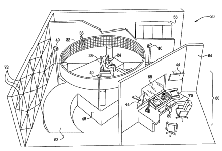
Fig. lA is schematic, pictorial illustration of a radio frequency medical
imaging and
therapy (RFIT) system 20, in accordance with an embodiment of the present
invention. The
description that follows refers mainly to the active imaging mode, for the
sake of clarity. The
other operational modes and the system configurations supporting them are
described further
below.
A patient 24 sits on a chair 28, which is located in the middle of a
cylindrical antenna
array 32. The antenna array comprises multiple antenna elements 36, which are
selectively
combined and actuated to form multiple effective antennas. The effective
antennas transmit
and -receive RF beams having different orientations to and from the patient's
body, in order to
image and/or apply treatment to a target organ or tissue. fn order to perform
RF ablation, local
heating or generate RF-induced pressure, however, some or all of antenna
elements 36 transmit
in unison towards a certain focal point. Although the embodiments described
below mainly
address cardiac imaging applications, the methods and systems described herein
can be used to
image and treat any other suitable target organ and tissue type.
In the exemplary system configuration of Fig. lA, array 32 has a diameter of
approximately 4 m and a height of approximately 80 cm. Six hundred thirty
elements 36 are
distributed around the cylinder perimeter, and the array has a height of forty
elements. Thus, in
total, the array comprises twenty-five thousand two hundred antenna elements.
In alternative
embodiments, array 32 may comprise a lower or higher number of elements and
may have any
other suitable shape or dimensions. In some embodiments, the system comprises
one or two
additional dome-shaped antenna arrays (not shown in the figure), which are
positioned above
13
CA 02616700 2008-01-24
WO 2007/017861 PCT/IL2006/000896
and/or below the patient. The additional arrays fiu-th.er improve the spatial
resolution of the
system, particularly when performing RF ablation.
Antenna elements 36 may comprise any suitable wideband radiating element
lcnown in
the art, such as flared-notch based elements, spiral or helical elements and
horn-based
elements. In some embodiments, array 32 may comprise eleinents transmitting in
different
polarizations, so as to enable the system to perform polarization-dependent
parameter
measurements. In some embodiments, elements 36 are active elements, which
comprise power
amplifiers for transmission, as well as low noise amplifiers and multiple A/D
converters for
reception.
Chair 28 typically comprises materials that cause little distortion to the RF
radiation,
i.e., materials having low reflectance aud absorption. The chair may comprise,
for example,
polystyrene foam, wood, artificial leather and cloth. Other objects, such as
various medical and
surgical tools and instruments, may be present in the vicinity of the patient.
These objects
should also comprise RF transparent materials or be covered with RF absorbing
material.
Typically, the target organ to be imaged should be positioned substantially at
the center
of the cylindrical antenna array, in both horizontal and vertical dimensions.
For this purpose,
chair 28 is typically adjustable and may also recline so the patient can lie
horizontally. The
chair may have multiple adjustable degrees of freedom. In some embodiments,
video cameras
40 are used for accurately positioning the patient at the center of the
cylinder. Cameras 40 are
mounted at different angles with respect to chair 28. Each camera is mounted
so that the center
of its field of view points to the center of the cylindrical array. The images
produced by
cameras 40 are displayed on one or more positioning displays 44. In order to
position the
patient correctly, chair 28 is remotely or locally adjusted until the region
of interest (e.g., the
patient torso) is seen at the center of the field of view of all caneras.
Array 32 may be mounted on an elevated platform 48. Some elements of system
20,
such as signal generation and reception circuitry, should be located near the
antenna array in
order to minimize RF losses. Such system elements may be located underneath
the elevated
platform. A ramp 52 enables wheelchair or gurney access to the platform. Other
system
elements, such as signal processing elements, can be located in a rack 56,
located further away
from array 32. Typically, a spacious area is left around the patient, so as to
allow easy access to
the patient by staff and equipment. For example, RF imaging and therapy can
take place in
parallel to other procedures, such as catheterization and imaging using other
modalities.
System 20 is controlled and operated from a control station 60, typically
separated
from array 32 by an RF absorbing wall 64. A window 68 comprising RF absorbing
material
14
CA 02616700 2008-01-24
WO 2007/017861 PCT/IL2006/000896
may be used for viewing the patient from the control station. The RF absorbing
wall and
window help to protect staff from RF radiation. RF leakage into and out of
system 20 can also
be reduced by covering external walls with radiation absorbing material, such
as RF absorbing
tiles 72. The room housing system 20 should be air-conditioned, in order to
dissipate the heat
produced by the RF energy, particularly when performing RF ablation.
Control station 60 comprises one or more imaging displays 76, which display
the
imaged target organ and other relevant information. The control station also
comprises input
devices 80, such as a keyboard, nlouse and/or traclcball, for providing input
and controlling the
system.
Fig. 1B is a schematic, pictorial illustration of an RFIT system, in
accordance with an
alternative embodiment of the present invention. In the system configuration
of Fig. IB,
cylindrical antenna array 32 is tilted at an angle typically in the range of
30-50 . Chair 28 is
positioned so that the area of interest, in the present example the torso of
patient 24, is located
at the center of the cylinder.
The configuration of Fig. 1B is particularly suitable for applications in
which staff
and/or equipment are present in the vicinity of the patient, but their effect
on the RF radiation
should be minimized by positioning them outside the beam paths. For example,
this
configuration may be used in different intra-operative imaging applications,
such as
catheterization procedures.
In the present example, a physician 82 sits or stands below the elevated area
of the
array, by the patient's feet. A display 83 displays the data acquired by the
system to the
physician. In some embodiments, radiation absorbing clothing can be worn by
the physician to
minimize radiation exposure.
OPERATIONAL MODES
System 20 supports several operational modes for imaging and/or therapy. Some
system configurations may support all modes, whereas other configurations may
support only a
single mode or a subset of the modes.
In some embodiments, the system supports an active imaging mode, in which the
target
organ is scanned with multiple beams. In the present example, the beams have
horizontal beam
widths of approximately 15 and vertical beam widths of approximately 4 ,
although other
radiation patterns can also be used. Using the exemplary array dimensions and
geometries
shown in Figs. lA and 1B above, these beam widths are suitable for irradiating
the patient
torso. In order to produce such beam widths, each effective antenna is
approximately 15 cm
CA 02616700 2008-01-24
WO 2007/017861 PCT/IL2006/000896
wide and 50 cm high. Assuming antenna elements 36 are spaced 2 cm apart, each
effective
antenna has approxiniately seven elements in the horizontal dimension and
twenty-five
elements in the vertical dimension.
The system measures multiple tissue paranieters at multiple locations in the
target
organ and displays them in three dimensions. Typically, the system directly
measures three
paranieters for each location, namely the local RF attenuation, local
reflection coefficient and
local time-delay caused by the decreased light velocity in tissue with respect
to free space. The
measured parameters, as well as the collected raw data, may also be used- for
evaluating
dielectric tissue properties, such as the local complex permittivity and local
conductivity of the
tissue.
Each of the parameters described above can be evaluated using the entire
system
bandwidth, or separately in multiple sub-bands. Paranleters can also be
evaluated for different
polarizations. Each parameter, or a combination of parameters, can be
displayed in 3-D. For
example, when performing cardiac imaging, the system can measure and display
the local
conductivity along the conduction pathways within the heart. As another
example, valve
calcification in the heart can be accurately detected based on a 3-D
measurement and display
of local permittivity.
The multiple tissue parameters can be jointly analyzed in order to accurately
and
reliably classify the tissue type, e.g., bone, muscle, fat or blood, at each
location in the target
organ. Using this analysis, a high-resolution display of the target organ,
with each tissue type
clearly marked and differentiated, can be provided to the physician. In the
active imaging
mode, the entire heart can be imaged at a typical frame rate of 100 Hz,
without gating. The
spatial resolution can reach 2 mm, and often 1 mm or better.
In some embodiments, certain dynamic mechardcal properties of the target organ
can
be evaluated by tracking frame-to-frame variations in the active imaging mode.
This sub-mode
is referred to herein as tissue tracking. For example, tissue tracking can
estimate the cardiac
wall motion velocity, as well as the local strain and local strain-rate. These
properties are
usually expressed as 3-D vectors.
Tracking inter-frame variations may involve known image processing techniques,
such
as optic flow methods. Additionally or alternatively, anatomical landmarks can
be identified in
the images, either manually or automatically. The variation in the coordinates
of these
landmarks can then be tracked in different frames.
In some embodiments, a contrast agent can be used during active imaging. The
contrast
agent is used to produce irregular values of one or more of the measured
parameters. For
16
CA 02616700 2008-01-24
WO 2007/017861 PCT/IL2006/000896
example, a contrast agent may comprise a highly reflective substance, such as
a ferrite-based
substance. Some contrast agents target a specific organ or tissue type, so as
to allow highly-
specific functional 'unaging. For exana.ple, organ-specific agents can be used
for myocardial
perfusion estimation, kidney performance evaluation and liver perfonnance
evaluation.
In some embodiments, system 20 supports a 3-D motion vector analysis mode,
which
measures the local motion vector for each location in the scanned target organ
(e.g., the local
blood velocity), as a function of time. In this mode, the system analyzes
multiple reception
beams simultaneously, and measures the Doppler shift with respect to each
beatn. The Doppler
shift, as measured for each point in space witli respect to several reference
points, is used to
determine the dominant velocity vector for each such point as a function of
time. The full
Doppler spectrum as a function of time and space may also be calculated for a
particular
component of the vectors.
Using the motion vector analysis mode, it is anticipated that imaging of the
entire
human heart can be performed at a frame rate on the order of 20 Hz, without
gating. The
spatial resolution in each axis is expected to be on the order of 1 mm. The
resolution of
velocity measurements in each beam is expected to be approximately 0.3 mis,
and the Nyquist
frequency (i.e., maximum unambiguous velocity) is expected to be approximately
3 m/s.
In some embodiments, system 20 supports a non-invasive RF ablation mode. In
this
mode, the antenna array focuses RF energy to a small region in the target
organ in order to
increase the local temperature by a factor on the order of 20 G. The local
temperature can be
measured in the active imaging mode. By combining the two modes, the system
can stabilize
the temperature in the target spot. RF ablation can be used for removing
tumors and cancerous
cells, as well as for performing non-invasive surgical operations such as
internal hemorrhage
reduction.
Typically, RF ablation in system 20 is performed in parallel to active
imaging. Unlike
known ablation methods, which use different modalities for imaging and
ablation, in system
20 no registration is usually needed between the coordinate systems used for
imaging and for
ablation.
The spatial resolution of the ablation mode is expected to be approximately 6
mm per
axis, at the 3 dB points of the ablation region. Using higher frequency bands
may allow
improving the spatial resolution by a factor of about two. When ablation is
guided by active
imaging, the frame rate is expected to be approximately 50 Hz, and the imaging
spatial
resolution is expected to be around 1 mm per axis.
17
CA 02616700 2008-01-24
WO 2007/017861 PCT/IL2006/000896
Simultaneous operation of the RF ablation mode with the tissue traclcing mode
enables
non-invasive ablation of moving organs, such as the destruction of ectopic
regions in the
cardiac muscle and the removal of lung cancer cells. When RF ablation is
combined witli
tissue tracking, the system adaptively adjusts the ablation region with a
refresh rate comparable
to the imaging frame rate.
In some embodiments, system 20 can be used to apply electromagnetically-
induced
pressure to target tissue. In some cases, applying instantaneous high-power
electromagnetic
pressure to a nerve pathway may induce an action potential either directly, or
due to thermal
effects. Thus, applying electromagnetic pressure may help to control the
innate frequency of
different regions within the cardiac conduction system, so as to achieve a
stable sinus rhythtn.
The pacing of other visceral organs, such as the gastro-intestinal system, may
also be affected
by electromagnetic pressure.
Additionally or alternatively, system 20 can be used to apply local heating to
tissue.
Local heating can be used, for example, to treat stressed muscles and to speed
the natural
healing of bruised or inflamed areas. In the local heating mode, a target
region can be defined
and visualized using the active imaging mode. The systeni can then locally
heat the selected
region by applying low-power RF energy to the region.
In some embodiments, system 20 performs high-speed passive imaging of
electrical
activity in the target organ. Various cells in the human body, such as nerve
and muscle cells,
are known to show substantial electrical activity. This activity may sometimes
be detected non-
invasively by a sensitive receiver. In the passive imaging mode, system 20
triangulates the
signals sensed by multiple reception beams, in order to determine the location
of the electrical
activity. The passive imaging mode is expected to reach a temporal resolution
of
approximately 1 s and a spatial resolution of approximately 1 mm. This
performance level
should enable the system to display electrical signals as they pass through
various
physiological systems.
Data processing in system 20 can be performed either in real-time, i.e.,
during data
acquisition, or off-line, i.e., after data acquisition is completed. In the
active and passive
imaging modes and in the motion vector analysis mode, data processing may be
performed
either in real-time or off-line. In the RF therapeutic modes, data processing
is typically
performed in real time in order to provide imaging guidance to the therapeutic
procedure.
SYSTEM COMPONENTS
Fig. 2 is a block diagram that schematically illustrates RFIT system 20, in
accordance
with an embodiment of the present invention. The system comprises a digital
exciter and
18
CA 02616700 2008-01-24
WO 2007/017861 PCT/IL2006/000896
receiver unit 84, which generates the signals used in the various imaging and
therapy modes.
The signal produced by the exciter is split into multiple signals aald
amplified by a transmitter
array 88. The amplified signals are distributed to individual antenna elements
36 in antenna
array 32 by a switching array 92, so as to form the appropriate radiation
beains. The signals are
transmitted towards the target organ by array 32.
The backscattered RF radiation is received by array 32. Switching array 92
selects the
appropriate subset of elements 36 corresponding to the currently-received
beam. The signal
corresponding to the specific beam is received by a digital receiver in unit
84. The signal
produced by the receiver is processed by a digital signal processor (DSP) unit
96. DSP unit 96
typically performs computationally-intensive calculations, such as operations
that are repeated
many times. These calculations may comprise, for example, compensation for
artifacts and
measurement distortions and 3-D image reconstruction.
A data processor and man-machine interface (MMI) unit 100 manages the various
real-
time processes performed by system 20 and controls other system elements, such
as transmitter
array 88, digital receiver and exciter 84 and switching array 92. The
management of real-time
processes may comprise mode selection, calculation of parameters to be used by
DSP unit 96
and other system elements, as well as general system timing.
Unit 100 also interacts with the user, in order to accept user input and
commands. In
some embodiments, unit 100 comprises a video/display processor (not sllown in
the figure),
which performs the transformations that translate the time-dependent 3-D
images and
calculated parameters, generated by DSP unit 96, to the desired viewing
configurations
presented on displays 76 and 83. In some embodiments, the video/display
processor may also
perforrn the final tissue classification, i.e., determining the tissue type
for each point in space
based on the 3-D images. Additionally or alternatively, the video/display
processor may carry
out tissue tracking.
Typically, DSP 96 and unit 100 comprise general-purpose or customer off the
shelf
computers, which are programmed in software to carry out the functions
described herein. The
software may be downloaded to the computers in electronic form, over a
network, for example,
or it may alternatively be supplied to the computers on tangible media, such
as CD-ROM. DSP
96 and unit 100 may be implemented in a single computing platform or in
separate platforms.
Some or all of the functions of DSP unit 96 may also be implemented in
hardware.
In some embodiments, in particular when performing passive imaging, the
thermal
noise level of antenna array 32 should be reduced. For this purpose, system 20
may comprise a
cooling system 104, which cools antenna array 32, switching array 92 and/or
the receiver in
19
CA 02616700 2008-01-24
WO 2007/017861 PCT/IL2006/000896
unit 84. System 20 is powered by a power supply 108. Appropriate environmental
conditions,
e.g., temperature and humidity, are maintained by an environmental control
unit 112.
Fig. 3 is a block diagram that schematically. illustrates transmitter array
88, in
accordance with an embodiment of the present invention. The signal produced by
the exciter in
unit 84 is split by a power splitter 116 and amplified by multiple power
amplifiers 120. The
amplified signals are provided to switching array 92. As will be shown below,
the transmitted
power of each effective antenna is set to around 3 kW peak power and 350 W
average power.
Assuming each effective antenna comprises 25x7=175 elements, the pealc power
of each
amplifier 120 is approximately 17 W and the average power is 2 W. These power
levels are
readily achievable using known solid-state devices.
Each amplifier 120 produces a signal that will ultimately drive a particular
element 36
in the currently-used effective antenna. Thus, the nuinber of amplifiers 120
in the transmitter
array should match the number of elements in the effective antenna. In
alternative
embodiments, a single amplifier can be allocated to a group of elements in the
effective
antenna.
In the RF ablation mode, the number of elements assigned to a particular
amplifier may
depend on the desired ablation region size and on the ability to control the
antenna array focal
point. In the passive imaging mode, the transmitter array is not used.
In some embodiments, the beam of the effective antenna is focused and shaped
by
applying different weights and/or different timing offsets to the different
elements. This
process is commonly referred to as apodization. In these embodiments,
transmitter array 88
comprises a timing and apodization module 124, which is controlled by the data
processor.
Module 124 adjusts the timing, gain and/or phase of amplifiers 120, in
accordance with the
apodization scheme used. In some embodiments, encoding of the transmitted
pulse sequence, a
fiinction that is described in detail further below, is also carried out by
module 124.
Fig. 4 is a block diagram that schematically illustrates switching array 92,
in
accordance with an embodiment of the present invention. Switching array 92 is
connected to
antenna array 32. An exemplary effective antenna 128 is shown in the figure.
On transmission,
switching array 92 accepts the signals from transmitter array 88 and routes
them to the
appropriate elements 36 in antenna array 32, in accordance with the currently-
used effective
antenna. On reception, switching array 92 routes the signals received by the
elements of the
currently-used effective antenna to the receiver in unit 84.
Switching array 92 comprises a switch matrix 132, which selects the
appropriate subset
of elements 36. The switch matrix is controlled by the data processor of unit
100. The
CA 02616700 2008-01-24
WO 2007/017861 PCT/IL2006/000896
switching array further comprises multiple duplexers 136. On transmission, the
duplexers
isolate the transmitted signals from the receiver. In some enibodiments, the
geometry and pulse
repetition frequency (PRF) of the system are configured so that transmissioii
and reception do
not occur simultaneously. lii these embodinlents, T/R switches or circulators
may be used
instead of duplexers 136.
The operation and/or configuration of the switching array may vary for
different
operational modes of system 20. For example, in the active imaging mode, a
single receiver
channel may be assigned to each element of the currently-used effective
antenna, to a group of
elements, or to the entire effective antenna. In the motion vector analysis
mode, a single
transmit beam and several (e.g., 10) receive beams are used. The receive beams
are often
narrower than in the active imaging mode, therefore the effective antennas
conlprise a higher
number of elements 36. During RF ablation, most or all elements participate in
the
transmission, and no reception is performed. In the passive imaging mode, no
transmission is
carried out, and several receive beams are used, either alternately or
simultaneously.
In order to achieve the desired frame refresh rate, system 20 performs liigh-
speed beam
switching, typically on the order of 9 MHz (i.e., scanning of 9,000,000 beams
per second). The
switching elements in switch matrix 132 should support this high switching
speed. For
example, PIN-diode switches, such as the S9H-79-3 device produced by GT
Microwave, Inc.
(Randolph, New Jersey), can be used for this purpose. These devices have a 30
ns switching
time. Further details regarding these PIN-diode switches are available at
www. glxnicr owave. com.
Fig. 5 is a block diagram that schematically illustrates digital receiver and
exciter unit
84, in accordance with an embodiment of the present invention. Unit 84
comprises an exciter
140, which produces the pulsed signal waveforms used for transmission and
provides the
signals to transmitter array 88. As shown in Fig. 2 above, on reception, unit
84 accepts the
received signals from elements 36 of the currently-used effective antenna via
switching array
92. The signals are combined using a power combiner 142.
A correlator module 144 correlates the received signal with the expected
signal, i.e.,
the signal waveform produced by the exciter. The pulse shape of the
transmitted pulses is
typically selected so that its temporal point spread function (PSF) has low
sidelobes. In some
embodiments, module 144 performs additional functions such as matched
filtering, down-
conversion to a suitable baseband or intermediate frequency (IF) and/or pulse
de-compression.
Module 144 may also apply time-dependent gain control (TGC) to the signal.
21
CA 02616700 2008-01-24
WO 2007/017861 PCT/IL2006/000896
In alternative embodiments, pulse de-compression can be perfornied using
suitable
software or digital hardware after the signal is digitized. 'Signal down-
conversion may also be
perfozmed on the digitized signal. In such configurations, the signal should
be digitized at a
sampling rate corresponding to the highest radio frequency used (e.g., 18
GHz).
As will be described in greater detail below, the range resolution of the
system is
achieved by sampling (digitizing) the signal using multiple analog-to-digital
(AID) converters
that sample the signal at incremental time offsets. For this purpose, the
signal produced by
module 144 is split by a 1:15 power splitter 146. The fifteen outputs of the
power splitter are
delayed by fifteen delay lines 148.
In the exemplary system configuration described herein, the signal produced by
module
144 enables a raw spatial resolution of 1.5 cm. Delay lines 148 divide this
range into 15
intervals. In other words, the delay difference between successive delay lines
is equivalent to a
1 mm range. The outputs of the delay lines are sampled by fifteen synchronized
A/D
converters 150. The sampled signals are provided to DSP unit 96.
The exemplary configuration of Fig. 5 is typically suitable for the active
imaging mode.
In the passive imaging mode, the system does not transmit. In this mode,
module 144
correlates the received signal with a synthetically-produced signal that
approximates the signal
waveform that is expected to be produced by the target tissue. In the RF
ablation mode, exciter
140 produces the ablating signal waveform, and the receiver is used only when
performing
imaging. In the motion vector analysis mode, the target organ is imaged
simultaneously by
several beams. An exemplary receiver configuration suitable for this mode is
described in Fig.
12 furt,her below.
. In alternative embodiments, some of antenna elements 36 can be defined as
transmit-
only elements, and other elements may be defined as receive-only elements.
Such
configurations reduce the number of duplexers and cables, and simplify the
system calibration.
Hybrid configurations in which some elements are transmit-only, some are
receive-only and
some perform both transmission and reception, are also feasible.
In some embodiments, the system configuration can be simplified by relaxing
some of
the systein requirements. For example, the system can be defmed to support
only a single
operational mode or a small subset of modes. Defining the system for a smaller
penetration
depth and/or slower refresh rate can also simplify the system. For example,
when using a
slower refresh rate, the system can use transmitted signals having simpler
waveforms, such as
waveforms based on stepped frequency, linear frequency modulation,
complementary coding
22
CA 02616700 2008-01-24
WO 2007/017861 PCT/IL2006/000896
and other phase coding methods. The system can use longer pulse sequences in
conjunction
with these wavefornis.
In alternative embodiments, the system can perform mechanical antenna scanning
instead of electronic scaiuling. For example, a single antenna can be steered
mechanically
around the patient in one or more axes, so as to produce the multiple beams
needed for
imaging. As another example, a pair of antennas can be positioned on opposite
sides of the
patient and steered mechanically around the patient, forming pairs of beams on
opposite sides
of the patient. Alternatively, two or more alitenna pairs can be used. Further
alternatively, the
antenna or antennas can be stationary, and the patient can be moved and/or
rotated with respect
to the anteiinas.
Additionally or alternatively, the distance between the antenna and the
patient can also
be changed incrementally by moving either the antenna or the patient. Using
multiple distances
between the antenna and patient is equivalent to using multiple A/D converters
having
incremental time offsets. Spherical scanning, or any other suitable scanning
geometry, can-be
used instead of cylindrical scanning, when using either mechanical or
electrical scanni.ng.
In system configurations based on mechanical scanning, the number of antennas
and
amplifiers is significantly reduced, and the switching array can be
significantly simplified or
altogether eliminated. Such configurations may be particularly suitable for
applications that do
not involve hospitalization, such as in dentistry, plastic surgery,
ophthalmology and orthopedic
applications. Configurations in which the antennas are stationary and the
subject is moved may
be useful in experimental and non-medical applications, such as in animal
experiments.
SPATIAL RESOLUTION
The spatial resolution expected to be achieved by system 20 in the active
imaging
mode is on the order of 1 mm per axis. In particular, when processing the
backscattered signal
of a certain beam, the system has a range resolution of 1 mm. This resolution
level is achieved
by a combination of (1) using a wideband transmitted waveform having a range
resolution of
-1.5 cm, and (2) dividing the 1.5 cm resolution into 1 mm effective range
gates by processing
the received signal using fifteen A/D converters having incremental time
offsets.
The transmitted signal produced by exciter 140 comprises a sequence of narrow
pulses
(narrow in time and wide spectrally). Each pulse has a bandwidth of
approximately 10 GHz,
i.e., a pulse width of approximately 0.1 ns. Typically, the spectral content
of the pulse covers
the range of 8-1 8 GHz. In alternative embodiments, higher bandwidths, such as
6-25 GHz,
may also be feasible.
23
CA 02616700 2008-01-24
WO 2007/017861 PCT/IL2006/000896
In order to improve signal-to-noise (SNR) ratio, exciter 140 produces a
sequence of
sixty-four successive pulses. The sequence is phase-encoded, i.e., each pulse
in the sequence is
given a certain phase shift. In the present example, bi-phase encoding is
used, in which the
phase shifts are either 0 or 180 . Alternatively, any other suitable encoding
scheme can be
used. On reception, the received pulse sequence is correlated with a reference
sequence, so as
to achieve the desired pulse compression gain.
Narrow, wideband pulses can be produced, for example, by chopping a narrowband
signal whose frequency approximately matches the desired transmit carrier
fiequency.
Alternatively, a baseband signal can be chopped and then up-converted to the
desired transmit
frequency. High speed chopping can be performed, for exaniple, using step
recovery diodes
(SRD). For example, Aeroflex/Metelix Inc. (Sunnyvale, California) offers a
silicon SRD
device denoted MMDB30-B11, which can be used for this purpose. These diodes
are capable
of generating 30 ps pulses. Further details are available at www.aeroflex-
metelics.com.
Since the recovery time of SRD devices is relatively long, a seqlience of
short pulses
can be generated by multiple SRD devices in parallel, which are actuated
sequentially. Each
SRD generates a single pulse in the sequence at the appropriate timing.
Figs. 6A and 6B are diagrams that schematically illustrate a pulse generation
circuit
152, which may be used by exciter 140 to generate the transmitted pulse
sequences, in
accordance with an embodiment of the present invention. Circuit 152 of Fig. 6A
comprises a
sequence of SRD-based switches 154. Circuit 152 shows only eight switches for
the sake of
simplicity, however similar circuits having different numbers of switches can
be used to
generate pulse sequences having any desired number of pulses. Each switch 154
has an input
156, which accepts a phase-encoded RF signal. In the present example, bi-phase
encoding is
used. The input to a switch whose pulse is encoded with a 0 phase shift is
marked "+", and the
input to a switch whose pulse is encoded with a 180 phase shift is marked "-
". The switches
are actuated sequentially by a timing logic circuit 158. The SRD outputs are
combined using a
combiner 160 to produce the desired pulse sequence.
Fig. 6B shows an exemplary circuit for generating the phase-encoded RF signal
used as
input to the different SRD switches, in accordance with an embodiment of the
present
invention. An oscillator 162 generates a continuous sinusoidal signal at the
desired transmit
frequency. The output of oscillator 162 is split by a power splitter 164. One
output of the
splitter is provided to the SRD stages encoded with a 0 phase shift (the
stages marked with
"+" in Fig. 6A). The other output of the splitter is phase-inverted using a
180 phase shifter
24
CA 02616700 2008-01-24
WO 2007/017861 PCT/IL2006/000896
166, and provided to the SRD stages encoded with a 180 phase shift (the
stages marked with
ti_ii ).
Fig. 7 is a diagram showing transmitted pulse sequences generated by exciter
140, in
accordance with an embodinient of the present invention. In the present
example, exciter 140
transmits sequences of 64 phase-encoded pulses. Each pulse is 0.1 ns wide, so
that the overall
transmitted signal has a length of 6.4 ns. The exciter transmits pulse
sequences at a pulse
repetition interval (PRI) of 55 ns. This PRI corresponds to a two-way range of
over 8 m in free
space, significantly more than the radius of antenna array 32, thus avoiding
range ambiguity. In
some embodiments, two or more pulse sequences are transmitted per beanz. In
these
embodiments, for each beam position, the data of each range gate may be
integrated over the
different pulses in order to enhance SNR. The 55 ns PRI is selected in order
to achieve a
temporal resolution of 100 Hz, as will be described in greater detail in Fig.
10 below.
As noted above, the reflected signal is sampled by fifteen parallel A/D
converters
having incremental time offsets equivalent to 1 mm. The fifteen A/D converters
effectively
divide the 1.5 cm range gates achieved by the wideband pulses into effective
range gates of 1
mm. In some embodiments, DSP unit 96 solves a set of linear equations based on
the outputs
of the fifteen A/D converters, and evaluates the received signal with a
resolution of 1 mm. The
DSP unit may apply known deconvolution methods for this purpose. This process
is typically
performed separately for each pulse in the pulse sequence. In some
embodiments, after
processing each pulse, DSP unit 96 integrates the data for each range gate
over the sixty-four
pulses in the sequence in order to improve the measurement SNR.
Module 144 in the receiver performs time-dependent gain control (TGC) prior to
digitization of the signal, in order to provide sufficient dynamic range at
the A/D converters.
The TGC process typically uses a fixed or pre-calibrated fu.nction that
specifies the attenuation
as a function of range. The function may be evaluated by occasionally
transmitting a narrow
calibration beam and measuring the attenuation as a function of range.
Typically, the TGC
process provides coarse gain control, and an additional fme gain control
process is performed
digitally, after the signal is sampled by the A/D converters.
The description above refers to a cardiac imaging application, which is highly-
dynamic
and imposes harsh refresh rate requirements. For organs other than the heart,
a 100 Hz refresh
rate may not be necessary. In these cases, longer pulse sequences, which may
comprise up to
1000 pulses or more, may be used. Using long pulse sequences significantly
increases the
achievable penetration depth.
CA 02616700 2008-01-24
WO 2007/017861 PCT/IL2006/000896
PARAMETER EXTRACTION AND MEASUREMENT ARTIFACT COMPENSATION
The reflected signal measurements performed by system 20 are often distorted
as a
result of the different physical properties of the imaged tissue. Different
tissue types differ
from one another, and fiom free space, by their light velocity, local
attenuation and signal
dispersion properties. In some embodiments, system 20 compensates for these
artifacts in
order to achieve high spatial resolution.
As noted above, system 20 scans the target organ using multiple beams from
multiple
directions. In some embodiments, the system compensates for tissue artifacts
by jointly-
analyzing the reflections measured by pairs of beatns that image the target
organ from opposite
directions.
Figs. 8A and 8B are graphs that schematically illustrate reflected signal
intensities
measured by opposite beams, in accordance with an embodiment of the present
invention. In
Fig. 8A, a curve 220 shows the reflection intensity as a function of range, as
measured by a
particular beam. Curve 220 shows three characteristic peaks 224A, 224B and
224C, which are
typically produced by tissue transitions (i.e., interfaces between different
tissue types). Peak
224A is a strong peak having the shortest range to the effective antenna. A
peak of this kind is
usually produced by the body "skin effect," i.e., the transition from free
space to tissue when
penetrating the skin. Peaks 224B and 224C relate to other transitions from one
tissue type to
another.
In Fig. 8B, a curve 228 shows the reflection intensity as a function of range,
as
measured by another beam, which is located at a 180 azimuth offset with
respect to the beam
of fig. 8A. In other words, the beam of Fig. 8B images a similar depth cross
section of the
target organ, but from the opposite direction. Note that the range
(horizontal) axis of Fig. 8B is
reversed, so as to enable curves 220 and 228 to be compared. Curve 228 also
shows three
peaks denoted 224D, 224E and 224F. Peaks 224D...F correspond to the same
tissue transitions
as peaks 224A...c, respectively, as seen from opposite directions.
In Figs. 8A and 8B the range coordinates o.f the peaks in the two opposite
beams are
shown to coincide with one another. In many practical cases, however, the
measured
coordinates of the peaks generally differ from one another in the opposite
direction
measurements because of the differences in local light velocity (local time
delay) across the
tissue. These differences are used to estimate and compensate for the local
tissue time delay.
Fig. 9 is a flow chart that schematically illustrates a method for extracting
tissue
properties from reflected signal intensity measurements of opposite beams, in
accordance with
an embodiment of the present invention. The method of Fig. 9 refers to a
particular depth cross
26
CA 02616700 2008-01-24
WO 2007/017861 PCT/IL2006/000896
section of the target organ, as imaged by a pair of beams at a 1800 offset.
The process of
integrating the data measured by multiple beam pairs at different azimuth
angles and heights
into a 3-D representation of the target organ is described fiuther below.
The method begins with system 20 acquiring raw reflection measurements using
two
beanis at opposite directions, at a data acquisition step 240. The intensity
measurements as a
function of range acquired by a particular beam are referred to hereinbelow as
a measurement
set. The system enhances the range resolution of the two measurement sets by
using multiple
A/D converters, as described above, at a range resolution enhancement step
244. After
reversing the range dimension of one of the measurement sets, the reflection
measurements of
the two beanls resemble the exemplary reflection measurements of Figs. 8A and
8B above.
In many cases, the skin-effect reflection, i.e., the reflection pealc having
the shortest
range to the effective antenna, is substantially stronger than other, deeper
reflections. Temporal
side-lobes generated by this peak may distort or maslc other reflection peaks.
The side-lobes
are a result of the point spread function (PSF) of system 20. In some
embodiments, the
distortion caused by the skin-effect peak can be reduced by detecting the true
position of the
skin-effect peak, and subtracting from the measured signal a replica of the
system PSF,
centered at the measured skin-effect pealc location. This process is typically
performed for each
beam position. The skin surface may be added after carrying out the different
artifact
correction procedures.
System 20 performs time delay estimation and compensation, at a time delay
correction
step 248. The system identifies in the two measurement sets pairs of
characteristic peaks that
correspond to the same tissue transition (interface), as viewed from the two
opposite directions
(such as, for example, the pair 224A and 224D in Figs. 8A and 8B above). As
noted above, the
measured peak locations are offset from one another in the two measurement
sets due to local
time delay differences. For each peak, the system calculates an estimated peak
location based
on the peak locations in the two measurement sets.
The system then applies piecewise translation and stretching transformations
to the
horizontal axes of the two measurement sets, which moves the peaks to the
estimated
locations. Thus, each peak is associated with two time delay offsets, i.e.,
the time offsets
between the original peak location in the two measurement sets and the
estimated location.
These time offsets are proportional to the cumulative local tissue time delay
from the effective
antenna and up to the location of the peak. In between peaks, the tissue is
assumed to be
relatively homogeneous, therefore the time delay offset is distributed
uniformly between
successive peaks.
27
CA 02616700 2008-01-24
WO 2007/017861 PCT/IL2006/000896
For each pair of characteristic peaks, the estimated peak location can be
determ.ined as
follows: The ranges, measured from the direction of one of the two effective
antennas,
corresponding to the i'th characteristic peak in the two measurenlent sets,
are denoted 7 Z1 and
rl2 , respectively. The estimated location of the i'tli peak is denoted ri.
The time delay, with
respect to the timing corresponding to the same range in free space, which is
caused by the
tissue between the (i-1)'th pealc and the i'tli peak, is denoted di, when
expressed in units of
range. Using this notation, it can be shown that:
ri =ri+d1
P
ri =ri- >, dz
i+1
wherein P denotes the number of characteristic pairs of pealcs. This set of
linear equations can
be solved to extract both ri and dl. These calculations assume there is no
time delay outside the
subject body.
After completing step 248, the system has an estimate of the local tissue time
delay for each
point (effective range gate) along the cross section of the target organ.
System 20 estimates the local tissue attenuation, at an attenuation estimation
step 252.
First, the system calculates the total side-to-side body attenuation along the
examined depth
cross section. The system identifies in the two measurement sets a pair of
peaks that
correspond to a particular skin transition (e.g., the pair 224A/224D or the
pair 224C/224F in
Figs. 8A and 8B above). The ratio between the reflection intensities of the
two peaks is used as
an estimate of the total body attenuation.
Using the known total attenuation, the cumulative attenuation from the sldn
and up to a
particular peak can be calculated. The cumulative attenuation is calculated by
considering the
following four equalities: (1) the total body attenuation is identical in the
two measurement
sets; (2) the measured peak intensity in the first measurement set (i.e.,
measured from the
direction of the first effective antenna) is equal to the true reflection at
the peak location, plus
the cumulative tissue attenuation from the skin and up to the peak from this
direction; (3) the
measured peak intensity in the second measurement set (i.e., measured from the
direction of
the second effective antenna) is equal to the true reflection at the peak
location, plus the
cumulative tissue attenuation from the skin and up to the peak from the
opposite direction, and
28
CA 02616700 2008-01-24
WO 2007/017861 PCT/IL2006/000896
(4) the sum of the two cumulative attenuations (from the skin and up to the
pealc) from the two
opposite directions is equal to the known total body attenuation.
The system solves the linear equations derived from these four equalities, and
extracts
the true reflection coefficient of each pealc, as well as the cumulative
attenuation between
every two successive peaks. The local tissue attenuation and reflection are
assumed to be
uniform between pealcs. Therefore, the cumulative attenuation is distributed
unifonnly
between pealcs. The system adjusts the intensity of the measurement sets, so
as to compensate
for the local attenuation. After completing step 252, the system has estimates
of the local tissue
attenuation and reflection coefficients for each point (effective range gate)
along the cross
section of the target organ.
Note that when measuring and mailipulating peak intensities, any gain control
applied
by the receiver should be taken into account. For example, wllen the receiver
employs time-
dependent gain control (TGC), the TGC value of each range gate should be taken
into
consideration, in order to measure the peak intensities correctly.
In some embodiments, the accuracy of the reflection coefficient calculation
can be
improved by averaging the two attenuation-corrected measurement sets.
Additionally or
alternatively, the estimation accuracy can be improved by performing several
iterations of time
delay and attenuation correction. In some embodiments, a background signal per
range gate,
typically pre-calibrated without the presence of tissue, can be subtracted
from the corrected
measurement of each range gate. An exemplary pre-calibration process is
described fiu-ther
below.
In some embodiments, the estimation process of steps 240-252 above can be
performed
separately in several spectral sub-bands in order to reduce the effects of
signal distortion. For
example, the 10 GHz signal bandwidth can be divided into 5-10 sub-bands of 1-2
GHz
bandwidth, and the local time delay, 'reflection and attenuation calculated
separately for each
sub-band. In order to process each sub-band, the reflected signal should be
filtered, so as to
retain only the spectral content in the currently-processed sub-band. Such
filtering can be
performed either before or after the AlD converters, using either analog or
digital filtering,
respectively. If desired, the corrected measurement sets can be summed over
all sub-bands to
produce equivalent wideband measurement sets.
The system now uses the three estimated local tissue parameters (local titne
delay,
reflection and attenuation) to evaluate the local dielectric properties along
the examined depth
cross section, at a dielectric evaluation step 256. Several models are known
in the art for
calculating dielectric properties based on such physical parameters. For
example, a single
29
CA 02616700 2008-01-24
WO 2007/017861 PCT/IL2006/000896
dielectric layer model is described by Yariv in "Optical Electronics," CBS
College Publishing,
1985, Chapter 4, pages 87-95, which is incorporated herein by reference.
The single layer model cited above is particularly suitable for continuous
transmission.
In some einbodiments, a more complex model that considers multiple dielectric
layers and
pulsed transmission can be used. The model analyzes a structure of multiple
adjacent layers
having different material composition, with the first layer representing free
space. For
example, consider a structure of three layers and three transitions (air to
first layer, first to
second layer, second to third layer). The three layers have dielectric
constants denoted E2, E3,
and 64. s1=se represents the dielectric constant of air. At the k'th layer
transition, the incident
wave is denoted Ik , and the wave progressing to the next layer is denoted Ik
. The incident
wave components can be written as
= 1
IR = I+ rJ)l
2 = I(1 + rl ) exp (j k2 2
_ J+ rl) exp ( j k2 2 ) (1 + r2)1
= l+ r1) exp (j k2d2 )(1 + r2) exp ( j k3d3),
= 1+ r1) exp (jk2 2) (1 + r2) exp (jk3A3) (1 + r3)I
wherein ri denotes the reflection coefficients of the i'th layer transitions,
dI denotes the
thickness of the i'th layer, and ki denotes the wave number in the i'th layer.
At each layer transition there exist reflected (outgoing) wave components from
both the
present layer transition aild from deeper layer transitions. In general, a
reflected component
originating from the i'th layer transition and having traversed (j-1) layers
after being reflected
is denoted Oi O. The outgoing components are given by
CA 02616700 2008-01-24
WO 2007/017861 PCT/IL2006/000896
01 - Ir1l
0~ = I+ rl) exp (jk2A2) E21
0~ = I+ r1 ) exp ( j k2A2) r3 exp ( j k2d2)(1 + r1)~
0~ = I(1+r2)exp(jk3d3)r3~
2 (1 + r1) exp (jk2A2) (1 + r2) exp (jk3A3) =
03 =
r3 exp (j k3A3 ) (1 + r2)
3 (1 + r1) exp (jk2A2) (1 + r2) exp (jk3A3) =
03 =
r3 exp (jk3d3) (1 + r2) exp (jk2A2) (1 + r1)
The total outgoing signal from the first layer transition is equal to OZ + 02
j+ 03 .
On can be written as
n
On = A exp H wtn + t~) = rn = ~ exp ~2jkm m~
m=2
= 11 (1+r1n)2
m=1
wherein A denotes the incident signal magnitude, 0 denotes the phase at the
first layer
transition, co denotes the signal angular velocity, and t, denotes the time
index at which the
signal wa.s received, corresponding to the two-way path of the beam. Using
this model, the
dielectric coefficients ei can be extracted from the measured results.
The models described above assume a planar incident wave front. In alternative
embodiments, the model may be adapted to take into account the spherical decay
of spherical
wave fronts, such as by dividing the signal by R2. In some cases, the incident
wave is not
perpendicular to the plane of layer transition, an effect which may also cause
the results to be
polarization-dependent.
When applying any suitable layer model to the measurements of system 20, the
layer
thicknesses correspond to distances between successive peaks, after
attenuation and time delay
correction. Additionally, the local attenuation and reflection coefficients
are also known, as
described above. The complex permittivity of -each layer can be calculated
(assuming
perpendicular incidence) using the well-known equations
31
CA 02616700 2008-01-24
WO 2007/017861 PCT/IL2006/000896
I ~ _ ~n +1 - ~n
rn
~n +1 +
F~n
lkni kp wherein r=j1 and kn respectively denote the reflection and attenuation
of the n'th layer. The
imaginary component of the permittivity, denoted s", and the conductivity,
denoted a-, roughly
obey the relationship d"=6/rv. Therefore, the conductivity o-can be derived
from the estimated
permittivity.
An alternative method for estimating the complex permittivity may be based on
the
signal within each layer separately. Assuming no losses, the light velocity v
in a particular
layer is given by:
w c 1
v = _
k n ,us
wherein n denotes the index of refraction, and tv is the signal angular
velocity. In the presence
of losses, k is complex. Defining k as k=~- j~, a and /.~ are given by the
equations
,8 2 a2 _ C02 Re
4 C2
2
~a - e2 Im ('61 This model is described in detail by Jackson in "Classical
Electrodynamics," John
Wiley & Sons Inc., New York, 1999, pages 295-316, which is incorporated herein
by
reference.
a and fj may be estimated by measuring the complex signal (before attenuation
correction and time-delay compensation) immediately before a reflection peak,
and comparing
it to the signal at a previous reflection peak. Using the estimated a and 6
values, both the real
and imaginary components of the permittivity can be calculated. Note that this
model is
particularly suitable for narrowband signals, such as when the signal is
confmed to a particular
sub-band of system 20.
Additionally or alternatively, any other suitable model can be used to
estimate the
dielectric tissue properties based on the measured time delay, reflection and
attenuation values.
32
CA 02616700 2008-01-24
WO 2007/017861 PCT/IL2006/000896
In summary, the method of Fig. 9 provides estimates of local tissue time
delay,
reflection and attenuation coefficients, as well as local dielectric
properties, based on
baclcscattering intensity measurements made by two beams that irradiate the
target organ from
opposite directions. Using this metliod, the reflected signal intensity
measurements acquired by
the different beams are corrected, to account for local time delay and
attenuation.
Most of the parameters measured by system 20 do not typically depend on the
geometry of the measurement. For example, Gabriel et al., in "The Dielectric
Properties of
Biological Tissues: I. Literature Survey," Physics in Medicine and Biology,
volume 41, 1996,
pages 2231-2249, which is incorporated herein by reference, refer to the
dielectric properties
of tissue as scalars and not as tensors. Reflection coefficient measurements,
on the other hand,
often do depend on the geometry of the measurement, e.g., on the angle of
incidence with
respect to the tissue interface that produces the reflection. In system 20,
however, every point
in space is measured from multiple directions. As a result, the geometry-
dependent effects are
inherently averaged and minimized.
The possible spatial dependency of the reflection coefficient, either due to
angular
dependency of the dielectric properties or due to the effect of the tissue
interface geometry, can
be used to extract additional clinical information. For example, 3-D
reconstructed images may
be produced using multiple beam configurations, with the beam configuration
changing
cyclically from one frame to another. The difference between successive frames
may be
indicative of the spatial dependency. The beam configurations may differ from
one another in
several aspects, such as the elevation of the beam with respect to the plane
of the cylinder.
SCANNING AND RECONSTRUCTION
System 20 integrates the extracted tissue parameters measured by the different
beams,
after artifact compensation, into 3-D representations of these parameters. In
principle, a
scanned volume containing the target organ is divided into horizontal slices,
at increments on
the order of 1 mm. Each horizontal slice is scanned from multiple directions.
Artifacts are
compensated for, and parameters are extracted using pairs of opposite beams,
as described
above. The system then performs two-dimensional (2-D) reconstruction of the
reflected signal
intensity across the slice, using the data collected by the multiple beams.
Fig. 10 is a diagram that schematically illustrates two-dimensional signal
reconstruction in a particular horizontal slice, in accordance with an
embodiment of the
present invention. Although the description that follows refers to the 2-D
reconstruction of
reflected signal intensity values, the method is similarly used for
reconstructing the other
33
CA 02616700 2008-01-24
WO 2007/017861 PCT/IL2006/000896
extracted tissue parameters across the slice. In other words, the method can
be used to
reconstruct the attenuation, reflection and/or time delay values across the 2-
D slice.
The time delay map may be directly translated into a map of local liglit
velocities. The
values of the local dielectric properties, as a function of time and space,
may be derived from
the above three reconstructed images (i.e., the attenuation map, the
reflection map and the time
delay map).
The diagram shows a top view of a particular slice. The area of the slice is
divided by a
grid 200 into multiple grid cells 204. Two beams 206 and 207 view the slice
from two
different directions. The two angular sectors shown in the figure indicate the
approximate
horizontal beam-widths of the beams.
As described above, for each beam, system 20 measures the reflected signal
intensity in
1 nun effective range gates, which are then corrected to account for local
time delay and
attenuation. In beam 206, a particular effective range gate 208 corresponds to
an arc having a
width of Ar=1 mm. A similar effective range gate 212 is shown for beam 207. As
can be seen
in the figure, each effective range gate covers multiple grid cells 204.
In order to perform 2-D reconstruction of the reflected signal intensity, DSP
unit 96
accumulates the intensities contributed to each grid cell 204 by the different
beams and
effective range gates. For each effective range gate of each beam, the system
previously
measured a particular intensity value. This intensity value is divided
uniformly among the grid
cells covered by the effective range gate in question. The process is repeated
for all grid cells,
beams and effective range gates. At the end of the process, each grid cell has
an accumulated
value, integrated over the different effective range gates of the different
beams scanning the
particular slice.
The 2-D reconstruction process described above refers to a particular
horizontal slice.
The process is repeated for all slices, to produce a 3-D grid. Although the
vertical increment
between neighboring slices is on the order of 1 mm, the thickness of each
slice is derived from
the elevation beam-width of the effective antennas used, typically on the
order of 15 cm. Thus,
there is significant overlap between the slices.
System 20 translates the measurements performed in the overlapping slices into
a set of
linear equations. Solving the equations achieves a spatial resolution on the
order of the
increment size (in the present example 1 mm) in the vertical dimension. The
equations may be
solved, for example, using deconvolution methods. Deconvolution methods are
often noise-
sensitive. Therefore, a sufficiently high SNR should be obtained.
34
CA 02616700 2008-01-24
WO 2007/017861 PCT/IL2006/000896
Accurately solving the equations often involves knowledge of the solution at
the
boundaries of the scaruied region. In sonie embodiments, protective RF
absorbing sheets
having a negligible reflection coefficient can be placed at the upper and
lower boundaries of
the subject's relevant body area, so as to force lcnown boundary conditions.
Thus, the
equations may be solved analytically.
In some embodiments, the contribution of each effective rage gate can be
divided non-
uniformly among the grid cells covered by the effective range gate. For
example, the
distribution can be range-dependent. Alternatively, the distribution can take
into account
angle-dependent gain differences of the effective antemia. Other lcnown
methods, such as
iterative back-projection and filtered back-projection, which are soinetimes
used in
tomography systems, can also be used for improving the spatial resolution in
system 20.
In the description above, effective range gates are geometrically represented
as
concentric arcs centered at the phase center of the effective antenna. In many
practical cases
this representation is inaccurate, since the light velocity in tissue is
different from the light
velocity in free space, and also between different types of tissue. As a
result, the actual
geometrical shape of an iso-time surface (i.e., the locus of all points having
a certain
propagation delay from the effective antenna) deviates from a perfect arc.
Estimating the actual
shape of the iso-time surfaces produces a more accurate representation of the
effective range
gates.
For example, the iso-time surfaces can be estimated by considering two
different ligllt
velocity values, a free space value and a representative light velocity value
in tissue. The shape
of the iso-time surfaces can be determined based on an estimation of the shape
of the patient's
outer body surface. Points determined to be within the patient body are
assigned the tissue
light velocity value, and points determined to be outside the body are
assigned the free space
value.
The shape of the outer body surface can be estimated, for example, using the
following
process:
= For each beam, measure the minimal distance at which a non-negligible
backscattering
intensity is received (i.e., measure the minimal distance from the effective
antenna
phase center to the body surface).
= The volume confined within the cylindrical antenna array is represented by a
3-D array,
whose values are initially set to zero. For each beam, all the elements whose
distance
from the effective antenna phase center matches the measured distance to the
surface,
CA 02616700 2008-01-24
WO 2007/017861 PCT/IL2006/000896
and that are within the main-lobe of the respective beam, are set to "1 ". The
resulting
set of coordinates for each beam typically resembles a dome-shaped surface.
= Assume that the point at the 3-D center of the cylinder is located within
the scanned
tissue. Let us now examine the values of the 3-D array along radial lines
originating
from the central of the cylinder and pointing to different 3-D angles
homogenously
spanning the 47u sphere. Along each such radial, the outer surface of the
patient body is
defined by the 3-D array element whose value is "1" and its distance from the
center of
the cylinder is minimal.
Additionally or alternatively, the iso-time surfaces can be adjusted according
to the
local time delay at each point within the patient body. This information is
typically available
after the reconstruction process (based on outer body surface estimation) is
completed. The
reconstruction process inherently produces a 3-D map of local time delays in
the scanned
volume, which can be used to recalculate the iso-time surfaces for each beam.
The time delay
values can then be used to refine the reconstructed image. Such a procedure
may be repeated
iteratively, for example until the incremental variations in each iteration
become negligible.
In the present example, system 20 scans a volume having a height of 25 cm, a
size
typical of cardiac imaging. The volume is divided into 250 horizontal slices
at 1 mm
increments. Each slice is scanned by 360 beams, which are distributed at 1
increments around
the perimeter of the cylindrical antenna array. ln each beam, two 64-pulse
sequences are
transmitted. Thus, a total of 250x360X2=180,000 pulse sequences are
transmitted in order to
scan the volume once. In order to achieve a temporal resolution of 100 Hz,
system 20 transmits
18,000,000 pulse sequences per second, yielding the PRI of 1/18,000,000=55 ns
shown in Fig.
7 above.
In the present example, the height dimension of the imaged volume is divided
into 250
slices using an array having forty antenna elements along the heiglit
diunension. When imaging
a particular slice, the beams are typically shaped so that their phase centers
fall in the plane of
the slice. As a result, some of the beams may not be perfectly parallel with
the base of the
cylinder, i.e., some beams may be slightly tilted in elevation. The tilt is
typically on the order
of several tenths of a degree, usually no greater than one degree.
In alternative embodiments, other scanning and reconstruction processes can be
used.
For example, instead of dividing the scanned volume into slices, the vertical
dimension can be
reconstructed similarly to the horizontal reconstruction method of Fig. 10,
since each beam has
a certain width in elevation. In general, the volume can be divided into a
three-dimensional
36
CA 02616700 2008-01-24
WO 2007/017861 PCT/IL2006/000896
grid comprising multiple 3-D grid cells. The reflected signal intensity of
each 3-D grid cell can
be evaluated by scanning over beams in azhnuth as well as elevation.
In some cases, some of the beam pairs used in the 2-D reconstruction process
contain
poor quality data, for example because of poor SNR due to higli penetration
depth. In these
cases, the overlap between the two beams, which view the same depth cross
section from
opposite directions, may be insufficient for performing artifact compensation.
When the
overlap region is not sufficient in some beam pairs, 2-D reconstruction of a
slice can be carried
out in two phases. First, 2-D reconstruction is performed using the beam pairs
that have
adequate overlap. When the beams of a particular beam pair do not have
sufficient overlap, the
attenuation and time delay of this beam pair can be corrected using the 2-D
reconstructed
values of the other beam pairs in the slice. In applications that can tolerate
a lower refresh rate,
such as non-cardiac applications, the penetration depth (and hence the
achievable overlap
between opposite beams) can be signiflcantly, improved by using longer pulse
sequences.
It should be noted that artifacts due to spatial aberrations and multi-path
are reduced in
system 20, because of the use of relatively wide beams, and because each
sample inherently
contains information averaged over a large volume. Multi-path artifacts are
also reduced due to
the relatively strong tissue attenuation involved.
In some embodiments, Icnown super-resolution methods can be used in post-
processing
to further improve the final spatial resolution of the system. Such methods
may comprise, for
example, Minimum Variance Methods (MVM), Burg and/or Yule-Wallcer methods.
Super-
resolution methods are described, for example, by Borison et al., in "Super-
Resolution
Methods for Wideband Radar," The Massachusetts Institute of Technology Lincoln
Laboratory
Journal, (5:3), 1992, pages 441-461, which is incorporated herein by
reference. Super-
resolution methods may be applied to each beam pair, to each effective slice
(i.e., to the data
corresponding to a specific height of the cylinder, after 2-D reconstruction),
or to the complete
3-D grid.
The 3-D reconstructed tissue parameters are displayed to a user by unit 100 on
displays
76 or 83. Different visualization methods and modes can be used. For example,
unit 100 can
display isometric views of the target organ or parts thereof, with selected
tissue parameters
shown as color-coded layers on the 3-D display. The display can change
dynamically, in
accordance with the refresh rate used.
Unit 100 may display selected 2-D slices, projections and otlier surfaces
based of the 3-
D information. Real-time 3-D data rendering can be used. Additionally or
alternatively, tissue
parameters can also be displayed numerically. Typically, the user can control
and customize
37
CA 02616700 2008-01-24
WO 2007/017861 PCT/IL2006/000896
the different display functions using input devices 80. In some embodiments,
unit 100 enables
the user to perform measurements, such as various length and velocity
measurements, based on
the 3-D display.
3-D MOTION VECTOR ANALYSIS MODE
In the motion vector analysis mode, system 20 tracks and displays tissue
dynamics,
sucli as blood flow. The system produces real-time images sllowing the 3-D
velocity vector of
each point in the scanned volume. Tissue velocity is measured using the
Doppler effect.
Fig. 11 is a flow chart that schematically illustrates a method for
calculating three-
dimensional motion vectors using Doppler measurements, in accordance with an
embodiment
of the present invention.
The method begins with system 20 scanning the target organ, at a scanning step
280.
The system scans the organ from multiple angles using pairs of opposite beams.
In each beam
position, several pulse sequences similar to the sequences of Fig. 7 above are
transmitted and
analyzed. The number of pulse sequences in each beam is on the order of ten,
although other
values can also be used.
In order to adequately image a relevant range of velocities, the measurements
should
have a Nyquist frequency of on the order of 3 m/s and a velocity resolution on
the order of 0.3
rnls. In order to reach this performance, a refresh rate on the order of 20 Hz
is used. In some
embodiments, the beams are scanned in an interleaved manner, transmitting a
single pulse
sequence at a time and returning several times to the same beam position,
until the desired
number of pulse sequences is transmitted. As a result, the pulse sequences are
distributed
evenly within the overall scanning cycle. Typically, narrow beams having
azimuth and
elevation beam widths on the order of 4 are used.
As described below, the Doppler shift measured with respect to a given
effective
antenna provides information related to a single component of the 3-D tissue
velocity vector.
In order to reconstruct the 3-D velocity vectors, three or more datasets are
acquired. Each
dataset provides information for each point in space, from a different viewing
angle. The
datasets can be acquired, for example, by changing the vertical inclination of
the beams from
one dataset to another.
For each angular beam position and for each range gate, the measurements are
de-
convolved to achieve a resolution on the order of I mm along the vertical
(cylinder height)
dimension. The DSP unit then performs artifact compensation on the
measurements of each
beam pair, at a compensation step 284. The compensation process is similar to
the process
carried out in steps 248 and 252 of Fig. 9 above.
38
CA 02616700 2008-01-24
WO 2007/017861 PCT/IL2006/000896
At the end of this process, at least three sets of measurements are available
in
cylindrical coordinates, having a spatial resolution on the order of 1 mm.
Since there is a
significant overlap in azimuth between adjacent beams, de-convolution can also
be performed
between adjacent horizontal beam angles, per range gate and height slice. De-
convolution may
also be performed between adjacent height slices, per horizontal beam angle
and range gate.
The system calculates the Doppler spectrum of each effective range gate in the
scanned
volulue, as viewed from the direction of each beam, at a spectrum calculation
step 288. Using
the well-known Doppler equation, the Doppler frequency shiftfd of a particular
tissue element
can be written as
1 -. -R s -Rz
fd A Vt rR, + V t Rl rI
wherein k denotes the signal wavelength, RS denotes the position vector of the
tissue element
with respect to the transmitting effective antenna, R. denotes the position
vector of the tissue
element with respect to the receiving effective antenna (which may be the same
as the
transmitting antenna), and Ut denotes the velocity vector of the tissue
element. The Doppler
spectrum of each tissue element can be calculated, for example, by applying a
16-bin discrete
Fourier transform (DFT) to the time-dependent data of each range gate in each
beam position.
Zero padding may be used to improve the DFT resolution. In some embodiments, a
window
function may be used to reduce the level of spectral sidelobes.
In some cases, the Doppler shifts measured by a particular effective antenna
may have
different values with respect to different elements of the effective antenna.
This effect may
cause some smearing in the Doppler spectrum. The smearing effect can be
estimated and
compensated for. For example, it may be assumed that the angular Doppler
velocity measured
by a certain antenna element varies in proportion to cos(O), wherein 9 denotes
the angle
between the normal to the antenna plane at the phase center of the effective
antenna and the
normal to the antenna plane at the location of the element in question. Using
this assumption,
suitable time-and-frequency dependent weighting can be applied to the DFT
coefficients.
In some cases, the Doppler spectrum is also smeared as a result of the large
signal
bandwidth. In order to compensate for this effect, the DFT result can be de-
convolved with a
point- spread function (PSF) that represents the smearing effect. The PSF can
be pre-calculated
using the Doppler equation, for any given spectral distribution of the
transrnitted signal.
39
CA 02616700 2008-01-24
WO 2007/017861 PCT/IL2006/000896
The system identifies dominant spectral components in the Doppler spectra, at
a
component identification step 292. Dominant Doppler components typically
comprise spectral
lines, or frequencies, having relatively strong intensities. Any suitable peak
detection method
known in the art ca.i1 be used to determine tlie peak frequencies. Centroid-
based methods can
be used to improve the calculation accuracy.
The system atteinpts to find dominant conZponents that are associated with one
another,
i.e., relate to the same tissue velocity vector, in different spectra measured
from different
directions. Using the associated dominant frequencies, the system solves tlie
Doppler equation
and calculates the 3-D motion vectors, such as using 3-D triangulation, at a
vector calculation
step 296.
In many practical cases, however, the Doppler spectra comprise multiple
dominant
components, and it is sometimes difficult to determine which components in the
different
spectra are associated with one another. Calculating motion vectors based on
Doppler
components that are not associated with one another usually produces false,
ambiguous motion
vectors.
The system detects and discards the false motion vectors, at a discarding step
300. The
system may use any suitable method or criterion for detecting false motion
vectors. For
example, it may be assumed that true motion vectors are continuous in both
time and space. In
other words, a newly-calculated motion vector should not significantly differ
from previously-
calculated vectors of the satn.e tissue element, and motion vectors in
adjacent tissue elements
should not differ significantly from one another. Using these assumptions,
motion vectors that
vary significantly over time and/or space can be discarded. When using this
method, care
should be taken at the boundaries of blood vessels, whose velocity is close to
zero.
The process described above produces the 3-D motion vectors of different
points in the
scanned volume. This information can be displayed to the physician either
independently or in
conjunction with the information of other imaging modes of the system.
As noted above, the beams used in motion vector analysis are relatively
narrow.
Scanning the target organ with narrow beams may limit the achievable refresh
rate. In order to
reduce the scanning time, in some embodiments the system transmits with a wide
beam and
receives with multiple narrow beams simultaneously. The narrow beams point
towards the
center of the cylinder and have different phase center locations. In order to
receive on multiple
beams simultaneously, the receiver of the system should comprise multiple
parallel receive
chains.
CA 02616700 2008-01-24
WO 2007/017861 PCT/IL2006/000896
The allocation of antenna elements 36 to each receive chain should support
data
acquisition using opposite beam pairs, in order to perform artifact
compensation. For this
purpose, in some embodiments the receive beams corresponding to different
receive channels
are configured to have different plzase centers, and the boresight of each
receive beam is
configured to be perpendicular to the antenna surface at the corresponding
phase center.
Fig. 12 is a block diagram that schematically illustrates a digital receiver
and exciter
unit 320, in accordance with another embodiment of the present invention. In
unit 320, the
received signal is processed simultaneously by multiple receive chains 324.
Each receive chain
324 is similar in structure to the receiver of Fig. 5 above. The switching
array is configured to
provide each receive chain with the signal of the appropriate receive
effective antemia.
Since some elements of antenna array 36 are used by multiple effective
antennas and
multiple receive chains, each of the signals received from switching array 92
is split, e.g.,
using a 1:10 splitter, and fed to a different combiner in each receive chain
324. Each such
combiner, corresponding to a specific receive chain, may use a different
apodization scheme,
so as to define the required receive beam pattern. In any given receive beam,
unused elements
36 are typically given a zero weight.
RF THERAPEUTIC MODES
In the RF therapeutic modes (RF ablation, local heating and electromagnetic
pressure
modes), system 20 applies concentrated RF energy to a 3-D target region in the
target organ.
The description that follows refers mainly to RF ablation. Generalization to
the other
therapeutic modes is straightforward.
Typically, system 20 performs RF ablation interleaved with active imaging of
the target
region and its vicinity, in order to visualize and guide the ablation process.
In some
embodiments, the system alternates in time between ablation frames and imaging
frames.
During the imaging frames, the system performs active 3-D imaging of the
target organ, as
described above. During the ablation frames, some or all of antenna elements
36 transmit high
frequency RF pulses focused on the target ablation region. The 3-D imaging
information is
used to adaptively track the focal point (i.e., the location of the target
region) to which ablation
energy is focused.
Adaptive tracking of the ablation region location enables ablation in dynamic
organs,
such as the cardiac muscle. In order to enable precise tracking, the maximum
frame-to-frame
motion of the target region should generally not exceed a certain fraction of
the ablation
resolution. Frame refresh rates on the order of 50 Hz for imaging and 50 Hz
for ablation are
typically sufficient.
41
CA 02616700 2008-01-24
WO 2007/017861 PCT/IL2006/000896
Since imaging and ablation are performed using the same coordinate system, and
since
the two modes are affected by the same physical artifacts, distortion in the
acquired 3-D image
typically has little influence on the focal point location accuracy.
Additionally, the time delay
between each element 36 and the target region, measured in the imaging mode,
can be used to
optimize the time delay of each transmitting element for ablation, so that
pulses from the
different elements reach the target region simultaneously.
According to simulated results, the 3 dB width of the ablation region in
approximately
0.6 by 0.6 by 3.6 cm, when using a transmission frequency of 18 GHz. Unlike
the pulse
sequences used for imaging, ablation may be performed using relatively long
pulses, so that
narrow bandwidths can be used.
In some embodiments, system 20 comprises one or two additional dome-shaped
antenna arrays. The combination of the dome-shaped arrays and the cylindrical
array
approximate a spherical array. hi a spherical array, the resolution is
substantially uniform in all
axes. Such a configuration is expected to provide a spatial ablation
resolution of approximately
0.6 cm in all axes.
Additionally or alternatively, system 20 can use directional radiating
elements for
ablation. The angle between the boresight of each directional element and the
normal to the
antenna plane at the center of the element can be optimized to maximize the
focusing of the
ablation beam, with minimal effect on the spatial resolution of the active
imaging mode. For
example, all directional elements may be oriented so that their boresights
point to the center of
the cylinder.
The spatial resolution of the ablation mode can also be improved by increasing
the
frequency of the ablation signal. On the other hand, signal absorption in
tissue increases with
frequency, so that the transmit power should be increased. It can be shown
that the radius of
array 32 has little effect on the ablation spatial resolution.
Unlike the other modes of system 20, in which processing may be performed off-
line,
the processing in the RF ablation mode is performed in real-time. Assuming a
volume of 5
cm2, which is imaged with a spatial resolution of 2 mm per axis, the number of
slices is
reduced from 250 to 25. Each slice comprises 360 beam positions. For each
pulse, only six
range gates are processed for each A/D converter. In order to obtain a 2 mm
resolution, 8 AID
converters should be used, providing 48 samples per pulse, or 96 samples for
each pulse
sequence (when using two pulse sequences in each transmission). For a frame
rate of F
42
CA 02616700 2008-01-24
WO 2007/017861 PCT/IL2006/000896
frames/second, the number of samples per second is 25-360-96=F, or 8.64=105 F
samples/second. For F=50 frames/second, the acquired data rate is 4.32= 107
samples/second.
Assuming the data rates given above, a representative digital signal processor
(DSP)
having a processing power of 15G floating-point operations per second (Gflop)
can perform
approximately 330 operations per sample, or 33000 operations per pulse. Higher
processing
power can be obtained by using multiple signal processors in parallel. Each
processor may
process a different set of pulses, and the resulting 3-D image matrices can be
summed, to
produce the final image.
The processing load per pulse sequence is made of three dominant factors:
(1) De-convolution per each effective range gate. There are forty-four
effective range
gates per pulse sequence, corresponding to a-J3 = 5 cm wide region. In some
cases, a
-~2 - 5 em width is also sufficient. A linear combination of eight A/D
saniples (8 additions
and 8 multiplications) is calculated for each pulse sequence. The results of
the two pulse
sequences are added (i.e., 16 operations x 2 pulse sequences, plus one
addition, per effective
range gate). Thus, a total of approximately 33x44=1452 operations are
performed per pulse
sequence.
(2) Time delay and attenuation correction: Approximately 500 operations are
performed per pulse sequence, i.e., 1000 operations per each pair of opposite
beams.
(3) 3-D reconstruction: Each of the twenty-five samples is spread over
approximately
25x25 grid cells, producing a total of 15625 operations per pulse sequence.
In total, the overall number of operations per pulse sequence is approximately
20000.
For F=50 frames/second, 25-360=20000=50, or approximately 9=109 operations are
performed
per second.
Once the 3-D image is produced, additional calculations are performed for
tissue
tracking in order to adaptively track the ablation focal point. Tracking may
be carried out by
cross-correlating 3-D blocks in successive frames. Altematively, tracking may
be performed
by determining the 3-D translation that minimizes the minimum mean-square
error (or
minimum mean absolute difference) between 3-D blocks in successive frames. For
each
possible motion vector, 253 multiplications and additions are performed (i.e.,
a total of 31250
operations). Thus, 1000 possible motion vectors (10 possibilities in each
axis) correspond to
approximately 31.25=106 operations per frame. For F=50 frames/second,
approximately
1.6=109 operations per second are added.
43
CA 02616700 2008-01-24
WO 2007/017861 PCT/IL2006/000896
In summary, a 15 Gflop DSP is typically capable of performing the conlbined
imaging
and ablation processing in real-time. Note, however, that if the overlap
between opposite
beams is insufficient, the processing becomes more complex. In such cases,
quiclcer, lower
precision time delay and attenuation correction methods can sometimes be used.
PASSIVE IMAGING MODE
Different cell types in the human body, suc11 as nerve and muscle cells, show
substantial electrical activity. The propagation velocity within neurons is
typically in the range
of 30-120 nVs. The duration of action potentials is usually in the range of 1-
10 ms, and their
amplitude ranges between 70 and 110 mV. The duration of neuron receptor-
potentials and
synaptic-potentials is typically between 5 and 100 ms, and their amplitude
ranges from 0.1 to
10 mV. These properties are described, for example, by Kandel et al., in
"Principles of Neural
Science," McGraw-Hill, New York, 2000, pages 19-35, which is incorporated
herein by
reference. The propagation velocities in muscle cells tend to be significantly
lower (e.g., it
takes about 50 ms for the signal generated by the sinoatrial node in the heart
to reach the
atrioventricular node).
This electrical activity generates electromagnetic fields. When operating in
the passive
imaging mode, system 20 senses and maps these fields. The system provides a
dynamic 3-D
display showing the propagation of electrical signals in a target organ, such
as along a nerve or
a muscle. The passive and active imaging modes may be combined, so that the
system displays
the electrical activity overlaid on a 3-D image of the organ produced by
active imaging.
Unlike the active imaging mode in which the signal is subject to two-way body
attenuation, in passive imaging the signal is only attenuated on its way from
the organ to the
receiving antenna. Furthermore, the antenna array and receiver front end may
be cooled, so
that their sensitivity is improved.
In order to detect the relatively weak electromagnetic fields generated by the
physiologic electrical activity, the receiver typically integrates over
relatively long time
intervals. For example, the receiver may use a single A/D converter operating
at a sampling
rate higher than 1 GHz, typically on the order of 10 GHz, and integrate the
data over intervals
on the order of 1 s.
In many practical scenarios, because of the low duty cycle of the sensed
electrical
activity signals, the receiver will typically sense no more than a single
signal within each 1
sec interval. Narrow receiving beams can be used to ftirther reduce the
probability of sensing
multiple signals simultaneously.
44
CA 02616700 2008-01-24
WO 2007/017861 PCT/IL2006/000896
In some embodiments, system 20 passively senses the electrical activity
signals using
three or more beams having different orientations. The beams can be received
either
simultaneously or in alternation. Based on these measurements, the system
estiinates the 3-D
coordinates of the source of the electrical activity, sucli as using
interferometry methods.
Although in some cases using three beams is sufficient, a higher number of
beams
(e.g., five beams) may be preferable, for example in order to reduce time
delay effects caused
by light velocity variations in tissue. In some embodimeilts, different
subsets of three beams
are used to calculate a single 3-D coordinate estimate. The center of mass of
the various
estimates is used as an estimate of the signal source coordinate.
Alternatively, a local time
delay map produced by the active imaging mode may be used to calculate an iso-
time surface,
which corresponds to the range measured by each beam. The intersection point
of the surfaces
corresponding to the three (or more) beams should provide a good estimate of
the signal
source coordinate.
In order to perform the interferometric triangulation calculation, the sensed
signal
should be processed at a narrow bandwidth. In some embodiments, the sensed
signal is filtered
using either digital or analog filtering.
TRANSM[T POWER CONSIDERATIONS AND EXPERIMENTAL RESULTS
The power level that should be used for transmission in system 20 depends
primarily
on the body attenuation in the relevant imaging scenarios. In the active
imaging and motion
vector analysis modes, the transmitted power should provide the system with
sufficient SNR at
the maximum desired penetration depth. On the otlier hand, the transmitted
power should not
exceed industry-standard safety recommendations.
In some embodiments, the instantaneous transmitted power of the system (i.e.,
the
instantaneous power summed over the elements of a particular effective
antenna) is set to 3
kW in the active imaging and motion vector analysis modes. As will be shown
below, this
power level is expected to achieve the desired system performance, and also
complies with
accepted radiation safety limits.
Using the pulse sequences described above, the transmission duty cycle is
approximately 11.6%, so that the average power is approximately 350 W.
Assuming the body
area exposed to the radiation is on the order of 1 m2, the average power per
unit body area is
mW/cm2. Instruction #6055.11 of the U.S. Department of Defense (DoD) entitled
"Protection of DoD Personnel from Exposure to Radiofrequency Radiation and
Military
Exempt Lasers," February 21, 1995, specifies for exposure durations shorter
than 0.1 hours a
CA 02616700 2008-01-24
WO 2007/017861 PCT/IL2006/000896
maximum permissible exposure level of 1/Texp mW/cm2 in any interval of Texp
hours. For
exposure durations longer than 0.1 hours, a maximum of 10 mW/cm2 is permitted.
Instruction
#6055.11 also states that the restrictions on maximal exposure levels may be
relaxed in cases
of partial body exposure. Since iniaging using the RFIT system only involves
several seconds
of exposure, the system power level is well within the recommended safety
limit.
In order to verify that the specified power level can achieve the desired
system
performance, the propagation of RF energy in various tissue types was measured
using an
experimental setup. The objectives of the experiment were (1) to quantify the
expected tissue
attenuation of different tissue types, and (2) to quantify the difference in
tissue attenuation,
reflection and time-delay values between the different tissue types.
The experiment was conducted in an anechoic chamber. Short RF pulses in the
range
8-18GHz were generated and analyzed by a time-domain network analyzer
connected to a horn
antenna. Different tissue samples were irradiated with the RF pulses. Nine
lamb tissue samples
were tested, namely blood, heart, lungs, bone, liver, kidney, intestine, brain
and thigh muscle.
Two additional samples containing air and water were also measured.
In each measurement, a particular sample was placed in the anechoic chamber
and
irradiated with RF pulses from a distance of approximately 65 cm. A double-
sided copper-
plated PVC sheet was placed behind the irradiated sample in order to reflect
the radiation back
to the horn antenna and networlc analyzer. The area of the reflecting sheet
approximately
matched the size of the sample and the beam-width of the antenna. In some of
the
measurements, a cascade of two different samples placed one behind the other
was tested.
The signal generated by the network analyzer was a stepped-frequency signal,
covering
the relevant frequency range in 801 pulses of different frequencies. Each
pulse was 192 ns
wide. The output power used was 1 mW. Different frequency ranges, such as 8-12
GHz, 15-18
GHz and 8-18 GHz, were tested. The network analyzer sampled the signal
reflected from the
sample and reflecting plate and reconstructed the signal as a function of
time. The setup was
pre-calibrated to enable measurement of the net attenuation of the sample.
The network analyzer measurements provided the reflection, attenuation and
time delay
characteristics of each tissue type. The measured results clearly show that
there are significant
differences between different tissue types in all three parameters. The
absolute attenuation
values measured support the feasibility of RFIT system 20 achieving adequate
SNR at
penetration depths on the order of 30 cm, enabling high-resolution cardiac
imaging.
46
CA 02616700 2008-01-24
WO 2007/017861 PCT/IL2006/000896
The experiment results were used to verify the transmit power rating of system
20.
Since some of the system parameters in the experiment are different from the
parameters the
RFIT system, the calculation should take these differences into account.
PtGa2 GcGi
According to the well-known radar equation, SNR oc , or, in
R4L
logarithmic representation, SNR = Pt + 2Ga + Gc + Gi - 4R - L + C, wherein
SNR denotes the signal to noise ratio of the system, Pt denotes the
transmitted power, G.
denotes the antenna one-way gain, Gc denotes the processing gain due to pulse
compression,
Gi denotes the gain due to integration over multiple pulses, R denotes the
range, and L denotes
the system and medium (air and tissue) losses.
The following table shows the parameters affecting the achievable SNR both in
the
experimental setup a.nd in system 20:
Parameter Experimental Experimental RFIT system RFIT
setup [linear] setup [dB] [linear] system
[dB]
Pt 10 W -30 dBW 3=103 W 34.8 dBW
Ga 16 12 dB 1508 31.8 dB
Go 1.54-10 6 62 dB 64 18 dB
Gl 1 0 dB 2 3 dB
R 0.6m -2.2 dB-m 1.7 m 2.3 dB-m
L
(nzedium only) 32 15 dB X XdB
Ae N/A N/A 0.075 m -11.2 dB
Preserving the SNR of the experimental setup in the RFIT system yields:
X c Z B = Pt +2Ga +Gc +Gi 4R-
(Pt + 2Ga + GC + G? - 4R - L)
= 34. 8 + 63. 6 + 18 + 3 - 9. 2-
(-30 + 24 + 62 + 0 + 8. 8 - 15) = 60. 4dB
47
CA 02616700 2008-01-24
WO 2007/017861 PCT/IL2006/000896
wherein the parameters marked - denote parameters of the RFIT system and
parameters not
marlced with - denote parameters of the experimental setup. Practically, the
SNR of the
experimental setup is significantly better tlian the = SNR required in system
20, so we may
safely assume Xdb=75 dB. Based on the experimental results, such a value
corresponds to a
penetration depth of approximately 30 cm. This penetration depth is suitable
for high
penetration depth applications, such as cardiac imaging.
In the RF ablation mode, the transmitted power of system 20 should enable
raising the
temperature of the target ablation region by approximately 20 C. As described
above, the
dimensions of the ablation region are approximately 0.6 by 0.6 by 3.6 cm, at
the 3 dB points:
Assuming the ablation region has the shape of an ellipsoid, its volume is
approximately
(47c/3)=0.3=0.3=1.8=0.68 em3. Assuming a characteristic tissue density of 1
g/cm3, the mass of
the ablation region is approximately 0.7 grams. The energy required to
increase the
temperature of this mass by 1 C is 2.9 Joules. Thus, approximately 59 Joules
are required for
increasing the temperature of the ablation region by 20 C.
Assuming the ablation procedure is 600 second long, the power dissipated in
the
ablation region should be somewhat greater than 59/600=0.098 W. After reaching
the target
temperature, the power used is normally decreased, so that the local
temperature remains stable
(i.e., so that the body heat dissipation mechanism and the RF heating
mechanism balance each
other).
According to the results of the experiment described above, the maximum one-
way
power attenuation is approximately 2 dB/cm. This attenuation was measured for
blood and
water. A pulse traveling a distance of 20 cm in tissue would therefore be
attenuated by no
more than 40 dB. According to computer simulation results, the ratio between
the transmitted
power per element and the power at the ablation region is 1:27712 for
continuous-wave
transmission. Thus, the transmission power per element P should be set to
approximately:
0.098 - 10000
F = W= 35. 4 mW
27712
In the simulated scenario, an array comprising 29964 elements is used, so that
the
overall RMS power is approximately 1060 W. Assuming the irradiated body area
is 1.5 m2,
the power per unit area is 70 mViT/cm2, which is well within the short-
exposure limits of the
DoD safety standard cited above. For longer durations, on the order of 600
seconds (0.1
hours), the power density is acceptable for treatment scenarios.
48
CA 02616700 2008-01-24
WO 2007/017861 PCT/IL2006/000896
The calculation above is based on worst-case attenuation assumptions, and in
many
practical cases the transmitted power can be significantly lower. When system
20 uses
additional dome-shaped antenna arrays, the dimensions of the ablation region
can be reduced
to approximately 0.6 by 0.6 by 0.6 cm. In such configurations, the energy
required for ablation
is reduced by a factor of six, and the power per unit body area is reduced to
approximately 11
mW/cm2. In some embodiments, sensitive body parts can be covered by radiation
absorbing
materials in order to fiirther reduce their exposure. It should be noted that
the heating effect
decays rapidly outside the 3 dB ablation region. Typically, immediately
outside the 3 dB
boundary, each tissue point is heated by 0.5 C or less for each 1 C
temperature increase inside
the ablation region.
The experimental setup described above was also used to verify the feasibility
of
enhancing the range resolution using multiple A/D converters. The reflecting
plate and tissue
sample were placed at multiple distances from the antenna. The distances were
spaced by
approximately 1.57 mm from one another, over a 15.7 mm range. The distance
offsets are
equivalent to the time delay offsets between A/D converters in system 20.
The data measured by the network analyzer was processed in off-line to extract
the
signal magnitude per each effective range gate. The results clearly showed
that the measured
signal changed significantly when the sample was moved by increments smaller
thati the raw
range resolution. The measured ranges to the different reflecting surfaces of
the sample and
plate approximately matched the manually-measured distances.
SYSTEM TEST AND CALIBRATION
In some embodiments, different test and calibration procedures may be
performed at
different life cycle stages of system 20. For example, during installation at
a particular site, the
installation quality and system integrity are evaluated and corrected if
necessary. Attention is
typically given to mechanical deformations in array 32, which may affect the
relative locations
of the various radiating elements. The cable lengths, the accuracy of delay-
lines and the
attenuation of cables and connectors, which may affect the beam-shaping of the
various
effective antennas used, are also sometimes tested at installation.
During everyday use of the system, the system may be tested both at the
radiating
element level and at the effective antenna level. Such tests may be performed
periodically or
when a fault is detected or suspected.
Radiating element level calibration and test may be performed using an
accurately-
manufactured calibration sphere, which is positioned at the center of the
cylindrical antenna
49
CA 02616700 2008-01-24
WO 2007/017861 PCT/IL2006/000896
array. For each element 36, pulses are transmitted and received using the
element. Receiving a
signal whose power is within an expected range is indicative of the integrity
of the entire
transmit-receive chain. The power of the received signal may also be used for
compensating
for the attenuation along the two-way path from the exciter to the element.
Furthermore, the
minimal range at which a non-negligible reflection is detected, corresponding
to the distance to
the sphere surface, can be used for minimizing range bias. Other calibration
procedures may
involve transmitting pulses from a particular element 36, and receiving the
signal at several
adjacent elements. The relative timing of the reflections can be used for
minimizing phase
misalignment between adjacent elements.
At the effective antenna level, a specially-manufactured phantom (i.e., an
artificial
body imitating an imaged object) can be placed at pre-determined locations
within the
cylindrical array. Pulses are transmitted from each possible effective
antenna. The reflection
from the phantom is received by the effective antenna, as a function of range.
Separate
measurements can be performed for each frequency sub-band. The minimal range
at which a
non-negligible reflection is detected can be used for range calibration.
Significant deviations
from the expected signal may indicate failure or performance degradation.
In some embodiments, as described above, the background signal (i.e., the
signal
output by the receiver without the presence of a reflecting object) is
subtracted from the output
of each received beam during image reconstruction. The background signal is
typically pre-
measured for each possible effective antenna. The background signal of each
effective antenna
is often measured as a function of range and for each frequency sub-band.
DATA ACQUISITION LOAD ESTIMATION
As noted above, measured data can be collected in system 20 either in real-
time or off
line (with the exception of RF therapeutic mode, in which data is typically
collected on-line).
When operating off-line, the acquired data is stored in DSP unit 96 or in data
processor unit
100. Typically, the duration of a complete off-line data acquisition cycle is
on the order of 1
second.
In the active imaging mode, fifteen A/D converters sample the signal
simultaneously.
Assuming a 10 GHz signal bandwidth, a range gate of 1.5 cm and a region of
interest that is 50
cm deep, the number of samples per pulse per A/D converter is 35. When using B
bits per
sample (e.g., B=20 bits, 10 bits per complex component), the number of bits
per pulse per A/D
converter is 35-B. For 15 simultaneous A/D converters, the number of bits per
pulse is 525-B.
For a pulse repetition frequency (PRF) of 18 MHz, the data acquisition rate is
9.45-B Gbit/sec.
CA 02616700 2008-01-24
WO 2007/017861 PCT/IL2006/000896
In the motion vector analysis mode, in comparison to the active imaging mode,
datasets
are collected from three different directions, at a lower refresh rate
(typically one fifth of the
refresh rate used for active imaging). Multiple pulse sequences, typically 10
sequences, are
used per beam position. The data acquisition rate is thus 3-10/5=6 times
higher than the data
rate of the active imaging mode, or 56.7-B Gbit/sec.
In the RF therapeutic modes, data acquisition is performed for real-time
imaging and
guidance. The frame rate used is typically half the rate used in active
imaging, and the number
of vertical slices used is typically one tenth of the number of slices used in
active imaging.
Moreover, a smaller region of only 5 cm3 region is typically imaged. The
nuinber of samples
per pulse is approximately 3.8 times lower than in active imaging, yielding a
data acquisition
rate of approximately 125 -B Mbit/sec.
In the passive imaging mode data is collected at a very high rate, typically
on the order
of 10 GHz. Data is collected from M beams simultaneously. Each sample uses B
bits. The
resulting data acquisition rate is 10=MB Gbit/sec.
Although the embodiments described herein mainly address cardiac applications,
the
methods and systems described herein can also be used for imaging and applying
treatment to
any other organ or system. RFIT methods and systems can be used for detecting
and removing
tumors, such as in the different fields of oncology. The tissue classification
capability can be
used for tissue analysis in pathology. Measuring local conductivity can be
used in different
neurological applications. Other applications may include veterinary
applications and general
clinical research.
Variations of the RFIT system can be used as all-in-one diagnosis and/or
treatment
tools in environments having limited access to hospitals, such as remote rural
areas, oil barges,
ships and space stations.
RFIT systems can apply additional types of treatment. For example, assuming
plaque
or other material deposited in arteries is particularly sensitive to heat in
comparison with the
surrounding tissue, a low power RFIT system can use RF ablation to treat
arterial stenosis,
such as in the coronary arteries. RF ablation can also be used to destroy
emboli. An additional
application of the local heating mode may be the activation of temperature-
activated drugs.
Such drugs can be introduced into the patient body and activated only in a
particular location
or organ by applying local heating.
Other applications of RFIT methods and systems may be in orthopedics and
sports
medicine. For example, the capability to perform 3-D imaging at extremely high
refresh rates
enables gathering kinetic data regarding skeletal motion and the motion of
muscles.
51
CA 02616700 2008-01-24
WO 2007/017861 PCT/IL2006/000896
The different RFIT modes, and in particular the local heating mode, can be
used in
different para-medical applications. For example, local heating can be used in
cosmetics, such
as for treating different dermatological conditions. Applications may also
exist in alternative
medicine.
RFIT methods and systems can also be used in non-medical applications. For
example,
an RFIT system can be used for 3-D modeling of temperature-sensitive materials
and 3-D
imaging of non-metallic objects in various industrial applications.
RFIT methods and systems can also be used for security applications. For
example, the
ability to measure conductivity with high spatial resolution can be used to
remotely detect
concealed weapons. A variation of the local heating mode can be used to
temporarily
incapacitate a person identified as carrying a concealed weapon. The tissue
characterization
capability of the system can be used to detect explosives, drugs and other
illegal substances.
The RFIT systems described hereinabove are based on backscattering
reflections. An
alternative high resolution imaging system may also be based on attenuation.
The attenuation-
based system can use a cylindrical array of radiating elements, which
transmits relatively wide
beams, approximately corresponding to the width of the imaged organ. The
attenuated signal is
received by multiple narrow beams concurrently. The receive beams may either
span several
locations in azimuth, or several locations in both azimuth and elevation. Both
transmit and
receive beams should point to the long axis of the subject, but receive and
transmit beams
should be located at different sides of the subject. The transmit beams may
span 180 or 360
around the circumference of the cylinder.
In principle, an attenuation-based system can provide a two-dimensional or one-
dimensional array of attenuation parameters for each transmit beam position.
Improved
performance may be obtained by transmitting at several elevations along the
cylinder height.
hnage reconstruction can use known methods, such as reconstruction methods
used in
computerized tomography (CT) imaging systems.
In order to reduce the effects of refraction, which cause the beams to deviate
from a
straight line, each receive beam may be relatively wide. In such cases,
adjacent receive beams
should have an overlapping mainlobe, and the angular resolution can be
achieved by de-
convolution procedures.
Attenuation-based systems can use narrowband RF signals, so as to simplify the
hardware used. In addition, the power used should only assume one-way
attenuation, so that
high penetration depths may be achieved. In some embodiments, the attenuation-
based system
can use mechanical antenna scanning. A wide-angle source (e.g., a horn
antenna) may be
52
CA 02616700 2008-01-24
WO 2007/017861 PCT/IL2006/000896
placed on one side of the subject, and an array of multiple receive beanzs
(e.g., an antenna with
digital beam-fornling or an array of discrete antennas) can be placed on the
opposite side. The
two antennas are mechanically-scanned around the subject in one or two
dimensions. In
applications in which a high refresh rate is not maiidatory, a single-beasn
receiving antenna
may be used, and the multiple receive beams can be generated by transmitting a
series of
pulses for each beam position, and moving the receive antenna from pulse to
pulse.
It will thus be appreciated that the embodiments described above are cited by
way of
example, and that the present invention is not limited to what has been
particularly shown and
described hereinabove. Rather, the scope of the present invention includes
both combinations
and sub-combinations of the various features described hereinabove, as well as
variations and
modifications thereof which would occur to persons skilled in the art upon
reading the
foregoing description and which are not disclosed in the prior art.
53