Note: Descriptions are shown in the official language in which they were submitted.
CA 02617173 2008-01-29
WO 2007/018973 PCT/US2006/027514
TISSUE PUNCTURE CLOSURE DEVICE WITH COILED
AUTOMATIC TAMPING SYSTEM
FIELD OF THE INVENTION
This invention relates generally to medical devices and more particula:rly to
devices for sealing punctures or incisions in a tissue wall.
BACKGROUND
Various surgical procedures are routinely carried out intravascularly or
intraluminally. For example, in the treatment of vascular disease, such as
arteriosclerosis, it is a common practice to invade the artery and insert an
instrument
(e.g., a balloon or other type of catheter) to carry out a procedure within
the artery.
Such procedures usually involve the percutaneous puncture of the artery so
that an
insertion sheath can be placed in the artery and thereafter instruments (e.g.,
catheter)
can pass through the sheath and to an operative position within the artery.
Intravascular and intraluminal procedures unavoidably present the problem of
stopping the bleeding at the percutaneous puncture after the procedure has
been
completed and after the instruments (and any insertion sheaths used therewith)
have
been removed. Bleeding from puncture sites, particularly in the case of
femoral
arterial punctures, is typically stopped by utilizing vascular closure
devices, such as
those described in U.S. Patent Nos. 6,179,963; 6,090,130; and 6,045,569 and
related
patents that are hereby incorporated by reference.
Typical closure devices such as the ones described in the above-mentioned
patents place a sealing plug at the tissue puncture site. Successful
deployment of the
1
CA 02617173 2008-01-29
WO 2007/018973 PCT/US2006/027514
sealing plug, however, requires that it be manually ejected from within a
device
sheath and tamped down to an outer surface of the tissue puncture using a
tamping
tube. The tamping procedure cannot commence until the device sheath (within
which
the tamping tube is located) has been removed so as to expose the tamping tube
for
manual grasping. Under certain conditions, removal of the sheath prior to
tamping
the sealing plug may cause the sealing plug itself to be displaced proximally
from the
tissue puncture, hindering subsequent placement of the sealing plug, and
resulting in
only a partial seal and associated late bleeding from the tissue puncture.
Accordingly, there is a need for improving the mechanism for deployment of the
sealing plug at the site of a tissue puncture.
SUMMARY
The present invention meets the above-described needs and others.
Specifically, the present invention provides methods and systems for closing
internal
tissue punctures. However, unlike prior systems, the present invention
provides
automatic tamping to a sealing plug as the closure device is retracted. In
addition,
the present invention allows the automatic tamping system to disengage,
facilitating
full retraction of the closure device and easy separation of the sealing plug
from the
remainder of the closure device.
In one of many possible embodiments, the present invention provides an
apparatus comprising a tissue puncture closure device, the tissue puncture
closure
device comprising an anchor, a sealing plug, a connector slidingly attaching
the
sealing plug to the anchor, and a coil operatively connected to the sealing
plug for
2
CA 02617173 2008-01-29
WO 2007/018973 PCT/US2006/027514
automatically tamping the sealing plug toward the anchor. The tissue puncture
closure device may further comprise a tamping tube disposed adjacent to the
sealing
plug, such that the tamping tube is driven by the coil to tamp the sealing
plug. The
tissue puncture closure device may further comprise a housing, a block
disposed in
the housing and receptive of at least a portion of the coil, and a driving
plate adjacent
to the coil. The block may comprise a curved channel, and the driving plate
may
comprise a drive pin extending into the curved channel adjacent to a first end
of the
coil.
According to some embodiment, the apparatus may comprise a spool
connected to the driving plate, where a portion of the filament is wound
around the
spool. The spool may be connected by a releasable clutch to the driving plate.
Some embodiments of the block may comprise a spiraled channel receptive of
at least a portion of the coil, and the driving plate may comprise a drive pin
extending into the spiraled channel adjacent to a first end of the coil. The
driving
plate may comprise a radially floating, angularly stable drive pin extending
into the
spiraled channel adjacent to the first end of the coil. The driving plate may
comprise
a radially compliant, angularly stable drive pin extending into the spiraled
channel
adjacent to the first end of the coil.
According to some embodiments, the block disposed in the housing comprises
a curved channel portion leading to a straight channel portion, the curved and
straight
channel portions receptive of at least a portion of the coil.
According to some embodiments, the coil is driven by a disengagable
automatic driving mechanism to tamp the sealing plug, and the selectably
3
CA 02617173 2008-01-29
WO 2007/018973 PCT/US2006/027514
disengagable automatic driving mechanism comprises a transducer for effecting
a
tamping force on the sealing plug via the coil upon withdrawal of the closure
device
from the tissue wall puncture.
Another aspect of the invention provides a tissue puncture closure device for
partial insertion into and sealing of a tissue puncture in an internal tissue
wall
accessible through a percutaneous incision. The device comprises an anchor for
disposition on a distal side of the internal tissue wall, a sealing plug for
disposition
on a proximal side of the internal tissue wall, a filament connected to and
anchored
at a distal end to the anchor and sealing plug for slidably cinching the
anchor and
sealing plug together about the tissue puncture, where the sealing plug is
slidably
disposed on the filament proximal to the anchor. The device also includes a
tamping
device disposed on the filament for driving the sealing plug along the
filament
distally towards the anchor, a storage spool onto which a proximal end of the
filament is wound, a driving plate connected to the storage spool, and a coil
operatively connected to the driving plate for providing a tamping force to
the
sealing plug. The device may further comprise a housing, and a block disposed
in
the housing comprising a curved channel receptive of at least a portion of the
coil,
where the driving plate is rotatably attached to the block, and the driving
plate
comprises a drive pin extending into the curved channel adjacent to a first
end of the
coil. The block disposed in the housing may comprise a spiraled channel
receptive of
at least a portion of the coil, and the driving plate may comprise a disk
rotatably
attached to the block, a slit in the disk, and a radially flexible
cantilevered finger in
the disk having a drive pin extending laterally into the spiraled channel at a
first end
4
CA 02617173 2008-01-29
WO 2007/018973 PCT/US2006/027514
of the coil. According to some embodiments the coil also comprises the tamping
device.
According to some embodiments, withdrawal of the closure device from the
tissue puncture with the anchor bearing against the internal tissue wall
unwinds the
filament from the storage spool. Further, the storage spool may rotate the
driving
plate, and the driving plate may drive the coil to directly or indirectly
provide a
tamping force to the sealing plug.
Another aspect of the invention provides a method of sealing a tissue puncture
in an internal tissue wall accessible through a percutaneous incision. The
method
comprises withdrawing a closure device from the tissue puncture, automatically
transducing a motive force generated by withdrawal of the closure device in a
first
direction to a cinching or tamping force from a coil in a second direction,
and
disabling the tamping force in the second direction. The cinching or tamping
force in
the second direction may be applied to a sealing plug.
The method may further comprise transferring the motive force to a driving
plate, and driving the coil with the driving plate. The coil may abut a
tamping tube
that is slidingly disposed about a filament, and the filament may be slidingly
connected to the sealing plug. The transferring may further comprise
automatically
unwinding the filament from a spool by deploying an anchor attached to the
filament
inside the tissue puncture, and withdrawing the closure device from the tissue
puncture. The transferring may also comprises driving a pin extending from the
driving plate along a channel holding the coil via the unwinding. The
disabling may
comprise disconnecting the spool from the driving plate.
5
CA 02617173 2008-01-29
WO 2007/018973 PCT/US2006/027514
Another method of sealing a tissue puncture in an internal tissue wall
accessible through a percutaneous incision may comprise providing a tissue
puncture
closure device comprising a filament connected at its distal end to an anchor
and to a
sealing plug located proximal of the anchor for disposition and anchoring
about the
tissue puncture, the tissue puncture closure device also comprising a coiled
automatic
tamping device, inserting the tissue puncture closure device into the
percutaneous
incision, deploying the anchor into the tissue puncture, at least partially
withdrawing
the closure device from the percutaneous incision, automatically tamping the
sealing
plug toward the anchor upon withdrawal of the closure device from the internal
tissue wall puncture with the coiled automatic tamping device, disengaging the
coiled
automatic tamping device, retracting the tissue puncture closure device,
exposing the
filament, cutting the filament, and leaving the anchor and the sealing plug at
the
tissue puncture. The coiled automatic tamping device may comprise a block
comprising a curved channel receptive of at least a portion of a coil, a
driving plate
rotatably attached to the block, the driving plate comprising a drive pin
extending
into the curved channel adjacent to a first end of the coil, and a spool
connected by a
releasable clutch to the driving plate, where a portion of the filament is
wound
around the spool.
Additional advantages and novel features of the invention will be set forth in
the description which follows or may be learned by those skilled in the art
through
reading these materials or practicing the invention. The advantages of the
invention
may be achieved through the means recited in the attached claims.
6
CA 02617173 2008-01-29
WO 2007/018973 PCT/US2006/027514
BRIEF DESCRIPTION OF THE DRAWINGS
The accompanying drawings illustrate various embodiments of the present
invention and are a part of the specification. The illustrated embodiments are
merely
examples of the present invention and do not limit the scope of the invention.
Fig. 1 is a partial cut-away view of a tissue closure device according to the
prior art.
Fig. 2 is a side view of the tissue closure device of Fig. 1 engaged with an
artery according to the prior art.
Fig. 3 is a side view of the tissue closure device of Fig. 1 being withdrawn
from an artery according to the prior art to deploy a collagen sponge.
Fig. 4 is a side view of the tissue closure device of Fig. 1 illustrating
tamping
of the collagen sponge according to the prior art.
Fig. 5A is a perspective assembly view of a tissue puncture closure device
with an automatic tamping or driving mechanism according to one embodiment of
the
present invention.
Fig. 5B is a side view of the tissue closure device of Fig. 5A inserted
through
a procedure sheath and shown engaged with an artery in a first position
according to
one embodiment of the present invention.
Fig. 5C is a detailed inset of Fig. 5B.
Fig. 5D is a side view of the tissue closure device of Fig. 5A shown engaged
with an artery in a second position and being retracted according to one
embodiment
of the present invention.
Fig. 5E is a detailed inset of Fig. 5D.
7
CA 02617173 2008-01-29
WO 2007/018973 PCT/US2006/027514
Fig. 5F is a side view of the tissue closure device of Fig. 5A shown engaged
with an artery in a third position tamping a sealing plug according to one
embodiment of the present invention.
Fig. 5G is a detailed inset of Fig. 5F.
Fig. 6 is illustrates one embodiment of the driving mechanism of Fig. 5A in a
bottom perspective assembly view according to the present invention.
Fig. 7 illustrates another embodiment of a driving mechanism in a top
assembly view according to one embodiment of the present invention.
Fig. 8 illustrates another embodiment of a driving mechanism in a top
assembly view according to one embodiment of the present invention.
Throughout the drawings, identical reference numbers designate similar, but
not necessarily identical, elements.
8
CA 02617173 2008-01-29
WO 2007/018973 PCT/US2006/027514
DETAILED DESCRIPTION
As mentioned above, vascular procedures are conducted throughout the world
and require access to an artery through a puncture. Most often, the artery is
a
femoral artery. To close the puncture following completion of the procedure,
many
times a closure device is used to sandwich the puncture between an anchor and
a
sealing plug. However, sometimes the sealing plug is difficult to eject from
the
sealing device and may not properly seat against an exterior situs of the
arteriotomy.
If the plug does not seat properly against the arteriotomy, there is a
potential for
elongated bleeding. The present invention describes methods and apparatus that
facilitate sealing plug ejection and proper placement of the sealing plug.
While the
vascular instruments shown and described below include procedure sheaths and
puncture sealing devices, the application of principles described herein are
not
limited to the specific devices shown. The principles described herein may be
used
with any medical device. Therefore, while the description below is directed
primarily to arterial procedures and certain embodiments of a vascular closure
device, the methods and apparatus are only limited by the appended claims.
As used in this specification and the appended claims, the term "tamp" or
"tamping" is used broadly to mean packing down by one or a succession of blows
or
taps or smooth, steady pressure, but not by excessive force. A "coil" is an
object
arranged in a curve, spiral, ring or winding capable of supporting a
compressive load.
A "spool" is a cylinder or other device on which something else is at least
partially
wound. A"tube?' is an elongated device with a passageway. The passageway may
be
enclosed or open (e.g. a trough). A "lumen" refers to any open space or cavity
in a
9
CA 02617173 2008-01-29
WO 2007/018973 PCT/US2006/027514
bodily organ, especially in a blood vessel. "Slidingly mounted" means movable
relative to an appropriate support. "Free floating" means able to move freely
according to at least one degree of freedom. "Free floating" movement is not
necessarily unlimited, and may include free movement only within a specified
range.
"Transduce" means to convert a force or other input energy in one form into
output
energy or forces of another form or direction. The term "effecting" means
producing
an outcome, achieving a result, or bringing about. The words "including" and
"having," as used in the specification, including the claims, have the same
meaning
as the word "comprising."
Referring now to the drawings, and in particular to Figs. 1-4, a vascular
puncture closure device 100 is shown according to the prior art. The vascular
puncture closure device 100 includes a carrier tube 102 with a filament or
suture 104
extending at least partially therethrough. The closure device 100 also
includes a first
or proximal end 106 and a second or distal end 107. External to a second or
distal
end 107 of the carrier tube 102 is an anchor 108. The anchor is an elongated,
stiff,
low profile member including an eye 109 formed at the middle. The anchor 108
is
typically made of a biologically resorbable polymer.
The suture 104 is threaded through the anchor 108 and back to a collagen pad
110. The collagen pad 110 may be comprised of randomly oriented fibrous
material
bound together by chemical means. The collagen pad 110 is slidingly attached
to the
suture 104 as the suture passes distally through the carrier tube 102, but as
the suture
traverses the anchor 108 and reenters the carrier tube 102, it is securely
slip knotted
CA 02617173 2008-01-29
WO 2007/018973 PCT/US2006/027514
proximal to the collagen pad 110 to facilitate cinching of the collagen pad
110 when
the closure device 100 is properly placed and the anchor 108 deployed (see
Fig. 4).
The carrier tube 102 typically includes a tamping tube 112 disposed therein.
The tamping tube 112 is slidingly mounted on the suture 104 and may be used by
an
operator to tamp the collagen pad 110 toward the anchor 108 at an appropriate
time
to seal a percutaneous tissue puncture.
Prior to deployment of the anchor 108 within an artery, the eye 109 of the
anchor 108 rests outside the distal end 107 of the carrier tube 102. The
anchor 108
may be temporarily held in place flush with the carrier tube 102 by a bypass
tube 114
disposed over the distal end 107 of the carrier tube 102.
The flush arrangement of the anchor 108 and carrier tube 102 allows the
anchor 108 to be inserted into a procedure sheath such as insertion sheath 116
as
shown in Figs. 2-4, and eventually through an arterial puncture 118. The
insertion
sheath 116 is shown in Figs. 2-4 inserted through a percutaneous incision 119
and
into an artery 128. However, the bypass tube 114 (Fig. 1) includes an
oversized head
120 that prevents the bypass tube 114 from passing through an internal passage
of the
insertion sheath 116. Therefore, as the puncture closure device 100 is
inserted into
the insertion sheath 116, the oversized head 120 bears against a surface 122
of
insertion sheath 116. Further insertion of the puncture closure device 100
results in
sliding movement between the carrier tube 102 (Fig. 1) and the bypass tube
114,
releasing the anchor 108 from the bypass tube 114 (Fig. 1). However, the
anchor 108
remains in the flush arrangement shown in Fig. 1 following release from the
bypass
tube 114, limited in movement by the insertion sheath 116.
11
CA 02617173 2008-01-29
WO 2007/018973 PCT/US2006/027514
The insertion sheath 116 includes a monofold 124 at a second or distal end
126 thereof. The monofold 124 acts as a one-way valve to the anchor 108. The
monofold 124 is a plastic deformation in a portion of the insertion sheath 116
that
elastically flexes as the anchor 108 is pushed out through the distal end 126
of the
insertion sheath 116. Typically, after the anchor 108 passes through the
distal end
126 of the insertion sheath 116 and enters the artery 128, the anchor 108 is
no longer
constrained to the flush arrangement with respect to the carrier tube 102 and
it
deploys and rotates to the position shown in Fig. 2.
Referring next to Figs. 3-4, with the anchor 108 deployed, the puncture
closure device 100 and the insertion sheath 116 are withdrawn together,
ejecting the
collagen pad 110 from the carrier tube 102 into the incision tract 119 and
exposing
the tamping tube 112. With the tamping tube 112 fully exposed as shown in Fig.
4,
the collagen pad 110 is manually tamped, and the anchor 108 and collagen pad
110
are cinched together and held in place with the self-tightening slip-knot on
the suture
102. Thus, the tissue puncture is sandwiched between the anchor 108 and the
collagen pad 110, thereby sealing the tissue puncture 118. The suture 104 is
then cut
and the incision tract 119 may be closed. The suture 104, anchor 108, and
collagen
pad 110 are generally made of resorbable materials and therefore remain in
place
while the puncture 118 heals.
Using the typical tissue puncture closure device 100 described above,
however, it may be difficult to tamp of the collagen pad 110. Tamping cannot
commence until the sheath 116 has been removed so as to expose the tamping
tube
112 for manual grasping. Under certain conditions, removal of the sheath 116
prior
12
CA 02617173 2008-01-29
WO 2007/018973 PCT/US2006/027514
to tamping the collagen pad 110 causes the collagen pad 110 to retract or
displace
proximally from the tissue puncture 118, creating an undesirable gap 120
between the
collagen pad 110 and the puncture 118. The gap 120 may remain even after
tamping
as shown in Fig. 4, and sometimes results in only a partial seal and bleeding
from the
tissue puncture 118.
Therefore, the present specification describes an apparatus such as a tissue
puncture closure device that is capable of automatically tamping the sealing
'plug
upon withdrawal of the tissue puncture closure device from the tissue puncture
site.
The mechanism for automatically driving the sealing plug may comprise a coil
operatively connected to the sealing plug, and the mechanism may be selectably
disengagable.
As described above, the general structure and function of tissue closure
devices used for sealing a tissue puncture in an internal tissue wall
accessible
through an incision in the skin are well known in the art. Applications of
closure
devices including those implementing principles described herein include
closure of
a percutaneous puncture or incision in tissue separating two internal portions
of a
living body, such as punctures or incisions in blood vessels, ducts or lumens,
gall
bladders, livers, hearts, etc.
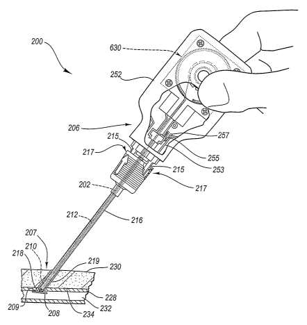
Referring now to Figs. 5A-5G, an apparatus, for example a tissue wall
puncture closure device 200, is shown according to one embodiment of the
present
invention. The closure device 200 is shown in an assembly view in Fig. 5A.
Figs.
5B-5G illustrate the closure device 200 assembled and inserted through a
procedure
sheath 216 and into a lumen 232. The closure device 200 has particular utility
when
13
CA 02617173 2008-01-29
WO 2007/018973 PCT/US2006/027514
used in connection with intravascular procedures, such as angiographic dye
injection,
cardiac catheterization, balloon angioplasty and other types of recanalizing
of
atherosclerotic arteries, etc. as the closure device 200 is designed to cause
immediate
hemostasis of the blood vessel (e.g., arterial) puncture. However, it will be
understood that while the description of the preferred embodiments below are
directed to the sealing off of percutaneous punctures in arteries, such
devices have
much more wide-spread applications and can be used for sealing punctures or
incisions in other types of tissue walls as well. Thus, the sealing of a
percutaneous
puncture in an artery, shown herein, is merely illustrative of one particular
use of the
closure device 200 according to principles of the present invention.
The closure device 200 includes a first or proximal end portion 206 and a
second or distal end portion 207. A carrier tube 202 extends from the proximal
end
portion 206 to the distal end portion 207 and includes an outlet 213 at the
distal end
portion 207. The distal end portion 207 may include a slit 209.
The carrier tube 202 may be made of plastic or other material and is designed
for insertion through the procedure sheath 216 (Fig. 5B). The procedure sheath
216
(Fig. 5B) is designed for insertion through a percutaneous incision 219 (Fig.
5B) in a
tissue layer 230 (Fig. 5B) and into the lumen 232 (Fig. 5B). According to
Figs. 5B-
5G, the lumen 232 comprises an interior portion of a femoral artery 228.
At the distal end portion 207 of the carrier tube 202 there is an anchor 208
and
a sealing plug 210. The anchor 208 of the present embodiment is an elongated,
stiff,
low-profile member arranged to be seated inside the artery 228 (Fig. 5B)
against an
artery wall 234 (Fig. 5B) contiguous with a puncture 218 (Fig. 5B). The anchor
208
14
CA 02617173 2008-01-29
WO 2007/018973 PCT/US2006/027514
is preferably made of a biologically resorbable polymer. The sealing plug 210
(Fig.
5B) is formed of a compressible sponge, foam, or fibrous mat made of a non-
hemostatic biologically resorbable material such as collagen, and may be
configured
in any shape so as to facilitate sealing the tissue puncture 218 (Fig. 5B).
The sealing plug 210 and anchor 208 are connected to one another by a
connector such as a filament or suture 204 that is also biologically
resorbable. The
anchor 208, the sealing plug 210, and the suture 204 are collectively referred
to as
the "closure elements" below. As shown in Fig. 5A, the anchor 208 is initially
arranged adjacent to and exterior of the distal end portion 207 of the carrier
tube 202,
while the sealing plug 210 (Fig. 5B) is initially disposed within the carrier
tube 202.
The anchor 208 is shown nested in its low profile configuration along the
carrier tube
202 to facilitate insertion into the lumen 232 (Fig. 5B) in Fig. 5A, and
deployed with
a first surface 236 abutting the artery wall 234 in Figs. 5B-5G. The suture
204
extends distally from the first end portion 206 of the closure device 200
through the
carrier tube 202. The suture 204 may be threaded through one or more
perforations
in the sealing plug 210, through a hole in the anchor 208, and proximally back
toward the carrier tube 202 to the sealing plug 210. The suture 204 is
preferably
threaded again through a perforation or series of perforations in the sealing
plug 210.
The suture 204 may also be threaded around itself to form a self-tightening
slip-
knot. The suture 204 may thus connect the anchor 208 and the sealing plug 210
in a
pulley-like arrangement to cinch the anchor 208 and the sealing plug 210
together
when the carrier tube 202 is pulled away from the anchor 208 and the sealing
plug
CA 02617173 2008-01-29
WO 2007/018973 PCT/US2006/027514
210. The anchor 208 and the sealing plug 210 sandwich and lock the anchor and
plug together, sealing the tissue puncture 218.
The carrier tube 202 may house a tamping device, such as a tamping tube 212,
for advancing the sealing plug 210 along the suture 204 and toward the anchor
208.
The tamping tube 212 is shown located partially within the carrier tube 202
and
proximal of the sealing plug 210. The tamping tube 212, however, also extends
through a handle or housing 252 of the closure device 200. The tamping tube
212 is
preferably an elongated tubular or semi-tubular member that may be rigid or
flexible
and formed of any suitable material. For example, according to one embodiment,
the
tamping tube 212 is made of polyurethane. The suture 204 extends through at
least a
portion of the tamping tube 212. For example, as shown in Figs. 5A-5G, the
suture
204 extends along the tamping tube 212 between the first and second end
portions
206, 207. However, the suture 204 is not directly connected to the tamping
tube 212.
Accordingly, the suture 204 and the tamping tube 212 may slide past one
another.
According to the embodiment of Figs. 5A-5G, the suture 204 attaches to an
automatic tamping assembly. The automatic tanlping assembly may include an
automatic driving mechanism 630 or other transducer and the tamping tube 212.
The
automatic driving mechanism 630 is located within the housing or handle 252 at
the
first end portion 206 of the closure device 200. Embodiments of the automatic
driving mechanism 630 are described in detail below with reference to Figs. 6 -
8
and may be selectively disengagable.
In practice, the carrier tube 202 of the closure device 200 (containing the
closure elements described above) is inserted into the insertion sheath 216,
which is
16
CA 02617173 2008-01-29
WO 2007/018973 PCT/US2006/027514
already inserted within the artery 228 (Figs. 5B-5C). As the closure device
200 and
the associated closure elements are inserted into the procedure sheath 216,
the anchor
208 passes through and out of the distal end of the procedure sheath 216 and
is
inserted into the artery lumen 232. As mentioned above and shown in Fig. 5A,
the
anchor 208 is initially arranged substantially flush with the carrier tube 202
to
facilitate insertion of the anchor 208 through the percutaneous incision 219
and into
the lumen 232.
After the anchor 208 passes out of the distal end of the procedure sheath 216,
however, it tends to deploy or rotate to the position shown in Figs. 5B-5C.
The
closure device 200 may also be partially withdrawn from the insertion sheath
216,
catching the anchor 208 on the distal end of the insertion sheath 216 and
rotating it
to the position shown in Figs. 5B-5C. However, the closure device 200
preferably
includes a pair of biased fingers 215 that are lockingly received by a
matching pair of
recesses 217 in the procedure sheath 216. The locking arrangement between the
biased fingers 215 and matching recesses 217 may fix the position of the
handle 252
relative to the procedure sheath 216.
Following deployment of the anchor 208, the handle 252 and the insertion
sheath 216 are withdrawn together. Withdrawing the handle 252 causes the
anchor
208 to anchor itself within the artery 228 against the artery wall 234. With
the
anchor 208 anchored within the artery 228 at the puncture site 218, further
retraction
of the handle 252 and insertion sheath 216 tends to pull the sealing plug 210
out from
the distal end portion 207 of the carrier tube 202, thereby depositing the
plug 210
within the incision or puncture tract 219. The slit 209 (Fig. 5A) in the
carrier tube
17
CA 02617173 2008-01-29
WO 2007/018973 PCT/US2006/027514
202 allows the distal end portion 207 of the carrier tube to flex or open,
facilitating
ejection of the sealing plug 210.
Referring to Figs. 5D-5E, the distal end portion 207 of the carrier tube 202
is
exposed (within the incision tract 219) as the handle 252 and the procedure
sheath
216 are retracted. The carrier tube 202 may retain its position relative to
the
puncture 218 until the handle 252 and the procedure sheath 216 have been
retracted a
predeterinined distance. Relative movement between the handle 252/procedure
sheath 216 and the carrier tube 202 may facilitated by a sliding mount
arrangement
between the automatic driving mechanisin 630 and the handle 252. However,
according to some embodiments the automatic driving mechanism 630 is fixed to
the
handle 252.
As shown by the combination of Figs. 5B-5G, the automatic driving
mechanism 630 (which is attached to the carrier tube 202) may be free floating
or
displaceable and slides relative to the handle 252 as the handle 252 and the
procedure
sheath 216 are retracted. However, the automatic driving mechanism 630 may be
initially held in a first position relative to the handle 252 as shown in Fig.
5B. For
example, as shown in Fig. 5B, the automatic driving mechanism 630 may comprise
a
temporary holder such as a stowage detent 255 slidingly inounted in a track.
The
track is shown in Fig. 5B as a webbing track 253. The webbing track 253 is
disposed
in the handle 252. The stowage detent 255 may include a finger 257 with a
protrusion to at least temporarily hold the automatic driving mechanism 630 in
the
first position shown in Fig. 5B, and prevent premature sliding within the
handle 252.
18
CA 02617173 2008-01-29
WO 2007/018973 PCT/US2006/027514
Although the finger 257 tends to hold or temporarily lock the automatic
driving mechanism 630 in the first position shown in Fig. 513, the finger 257
releases
when a sufficient predetermined force is applied between the handle 252 and
the
automatic driving mechanism 630. For example, with the anchor 208 deployed, a
retraction force provided by a user to the handle 252 causes the finger 257 to
deflect
inward and release. Thereafter, the finger 257 provides very little resistance
to
sliding movement between the automatic driving mechanism 630 and the handle
252.
Accordingly, retraction of the handle 252 may retract the procedure sheath 216
(which is fixedly connected to the handle 252), but the automatic driving
mechanism
630 and the carrier tube 202 may slide relative to the handle 252 and
therefore
remain in position with respect to the puncture 218 as shown in Fig. 5D. The
automatic driving mechanism 630 may slide a predetermined distance with
respect to
the handle 252 until the automatic driving mechanism 630 reaches a stop. The
predetermined distance may be at least long enough to fully expose the slit
209 (Fig.
5A) in the carrier tube 202.
When the automatic driving mechanism 630 reaches the stop, further
retraction of the handle 252 withdraws the carrier tube 202 as well, ejecting
and
tamping the sealing plug 210 automatically as shown in Figs. 5F-5G. Unlike
previous closure devices that require a separate, manual tamping procedure
following
the deposition of the sealing plug 210, the closure device 200 of the present
invention automatically tamps the sealing plug 210. The sealing plug 210 may
be
tamped while the carrier tube 202 is being withdrawn, reducing or eliminating
any
19
CA 02617173 2008-01-29
WO 2007/018973 PCT/US2006/027514
gaps that may otherwise occur between the sealing plug 210 and the puncture
218 in
the femoral artery 228.
In addition, by placing tension on or pulling the suture 204 away from the
puncture tract 219, the suture 204 may cinch and lock (with a slip knot or the
like)
together the anchor 208 and the sealing plug 210, sandwiching the artery wall
234
between the anchor 208 and sealing plug 210. The force exerted by the tamping
tube
212 and the cinching together of the anchor 208 and sealing plug 210 by the
filament
204 also causes the sealing plug 210 to deform radially outward within the
puncture
tract 219 and function as an anchor on the proximal side of the tissue
puncture site
218 as shown in Figs. 5F-5G.
The tamping tube 212 is automatically driven toward the sealing plug 210 by
the automatic driving mechanism 630. One embodiment of the automatic driving
mechanism 630 is shown in detail in Figs. 5A and 6. The automatic driving
mechanism 630 may comprise a coil assembly 629 and may be selectably
disengageable. According to the embodiment of Figs. 5A and 6, once the
automatic
driving assembly 630 contacts the stop, further retraction of the closure
device 200
automatically effects tamping of the sealing plug 210 (see Figs. 5F-5G).
According to Figs. 5A and 6, the coil assembly 629 comprises a coil 633
having a first end 635. The coil 633 is operatively connected to the sealing
plug 210
to automatically tamp the sealing plug 210 toward the anchor 208. The coil 633
may
abut the tamping tube 212, or the coil 633 may comprise the tamping tube 212.
The
coil 633 may be semi-flexible and is capable of taking the shape of a track
and also
providing a compression force to the sealing plug 210.
CA 02617173 2008-01-29
WO 2007/018973 PCT/US2006/027514
The coil assembly 629 may further comprise a block such as plate block 634
that is disposed in the housing 252. The plate block 634 may comprise a
generally
planar first surface 636 that is receptive of a least a portion of the coil
633. The
plate block 634 may thus include a channel such as a curved channel 638 shaped
similarly to the coil 633 and may be recessed sufficiently to entirely receive
the coil
633. The curved channel 638 may, however, exhibit a generally straight portion
639
as it leads out of the block 634. The plate block 634 may also comprise a
protrusion
640 receptive of other components of the automatic driving mechanism 630. For
example, the plate block 634 may be receptive of a driving plate 642.
The driving plate 642 may comprise a disk or circular shape as shown,
although the driving plate 642 may include other shapes as well. The driving
plate
642 may be rotatably attached to the plate block 634 as shown. The driving
plate 642
includes a first generally planar surface 644 (Fig. 5A) and a second generally
planar
surface 646 (Fig. 6). A drive pin 648 extends laterally or normally from the
second
generally planar surface 646. The radial position of the drive pin 648
corresponds to
a radius of curvature of the curved channel 63 8.
According to the embodiment of Figs. 5A and 6, the drive pin 648 is rigidly
fixed to or unitarily formed with the driving plate 642. When the automatic
driving
mechanism 630 is assembled, the drive pin 648 extends into the curved channel
638
adjacent to the first end 635 of the coil 633. Therefore, when the driving
plate 642
rotates, the drive pin 648 engages or contacts the first end 635 of the coil
633 and
provides a driving force to move the coil 633 along and out of the curved
channel
638. The coil 633 is arranged adjacent to (or may even comprise) the tamping
tube
21
CA 02617173 2008-01-29
WO 2007/018973 PCT/US2006/027514
212, and therefore advancing the coil 633 by the drive pin 648 may effect
tamping of
the sealing plug 210.
As shown in Fig. 5A, the driving plate 642 may be connected to a spool 632.
The suture 204 is connected to and partially wound about the spool 632. The
driving
plate 642 tends to rotate at the same angular rate as the spool 632, however,
a clutch
650 may selectively connect and release the driving plate 642 from the spool
632.
One embodiment of the clutch 650 is described in detail below, however, any
clutch
may be used.
Withdrawal of the closure device 200 (Fig. 5F) from the tissue puncture site
218 (if the anchor 208 (Fig. 5F) is deployed and the automatic driving
mechanism
630 has contacted the stop) causes the suture 204 to unwind from the spool
632. The
spool 632 rotates as the suture 204 unwinds and provides a torsional motive
force
that is transduced to a linear tamping force.
The torsional motive force provided by the spool 632 is transduced into the
linear tamping force by the coil assembly 629 according to the embodiment of
Figs.
5A and 6. The coil assembly 629 includes the coil 633 and the driving plate
642
arranged coaxially with the spool 632. When the spool 632 rotates, it drives
the
driving plate 642, which in turn drives the coil 633. The coil drives the
tamping tube
212, which in turn tamps the sealing plug 210.
The tamping tube 212 is preferably tubular or semi-tubular partially disposed
about the suture 204 along its longitudinal axis. If the coil 633 also
comprises the
tamping tube, the coil 633 may comprise a semi-tubular shape having a
generally U-
shaped cross section, to provide a trough through which the suture 204 may
enter and
22
CA 02617173 2008-01-29
WO 2007/018973 PCT/US2006/027514
exit laterally. An open trough would permit the suture and the coil 633 to
merge as
the spool 632 unwinds. Accordingly, with the anchor 208 deployed, as the
closure
device 200 is retracted in a first direction, the suture 204 unwinds from the
spool
632, which drives the driving plate 642. The driving plate 642 drives the coil
633,
and the coil 633 drives the tamping tube 212 in a second, opposite direction.
The
tamping tube tamps the sealing plug 210.
In embodiments including a clutch, the clutch 650 may comprise a plurality of
release fingers 661 as shown in Fig. 5A. The release fingers 661 are arranged
substantially in a circle. A first component 663 of the release fingers 661 is
cantilevered from a base 637 and extends normal therefrom. A protrusion 665 of
the
first component 663 extends radially outward and is received by a mating
internal
recess 667 of the spool 632. A second component 669 of the release fingers 661
arcs
substantially normal to the first component 663 and the base 637. The second
component 669 of each of the release fingers 661 extends through a central
hole 671
of the spool 632. An actuator button 651 fits over and contacts the second
components 669 of each of the release fingers 661.
The fit of the protrusions 665 of the base 637 with the mating recesses 667 of
the spool 632 causes the base 637 (and thus the driving plate 642 to which the
base
637 is fixedly attached) and spool 632 to rotate together at an identical
angular
velocity. However, when the actuator button 651 is depressed, the actuator
button
slides along the arcs of the second component 669, forcing each of the release
fingers
661 radially inward. The radial inward displacement of the release fingers 661
at
least partially removes the protrusions 665 from the mating recesses 667,
allowing
23
CA 02617173 2008-01-29
WO 2007/018973 PCT/US2006/027514
independent rotation of the spool 632 with respect to the driving plate 642.
Therefore, after the sealing plug 210 is driven toward the anchor 208, the
selectably
disengagable automatic driving mechanism 630 is disengaged or disabled,
allowing
the suture 204 to safely unwind without further tamping. The suture 204 is
then
exposed to the operator for convenient cutting.
As shown in Figs. 5A and 6, the block 634 may also be receptive of a closing
mold 670. The closing mold 670 matingly fits over the block 634 and provides a
suture and coil path 672 therethrough leading to the carrier tube 202.
Another embodiment of the automatic driving mechanism 630 is illustrated in
Fig. 7. The automatic driving mechanism 630 of Fig. 7 may replace the
mechanism
630 of Fig. 5A. The automatic driving mechanism 630 of Fig. 7 is similar to
the
embodiment of Fig. 5A, however, the coil assembly 629 of Fig. 7 comprises a
spiral
shaped coil 733. The spiral shaped coil 733 is operatively connected to the
sealing
plug 210 (Fig. 5A) to automatically tamp the sealing plug 210 (Fig. 5A) toward
the
anchor 208 (Fig. 5).
In addition, as shown in Fig. 7, block 734 may comprise a spiral channel 738
shaped like the spiral shaped coil 733. The spiral channel 738 may, however,
exhibit
a generally straight portion 739 as it leads out of the block 734. Driving
plate 742
may be rotatably attached to the block 734. The driving plate 742 comprises,
however, a radial slot 754 in which a sliding drive pin 748 rides. The sliding
drive
pin 748 extends normally from the driving plate 742 and into the spiral
channel 738
at a first end 635 of the spiral coil 733. The sliding drive pin 748 is
radially free
floating in the slot 754, and it is angularly stable. Therefore, as the
driving plate 742
24
CA 02617173 2008-01-29
WO 2007/018973 PCT/US2006/027514
rotates, the sliding drive pin 748 advances the spiral coil 733 along the
spiral channel
738, and the sliding drive pin 748 slides radially to remain in the spiral
channel 738
while continuing to advance the spiral coil 733. The spiral coil 733 effects
tamping
of the sealing plug 210 (Fig. 5A). The remaining components of the automatic
driving mechanism 630 may be similar or identical to the embodiment of Fig.
5A.
Another embodiment of an automatic driving mechanism 630 is illustrated in
Fig. 8. The automatic driving mechanism 630 of Fig. 8 may replace the
mechanism
630 of Fig. 5A. The automatic driving mechanism 630 of Fig. 8 is similar to
the
embodiment of Fig. 7, however, the coil assembly 629 of Fig. 8 comprises a
disk
such as a compliant plate 870. The compliant plate 870 is fixed to the driving
plate
742, but may also be clutched thereto with a spiral connection 880. The
compliant
plate 870 comprises an open slit 872 of variable width and a cantilevered
finger 850
coplanar with the compliant plate 870 and extending along the open slit 872.
The
cantilevered finger 850 is radially flexible within the open slit 872 and
includes a
lateral drive pin 848 at a distal end thereof. The drive pin 848 is thus free
to move
radially within the open slit 872, and it is angularly stable. Therefore, as
the driving
plate 742 rotates, it drives the compliant plate 870, and the compliant plate
870
comprising the drive pin 848 advances the spiral coil 733 along the spiral
channel
738. The drive pin 848 moves radially as the compliant plate 870 rotates to
remain
in the spiral channel 738 while continuing to advance the spiral coil 733, and
the
spiral coil 733 effects tamping of the sealing plug 210 (Fig. 5A). According
to some
embodiments, the compliant plate 870 also comprises a driving plate and the
driving
CA 02617173 2008-01-29
WO 2007/018973 PCT/US2006/027514
plate 742 may be omitted. The remaining components of the automatic driving
mechanism 630 may be similar or identical to the embodiment of Fig. 5A.
It will be understood by those of skill in the art having the benefit of this
disclosure that the coil assembly 629 configurations shown in Figs. 5A, 6, 7,
and 8
are exemplary in nature, and not limiting. Any configuration may be used to
advance
a coil within a channel to provide an automatic driving force to the sealing
plug 210
(Fig. 5F).
Operation of the embodiment of Figs. 5A-8 is as follows. As the handle 252
of the closing device 200 is retracted from the puncture tract 219 as shown in
Fig.
5B, the detent 255 releases. The automatic tamping mechanism 630 and carrier
tube
202 may remain stationary and therefore float relative to the handle 252. The
procedure sheath 216 is retracted as the handle 252 is withdrawn, exposing the
distal
end 207 of the carrier tube 202. The automatic tamping mechanism 630
eventually
contacts a stop (or, in some embodiments, the automatic tamping mechanism is
fixed), and further retraction causes the automatic tamping mechanism 630 and
carrier tube 202 to retract as well. As the automatic tamping mechanism 630
retracts, the suture 204, which is threaded through the anchor 208, unwinds
from and
causes rotation of the spool 632. The spool 632 drives the driving plate
642/742 or
the compliant plate 870 as it rotates via a coaxial connection between. As the
driving plate 642/742 and/or the compliant plate 870 rotate, the coil 633/733
is
advanced along the channel 638/738. The coil 633/733 drives the tamping tube
212,
or the coil 633/733 may be long enough to operate as a tamping tube itself.
The
tamping tube 212 tamps the sealing plug 210. According to spiral or non-
circular
26
CA 02617173 2008-01-29
WO 2007/018973 PCT/US2006/027514
coil designs, the drive pin 748 of the driving plate 742 or the drive pin 848
of
compliant plate 870 may migrate radially to remain in the spiral channel 738
and
advance the coil 733.
Therefore, as the closing device 200 is retracted from the puncture tract 219,
the procedure sheath 216 may be retracted (Figs. 5D-5E), and the sealing plug
210 is
automatically tamped (Figs. 5F-5G). The sealing plug 210 is more likely to
create a
sufficient arterial seal without a gap relative to the anchor 208, as may
otherwise
occur with a separate manual tamping procedure.
Moreover, when the sealing plug 210 has been sufficiently tamped, the
automatic driving mechanism 630 may be disengaged, enabling further retraction
of
the closure device 200 without additional tamping. With the sealing plug 210
fitlly
tamped, there may be little or no portion of the suture 204 extending outside
of the
tissue layer 230 and exposed to an operator. Therefore, it may be difficult
for an
operator to separate the sealing plug 210 and anchor 208 from the remainder of
the
closure device 200. In addition, too much retraction with the selectably
automatic
driving mechanism 630 enabled could potentially overtamp the sealing plug 210
into
the artery 228. Accordingly, the automatic driving mechanism 630 may be
advantageously disabled by activating the actuator 651 through the access hole
253.
Activating the actuator 651 allows the suture 204 to fully unwind from the
spool 632
without driving the tamping tube 212. Unwinding the spool 632 exposes a
sufficient
length of the suture 204 to allow an operator to easily cut it and separate
the sealing
plug 210 and anchor 208 from the remainder of the closure device 200.
27
CA 02617173 2008-01-29
WO 2007/018973 PCT/US2006/027514
The preceding description has been presented only to illustrate and describe
exemplary embodiments of invention. It is not intended to be exhaustive or to
limit
the invention to any precise form disclosed. Many modifications and variations
are
possible in light of the above teaching. It is intended that the scope of the
invention
be defined by the following claims.
28