Note: Descriptions are shown in the official language in which they were submitted.
CA 02617317 2008-01-30
WO 2007/019274 PCT/US2006/030369
SYSTEMS FOR INTRODUCING IMPLANTS
CROSS-REFERENCE TO RELATED APPLICATION
This application is related to copending U.S provisional patent applications
entitled
"Introducer Needle with Extendable Implant Lasso", filed on December 28, 2005,
and
accorded serial number 60/754,265, and "Introducer Needle With Implant
Retrieval Snare",
filed on April 19, 2006, and accorded serial number 60/745,140, both of which
are entirely
incorporated herein by reference.
BACKGROUND
Surgical devices referred to as "introducers" are often used to implant or
"introduce"
implantable devices within the body. For example, such introducers can be used
to position
within the pelvis mesh implants intended for treating urinary incontinence or
performing
prolapse repair.
Positioning an implant within the human body, such as within the pelvis, can
be
challenging due to the anatomy of the body and the placement of the implant
that may be
required to treat a given ailment. For instance, the treatment of rectocele, a
condition in
which the rectum encroaches on the vagina, may require accessing the vaginal
vault from a
position deep within the pelvis so as to form a passage in which a portion,
such as an
anchoring arm, of the implant can be placed. Formation of such a passage
typically requires a
relatively high degree of skill.
Further complicating implantation of a rectocele implant, or other such pelvic
implant,
is the need to draw the implant into the body and through the formed passage.
In present
CA 02617317 2008-01-30
WO 2007/019274 PCT/US2006/030369
te '""" t~~f:'s.: h a pelvic incision, through the soft tissue of the pelvis,
~~q~f
' ~ into the vagina, down through the vagina, and out the vaginal introitus to
enable the implant
to be connected to the needle so that the needle may then be withdrawn with
the implant in
tow to position the implant within the formed passage. Given the configuration
and
dimensions of the human pelvis and its organs, it can be difficult to navigate
a needle through
such a tortuous path without causing damage to or otherwise disrupting the
tissues of the
pelvis, such as the pelvic floor muscles.
BRIEF DESCRIPTION OF THE DRAWINGS
The disclosed systems can be better understood with reference to the following
drawings. The components in the drawings are not necessarily to scale.
FIG. 1 illustrates a first embodiment of an introducer system.
FIG. 2 is a perspective view of an introducer shown in FIG. 1.
FIG. 3 illustrates passage of a snare shown in FIG. 1 through the introducer
shown in
FIG. 2.
FIG. 4 illustrates a second embodiment of an introducer system.
FIG. 5 is a perspective view of an introducer shown in FIG. 4.
FIG. 6 illustrates passage of a snare shown in FIG. 1 through the introducer
shown in
FIG. 5.
FIGs. 7A-7K illustrate steps performed in a first embodiment of a method for
implanting
a pelvic implant within the body.
FIGs. 8A and 8B illustrate steps performed in a second embodiment of a method
for
implanting a pelvic implant within the body.
FIG. 9 is a side view of an alternative embodiment of a snare that can be used
in an
introducer system.
2
CA 02617317 2008-01-30
WO 2007/019274 PCT/US2006/030369
of the snare of FIG. 9, illustrating an implant coupling
element of the introducer.
FIG. 11 is a perspective view of an alternative embodiment of an introducer
that can
be used in an introducer system.
FIG. 12 illustrates securing of a snare to the introducer shown in FIG. 11
using a cleat of
the snare.
DETAILED DESCRIPTION
As described above, it can be difficult to position an implant within the
body. That
may particularly be the case in relation to positioning a pelvic implant
intended for use in
treating incontinence or performing prolapse repair. For example, as described
above, a
surgeon may need to access a point deep within the pelvis, such as the vaginal
vault, with an
introducer and connect an implant to the introducer at a point outside of the
body to enable
the implant to be drawn through a passage formed in the soft tissues of the
pelvis by the
introducer. It is difficult to perform such a procedure with current
introducers given that the
introducer must traverse a tortuous path to extend outside of the body from a
point deep
within the pelvis.
Disclosed herein are introducer systems that simplify implantation of an
implantable
device, such as a pelvic implant. In some embodiments, the introducer system
comprises a
snare that can be extended from a tip of an introducer needle to a position
outside of the body
when the tip is positioned at a point within the body, such as within vagina.
In such a case, an
implant can be coupled to the extended snare and the snare can then be
retracted to pull the
implant through the body and at least to the tip of the introducer needle. In
some
embodiments, both the snare and the implant can further be drawn through the
introducer
needle such that the implant traverses the passage formed by the introducer
needle without
3
CA 02617317 2008-01-30
WO 2007/019274 PCT/US2006/030369
digsqt;016ipassage, thereby reducing irritation to the soft tissues in
which the passage is formed.
In the following, various embodiments of systems and methods are described in
detail.
Although specific embodiments are presented, those embodiments are mere
exemplary
implementations of the disclosed systems and methods and it is noted that
other embodiments
are possible. All such embodiments are intended to fall within the scope of
this disclosure.
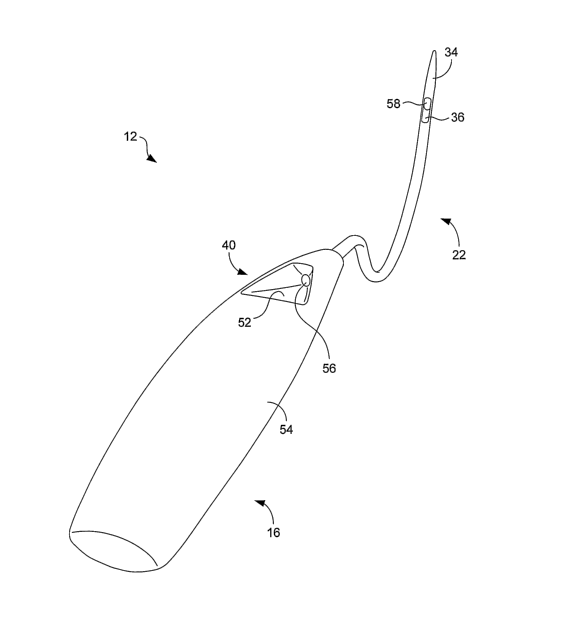
FIG. 1 illustrates a first embodiment of an introducer system 10. The
introducer
system 10 is well suited for use in performing prolapse repair, such as
anterior prolapse repair
and treating cystocele. As indicated in FIG. 1, the system 10 includes an
introducer 12 and a
snare 14. The introducer 12 comprises a handle 16 that includes a proximal end
18 and a
distal end 20. The handle 16 is generally sized and shaped to fit within a
surgeon's hand and,
as depicted in FIG. 1, can be curved to facilitate firm gripping.
A needle 22 extends from the distal end 20 of the handle 16. As shown in FIG.
1, at
least a portion of the needle 22 is curved. In the embodiment of FIG. 1, the
needle 22
comprises a first generally straight portion 24 adjacent its proximal end 26,
a curved portion
28 in a central region, and a second generally straight portion 30 adjacent
its distal end 32.
Fonned at the distal end 32 is a blunt point or tip 34 that is configured to
dissect soft tissue as
the needle 22 is passed through the body.
The needle 22 is hollow so as to form a cannula through which the snare 14 can
be
passed. More particularly, the needle 22 forms an inner lumen that extends
from a first
opening 36 of the needle to a second opening 38 of the needle. In the
embodiment shown in
FIG. 1, the first opening 36 is positioned adjacent the distal end 32 and the
second opening 38
is positioned adjacent the proximal end 26. The second opening 38 is in open
communication
with a port 40 that is formed in the handle 16. As is described in greater
detail below, the
4
CA 02617317 2008-01-30
WO 2007/019274 PCT/US2006/030369
i;~~~~ t~Gj"~l~ ~ti;lport 40 and the second opening 38 to position the snare
within the needle 22. The configuration of the port 40 is described in
relation to FIG. 2.
In terms of materials, the handle 16 can be constructed of any suitable rigid
material,
such as a metal or a polymeric material. The needle 22 can be constructed of a
biocompatible, strong material, such as stainless steel. In some embodiments,
the handle 16
and needle 22 can be composed of the same material and may even be unitarily
formed
together so as to have a monolithic configuration.
With continued reference to FIG. 1, the snare 14 comprises an elongated shaft
42
having a proximal end 44 and a distal end 46. The shaft 42 is flexible so as
to enable the
shaft to easily adapt to the contours of the needle inner lumen and any body
passages along
which the snare is to travel. In some embodiments, the shaft 42 comprises a
hollow tube
through which a wire passes. In such cases, the shaft 42 can be constructed of
a suitable
flexible biocompatible material, such as a polymeric material. In other
embodiments, the
shaft 42 is solid and can be made of a polymeric material or a metal material,
such as stainless
steel or nitinol.
Provided at the proximal end 44 of the snare 14 is a grip element 48 that, as
described
below, is used to manipulate the snare relative to the introducer 12. Provided
at the distal end
46 of the snare 14 is an implant coupling element 50 that is configured to
couple to and
secure an implant that is to be positioned with the body. In the illustrated
embodiment, the
coupling element 50 is formed as a loop. Such a loop can be formed from a
flexible wire
constructed of a polymeric or metal material. In such a case, the wire can
extend from the
gripping element 48, through the shaft 42, and terminate in a loop. In some
embodiments,
nitinol is suitable for the construction of the coupling element 50 due to
nitinol's shape
memory characteristics. In particular, when nitinol is used, the coupling
element 50 can
easily be compressed to pass through the needle inner lumen, but can readily
spring back to
5
CA 02617317 2008-01-30
WO 2007/019274 PCT/US2006/030369
emerging from the needle 22. In some embodiments,
the shaft 42 and the coupling element 50 comprise a unitarily-formed element,
such as an
elongated wire that extends from the gripping element 48 and terminates in a
loop. In such
cases, the shaft 42 need not comprise a tube.
FIG. 2 is a perspective view of the introducer 12. As indicated in that
figure, the port
40 of the handle 16 is formed by one or more surfaces 52 that extend inwardly
from an outer
surface 54 of the handle to an orifice 56 that is aligned with the second
opening 38 of the
needle 22 (FIG. 1). As is also visible in FIG. 2, the needle 22 includes a
snare-deflecting
surface 58 positioned within the first opening 36 that urges the snare 14
(FIG. 1) out from the
needle when the snare is pushed against the surface.
With the above-described system configuration, the snare 14 can be inserted
through
the port 40 and orifice 56 of the introducer handle 16, moved into the inner
lumen of the
introducer needle 22, pushed through the needle inner lumen, and made to exit
the needle
through the first opening 36. The result of that process is illustrated in
FIG. 3.
FIG. 4 illustrates a second embodiment of an introducer system 100. The
introducer
system 100 is similar to the system 10 described in relation to FIGs. 1-3,
although the system
100 is configured for use in performing posterior prolapse repair and treating
rectocele. As
indicated in FIG. 4, the system 100 includes an introducer 102 and a snare
104. The
introducer 102 comprises a handle 106 that includes a proximal end 108 and a
distal end 110.
The handle 106 is generally sized and shaped to fit within a surgeon's hand
and, as depicted
in FIG. 4, can be curved to facilitate firrn gripping.
A needle 112 extends from the distal end 110 of the handle 102. As shown in
FIG. 4,
at least a substantial portion of the needle 112, like needle 22 (FIG. 1) is
curved. In the
embodiment of FIG. 4, however, the needle 112 is longer and straighter to
enable passage of
the needle deep into the pelvis. The needle 112 comprises a first generally
straight portion
6
CA 02617317 2008-01-30
WO 2007/019274 PCT/US2006/030369
curved portion 118 in a central region, and a second
generally straight portion 120 adjacent its distal end 122. Formed at the
distal end 122 is a
blunt point or tip 124 that is configured to dissect soft tissue as the needle
112 is passed
through the body.
The needle 112 is hollow so as to form a cannula through which the snare 104
can be
passed. More particularly, the needle 112 forms an inner lumen that extends
from a first
opening 126 of the needle to a second opening 128 of the needle. In the
embodiment shown
in FIG. 4, the first opening 126 is positioned adjacent the distal end 122 and
the second
opening 128 is positioned adjacent the proximal end 116. The second opening
128 is in open
communication with a port 130 that is formed in the handle 106. As is
described in greater
detail below, the snare 104 can be passed through the port 130 and the second
opening 128 to
position the snare within the needle 112. The configuration of the port 130 is
described in
relation to FIG. 5.
In terms of materials, the handle 106 can be constructed of any suitable rigid
material,
such as a metal or a polymeric material. The needle 112 can be constructed of
a
biocompatible, strong material, such as stainless steel. In some embodiments,
the handle 106
and needle 112 can be composed of the same material and may even be unitarily
formed
together so as to have a monolithic configuration.
With continued reference to FIG. 4, the snare 104 comprises an elongated shaft
132
having a proximal end 134 and a distal end 136. The shaft 132 is flexible so
as to enable the
shaft to easily adapt to the contours of the needle inner lumen and any body
passages along
which the snare is to travel. In some embodiments, the shaft 132 comprises a
hollow tube
through which a wire passes. In such cases, the shaft 132 can be constructed
of a suitable
flexible biocompatible material, such as a polymeric material. In other
embodiments, the
7
CA 02617317 2008-01-30
WO 2007/019274 PCT/US2006/030369
., , ., .
sh.. ~~ti~:1~3~~t-,is ~= ~ Y.==~h~. ca~h~l;~le:::lr~i~ of a polymeric material
or a metal material, such as
stainless steel or nitinol.
Provided at the proximal end 134 of the snare 104 is a grip element 138 that,
as
described below, is used to manipulate the snare relative to the introducer
102. Provided at
the distal end 136 of the snare 104 is an implant coupling element 140 that is
configured to
couple to and secure an implant that is to be positioned with the body. In the
illustrated
embodiment, the coupling element 140 is formed as a loop. Such a loop can be
formed from
a flexible filament, such as a wire, constructed of a polymeric or metal
material. In such a
case, the wire can extend from the gripping element 138, through the shaft
132, and terminate
in a loop. In some embodiments, nitinol is suitable for the construction of
the coupling
element 140 due to nitinol's shape memory characteristics. In particular, when
nitinol is
used, the coupling element 140 can easily be compressed to pass through the
needle inner
lumen, but can readily spring back to its original shape (e.g., loop shape)
after emerging from
the needle 112. In some embodiments, the shaft 132 and the coupling element
140 comprise
a unitarily-formed element, such as an elongated wire that extends from the
gripping element
138 and terminates in a loop. In such cases, the shaft 132 need not comprise a
tube.
FIG. 5 is a perspective view of the introducer 102. As indicated in that
figure, the port
130 of the handle 106 is formed by one or more surfaces 142 that extend
inwardly from an
outer surface 144 of the handle to an orifice 146 that is aligned with the
second opening 128
of the needle 112 (FIG. 4). As is also visible in FIG. 5, the needle 112
includes a snare-
deflecting surface 148 positioned within the first opening 126 that urges the
snare 14 (FIG. 4)
out from the needle when the snare is pushed against the surface.
With the above-described system configuration, the snare 104 can be inserted
through
the port 130 and orifice 146 of the introducer handle 106, moved into the
inner lumen of the
8
CA 02617317 2008-01-30
WO 2007/019274 PCT/US2006/030369
intmc~ u:,:duc~n=~i ~~õ:~, ~:,i6dd~~.,, 1111111 ;, ;~j~~ ,~ =
~e ;,=p~i~~;gh the needle inner lumen, and made to exit the needle
through the first opening 126. The result of that process is illustrated in
FIG. 6.
FIGs. 7A-7K illustrate a process for implanting an article using a system of
the
disclosure. More particularly, FIGs. 7A-7K illustrate a procedure for
implanting a posterior
prolapse repair implant between the vagina and the rectum using the introducer
system 100
shown in FIG. 4. Although a posterior repair procedure is depicted in FIGs. 7A-
7K and is
described in detail in the following for purposes of describing the manner in
which the
disclosed introducer systems can be used to introduce an implant, it is to be
understood that
the procedure is described for purposes of example only. As stated above,
similar systems
1o may be used to implant other implants in other surgical procedures, such as
anterior prolapse
repair or treatment of urinary incontinence.
Beginning with FIG. 7A, small pararectal incisions 200 are made on either side
of the
anus 202 with a sharp device, suc11 as a scalpel 204. By way of example, the
incisions 200
are made 2-3 centimeters (cm) posterior and lateral to the anus 202. In
addition, a midline
incision is made in the posterior vaginal wall 206 to form an opening 208 that
extends from
the vaginal introitus to the vaginal apex to provide access to the space
between the vagina and
the rectum. The vaginal mucosa may then be dissected away from the rectum
using blunt
and/or sharp dissection.
Turning to FIG. 7B, the tip 124 of the introducer needle 112 is positioned at
one of the
incisions 200 with the introducer 102 oriented so that the handle 106 is
substantially vertical
and the second straight portion 120 of the needle is substantially parallel to
the vagina 210.
Referring next to FIG. 7C, the introducer needle 112 is passed through the
incision 200 and
through the soft tissue of the pelvis toward the ischial spine (not shown). As
the needle 112
passes through the soft tissue, the introducer 102 is rotated so that the
second straight portion
120 approaches a vertical orientation, as indicated in the figure. The needle
tip 124 is
9
CA 02617317 2008-01-30
WO 2007/019274 PCT/US2006/030369
advp~-,cef t 1,~~t s al wall and into the vaginal vault 212 such that the tip
is
~~i~~i~LApi~Q positioned within the vagina. That process can be aided by
placing a finger within the vagina
to guide the needle tip 124 into position.
With reference to FIG. 7D, the snare 104, which can have been positioned
already
within the introducer 102 or later inserted therein, is extended from a
retracted position in
which the implant coupling element 140 is contained within the inner lumen of
introducer
needle 112 to an extended position in which the coupling element extends
beyond the first
opening 126 (FIG. 4) of the needle. The snare 104 is then extended through the
introducer
102, for example using the gripping element 138, until the implant coupling
element 140
passes out from the vaginal introitus 213, as indicated in FIG. 7D.
Referring next to FIG. 7E, a relatively long anchoring arm. 214 of an implant
216 is
coupled to the implant coupling element 140. By way of example, the implant
216 comprises
a flexible mesh implant such that the ann 214 can be simply passed through the
loop of the
coupling element to secure the implant to the snare 104.
Turning to FIG. 7F, the snare 104 is retracted back into the introducer needle
112, for
example using the gripping element 138, such that the implant coupling element
140 is again
contained within the inner lumen of the needle. Due to the coupling between
the implant 216
and the snare 104, a portion of the anchoring arm 214 may also be contained
within the
needle inner lumen. In some embodiments, a stop mechanism (not shown) can be
provided
within the needle inner lumen so as to limit the extent to which the snare 104
can be retracted
into the needle inner lumen. For example, a stop (not shown), such as a
bulbous portion, can
be provided along the snare 104 adjacent the implant coupling element 140 that
will abut a
mating surface within the needle inner lumen, such as a constriction, adjacent
the needle tip
124 so that the implant coupling element can be drawn into the needle inner
lumen, but not
farther through the needle inner lumen. Such a stop mechanism facilitates
simultaneous
CA 02617317 2008-01-30
WO 2007/019274 PCT/US2006/030369
tl~q, needle 112. In other embodiments, the snare 104 need
,1 c~
wO~~ra~v~a~=' c~5 19, 1
,
not be retracted back into the introducer needle 112 at all. In such an
embodiment, the needle
112 and snare 104 can be withdrawn from the patient together with the snare in
the extended
position, if desired.
With reference next to FIG. 7G, at least a portion of the implant arm 214 can
be pulled
through the inner lumen of the introducer needle 112 so as to position the
anchoring arm 214
in the passage that extends between the incision 200 and the vagina 210, which
was formed
by the needle. Notably, because the implant arm 214 is placed into that
position while still
contained within the needle 112, damage to the soft tissues in which the
passage has been
formed is reduced, as is the friction that resists such positioning. As shown
in FIG. 7G, the
snare 104 can be retracted to the point at which the implant coupling element
140 and the
anchoring arm 214 exit the introducer handle 106. At that point, the anchoring
arm 214 has
been properly positioned within the body for subsequent adjustment, if
necessary. As
mentioned above, however, the snare 104 can, alternatively, be retracted to a
limited extent
due to the provision of a stop mechanism, or can not be retracted at all, as
desired by the
surgeon perfonning the procedure.
Assuming the snare 104 is retracted to the point at which it exits the
introducer
handle, the anchoring arm 214 is released from the implant coupling element
140, as
indicated in FIG. 7H. Then, as indicated in FIG. 71, the introducer needle 112
can be
withdrawn from the body through the incision 200, thereby leaving the
anchoring arm 214 in
place within the tissues of the pelvis with a portion of the arm extending out
from the
incision. As mentioned above, the snare 104 can be withdrawn from the body
simultaneous
to withdrawal of the needle 112 in cases in which the snare is not withdrawn
from the needle
inner lumen (e.g., due to provision of a stop mechanism) or in cases in which
the snare is not
retracted back into the needle inner lumen after extension at all. The same
result is
11
CA 02617317 2008-01-30
WO 2007/019274 PCT/US2006/030369
, va e; i- given that the anchoring arm 214 can still be drawn
ac~i41 . 7
through the passage formed by the needle 112 until a portion of the arm
extends from the
incision 200. The primary difference in such cases is that the anchoring arm
214 is in direct
contact with the soft tissue of the passage as it passes through the passage
instead of
travelling through the needle inner lumen.
At this point, a similar procedure can be followed for positioning the
opposite arm of
the implant 216 using the other pararectal incision 200. That is, the
introducer needle 112 can
be passed through the incision 200 to the vaginal vault 212 on the opposite
side of the vagina
210 and the opposite implant arm can be positioned in the passage formed by
the needle. In
addition, the relatively short arms of the implant can be positioned in other
passages
extending from the incisions on opposite sides of the vagina 210 to a position
adjacent the
vaginal introitus 213. Once that has been completed, a portion of a relatively
short ann 218
and a portion of a relatively long arm 220 extends out from each pararectal
incision 200, as
indicated in FIG. 7J, and a central body 222 (FIG. 7K) of the implant 216 can
be positioned
between the vagina 210 and the rectum 224 to provide a support structure that
prevents
encroachment of the rectum into the vaginal space. Finally, the implant arms
218, 220 can be
appropriately tensioned, for example by pulling excess length out from the
incisions 200, and
the portions of the arms that extend outside of the body trimmed. The final
result of the
implantation is illustrated in FIG. 7K, with the implant body 222 positioned
between the
vagina 210 and the rectum 224.
As described above, other implantation procedures can be performed using
similar
introducer systems. For example, anterior prolapse repair can be performed. To
perform
such a procedure, similar steps to those described above are completed. The
primary
differences include the shape of the implant, the location of the incisions
made in the pelvis,
and the positioning of the implant within the pelvis. As shown in FIG. 8A
superior and
12
CA 02617317 2008-01-30
WO 2007/019274 PCT/US2006/030369
ine~or~~inGilip~~ i~ipõ~l made in the paravaginal region 304 in alignment with
~
the obturator foramina 306 of the pubic bone. Again, those incisions 300 and
302 can be
made with a sharp device, such as a scalpe1308. In addition, a midline
incision 310 can be
made in the anterior vaginal wall 312 to provide access to the space between
the vagina and
the urethra. Each of four arms can be positioned within passages that extend
from the
incisions 300 and 302 to the vagina to position a body of the implant between
the vagina and
the urethra. As shown in FIG. 8B, portions 314 of the arms extend from the
incisions 300
and 302 can then be trimmed as described above in relation to the posterior
prolapse repair
procedure.
As is also described above, the introducer systems can be used to treat
urinary
incontinence. In such a procedure, similar steps are performed except that the
implant can
comprise a urethral sling that is positioned below the urethra to provide
support to the urethra.
The ends of the sling can, for example, be passed through and/or embedded in
the obturator
foramina, or can be otherwise secured to hard or soft tissue of the pelvis.
FIGs. 9 and 10 illustrate an alternative embodiment of a snare 400. Referring
first to
FIG. 9, the snare 400 can be formed as a wire constructed of a suitable metal
material, such as
stainless steel or nitinol. The snare 400 is pre-shaped to have a bend 402
that facilitates
manipulation of the snare when positioned within the vagina or other body
passage in which
it is used. In particular, the bend 402 provides steering capability to the
snare 400 so that the
implant coupling element 404 of the snare can be moved in a desired direction,
for exaniple
by twisting the snare using a grip element of the snare (not shown). As
indicated in FIG. 9,
the implant coupling element 404 comprises a further bend 406 that reduces the
likelihood of
snagging of the snare 400 within the vagina once the snare has been extended
from its
introducer needle.
13
CA 02617317 2008-01-30
WO 2007/019274 PCT/US2006/030369
~i;;~h, i~~~~~t coupling element 404 comprises a loop 408 and a
.
constriction 410 that is, for example, positioned at a distal end of the loop.
With such a
configuration, an implant can be securely held by the implant coupling element
404 by first
passing a portion of the implant through the loop 408 and then passing the
implant portion
into the constriction 410, such that the implant is securely clamped by the
constriction. As is
apparent from FIG. 10, the implant coupling element 404 can be formed from a
wire that
extends from a shaft 412 and forms the loop 408 and the constriction 410. In
an alternative
arrangement, the snare 400 can only comprise one or more wires that form the
loop 408 and
constriction 410, as well as the shaft.
FIG. 11 illustrates a further embodiment of an introducer 500. As indicated in
that
figure, the introducer 500 comprises a handle 502 and a needle 504. As with
the previously-
described embodiments, the handle includes a port 506 that defines an orifice
508, which
leads to an inner lumen of the needle 504. The needle 504 includes an opening
510 in
communication with the inner lumen that enables a snare to be extended from
the needle.
Unlike the previously-described embodiments, however, the introducer 500
includes a cleat
512 comprising opposing inner surfaces 514 that are adapted to secure a snare
relative to the
introducer such that snare is positioned in a desired position along the inner
lumen of the
needle 504 when so secured.
Turning to FIG. 12, securing of a snare 516 with the introducer 500 is
depicted. As
shown in that figure, the snare 516 is pushed into the cleat 512 such that the
snare is securely
clamped by the opposing inner surfaces 514 of the cleat. In some embodiments,
the snare 516
can comprise indicia (not shown) that indicate what portion of the snare is to
be secured
within the cleat 512 such that indexing is provided as to important positions
of the snare
within the introducer 500. For example, indicia can be provided on the snare
516 at a
position that, when aligned with the cleat, correspond to a position at which
an implant
14
CA 02617317 2008-01-30
WO 2007/019274 PCT/US2006/030369
coMIinO"e16A, iffi.549:itof ffidl;~~&Sis";~&sitioned just within the opening
510 of the needle 504.
In other embodiments, the snare 516 can comprise a complementary feature (not
shown), such
as mating indentations or protuberances, that are specifically adapted to
interface with the
cleat 512.