Note: Descriptions are shown in the official language in which they were submitted.
CA 02617904 2008-02-04
WO 2007/019152 - 1 -
PCT/US2006/030022
BIOPSY DEVICE WITH REPLACEABLE PROBE AND
INCORPORATING VIBRATION INSERTION ASSIST AND STATIC
VACUUM SOURCE SAMPLE STACKING RETRIEVAL
FIELD OF THE INVENTION
[own] The present invention relates in general to biopsy devices, and more
particularly
to biopsy devices having a cutter for severing tissue, and even more
particularly to biopsy
devices for multiple sampling with a probe remaining inserted.
BACKGROUND OF THE INVENTION
[0002] When a suspicious tissue mass is discovered in a patient's breast
through
examination, ultrasound, MRI, X-ray imaging or the like, it is often necessary
to perform
a biopsy procedure to remove one or more samples of that tissue in order to
determine
whether the mass contains cancerous cells. A biopsy may be performed using an
open or
percutaneous method.
[0003] An open biopsy is performed by making a large incision in the breast
and
removing either the entire mass, called an excisional biopsy, or a substantial
portion of it,
known as an incisional biopsy. An open biopsy is a surgical procedure that is
usually
done as an outpatient procedure in a hospital or a surgical center, involving
both high cost
and a high level of trauma to the patient. Open biopsy carries a relatively
higher risk of
infection and bleeding than does percutaneous biopsy, and the disfigurement
that
sometimes results from an open biopsy may make it difficult to read future
mammograms. Further, the aesthetic considerations of the patient make open
biopsy even
less appealing due to the risk of disfigurement. Given that a high percentage
of biopsies
show that the suspicious tissue mass is not cancerous, the downsides of the
open biopsy
procedure render this method inappropriate in many cases.
[0004] Percutaneous biopsy, to the contrary, is much less invasive than
open biopsy.
Percutaneous biopsy may be performed using fine needle aspiration (FNA) or
core needle
biopsy. In FNA, a very thin needle is used to withdraw fluid and cells from
the suspicious
CA 02617904 2013-08-27
- 2 -
ti-Ssiie mass. This method has an advantage in that it is very low-pain, so
low-pain that
local anesthetic is not always used because the application of it may be more
painful than
the FNA itself. However, a shortcoming of FNA is that only a small number of
cells are
obtained through the procedure, rendering it relatively less useful in
analyzing the
suspicious tissue and making an assessment of the progression of the cancer
less simple if
the sample is found to be malignant.
100051 During a core
needle biopsy, a small tissue sample is removed allowing for a
pathological assessment of the tissue, including an assessment of the
progression of any
cancerous cells that are found. The following patent documents disclose
various core
biopsy devices: 6,273,862
issued Aug. 14, 2001; US 6,231,522 issued May 15, 2001; US 6,228,055 issued
May 8,
2001; US 6,120,462 issued September 19, 2000; US 6,086,544 issued July
11,2000; US
6,077,230 issued June 20, 2000; US 6,017,316 issued Jan. 25, 2000; US
6,007,497 issued
Dec. 28, 1999; US 5,980,469 issued Nov. 9, 1999; US 5,964,716 issued Oct. 12,
1999;
US 5,928,164 issued July 27, 1999; US 5,775,333 issued July 7, 1998; US
5,769,086
issued June 23, 1998; US 5,649,547 issued July 22, 1997; US 5,526,822 issued
June 18,
1996; and US Patent Application 2003/0199753 published Oct. 23, 2003 to Hibner
et al.
[00061 At present, a
biopsy instrument marketed under the tradename MAIVEVIOTOME is
commercially available from ETHICON ENDO-SURGERY, INC. for use in obtaining
breast biopsy samples. These devices generally retrieve multiple core biopsy
samples
from one insertion into breast tissue with vacuum assistance. In particular, a
cutter tube is
extended into a probe to cut tissue prolapsed into a side aperture under
vacuum assistance
and then the cutter tube is fully retracted between cuts to extract the
sample.
[00071 With a long
probe, the rate of sample taking is limited not only by the time
required to rotate or reposition the probe but also by the time needed to
translate the
cutter. As an alternative to this "long stroke" biopsy device, a "short
stroke" biopsy
device is described in the following commonly assigned patent applications: US
Patent
Application 10/676,944, "Biopsy Instrument with Internal Specimen Collection
Mechanism" filed September 30, 2003 in the name of Hibner et al.; and US
Patent
Application 10/732,843, "Biopsy Device with Sample Tube" filed December 10,
2003 in
the name of Cicenas et al. The cutter is cycled across the side aperture,
reducing the
sample time. Several alternative specimen collection mechanisms are described
that draw
CA 02617904 2008-02-04
WO 2007/019152 - 3 -
PCT/US2006/030022
tlifoUilinifie cutter tube, all of which allow for taking multiple samples
without
removing the probe from the breast.
[0008] Even given the many advantages of such multiple sample taking core
biopsy
devices, in certain applications some surgeons continue to use less expensive
biopsy
devices guided in real time by an ultrasonic system. These simple biopsy
systems omit a
full function control console that operates the cutter and vacuum assistance.
Instead, a
manually controlled hand piece advances a cutter by either stored spring
force, a constant
pneumatic pressure source, or motor power. Then the surgeon activates a cutter
motor to
effect the tissue sample. Thus, the surgeon is challenged to maintain the
biopsy probe at a
desired surgical site while manipulating the patient's breast.
[0009] Consequently, it would be desirable to provide for a core biopsy
device with a
motorized cutter that provides increased functionality such as one-handed
operation with
assisted multiple sample retrieval with only one insertion of the probe, yet
be able to
retain the economical aspects of simple core biopsy devices that lack
elaborate remote
control systems.
[0010] Spring-fired core needle biopsy devices rely upon a firing mechanism
that thrusts
forward a needle and a cutter to penetrate the tissue and to obtain a tissue
sample rather
than palpitating tissue to prolapse into a side aperture of a probe.
Frequently, a surgeon
may encounter an area of dense tissue that is more difficult to penetrate than
the
surrounding tissue during core needle biopsy. In particular, the lesion or
tissue mass being
targeted in the biopsy procedure may be difficult to penetrate, requiring the
physician to
push the biopsy needle with considerable force and/or speed in an attempt to
penetrate the
lesion and collect a sample.
[0011] When encountering such an area of dense tissue, it is common for
surgeons using
the type of firing core needle biopsy device described above to fire the
device in order to
penetrate the lesion and obtain a sample. However, due to the length of the
firing stroke
of such devices, which may be as long as 0.75 inches, it is nearly impossible
for the
surgeon to control the travel of the needle after firing. Consequently, the
long needle
stroke may cause uncertainty as to the needle tip location post fire. This may
cause the
surgeon to obtain a sample from the wrong area. In addition to missing the
targeted tissue,
long firing strokes may cause the needle to puncture the chest wall or pierce
the skin,
particularly when the targeted area is near the patient's chest wall. Even if
the skin is not
CA 02617904 2013-08-27
-4-
riofced, the long travel of the needle, along with the likelihood that the
needle will be
pushed off course by the force of the firing stroke, may lead to needlessly
increased
trauma for the patient. These spring-fired biopsy devices also yield a single
sample per
insertion, thus limiting the amount of diagnostic and therapeutic treatment
that may be
achieved without the increased discomfort and tissue trauma from repeated
insertions.
Based on surgeons' use of the long firing stroke feature of current devices to
aid in
penetrating tissue lesions, it is clear that the medical community sees the
benefit of firing
assistance when inserting a probe to the desired location.
(0012) In commonly-owned and co-pending US Pat. Application No.
11/035,873,
BIOPSY INSTRUMENT WITH IMPROVED NEEDLE PENETRATION to Beckman,
et al., filed on January 10, 2005, manual mechanisms are disclosed that impart
small
reciprocating motions to the probe of a core biopsy device to render
assistance in
penetrating tissue, yet cutting is performed after the probe is properly
positioned, thus
avoiding taking samples from the wrong location. While there are advantages to
having
such cutting assistance imparted by manual actuation, it is generally
desirable to alleviate
the need for the surgeon to perform this additional action while having to
manually
position the biopsy device.
[0013] Additionally, it would be desirable to provide for a hand-held
core biopsy device
that automatically imparts a motion to the probe that assists in penetrating
dense tissue yet
does not take a sample.
SUMMARY OF THE INVENTION
In one embodiment, there is provided a biopsy device, comprising: (a) a body;
(b)
a needle extending distally from the body, wherein the needle includes: (i) a
closed tip,
(ii) a transverse tissue receiving aperture proximal to the tip, and (iii) a
first lumen in
communication with the transverse tissue receiving aperture; (c) a cutter
disposed in the
first lumen, wherein the cutter is movable within the first lumen to sever
tissue protruding
through the transverse tissue receiving aperture, wherein the needle further
includes a
second lumen adjacent to the cutter; and (d) a valve assembly in fluid
communication
with the second lumen, the valve assembly comprising: (i) a translating
member, and (ii)
a valve body, wherein the translating member is movable relative to the valve
body to
DOCSTOR 2792168 k1
CA 02617904 2013-08-27
-4a-
change a pneumatic state of the second lumen based at least in part on the
position of the
cutter in the first lumen.
In another embodiment, there is provided a biopsy device, comprising:(a) a
body;
(b) a needle extending distally from the body, wherein the needle includes:
(i) a closed
tip, and (ii) a transverse tissue receiving aperture proximal to the tip; (c)
a cutter, wherein
the cutter is movable relative to the needle to sever tissue protruding
through the
transverse tissue receiving aperture; and (d) a valve assembly in fluid
communication
with the needle, the valve assembly comprising: (i) a translating member, and
(ii) a valve
body, wherein the translating member is movable relative to the valve body to
change a
pneumatic state of the needle based at least in part on the position of the
cutter relative to
the needle.
In another embodiment, there is provided a biopsy device, comprising:(a) a
body;
(b) a needle extending distally from the body, wherein the needle includes:
(i) a closed
tip, (ii) a transverse tissue receiving aperture proximal to the tip, and
(iii) a first lumen in
communication with the transverse tissue receiving aperture; (c) a cutter
disposed in the
first lumen, wherein the cutter is movable within the first lumen to sever
tissue protruding
through the transverse tissue receiving aperture, wherein the needle further
includes a
second lumen adjacent to the cutter; and (d) a valve assembly in fluid
communication
with the second lumen, the valve assembly comprising: (i) a translating
member, and (ii)
a valve body defining a bore and having a first port and a second port in
fluid
communication with the bore, wherein the first port is further in fluid
communication
with the second lumen, wherein the second port is further in fluid
communication with
atmospheric air, wherein the translating member is movable within the bore of
the valve
body to selectively couple the first port with the second port based on the
longitudinal
position of the translating member in the bore.
DOCSTOR 2792168\1
CA 02617904 2013-08-27
-4 b-
[0014] In one aspect, there is disclosed
a core biopsy device having a probe assembly with a probe support structure
that holds a probe having a side aperture. A cutter tube is slidingly received
by the probe
and sized to translate across the side aperture to sever prolapsed tissue. A
hand piece
includes a hand piece support structure having a lateral engaging portion that
receives the
probe assembly. A lead screw is attached for rotation to the hand piece
support structure.
A cutter carriage is longitudinally translated by rotation of the lead screw
thereby
translating the cutter tube. Thereby, an economical incorporation of a
replaceable probe
and cutter tube into a laterally mounted assembly allows reuse of a powered
hand piece.
DOCSTOR 2792168\1
CA 02617904 2013-08-27
- 5 -
[0.6151 Also disclosed is a biopsy device that
includes a frame supported core biopsy probe, the frame spring biased to a
housing. A
motor driven cam wheel coupled to the housing urges the frame against the
spring bias,
imparting a reciprocating longitudinal movement to the core biopsy probe to
assist in
penetrating dense tissue.
Also disclosed is a biopsy device that
[0016] includes the
replaceable probe
assembly that engages a motor-driven carriage assembly that sequences distal
translation
of a rotated cutter tube with vacuum assistance sequenced from a constant
vacuum source
by the position of the cutter tube. Thereby, advantages of consistent prolapse
of tissue
into the probe is achieved with a commonly available vacuum source.
[0017] Also disclosed
is a biopsy device that obtains tissue samples that
prolapse into a sample aperture in a probe needle that are then severed by a
translating
cutter tube received in the probe needle. A sample straw is proximally
received in the
cutter tube to capture these severed tissue samples. As these severed tissue
samples are
sequentially stacked in the sample straw, an indicator tube is forced
proximally out of the
sample straw to give a visual indication as to the number of tissue samples
obtained. The
stored tissue samples advantageously are maintained in the order taken, which
aids in
further diagnostic assessment.
[0018] Also disclosed
is a biopsy device that obtains tissue samples that
prolapse into a sample aperture in a probe needle that are then severed by a
translating
cutter tube received in the probe needle. A storage tube communicates with a
proximal
end of the cutter tube so that a vacuum control may apply a vacuum through the
storage
tube and the cutter tube to retract severed tissue samples there through. The
stored tissue
samples are also advantageously maintained in the order taken to aid in
further diagnostic
assessment.
[0019] Also disclosed
is a hand piece has a hand piece support
structure having a lateral engaging portion operatively configured to engage a
probe
support structure of a selected one of a first and second probe assemblies. A
lead screw
translates a cutter carriage that advances a cutter tube within a probe needle
of the
selected probe assembly. One probe assembly includes a sample straw that is
proximally
advanced by a cutter carriage of the hand piece that is longitudinally
translated by rotation
of the lead screw to retract tissue samples. The other probe assembly has a
storage tube
CA 02617904 2013-08-27
-6-
that communicates with the cutter tube for pneumatically retracting tissue
samples. Thereby,
economical incorporation of a common hand piece may be realized while
providing the
clinical flexibility of choosing a disposable probe assembly with a desired
approach to tissue
sample retraction.
[0020] Also disclosed is a method of obtaining core biopsy samples that
advantageously maintains samples taken in a sequential stack to enhance
diagnostic
assessment thereof. This orientation is achieved by inserting a core biopsy
needle into tissue,
prolapsing tissue into an opening of the core biopsy needle and then
translating a cutter tube
through the core biopsy needle to sever the prolapsed tissue to form a first
tissue sample.
These steps are repeated with each tissue sample being sequentially urged into
a sample
lumen that proximally communicates with the cutter tube. Thereby, the
sequential stacking
is maintained for lateral retrieval and analysis.
BRIEF DESCRIPTION OF THE DRAWINGS
[0022] While the specification concludes with claims particularly pointing out
and distinctly
claiming the present invention, it is believed the same will be better
understood by reference
to the following description, taken in conjunction with the accompanying
drawings in
which:
[0023] FIGURE 1 is a top perspective view of a biopsy device with a disposable
probe
assembly detached from a reusable hand piece, the latter with a housing shown
in phantom;
[0024] FIGURE 2 is a bottom perspective view of the biopsy device of FIG. 1 ;
[0025] FIGURE 3 is a disassembled perspective view of the disposable probe
assembly of
FIG. 1;
[0026] FIGURE 4 is a disassembled perspective view of the reusable hand piece
of FIG. 1;
[0027] FIGURE 5 is a top view of an assembled biopsy device of FIG. 1 ;
[0028] FIGURE 6 is a front view of the biopsy device of FIG. 5;
Docs roR 1416584\2
CA 02617904 2008-02-04
WO 2007/019152 - 7 -
PCT/US2006/030022
¨FIGURE7 is a left side view in elevation of the biopsy device of FIG. 5;
[0030] FIGURE 8 is a bottom view of the biopsy device of FIG. 5;
[0031] FIGURE 9 is a front view of the biopsy device of FIG. 7 taken in
cross section
along lines 9-9 through a distal cutter carriage engagement to a cutter gear;
[0032] FIGURE 10 is a front view of the biopsy device of FIG. 7 taken in
cross section
along lines 10-10 through a proximal straw carriage and stacking straw
assembly;
[0033] FIGURE 11 is a front view of the biopsy device of FIG. 7 taken in
cross section
along lines 11-11 through a bayonet locking member disengaged from the
stacking straw
assembly by attaching the disposable probe assembly to the reusable hand
piece;
[0034] FIGURE 12 is a bottom view of the biopsy device of FIG. 7 taken in
horizontal
cross section along lines 12-12 through the probe and stacking straw assembly;
[0035] FIGURE 13 is a detail perspective view of a slide button, sliding
spur gear, and
tissue penetration gear of the biopsy device of FIG. 5;
[0036] FIGURE 14 is a left side view of the probe inserted into tissue of
the biopsy
device of FIG. 12 in longitudinal cross section exposing the distally
translated cutter tube,
elongate straw, and indicator tube;
[0037] FIGURE 15 is a left perspective view of the biopsy device of FIG. 12
with the
housing removed;
[0038] FIGURE 16 is a bottom view of the biopsy device of FIG. 6 taken in
cross section
along staggered lines 16-16 through a lead (translation) screw and a slide pin
engaged to
the cutter and straw carriages;
[0039] FIGURE 17 is a bottom view of the biopsy device of FIG. 6 taken in
horizontal
cross section along lines 17-17 through a pneumatic valve that sequences
vacuum
assistance corresponding to cutter position;
[0040] FIGURE 18 is a bottom of the biopsy device of FIG. 16 in cross
section after
proximal retraction of the straw carriage;
[0041] FIGURE 19 is a left perspective detail view of the carriages, lead
screw, and
sliding pin of the biopsy device of FIG. 18 with the housing removed;
CA 02617904 2008-02-04
WO 2007/019152 - 8 -
PCT/US2006/030022
[81AiP i t1IGVIZV"20"is a left view in elevation of the probe in longitudinal
cross section of
the biopsy device of FIG. 18 with the elongate straw and indicator tube
retracted;
[0043] FIGURE 21 is a bottom of the biopsy device of FIG. 18 in cross
section with both
the cutter carriage and straw carriage retracted;
[0044] FIGURE 22 is a left perspective detail view of the carriages, lead
screw, and
sliding pin of the biopsy device of FIG. 21;
[0045] FIGURE 23 is a left view in elevation of the probe in longitudinal
cross section of
the biopsy device of FIG. 21 with vacuum assistance prolapsing tissue into the
side
aperture;
[0046] FIGURE 24 is a bottom view of the pneumatic valve in horizontal
cross section of
the biopsy device of FIG. 21;
[0047] FIGURE 25 is a left perspective detail view of the carriages, lead
screw and
sliding pin of the biopsy device of FIG. 21 after distal translation of the
cutter carriage;
[0048] FIGURE 26 is a left side view of the probe in longitudinal cross
section of the
biopsy device of FIG. 25 after severing tissue;
[0049] FIGURE 27 is a left side view of the probe in longitudinal cross
section of the
biopsy device of FIG. 26 with distally translated cutter and straw carriages
after taking
two samples held in the elongate straw by bent up tabs with corresponding
proximal
extrusion of the indicator tube;
[0050] FIGURE 28 is a left side detail view in elevation of a proximal
portion of the
stacking straw assembly including a mechanical diode preventing distal
movement of the
indicator tube into the elongate straw;
[0051] FIGURE 29 is a perspective view of the straw carriage and an engaged
stacking
straw assembly;
[0052] FIGURE 30 is a perspective view of the straw carriage and a
disengaged stacking
straw assembly;
[0053] FIGURE 31 is an aft view in elevation of the biopsy device of FIG.
30 with the
disengaged stacking straw assembly;
CA 02617904 2008-02-04
WO 2007/019152 - 9 -
PCT/US2006/030022
[0054] FIGURE . = is an aft view in elevation of the biopsy device of FIG.
29 with the
stacking straw assembly rotated a quarter turn into engagement;
[0055] FIGURE 33 is a perspective view of the stacking straw assembly of
the biopsy
device of FIG. 1 after removal and peeling apart to access samples;
[0056] FIGURE 34 is a top perspective view of an alternative probe assembly
with
omitted vacuum assistance instead relying on external hand palpitation of
tissue to
prolapse the tissue into the side aperture of the probe for the biopsy device
of FIG. 1 to
acquire tissue samples;
[0057] FIGURE 35 is a bottom perspective view of the alternative probe
assembly of
FIG. 34;
[0058] FIGURE 36 is a disassembled perspective view of the alternative
probe assembly
of FIG. 34;
[0059] FIGURE 37 is a disassembled perspective view of an alternative
disposable
assembly with a straw assembly having a luer fitting for the reusable hand
piece of FIG.
1;
[0060] FIGURE 38 is a left side view of an alternative probe inserted into
tissue for the
reusable hand piece of FIG. 1 in longitudinal cross section exposing the
distally translated
cutter tube, elongate straw, and indicator tube and with through holes in a
probe tube;
[0061] FIGURE 39 is a left side view of another alternative probe inserted
into tissue for
the hand piece of FIG. 1 that employs pneumatic pressure to retrieve tissue
samples
through the cutter tube rather than a straw assembly;
[0062] FIGURE 40 is a top left perspective view of an alternative proximal
stacking
disposable assembly incorporating the probe of FIG. 39 and being in an initial
state
before use;
[0063] FIGURE 41 is a bottom right perspective view of the alternative
proximal
stacking disposable assembly of FIG. 40;
[0064] FIGURE 42 is a disassembled perspective view of the alternative
proximal
stacking disposable assembly of FIG. 40;
CA 02617904 2008-02-04
WO 2007/019152 - 10 -
PCT/US2006/030022
[1646-51 µIGURE
43 is a top left perspective view of the alternative proximal stacking
disposable assembly of FIG. 40 with a retrieved tissue sample and a retracted
cutter;
[0066] FIGURE
44 is a bottom right perspective view of the alternative proximal
stacking disposable assembly of FIG. 43;
[0067] FIGURE
45 is a top left perspective view of a flexible, peel-apart external tissue
lumen after actuating a lumen peel-apart tab to separate an inwardly open
channel holding
retrieved tissue samples from an elongate seal;
[0068] FIGURE
46 is a left aft perspective view of the sample holding portion of the
alternative proximal stacking disposable assembly of FIG. 40 with a distal
portion
transversely cut away to expose vacuum and tissue lumens;
[0069] FIGURE
47 is a disassembled perspective of the sample holding portion of the
alternative proximal stacking disposable assembly of FIG. 40;
[0070] FIGURE
48 is a left perspective view of the sample holding portion of the
alternative proximal stacking disposable assembly of FIG. 40 formed of a
transparent
material exposing retrieved tissue samples;
[0071] FIGURE
49 is a top right perspective view of a reciprocating member of the
sample holding portion of the alternative proximal stacking disposable
assembly of FIG.
40;
[0072] FIGURE
50 is a perspective view of a translating flexible rod of the sample
holding portion of the alternative proximal stacking disposable assembly of
FIG. 40;
[0073] FIGURE
51 is a left side view in longitudinal cross section taken through the
translating flexible rod of the sample holding portion of the alternative
proximal stacking
disposable assembly of FIG. 40;
[0074] FIGURE
52 is a left perspective view of the sample holding portion of the
alternative proximal stacking disposable assembly of FIG. 40 with a retracted
reciprocating portion;
[0075] FIGURE
53 is a left perspective view of the sample holding portion of the
alternative proximal stacking disposable assembly of FIG. 40 with the
reciprocating
portion subsequently distally advanced; and
CA 02617904 2008-02-04
- 11 -
WO 2007/019152
PCT/US2006/030022
foti76r PIGtIgg" 54-1 '' a left perspective view of the sample holding
portion of the
alternative proximal stacking disposable assembly of FIG. 40 with the
reciprocating
portion subsequently proximally retracted.
DETAILED DESCRIPTION OF THE INVENTION
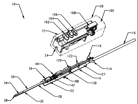
[0077] In FIGS. 1-4, a biopsy device 10 has a reusable hand piece 12 and a
disposable
probe 14 that enables economical taking of multiple percutaneous core biopsy
samples by
accessing a standard medical vacuum pump or wall-mounted vacuum access port
(not
shown) through an interfacing vacuum conduit 16. In the illustrative version,
the hand
piece 12 is self-powered and suitable for use in conjunction with ultrasonic
diagnostic
imaging. The disposable probe 14 reduces the portion of biopsy device 10 that
requires
protective packaging to avoid contact with sharp surfaces and to keep it
sterile prior to
use. Further economy is accomplished by reducing the portion of the biopsy
device 10
that is disposed as medical waste between uses. Movable components of the
disposable
probe 14 are advantageously locked until mounted in an access trough 18 formed
in a
housing 20 of the reusable hand piece 12. It should be appreciated that one or
more
standard mechanical, pneumatic, or electrical latches (not shown) may be
integrated into
the biopsy device 10 to secure the disposable probe 14 to the reusable hand
piece 12.
[0078] With particular reference to FIG. 3, the disposable probe assembly
14 includes a
substantially rectangular cover 22 sized to close the access trough recess 18
(FIGS. 2, 4).
An end slot 24 formed in the cover 20 (FIGS. 1-2, 4) is closed by a probe
union sleeve 26
attached to an inner surface 27 of the substantially rectangular cover 22. A
core biopsy
needle ("probe") assembly 28 passes longitudinally through the probe union
sleeve 26
and is formed by a probe tube 30 with underlying vacuum lumen 32 that
communicates
with a side aperture 34 through holes 35 (FIG. 23) near a distal opening 36 of
the probe
tube 30 that is closed by a piercing tip 38. A cutter tube 40 is sized to
closely fit and
translate within an inner diameter (i.e., cutter lumen) of the probe tube 30
with a length
sufficient to close the side aperture 34 with a proximal end 42 extending from
the probe
union sleeve 26 to attach to a cutter gear 44, as depicted in FIG. 1.
[0079] Proximal to the probe union sleeve 26 is an elongate slot 50 that is
part of a
vacuum assist valve assembly 52. The cutter gear 44 includes distal and
proximal annular
recesses 54, 56 flanking spur gear teeth 58 that engage the reusable hand
piece 12 as
described below. A more distal annular recess 60 is gripped by a post 62 that
is engaged
CA 02617904 2008-02-04
- 12 -
WO 2007/019152
PCT/US2006/030022
to IthigitudiliallFtiansTate in an elongate post slot 64 of a distal portion
66 of a vacuum
valve actuator 68. A cylindrical proximal portion 70 of the vacuum valve
actuator 68 has
distal and proximal 0-ring grooves 72, 73 that respectively retain distal and
proximal
dynamic 0-ring seals 74, 75 that move within a distally open cylindrical valve
bore 76 of
a valve body 78 molded onto an outer surface 79 of the substantially
rectangular cover 22
(FIG. 2).
[0080] As described below, the vacuum valve actuator 68 selectively allows
communication between a proximal port 80, a center port 82, and a distal port
84 (FIG.
2). In particular, with the cutter gear 44 retracted, the proximal and center
ports 80, 82 are
in communication. With the cutter gear translated distally, the center and
distal ports 82,
84 communicate. The center port 82 is attached to a distal vacuum conduit 86
whose
other end is connected through the rectangular cover 22 to the probe union
sleeve 26. It
should be appreciated that the probe union sleeve 26 includes pneumatic
passages that
communicate between a proximal end of the vacuum lumen 32 and the distal
vacuum
conduit 86. The distal port 84 is attached to a hose nib 88 that is exposed to
atmospheric
pressure. Hose nib 88 may include an air and/or saline filter. Alternatively,
hose nib 88
may be connected to a positive pressure source (e.g., fluid pump) or a
negative pressure
source (e.g., vacuum pump, syringe) to aspirate fluids. Likewise, hose nib 88
may be used
to lavage the tissue cavity with saline, pain medication, or bleeding control
fluids.. The
proximal port 80 communicates through a proximal vacuum conduit 90 to the
interfacing
vacuum conduit 16.
[0081] With further reference to FIG. 3, a sample extraction feature is
incorporated so
that multiple samples may be made without the need to remove the probe
assembly 28
from tissue nor even to full retract the cutter tube 40 to retract a tissue
specimen to the
reusable hand piece 12. In the illustrative version, this feature is
accomplished with a
stacking straw assembly100. An elongate straw 102 is scored down its length on
opposite
sides by grooves 104 defining first and second straw halves 106, 108, whose
respective
proximal, outer surfaces 110, 112 are attached to triangular grips 114, 116,
respectively.
A locking strip 118 extends distally from one triangular grip 114 and is
attached along a
proximal portion of the first straw half 106.
[0082] Distal and proximal tabs 120, 122 extend from the inner surface 27
of the
substantially rectangular cover 22, each having a respective through hole 124,
126
CA 02617904 2008-02-04
WO 2007/019152 - 13 -
PCT/US2006/030022
the '' stacking straw assembly 100 is inserted. The through holes 124, 126
are shaped to allow the locking strip 118 to rotate ninety (90) degrees. A
bayonet locking
member 130 also extends from the inner surface 27 of the substantially
rectangular cover
22 just distal and laterally offset from the through hole 124 of the distal
tab 120 to lock
into an alignment locking slot 132 in the locking strip 118 when laterally
rotated. The
bayonet locking member 130 prevents axial movement of the stacking straw
assembly
100. The cutter gear 44 and cutter tube 40 cannot move proximally due to
contact with
the stacking straw assembly 100 and cannot move distally due to contact with
the probe
union sleeve 26. By securing both the cutter gear 44 and the stacking straw
assembly 100
in a full distal axial position, the disposable probe 14 is aligned to engage
the components
of the reusable hand piece 12 as described below. Distal to the alignment
locking slot
132, a rectangular recess 134, formed in the locking strip 118, defmes a
distal-most
locking finger 136 for engaging components of the reusuable hand piece 12 that
positions
the stacking straw assembly 100 as described below. An indicator tube 150 has
a stacked
cone-shaped outer surface 152 (FIG. 14) that slides within the elongate straw
104 that in
turn slides within the cutter tube 40.
[0083] With particular reference to FIG. 4, the reusable hand piece 12
includes four user
controls aligned on a top surface 160 of the housing 20, specifically from
most distal to
most proximal: a forward motor rotation key 162, a reverse motor rotation key
164, a
saline flush key 166 and a slide button 168 for selecting insertion mode or
sample taking
mode. The keys 162-166 control a control circuit 170, which may include
integral power
storage (e.g., batteries, fuel cell, etc.) for untethered use. The forward
motor rotation key
162 causes a DC motor 172 to rotate its motor output shaft 174 in a forward
rotation. A
slide spur gear 176 includes an internal keyed engagement with a longitudinal
key groove
178 on the motor output shaft 174 that allows longitudinal positioning by the
slide button
168. In particular, fore and aft brackets 180, 182 of the slide button 168
engage distal and
aft annular grooves 184, 186 that flank spur gear teeth 188 of the slide spur
gear 176.
[0084] When the slide button 168 is moved distally, the slide spur gear 176
engages a
tissue penetration gear 190 that spins on a common shaft centerline 192
forward of a
gearbox input gear 196. Gearbox input gear 196 consists of a distal small gear
198 and a
proximal large gear 200. The tissue penetration gear 190 has spur gear teeth
206 that
engage the slide spur gear 176. A frame hub 212 projects proximally from the
frame 204
with a strike pin 214 projecting upwardly from the frame hub 212. In FIG. 4
and 13, a
CA 02617904 2008-02-04
WO 2007/019152 - 14 -
PCT/US2006/030022
circular cam wheel 216 is attached to a distal side of the tissue penetration
gear 190.
Rotating the tissue penetration gear 190 urges the strike pin 214, and thus
the frame 204,
proximally. In FIG. 12, left and right spring cavities 218, 220 (when viewed
from above),
formed longitudinally in distal corners of the frame 204, respectively receive
inwardly
projecting left and right tabs 222, 224 from the cover 20 and receive left and
right
compression springs 226, 228. Movement of the frame 204 proximally compresses
these
compression springs 226, 228 that thereafter assert a restoring force.
[0085] When the slide button 168 is moved proximally into engagement with
the gearbox
input gear 196, specifically the distal small gear 198, also engages and turns
a translation
large input gear 230 whose shaft 232 passes through an aft wall 234 of the
frame 204. The
proximal large gear 200 of the gearbox input gear 196 engages and turns a
rotation small
input gear 236 whose shaft 238 passes through the aft wall 234. The frame 204
includes a
carriage recess 240, defined between a partition 242 and the aft wall 234,
that contains
longitudinally aligned left side lead (translation) screw 244 and right-side
rotation spur
gear 246 that are attached for rotation respectively with the shafts 232, 238.
The partition
242 is positioned aft of the left and right tabs 222, 224 of the cover 20 and
also defines in
part the left and right spring cavities 218, 220. An unlocking cam 247
projects proximally
from and is longitudinally centered on the aft wall 234 above the position of
the lead
(translation) screw 244 and rotation spur gear 246.
[0086] The rotation spur gear 246 engages the cutter gear 44 when the
disposable probe
14 is inserted, imparting a rotation as the cutter tube 40 and cutter gear 44
translate
longitudinally in response to the rotation of the lead (translation) screw
244. This
translation is caused by lead screw threads 248. In particular, a distal
cutter carriage 250
is longitudinally moved on the lead screw threads 248. Distal and proximal J-
hook
extensions 252, 254 project downwardly from the distal cutter carriage 250 to
engage the
distal and proximal annular recesses 54, 56 of the cutter gear 44 (FIG. 3).
Distal of the
cutter carriage 250, a biasing spring 256 urges against the cutter carriage
250, which
assists in engagement of the lead screw threads 248 with the distal cutter
carriage 250.
With reference to FIGS. 4 and 19, a sliding pin 260 has a proximal carriage
sliding pin
retainer 266 attached to a proximal straw carriage 258. Shaft 264 also passes
through a
distal carriage sliding pin retainer 270 attached to the distal cutter
carriage 250. Sliding
pin 260 has a proximal end 262 and a distal end 268 to prevent the sliding pin
260 from
disengaging from the carriage sliding pin retainers 266, 270. A sliding pin
spring 272
CA 02617904 2008-02-04
WO 2007/019152 - 15 -
PCT/US2006/030022
faiag 'on-the sliding pin 260 and is constrained at each end by carriage
sliding pin
retainers 266, 270.
[0087] With the components FIGS. 1-4 now introduced, a sequence of use of
the biopsy
device 10 will be described. The interfacing vacuum lumen 16 is attached to
the
disposable probe assembly 14 (FIGS. 1-2). The disposable probe assembly 14 is
installed
into the reusable hand piece 12 (FIGS. 5-8). In so doing, the distal cutter
carriage 250
engages the cutter gear 44 (FIG. 9), the proximal straw carriage 258 engages
the locking
strip 118 of the stacking straw assembly 100 (FIG. 10), and the bayonet
locking member
130 is deflected by the unlocking cam 247, longitudinally unlocking from the
alignment
locking slot 132 of the locking strip 118 (FIG. 11) allowing longitudinal
movement of the
cutter tube 40 and the straw stacking assembly 100.
[0088] In FIGS. 12, 14, the cutter and straw carriages 250, 258 may
initially be distally
advanced to close the side aperture 34 of its probe tube 30 with the cutter
tube 40 and the
stacking straw assembly 100 also fully distally advanced to minimize proximal
extension
of its elongate straw 102.
[0089] In FIG. 13, the piercing tip 38 of the core biopsy needle (probe)
assembly 28 is
assisted in penetrating tissue by moving the slide button 168 distally to a
"tissue insertion
mode" wherein the slide spur gear 176 engages the tissue penetration gear 190.
Depression of the forward motor rotation key 162 turns these gears 176, 190
causing the
circular cam wheel 216 to turn against strike pin 214 that creates proximal
longitudinal
motion of frame 204 and core biopsy needle (probe) assembly 28 of
approximately 0.1
inch at a rotation rate of 7 cycles per second. Left and right compression
springs 226, 228
provide the restoring distal longitudinal motion to frame 204 and disposable
probe 14 as
left and right compression springs 226, 228 are repeatedly compressed between
the
forward surface of the left and right spring cavities 218, 220 as the frame
204 and the left
and right tabs 222, 224 of the housing 20. The restoring distal longitudinal
motion to
frame 204 and core biopsy needle (probe) assembly 28 result in a corresponding
distal
motion of piecing tip 38 that assists in penetrating tissue.
[0090] In FIG. 15, with the side aperture 40 positioned within the tissue
to take samples,
the slide button 168 is moved proximally to engage the slide spur gear 176
with the distal
small gear 198 of the gearbox input gear 196. When the forward motor rotation
key 162 is
depressed, the DC motor 172 rotates in a forward direction, turning the slide
spur gear
CA 02617904 2008-02-04
WO 2007/019152 - 16 -
PCT/US2006/030022
the distal small gear 198 that directly turns the translation large input
gear 230 that is connected by the shaft 232 through the aft wall 234 of the
frame 204 to
the lead (translation) screw 244. Meanwhile, the proximal large gear 200 of
the gearbox
input gear 196 rotates the small input gear 236 that turns shaft 238 through
aft wall 234 to
turn the rotation spur gear 246.
[0091] With the carriages 250, 258 distally advanced as depicted in FIGS.
15-16, the
cylindrical proximal portion 70 of the vacuum valve actuator 68 is also
distally positioned
as depicted in FIG. 17. The hose nib 88 is thus in fluid communication through
the distal
port 84, through the distally open cylindrical valve bore 76 between distal
and proximal
dynamic 0-ring seals 74, 75 to the center port 82 through the distal vacuum
conduit 86 to
the vacuum lumen 32.
[0092] In FIGS. 18-19, depression of the reverse motor rotation key 164
causes the lead
(translation) screw 244 to rotate in a reverse direction. Sliding pin spring
272 between the
distal cutter carriage 250 and the proximal straw carriage 258 urges the
proximal straw
carriage 258 into engagement with the lead screw thread 248, causing the straw
carriage
258 to move proximally as the cutter carriage 250 free wheels on an unthreaded
distal
portion of the lead screw 244. The straw carriage 258 draws back the elongate
straw 102
and the indicator tube 150 (FIG. 20). As the straw carriage 258 approaches the
proximal
portion of the lead screw 244, the distal end 268 of sliding pin 260 contacts
the distal
carriage sliding pin retainer 270 on distal cutter carriage 250, pulling the
distal cutter
carriage 250 onto the lead screw thread 248. Thereafter, the cutter carriage
250 and the
cutter tube 40 are retracted as the straw carriage 258 free wheels (FIGS. 21-
22).
[0093] Alternately, sliding pin spring 272 may be replaced with a ball
detent mechanism
(not shown) located on frame 204 that would engage with a small depression in
proximal
straw carriage 258. This alternate mechanism in conjunction with biasing
spring 256
would cause both the distal cutter carriage 250 and proximal straw carriage
258 to retract
simultaneously from their fully distal position and to advance sequentially
from their fully
proximal position (i.e., cutter carriage 250 would fully advance and then the
straw
carriage 258 would advance).
[0094] At the end of the proximal movement of the cutter tube 40, vacuum
valve actuator
68 is moved proximally such that the distal and proximal dynamic 0-ring seals
74, 75
bracket the proximal port 80 and center port 82 of the distally open
cylindrical valve bore
CA 02617904 2008-02-04
WO 2007/019152 - 17 -
PCT/US2006/030022
76.-Thereby, the interfacing vacuum conduit 16 draws air through the proximal
vacuum
conduit 90, through the valve body 78, through the distal vacuum conduit 86,
and
ultimately from the vacuum lumen 32 (FIG. 24). In FIG. 23, this suction draws
tissue 280
into the side aperture 34 of the probe assembly 28.
[0095] It should be appreciated that in the illustrative version, the
distal cutter carriage
250 does not freewheel (FIG. 21) in its proximal-most position. Instead,
rotation of the
motor is stopped as the distal cutter carriage 250 is about to contact the
proximal straw
carriage 258 with closed-loop control based on an encoder (not shown) coupled
to the DC
motor 172 enabling accurate positioning of the motor output shaft 174.
Alternatively,
freewheeling may be incorporated at the proximal-most position of the distal
cutter
carriage 250 by adding a section of no helical threads to the proximal end of
the lead
(translation) screw 244 equal to the longitudinal thickness of the distal
cutter carriage
250.
[0096] It should further be appreciated that free wheeling may be provided
for cutter
translation even without stacking straw sample retraction to avoid reliance
upon other
structures to block further translation or more elaborate closed loop position
control.
[0097] The forward motor rotation key 162 is depressed to advance the
cutter tube 40,
rotating lead (translation) screw 244 and rotation spur gear 246, as depicted
in FIG. 25.
Due to sliding pin spring 272 between carriages 250, 258, only the distal
cutter carriage
250 engages with the lead screw threads 248 of the lead (translation) screw
244 and
translates distally initially cutting tissue 280, as depicted in FIG. 26. Once
the distal cutter
carriage 250 approaches its distal-most position, the sliding pin 260 pulls
the proximal
straw carriage 258 into engagement with the lead screw threads 248 of the lead
(translation) screw 244. As the cutter carriage 250 freewheels, the elongate
straw 102 is
distally translated to encompass a first severed tissue sample 280a,
displacing proximally
the indicator tube 150 a corresponding amount.
[0098] At this point, depression of the reverse motor rotation key 164
causes retraction of
the proximal straw carriage 258 (FIG. 18) with the side aperture 134
communicating with
atmospheric pressure (FIG. 17) as previously discussed so that the first
severed tissue
sample 280a remains within the elongate straw 280a. It should be appreciated
that
repeating the retraction and advancement of the cutter carriage 250 thereafter
results in a
second severed tissue sample 280b being encompassed by the elongate straw 102
and the
CA 02617904 2008-02-04
WO 2007/019152 - 18 -
PCT/US2006/030022
tilbrf5t1ieliig further proximally displaced thereby as depicted in FIG. 27.
An
additional retention feature is depicted in FIG. 27 wherein small bent-up,
proximally
directed tabs 284 formed in the elongate straw 102 resist distal movement of
the severed
tissue samples 280a, 280b. This automated sequencing of the cutter and straw
carriages
250, 258 during retraction and advancement may be repeated a number of times
to take a
plurality of samples without withdrawing the probe assembly 28 from tissue
280. The
surfaces of the elongate straw 102 may be coated with lubricous materials to
aid in
proximal movement of tissue through the elongate straw 102 and to reduce
friction
between the elongate straw 102 and the cutter tube 40. Likewise, to aid in
proximal
movement of tissue through the elongate straw 102, the diameter of the
elongate straw
102 and the cutter tube 40 may be increased slightly some distance proximal
from their
distal end to reduce the friction of the tissue through the elongate straw
102.
[0099] In FIG. 28, a proximal end of the stacking straw assembly 100
includes a one-way
latch (mechanical diode) 290 that engages the stacked cone shaped outer
surface 152 of
the indicator tube 150 as it proximally extends out of the elongate straw 102
preventing
its being pneumatically drawn back into the elongate straw 102 when
subsequently
exposed to vacuum pressure.
[0om] In FIGS. 29, 30, the proximal straw carriage 258 is shown to include
distal and
proximal J-hooks 300, 302 that encompass on three sides the stacking straw
assembly
100. In particular, the rectangular recess 134 formed in the locking strip 118
is sized to
longitudinally bracket the J-hooks 300, 302 with the distal locking finger 136
preventing
retraction as depicted in FIG. 29 when the triangular grips 114, 116 are
positioned
horizontally (FIG. 31), as would be typical before and during use of the
biopsy device 10.
The surgeon may wish to segregate samples as they are taken or to take more
samples
than possible within one stacking straw assembly 100. Extraction and
replacement of the
stacking straw assembly 100 is allowed by rotating the triangular grips one
quarter turn
counterclockwise (as viewed proximally) as depicted in FIG. 32, which rotates
the
locking finger 136 out of alignment with the J-hooks 300, 302 of the straw
carriage 258
(FIG. 30). A new stacking straw assembly 100 is then reinserted in reverse
fashion.
[00101] In FIG. 33, samples contained in the removed stacking straw
assembly 100 may
be accessed by pulling apart the triangular grips 114, 116 causing the grooves
104 to peel
apart the first and second straw halves 106, 108, which need not be symmetric.
The
CA 02617904 2008-02-04
WO 2007/019152 - 19 -
PCT/US2006/030022
'sErifiles may be removed individually or the samples and the straw half 106
portion of the
straw 102 in which they are located may be put directly into a formalin
solution for
pathological preparation. Alternately, the samples contained in the stacking
straw
assembly 100 can be removed from the elongate straw 102 with a simple plunger-
like rod
(not shown) eliminating the need to peel apart the straw to access the tissue
samples.
[00102] Although the integral vacuum assistance supported by a medical
vacuum pump
may often be advantageous, some surgeons may desire to palpitate tissue into a
side
aperture of a probe assembly without the assistance of vacuum. To that end, in
FIGS. 34-
36, an alternative disposable probe 414 is depicted that omits a vacuum valve
capability
that responds to the cutter position but is otherwise identical to the afore-
described
disposable probe 14. The modified components of the disposable probe assembly
414
include a substantially rectangular cover 422 sized to close the access trough
recess 18 of
the reusable hand piece 12 (not shown in FIGS. 34-36). The probe union sleeve
26,
attached to the inner surface 27 of the substantially rectangular cover 422,
communicates
through a short pneumatic conduit 425 that terminates on the outer surface 79
at a hose
nib 427. Hose nib 427 may include an air and/or saline filter. Alternatively,
hose nib 427
may be connected to a positive pressure source (e.g. fluid pump) or a negative
pressure
source (e.g., vacuum pump, syringe) to aspirate fluids. Hose nib 427 could
also be used to
lavage the tissue cavity with saline, pain medication, or bleeding control
fluids. A core
biopsy needle ("probe") assembly 428 that passes longitudinally through the
probe union
sleeve 26 differs in that a cutter gear 444 needs only engage and respond to
the distal
cutter carriage 250 (not shown in FIGS. 34-36) and not also position a
pneumatic valve.
Cutter guide tab 445 extends out from the inner surface 27 to provide a distal
stop for
cutter gear 444. Prior to insertion of the disposable probe 414 into the
reusable hand piece
12 (not shown in FIGS. 34-36), the bayonet locking member 430 prevents axial
movement of the stacking straw assembly 100. The cutter gear 444 and cutter
tube 40
cannot move proximally due to contact with the stacking straw assembly 100 and
cannot
move distally due to contact with the cutter guide tab 445. By securing both
the cutter and
straw in a fully distal axial position, it insures that when the disposable
probe 414 is
inserted into the reusable hand piece 12 that the cutter gear 444 and stacking
straw
assembly 100 align and engage with the correct components within the reusable
hand
piece 12.
CA 02617904 2008-02-04
WO 2007/019152 - 20 -
PCT/US2006/030022
fOtiiaj 'in-PTC:Ti: an alternative disposable assembly 514 is, as described in
FIG. 3 but
with the stacking straw assembly 100, replaced with a straw assembly 516
having distal
tube 518 attached to a proximally attached luer fitting 520. The straw
assembly 516 may
be used to flush the cavity (via side aperture 34) with saline, epinephrine
(or similar
substances that reduce bleeding), or lidocane (or similar substances that
reduce pain) by
attaching a syringe or similar device (not shown) to the luer fitting 520. To
remove the
saline, epinephrine, or lidocane from the tissue, the cutter tube 40 may be
fully or
partially retracted to insure that the valve assist valve assembly 52 is
positioned to
connect the lateral lumen (distal vacuum conduit 86) with the vacuum source (
and not
simply atmospheric pressure) as depicted in FIG. 24. The fluid would then be
drawn from
the tissue cavity (via side aperture 34), through the lateral lumen (distal
vacuum conduit
86) and into a canister located in line with the vacuum source (not shown).
[00104] In FIG. 38, an alternative biopsy needle (probe) assembly 628 is
identical to that
depicted in FIG. 14 with the exception of a probe tube 630 with through holes
631 placed
proximate to the side aperture 34. The vacuum lumen 32 thus communicates with
the
holes 631 in the probe tube 630 as an alternate means to apply saline,
epinephrine, or
lidocane to the tissue cavity. These through holes 631 allow the fluid to
reach the cavity
while the elongate straw 102 and indicator tube 150 remain distally positioned
in the
cutter tube 40 (i.e., during the middle of a biopsy sampling procedure). In
this case, the
syringe would be attached to the hose nib 88 via a stopcock fitting (not
shown). With the
stopcock valve positioned to connect the syringe directly to the needle's
lateral lumen
(distal vacuum conduit 86), when the syringe is depressed the fluid will enter
the lateral
lumen (distal vacuum conduit 86) and then flow into the tissue through the
through holes
631 in the wall of the probe tube 630. The cutter tube 40 would be positioned
distally
(side aperture 34 closed) while the fluid is being inserted into the cavity to
prevent the
tissue indicator tube 150 from being moved proximally due to the fluid
pressure. During
subsequent sampling cycles, the fluid would then be aspirated from the tissue
cavity.
[00105] In FIGS. 39-45, an alternative proximal stacking disposable
assembly 702 is
depicted that may also be used with the reusable hand piece 12. Pneumatic
force is
employed to retrieve tissue samples rather than a mechanical movement from the
reusable
hand piece 12 that actuates a straw assembly. To that end, in FIG. 39, a core
biopsy
needle ("probe") assembly 704 is formed by a probe tube 706 with a distally
positioned
side aperture 708. A cutter tube 710 is sized to closely fit and translate
within an inner
CA 02617904 2008-02-04
WO 2007/019152 - 21 -
PCT/US2006/030022
Ziainel& (I.Z.Tcutter lumen) 712 of the probe tube 706 with a length
sufficient to close the
side aperture 708. The probe assembly 704 includes an underlying vacuum lumen
714
that communicates with the cutter lumen 712 via through holes 716 underlying
the side
aperture 708. Both the probe tube 706 and vacuum lumen 714 distally terminate
in open
ends that communicate with each other via a curved manifold 718 defined inside
of a
piercing tip 720 that is attached as a distal-most portion of the probe
assembly 704. A
distal tissue stop 722 projects from the piercing tip 720 into the distal open
end of the
probe tube 706 to maintain prolapsed tissue inside a sampling bowl 724 under
the side
aperture 708 within the cutter lumen 712. Prolapsing occurs under the urging
of axial
vacuum force through the cutter lumen 712 and lateral vacuum force through the
vacuum
lumen 714 converging at the side aperture 708. After distal translation of the
rotated
cutter tube 712, a tissue sample 726 resides within a distal portion of the
cutter tube 712,
wherein an inner diameter of the cutter tube 712 defines a tissue sample lumen
728 for
guiding retrieval of samples 726. Rather than subsequently distally advancing
a straw to
encompass and retract the tissue sample 726, axial vacuum pressure as depicted
by arrow
730 is asserted against a proximal face of the tissue sample 726 through the
tissue sample
lumen 728 with the cooperation of lateral pneumatic pressure as depicted by
arrow 732
through vacuum lumen 14 and curved manifold 718 to a distal face of the tissue
sample
726.
[00106] In FIGS. 40-45, the portions of the alternative proximal stacking
disposable
assembly 702 capture these tissue samples 726. A proximal end of the cutter
tube 710
extends through a probe union sleeve 734 to attach to a cutter gear 736. A
proximal end
of the vacuum lumen 714 terminates within the probe union sleeve 734. The
alternative
proximal stacking disposable assembly 702 includes a substantially rectangular
cover 738
sized to close the access trough recess 18 (FIGS. 2, 4), and omits pneumatic
valve
features. Instead, the distally positioned probe union sleeve 734 attached to
an inner
surface 740 of the substantially rectangular cover 738 communicates to a
distal hose nib
742 formed on an outer surface 744 of the rectangular cover 738 and to the
vacuum
lumen 714. A hose 746 is attached to the distal hose nib 742 to selectively
provide
pneumatic vacuum, pneumatic pressure, or fluid transfer (not shown). The
alternate
proximal stacking assembly 702 could likewise have a vacuum assist valve
assembly 052
as depicted in FIG. 2 to selectively provide pneumatic vacuum, pneumatic
pressure, or
fluid transfer to the vacuum lumen 714.
CA 02617904 2008-02-04
-
WO 2007/019152 - 22
PCT/US2006/030022
[00107] with particular reference to FIGS. 40, 42, a rear tube 748 is
aligned proximally to
the cutter tube 710 and coupled for longitudinal movement thereto, although
the rear tube
748 is disengaged from the rotational movement of the cutter tube 710. This
coupled
movement may be achieved by an actuator that engages the distal cutter
carriage 250
(FIG. 4) or by a circular lip and groove engagement between the cutter tube
710 and rear
tube 748. The inner surface 740 of the rectangular cover 738 includes four
support
surfaces. First, a cutter guide 750 supports the cutter tube 710 proximal to
the probe union
sleeve 734 and distal to a most distal position of the cutter gear 736. A
distal rear tube
guide 752, is proximal to the most proximal position of the cutter gear 736,
and a
proximal rear tube guide 754, and distal to a most distal position of a
proximal locking
flange 756 of the rear tube 748, to maintain alignment of the rear rube 748. A
bottom
half-cylinder locking flange 758 at a proximal end of the rectangular cover
738
cooperates with the proximal locking flange 756 of the rear tube 748 to lock
to a sample
holding portion 760 of the alternative proximal stacking disposable assembly
702. The
sample holding portion 760 extends proximal to the rectangular cover 738 and
the
reusable hand piece 712 and thus may be readily replaced during a biopsy
procedure.
[00108] A distal locking half cylindrical portion 762 engages the bottom
half-cylinder
locking flange 758. The distal locking half cylindrical portion 762 is
attached to a
proximal half cylindrical portion 764 to form an outer sleeve 766. A
reciprocating
member 768, which engages the proximal locking flange 756 of the rear tube 748
and is
partially encompassed by the outer sleeve 766, engages and distally advances a
more
proximal rod 770 out of an external vacuum lumen 772 defined as an inner
diameter of an
external vacuum tube 773. The rod has a down turned distal end 774 that exits
an opening
776 in the proximal half cylindrical portion 764. A flexible, peel-apart
external tissue tube
777 defining an external tissue lumen 778 is formed from an inwardly open
channel 780
closed by an elongate seal 782.
[00109] Rod 770 may be formed of a fluoropolymer resin material such as
TEFLONTm or
other suitable flexible material having a low coefficient of friction. Rod 770
may be sized
and shaped to conform closely to the inner diameter (i.e., vacuum lumen 772)
of vacuum
tube 773. The close fit between rod 770 and vacuum lumen 773, as well as the
low
friction properties of the rod 770, enable the rod 770 to translate easily
within the vacuum
lumen 772 without any loss of vacuum force through the distal end of the
vacuum lumen
772. The inwardly open channel 780 may advantageously be formed of polyvinyl
CA 02617904 2008-02-04
WO 2007/019152 - 23 -
PCT/US2006/030022
hrdrideor another similar type of flexible, water insoluble material so that
stacked tissue
samples may be visible. A proximal end of the open channel 780 is attached to
and closed
by a lumen peel tab 784. A proximal end of the external vacuum lumen 772 is
attached to
a vacuum line 786 via a tubing connector 788.
[00110] In FIGS. 40, 41, the alternative proximal stacking disposable
assembly 702 is in
an initial condition with the rod 770 at its proximal most position in the
external vacuum
lumen 772. The cutter gear 736 and thus the rear tube 748, reciprocating
member 768 and
flexible, peel-apart external tissue lumen 778 are in their distal most
position. In FIGS.
43, 44, the rod 770 has extruded distally out of the opening 776 in the
proximal half
cylindrical portion 764 of the outer sleeve 766, denoting reciprocating cycles
to retract at
least one tissue sample (not shown) that is held within a proximal portion of
the external
tissue lumen 778. The cutter gear 736 and thus the rear tube 748,
reciprocating member
768 and flexible, peel-apart external tissue lumen 778 are in their proximal
most positions
relative to the outer sleeve 766 and rectangular cover 738. The relative
change causes the
flexible, peel-apart external tissue lumen 778 to bow away from the outer
sleeve 766. In
FIG. 45, the lumen peel tab 784 has been pulled to separate the inwardly open
channel
780 from the elongate seal 782 to reveal and possibly access stored tissue
samples (not
shown).
1001111 In FIGS. 46-48, the sample holding portion 760 is depicted in
greater detail. The
distal locking half cylindrical portion 762 of the outer sleeve 766 includes
upper lateral
locking arms 790 that lock into the bottom half-cylinder locking flange 758 at
the
proximal end of the rectangular cover 738. In FIGS. 46, 47, aligned below
these, lower
lateral locking arms 792 of a distal interface portion 794 of the
reciprocating member 768
lock into the proximal locking flange 756 of the rear tube 748. The distal
interface portion
794 of the reciprocating member 768 includes an axially-extending bore 796 for
connecting the external tissue lumen 778 of the sample holding portion 760 to
the rear
tube 748, maintaining generally coaxial alignment of the probe assembly 702,
tissue
sample lumen 728, rear tube 748, bore 796, and external tissue lumen 778 to
provide an
unobstructed passageway for the aspiration of tissue samples from the cutter
tube 710.
[00112] In FIGS. 48, 50-51, the flexible rod 770 may be advanced distally
within the
external vacuum lumen 772 by the interaction between side ratchet teeth 798
and a pawl-
type latching mechanism 800 on the reciprocating member 768, which is shown in
greater
CA 02617904 2008-02-04
-
WO 2007/019152 - 24
PCT/US2006/030022
FI49': 'Reciprocating member 768 may be supported on lower lateral latch
arms 792 and reciprocate as cutter tube 710 is advanced and retracted.
Reciprocating
member 768 may have a bifurcated proximal end with proximally extending
portions 802
separated by an axially extending slot 804. A ramped surface 806 is formed
between
portions 802 at a distal end of slot 804. Ramped surface 806 may serve to
deflect the
distal end 774 of rod 770 through the opening 776 in the outer sleeve 766 as
the rod 770
is ratcheted out of external vacuum lumen 772. Unidirectional engagement pawls
808
formed to inwardly extend from the proximally extending portions 802 into the
axially
extending slot 804 engage side ratchet teeth 798 on rod 770 as the rod 770
extends
through the axially extending slot 804. The engagement between pawls 808 and
side
ratchet teeth 798 advances rod 770 distally through vacuum lumen 772.
[00113] In FIG. 51, a plurality of small holes 810 may be formed in a
center wall divider
812 of the external vacuum tube 773 between external vacuum lumen 772 and
tissue
lumen 778. Small holes 810 enable vacuum from a source (not shown) connected
to
vacuum line 786 to communicate from external vacuum lumen 772 into external
tissue
lumen 778, to provide vacuum in tissue sample lumen 728 in cutter tube 710.
Small holes
810 may be spaced along the longitudinal axis of tube vacuum tube 773 and
separated by
a distance in the range of 0.1 to 4 centimeters. Holes 810 may be oriented at
an angle
relative to the longitudinal axis of vacuum tube 773. The angle in holes 810
may function
as a mechanical diode, in that the edge of the holes 810 opening into the
tissue lumen 778
may aid in preventing motion of tissue samples 726 in a distal direction,
while permitting
tissue samples 726 to move proximally in tissue lumen 778 under vacuum force
provided
by the vacuum line 786. A tissue sample 726 may continue to slide proximally
through
the tissue lumen 778 until the sample 726 contacts either a proximal tissue
stop 812
attached to the lumen peel tab 784 or a preceding tissue sample 726.
[00114] With further reference to FIG. 51, small holes 810 may be formed
between
lumens 772, 778 by boring top holes 813 into an upper surface 814 of external
vacuum
tube 773 with the sharpened tip of a drill or other appropriate instrument.
The tip of the
drill bit or other boring instrument may be directed to pass through vacuum
lumen 772 to
penetrate the center wall divider 812 that separates the two lumens 772, 778.
The
proximal half cylindrical portion 764 of the outer sleeve 766 may be securely
attached to
the upper surface 814 of the external vacuum tube 773 following the drilling
of vacuum
communication small holes 810 to seal top holes 813. For instance, outer
sleeve 766 may
CA 02617904 2008-02-04
-
WO 2007/019152 25 -
PCT/US2006/030022
-be-ittiChed to the external vacuum tube 773 by an adhesive or other
appropriate type of
attachment mechanism.
[00115] As tissue samples 726 are stored in tissue lumen 778, the stack of
samples 726
will grow in length distally in tissue lumen 778. The samples 726 will tend to
block or
otherwise restrict flow communication through small holes 810 as the stack of
samples
726 extends distally in tissue lumen 778. The translating flexible rod 770 is
shown
disposed at least partially in vacuum lumen 772. Rod 770 extends axially
through vacuum
lumen 772 to selectively cover or otherwise block at least some of the small
holes 810.
Rod 770 may be manipulated, such as by axial movement of rod 770, to
selectively
expose small holes 810 in the vacuum tube 773 in compensation for those holes
810
blocked by stacked tissue samples 726. For instance, during each cutting
cycle, rod 770
may be advanced distally within vacuum lumen 772 to expose or otherwise
unblock/open
additional small holes 810 as additional samples 726 are stored in tissue
lumen 778. The
movement of rod 770 maintains a predetermined number of small holes 810 open
to
provide flow communication between vacuum and tissue lumens 772 and 778 as
additional tissue samples 726 are added to the stack of tissue samples 726 in
tissue lumen
778, thereby facilitating a generally consistent vacuum force, depicted as
arrow 816, in
tissue sample lumen 728 in the probe assembly 704 (FIG. 39) throughout
multiple cutting
cycles.
[00116] Initially as depicted in FIG. 52, flexible rod 770 may be inserted
within vacuum
lumen 772 such that rod 770 is axially offset within vacuum lumen 772 so as to
cover or
otherwise block most, but not all, of the small holes 810. For instance, prior
to storing any
samples 726 in tissue lumen 778, rod 770 may be offset distally within vacuum
lumen
772 a distance that is slightly longer than the length of side aperture 708
(FIG. 40).
Offsetting rod 770 distally within the vacuum lumen 772 ensures an initial set
of small
holes 810 are exposed to communicate axial vacuum force 730 to side aperture
708 when
cutter tube 710 is in the fully proximal position prior to tissue sampling.
The axial
vacuum force 730 communicated through the exposed small holes 810 aids in
prolapsing
tissue into side aperture 708 prior to cutting, as well as pulling the tissue
sample 726
proximally into tissue lumen 778 after cutting. As a tissue sample 726 is
drawn into and
stacked within tissue lumen 778, the tissue sample 726 blocks the previously
exposed
small holes 810, preventing vacuum from passing into the tissue lumen 778. Rod
770 may
be selectively moved a predetermined distance distally that is slightly longer
than the
CA 02617904 2013-08-27
-26-
length of Side aperture 708 to expose additional small holes 810 immediately
distal of the
most recently acquired tissue sample 726. Rod 770 may be adapted to be
automatically
advanced distally by the translation of the cutter carriage 250. The newly
exposed small
holes 810 continue the communication of vacuum force 816 into tissue lumen 778
for the
next cutting cycle. As reciprocating member 768 retracts proximally,
unidirectional
bottom ratchet teeth 818 located on the bottom side of flexible rod 770 engage
the small
holes 810 within vacuum lumen 772. The engagement between the bottom ratchet
teeth
818 and small holes 810 prevents rod 770 from moving proximally within vacuum
lumen
772. As pawls 808 move proximally relative to rod 770, the pawls 808 engage
the next
proximal set of side ratchet teeth 798 on rod 770. This engagement with the
next set of
side ratchet teeth 798 causes rod 770 to again advance distally when the
reciprocating
member 768 advances distally during the next cutting cycle to expose
additional small
holes 810. In the event that the cutter tube 710, and thus the reciprocating
member 768, is
advanced and retracted without the probe assembly 704 in tissue, the result is
that the
flexible rod 770 advances too far distally relative to the tissue samples 726;
the flexible
rod 770 may be rotated a fraction of a turn about its longitudinal axis to
disengage side
ratchet teeth 798 and pawls 808 allowing the flexible rod 770 to be
repositioned
proximally within the vacuum lumen 772.
100117] A similar sample holding portion is described in five commonly-
owned and
co-pending U.S. Pat. Appin. Pub. No. 20060074345, "Biopsy Apparatus and
Method";
Pub. No. 20060074346, "Improved Biopsy Apparatus and Method"; Pub. No.
20060074344, "Fluid Control for Biopsy Device"; Pub. No. 20060074343, "Biopsy
Device with Sample Storage"; and Pub. No. 20060074342, "Cutter for Biopsy
Device", all
to Hibner et al. and filed on 29 September 2004.
[00118] While preferred embodiments of the present invention have been
shown and
described herein, it will be obvious to those skilled in the art that such
embodiments are
provided by way of example. Numerous variations, changes, and substitutions
will occur
to those skilled in the art. The scope of the claims should be given the
broadest
interpretation consistent with the description as a whole.
DOCSTORs 2792629\1
CA 02617904 2008-02-04
WO 2007/019152 - 27 -
PCT/US2006/030022
1061141 -TO r example, one or more sensors may be incorporated into the hand
piece 12 to
sense the actual position of each carriage or to sense the particular
disposable probe
assembly assembled into the hand piece 12.