Note: Descriptions are shown in the official language in which they were submitted.
CA 02618125 2008-01-22
Hydro2el Proximal Interphalangeal Implant
[0001] The present invention relates to the field ofjoint replacement.
Specifically, the
present invention relates to a joint prosthesis for proximal interphalangeal
joints.
[0002] The replacement of damaged or diseased joints in the human body has
been
known for some time. Devices utilized to replace natural joint structures
generally mimic
natural movement of the joint. In addition, such devices are often configured
to provide for a
natural "at rest" position similar to that of the natural joint.
[0003] Known proximal interphalangeal joint prosthetics typically employ two
stems
or arms with an intermediate pivoting structure. In some devices, the entire
prosthetic is
manufactured from a single elastomer material or from metal alloy.
[0004] The present invention relates to a prosthetic used to replace a damaged
joint,
such as a pivotal interphalangeal joint, for example. The prosthetic may
include a body
portion and an outer weave portion. The body portion may include an
intermediate portion
and a pair of stems connected to, and extending from, the intermediate
portion.
[0005] The body portion niay be formed from a hydrogel material, which may
expand
upon absorption of water. In addition, the outer weave portion may include a
plurality of
layers including a polymer layer and a metal layer. The polymer layer may be
located
intermediate the metal layer.
[0006] The intermediate portion may include a recess, which may be formed in
the
palmar side of the intermediate portion.
[0007] In one form thereof, the present invention provides a prosthetic used
to replace
a damaged joint including a body portion including an intermediate portion and
a pair of
stems connected to the intermediate portion; and an outer weave encompassing
the body
portion.
CA 02618125 2008-01-22
[0008] In another form, the present invention provides a prosthetic used to
replace a
damaged joint including a body portion including an intermediate portion and a
pair of stems
connected to the intermediate portion; wherein the body portion is formed from
hydrogel.
[0009] In another form, the present invention provides a prosthetic used to
replace a
damaged joint including a body portion formed from a hydrogel material and
including a pair
of interconnected stems; and an outer weave at least partially encompassing at
least one of the
stems.
[0010] The above-mentioned and other features and advantages of this
invention, and
the manner of attaining them, will become more apparent and the invention
itself will be
better understood by reference to the following description of an embodiment
of the invention
taken in conjunction with the accompanying drawings, wherein:
[0011] Figure l is a perspective view of a prosthetic device embodying the
present
invention;
[0012] Figure 2 is a perspective view of the prosthetic device of Figure 1
with a
portion of the outer weave omitted for illustrative purposes;
[0013] Figure 3 is a side view of a body portion of the prosthetic device,
illustrating
exemplary ranges of motion thereof from a substantially non-flexed or neutral
position shown
in solid lines;
[0014] Figure 4 is a side view of the body portion depicted in Figure 3 in a
flexed
position; and
[0015] Figures 5-7 are side views of a finger illustrating an exemplary
surgical
method of implanting the prosthetic of Figure 1.
[0016] Corresponding reference characters indicate corresponding parts
throughout
the several views. The exemplification set out herein illustrates one
preferred embodiment of
the invention, in one form, and such exemplification is not to be construed as
limiting the
scope of the invention in any manner.
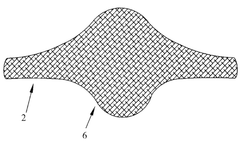
[0017] Figures 1 and 2 depict different views of a joint prosthetic, generally
indicated
by numeral 2, representing an exemplary embodiment of the present invention.
Prosthetic 2
2
CA 02618125 2008-01-22
includes a body portion 4 and a cover or weave portion 6 encompassing body
portion 4. In
Figure 2, a portion of weave portion 6 has been omitted in order to illustrate
body portion 4
with respect to weave portion 6. The depicted embodiment of prosthetic 2 is
configured to be
utilized in the proximal interphalangeal (PIP) joint of a human finger 30, as
shown in Figure
7.
[0018] With reference to Figures 3 and 4, body portion 4 includes a first stem
8, a
second stem 10 and an intermediate portion 12. Stems 8, 10 and the
intermediate portion 12
may be formed with a unitary one-piece construction. In the present
embodiment, body
portion 4 may be formed from any suitable hydrogel material.
[0019] A hydrogel is a network of polymer chains that are water-soluble but
made
insoluble through physical and/or chemical crosslinks. These materials are
sometimes found
as a colloidal gel in which water is the dispersion medium. Hydrogels are
generally formed
from natural or synthetic polymers. Hydrogels may be classified as
"superabsorbent" and
may contain over 99% water, by weight. In addition, hydrogels may have the
abilty to swell
due to water absorption. Hydrogels may also possess a degree of flexibility
very similar to
natural tissue, due to their significant water content. Suitable hydrogels
include hyaluronic
acid, polypropylene fumarate, and Poly(ethylene glycol)-co-polylactide, methyl
cellulose, and
carboxy methyl cellulose.
[0020] In general, the stems 8, 10 are sized and configured to be received
within
intramedullary recesses or bores of adjacent bones. For example, in the
exemplary
implantation depicted in Figure 7, the first stem 8 is sized and configured to
be received into a
bore 42 of the middle phalanges 34 of the finger 30. Similarly, the second
stem 10 is sized
and configured to be received into a bore 44 of the proximal phalanges 36 of
finger 30.
[0021] Intermediate portion 12 is configured to provide flexion motion between
the
first stem 8 and the second stem 10. With reference again to Figures 1 through
4,
intermediate portion 12 includes a first surface 14 and a second surface 16.
In the depicted
embodiment, first surface 14 is substantially planar while second surface 16
includes a
concave area or recess generally indicated by numeral 18.
3
CA 02618125 2008-01-22
[0022] In the present embodiment, the concave area 18 is located on the palmar
side
of the prosthetic 2 and includes a bending portion defined by arcuate surface
20. Arcuate
surface 20 extends medial-laterally.
[0023] Second surface 16 also includes two flanges 22, 24. The flanges extend
in the
palmar direction on opposite sides of arcuate surface 20. As depicted in the
figures, the
flanges 22, 24 travel toward each other during flexion movement. The flanges
22, 24 are
configured to engage during flexion movement in order to inhibit over-flexion,
as shown in
Figure 4.
[0024] For illustrative purposes, the first stem 8 defines a central axis,
generally
indicated by numeral 26, which extends longitudinally through the center of
first stem 8.
Similarly, second stem 10 defines a central axis, generally indicated by
numeral 28, which
extends longitudinally through the center of second stem 10. When in a neutral
or rest
position depicted in solid lines in Figure 3, the first stem 8 extends at a
slight angle with
respect to the second stem 10. Accordingly, in the rest position, the first
stem 8 and the
second stem 10 do not extend along a straight line, rather, axis 26 and axis
28 are positioned
at an angle of approximately 15 with respect to each other when the
prosthetic 2 is "at rest"
or under no significant external forces, or stress. The at rest angle may be
any angle suitable
for a given usage of prosthetic 2.
[0025] With reference specifically to Figures 3 and 4, the intermediate
portion 12
allows for infinite flexion motion to any position intermediate the positions
depicted in
phantom in Figure 3. As shown in Figure 3, in the depicted embodiment,
intermediate portion
12 may allow for infinite flexing between about 0 and about 108 as defined
by the axes 26,
28. Figure 4 depicts the prosthetic 2 in a flexion position.
[0026] The slight angle defined by the axes 26, 28 generally conforms to the
naturally-biased position of the phalanges 34, 36, which generally extend at
angles ranging
from about 10 to about 50 , depending on the location of the joint. For
example, the natural
bias of the PIP in a typical index finger differs from the natural bias of a
PIP in a ring finger.
Those possessing ordinary skill in the art may readily determine a suitable
angle to
accommodate the natural bias of any extremity at rest.
4
CA 02618125 2008-01-22
[0027] It should be noted that the normally biased attitude of the two stems
8, 10 is at
an angle that accommodates the natural bias in the joints. Thus, the bias of
the prosthetic 2
will not tend to force a finger in which the prosthetic 2 is implanted into an
unnatural straight
position or an unnatural overly bent position.
[0028] With reference still to Figures 1 and 2, in the present embodiment,
weave
portion 6 may comprise multiple braided layers of suitable material. For
example, in the
embodiment depicted in Figure 2, weave portion 6 may include an outer metal
layer 7 and a
polymer 9. The polymer layer 9 may be arranged intermediate the hydrogel
surface of body 4
and the outer metal layer 7 of the weave 6. The inclusion of the polymer layer
9 of the weave
6 reduces the potential for the outer metal layer 7 to damage the hydrogel
surface of body 4.
If necessary, additional layers of material may be utilized intermediate the
hydrogel surface of
body portion 4 and the outer metal layer 7 of weave portion 6 to further
reduce the potential
for damage to body portion 4.
[0029] Weave portion 6 may be formed in any suitable manner, such as by way of
braiding, for example, and may be interconnected to body portion 4 in any
known manner.
For example, weave portion 6 may be woven around body portion 4 by way of
insert braiding.
Also, weave portion 6 may be woven in any suitable manner that restricts the
motion of the
prosthetic 2 in order to ensure the prosthetic does not flex in a direction
incompatible with the
normal direction of flexion of a joint. In addition, the formation of the
weave portion 6 may
constrain the motion of the prosthetic to that of a normal joint.
[0030] Figures 5 through 7 depict the various stages of an exemplary surgical
method
for implanting prosthetic 2 in a PIP joint. Figure 5 depicts a finger 30
including distal
phalanges 32, middle phalanges 34, proximal phalanges 36, and a natural PIP
joint 38. In an
exemplary method of implantation of prosthetic 2, a gradual curving dorsal
incision may be
made over the PIP joint 38. Througll suitable dissection, skin flaps (not
shown) may be
gently elevated in order to expose a portion of the extensor tendon mechanism
(not shown).
An additional incision may be made interniediate the central tendon (not
shown) and the
lateral band (not shown) on the opposite side of finger 30. The dorsal capsule
(not shown)
may then be incised in order to expose the PIP joint 38.
CA 02618125 2008-01-22
[0031] After suitable incision and preparation has been accomplished, a
surgeon may
remove the natural PIP joint 38. In particular, the central tendon (not shown)
may be
protected with retractors (not shown) while a micro-oscillating saw (not
shown) is used to
resects the proximal phalanges 36 at a position that results in the removal of
the PIP joint 38.
A rongeur (not shown) may also be utilized to remove spurs from the middle
phalanges 36
thereby flattening out the niiddle phalanges.
[0032] As depicted in Figure 6, the removal of the PIP joint 38 results in
void 40
having a size predetermined to receive prosthetic 2. The surgeon may remove
additional bone
structure on the proximal phalanges 36 and the middle phalanges 34, as
necessary, such that
void 40 is large enough to receive the intermediate member 12 of the
prosthetic 2.
[0033] The surgeon may then create a start hole (not shown) in the exposed
intrameduallary tissue of the remainder of the middle phalanges 34 using a
known instrument
(not shown) such as a reamer or a sharp awl. The surgeon thereafter removes
the
intrameduallary tissue in order to create a bore 42 in the middle phalanges 34
configured to
receive first stem 8 of prosthetic 2. The surgeon may employ a series of
sequentially sized
broaches (not shown) with the final size corresponding to that of first stem
8. The surgeon
may prepare the proximal phalanges 36 in a similar manner thereby resulting in
bore 44.
[0034] The surgeon may optionally attempt a trial fit of the prosthetic 2. The
trial fit
may result in additional sizing or shaping of the bores 42, 44. In addition,
the trial fit may
determine if additional portions of the proximal phalanges 36 or the middle
phalanges 34
should be removed. Furthermore, the trial fit may be used to determine if a
different sized
prosthetic 2 is required. A correctly sized prosthetic 2 should seal well
against the middle
phalanges 34 and the proximal phalanges 36 and be stable.
[0035] The surgeon may then insert the prosthetic 2 and attempt flexion and
extension
movement on the finger 30 in order to detennine if the movement falls within
an acceptable
range of motion, such that flexion and extension occurs relatively uninhibited
over a
predetermined range of motion. Those with ordinary skill in the art may
determine the
acceptable threshold amount of uninhibited range of motion for a given
patient. In order to
insert the component, the surgeon may insert first stem 8 into bore 42 of the
middle phalanges
6
CA 02618125 2008-01-22
34. Second stem 10 may then be inserted into bore 44 of the proximal phalanges
36, as
depicted in Figure 7.
[0036] Once the prosthetic 2 has been implanted, the surgeon may close the
site using
techniques known in the art. Generally, the capsule may be sutured, if
necessary. In addition,
the exterior mechanism may also be sutured.
[0037] After implantation, the hydrogel composition of the stems 8, 10 allows
the
stems 8, 10 to swell within the finger 30 as the prosthetic absorbs water.
Accordingly, less
reaming of the phalanges 34, 36 is necessary since the stems 8, 10 will
initially be relatively
short but grow in size and extend into the bores 42, 44 of the phalanges 34,
36 as water is
absorbed by the prosthetic to provide initial fixation. In addition, the outer
layer of metal
comprising the weave portion 6 represents a substantially open cell or porous
structure
promoting osseointegration into which the bone of the phalanges 34, 36 may
grow into after
the implant has been implanted for long-term fixation. It should be noted that
the expansion
of stems 8, 10 due to the absorption of water will force the outer metal layer
of weave portion
6 into contact with the bone of the phalanges 34, 36, thereby aiding in the
interconnection of
the growing bone and the weave 6. Furthermore, the general properties of the
hydrogel
comprising body portion 4 functions to cushion the joint in which the
prosthetic 2 is inserted.
[0038] While this invention has been described as having exemplary designs,
the
present invention may be further modified within the spirit and scope of the
disclosure. This
application is therefore intended to cover any variations, uses, or
adaptations of the invention
using its general principles. Further, this application is intended to cover
such departures
from the present disclosure as conie within known or customary practice in the
art to which
this invention pertains.
7