Note: Descriptions are shown in the official language in which they were submitted.
CA 02618580 2008-02-08
WO 2007/017915 PCT/IT2006/000600
1
USE OF COMMON y CHAIN CYTOKINES FOR THE VISUALIZATION, ISOLATION
AND GENETIC MODIFICATION OF MEnIIORY T LYMPHOCYTES.
INTRODUCTION
The repertoire of antigen (Ag)-specific T-cells is tightly regulated by
homeostatic mechanisms
that ensure their persistence and itinctionality even in the absence of the
antigen. Following Ag
encounter, naive T cells undergo rapid clonal expansion and differentiate into
effector T-cells (1,
2). The life-span of effector T-cells is limited by cell death which can occur
upon further Ag
encounter (activation induced cell death) or due to the lack of survival
factors. During an
immune response, memoiy T-cells are also generated. Memory T-cells can survive
throughout
life, thus providing long-lasting protection against re-call pathogens (3).
The frequencies of
antigen-specific memory T-cells in most biological samples, however, remain
below the limit of
detection of Ag/MHC (Major Histocompatibility Complex) tetranler staining and
functional
assays, such as intracellular cytokine staining and ELISpot (4, 5). Ag-
specific CD4+ T cells in
pai-ticular are mostly undetectable ex vivo and thus analyzed after multiple
rounds of in vitro Ag-
driven T cell expansion. In vitro re-stimulation however, is likely to favor
tern-iinal
differentiation of the cells, hampering their lon,--terin survival. As a
consequence, ifi vitro Ag re-
stimulated T cells might also exhibit a phenotype not entirely representative
of the one found in
vivo. For these reasons alternative strategies improving the ex vivo detection
of Ag-specific T
cells are needed to better characterize ongoing inimune responses, and
evaluate the immuno-
conipetence of patients with immune-related disorders.
Several studies have shown that the establishment and the maintenance of T
cell memory is
controlled by cell associated (Ag/MHC complex) and soluble (cytokines) driven
signals (3, 6, 7).
Triggering of the TCR by self and non self Ag/MHC complexes regulates the
transition from
naive to memory cells, the survival and the proliferation of inemory cells.
The pool of memory
lymphocytes is possibly highly heterogeneous. Recently, two types of memory T-
cells have been
identified: effector memory T-cells (CD45RA- CCR7-, CD62L-) and central memory
T-cells
that are CD45RA negative cells characterized by the expression of CCR7 and
CD62L, two
molecules required for homing in T-cell areas of secondary lymphoid organs.
Upon antigenic
stimulation, central memory T-cells produce low levels of effector cytokines
such as IL-4 and
IFN-y, but high levels of IL-2, which is able to sustain their rapid and
consistent proliferation.
Upon antigen encounter central meniory T-cells undergo: 1) Proliferation,
resulting in an auto-
regenerative process, aimed at increasing their pbol, and 2) differentiation,
resulting in the
generation of effector memory T-cells, which are characterized by a low
proliferative potential
CA 02618580 2008-02-08
WO 2007/017915 PCT/IT2006/000600
2
but are able to migrate to inflamed non-lymphoid tissues and mediate the
effector phase of the
immune response (8). Ag withdrawal is critical to avoid excessive TCR
stimulation and
activation-induced cell death, and for the generation of central memory T
cells. Appropriate T-
cell homeostasis is ensured by cytokines tightly regulating survival,
proliferation and apoptosis
of human and murine T lymphocytes. Among the soluble factors the common y
chain-binding
cytolcines such as IL-2, IL-4, IL-7, IL-9, IL-15 and IL-21 promote cell
survival and homeostatic
proliferation (2). In particular, IL-2 sustains both T-cell proliferation and
apoptosis upon antigen
encounter. TCR as well as IL-7-generated signals control proliferation and
survival of natve and
memory cells (7, 9-14). In the absence of TCR engagement, IL-7 renders mature
human naive
and memory CD4+ T cells less susceptible to Fas-induced cell death (15).
Moreover, by inducing
the upregulation of Bc.l-2 it favors the transition of Ag-experienced CD4+ T
cells to resting
memory cells (9, 11). Finally, IL-15, combines an anti-apoptotic activity with
a consistent effect
in promoting the proliferation of naive and memory T cells (16). For these
reasons the common
y chain-binding cytokines have been previotisly used in combination with Ag-
driven cell
expansion for the in vitro maintenance and exp~,nsion of Ag-specific T cell
lines. In some
instance common 7 chain-binding cy-tokines were also used to ameliorate the
detection of Ag-
specific T cells.
US patent application US2005/0074822 refers to a method of detecting an
antigen specific T cell
population wherein the cells are exposed to the antigen in the presence of
common y chain-
binding cytokines. This method does not allow the expansion, or the
ein=ichement of Ag-specific
memory cells.
DESCRIPTION OF THE INVENTION
On the contrary in the present invention, the authors have investigated
whether IL-7 and/or IL-
15, would allow:
1) Memory T cells to accumulate in an Ag-free environment, (and, can be thus
used in short-terni
in vitro culture in the absence of Ag-driven cell expansion to eiirich for
rare populations of in
vivo-primed antigen-specific T cells each of one being specific for an antigen
encountered in
vivo) granting the identification of pathogen/turllor/allergen/self-specific T
cells. 2) Central
rnemory cells to expand while maintaining their functional phenotype (and can
thus be used to
promote gene-modification of central memory lymphocytes by viral vectors).
To validate the invention, the authors took advantage of tlu=ee unrelated
preclinical animal
models, and itlrthermore validation on lnunan samples was generated. The first
two models
allow the enumeration of Ag-specific T cells at the single cell level in the
context of tumor
disease (17) and of dendritic cell-based vaccination (18). These models allow
validating the
CA 02618580 2008-02-08
WO 2007/017915 PCT/IT2006/000600
3
concept of T cell accumulation in vitro in an Ag-fi=ee enviromnent. The third
model is based on
the engraftment of human T cells in immunedeficient mice, thus allowing to
evaluate the
immune competence of genetically modified central memory T cells.
Model 1. To study tumor-specific T cell responses, the authors took advantage
of an animal
model recently developed (17). In this model, TS/A-LACK tumors (TS/A
adenocarcinoma tumor
cells expressing the Leishinania 11ajo7--derived antigenic protein LACK) are
grown in syngeneic
BALB/c mice and LACK-specific T cells are studied in the peripheral lynlphoid
organs (lyinph
nodes, spleen or blood) by flow cytometry with fluorescent LACK-peptide/MHC
class II
tnultimers. In this model, LACK-specific T cells c'an also be independently
characterized by Ag-
induced intracellttlar cytokine release. TS/A-LACK-specific T cells can be
traced in BALB/c
mice and in 16.2p TCR transgenic mice, which express a transgenic TCR (3 chain
specific for
LACK allowing an easier characterization of LACK-specific CD4 T-cell response.
Furthennore,
TS/A cells naturally express the envelope protein gp70 of an endogenous MuLV
for which an
immttnodominant epitope was previously descr-ibed (AH-1, (19)). Thus in
addition to the LACK-
specific CD4 T cell response, also the AH-1-specific T cell responses can be
traced in TS/A-
LACK tumor-bearing mice.
Model 2. To study vaccine-specific T cell responses, the authors used bone-
man=ow-derived
dendritic cells (DC) pulsed with the viral SV40-derived antigenic peptide Tag
IV and vaccined
syngeneic C57BL/6 mice (1 S). In this model, Tag IV-specific T cells were also
characterized by
antigen-specific cytokine secretion assay ex vivo in order to enumerate TAG IV-
specific CDS T
cell response.
The study of Ag-specific CD4 (LACK) and CD8 EAH-1, Tag IV) T cell response was
perfortned
ex vivo and after the proposed short-teim in vitro culture performed in the
presence of IL-7 in
the absence of Ag-driven cell stimulation. Optimal amounts of IL-7 and, as
comparison IL-2, or
IL-15, and as negative controls, IL-6, IL-10 and TNF-a were used. In all the
experimental
conditions the authors found that a simple short-tertn culture in optimal
amounts of IL-7 allowed
the accumulation of in vivo-primed Ag-specific T lymphocytes bypassing the
need of Ag-driven
cell expansion and maintaining the lymphocyte original phenotype. Most
importantly, the shot-t-
tetln culture in IL-7 in some instances umnaslced rare population of Ag-
specific T cells,
otherwise undetectable by conventional assays.
As for validating results on human samples T lymphocytes were derived from
hea1t11y donors
and 10cobcrctei-izun trrbei~culosis infected patients and analyzed ex vivo and
after an IL-7-driven
short-tetm culture by antigen-specific cytokine release. In all cases, IL-7
favored the
accumulation of antigen-specific IL-2 and IFN-y-producing intermediate memory
T cells by
-
CA 02618580 2008-02-08
WO 2007/017915 PCT/IT2006/000600
4
sustaining their in vitro proliferation and survival. IL-7 efficacy relied on
in vivo antigen
encounter, optimal cytokine amounts, and high cell density conditions, and was
prevented by
anti-LFA-1 antibody and by Cyclosporin A. IL-7 was markedly more efficient
than IL-2 and IL-
15 for CD4 memory T cell expansion, while IL-15 and IL-2 favored CD8 memory T
cell
expansion.
Results from the study show that:
1) A short-teim culture in high cell density, and optimal IL-7, or IL-15
amounts is suitable for
the maintenance and the selective expansion of a population of in vivo primed
memory CD4 and
CD8 T cells. These cells are best defined as capable of IL-2 and IFN-g
secretion and of fast
proliferation in response to IL-7 (CD4) or IL-15 (CD8).
2) The short-tei-in culture in IL-7 (and to some extent IL-15 or IL-2) allows
the detection of in
vivo primed rare Ag-specific CD4+ or CD8+ T cells possibly undetectable by
conventional
methods, bypassing the need for in vitro Ag-driven expansion.
3) The culture in IL-7 (and to some extent in IL-2, or IL-15) eiuiches both
the frequency and
total number of in vivo primed Ag-specific T cells in an Ag-independent
manner.
4) The short-tei-m culture in IL-7 (and not IL-2) preserves all of the
lyrnphocyte subsets
independently from their activation status, does not favour terminal
differentiation of the cells,
and maintains the original phenotype of in vivo pr-iiped T cells.
5) The short-term culture in IL-7 in the absence of the Ag allows the
accumulation of CD4+
effector and central memory T lyinphocytes capable of Ag-specific responses
and long-tenn
suivival. 6) IL-7/IL-15-expanded cells are of clinical relevance as they are
capable of delaying tumor
growth when transferred into naive animals.
The advantage of the proposed strategy over the existing protocols lies on:
A) The possibility to enrich biological samples for antigen-specific memory T
cells in the
absence of TCR engagement (i.e. Ag-stimulation). Differently from existing
strategies, this
protocol does not alter the surface and ftinctional phenotype of the cells. By
coupling this new
approach to the spreading tecliniques of peptide/MHC I or MHC II multimer
staining it would be
possible to enumerate Ag-specific T cell in biological sample and evaluate
their in vivo
frequency.
B) The possibility to reveal rare antigen-specific T cells otherwise
undetectable ex vivo by
conventional tecluiiques. This Nvill be critical for all those clinical
condition for whicli the
enumeration of rare antigen-specific in vivo prinied CD4 and CD8 T cells is of
diagnostic and
CA 02618580 2008-02-08
WO 2007/017915 PCT/IT2006/000600
prognostic interest, and currently relies on repeated and time-consuming in
vitro Ag-driven cell
expansion.
C) The possibility to expand effector, central and intermediate memory T
lymphocytes. No
protocols are cui-rently available to maintain central memory lympocytes in
vitro. The present
5 invention has an impact on adoptive immunotherapetitic strategies. Indeed
while available
strategies require the transfer of high numbers of short-lived effector cells,
comparable or even
improved clinical results are likely to be achieved by the transfer of limited
numbers of
renewable long-lived memory IL-7/IL-15-cultured cells.
Overall, the present invention has both diagnostic and therapeutic
implication. On one hand it
will aid the identification of rare populations of clinically relevant
pathogen/tumor-specific T
cells, and on the other hand it will also ameliorate current adoptive
immunotherapeutic
strategies.
It is expected that the defined in vitro culture will be applicable for the
study of several
infectious and immune-mediated diseases such as HIV, CMV, RSV, Flu, HBV, HPV,
Cancer,
Diabetes, Rheumatoid Artlu=itis, Lyme Artlu-itis, Multiple Sclerosis, Celiac
Disease).
Model 3. Another aspect of this invention relies on the concept that central
memory cells, upon
TCR triggering in the presence of co-stimulation and culture with gamma-
cytokines, can expand
in vitro and be genetically modified by a viral vector, while maintaining
their fitnctional
phenotype,
It is believed that cellular therapy with T lymphocytes has a tremendous
potential to cure cancer,
infections, immuno-deficiencies and autoimmunity. Moreover, it can be used to
modulate the
immune responses occurring in the context of transplantation. Genetic
modification is aimed at
broaden the therapeutic interval of T lymphocytes by increasing their efficacy
and/or limiting
their toxicity. This is achieved by the transfer of genes encoding for novel
receptors, biologically
active products, resistance and control factors. Control factors are expected
to provide selective
-
in Wvo elimination/inac.tivation of gene-modified cells if a toxic/unwanted
effect ensues. Suicide
gene therapy in the context of allogeneic hematopoietic cell transplantation
(allo-HCT) is the
clearest example of how genetic modihcation of T-cells with a control factor
achieves a
therapeutic benefit, In allo-HCT, the innnune recogiiition of host antigens by
donor T-cells is a
"double-edged" sword, leading to specific beneficial effects: T cells 1)
mediate a direct anti-
tunlor effect (graft-versus-leukemia-GvL); 2) promote the engraftment of
hematopoietic
precursors; 3) provide an intact immune system to transplanted patients thus
allowing to abate
the incidence and severity of post-transplant infections. Unfortunately donor
T-cells may also
react against healthy host tissues, thus leading to the life-tlireatening
graft-versus-host disease
CA 02618580 2008-02-08
WO 2007/017915 PCT/IT2006/000600
6
(GvHD) (20). Genetic modification of T-cells with a retroviral vector
expressing the Herpes
Siniplex Vints-thymidine kinase (TK) suicide gene confers selective
sensitivity to the pro-di-ug
ganciclovir (GCV). In patients, the infusion of TK+ lyniphocytes and the
subsequent
administration of GCV resulted in a time-wise modulation of anti-host
reactivity for the
presetvation of T-cell benefits, and a selective control of GvHD (21-23).
The success of T-cell therapy and T-cell gene therapy depends on the ability
of T-cells to
proliferate and survive long-tet-m irz vivo. To achieve this goal, T-cells
need to properly home to
secondary lymphoid organs, where appropriate encounter with the antigen occurs
and induces T-
cells to acquire effector fttnctions. It is becoming increasingly recognized
that these attributes
tends to segregate at early stages of mature T-cell 4ifferentiation, and in
particular in the central
memory compartment. Genetic modification with viral vectors may alter T-cell
physiology. In
particular, genetic modification through retroviral vectors (RV) requires
cellular proliferation.
This is currently achieved by activation with polyclonal stimuli and cttlture
in the presence of
high doses of recombinant human IL-2. The authors found that gene-modified
human T
lymphocytes generated with current protocols, i.e. activation with soluble
anti-CD3 antibodies
and culture in the presence of IL-2, are mainly effector memory cells, that
readily display
effector fiulctions in vitro but that poorly engraft in conditioned
immunodeficient hosts. Since
expansion and persistence of human T cells is a crucial pre-requisite for an
effective T-cell based
gene therapy, the present invention provides a method of T cell culture and
transduction able to
generate genetically modified central memory T cells. To this putpose, the
authors combined:
- activation of T cells with beads conjugated with anti-CD3 and anti-CD2S
antibodies
- culture with IL-7 and IL-15 at low doses
- transduction with a retroviral vector,
Results indicate that the production of gene-modified lymphocytes with beads
in the presence of
IL-7 and IL-15 is feasible and that these cells have a physiologic CD4/CD8
ratio and a central
memoty fitnctional phenotype, as defined by i) an absence of CD45RA expression
and presence
of CD62L expression, ii) a co-expression of the molecules CD27 and CD28 and
iii) a production
of IL-2 in the absence of IFN-y and/or IL-4.
Furthermore, the authors obset-ved that genetically modified central memory T-
cells infused in
conditioned immunodeficient hosts i) engraft and expand at significantly
higher levels than
effector memory genetically modified T cells and ii) are more potent than
effector memory
genetically modified lytnphocytes at inducing an inimune response to host
antigens.
These results demonstrate that fitlly functional central memory recombinant
lytnphocytes can be
obtained and exploited for the cure of human diseases.
CA 02618580 2008-02-08
WO 2007/017915 PCT/IT2006/000600
7
In the present invention, fully fiinctional central memory recombinant
lymphocytes means
central memory T-cells with long-teim survival potential, able to home to
peripheral lyYnphoid
organs, and to differentiate into effector cells upon antigen re-encounter in
vivo.
Therefore it is an object of the instant invention an in vitro method for
expanding rare
populations of antigen specific memory T cells in a sample comprising the step
of exposing said
sample to an effective amount of at least one cytokine receptor agonist able
to selectively expand
said rare populations of antigen specific memory Tbells. Preferably the
cytokine receptor agonist
is a cytokine or a derivative thereof.
In a preferred embodiment the at least one cytokine receptor agonist is a IL-7
receptor agonist or
a IL-15 receptor agonist, preferably a IL-15 receptor agonist or a IL-7
receptor agonist is also
present, respectively.
In a preferred embodiment the rare populations of antigen specific memory T
cells comprise
CD4+ and/or CDS+ and/or 78 and/or NKT T cell populations.
In a preferred embodiment said sample is a biological sample belonging to the
group of: blood
and other liquid samples of biological origin, solid tissue samples, tissue
cultures of cells
derived therefrom and the progeny thereof, isolated cells fiom biological
samples as i.e. PBMCs.
It is a ftirther object of the invention an in vitro method for detecting a
rare population of antigen
specific memory T cells in a sample comprising the steps of:
a) exposing said sample to an effective amount of at least one cytokine
receptor agonist able to
selectively expand rare populations of antigen specnfic memory T cells as
previously described;
b) incubating said sample with at least one ligand, being the ligand specific
for one of said
expanded rare populations of antigen specific memory T cells;
c) detecting the expanded rare population of antigen specific memory T cells
bound to the
specific ligand.
Preferably said specific ligand is the specific antigen, or a derivative
thereof for one of said rare
populations of antigen specific memory T cells, more preferably the specific
antigen is
associated to a microbial pathogen including but not limited to Mycobacterium,
Pneumocystic
carinii, Plasmodium falciparum, Candida, Toxoplasma, CMV, EBV, BPV, HCV, HBV,
HIV.
Alternatively the antigen is a tumor-associated antigen. Alternatively the
antigen is an allergen.
Alternatively the antigen is a self-antigen.
In a prefeiTed embodiment the specific antigen is present as an antigen-MHC
complex, or a
derivative thereof.
In a preferred embodiment the detecting of said qxpanded rare populations of
antigen specific
memory T cells is performed by a binding assay. Alternatively the detecting of
said expanded
CA 02618580 2008-02-08
WO 2007/017915 PCT/IT2006/000600
8
rare populations of antigen specific memory T cells is perfornied by a
cytokine release assay.
Altematively the detecting of said expanded rare populations of antigen
specific memory T cells
is performed by a proliferation assay.
In a preferred embodiment cells are labeled with a fluorescent vital dye
before incubating the
sample with the specific ligand and the detecting step is performed by a dye
dilution assay.
It is a ftirther object of the invention a kit for cai7ying out the method for
detecting a rare
population of antigen specific meniory T cells in a sample as above described
comprising at least
one cytokine receptor agonist; at least one ligand specific for the rare
populations of antigen
specific memory T cells; detecting means.
It is a ftu-ther object of the invention an in vitro method for isolating a
rare population of antigen
specific memory T cells in a sample comprising the steps of:
a) exposing said sample to an effective amount of at least one cytokine
receptor agonist able to
selectively expand rare populations of antigen speci li c memory T cells as
above described;
b) incubating said sample with at least one ligand, being the ligand specific
for one of said
expanded rare populations of antigen specific memory T cells;
c) isolating the expanded rare population of antigen specific memory T cells
bound to the
specific ligand.
Preferably said specific ligand is the specific antigen or a derivative
thereof for one of said rare
populations of antigen specific memory T cells; more preferably the specific
antigen is
associated to a microbial pathogen including but not limited to Mycobacterium,
Pneumocystic
carinii, Palsmodiunl falciparum, Candida, Toxoplasma, CMV, EBV, BPV, HCV, HBV,
HIV.
Altematively the antigen is a tumor-associated antigen. Alternatively the
antigen is an allergen.
Altematively the antigen is a self-antigen.
In a preferred embodiment the specific antigen is present as an antigen-MHC
complex, or a
derivative thereof.
In a preferred embodiment the isolating of said expanded rare populations of
antigen specific
memoiy T cells is performed by a binding step. Altematively the isolating of
said expanded rare
populations of antigen specific memory T cells is performed by measuring
cytokine and
cytotoxin production, including but not limited to ELISPOT assay, ELISA assay,
flow cytometry
cytokine detection assay for IL-2, IFN-g, IL-4, IL-5, IL-10, TNF-alfa, TGF-
beta, granzynies.
It is a ftirther object of the invention the in vitro methods as described for
the diagnostic and/or
prognostic clinical investigation of immune-, infectious-, cancer-, allergy-,
auto-immune-related
pathologies.
CA 02618580 2008-02-08
WO 2007/017915 PCT/IT2006/000600
9
It is a further object of the invention the use of the rare T cell populations
isolated according to
the method as described above for the treatment and/or the prevention of
immune-, infectious-,
cancer-, allergy-, auto-immune-related pathologies. In a particular embodiment
said rare T cell
populations are genetically modified.
It is a further object of the invention an ifz vitro method for obtaining a
genetically modified
memory T cell population, comprising the steps of:
a) activating lyinphocytes with at least two specific activating receptor
agonists, including but
not limited to agonist antibodies, recombinant ligands and derivatives
thereof, able to drive
lymphocyte activation;
b) exposing activated lymphocytes to an effective amount of at least one
cytokine receptor
agonist, able to selectively expand populations of memory T cells;
c) inserting and expressing an exogenous gene by means of an appropriate
vector into cells as
obtained in b).
Preferably the populations of memory T cells comprise CD4+ and/or CDB+ and/or
y8 and/or NKT
T cell populations.
Preferably lymphocytes are derived fi=om a biological sample belonging to the
group of: blood
and other liquid samples of biological origin, soli,d tissue samples, tissue
cultures of cells derived
therefrom and the progeny thereof, isolated cells fi=om biological samples as
i.e. PBMCs.
Preferably the specific lymphocyte activating receptor agonist is conjugated
to cell-mimicking
supports, more preferably the cell-mimicking supports are paramagnetic beads.
In a prefen=ed embodiment one of the lynlphocyte activating receptor agonists
is specific for the
CD3 polypeptide, preferably another of the lyinphocyte activating receptor
agonists is specific
for a costimulatory receptor, i.e. CD28.
In a preferred embodiment the at least one cytokine receptor agonist is a IL-7
receptor agonist or
a IL-15 receptor agonist, preferably a IL-15 receptor agonist or a IL-7
receptor agonist is also
present, respectively.
In a preferred embodiment the vector is a viral vector.
In a preferred embodiment the exogenous gene encodes for a suicide gene,
and/or a marker gene,
and/or a biologically active molecule, and/or a receptor, and/or a soluble
factor retained in the
cell or released outside the cell, and/or a gene coiafen=ing resistance to a
prodrug.
It is a further object of the invention the use of the genetically modified
memory T cell
population generated according to the method above described for the treatment
and/or the
prevention of cancer, infections, immunodeficiencies or autoimmunity or for
transplantation of
hematopoietic precursors or solid organs.
CA 02618580 2008-02-08
WO 2007/017915 PCT/IT2006/000600
The invention will be now described by means of non-limiting examples, making
reference to
the following figures:
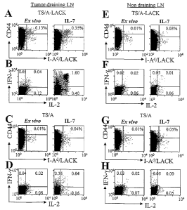
FIGURE 1. IL-7 favors the accumulation of tumor-specific memoly CD4+ T cells
without
5 the need of Ag-stimulation. BALB/c mice (5 per group) were challenged with 3
X 105 TS/A-
LACK or TS/A tumor cells and sacrificed 21 days later. Cells from pools of
tumor-draining LN
(A-D), and non draining LN (E-H) were analyzed ex vivo and after 7 days in
culttire with IL-7
alone. A, C, E, G) cells were stained as described in Material and Methods.
Representative flow
cytometry profiles are shown after gating on viable CD4+, B220", CD8-, CD11b-,
TOPRO-3-
10 cells. The frequency of CD44""g" I-Ad/LACK+ CD4+ cells is indicated. B, D,
F, H) lymphocytes
were stimulated with LACK aAPC (see Materials and Methods), fixed, pei-
nieabilized, stained
with anti-CD4 mAb, anti-IL-2 and anti-IFN-y mAbs, and analyzed by flow
cytometry.
Representative dot plots showing IL-2 and IFN-y production by CD4+ are shown.
The fi=equency
of cytokine-producing cells is reported in each quadrant. The experiment is
representative of 6
independent deter-minations. In some cases, LACK-specific IFN-7 release was
detected in IL-7-
treated LN culture of TS/A-tumor-bearing mice. ~ven though the nature of these
cells remains to
be elucidated, these cells might be specific for the LACK homologue mammalian
RACK (24).
FIGURE 2. IL-7 and IL-2, but not Ag, IL-15 and IL-6 elicit the accumulation of
tumor-
specific CD4+ T cells. Pools of LN cells recovered from TS/A-LACK tumor-
bearing BALB/c
mice (n=5) were cultured with irradiated splenocytes in the absence (APC) or
in the presence of
the LACK peptide (Ag/APC) or with IL-7, IL-2, IL-15 and IL-6 alone. After 7
days, cells were
recovered and surface stained to deteimine the frequency of Ag-experienced
LACK-specific T
cells (A, C), or stimulated with LACK aAPC to evaluate LACK-specific
intracellular cytokine
release (B, D), as described in Figure 1. A) Representative flow cytometry
profiles are shown
after gating on viable CD4+, B220", CD8-, CDllb", TO-PRO-3" cells, The
fi=equency (A) and
total number (C) of CD44""g" I-Ad/LACK+ CD4+ cells aniong CD4+ cells is
indicated. B)
Representative dot plots depict IL-2 and IFN-y production by CD4* T cells. The
frequency (B)
and total number (D) of LACK-specific cytokine-producing cells among CD4+ T
cells is
reported. The experiments are representative of 3to 5 independent
determinations.
FIGURE 3. IL-7 and IL-2 sustain the Ag-independent proliferation of in vivo-
primed
tunlor-specific CD4+ T cells. A) Pools of LN cells recovered from naYve or
TS/A-LACK tumor-
bearing BALB/c mice (n=5) were labeled with the CFSE vital dye and cultured
for a week in
plain inedium. Representative dot plots of viable CD4+ T cells are shown. B-D)
CFSE-labeled
LN cells derived fi=om TS/A-LACK-tumor-draining LNs were cultured without
(nil) or with IL-
CA 02618580 2008-02-08
WO 2007/017915 PCT/IT2006/000600
11
7, IL-2, IL-15 and IL-6 for 7 days. Cells were then stimulated with LACK aAPC
(B, D) or
control aAPC (C), and analyzed by flow cytonletry for intracellular cytokine
release as described
in Figure 1. Representative dot plots showing the CFSE content and IL-2 or IFN-
y production by
viable CD4+ T cells are shown in B and C. In D, the total number of CFSEd'm
CD4+ T cells
producing IL-2 and/or IFN-y is depicted. The experiment is representative of 3
independent
deterniinations.
FIGURE 4. IL-2, but not IL-15 and IL-6, IL-10, TNF-oc niimies IL-7 and
enriches cell
cultures for tumor-specific meinory CD4+ T cells in the absence of Ag. TS/A-
LACK and
TS/A-tumor-draining LN derived fi=om 16.2(3 transgenic mice were cultured for
a week in the
absence (-) or in the presence of the indicated recombinant cytokine alone and
analyzed by flow
cytometry as described in Fig. 1. A) Representative dot plots are shown after
gating on viable
CD4+, B220", CD8-, CD11b', TOPRO-3- cells. The frequency of I-Ad/LACK+ CD4+
cells is
indicated. B-D) Lynlphocytes derived from TS/A-LACK- (B, C) and TS/A- (D)-
tumor draining
LN ctlltures were stimulated with LACK aAPC (B, D) or control aAPC (C) to
detect intracellular
IL-2, IFN-y and IL-4. Representative dot plots showing IL-2 and IFN-y
production by CD4+ are
shown. IL-4+ cells were within background levels in all experiments. The
fi=equency of IL-2+,
IFN-y + cytokine-producing cells is reported in each quadrant. E) Tumor-
draining LN cells fi=om
TS/A-LACK-tumor-bearing 16.2(3 mice were labeled with CFSE, and cultured in
the absence (-)
and in the presence of the indicated cytokines for a week. Thereafter the
cells were re-stimulated
with LACK aAPC ad analyzed by flow cytometry. Representative dot plots showing
the CFSE
content and IL-2, and IFN-y production by CD4+ are shown. The experiment is
representative of
3 independent determinations.
FIGURE 5. IL-7 and IL-2 favor T cell survival and the optimal expression of
Bcl-2. Cells
from TS/A-LACK tumor draining LN were labeled with the CFSE vital dye and
cultured in the
absence (-) or in the presence of IL-7, IL-2, IL-15 and IL-6 alone. After 7
days, cells were
recovered and stained with anti-CD4 mAb and TO-PRO-3 (A) and with anti-Bcl-2
mAb (B, C).
A) representative dot plots of total CD4+ cells are shown. The fi-equencies of
total CD4+
TOPRO-3+ (brackets) and of CFSE dim, TO-PRO-3- cells (bold) are reported. B-C)
events are
shown after physical gating on viable CD4+ T lympllocytes. C) thin line:
isotype control; thin
lines-shaded profile: anti-Bcl-2 Ab, mediuin cultured cells; thick lines: anti-
Bcl-2 Ab, cytokine-
cultured cells. The experiment is representative of 2 independent
determinations.
FIGURE 6. Phenotype and subset representation of lymphocytes maintained in IL-
7 and
IL-2. CFSE-labeled TS/A-LACK-turnor-drainin,g LN cultures were maintained for
a week in the
presence of IL-7 or IL-2. Thereafter the cells were stained with anti CD4,
CD44, CD25, CD127,
CA 02618580 2008-02-08
WO 2007/017915 PCT/IT2006/000600
12
and CD132 mAbs, A) representative dot plots report the expression levels of
CD44, CD62L,
CD25, CD127, and CD132 of viable CD4+. B) ov'erlay of CD4+, CFSE dim cells
derived from
IL-7 (thin lines) and IL-2 (thick lines) cultures in the absence of Ag-
stimulation are shown. C)
CFSE-labeled cells were surface stained for CD4 and CD62L surface levels, and
for intracellular
Bcl-2. Dot plots are shown after gating on CD4+ CFSE dim cells. The experiment
is
representative of 3 independent determinations.
FIGURE 7. IL-7-cultured cells are comparable to the ones found at the time of
sacrifice.
TS/A-LACK-tumor-draining LN derived from 16.2p transgenic mice were analyzed
by I-
Ad/LACK staining as described in Fig. 1 e.x i4vo and after the short-tenn
culture in IL-7 or IL-2
in the absence of Ag-stimulation. A) representative dot plots report the
expression levels of
CD44, CD25, CD127, and CD132 of viable CD4+, B220", CDS-, CD11b", TOPRO-3'
cells. B)
overlay of CD4+, I-Ad/LACK+ and CD4+, I-Ad/LACK" after gating on B220", CD8",
CD 11 b-,
TOPRO-3" lyinphocytes are showm. Ex vivo: dotted lines; thin line: IL-7; thick
line: IL-2.
FIGURE 8. IL-7-, IL-2- or IL-15-driven cultures are enriched for in vivo-
primed TAG IV-
specific meniory CD8+ T cells. C57BL/6 micei were immunized with bone man=ow-
derived
dendritic cells pulsed with the Tag IV peptide. Fourteen days later axillary,
brachial and inguinal
LN cells were recovered and analyzed ex vivo and after a week in culture in
the absence or in the
presence of IL-7, IL-2, IL-15, IL-6, The total number of Tag IV- specific
cytokine-producing
CD45.1- CDS+ T cells is reported. The experiments are representative of 2
independent
determinations.
FIGURE 9. IL-7-, IL-2- or IL-15-driven cultures rescue comparable numbers of
CD8+ T
cells. C57BL/6 mice were immunized with bone mairow-derived dendritic cells
pulsed with the
Tag IV peptide. Fourteen days later axillary, brachial and inguinal LN cells
were recovered and
analyzed ex vivo and after a week in culture in the absence or in the presence
of IL-7, IL-2, IL-
15, IL-6. The total number of CD45.1- CDS+ T cells is reported. The
experiments are
representative of 2 independent deteiminations.
FIGURE 10. IL-7 favors the accuniulation of tumor-specific niemory CD8+ T
cells
otherwise uudetectable ex vivo, A) BALB/c miq (5 mice per group) were
challenged with 3 X
105 TS/A-LACK tumor cells and sacrificed 21 days later. Cells from pools of
tumor draining LN
(A-C) were analyzed e.x vivo (A) and after 7 days in culture in the absence or
in the presence of
IL-7, IL-2, IL-15, IL-6 (B-C). A) Representative dot plots depict IL-2 and IFN-
y production by
ILJ1.26- CDS+ T cells, The frequency (A, B) and total number (C) of AH-1-
specific cytolcine-
producing ILJ1,26" CDS+ T cells is reported.
CA 02618580 2008-02-08
WO 2007/017915 PCT/IT2006/000600
13
FIGURE 11. Intermediate meniory CD4+ T cells accumulate in the presence of IL-
7 in a
cell-density dependent, CsA-sensitive manner. Cells der-ived from the
axillary, brachial and
inguinal LN of naYve (A, B) and TS/A-LACK tumor-bearing (C-E) 16.2p mice were
labeled with
CFSE and respectively cultured for 7 days in plain medium, in the presence of
LACK peptide
(Ag) (A, B) or IL-7 (C-E) in the absence (nil) or in the presence of the
indicated inhibitors. At
the end of the culture the cells were stimulated for 5h with L/28 aAPCs, and
intracellular
cytokine release was detei-mined. A-D) Histograms show the CFSE dilution
profile of equivalent
numbers of (7x10⁴) CD4+ T cells. In A and C the thin lines reflect the CFSE
profile of CD4+ T
cells cultured in plain medium, while the thick lines depict the CFSE profile
of CD4+ T cells
cultured in Ag or IL-7, respectively. In B and D, thin line: cells cultured in
the absence of the
inhibitor, thick line: cells cultured in the presence of the inhibitor. E) Dot
plots are shown after
gating on viable CD4+ T cells. Percentages indicate the frequency of LACK-
specific cytokine
secreting cells.
FIGURE 12. IL-7 sustains the fast, Ag-independent proliferation of a fraction
of human
peripheral blood CD4+ T cells. Human PBMCs fi=om healthy donors were labeled
with the
CFSE vital dye and cultured for 7 days in the absence (nil) or in the presence
of recombinant
human IL-7 (100 ng/ml) at the indicated cell densities. Representative dot
plots of viable CD4+ T
cells are depicted. The fi=equency of CFSEd'm CD4+ T cells is indicated. B)
Histogram overlays of
CD4+ T cells cultured at the indicated cell density (N:non proliferating
cells, S and F: slow and
fast-proliferating cells).
FIGURE 13. IL-7 sustains the accumulation of fast-dividing cells in autologous
serum.
Human PBMCs from a healthy donor were labeled with the CFSE vital dye and
cultured for 7
days in coulture medium addition of 10% autologous serum in the absence (nil)
or in the
presence of recombinant human IL-7 (100 ng/ml) at the indicated cell
densities. Representative
dot plots of viable CD4+ T cells are depicted. The frequency of non
proliferating (N), and slow-
(S) and fast- (F) proliferating CFSEd"" CD4+ T cells is indicated.
FIGURE 14. IL-7-driven accumulation of fast-dividing CD4 T cells is dose
dependent.
Human PBMCs from healthy donors were lat~eled with the CFSE vital dye and
cultured for 7
days in the presence of the indicated amounts of recombinant human IL-7.
Representative dot
plots of viable CD4+ T cells are depicted. Non proliferating (N), and slow-
(S) and fast- (F)
proliferating CFSE.d"" CD4+ T cells are indicated.
FIGURE 15. IL-7 sustains the accuniulation of fast-dividing IFN-y-producing
memory
CD4+ T cells. Human PBMCs from healthy donors were labeled with the CFSE vital
dye and
cultured for 7 days in the presence of the indicated amounts of recombinant
human IL-7. After 7
CA 02618580 2008-02-08
WO 2007/017915 PCT/IT2006/000600
14
days, cells were hai-vested and re-stimulated with PMA and Ionomycin for 6
hours. Cells were
then surface stained, fixed and stained with anti-IFN-y mAb. Representative
dot plots of viable
CD4+ T cells are depicted.
FIGURE 16. IL-7-driven accumulation of IL-2/IFN-y+ CD4+ T cells is dose
dependent.
Human PBMCs fi-om healthy donors were cultured for 7 days in the presence of
the indicated
amounts of recombinant human IL-7. After 7 days, cells were harvested and re-
stimulated with
PMA and Ionomycin for 6 hours. Cells were then surface stained, fixed and
stained with anti-
IFN-y mAb. Representative dot plots of viable CD4+ T cells are depicted.
FIGURE 17. IL-7 best expands fast-dividing memory CD4+ T lymphocytes, while IL-
15
drives the accumulation of fast-dividing memory CD8+ T lymphocytes. CFSE-
labeled human
PBMCs were cultured in plain medium (nil) or in the presence of human
recombinant IL-7, IL-2,
IL-15, and IL-6 for 7 days, then stained with anti-CD4 and anti-CD8 mAb, and
analyzed by flow
cytometry. Histogram overlays show the CFSE content within the same number of
CD4+ (A) and
CDB+ (B ) lyinphocytes. Fast- (F), Slow-(S), and Non-(N) proliferating cells
are indicated.
FIGURE 18. IL-7, IL-2 and IL-15 di-ive optimal Bcl-2 expression on cultured
human cells.
Human PBMCs from healthy donors were labeltd with the CFSE vital dye and
cultured in the
absence (nil) or in the presence of human recombinant IL-6, IL-7, IL-2 and IL-
15 for 7 days.
Cells were then stained with anti-CD4 mAb, fixed and intracellular Bcl-2
levels were deteimined
by intracellular staining. Events are depicted after gating on CD4+ T cells.
FIGURE 19. IL-7-driven CD4 T cell proliferation of human peripheral blood
memory CD4
T cells relies on CsA and LFA-1/ICAI\/I-dependent signaling. Human PBMCs fi-om
healthy
donors were labeled with the CFSE vital dye and cultured in the presence of
human recombinant
IL-7 for 7 days in the absence (nil) or in the presence of the indicated
inhibitors. Cells were then
stained with anti-CD4, antiCD45RA and anti-CD62L inAb and analyzed by flow
cytometry. A)
Histograms depict the overlay of the CFSE profiles of viable CD4+ T cells. B)
Dot plots depict
viable CD4+ T cells. Fast (F), Slow (S), and Non (N) proliferating cells were
electronically
defined and are shown in C. The relative representation of naive and memory T
cells is indicated
in the tigtire. Results are representative of 2 independent determinations.
FIGURE 20. Alycobacterium tubercrclosis spGcific CD4+ T cells are enriched for
by IL-7-
driven cultures. PBMC fi-om three TB patients (Pt. #1, Figure 20 A; Pt#2,
Figure 20 B; and
Pt#3, Figure 20 C) were analyzed for MTP-specific IFN-y release by an ELISPOT
assay at the
time of thawing (crio-preserved) and after a 7 days culture in the absence
(nil) or in the presence
of human recombinant IL-7 (cultured). Background IFN-y release was measured in
unpulsed
control wells (ctr). B) The total number of MTP-specific IFN-y-producing cells
is depicted.
CA 02618580 2008-02-08
WO 2007/017915 PCT/IT2006/000600
Figure 21. The IL-7-driven culture facilitates the identification of
Mycobacterium
titbei=culosis specific CD4+ T cells. MTP-specific IFN-y production by crio-
preserved and IL-7
cultured PBMCs derived from 8 healthy donors and 5 TB-patients were analyzed
by ELISPOT.
Statistical significance was evaluated by a paired two-tail T-test.
5 FIGURE 22. Mycobacterium tubei-culosis specific IL-2/IFN-y+ CD4+ T cells
accumulate in
IL-7 driven cultures. Pt#1 cells were cultured in the absence (nil) or in the
presence of human
recombinant IL-7 (cultured), A) MTP-specific IFN-y release detected by ELISPOT
(also
depicted in Fig. 20A), B) A parallel set of cultured cells were also re-
stimulated for 6 hours with
MTP-pulsed autologous irradiated PBMC, surface stained with anti-CD4 mAb,
fixed and fiu-ther
10 stained with anti-IFN-y mAb. Events are depicted 4fter gating on viable
CD4+ T lyinphocytes.
FIGURE 23. Mycobacter=ircin tccbei-culosis specific CD4+ T cells proliferate
in IL-7-driven
cultures. A) Pt#1 cells were cultured in the absence (nil) or in the presence
of human
recombinant IL-7 (cultured). MTP-specific IFN-y release detected by ELISPOT
(also depicted in
Fig. 20A). B) Parallel cultures were set up with CFSE-labeled PBMCs. After 7
days the cells
15 were stimulated with MTP-pulsed autologous irradiated PBMCs, and
intracellular IFN-y release
was deteimined by flow cytometry. Events are shown after gating on viable CD4+
T cells.
FIGURE 24. Candida Albicans specific IFN-y+ memory T cells accuniulate in IL-7-
di-iven
cultui-es. A) C. Albicans-specific IFN-y release an ELISPOT assay by Pt#1
cells either crio-
preserved or cultured for 7 days in the absence (nil) or in the presence of
human recombinant IL-
7 is depicted. Background IFN-y release was measured in unpulsed control wells
(ctr). B) The
total number of IFN-y-producing cells is depicted.
FIGURE 25. IL-7/IL-15 cultured meinory cells delays tumor growth upon adoptive
cell
tt-ansfer in vivo. Lymph nodes cells derived fTm control (naive) and TS/A-LACK
tumor-
bearing mice were cultured for 7 days in high cell density (5x10⁶ cells/ml) in
the presence of IL-
7 and IL-15 (both at 50 ng/ml). Thereafter 10' cultured cells adoptively
transferred into naYve
BALB/c mice. 48 hours later mice were challenged with 300.000 TS/A-LACK cells
and tumor
growth was monitored overtime. Statistical significance was evaluated by a
paired two-tail T-
test.
FIGURE 26. Scheniatic representation of the proposed strategy and its
diagnostic and
therapeutic implications.
FIGURE 27. Activation with beads conjugated with anti-CD3 and anti-CD28
antibodies
(ba CD3/CD28) and culture with IL-7 and IL-15 proniote T cell expansion and
efficiently
generate genetically niodified human lymphocytes with a preserved CD4/CD8
ratio. 5x10⁶
PBMC were stimulated either with aCD3 and cultured with IL-21 or with
baCD3/CD28 and
CA 02618580 2008-02-08
WO 2007/017915 PCT/IT2006/000600
16
cultured with IL-7 and IL-15. At day 14, cells were counted by trypan blue
exclusion, (A)
Averages of T cell fold expansion in the two stimulation and culture
conditions are reported (77=4
donors). 48 and 72 h after initial stimulation, cells were transduced by the
SFCMM3 retroviral
vector. At day 6, genetically modified cells were quantified by flow-cytometry
after staining
with anti-LNGFR antibodies. (B) Averages of titransduction efficiency (in %)
in the two
stimulation and culture conditions are reported (fa=4 donors). At day 14,
cells were analyzed by
flow-cytometry for ALNGFR expression and for the expression of CD4 and CD8.
(C) Averages
of CD4/CD8 ratio in genetically modified cells generated with the two
protocols are reported
(n=4 donors).
FIGURE 28. Activation with baCD3/CD28 and culture with IL-7 and IL-15 generate
TK+
human lymphocytes with central meinory phenotype. At day 14, Th+ cells
generated with
aCD3 and cultured with IL-2 or generated with baCD3/CD28 and cultured with IL-
7 and IL-15
were analyzed for memory phenotype. After gating for ALNGFR expression, cells
were analyzed
by flow-cytometry for CD45RA and CD62L co-expression. Averages of the relative
distribution
(y axis, %) of CD45RA+CD62L+ (black bars), CD45RA" CD62L + (dark grey bars),
CD45RA-
CD62L "(light grey bars) or CD45RA+ CD62L "(white bars) are reported (A) for
CD4+ and (B)
for CD8+ cells obtained from n=4 donors. Cells were also analyzed by flow-
cytometry for CD27
and CD28 co-expression. Averages of the relati1 e distribution (y axis, %) of
CD28+CD27+
(black bars), CD28"CD27+ (dark grey bars), CD28"CD27" (light grey bars) or
CD2S+CD27-
(white bars) are reported (C) for CD4+ and (D) for CDS+ cells obtained from
n=4 donors.
FIGURE 29. Central niemory TK+ hunian lyniphocytes are un-polarized cells. At
day 14,
TIC+ cells generated with aCD3 and cultured with IL-2 or generated with
baCD3/CD28 and
cultured with IL-7 and IL-15 were analyzed for cytokine production. After
gating for OLNGFR
expression, cells were analyzed by flow-cytometry for IFN-y and IL-4
production. Averages of
the relative distribution (y axis, %) of IL-4+IFN-y "(white bars), IL-4+IFN-y
+(grey bars) or IL-4-
IFN-y + (black bars) are reported (A) for CD4+ and (B) for CD8+ cells.
FIGURE 30. Activation with beads conjugated with anti-CD3 and anti-CD28
antibodies
and culture with IL-7 +/- IL-15 or IL-2 promote T cell expansion and
efficiently generate
genetically niodified human lymphocytes with a preserved CD4/CD8 ratio. PBMC
were
stimulated either with beads conjugated with anti-CD3 and anti-CD2S (named
"B") and cttltured
with no cytokines, IL-7 + IL-15, IL-2, or IL-7, qr were stimulated with
soluble anti-CD3 and
cultured with IL-2. 48 and 72 h after initial stimulation, cells were
transduced by the SFCMM3
retroviral vector. A) At day 6, genetically modified cells were quantified by
flow-cytometry after
staining with anti-LNGFR antibodies. Averages of transduction efficiency (in
%) in the different
CA 02618580 2008-02-08
WO 2007/017915 PCT/IT2006/000600
17
stimulation and c.ulture conditions are reported. B) At day 10, cells were
analyzed by flow-
cytometry for ALNGFR expression and for the expression of CD4 and CDB.
Averages of
CD4/CD8 ratio in genetically modified cells generated with the different
protocols are reported.
C) At days 6 and 9, cells were counted by trypan blue exclusion. Averages of T
cell fold
expansion in the different stimulation and culture conditions are reported
(77=5 donors). p <
0.05 ** = p <0.01.
FIGURE 31. Activation with beads conjugated with anti-CD3 and anti-CD28
antibodies
and culture with IL-7 +/- IL-15 or IL-2 generate transduced human lyniphocytes
with
central memory phenotype. At day 10 transduced lynzphocytes generated either
with beads
conjugated with anti-CD3 and anti-CD28 and cultured with no cytokines, IL-7 +
IL-15, IL-2, or
IL-7, or stimulated with soluble anti-CD3 and cultured with IL-2 were analyzed
for memory
phenotype. Cells were analyzed by flow-cytomktry for CD45RA and CD62L co-
expression.
Averages of the relative distribution (y axis, %) of CD45RA+CD62L+ (naive
cells), CD45RA"
CD62L +(central memory cells), CD45RA" CD62L "(effector memory cells) or
CD45RA+
CD62L "(tei-minally differentiated effectors) are reported on CD3+ OLNGFR+
cells. (n=6
donors). * = p < 0.05 * } = p <0.01.
FIGURE 32 kinetic of expression of IL-2/15 receptor P (CD122) does not depend
from the
activation, culture and transduction procedure. At days 0, 2, 4, 9 and 13,
after initial
stimulation, transduced lymphocytes generated either with beads conjugated
with anti-CD3 and
anti-CD28 and cultured with no cytokines, IL-7 + IL-15, IL-2, or IL-7, or
stimulated with soluble
anti-CD3 and cultured with IL-2 were analyzed for the expression of CD122. The
% of CD3+
cells (at days 0, 2 and 4) and of transduced cells (at days 9 and 13)
expressing CD122 at different
time-points are reported on A) CD8+ and B) CD4+ cells. (77=4 donois).
FIGURE 33 Activation with beads conjugated with anti-CD3 and anti-CD28
antibodies
and culture with IL-7 +/- IL-15 or IL-2 pron~ote intense and prolonged expi-
ession of IL-2
receptor a (CD25) on transduced lymphocytes. At days 0, 2, 4, 9 and 13, after
initial
stimulation, transduced lymphocytes generated either with beads conjugated
with anti-CD3 and
anti-CD28 and cultured with no cytokines, IL-7 + IL-15, IL-2, or IL-7, or
stimulated with soluble
anti-CD3 and cultured with IL-2 were analyzed for the expression of CD25. The
% of CD3+
cells (at days 0, 2 and 4) and of transduced cells (at days 9 and 13)
expressing CD25 at different
time-points are reported on A) CDS+ and B) CD4+ cells. (n=4 donors). *= p <
0.05 p
<0.01.
FIGURE 34 Activation with beads conjugated with anti-CD3 and anti-CD28
antibodies
and culture with IL-7 promotes intense and prolonged expression of IL-7
receptor
CA 02618580 2008-02-08
WO 2007/017915 PCT/IT2006/000600
18
a (CD127) on transduced lymphocytes. At days 0, 2, 4, 9 and 13, after initial
stimulation,
transduced lymphocytes generated either with beads conjugated with anti-CD3
and anti-CD2S
and cultured with no cytokines, IL-7 + IL-15, IL-2, or IL-7, or stimulated
with soluble anti-CD3
and cultured with IL-2 were analyzed for the expression of CD127. The % of %
of CD3+ cells
(at days 0, 2 and 4) and of transduced cells (at days 9 and 13) expressing
CD127 at different
time-points are reported on A) CDS+ and B) CD4+ cells. (n=4 donors), *= p <
0.05 p
<0.01.
FIGURE 35 Activation with beads conjugated with anti-CD3 and anti-CD28
antibodies
and culture with IL-7 preserves a high alloreactive proliferative potential
and a low
sensitivity to apoptotic signals in transduced cells.
Transduced T cells generated either with beads conjugated with anti-CD3 and
anti-CD28 and
cultured with IL-7 or IL-2, or by activation with soluble anti-CD3 and culture
in IL-2 were
stained with CFSE at day 9 after initial stimulation, and were co-cultured
with irradiated
allogeneic PBMCs. Unmanipulated peripheral blood lyinphocytes (PBL) from the
same donors
were stained with CFSE, co-cultured with the same in=adiated allogeneic PBMCs
and used as
controls. After 7 days cells were counted, stained with To-pro3, and analysed
by FACS to
evaluate the percentage of dividing and/or dying cells. A) Percentage of
dividing cells, B) Total
number of dying cells. Activation induced cell death was calculated on
dividing cells (white,
lower part of histograms) and death for neglect was calculated on non-dividing
cells (black,
upper part of histograms). * = p < 0.05 ** = p<0.01.
FIGURE 36 Self-renewal capacity of central memory genetically modified cells
generated
with beads conjugated with anti-CD3 and anti-CD28 antibodies and culture with
IL-7,
after allogeneic stimulation.
A) Cells treated as described in figure 35 were analysed for the expression of
CCR7 and IL-7Ra
7 days after the MLR was started. The percentage of CCR7+ (upper panel) and IL-
7Ra+ (lower
panel) dividing cells is shown in the left upper quadrant. B) Cells were then
re-challenge in vitro
with the same allogeneic stimulators, following the same culture conditions
utilized in the first
stimulation. The percentage of dividing cells after the second stimulation is
shown.
Figure 37. Transduced lymphocytes generated with with beads CD3/CD28
antibodies
rapidly engraft in NOD/scid mice
NOD/scid mice were conditioned and transplanted with human skin, were infused
i.v. with
20x106 transduced lymphocytes generated with beads CD3/CD2S and culture with
IL7 or IL2, or
with OKT3 and culture with IL-2. Hunlan chimerism was assessed weekly by flow-
cytometry
after staining for human CD3 and mouse CD45. Quadrants and percentages were
set according
CA 02618580 2008-02-08
WO 2007/017915 PCT/IT2006/000600
19
to isotype control staining. Kinetic of human chimerism is shown (n=4 donors).
Figure 38. Transduced lymphocytes generated with beads CD3/CD28 and cultured
with
IL-7 induce severe xenogenic GvHD in NOD/scid mice
NOD/scid mice were conditioned and transplanted with human skin, were infused
i.v, with
20x 106 transduced lymphocytes generated with beads CD3/CD28 and culture with
IL7 or IL2, or
with OKT3 and culture with IL-2. Mice were monitored for xenogenic GvHD
according 1)
Wight loss. 2) GvHD clinical score (described in material and methods).
Controls: animals that
ti
did not receive infiisions of lymphocytes.
Figure 39. Transduced lymphocytes generated with with beads CD3/CD28 and
cultured
with IL-7 induce seve--e allogenic GvHD in NOD/scid mice
Tlu=ee weeks after human T-cell inftision, NOD/Scid chimera mice were
sacrificed and human
skin was removed bilaterally. All biopsies were subsequently blindly analysed
by pathologists
through ematossilin-eosin (EE) and anti human CD3 staining (ahCD3).
Representative sections
are reported.
Materials and Methods
Experimental protocols were approved by the Ethical Committee of the San
Raffaele Scientific
Institute and performed according to its guidelines.
Mice and Tumor Cells
Seven to 8-week old BALB/c and CD45.2+ C57BL/6 mice were purchased from
Charles River
(Charles River Italia, Milano, Italy). CD45.1+ C57BL/6, DO11.10 and 16.2(3
Transgenic (Tg)
(BALB/c background) (25) mice were bred in the Institute specific pathogen
free facility. TS/A
and TS/A-LACK mouse mammary adenocarcinoma were previously described (17, 19,
26).
Exponentially growing 4x10⁵ tumor cells were subcutaneously injected in 100 l
of PBS in the
right flai-llc of syngeneic mice (BALB/c). Typically, five mice per group were
used in each
experiment.
T cell primary cultures
Twenty days after tumor cell injection mice were sacrificed and the axillary,
brachial and
inguinal peripheral lyinph nodes (LN) draining and distal (non draining LN) to
the site of tumor
growth were recovered. In the experiments of dendritic cell (DC) vaccination,
mice were
sacrificed fourteen days after DC administration and the axillary, brachial
and inguinal LN were
surgically excised. LN cells were cultured in 24 well plates at the density of
5x10⁶ in coinplete
medium in the absence or in the presence of recombinant murine IL-7 (200
ng/ml), IL-2 (20
ng/ml), IL-6 (45 ng/ml), or IL-15 (100 ng/ml) (all from Peprotech). In
parallel cultures, cells
were in vitro stimulated with the LACK-derived' MHC II-restricted peptide (5
M, (25)) and
CA 02618580 2008-02-08
WO 2007/017915 PCT/IT2006/000600
5x 10b iiTadiated syngeneic splenocytes. As a control similar cultures Nvere
set up from syngeneic
naive mice. In some experiments, LN cells were l~beled with the fluorescent
dye CFSE (5-(and-
6)-carboxyfluorescein diacetate, succinimilyl ester) at the final
concentration of l M
accordingly to manufacturer instruction.
5 Dendritic cell (DC) preparation
Bone Marrow derived DC were obtained as previously described (18). Briefly,
CD45.2+
C57BL/6 bone marTow precursors were propagated for 7 days in complete Iscove's
medium
containing 25 ng/ml recombinant murine GM-CSF and 5 ng/ml recombinant murine
IL-4
(Pharmingen, San Diego, CA). Then, BMDC were matured at 37 C in the presence
of LPS (1
10 g/ml, Sigma, Milan, Italy) for 8 liours and pulsed for 1 hour with 10 g/ml
of the large T Ag-
derived Tag IV peptide (18). DC maturation and purity were routinely evaluated
by flow
cytometry after staining with mAb recognizing CD 11 c, MHC class II, B7.1,
B7.2 and CD40
molecules (all from Phaimingen). 2x10⁵ pulsed mature DC were subcutaneously
injected in 200
l of PBS in the right flanlc of syngeneic C57BL/6 mice.
15 Flow Cytometry analysis
I-Ad/LACK multimer staining was previously described. Briefly I-Ad/LACK dimers
(MHC II-
peptide complexes, 3 g/sample.) are multimerized by the addition of Alexa 488-
coupled protein
A (Molecular Probes Inc., Eugene, 0.3 g/sample) in PBS for 30 minutes at room
temperature.
Free protein A binding sites were saturated by the addition of total IgG (1
g/sample). 6x 10⁵ cells
20 were first incubated with a bloclcing buffer (5% rat serum + 95% culture
supernatant of 2.4G2
anti-FcR mAb-producing hybridoma cells, 20 minutes) and then stained with the
multimers (lh
at 4 C, in PBS supplemented with 0.5% BSA). The cells were then stained with
anti-CD4, anti-
CD44, anti-CDl lb, anti-B220, anti-CDSa mAbs (PharMingen, San Diego, CA, USA)
and TO-
PRO-3 (1 n1Vl, Molecular Probes). 3x10⁵ CD4+ or 103 CD4+ I-Ad/LACK+ events
were collected
by exclttding all of the anti-CDllb+, anti-B220+, anti-CD8a+ and TO-PRO-3+
events. Where
indicated the cells were surface stained with anti-CD4 or anti-CD8 mAb, and
anti-CD44, anti-
CD127, anti-CD25, anti-CD132 and anti-CD62L mAbs (all from PharMingen except
anti-
CD127 Ab, A7R34 clone, fi=om Bioscience), and fixed, permeabilized and further
stained with
anti-Bcl-2 mAb according to to manufacturer instniction.
LACK-specific artificial antigen presenting cells (aAPC) and cytokine
secretion assays
5 m polystyrene sulfate latex beads (Interfacial Dynamics) were coated with I-
Ad/LACK dimers
(20 g/rnl) and anti-CD28 niAb (37.51; 2 g/ml) (LACK aAPC) or with anti-CD28
mAb only
(control aAPC). Coating of the proteins was monitored by flow cytometry
analysis. Typically
5x 10$ LN cells were cultured with 5x 10⁶ aAPC for 5 hours at 37 C. Brefeldin
A (5 g/ml,
CA 02618580 2008-02-08
WO 2007/017915 PCT/IT2006/000600
21
Sigma) was added to the cultures for the last 2 hours. Cytokine release
induced by LACK aAPC
Nvas comparable to the one induced by LACK-pulsed syngeneic splenocytes (not
shown). In the
case of AH-1 and Tag IV-induced cytokine production, splenocytes were derived
from DO11.10
and CD45,1+ C57BL/6 mice and used as antigen presenting cells. Splenocytes
(3x10⁷ cells/ml)
were pulsed with 1 M AH-1 (19) and 10 l.Lg/rnl Tag IV (18) peptides for 1
hour at 37 C and
then used to stimulate syngeneic LN cells derived fi=om the LN of tumor-
bearing mice and DC-
vaccinated mice, respectively. Thereafter the cells were stained with anti-
CDB, and IL-2 and
IFN-y as described above and with the anti-clonotipic KJ 1.26 mAb, and the
anti-CD45.1+
marker to exclude T cells of APC origin. CD4+, KJ 1.26" or CD8+ CD45.1 '
events were then
collected on a FACS Calibur. The total number of Ag-specific IL-2+/IFN-y+ T
cells was
determined by multiplying the percentage by the total number of Trypan Blue-
negative LN cells.
Human T cell cultui=es and ELISPOT assay.
Peripheral blood mononuclear cells (PBMC) wer~ obtained from patients with
tuberculosis (TB)
and healthy donors by blood centrifugation over Fycoll-Hypaque density
gradient and analyzed
right away or frozen for fiiture analysis. Where indicated cells were stained
with CFSE (1 M).
Cells were cultured in the absence or in the presence of human IL-7 (200
ng/ml), IL-2 (20 ng/ml,
IL-15 (100 ng/ml) or IL-6 (45 ng/ml) for 7 days. Where indicated Ciclosporin
A(CsA)
(0.5 g/hnl) or anti-LFA-1 (5 g/ml) blocking antibody were added. Cells were
then harvested and
stained with CD4, CDS, CD3, CD56, CD45RA, CD62L mAbs (all fi=om PharMingen)
and
analyzed by flow cytometry.
The ELISPOT assay for IFN-y secretion was performed as previously reported
(28). Briefly cells
Nvere seeded in duplicate at 5x10⁴ cells/well in 96-well plates (MAIl'S4510;
Millipore, Bedford,
Mass.) pre-coated with anti-IFN-y capture niAb (B-B1; Diaclone, Besangon,
France) in the
presence of autologous in=adiated PBMC (5x10⁴ cells/well), and a pool of MTP
peptides for 18 h
at 37 C in air plus 5% COZ. Biotinylated anti-IFN-y detection mAb (B-Gl;
Diaclone) was added
.for 4 h, followed by the addition of streptavidin-all.aline phosphatase
conjugate (Amersham
Phannacia Biotech Europe GmbH, Freiburg, Germany) for 1 h. After a washing
step, the
nitroblue tetrazolium-BCIP (5-bromo-4-chloro-3-indolylphosphate; Sigma, St.
Louis, Mo.)
clu'omogenic substrate was added. Individual spot fonning cells (SFC) were
counted using an
automated image analysis system ELISPOT reader (AID-GmbH, Strassberg,
Germany). A pool
of six synthetic A'Iycobacteriian tuberculosis peptides (MTP; Primm srl,
Milano, Italy) with a
length of 20 amino acids, >70% purified, derived from the sequences of ESAT-6
and CFP-10
secretory proteins of R~L tasber=culosis were used at a final concentration of
2 g/ml per peptide
for the detection of a specific response .(28). PBMCs in medium alone or
stimulated with
CA 02618580 2008-02-08
WO 2007/017915 PCT/IT2006/000600
22
phytohemagglutinin (PHA-P; Sigma) 5~Lg/m1 were respectively used as negative
and positive
controls.
In some instances, MTP-specific IFN-y release was analyzed at the single cell
level by
intracellular cytokine secretion assay, Briefly, 0.6x10⁶ CFSE labeled cells -
were re-stimulated
for 6 hours in the presence of human anti-CD2S stimulating mAb (2 g/ml) and
3x10⁶
autologous irradiated (5000 rad) PBMCs pulsed with HLA-DR-restricted MTP (4
g/ml) or left
un.pulsed, in negative controls. In the last 5 houis $refeldin A (10 g/ml)
was added to the cells.
Thereafter the cells were fixed, permeabilized and stained with anti-CD4, anti-
IL-2 and anti-IFN-
y mAbs and analyzed on a FACS Calibur.
Classification of TB patients.
Five HIV-seronegative patients with active TB (clinic and culture confinned)
were recovered at
the Clinic of Infectious Diseases, S. Raffaele Hospital. They underwent
tuberculin skin test
(TST) administered by the Mantoux method with 0.1 ml (5 tuberculin units) of
Biocinetest-PPD
tuberculin (Chiron Italia srl, Milano, Italy). The size of induration was
evaluated after 48-72
hours (an induration >_10 mm was classified as positive). Peripheral blood was
withdrawn before
starting any therapy and with a previous written informed consent. Healthy
controls (n=S) were
selected among HIV-seronegative individuals with no history of TB exposure, no
infection and
with negative reaction to the TST.
Activation, culture and retroviral transduction of human T-cells
Peripheral blood mononuclear cells (PBMC) wei;e isolated by Lyinphoprep
gradient separation
from buffy coats of healthy donors obtained after infonned consent (Fresenius,
Oslo, Noiivay).
PBMC were cultured in RPMI1640 medium (GIBCO-BRL, Gaithersburg, MD)
supplemented
with antibiotics, glutamine and with 10% heat-inactivated FBS (BioWhittaker-
Italia, Milano,
Italy). PBMC were seeded in 6-well plates (1x10⁶/ml) and activated with anti-
aCD'3 (OKT3
30ng/ml, OrthoBiotech, Raritan, NJ) or para-magnetic baCD3/CD28 (3:1 beads/T-
cell)
(Dynabeads, Dynal Biotech, or Xcyte Therapies Ine., Seattle WA, USA,
Invitrogen). T-cells
were enriched by baCD3/CD28 before culture. Cells activated with aCD3 were
cultured with
human recombinant IL-2 at 600 IU/ml (Chiron, Emeryville, CA).
Cells activated with baCD3/CD28 were cultured: 1. In the absence of cytokines;
2. with human
recombinant IL-7 at the minimal concentration of 5 ng/ml (Peprotech, London,
UK); 3. with
human recombinant IL-7 and IL-15 both at the miniinal concentration of 5 ng/ml
each
(Peprotech, London, UK). At day 2 and 3, cells were transduced Nvith the
SFCMM3 (39-40)
retroviral supeinatant by spinoculation at 2400 rpm for 2h at 37 C with S g/ml
polybrene
(Sigma, St Louis, IVIO). The SFCMM3 retroviral vector encodes for the TK
suicide gene under
CA 02618580 2008-02-08
WO 2007/017915 PCT/IT2006/000600
23
the LTR promoter and for a tnlncated form of thei low affinity receptor for
nerve growth factor
(ALNGFR) under the SV40 promoter (27). The retroviral supematant was provided
by Molmed
s.p.a. At day 6 after T-cell activation, cell-sized beads used for the
stimulation were removed
fi=om T-cells, according to manufactureur's instructions, hi some experiments,
at day 7 after T-
cell activation, transduced lyniphocytes were positively selected according to
the protocol that
follows (protocol entitled: Positive iirununeselection of transduced
lymphocytes).
Cells were cultured up to 14 days. At day 14, fold expansion was calculated by
multiplying the
percentages of LNGFR+ cells deteimined by flow-cytometry with the trypan blue
counts.
Medium and cytolcines were replaced, according to the initial protocol, every
3-4 days. At
selected time-points, fold expansion was calculated by multiplying the
percentages of ALNGFR+
cells detei-mined by flow-cytometry with the trypan blue counts.
Positive immuneselection of transduced lymphocytes
At day 7 after T-cell activation transduced lymphocytes were positively
selected. Since
transduced cells expressed ALNGFr on their surface, anti-LNGFr antibodies
covered with
-
magnetic beads were used to separate transduced from untransduced lymphocytes.
Cells were collected from plates in tubes, washed (1500 rpm, 10 min. at room
temperature-RT)
and resuspended in IATB at a final concentration of 5x10⁶/ml. Anti-LNGFr
antibody 20.1 was
then added to the cell suspension (10 g/20x106) and cells were placed to
rotate at 10 rpm for 30
min at RT. T-cells were then washed once and resuspended in WB at 25x106/ml.
Dynabeads M-
450 Sheep anti-Mouse IgG were then added (5x10⁶ beads/106 positive cells) and
cells were
placed to rotate at 10 rpm for 30' at room temperature. Transduced cells were
then magnetically
selected. To this purpose, tubes were placed near the magnet for 3 min and the
negative fraction
was discarded. This procedure was repeated for a total of three times. Finally
the fi=action of cells
bound to the beads was removed fi=om the magnet, washed, and resuspended in
fi=esh medium
with the appropriate cytokine cocktail at a concentration of 1 x 10bcells/ml.
Floiv cytometa=y of transduced T-cells
Flow cytometry was used for analysis of surface phenotype, transduction
efficiency, cell cycle,
i '
and cytokine production. The following antibodies (Pharmingen) were used: FITC-
conjugated
mAb to human CD4, CDS, CD45RA, CD27 and IFN-7, (PE)-conjugated mAb to human
CD4,
CDS, LNGFR, CD62L, CD2S and IL-4, peridinin chlorophyll-a protein (PerCP)-
conjugated
mAb to mouse CD45 (Ly5.1), and allophycocyanin (APC)-conjugated mAb to human
CD3
Samples were run through a Facscalibur flow-cytometer (Becton Dickinson,
Mountain View,
CA) after isotype-matched fluoroclu-ome-conjugated irrelevant mAb-stained
control and data
were analyzed using CellQuest Software (Becton Dickinson).
CA 02618580 2008-02-08
WO 2007/017915 PCT/IT2006/000600
24
Fluoroclu-ome-conjugated antibodies to CD127, CD122, CCR7, and to mouse CD45
(Phai-mingen, San Diego, CA, USA) were also utilized to stain lymphocytes
Cytokine production
For detennination of cytokine production, cells were seeded in 24-well plates
(1x10⁶/ml) and
stimulated with 50ng/ml PMA (Sigma) and 1 g/ml ionomycin (Signla). After 4h,
brefeldin A
(Sigma) were added for additional 2h (10 g/ml). Cells were then stained with
the appropriate
fluorocl-irome-conjugated anti-surface marker antibodies and fixed with 1%
para-formaldehyde
at 4 C for 10 min. Intracellular staining was pfforined with the appropriate
fluorochrome-
conjugated anti-cytokine antibodies after incubation for 20 min at RT in PBS
2% FBS containing
0,05% saponin (Sigina).
CFSE dilution assay and mixed lymphocyte reaction (MLR)
Analysis of T-cell alloreactivity was perfoi-med at day 10 after initial
culture of lymphocytes.
Transduced T-cells were stained with CFSE at day 10, and then cultured with
irradiated
allogeneic PBMCs. CFSE consists of a fluorescein molecule containing a
succinimidyl ester
ftuzctional group and two acetate moieties. CFSE diffuses freely into cells
and intracellular
esterases cleave the acetate groups converting it to a fluorescent, membrane
impermeant dye.
The dye is not transferred to adjacent cells. CFSE is retained by the cell in
the cytoplasm and
does not adversely affect cellular function. During each round of cell
division, the relative
intensity of the dye decreases by half.
CFSE stainingprocedure:
Cells were washed twice in PBS (in the absence.of serum) and adjusted to
2x10⁷/ml. CFSE was
diluted to 1~LM in PBS and mixed with the cell suspension at a 1:1 ratio,
Cells were vortexed
quiclcly, and mixed for 8 min. at RT. FBS was then added at a 1:1 ratio and 1
minute later cells
were centr-iftiged at 2000 rpm for 2 rnin. Supernatant was then discarded,
cells washed twice with
a 10% FBS containing solution (PBS or medium).
MLR
At the end of the procedure, CFSE-stained transduced T-cells (responders) were
placed in culture
in 24-well-plates with 2000 cGy irradiated allogeneic PBMCs (stimulators) in a
1:1 ratio, No
cytokines were added to cell culture. CFSE-stained transduced T-cells placed
in culture in the
absence of stimulators were used as negative control. Cells placed in culture
with soluble anti-
CD3 antibody were used as positive control.
Read-outs
At selected time points after stimulation, cells samples were collected and
stained with
fluorochome-conjugated anti-surface markers monoclonal antibodies. hnmediately
before FACS
CA 02618580 2008-02-08
WO 2007/017915 PCT/IT2006/000600
acquisition, 1 l To-Pro-3 solution was added to each FACS sample. To-Pro-3 is
a high red
intercalating DNA dye, detectable in fluorescence 4, whicll offers the
possibility to co-stain cells
with FITC, PE and APC conjugated antibodies. Its function is to reveal the
dead cell fraction.
In vivo analyses
5 Mice witll in-imunological defects in the adaptive (scid, recombination-
activating genes") as well
as in the im-iate compartment (NOD, common y chaiii ) are commonly used to
study human
lyznphocyte biology in vivo. We utilized NOD/scid mice to test the activity of
central memory
genetically modified lyniphocytes in vivo. Six- to S-week-old female NOD/scid
mice were
obtained from Charles-River Italia (Calco, Italy). The experimental protocol
was approved by the
10 intenlal committee for animal studies of our Institute (Institutional
Animal Care and Use
Conunittee [IACUC]). Mice were treated according to the following protocols:
Xenogenic Graft-versus-Host Disease model
6-8 weeks old female NOD/scid mice were obtained from Charles-River Italia
(Calco, Italy).
One week before infusion, mice were transferred fi=om laminar-flow isolators
to noimal cages
15 and kept under specific pathogen-free conditions receiving sterile water
and in=adiated pellets ad
ti
libitum. The day before the experiment, mice were given lmg blocking anti-
mouse IL-2Rp
monoclonal antibody i.p. to neutralize residual NK activity. The anti-IL2RP
antibody was
produced as described from the TM[3-1 hybridoma kindly provided by Prof.
Tanaka (Osaka
University, Japan). At day 0, mice received total body in=adiation with a
single dose of 350 cGy
20 (gamma iiradiation fi=om a linear accelerator) and were immediately
infizsed with unmodified
PBL or human lyinphocytes transduced with the SFCMM3 retroviral vector (28).
Unmodified
PBL were obtained from PBMC after the depletion of contaminating monocytes, B-
and NK-
cells with Pan T-cell isolation kit (Miltenyi, Bergisch Gladbach, Gennany).
Cells were re-
suspended in 500 1 X-VIVO15 medium and infused i.p. Mice were then monitored
for GvHD by
25 calc.ulating weight loss. Moribund mice were sacrificed for ethical
reasons. Human chimerism
was determined weekly by flow-cytometry after bleeding from the tail vein.
Human chimerism
was calculated as follows: human chimerism (%) _[huCD3+ / (huCD3+ + mCD45+)] x
100.
Analysis of xenogenic GvHD
At week 1, 2 and 3 after T-cell infusion, mice were weighted and evaluated for
xenogeneic
GvHD according to the following score: weiglit loss (0 for weight loss <10%, 1
for 10%-25%, 2
for >25%), hunching (0-2), activity (0-2), fur texture (0-2), and skin
integrity (0-2), maximum
index 10. Weight loss was also estimated as an independent variable, since it
was considered the
most objective criterion (Table 1).
CA 02618580 2008-02-08
WO 2007/017915 PCT/IT2006/000600
26
Table 1: Assessment of clinical xeno-GvHD in human T-cells infused mice,
Criteria Grade 0 Grade 1 Grade 2
Weight loss < 10 io 10-25% >25%
Posture Normal Hunching noted only at rest Severe hunching impairs
movement
Activity Nonnal Mild to moderately Stationary tn-Jess stimulated
decreased
Fur texture Nonnal Mild to moderately iliffliqlg Severe i-uffling/poor
grooming
Sltin integrity Normal Scaling of paws/tail Obvious areas of denuded skin
Allogeneic GvHD niodel
In the allogeneic GvHD model, mice were transplanted with human skin and
infitsed with
allogeneic genetically modified lymphocytes to evaluate their ability to home
to the human skin
and mediate an allogenic GvH reaction. One week before transplantation, mice
were transfen=ed
fi=om laminar-flow isolators to noin-ial cages and kept under specific
pathogen-fi=ee conditions
receiving sterile water and irradiated pellets ad libitum. Around three weeks
before human T-cell
inftision, mice are anesthetized with 12-18 mg avertin/mouse
intraperitoneally. They were then
depilated on the back, and an horizontal skin incision Nvas performed
bilaterally on the animal's
back. A subcutaneous pocket was then opened, and a small piece of human
abdominal epidernzis
(deprived from dei-mal fat and coiuiective tissue) Nvas introduced. At the end
of the procedure,
the wound was sutured. Since mice temperature iprogressively decreases during
the operation,
animals were placed in a heated box for about 30 min. and finally transfen=ed
into their cages,
Human T-cell infusion
To facilitate engraftment of human lymphocytes in NOD/scid mice, we
functionally inactivated
NK cells with anti-mouse IL-2 receptor P (TM(3-1) antibodies prior to
lymphocytes transfer. The
antibody was produced fi=om the TMP-1 hybridoma kindly provided by Prof Tanaka
(Osaka
University, Japan), At day 0, mice received total body irradiation with a
single dose of 300 cGy
(y irradiation from a linear accelerator). Animals were then weighted and
immediately infused
with transduced human lymphocytes that had been harvested at day 9 after
initial stinnilation.
Cells were inftised intravenously in 250 1.LL saline solution,
Analysis of T cell engraftnient
At week 1, 2 and 3 after T-cell infiision, about 300 l blood/mouse was
haivested from a little
incision in the tail vein and collected in heparin-containing tubes. Red blood
cells were lysed
CA 02618580 2008-02-08
WO 2007/017915 PCT/IT2006/000600
27
with a 3 min. exposure to ACK and then stained for cytofluorimetric analysis
as described in the
paragraph entitled "Staining for surface markers and cytofluorimentric
analysis".
Analysis of allogeneic GvHD . %
At week 3 after T-cell infusion, or earlier in case of severe GvHD, mice were
sacrificed and the
two pieces of human skin removed bilaterally. Formalin-fixed, paraffin-
embedded skin was cut
in 4- m thick sections and stained with hematoxylin and eosin for moiphologic
evaluation.
Immunohistochemical assessment for the presence of human T lymphocytes was
carried out with
monoclonal anti-human CD3 antibody (Dalco, Glostrup, Denniark) at 1:100
dilution, by way of
the avidin/biotin peroxidase complex method using an automized Dako
immunostainer, Staining
reaction was revealed by the tetrahydrochloride chromogen method and sections
were
counterstained with hematoxylin. Pictures were taken with a Zeiss Axiocam HRC.
Statistical analyses
For each variable considered in this study, mean, median and standard devation
were calculated.
All statistical analyses were perfoi-nled by using Microsoft Excel 2003
(Microsoft, Redmond,
WA) and its add-in form Statcel2 (OMS pttblish, Saitama, Japan). Scheffe's F
test following
analysis of variance (ANOVA) was performed for parametric data, and Mann-
Whitney's U test
-
following Kruskal-Wallis test was perfoimed for non-parametric data.
Results and Discussion
IL-7 favors the detection of rare tumor-specific CD4+ T cells without the need
of Ag-driven
cell expansion.
The enumeration of Ag-specific T cells might be critical to several clinical
conditions, for which
the presence of Ag-specific T cells, is of diagnostic and prognostic interest.
The attthors recently
developed a preclinical mouse model of tumor-disease with TS/A tumor cells
expressing the
Leislunania Major-derived model Ag LACK (TS/A-LACK). While LACK-specific natve
CD4+
T cells can not be identified in uiunanipulated BALB/c mice (29), the authors
recently identified
LACK-specific Ag-experienced CD44h'gl' CD4+ T lymphocytes in TS/A-LACK tumor-
bearing
mice by fluorescent MHC class II/Ag multimers staining and by LACK-specific IL-
2 and IFN-y
intracellular release (20).
In the effoi-t of improving the detection and cloning of Ag-specific T cells,
the authors
investigated whether IL-7, laiown to favor survival of memory CD4+ T cells (7,
9-14) might
eiu-ich the frequency of tumor-specific CD4+ T cells. Control TS/A and TS/A-
LACK tumor cells
were subcutaneously injected in BALB/c mice. Twenty days after tumor cell
injection, all the
mice liad developed measurable tumors. Mice were sacrificed and the tumor
draining and non
draining LN were surgically excised. Wliile the foimers contained a population
of LACK-
CA 02618580 2008-02-08
WO 2007/017915 PCT/IT2006/000600
28
specific Ag-experienced CD44n'gh CD4+ T lymphocytes capable of IL-2 and IFN-y
production,
the latter remained ignorant of the tumor and present LACK-specific natve CD4+
T lymphocytes
(31). The frequency of LACK-specific CD4+ T cells was analyzed ex vivo, and
after 7 days in
culture in the presence of recombinant IL-7 without any fiuther Ag stimulation
(Fig. 1). Ex vivo,
the draining LN of TS/A-LACK-tumor bearing animals showed a low but
significant frequency
of CD4+ I-Ad/LACK+ cells expressing high levels of CD44 (Fig. lA), and capable
of producing
IL-2 and/or IFN-y upon LACK stimulation (Fig. 1B). After 7 days in culture
with IL-7 alone, the
fi-equencies of CD4+ I-Ad/LACK+ CD44""gh and LACK-specific cytokine-producing
cells (Fig.
IA, B), as well as their total number (not shown, and Fig. 2, 4) was markedly
increased. Among
cytokine secreting cells, IL-2 and IFN-y-producing cells, possibly
representing intermediate
memory T lymphocytes previously described as poly-fiulctional (30-33), were
mostly enriched
for. The fi=equency of LACK-specific T cells in the tumor-draining LN of
control TS/A-tumor
bearing mice and from the non draining LN of TS/A as wells as TS/A-LACK mice
was
comparable to the one found in naive BALB/c mice ex vivo (Fig. 1 C, D and not
shown), and
remained within bacl:ground measures after the culture in IL-7 (Fig. 1 C, D).
Thus, IL-7 enriches
LN cultures for in vivo-primed tumor-specific Ag-experienced CD4+ T cells thus
favoring their
enumeration bypassing the need of in vitro Ag-driven cell expansion .
IL-7 and IL-2, but not antigenic stimulation favor the accumulation of tumor-
specific CD4+
T cells.
Re-stinnilation with Ag is most commonly used to expand, and in some instances
to identify Ag-
specific CD4+ T cells (34). Furthennore, in addition to IL-7, also IL-2 and IL-
15 control memory
T cells proliferation (13, 35-37). Hence, LN cells fi=om TS/A-LACK tumor-
bearing mice were
cultured in the presence of irradiated singeneic APC unpulsed (APC) or pulsed
with the LACK-
derived peptide (Ag/APC) or in the presence of~optimal amounts of IL-7, IL-2,
IL-15 and IL-6,
as control, and analyzed by flow cytometry. The frequency of CD4+ I-Ad/LACK+
CD44""9" T
cells was slightly higher in cultured cells when compared to the one found ex
vivo (Fig. lA), but
no difference was detected among control (APC) and Ag-stimulated cultures
(Ag/APC) (Fig.
2A). In contrast, culturing the cells in IL-7 increased the frequency (Fig.
2A, B) as Nvell as the
total number (Fig. 2C, D) of LACK-specific CD4+ T cells above the ones found
in control (not
shown) and IL-6-driven culttu=es. Similarly to IL-7, also IL-2 eru=iched LN
cultures of CD4+ I-
Ad/LACK+ CD44"""l' T cells (Fig. 2A, C) and of CD4+ cells capable of IL-2 and
IFN-y secretion
upon Ag-specific stimulation (Fig. 2B, D). In IL-15-driven cultures, the
frequency of CD4+ I-
Ad/LACK+ CD44"'I" T cells remained comparable to the one of control culture,
but LACK-
specific CD4+ T cells capable of cytokine-secretion were eiu=iched for in
several independent
CA 02618580 2008-02-08
WO 2007/017915 PCT/IT2006/000600
29
experiments. Again, among the LACK-specific cytokine-producing cells, IL-2/IFN-
y-secreting
CD4* T cells were mostly enriched for, and better favored in IL-7-driven
culture (Fig. 2C,D).
IL-7 and IL-2 sustain the Ag-independent spontaneous proliferation and
survival of irt
vivo-primed tumor-specific CD4+T cells. -
The authors next investigated the mechanism by which IL-7 and IL-2 favor the
accumulation of
in vivo-primed LACK-specific CD4+ T cells, First the authors analyzed the
ability of these
cytokines to support the expansion of these cells in vitro. LN cells were
labeled with the CFSE
vital dye, and cultured for a week in the absence or in the presence of the
recombinant cytokines.
In the absence of exogenous cytokines LN cells derived from naive mice did not
proliferate and
retained their original CFSE content (Fig, 3A, left panel). In contrast, a
detectable population of
CD4+ LN cells derived from TS/A-LACK-tumor-bearing mice proliferated and
diluted its CFSE
content in vitro in the absence of additional stimulation (Fig. 3A right
panel). LACK-specific
memory T cells, identified by their ability to secrete IL-2 and IFN-y upon in
vitro LACK
restimulation were mainly contained among fast-proliferating CFSEd"" cells
(Fig. 3B, nil, MFI:
66), suggesting that they were committed to proliferate by a recent in vivo
tumor-Ag encounter.
In the presence of IL-7, a higher fi-equency of cells derived from tumor-
draining LN completed
1-3 cell division, and LACK-specific T cells, ideiltified by their ability to
secrete IL-2 and IFN-g
upon LACK-specific restimulation were markedly enriched for and had a lower
CFSE content
(MFI: 44) (Fig. 3B and C). Indeed, LACK-specific CD4+ T cells had performed
more than 3-4
cell cycles, which distinguishes them from cells undergoing slow homeostatic-
like cell division
(less than 4 cell cycles). IL-7-driven homeostatic cell division was also
found in the LN of naive
control mice (Fig. 5C). However, LACK-specific CD4+ T cells were not em-iched
for in these
culttires (not shown). Because of the spontaneous in vitro proliferation, and
of the ability to
produce both IL-2 and IFN-g upon restimulation, LACK-specific T cells found in
tumor-draining
LN and accumulating in vitro in response to IL-7 possibly represent recently
primed unpolarized
intermediate memory T cells, also found in vivo in chronically infected
patients (32).
In addition to IL-7, also IL-2 supported the in vitro proliferation of a
fraction of CD4+ T cells
and increased the number of LACK-specific memory lymphocytes. In contrast, IL-
15 and IL-6
failed to support either proliferation of the cells, or the accumulation of
LACK-specific CD4+ T
cells over the one found in control (nil) cultures (Fig. 3B, and D). Thus, IL-
7 and IL-2 are
capable of supporting the in vitro expansion of in vivo Ag-experienced memory
T cells.
In parallel experinlents the authors showed that similar results were obtained
by analyzing LN
cultures derived from TS/A-and TS/A-LACK-tumor bearing 16.2R transgenic mice
which
contain a sizeable frequency of I-Ad/LACK+ CD4+ naive T cells (25). Indeed as
in the case of
CA 02618580 2008-02-08
WO 2007/017915 PCT/IT2006/000600
cultures derived from tumor-bearing BALB/c mice, the cultures derived from
16.2(3 mice contain
LACK-specific CD4+ expressing high levels of CD44 (Fig. 4A) and capable of IL-
2 and IFN-y
LACK-specific release (Fig. 4B, 4C). IL-7, IL-2 and to a reduced extent IL-15,
but not IL-6,
TNF-a, and IL-10 enriched the cultures for these cells. LACK-specific CD4+ T
cells capable of
5 IL-2 and IFN-y LACK-specific release were contained again within CFSE dim
cells (Fig. 4E).
Even in this unrelated model, IL-7 and IL-2 em=ich LN cultures of Ag-
experienced CD4+ T cells
by sustaining their in vitro proliferation bypassing the need of Ag-
stimulation.
To fiirther characterize the relative potency of IL-7 and IL-2 in promoting
the accumulation of
Ag-experienced CD4+ T cells, CFSE labeled cells were stained with the
fluorescent dye TO-
10 PRO-3, able to identify viable and dead cells within proliferating cells.
IL-7 best preserved the
viability of the cultures with only 15% of TO-PRb-3+, dead cells after a week.
At difference, up
to 47% of the cells maintained in IL-2 and 60%, 57%, and 73% of the cells
cultured in the
absence of exogenous cytokine or in the presence of IL-15 and IL-6 resulted TO-
PRO-3+ (Fig.
5A). Moreover, while only 40% of CFSE dim cells excluded the dye in the
presence of IL-2, up
15 to 72% of these cells remained TO-PRO-3- in the presence of IL-7. This
indicates that cells
actively proliferating in the presence of IL-7 alone are more viable and
possibly less susceptible
to cell death when compared to the one cultured in IL-2 alone.
The ability of IL-7 and IL-2 to favor T cell survival is linlced to their
capacity to regulate the
expression of the anti-apoptotic factor Bcl-2 (10, 38). Thus, the authors
analyzed Bcl-2 levels in
20 CFSE-labeled LN cultures maintained in the absence and in the presence of
recombinant
cytokines. In every culture conditions, with the exception of IL-15, CFSE dim
cells expressed
optimal levels of Bcl-2 (Fig. 5B), suggesting that cells primed in vivo had
received a pro-survival
signal. IL-7 however, was unique in favoring the survival of both CFSE dim and
CFSE biight
cells. Indeed, up to 82.5% of CD4+ T cells exprossed optimal Bcl-2 levels in
the presence of IL-
25 7, whereas only 41.3%, 16.7%, 3S%, and 10.5% of the cells respectively
cultured in control
medium, IL-2, IL-15 and IL-6 maintained high Bcl-2 expression (Fig. 5B, C). In
the presence of
IL-2 and IL-15 Bcl-2 expression in the majority of the cells was even reduced
(Fig. 5C).
Together these findings support the superior ability of IL-7 to favor CD4+ T
lymphocyte
survival, when compared to IL-2 or IL-15 and suggest that IL-7 might be unique
in preserving all
30 the CD4+ T lyinphocyte subsets, regardless of their activation status.
The authors ftirther analyzed the surface phenotype in the tumor-draining LN
derived cultures in
order to assess whetller IL-7 is more powerfiil than IL-2 in preserving all
the subsets of in vivo-
generated tumor-specific CD4 T+ cells and consequently if IL-7 is a major
interesting tool to
follow and detect rare in vivo Ag-experienced CD4+ T cells. TS/A-LACK tumor-
draining LN
CA 02618580 2008-02-08
WO 2007/017915 PCT/IT2006/000600
31
cells were labeled with the CFSE vital dye and cultured for a week in the
presence of IL-7 or IL-
2 alone. Among CFSE dim cells tnaintained in IL-7 alone, a fraction of CD4+ T
cells,
comparable to the one found ex vivo (Fig. 6A) retained high or low expression
of CD44 and of
CD62L (Fig. 6A, B). These cells are possibly representative of naYve, effector
and central
memory lylnphocytes (8, 39-41). IL-7-treated CFSE dim cells expressed intei-
nlediate levels of
CD25, and CD132, and downregulated the surface expression of CD127, as
previously reported
(42). At difference, most of CFSE dim cells maintained in IL-2 alone expressed
a fully activated
surface phenotype (CD44""9h, CD25""gh, CD62L10"', Bcl-2'0w, Fig. 6A), and
cultures were enriched
for CD44 ""g" T cells (Fig. 6B and 7B). In cultures derived from 16.2p
transgenic mice, while
both IL-7 and IL-2 enriched for Ag-experienced CD4+ T cells with a surface
phenotype
comparable to the one found ex: vivo (Fig. 7A), only IL-7 maintained the
original ratio between
CD44 high and low cells among I-Ad/LACK+ and I-Ad/LACK" CD4+ T cells (Fig.
7B). The
ability to preserve the relative lymphocyte representation might be relevant
when attempting to
exploit the short-tenn culture to deternline the fi=equency of Ag-specific T
cells in biological
samples. Moreover, while most of Bcl-2+ cells expressed low levels of the LN
homing molecule
CD62L in IL-2-cultured cells, in the presence of IL-7 up to 53% of CFSE dim
CD4+ T cells
maintained an optimal expression of Bcl-2 and CD62L (Fig. 6C).
IL-7, IL-2 and IL-15 expand Ag-specific $pecific menio-y CD8+ T cells in an Ag-
independent nianner.
To further address the general usefiilness of the Ag-independent short-term
culture in IL-7 to
reveal in vivo generated Ag-specific T cells the authors investigated whether
Ag-specific CD8+
T lymphocytes could be traced in a different context from tumor disease. The
major aims were:l)
to evaluate the applicability of the invention for the tracing of in vivo Ag-
expeiienced CDB+ T
cells, and not only in vivo Ag-experienced CD4+ T cells, 2) to evaluate the
applicability of the
invention in a clinical setting (active vaccination), different from the
diagnosis of anti-tumor
innnune responses.
In an attempt to investigate whether IL-7 could be used to reveal Ag-specific
T cells in a clinical
setting different from the one of tumor-disease, we analyzed peptide-specific
CD8+ T cells
induced by a dendritic cell (DC)-based vaccine. C57BL/6 mice were vaccinated
with bone
mai7=ow derived DC pulsed with the MHC class I restricted Tag IV peptide (DC-
Tag) derived
fi-om the SV40 Large T antigen (18). FourCeen lays later LN cells were
analyzed by Ag-specific
intracellular cytokine release ex vivo and after the cytokine-driven cultures.
As a control LN cells
were also derived from naYve unvaccinated C57BL/6 mice. Ex vivo, Tag IV
specific CDS+ T
cells capable of producing only IFN-y or IL-2 and IFN-7 after Ag re-
stimulation were
CA 02618580 2008-02-08
WO 2007/017915 PCT/IT2006/000600
32
undetectable in naYve mice, and detectable at low frequencies in DC-vaccinated
mice (0% and
0.37%, respectively). After 7 days in culture with IL-7, IL-2 and IL-15 in the
absence of Ag re-
stinnilation the fi=equency (3.94%, 1.83% and 1.95%, respectively) as well as
the total number
(Fig. 8) of Tag IV-specific CDB+ T cells were markedly increased in the
cultures derived from
DC-vaccinated mice and not from naive mice (not shown).
In the same cultures the authors analyzed the relative em=ichment of Tag IV-
specific CD8+ T
cells in comparison to the total CD8+ T cells (Fig. 9). Aniong all of the
different conditions, the
culture in IL-7, IL-2 and IL-15, but not IL-6 alone or plain medium increased
the total counts of
CDB+ T cells by doubling their absolute number if compared to the ex vivo
analisys (Fig. 9). By
contrast despite the two-fold increase in total CD8+ T cells, Tag IV specific
CDS+ T cells had
undergone a 5-7-fold-increase (Fig. 8) meaning that the in vivo Tag IV-vaccine
experienced CD8
T cells have a major advantage among other ~D8+ T cell populations and are
selectively
em-iched by the culture in the presence of pro-survival cytokines such as IL-
7, IL-2 or IL-15.
Thus IL-7 can be used to reveal tumor-and vaccine-induced CD8+ T cell
responses even in the
absence of Ag re-stimulation.
IL-7 reveals antigen-specific CD8+T cells otherwise undetectable ex vivo.
TS/A cells naturally express the envelope protein gp70 of an endogenous MuLV
for which an
immunodominant epitope was previously described (AH-1, (19)). In their further
attempt to
address if the culture in IL-7 might also aid the identification of rare Ag-
specific CD8+ T cells,
the authors compared the AH-1-specific CD8+ T cell responses ex vivo and after
a week in
culture without or with IL-7, IL-2, IL-15 and IL-6 in the absence of Ag re-
stimulation.
Lyniphocytes were analyzed by intracellular cytokine release upon stimulation
with unpulsed
and AH-1-pulsed syngeneic splenocytes (Figure 10). In tumor-bearing mice AH-1-
specific CD8+
T cells were undetectable ex vivo since the frequency of cytokine producing
cells remain within
background levels (Figure l0A) and was comparhble to the one found in na;fve
mice (not shown).
After 7 days, LN cultures derived fi=om TS/A-LACK tumor-bearing LN and
maintained with or
without recombinant cytokines contained a variable frequency of cells
producing IFN-y
independently fi=om AH-1 re-stimulation (Fig l OB). In the presence of IL-7
the cultures were
enriclied for AH-1-specific CDS+ T cells able to produce only IFN-y or IL-2
and IFN-y upon Ag
re-stiniulation (Figure 10B). While also IL-2 increased the frequency and
total number of AH-1-
specific CD8+ T cells (Figure lOB and C), these were not increased by IL-15-
and IL-6 where the
frequency remained comparable to the one found in cultures derived from naive
mice and within
background levels (data not shown). Thus, IL-7, and IL-2 are able to reveal
tumor-specific CDS+
T cells otllerwise undetectable ex vivo, bypassing the need for ifa vitro Ag-
driven cell expansion.
CA 02618580 2008-02-08
WO 2007/017915 PCT/IT2006/000600
33
Interleukin-7 synergizes with a Cyclosporin A-sensitive signal for the
selective expansion of
meniory CD4+ T cells.
Proliferation and suitlival of memory CD4+ T cells in vivo relies on botli IL-
7 as well as TCR-
driven events (9). In vitro, TCR-driven proliferation of human memoiy T cells
requires intact
ERK activity, while cytokine-driven homeostatic cell division relies on p38
and is insensitive to
Cyclosporin A (CsA) (13), The authors thus aqalyzed the requirements for the
IL-7-driven
accunnilation of intennediate memory T cells by culturing lymphocytes in the
presence of
blocking antibodies or of inhibitors of selected signaling pathways. Cells
were derived fi=om the
LN of naive or TS/A-LACK-tumor bearing16.2(3 mice, which are transgenic mice
having a
sizeable fi=equency of LACK-specific naive CD4+ T cells (25), labeled with
CFSE and cultured
in the presence of optimal IL-7 amounts, and the indicated inhibitory agents.
As control, naYve T
cells were stimulated with LACK-pulsed antigen presenting cells (APC) in the
absence or in the
presence of the selected inhibitors (Fig. 11A, B). Ag-driven T cell
proliferation was markedly
inhibited by anti-MHC class II mAb, and CsA, and partially hampered by anti-
ICAM-1, anti-
LFA-1 mAb (Fig. 11A, B). As expected Rapamycin (mTOR inhibitor) only delayed
Ag-driven T
cell division (Fig. 11 A, B), while SB202190 (a p38 ii-Ahibitor) failed to
inhibit Ag-stimulated T
cell division, as previously reported (13). In the presence of IL-7 a fraction
of CD4+ T cells in
cultures derived from control naive mice perfoi-med 1-2 cycles of slow
homeostatic cell division
(Fig. 11 C, nil, thin line), while a population of fast proliferating CD4+
CFSEd'r" cells
accumulated in cultures derived fi=om tumor-draining LN (Fig. 11 C, nil, thick
line). Again,-
LACK-specific T cells capable of IFN-y (Fig. 11 E) and IL-2 (not shown)
release were found
mainly among the fast proliferation CD4 T cells. The addition of anti-MHC
class II mAb to the
IL-7-driven cultures of tumor-draining LN had a slight effect on the
accumulation of fast
proliferating CD4+ CFSEd"" cells (Fig. 11D, thick line), and reduced the
frequency of LACK-
specific IFN-y producing cells by 50% (Fig. 11E). Anti-ICAM-1, or anti-LFA-1
mAb, as well as
CsA inhibited the IL-7-driven accumulation of fast-dividing CFSEd'n' CD4+ T
cells, and of
LACK-specific IFN-y producing cells, leaving unchanged the IL-7-driven
homeostatic cell
division (Fig. 11D, thick lines and Fig. 11E). While SB202190, or PP2 only
partially inhibited
IL-7-driven accumulation of fast-proliferating LACK-specific CD4+ T (Fig. 11D,
E), Rapamycin
completely abolished the IL-7-driven slow and fast cell division (Fig. 11D,
thick lines) and the
IL-7-driven accumulation of LACK-specific T cells (Fig. 11E).
Together these findings indicate that IL-7 is able to drive the accumulation
of fast proliferating
IL-2/IFN- y+ intei-inediate memory CD4+ T cells by the synergy with a cell-
derived CsA sensitive
signal possibly mediated by adhesion molecules and/or self peptide/MHC
interaction.
CA 02618580 2008-02-08
WO 2007/017915 PCT/IT2006/000600
34
IL-7 sustains the selective accumulation of fast-dividing peripheral blood
human CD4
memory T lyniphocytes.
It was previously reported that IL-7 and IL-15 sustain a slow homeostatic-like
cell division of
both central memory and effector human memory T cells (13). The authors
investigated whether
high-density culture conditions and optimal IL-7 amounts could instead reveal
a population of
fast-dividing intermediate memory T cells among peripheral blood-derived T
lymphocytes. To
this aim PBMC were derived from healthy donors, labeled with CFSE and cultured
for 7 days at
different cell densities in the absence or the presence of optimal IL-7
amounts (Fig. 12). At low
cell density (106 cells per 24 well/ml), and in the absence of IL-7, all the
cells failed to proliferate
and retained the original CFSE content. At high cell density (5x 10⁶ cells per
24 well/ml), a small,
but measurable population of fast proliferating CFSEa'm CD4+ T cells able to
perfoim more than
4 cell division in the 7 days of culture appeared in control cultures
maintained in Fetal Bovine
Seilim (Fig. 12, nil, top panels) or autologous seium (Fig. 13). In the
presence of IL-7, a higher
fraction of CD4 T cells proliferated, completing 1-6 cell division (Fig. 12,
IL-7, bottom panels).
Fast-proliferating cells able to perform more than 5 cell cycles were best
revealed in high cell
density cultures (Fig. 12B), and in optinial IL-7 amounts (Fig. 14). These
fast-dividing cells
mostly contained intermediate memory cells, as a large fraction of them
prodticed IFN-y (Fig.
15) and IL-2 (not shown) upon PMA and Ionomycin stimulation. IL-2/IFN-7+ T
cells best
accumulated both in frequencies (Fig. 16), and in total number (not shown) at
concentration
above 50 ng/ml.
In addition to IL-7, also IL-2 and IL-15 sustained the irz vitro proliferation
of human CD4+ T
cells (Fig. 17A), as also reported elsewhere (13), and the upregulation of Bcl-
2 expression (Fig.
18). However, most of the cells proliferating in response to IL-2 and IL-15
completed 1-4 cycle
of slow homeostatic-lilce cell division, and fast-proliferating memory T cells
failed to
accumulate. When CD8+ T cells were analyzed in the same cultures, IL-7 mostly
supported the
slow honleostatic division of a fraction of CDS+ T lymphocytes, while IL-2 and
IL-15 allowed
the accumulation of a population of fast-dividing CDS+ T lymphocytes (Fig.
17B) suggesting an
analogous role for these cytokines on the two different T cell subsets.
IL-7 driven T cell expansion of peripheral blood human T lymphocytes is
sensitive to
Cyclosporin A.
The IL-7 and IL-15-driven slow homeostatic-like cell division of human memory
T cells was
repor-ted to be insensitive to CsA, and instead rely on p38-dependent
signaling (13).
The authors next investigated the signaling required for the IL-7 driven
expansion of the
intennediate memory fast-proliferating human T cells, As in the case of mouse
cells, the IL-7-
CA 02618580 2008-02-08
WO 2007/017915 PCT/IT2006/000600
driven accumulation of fast-proliferating CD4+ T cells was sensitive to CsA,
and to RAPA, and
to a lesser extent to SB (Fig. 19A) and PP2 (not shown), CsA did not prevent
the cytokine-driven
slow proliferation consistently with previous report (13). The expansion of
fast-proliferating
memory cells also relied on LFA-1-dependent interactions, as the addition of
an anti-LFA-1
5 mAb ii-diibited their accumulation (Fig. 19B). Fast-proliferating CD4 T
cells expanding in high
cell density conditions, and in IL-7-driven cultures were represented by
CD45RA-, CD62L' T
lymphocytes (effector memory) and by CD45RA", CD62L+ (central memory) CD4 T
lymphocytes. Of note, CD45RA-, CD62L+ memory T cells appeared to be the
largest fraction of
spontaneously proliferating CD4 T cells, were mostly enriched for by the ex
vivo expansion
10 protocol, and were most sensitive to CsA inliibition (Fig. 19B and C).
Thus, the authors identified a population of CD45RA-, CD62L+ memory CD4+ T
cells able of
spontaneous in vitro fast proliferation in the peripheral blood of healthy
donors. The
accumulation of these memory CD4+ T cells was favored by IL-7, was cell
density-dependent
and CsA-sensitive.
15 IL-7-driven short-term cultures aid the enumeration of Mycobacter-iarrna
tuberculosis-specific
CD4+ T cells in human subjects.
Having detern-iined that IL-7 sustains the in vitro expansion of a fast-
proliferating memory T
cells possibly prograrruned in vivo to proliferate in vitro, the authors
investigated whether this
could be exploited for the clinical investigation of immune-related
pathologies. To this aim, ciio-
20 preserved PBMC samples of M. tzrbei-cztlosis-infected (TB) patients were
analyzed at the time of
thaw or after a week in culture with optimal IL-7 amounts, by MTP-specific
ELISPOT analysis
(43). Patients were chosen based on their clinical history and manifestation
of acute TB (clinic
and culture confirmed), on their positive reaction to the TST, and on the
ability to respond to
MTP in the ELISPOT-IFN-y assay. Crio-preserved PBMC fi-om not infected healthy
donors were
25 also analyzed as control. Pt#l showed a sizeable number of IFN-Y+ spots
upon MTP-specific re-
stimulation (Fig. 20A), at the time of thawing (Crio-preserved: 950 SFCx 106
PBMC). Upon
culturing the cells in control medium (Nil) the number of IFN-y+ MTP-specific
cells doubled
(1890 SFCx 106 PBMC), reflecting the increased fi=equency of CD4+ T cells in
cultured cells
(numbers in brackets in Fig. 20A). In the presence of IL-7 the number of IFN-
y+ MTP-specific
30 cells (3930 SFCx 106 PBMC) was 4 fold higher than the one found in crio-
preserved/thawed
samples, and doubled wlien compared to control cultures. Pt#2 and Pt#3 had
detectable MTP-
specific T cells at the time of sample collection (not shown), but not in crio-
preserved/thawed
samples (Fig. 20B, 20C). Upon the IL-7-driven culture, IFN-y+ MTP-specific
spots were instead
revealed. IL-7 resulted in the increase in absolute numbers of MTP-specific T
cells in all the TB-
CA 02618580 2008-02-08
WO 2007/017915 PCT/IT2006/000600
36
patients analyzed, and as a result, the difference o4nong healthy donors and
TB patients in the
number of MTP-specific IFN-y producing cells gained in significance (Fig. 21).
MTP-specific T
cells accumulating in response to IL-2 were mostly represented by IL-2/IFN-y+
memory CD4 T
cells as deteimined by MTP-induced intracellular cytokine secretion (Fig. 22).
Furthennore, the
expansion of MTP-specific IL-2/IFN-y+ memory CD4 T cells was prevented in the
presence of
CsA (Fig. 23).
IL-7-driven short-term cultures aid the enumeration of Candida antigen-
specific human T
lyniphocytes.
In addition to MTP-specific T cells, the authors also investigated whether IL-
7 could ei-Aiance the
identification of T 13nnphocytes specific for Candida Albicans-derived
antigens. To this aim,
PBMCs fi=om Pt#1 were analyzed at the time of thawing or after a 7 days
culture in plain medium
or in the presence of IL-7, by an ELISPOT assay perfoi-med with unpulsed or
C.Albicans-derived
Ag-pulsed in=adiated autologous PBMC. As in the case of M. Tuberculosis-
specific T cell
responses, also C.Albicans-specific T cells capable of IFN-y release were em-
iched for by the
short-tenn culture in IL-7 (Fig. 24A, B).
The adoptive cell therapy with IL-7/IL-15 cultured memory T cells delays tumor
growth in
vivo.
Having determined that IL-7 determines the accumulation of in vivo primed
memory CD4 T
cells, and that IL-15 best drives the expansion of CD8 memory T cells, the
authors evaluated
whether the expanded populations have a clinical relevance. To address this
point lyinph nodes
were derived from TS/A-LACK tumor-bearing mice and cultured for 7 days in
optimal cell
density (5x10⁶ cells/ml) and optimal cytokines amounts (50 ng/ml). As a
control naive T cells
derived from a control mouse were also cultured in the same conditions.
Thereafter 107 cultured
cells bearing comparable frequencies of CD4 and CD8 T cells were adoptively
transferred into
nafve BALB/c mice. 4S hours later mice were challenged with 300.000 TS/A-LACK
cells and
tumor growth was monitored overtime. As shown in Fig. 25, TS/A-LACK tumors
rapidly
developed in control mice and in mice adoptively transferred with cytokine-
treated naive T cells.
In contrast, tumor growth was significantly delayed in mice adoptively
transferred with cytokine-
treated tumor-bearing mice derived T cells (Fig. 25). This indicates that the
IL-7/IL-15-driven
cultures detennined the expansion of a population of clinically relevant
nzenlory T cells.
Generation of gene-modified central memory hunian T-cells
Cell proliferation is required for retroviral transduction of T lymphocytes.
The authors activated
PBMC with aCD3 or baCD3/CD28. Cells were activated with baCD3/CD28 and
cultured with
IL-7 and IL-15 or Nvith aCD3 and cultured with IL-2 (Figure 27A). Cells were
transduced at day
CA 02618580 2008-02-08
WO 2007/017915 PCT/IT2006/000600
37
2, by spinoculation, with the SFCMM3 retroviral vector. Transduction was
performed at the
same time and following the same protocol. Activation with baCD3/CD28 in the
presence of IL-
7 and IL-15 promoted a higher T cell expansion (Figure 27A) and led to a
higher transduction
efficiency than activation with aCD3 in the presence of IL-2 (Figure 27B). In
addition, at the end
of the culture period, the physiological CD4/CD8 ratio was analyzed and found
to be better
maintained in transduced cells activated with baCD3/CD28 and cultured with IL-
7 and IL-15
than with cells activated with aCD3 and cultured with IL-2 (Figure 27C).
Polyclonal activation required for retroviral tl-ansduction of T lymphocytes
enriches for
memory cells.
To deter-mine the relative distribution of niemoiy subsets in human T-cells
transduced with the
retroviral vector, the authors analyzed CD45RA/CD62L co-expression. At day 14,
transduced T-
cells activated with aCD3 and cultured with IL-2 were mainly CD45RA-CD62L",
i.e, effector
memory cells. On the contrary, transduced CD4+ T-cells activated with
baCD3/CD28 and
cultured IL-7 and IL-15 were highly enriched for CD45RA"CD62L+, i.e., central
memory cells
(Figure 28 A). An enrichment of CD45R.A-CD62L+ central memory cells in the
case of cells
activated by baCD3/CD28 and cultured with IL-7 and IL-15 was also observed for
transduced
CD8+ cells (Figure 28B). To better define the memory phenotype, we also
analyzed CD28/CD27
co-expression. Whereas for transduced CD4+ cells there was no difference in
the relative
distribution of the subsets between the two conditions (Figure 2SC), for
transduced CD8+ cells,
activation with baCD3/CD28 and culture with IL-7 and IL-15 advantaged
CD28+CD27+ T-cells
(Figure 28D).
Functional correlates of gene-modified central memory human T-cells
Central and effector memory human T lynlphocytes, as identified by surface
phenotype, differ in
the ability to produce effector cytokines. At day 14, the authors re-
stimulated the two populations
of gene-modified T-cells and analyzed them for cytokine production. CD4+ T-
cells stimulated
with aCD3 and cultured with IL-2 efficiently produced the prototypical
effector cytokine IFN-7 .
In sharp contrast, the majority of CD4+ T-cells stimulated with baCD3/CD28, IL-
7 and IL-15
were un-polarized cells and neither produced IFN-y nor IL-4 (Figure 29A). A
similar result was
obtained with CD8+ T-cells (Figure 29B).
GvHD potential of gene-modified central memory human T-cells.
Different xenograft models llave been proposed to study GvHD induced by hitman
lyrnphocytes.
In order to evaluate the relative anti-host reactivity of the two suicide gene-
modified central and
effector memory human T lymphocytes ifa vivo, these populations were inftlsed
into NOD/scid
mice conditioned with non-lethal in=adiation an~l anti-NK antibodies. Control
mice were infused
CA 02618580 2008-02-08
WO 2007/017915 PCT/IT2006/000600
38
with human'purified syngeneic PBL. The authors observed that central memory
gene-modified
lymphocytes were more efficient at engrafting th4n their effector memory
counterpart (human
chimerism at week 1: average 0,45% range 0,2-1,1 for effector memory
genetically modified
cells vs average 4,5% range 4,1-5,2 for central memoiy genetically modified
cells, Table 2. 5 out
of 6 mice infused with effector memory gene-modified T-cells presented a
decreased human
chimerism after week 1 and did not show GvHD. On the other hand, persistent
human chimerism
was observed in the majority of mice inftised with central memory suicide gene-
modified T-cells
and 4 mice out of 6 developed severe GvHD.
Table 2. Engraftment and graft-versus-host disease
PBL Effector menioi-y Central memory
TK+ cells TK+ cells
Human chimerism in %(range)a 3,6 (2,5-5) 0,45 (0,2-1,1) 4,5 (4,1-5,2)
GvHD incidenceb 6/6 1/6 4/6
Average (range) at week 1 after infusion
bDefined as weight loss >10% from initial body weight
Generation of gene-modified central memory human T cells
To deter-mine the minimal requirements to obtain a number of gene-modified
central memory
human T-cells suitable for clinical application, we compared the following
five T-cell
transduction conditions:
1. soluble anti-CD3 antibodies (OKT-3) + high doses of interleukin 2 (600
UI/ml);
2. anti CD3/CD28 cell sized beads without any cytokine;
3. anti CD3/CD2S cell sized beads + low doses of IL-2 (200 IU/ml);
4. anti CD3/CD28 cell sized beads + low doses of IL-7 (5 ng/ml);
5. anti CD3/CD28 cell sized beads + low doses of IL-7 (5 ng/ml) + low doses of
IL-
15 (5 ng/ml).
Cells were transduced and cultured following protocols described in materials
and methods.
These experiments produced the following results:
-
1. Activation with anti CD3/CD28 beads allows higher transduction efficiency
than activation
with soluble anti-CD3 antibodies
As shown in figure 30A retroviral transduction efficiency was significantly
higher after
stimulation with cell sized magnetic beads than after stimulation with the
soluble anti-CD3
antibody. This result was independent from the use of cytokines in the culture
cocktail.
CA 02618580 2008-02-08
WO 2007/017915 PCT/IT2006/000600
39
2. Activation with CD3/CD28 beads followed by culture in the presence of
cytolcine (IL2,
IL7+IL15 or IL7~preserves the physiological CD4/CDS ratio in transduced T
lymphocytes.
To evaluate the ability of our transduction protocols in preserving the
physiologic CD4/CDS
ratio, we analysed the CD4/CD8 ratio of transduced cells produced by different
protocols, 10
days after initial stimulation. As shown in figure 30B, only stimulation with
magnetic beads
followed by culture in cytokines (IL2, IL7 or IL7+IL15) maintained a
physiological CD4/CD8
ratio in transduced T-cells, while the average CD4/CDS ratios obseived with
other culture
conditions did not exceed 1/1.
3. Activation with CD3/CD28 beads followed 9y culture in the presence of
cytokine (IL2,
IL7+IL15 or IL7) induces a sigilificantl y hi herproliferation rate of
transduced cells than other
culture conditions.
Protocol of ex vivo gene transfer designed for clinical application must
fulfil to the relevant
criteria related to feasibility: one of the major feasibility issue in the
clinical translation of a gene
therapy approach relates to cell number and cell expansion in vitro. As shown
in figure 30C, a
statistically significant difference was observed in cell numbers obtained
between genetically
modified cells stimulated with beads and cultured with cytokines, compared to
the other
conditions (beads alone and OKT3+IL-2). These results show that transduced
lylnphocytes
obtained with anti-CD3/CD28-conjugated beads and culture with IL-7, IL-7+IL-15
or IL-2
rapidly expand in vitro to numbers suitable for clinical application.
4. Activation with anti CD3/CD28 beads generates mainly central memory CDS+
and CD4+
transduced lymphocytes
We investigated the immunophenotype of tra,nsduced cells obtained by different
culture
conditions through FACS staining for CD3, CD4, CD8, CD45RA, and CD62L 10 days
after
initial stimulation. We observed that a very high fraction (about 80%) of both
CD8+ and CD4+ T-
cells, which had been stimulated with anti CD3/CD28 beads, was CD45RA-CD62L+:
this pattern
corresponds to central memory T-lymphocytes. On the contrary, OKT3-stimulated
T-cells
showed 60% CD8+ and 45% CD4+ effector memory T-cells (CD45RA-CD62L-) versus
30%
CDS+ and 50% CD4+ central memory TK+ lymphocytes, (Fig 31).
y-chain receptor expression during culture of transduced lymphocytes
Cytokine receptors' expressions are tightly regulated during T-cell
stimulation. We analysed the
expression kinetic of 7-chain cytokine receptors during the different T cell
culture and
transduction protocols, as a measure of T cell ftinctions and potential. To
this puipose, we
perfonned cytofluorimetric analysis after staining transduced cells with
fluorclu=ome-labeled
CA 02618580 2008-02-08
WO 2007/017915 PCT/IT2006/000600
antibodies to CD122 (IL-2/15 receptor conimon (3 chain), CD25 (IL-2 receptor a
chain) and
CD 127 (IL-7 receptor a chain) at different time-points after first
stimulation.
1. IL-2/15 receptor P chain (CD122) expression does not change among the
different
transduction protocols
5 During the course of immune responses IL-2/IL-15 receptor (3 expression
increases after T cell
activation and then decreases to an intermediate level of expression that is
retained tliroughout
the memory-cell phase (13)
Figure 32 shows the expression kinetic of CD122 in transduced lyinphocytes
during 13 days of
culture. We did not obseive any differences in tenns of CD122 expression on T
cells cultured
10 among the five protocols. In all cases, T-cells, and in particular CD4+
cells, up-regulated CD122
after activation, reaching a peak around day 4, when almost 100% of
genetically modified cells
expressed the molecule. Cells then slowly down-regulated CD122 expression,
reaching the same
level of expression observed before stimulation 13 days after the begimling of
T-cell culture,
when cells had reached the resting state.
15 2. Stimulation with beads CD3/CD28 promotes an intense and prolonged
expression of IL-2
receptor a in transduced T-cells
IL-2 receptor a(CD25) is a relevant activation marker for T-1}miphocytes. In
physiological
conditions, naYve T cells do not express CD25; however, its expression is
rapidly upregulated by
T-cell activation and usually declines before the proliferative peak of the
response.
20 In transduced cells activated by beads, independently fi=om subsequent
culture conditions, flow
cytometry analysis showed that at day 2 after stimulation the majority of T-
cells undenvent a
significant increase in IL-2 receptor a expression. On the contrary, only 40%
of transduced cells
activated with soluble OKT3 up-regulated the receptor (figure 33), suggesting
that the majority
of T cells were not properly activated with this stimulation procedure. As
long as CD25 is
25 expressed, T cells can proliferate to IL-2, thus actively participating to
the immune response. In
transduced cells produced after beads activation, the expression of CD25
remained high in the
majority of cells up to day 13. On the contrary, cells that up-regulated CD25
after stimulation
with soluble anti-CD3 antibody reached a peak of expression 2 days later than
cells activated
with beads and then rapidly down-regulated the 6_%'D25 molecule,
30 3. Transduced cells activated with anti-CD3/CD28 beads + IL-7 show the
maximal expression of
IL-7 receptor a., a marker of lone-surviving meinorY T-cells
In pli_ysiological conditions, IL-7 receptor a(CD127) is constitutively
expressed by naive T
cells. Its expression is dowm=egulated by T-cell activation (in a specular
manner compared to
CD25) and such down-regulation might promote cell death. Conversely,
expression of CD127
CA 02618580 2008-02-08
WO 2007/017915 PCT/IT2006/000600
41
increases as the inunune response proceeds, reaching high levels in memory T
cells. IL-7 is a
potent survival factor for memory T-13nnphocytes: triggering of the receptor
by IL7 promotes T
cell survival and proliferation and protects cells from apoptosis through
different intracellular
signal pathways. We analysed the kinetic of CD127 expression in our transduced
cells. IL-7
receptor a undenvent a deep down-regulation after stimulation (approximately
between day 1
and day 6). From day 7, its expression progressively increased and
interestingly, we observed a
significant difference in the proportion of CD 127+ transduced cells obtained
with beads
CD3/CD28 and IL-7 compared to all other conditions (figure 34). From day 9,
more than 80%
of both CD8+ and CD4+ TIL+ cells, which had been stimulated with cell-sized
beads and IL-7,
were positive for IL-7 receptor a. This suggests that IL-7 is responsible for
the maintenance of
high levels of CD127 expression in the majority of T cells after activation,
thus providing an
exclusive long-tei-m sui-vival advantage to transduced cells.
Transduced lymphocytes generated with beads + IL-7 have the highest alloi-
eactive
potential
From the results previously shown, it emerges that activation with anti-
CD3/CD28 magnetic
beads and the adjuvant effect of cytokines (in particular IL-7) are important
factors for the
generation of central memoiy transduced T-lymphocytes, with a high survival
potential.
Our next aim was to investigate whether these CM transduced T-lymphocytes were
actually able
to elicit a powerftil and effective immune response. We addressed this issue
in vitro by
stimulating transduced cells with allogeneic antigens and we obtained the
following results:
1. Transduced lymphoc es generated with beads + IL-7 have the hi ng est
proliferative potential,
when stimulated with an allogenic antigen.
Transduced T cells generated with each of the five conditions were stained
with CFSE and co-
cultured with iiTadiated allogeneic PBMCs. After~l week we counted cell
numbers and analyzed
CFSE dilution by FACS to evaluate the percentage of dividing cells. As shown
in figure 35A, we
found a statistically significant difference between IL-7-containing
conditions and the other
protocols, in CD3+ and CD8+ T-cells: Indeed, a high percentage (40%) of
transduced cells
generated with beads CD3/CD28 + IL-7 had divided in 1 week after allogeneic
stimulation. On
the contrary, only 20% of transduced cells generated with OKT3- had divided in
the same culture
conditions.
2. Transduced ljn} phocytes generated with beads + IL-7 have the lowest
sensitivity to death.
In order to maintain T-lymphocytes homeostasis, massive T-cell activation in
response to an
allogeneic challenge is usually followed by an extensive apoptotic program, IL-
2 being the main
player in the so called "activation induced cell death" (AICD). Mechanisms to
counteract AICD
CA 02618580 2008-02-08
WO 2007/017915 PCT/IT2006/000600
42
are required for the development of an efficient and long-lasting
immunological memory after
primary immune responses. To investigate the sensitivity of transduced cells
to AICD, we
stained allo-stimulated CFSE+ lymphocytes with To-Pro 3, a fluorescent dye,
which selectively
binds to dead cells. We calculated the number of dead cells in dividing and
non dividing
transduced cell populations, in order to evaluate respectively activation
induced cell death and
death by neglect (figure 35B). Transduced cells cultured in IL2 (independently
from the
activation signal) proved highly sensitive to AICD and death by neglect. On
the contrary,
transduced cells generated with beads CD3/CD2S + IL-7 displayed the lowest
mortality,
comparable to that of umnodified lyniphocytes. This observation may be related
to the persistent
expression of CD127 on a high propoi-tion (33% of CD8+ and 52% of CD4+) of
genetically
modified lymphocytes generated with beads CD3/CD28 and IL-7.
In accordance with this observation, transduced lymphocytes generated with
beads CD3/CD28
and IL-2, who proved highly sensitive to cell death, showed the lowest
proportion of cells
expressing CD127+ cells (30%). FACS plots in figtire 36A show a representative
example of
CD127 detection in CFSE-stained transduced T-lymphocytes 10 days after
allogeneic
stimulation.
3. Transduced Iymphoc es generated with beads + IL-7 preserve a central
memoryphenotype
after allogeneic stimulation.
Immunological memory is ensured by a self-renewal capacity of memory cells,
that, upon
antigen re-enconteer, divide, and generate both effectors, able to directly
eliminate the pathogen,
and memory cells, able to protect the host long-tern7. To verify whether
central memory
genetically modified lymphocytes had this self-renewal capacity, we analysed
the expression of
CCR7 (a marker of central memory cells) on CFSE-stained cells, at day 10 after
allogeneic
stimulation. As shown in figure 36A, our data show that 59% of CD3+
genetically modified
lyinphocytes generated with beads CD3/CD28 and IL-7, that had undergone at
least one division,
were positive for CCR7 expression. On the contrary, only 17% of CD3+
genetically modified
lylnphocytes generated with OKT3 expressed CCR7. Culture in the presence od IL-
7 was
essential for preserving the self-renewal capacity of central memory
lymphocytes, since only
36% of CD3+ genetically modified lynlphocytes generated with beads CD3/CD2S
and IL-2
nlaintain the expression of CCR7 after allogeneic stimulation. According to
this result, when
cells were re-challenged in vitro with the same allogeneic stimulators,
following the same culture
conditions utilized in the first stimulation, more than 70% of transduced
cells generated with
CD3/CD28 beads and IL-7 and only 50% of transduced cells generated with OKT3
proliferated
-
in the following week (figure 36B).
CA 02618580 2008-02-08
WO 2007/017915 PCT/IT2006/000600
43
Transduced lymphocytes generated with beads + IL-7 have the highest
alloreactive
potential in vivo
To evaluate the efficacy of central memory transduced T-cells in vivo, we
established a new
chimeric model, based on NOD/Scid mice transplanted with human skin. Based on
results
obtained in vitro we decided to investigate the potency of genetically
modified central memory
lymphocytes generated with beads CD3/CD28 + IL-7, beads CD3/CD28 + IL-2 and to
compare
the ftulctional activity of these cells with genetically modified effector
memory lymphocytes,
generated with OKT-3 and IL-2, and currently utilized in clinical trials.
After infusion of
transduced cells, xenogeneic T-cell reactivity was detei-mined by clinical
obsei-vation, while
allogeneic GvHD was evaluated histologically and correlated to the analysis of
human skin
infiltration by transduced cells. Results of these studies are summarized here
below:
Genetically modified cells generated with CD3/CD28 and IL-7 rapidly engraft in
skin-
transplanted NOD/Scid mice
The number of human T-1}mlphocytes in mice peripheral blood increased fl-om
week one to week
2 after infusion. Transduced cells generated with beads engrafted at higher
extent than
transduced cells generated with OKT-3. The difference was more evident at the
second week
after T cell infiision (Fig 37).
Activation with beads CD3/CD28 and IL-7 stimulation confer to transduced cells
the
hij4hest reactivity against xenogeneic antiaens. Xenogeneic GvHD was monitored
according
to a clinical score described in inaterial and methods and by measuring weight
loss. Infiised
NOD/Scid mice progressively lost their weight and some of them eventually died
for xenogeneic
GvHD or were sacrificed for ethical reasons. The most xeno-reactive T-cells
were those
stimulated with anti CD3/CD28 beads and cultured with IL-7, followed by
transduced cells
generated with CD3/CD28 beads and IL-2. The stimulation with soluble anti CD3
antibodies did
not generate strongly xeno-reactive T-cells, as mice inftised with these
lymphocytes did show
neither a significant body weight loss, nor the appearance of other clinical
xeno-GvHD signs
(Fig 38). %
Activation with beads CD3/CD28 and IL-7 stimulation confer to transduced cells
the highest
reactivity against allogeneic anti ens
Our NOD/Scid human skin chimera mouse model consists of NOD/Scid mice, that
had
undergone skin transplantation, tlirough the insertion of two pieces of hunlan
abdominal
epidei-inis into two subcutaneous pockets on the mouse back and that were
subsequently infused
intravenously with genetically modified cells. Human skin transplantation
allows to investigate
T-cell reactivity against allogeneic antigens, by histological studies.
Transplanted human skin,
CA 02618580 2008-02-08
WO 2007/017915 PCT/IT2006/000600
44
indeed, contains not only epideimal and stromal cells, but also some antigen
presenting cells,
which are able to attract circulating lymphocytes and possibly promote their
activation. Three
weeks after human T-cell infusion, we sacrificed NOD/Scid chimera mice and
removed human
skin bilaterally. All biopsies were subsequently blindly analysed by
pathologists through both
ematossilin-eosin (EE) and anti human CD3 staining. We observed a massive
human T-cell
infiltration in the context of human skin, only in mice that had been infiised
with "beads + IL-7"
stimulated T-lymphocytes. In "OKT-3" conditions the level of T-cell tissue
infiltration, if any,
was clearly spare. Figure 39 shows representative examples of these results.
Conclusion I %
Although it is generally recognized that T cells play a central role in the
generation of immunity
to pathogens, to tumors, and to immuno-deficiencies and in autoimmune
disorders, it has been
difficult to use them as diagnostic and prognostic markers of immunocompetence
in humans.
Furthei-nlore, techniques suitable for the expansion of non polarized poly-
ftinctional intennediate
and central memory lyrnphocytes are currently missing.
The results described in this report of invention identify new culture
conditions which:
A) allow the Ag-independent accumulation of in vivo primed memory T cells;
B) guide expansion of central memory lyinphocytes in spite of lyinphocyte
polyclonal
stimulation and genetic manipulation.
Results indieate that IL-7, and IL-15 can be used to eiu=ich biological
samples, such as peripheral
LN, blood, and tumor for in vivo primed Ag (tumor/pathogens/allergens/self
antigens)-specific
CD4+ or CD8+ T cells. When compared to IL-2, IL-7 better maintained the
original lymphocyte
phenotype and representation and better favored the survival of all T
lymphocyte subsets,
-
allowing the detection and expansion of rare CD4+ T memory lyrnphocytes.
The ability to enumerate Ag-specific CD4+ or CD8+ T cells in the context of
clironic viral
infection, autoimmune disease, vaccination or inimunotherapy would provide a
direct measure
for the patient immunocompetence or disease and assist clinicians in the
choice of the most
appropriate therapy. Furthennore, the possibility to em-ich in vivo-primed
memory T cells
without altering their phenotype might improve their characterization, as well
as their
exploitation for the immune response in adoptive immunotherapy strategies.
Finally, genetically
modified central memory T-cells can be obtained upon CD3/CD28 activation and
culttre with
homeostatic cytokines. When inftised in conditioned immunodeficient llosts
genetically modified
central memory T-cells i) engraft and expand at significantly higher levels
than effector memory
genetically modified T cells and ii) are more potent than effector memory
genetically modified
lymphocytes at inducing an immune response to host and allogeneic antigens.
CA 02618580 2008-02-08
WO 2007/017915 PCT/IT2006/000600
These results demonstrate that fully functional central meinory gene-modified
lymphocytes can
be obtained and exploited for the cure of human diseases.
REFERENCES
1. Jenlcins, M.K., A. Itihon.its, E. Ingulli, D.L. Mueller, S.J. McSorley,
R.L. Reinhardt, A.
5 Itano, and K.A. Pape. 2001, In vivo activation of antigen-specific CD4 T
cells. Ajzntr. Rev
Iriii7iuraol 19:23-45.
2. Sprent, J., and C.D. Surh. 2002. T cell memory. Annu Rev I anazuzol 20:551-
579.
3. Man=ack, P., J. Bender, D. Hildeman, M. Jordan, T. Mitchell, M. Murakami,
A.
Sakamoto, B.C. Schaefer, B. Swanson, and J. Kappler. 2000. Homeostasis of
alpha beta
10 TCR+ T cells. Nctt hnrnwiol 1:107-111.
4. Novak, E.J., A.W. Liu, G.T. Nepom, and W.W. Kwok. 1999. MHC class II
tetramers
identify peptide-specific human CD4(+) T cells proliferating in response to
influenza A
antigen. J Cliiz Invest 104:R63-67.
5. Mallone, R., and G.T. Nepom. 2004. MHC Class II tetramers and the pursuit
of antigen-
15 specific T cells: define, deviate, delete. Cli~r Iriiirrztinol 110:232-242.
6. Boursalian, T.E., and K. Bottomly. 1999. Suivival of naive CD4 T cells:
roles of
restricting versus selecting MHC class II and cytokine milieu. Jlmnzaefrol
162:3795-3801.
7. Seddon, B., and R. Zamoyska. 2002. TCR and IL-7 receptor signals can
operate
independently or synergize to promote lyinphopenia-induced expansion of naive.
T cells.
20 J177z77z117701 169:3752-3759.
8. Sallusto, F., D. Lenig, R. Forster, M. Lipp, and A. Lanzavecchia. 1999. Two
subsets of
memory T lyrnphocytes with distinct homing potentials and effector functions.
Natztre
401:708-712.
9. Seddon, B., P. Tomlinson, and R. Zamoyska. 2003. Interleukin 7 and T cell
receptor
25 signals regulate homeostasis of CD4 memory cells. Nat I a runol 4:680-686.
10. Kondrack, R.M., J. Harbertson, J.T. Tan, M.E. McBreen, C.D. Surh, and L.M.
Bradley.
2003. Interleukin 7 regulates the survival and generation of memory CD4 cells.
J Exp
A~Ied 198:1797-1806, 11. Li, J., G. Huston, and S.L. Swain. 2003. IL-7
promotes the transition of CD4 effectors to
30 persistent memory cells. JE.rp A~Iecl 198:1807-1815.
12. Lenz, D.C., S.K. Kurz, E. Lemmens, S.P. Sclloenberger, J. Sprent, M.B.
Oldstone, and D.
Homaiul. 2004. IL-7 regulates basal homeostatic proliferation of antiviral
CD4+T cell
memory. Proc Ntitl Acad Sci L1 SA 101:9357-9362.
CA 02618580 2008-02-08
WO 2007/017915 PCT/IT2006/000600
46
13. Geginat, J., F. Sallusto, and A. Lanzavecchia. 2001. Cytokine-driven
proliferation and
differentiation of human naive, central memory, and effector memory CD4(+) T
cells. J
Exp 1lIed 194:1711-1719.
14. Rivino, L., M. Messi, D. Jarrossay, A. Lanzavecchia, F. Sallusto, and J.
Geginat. 2004.
Chemokine receptor expression identifies Pre-T helper (Th)1, Pre-Th2, and
nonpolarized
cells among human CD4+ central memory T cells. JExp A~led 200:725-735.
15. Jaleco, S., L. Swainson, V. Dardalhon, M. Burjanadze, S. Kinet, and N.
Taylor. 2003.
Homeostasis of naive and memory CD4+ T cells: IL-2 and IL-7 differentially
regulate the
balance between proliferation and Fas-mediated apoptosis. Jlrnrnunol 171:61-
68.
16. Jameson, S.C. 2002. Maintaining the nonni T-cell homeostasis. Nat Rev
hnrnunol 2:547-
556.
17. Benigni, F., V.S. Zimmermann, S. Hugues, S. Casea-ta, V. Basso, L. Rivino,
E. Ingulli, L.
Malherbe, N. Glaichenliaus, and A. Mondino. 2005. Phenotype and homing of CD4
tumor-specific T cells is modulated by tumor bulk. JI nrazcjiol 175:739-748.
18. Degl'Innocenti, E., M. Grioni, A. Boni, A. Camporeale, M.T. Bertilaccio,
M. Freschi, A.
Momlo, C. Arcelloni, N.M. Greenberg, and M. Bellone. 2005. Peripheral T cell
tolerance
occurs early during spontaneous prostate cancer development and can be rescued
by
dendritic cell immunization. EairJlrnmunol 35:66-75.
19. Rosato, A., S.D. Santa, A. Zoso, S. Giacomelli, G. Milan, B. Macino, V.
Tosello, P.
Dellabona, P.L. Lollini, C. De Giovanni, and P. Zanovello. 2003. The cytotoxic
T-
lymphocyte response against a poorly inununogenic mammary adenocarcinoma is
focused on a single immunodominant class I epitope derived from the gp70 Env
product
of an endogenous retrovirus. Cancer Res q3:2158-2163.
20. Appelbaum, F.R. 2001. Haematopoietic cell transplantation as
immunotherapy. Nature
411:3S5-389.
21. Bonini, C., G. Ferrari, S. Verzeletti, P. Servida, E. Zappone, L.
Ruggieri, M. Ponzoni, S.
Rossini, F. Mavilio, C. Traversari, and C. Bordignon. 1997. HSV-TK gene
transfer into
donor lymphocytes for control of allogeneic graft-versus-leukemia. Science
276:1719-
1724.
22. Fehse, B., F.A. Ayl.ik, N. Kroger, L. Fang, K. Kuhlcke, M. Heinzelmaiui,
T. Zabelina,
A.A. Fauser, and A.R. Zander. 2004. Evidence for increased risk of secondary
graft
failure after in vivo depletion of suicide gene-modified T lymphocytes
transplanted in
conjunction with CD34+-enriched blood stem cells. Blood 104:3408-3409.
CA 02618580 2008-02-08
WO 2007/017915 PCT/IT2006/000600
47
23. Tiberghien, P., C. Ferrand, B. Lioure, N. Milpied, R. Angonin, E.
Deconinck, J.M.
Certoux, E. Robinet, P. Saas, B. Petracca, C. Juttner, C.W. Reynolds, D.L.
Longo, P.
Herve, and J.Y. Cahn. 2001. Administration of herpes simplex-thymidine kinase-
expressing donor T cells with a T-cell-depleted allogeneic marrow graft. Blood
97:63-72.
24. Mougneau, E,, F. Altare, A.E. Wakil, S. ZIleng, T. Coppola, Z.E. Wang, R.
Waldmann,
R.M. Locksley, and N. Glaichenhaus. 1995. Expression cloning of a protective
Leishmania antigen. Scieitce 268:563-566.
25. Malherbe, L., C. Filippi, V. Julia, G. Foucras, M. Moro, H. Appel, K.
Wucheipfennig,
J.C. Guery, and N. Glaicher-diaus. 2000. Selective activation and expansion of
high-
affinity CD4+ T cells in resistant mice upon infection with Leislunania major.
Im aunity
13:771-782.
26. Lollini, P.L., C. de Giovanni, V. Eusebi, G. Nicoletti, G. Prodi, and P.
Namli. 1984.
High-metastatic clones selected in vitro fi=om a recent spontaneous BALB/c
mammary
adenocarcinoma cell line, Cliri E.xp Metastasis 2:251-259.
27. Verzeletti, S., C. Bonini, S. Marktel, N. Nobili, F. Ciceri, C.
Traversari, and C.
Bordignon. 1998. Herpes simplex virus thymidine kinase gene transfer for
controlled
graft-versus-host disease and graft-versus-leukemia: clinical follow-up and
improved new
vectors. Huin Geiae Thei- 9:2243-2251.
28. Bondanza, A., V. Valtolina, Z. Magnani, M. Ponzoni, K. Fleisclihauer, M.
Bonyhadi, C.
Traversari, F. Sanvito, S. Toma, M. Radrizzani, S. La Seta-Catamancio, F.
Ciceri, C.
Bordignon, and C. Bonini. 2005. Suicide gene therapy of graft-versus-host
disease
induced by central memory human T lyinphocytes. Blood
29. Stetson, D.B., M. Mohrs, V. Mallet-Designe, L. Teyton, and R.M. Locksley.
2002. Rapid
expansion and IL-4 expression by Leislunania-specific naive helper T cells in
vivo.
Ijnrazsnit>> 17:191-200.
30. lezzi, G., K. ILaijalainen, and A. Lanzavecchia. 1998. The duration of
antigenic
stimulation deteimines the fate of naive and effector T cells. I z iwaity 8:89-
95.
31. Gett, A.V., F. Sallusto, A. Lanzavecchia, and J. Geginat. 2003. T cell
fitness deteimined
by signal strength. Nat Immaoiol 4:355-360.
32. Harari, A., S. Petitpiei7=e, F. Vallelian, and G. Pantaleo. 2004. Skewed
representation of
functionally distinct populations of vinis-spec.ific CD4 T cells in HIV-1-
infected subjects
with progressive disease: clianges after atitiretroviral therapy. Blood
103:966-972.
CA 02618580 2008-02-08
WO 2007/017915 PCT/IT2006/000600
48
33. Harari, A., F. Vallelian, P.R. Meylan, and G. Pantaleo. 2005. Functional
heterogeneity of
memory CD4 T cell responses in different conditions of antigen exposure and
persistence. J hn azrfaol 174:1037-1045.
34. Jackson, H.M., N. Dimopoulos, Q. Chen, T. Luke, T. Yee Tai, E.
Maraskovsky, L.J. Old,
I.D. Davis, J. Cebon, and W. Chen. 2004. A robust human T-cell culture method
suitable
for monitoring CDB+ and CD4+ T-cell responses from cancer clinical trial
samples. J
li7r77arnol Metllods 291:51-62.
35. Unutmaz, D., F. Baldoni, and S. Abrignani. 1995. Human naive T cells
activated by
cytokines differentiate into a split phenotype with functional features
inteimediate
between naive and memory T cells. Int Imrrrzozol 7:1417-1424.
36. Unutmaz, D., P. Pileri, and S. Abrignani. 1994. Antigen-independent
activation of naive
and memory resting T cells by a cytokine oombination. JExp Med 180:1159-1164.
37. Dudley, M.E., J.R. Wunderlich, T.E. Shelton, J. Even, and S.A. Rosenberg.
2003.
Generation of tumor-infiltrating l}nnphocyte cultures for use in adoptive
transfer therapy
for melanoma patients. J Inwrramother 26:332-342.
38. Hassan, J., and D.J. Reen. 1998. IL-7 promotes the survival and maturation
but not
differentiation of human post-thynlic CD4+ T cells. Eari- JI z iatrtol 28:3057-
3065.
39. Reinhardt, R.L., A. Khoruts, R. Merica, T. Zell, and M.K. Jenl:ins. 2001.
Visualizing the
generation of memory CD4 T cells in the whole body. Nature 410:101-105.
40. Roman, E., E. Miller, A. Hai-msen, J. Wiley, U.H. Von Andrian, G. Huston,
and S.L.
Swain. 2002. CD4 effector T cell subsets in the response to influenza:
heterogeneity,
migration, and function. J Exp 1l~Ied 196:957-968.
41. Sallusto, F., J. Geginat, and A. Lanzavecchia. 2004. Central memory and
effector
memory T cell subsets: function, generation, and maintenance. Aimu Rev Iin
umol
22:745-763.
42. Park, J.H., Q. Yu, B. Erman, J.S. Appelbaum, D. Montoya-Durango, H.L.
Grimes, and A.
Singer. 2004. Suppression of IL7Ralpha transcription by IL-7 and other
prosurvival
cytokines: a novel mechanism for maximizing IL-7-dependent T cell survival.
Irnfriirnity
21:289-302.
43. Scarpellini, P., S. Tasca, L. Galli, A. Beretta, A. Lazzarin, and C.
Fortis. 2004. Selected
pool of peptides fi-om ESAT-6 and CFP-10 proteins for detection of
Mycobacterium
tuberculosis infection. J Cliii Microbiol 42:3469-3474.