Note: Descriptions are shown in the official language in which they were submitted.
CA 02618731 2016-07-04
DECELLULARIZATION AND
RECELLULARIZATION OF ORGANS AND TISSUES
TECHNICAL FIELD
This invention relates to organs and tissues, and more particularly to
methods and materials for decellularizing and recellularizing organs and
tissues.
BACKGROUND
Biologically derived matrices have been developed for tissue engineering
and regeneration. The matrices developed to date, however, generally have a
.. compromised matrix structure and/or do not exhibit a vascular bed that
allows
for effective reconstitution of the organ or tissue. This disclosure describes
methods for decellularization and recellularization of organs and tissues.
SUMMARY
The invention provides for methods and materials to decellularize an
organ or tissue as well as methods and materials to recellularize a
decellularized
organ or tissue.
In one aspect, the invention provides for a decellularized mammalian
heart. A decellularized mammalian heart includes a decellularized
extracellular
matrix of the heart that has an exterior surface. The extracellular matrix of
a
decellularized heart substantially retains the morphology of the extracellular
matrix prior to decellularization, and the exterior surface of the
extracellular
matrix is substantially intact.
Representative hearts include but are not limited to rodent hearts, pig
hearts, rabbit hearts, bovine hearts, sheep hearts, or canine hearts. Another
representative heart is a human heart. The decellularized heart can be
cadaveric.
In some embodiment, the decellularized heart is a portion of an entire heart.
For
example, a portion of an entire heart can include, without limitation, a
cardiac
patch, an aortic valve, a mitral valve, a pulmonary valve, a tricuspid valve,
a
right atrium, a left atrium, a right ventricle, a left ventricle, septum,
coronary
vasculature, a pulmonary artery, or a pulmonary vein.
CA 02618731 2008-02-11
WO 2007/025233
PCT/US2006/033415
In another aspect, the invention provides for a solid organ. A solid organ
as described herein includes the decellularized heart described above and a
population of regenerative cells attached thereto. In some embodiments, the
regenerative cells are pluripotent cells. In some embodiment, the regenerative
cells are embryonic stem cells, umbilical cord cells, adult-derived stem or
progenitor cells, bone marrow-derived cells, blood-derived cells, mesenchymal
stem cells (MSC), skeletal muscle-derived cells, multipotent adult progenitor
cells (MAPC), cardiac stem cells (CSC), or multipotent adult cardiac-derived
stem cells. In some embodiments, the regenerative cells are cardiac
fibroblasts,
cardiac microvasculature cells, or aortic endothelial cells.
Generally, the number of the regenerative cells attached to the
decellularized heart is at least about 1,000. In some embodiments, the number
of
the regenerative cells attached to the decellularized heart is about 1,000
cells/mg
tissue (wet weight; i.e., pre-decellularized weight) to about 10,000,000
cells/mg
tissue (wet weight). In some embodiments, the regenerative cells are
heterologous to the decellularized heart. Also in some embodiments, the solid
organ is to be transplanted into a patient and the regenerative cells are
autologous to the patient.
In yet another aspect, the invention provides a method of making a solid
organ. Such a method generally includes providing a decellularized heart as
described herein, and contacting the decellularized heart with a population of
regenerative cells under conditions in which the regenerative cells engraft,
multiply and/or differentiate within and on the decellularized heart. In one
embodiment, the regenerative cells are injected or perfused into the
decellularized heart.
In still another aspect, the invention provides for a method of
decellularizing a heart. Such a method includes providing a heart, cannulating
the heart at one or more than one cavity, vessel, and/or duct to produce a
carmulated heart, and perfusing the cannulated heart with a first cellular
disruption medium via the one or more than one carmulations. For example, the
perfusion can be multi-directional from each cannulated cavity, vessel, and/or
duct. Typically, the cellular disruption medium comprises at least one
detergent
such as SDS, PEG, or Triton X.
2
Such a method also can include perfusing the cannulated heart with a second
cellular
disruption medium via the more than one cannulations. Generally, the first
cellular disruption
medium can be an anionic detergent such as SDS and the second cellular
disruption medium can
be an ionic detergent such as Triton X. In such methods, the perfusing can be
for about 2 to 12
hours per gram (wet weight) of heart tissue.
In an aspect, the invention provides a decellularized pig, bovine, sheep,
canine or human
organ or a decellularized vascularized pig, bovine, sheep, canine or human
tissue, comprising a
decellularized extracellular matrix of said organ or tissue, wherein said
extracellular matrix of
said organ comprises an intact exterior surface and a vascular tree, wherein
said extracellular
matrix of said tissue comprises a vascular tree, wherein said decellularized
extracellular matrix
of said organ or tissue retains a majority of fluid introduced to the
decellularized extracellular
matrix vascular tree.
In another aspect, the invention provides for an ex vivo method of making an
organ, or
tissue, comprising providing the decellularized organ or tissue of as
described above, and
contacting said decellularized organ or tissue with a population of
regenerative cells under
conditions in which said regenerative cells engraft, multiply and/or
differentiate within and on
said decellularized organ or tissue or with one or more compounds to assist or
stimulate cells
during the recellularization process.
In a further aspect, the invention provides for a method of decellularizing a
pig, bovine,
sheep, canine or human organ or a pig, bovine, sheep, canine or human
vascularized tissue,
comprising providing said organ or vascularized tissue thereof; cannulating
said organ or
tissue at one or more cavities, vessels, and/or ducts, thereby producing a
cannulated organ or
tissue; and perfusing said cannulated organ or tissue with a first cellular
disruption medium so as
to yield a decellularized pig, bovine, sheep, canine or human organ comprising
extracellular
matrix having an exterior surface and a vascular tree or a decellularized pig,
bovine, sheep,
canine or human tissue comprising extracellular matrix, and wherein said
decellularized
extracellular matrix of said organ or tissue retains a majority of fluid
introduced to the
decellularized extracellular matrix vascular tree.
In an aspect, the invention also provides a perfusion decellularized
extracellular matrix of
a mammalian organ prepared by: perfusing a mammalian organ from a pig, bovine,
sheep, canine
or human through one or more cavities, vessels, and/or ducts with a first
cellular disruption
medium, so as to yield a decellularized pig, bovine, sheep, canine or human
organ comprising
3
CA 2618731 2017-09-26
CA 02618731 2013-09-27
extracellular matrix having an exterior surface and a vascular tree, wherein
said decellularized
extracellular matrix of said organ retains a majority of fluid introduced to
the decellularized
extracellular matrix vascular tree.
In an aspect, the invention further provides for an ex vivo method of making
an organ,
comprising providing the perfusion decellularized extracellular matrix of the
organ as described
above, and contacting the perfusion decellularized extracellular matrix of the
organ with a
population of regenerative cells under conditions in which the regenerative
cells engraft and/or
differentiate within and on the perfusion decellularized extracellular matrix
or with one or more
compounds to assist or stimulate cells during the recellularization process.
Furthermore, in an aspect, the invention provides a decellularized
extracellular matrix of
a portion of a pig, bovine, sheep, canine or human organ, wherein said
extracellular matrix
portion comprises a vascular tree, wherein said decellularized extracellular
matrix of said portion
retains a majority of fluid introduced to said decellularized extracellular
matrix vascular tree.
In another aspect, the invention provides for an ex vivo method of
recellularizing a
perfusion decellularized extracellular matrix of a portion of an organ,
comprising providing said
decellularized extracellular matrix portion of as described above, and
contacting said
decellularized extracellular matrix portion with a population of regenerative
cells under
conditions in which said regenerative cells engraft, multiply and/or
differentiate within and on
said decellularized extracellular matrix portion or with one or more compounds
to assist or
stimulate cells during the recellularization process.
In an aspect, the invention also provides for a method of decellularizing a
portion of a
pig, bovine, sheep, canine or human organ, comprising providing said organ
portion; cannulating
said organ at one or more cavities, vessels, and/or ducts, thereby producing a
cannulated portion;
and perfusing said carmulated portion with a first cellular disruption medium
so as to yield a
decellularized extracellular matrix portion comprising a vascular tree,
wherein said
decellularized extracellular matrix portion retains a majority of fluid
introduced to said
decellularized extracellular matrix vascular tree.
In an aspect, the invention provides a perfusion decellularized extracellular
matrix
portion of a pig, bovine, sheep, canine or human organ prepared by the method
of as described
above, wherein said extracellular matrix portion has a vascular tree, wherein
said decellularized
3a
CA 02618731 2013-09-27
extracellular matrix portion retains a majority of fluid introduced to said
decellularized
extracellular matrix vascular tree.
Unless otherwise defined, all technical and scientific terms used herein have
the same
meaning as commonly understood by one of ordinary skill in the art to which
this invention
belongs. Although methods and materials similar or equivalent to those
described herein can be
used in the practice or testing of the present invention, suitable methods and
materials are
described below. In addition, the materials, methods, and examples are
illustrative only and not
intended to be limiting. All publications, patent applications, patents, and
other references
mentioned herein are incorporated by reference in their entirety. In case of
conflict, the present
specification, including definitions, will control.
The details of one or more embodiments of the invention are set forth in the
accompanying drawings and the description below. Other features, objects, and
advantages of
the invention will be apparent from the drawings and detailed description, and
from the claims.
DESCRIPTION OF DRAWINGS
Figure 1 is a schematic showing the initial preparation for the
decellularization of a heart.
The aorta, pulmonary artery, and superior caval vein are cannulated (A, B, C,
respectively), and
the inferior caval vein, brachiocephalic artery, left common carotid artery,
and left subclavian
artery are ligated. Arrows indicate the direction of perfusion in antegrade
and retrograde.
Figure 2 is a schematic of one embodiment of a decellularization /
recellularization
apparatus.
Like reference symbols in the various drawings indicate like elements.
DETAILED DESCRIPTION
Solid organs generally have three main components, the extracellular matrix
(ECM), cells
embedded therein, and a vasculature bed. Decellularization of a solid organ as
described herein
removes most or all of the cellular
3b
CA 02618731 2008-02-11
WO 2007/025233
PCT/US2006/033415
components while substantially preserving the extracellular matrix (ECM) and
the vasculature bed. A decellularized solid organ then can be used as a
scaffold
for recellularization. Mammals from which solid organs can be obtained
include, without limitation, rodents, pigs, rabbits, cattle, sheep, dogs, and
humans. Organs and tissues used in the methods described herein can be
cadaveric.
Solid organs as referred to herein include, without limitation, heart, liver,
lungs, skeletal muscles, brain, pancreas, spleen, kidneys, uterus, and
bladder. A
solid organ as used herein refers to an organ that has a "substantially
closed"
vasculature system. A "substantially closed" vasculature system with respect
to
an organ means that, upon perfusion with a liquid, the majority of the liquid
is
contained within the solid organ and does not leak out of the solid organ,
assuming the major vessels are cannulated, ligated, or otherwise restricted.
Despite having a "substantially closed" vasculature system, many of the solid
organs listed above have defined "entrance" and "exit" vessels which are
useful
for introducing and moving the liquid throughout the organ during perfusion.
In addition to the solid organs described above, other types of
vasculatized organs or tissues such as, for example, all or portions of joints
(e.g.,
knees, shoulders, or hips), trachea, or spinal cord can be decellularized
using the
methods disclosed herein. Further, the methods disclosed herein also can be
used to decellularize avascular tissues such as, for example, cartilage or
cornea.
A decellularized organ or tissue as described herein (e.g., heart or liver)
or any portion thereof (e.g., an aortic valve, a mitral valve, a pulmonary
valve, a
tricuspid valve, a pulmonary vein, a pulmonary artery, coronary vasculature,
septum, a right atrium, a left atrium, a right ventricle, or a left
ventricle), with or
without recellularization, can be used for transplanting into a patient.
Alternatively, a recellularized organ or tissue as described herein can be
used to
examine, for example, cells undergoing differentiation and/or the cellular
organization of an organ or tissue.
Decellularization of Organs or Tissues
The invention provides for methods and materials to decellularize a
mammalian organ or tissue. The initial step in decellularizing an organ or
tissue
is to cannulate the organ or tissue, if possible. The vessels, ducts, and/or
cavities
4
CA 02618731 2008-02-11
WO 2007/025233
PCT/US2006/033415
of an organ or tissue can be cannulated using methods and materials known in
the art. The next step in decellularizing an organ or tissue is to perfuse the
cannulated organ or tissue with a cellular disruption medium. Perfusion
through
an organ can be multi-directional (e.g., antegrade and retrograde).
Langendorff perfusion of a heart is routine in the art, as is physiological
perfusion (also known as four chamber working mode perfusion). See, for
example, Dehnert, The Isolated Perfused Warm-Blooded Heart According to
Langendorff, In Methods in Experimental Physiology and Pharmacology:
Biological Measurement Techniques V. Biomesstechnik-Verlag March GmbH,
West Germany, 1988. Briefly, for Langendorff perfusion, the aorta is
cannulated
and attached to a reservoir containing cellular disruption medium. A cellular
disruption medium can be delivered in a retrograde direction down the aorta
either at a constant flow rate delivered, for example, by an infusion or
roller
pump or by a constant hydrostatic pressure. In both instances, the aortic
valves
are forced shut and the perfusion fluid is directed into the coronary ostia
(thereby
perfusing the entire ventricular mass of the heart), which then drains into
the
tight atrium via the coronary sinus. For working mode perfusion, a second
carmula is connected to the left atrium and perfusion can be changed from
retrograde to antegrade.
Methods are known in the art for perfusing other organ or tissues. By
way of example, the following references describe the perfusion of lung,
liver,
kidney, brain, and limbs. Van Putte et al., 2002, Ann. Thorac. Surg.,
74(3):893-
8; den Butter et al., 1995, Transpl, Int., 8:466-71; Firth et al., 1989, Clin.
Sci.
(Lond.), 77(6):657-61; Mazzetti et al., 2004, Brain Res., 999(1):81-90; Wagner
et al., 2003, J. Artif. Organs, 6(3):183-91.
One or more cellular disruption media can be used to decellulatize an
organ or tissue. A cellular disruption medium generally includes at least one
detergent such as SDS, PEG, or Triton X. A cellular disruption medium can
include water such that the medium is osmotically incompatible with the cells.
Alternatively, a cellular disruption medium can include a buffer (e.g., PBS)
for
osmotic compatibility with the cells. Cellular disruption media also can
include
enzymes such as, without limitation, one or more collagenases, one or more
dispases, one or more DNases, or a protease such as trypsin. In some
instances,
cellular disruption media also or alternatively can include inhibitors of one
or
5
CA 02618731 2008-02-11
WO 2007/025233
PCT/US2006/033415
more enzymes (e.g., protease inhibitors, nuclease inhibitors, and/or
collegenase
inhibitors).
In certain embodiments, a cannulated organ or tissue can be perfused
sequentially with two different cellular disruption media. For example, the
first
cellular disruption medium can include an anionic detergent such as SDS and
the
second cellular disruption medium can include an ionic detergent such as
Triton
X. Following perfusion with at least one cellular disruption medium, a
cannulated organ or tissue can be perfused, for example, with wash solutions
and/or solutions containing one or more enzymes such as those disclosed
herein.
Alternating the direction of perfusion (e.g., antegrade and retrograde) can
help to effectively decellulatize the entire organ or tissue.
Decellularization as
described herein essentially decellularizes the organ from the inside out,
resulting in very little damage to the ECM. An organ or tissue can be
decellularized at a suitable temperature between 4 and 40 C. Depending upon
the size and weight of an organ or tissue and the particular detergent(s) and
concentration of detergent(s) in the cellular disruption medium, an organ or
tissue generally is perfused from about 2 to about 12 hours per gram of solid
organ or tissue with cellular disruption medium. Including washes, an organ
may be perfused for up to about 12 to about 72 hours per gram of tissue.
Perfusion generally is adjusted to physiologic conditions including pulsatile
flow, rate and pressure.
As indicated herein, a decellularized organ or tissue consists essentially
of the extracellular matrix (ECM) component of all or most regions of the
organ
or tissue, including ECM components of the vascular tree. ECM components
can include any or all of the following: fibronectin, fibrillin, laminin,
elastin,
members of the collagen family (e.g., collagen I, III, and IV),
glycosaminoglycans, ground substance, reticular fibers and thrombospondin,
which can remain organized as defined structures such as the basal lamina.
Successful decellularization is defined as the absence of detectable
myofilaments, endothelial cells, smooth muscle cells, and nuclei in histologic
sections using standard histological staining procedures. Preferably, but not
necessarily, residual cell debris also has been removed from the
decellularized
organ or tissue.
6
CA 02618731 2008-02-11
WO 2007/025233
PCT/US2006/033415
To effectively recellularize and generate an organ or tissue, it is
important that the morphology and the architecture of the ECM be maintained
(i.e., remain substantially intact) during and following the process of
decellularization. "Morphology" as used herein refers to the overall shape of
the
organ or tissue or of the ECM, while "architecture" as used herein refers to
the
exterior surface, the interior surface, and the ECM therebetween.
The morphology and architecture of the ECM can be examined visually
and/or histologically. For example, the basal lamina on the exterior surface
of a
solid organ or within the vasculature of an organ or tissue should not be
removed
or significantly damaged due to decellularization. In addition, the fibrils of
the
ECM should be similar to or significantly unchanged from that of an organ or
tissue that has not been decellularized.
One or more compounds can be applied in or on a decellularized organ or
tissue to, for example, preserve the decellularized organ, or to prepare the
decellularized organ or tissue for recellularization and/or to assist or
stimulate
cells during the recellularization process. Such compounds include, but are
not
limited to, one or more growth factors (e.g., VEGF, DKK-1, FGF, BMP-1,
BMP-4, SDF-1, IGF, and HGF), immune modulating agents (e.g., cytokines,
glucocorticoids, IL2R antagonist, leucotriene antagonists), and/or factors
that
modify the coagulation cascade (e.g., aspirin, heparin-binding proteins, and
heparin). In addition, a decellularized organ or tissue can be further treated
with,
for example, irradiation (e.g., UV, gamma) to reduce or eliminate the presence
of
any type of microorganism remaining on or in a decellularized organ or tissue.
Recellularization of Organs or Tissues
The invention provides for materials and methods for generating an
organ or tissue. An organ or tissue can be generated by contacting a
decellularized organ or tissue as described herein with a population of
regenerative cells. Regenerative cells as used herein are any cells used to
recellularize a decellularized organ or tissue. Regenerative cells can be
totipotent cells, pluripotent cells, or multipotent cells, and can be
uncommitted
or committed. Regenerative cells also can be single-lineage cells. In
addition,
regenerative cells can be undifferentiated cells, partially differentiated
cells, or
fully differentiated cells. Regenerative cells as used herein include
embryonic
7
CA 02618731 2008-02-11
WO 2007/025233
PCT/US2006/033415
stem cells (as defined by the National Institute of Health (NIE1); see, for
example, the Glossary at stemcells.nih.gov on the World Wide Web).
Regenerative cells also include progenitor cells, precursor cells, and "adult"-
derived stem cells including umbilical cord cells and fetal stem cells.
Examples of regenerative cells that can be used to recellularize an organ
or tissue include, without limitation, embryonic stem cells, umbilical cord
blood
cells, tissue-derived stem or progenitor cells, bone marrow-derived step or
progenitor cells, blood-derived stem or progenitor cells, rnesenchymal stem
cells
(MSC), skeletal muscle-derived cells, or multipotent adult progentitor cells
(MAPC). Additional regenerative cells that can be used include cardiac stem
cells (CSC), multipotent adult cardiac-derived stem cells, cardiac
fibroblasts,
cardiac microvasculature endothelial cells, or aortic endothelial cells. Bone
marrow-derived stem cells such as bone marrow mononuclear cells (BM-MNC),
endothelial or vascular stem or progenitor cells, and peripheral blood-derived
stem cells such as endothelial progenitor cells (EPC) also can be used as
regenerative cells.
The number of regenerative cells that is introduced into and onto a
decellularized organ in order to generate an organ or tissue is dependent on
both
the organ (e.g., which organ, the size and weight of the organ) or tissue and
the
type and developmental stage of the regenerative cells. Different types of
cells
may have different tendencies as to the population density those cells will
reach.
Similarly, different organ or tissues may be cellularized at different
densities.
By way of example, a decellularized organ or tissue can be "seeded" with at
least
about 1,000 (e.g., at least 10,000, 100,000, 1,000,000, 10,000,000, or
100,000,000) regenerative cells; or can have from about 1,000 cells/mg tissue
(wet weight, i.e., prior to decellularization) to about 10,000,000 cells/mg
tissue
(wet weight) attached thereto.
Regenerative cells can be introduced ("seeded") into a decellularized
organ or tissue by injection into one or more locations. In addition, more
than
one type of cell (i.e., a cocktail of cells) can be introduced into a
decellularized
organ or tissue. For example, a cocktail of cells can be injected at multiple
positions in a decellularized organ or tissue or different cell types can be
injected
into different portions of a decellularized organ or tissue. Alternatively, or
in
addition to injection, regenerative cells or a cocktail of cells can be
introduced
8
CA 02618731 2008-02-11
WO 2007/025233
PCT/US2006/033415
by perfusion into a carmulated decellularized organ or tissue. For example,
regenerative cells can be perfused into a decellularized organ using a
perfusion
medium, which can then be changed to an expansion and/or differentiation
medium to induce growth and/or differentiation of the regenerative cells.
During recellularization, an organ or tissue is maintained under
conditions in which at least some of the regenerative cells can multiply
and/or
differentiate within and on the decellularized organ or tissue. Those
conditions
include, without limitation, the appropriate temperature and/or pressure,
electrical and/or mechanical activity, force, the appropriate amounts of 02
and/or
CO2, an appropriate amount of humidity, and sterile or near-sterile
conditions.
During recellularization, the decellularized organ or tissue and the
regenerative
cells attached thereto are maintained in a suitable environment. For example,
the
regenerative cells may require a nutritional supplement (e.g., nutrients
and/or a
carbon source such as glucose), exogenous hormones or growth factors, and/or a
particular pH.
Regenerative cells can be allogeneic to a decellularized organ or tissue
(e.g., a human decellularized organ or tissue seeded with human regenerative
cells), or regenerative cells can be xenogeneic to a decellularized organ or
tissue
(e.g., a pig decellularized organ or tissue seeded with human regenerative
cells).
"Allogeneic" as used herein refers to cells obtained from the same species as
that
from which the organ or tissue originated (e.g., related or unrelated
individuals),
while "xenogeneic" as used herein refers to cells obtained from a species
different than that from which the organ or tissue originated.
In some instances, an organ or tissue generated by the methods described
herein is to be transplanted into a patient. In those cases, the regenerative
cells
used to recellularize a decellularized organ or tissue can be obtained from
the
patient such that the regenerative cells are "autologous" to the patient.
Regenerative cells from a patient can be obtained from, for example, blood,
bone
marrow, tissues, or organs at different stages of life (e.g., prenatally,
neonatally
or perinatally, during adolescence, or as an adult) using methods known in the
art. Alternatively, regenerative cells used to recellularize a decellularized
organ
or tissue can be syngeneic (i.e., from an identical twin) to the patient,
regenerative cells can be human lymphocyte antigen (HLA)-matched cells from,
for example, a relative of the patient or an HLA-matched individual unrelated
to
9
CA 02618731 2008-02-11
WO 2007/025233
PCT/US2006/033415
the patient, or regenerative cells can be allogeneic to the patient from, for
example, a non-HLA-matched donor.
Irrespective of the source of the regenerative cells (e.g., autologous or
not), the decellularized solid organ can be autologous, allogeneic or
xenogeneic
to a patient.
In certain instances, a decellularized organ may be recellularized with
cells in vivo (e.g., after the organ or tissue has been transplanted into an
individual). In vivo recellularization may be performed as described above
(e.g.,
injection and/or perfusion) with, for example, any of the regenerative cells
described herein. Alternatively or additionally, in vivo seeding of a
decellularized organ or tissue with endogenous cells may occur naturally or be
mediated by factors delivered to the recellularized tissue.
The progress of regenerative cells can be monitored during
recellularization. For example, the number of cells on or in an organ or
tissue
can be evaluated by taking a biopsy at one or more time points during
recellularization. In addition, the amount of differentiation that
regenerative
cells have undergone can be monitored by determining whether or not various
markers are present in a cell or a population of cells. Markers associated
with
different cells types and different stages of differentiation for those cell
types are
known in the art, and can be readily detected using antibodies and standard
immunoassays. See, for example, Current Protocols in Immunology, 2005,
Coligan et al., Eds., John Wiley & Sons, Chapters 3 and 11. Nucleic acid
assays
as well as morphological and/or histological evaluation can be used to monitor
recellularization.
Controlled System for Decellularizing and/or Recellularizing An Organ or
Tissue
The invention also provides for a system (e.g., a bioreactor) for
decellularizing and/or recellulaiizing an organ or tissue. Such a system
generally includes at least one cannulation device for cannulating an organ or
tissue, a perfusion apparatus for perfusing the organ or tissue through the
cannula(s), and means (e.g., a containment system) to maintain a sterile
environment for the organ or tissue. Cannulation and perfusion are well-known
techniques in the art. A cannulation device generally includes size-
appropriate
CA 02618731 2008-02-11
WO 2007/025233
PCT/US2006/033415
hollow tubing for introducing into a vessel, duct, and/or cavity of an organ
or
tissue. Typically, one or more vessels, ducts, and/or cavities are cannulated
in an
organ. A perfusion apparatus can include a holding container for the liquid
(e.g.,
a cellular disruption medium) and a mechanism for moving the liquid through
the organ (e.g., a pump, air pressure, gravity) via the one or more cannulae.
The
sterility of an organ or tissue during decellularization and/or
recellularization can
be maintained using a variety of techniques known in the art such as
controlling
and filtering the air flow and/or perfusing with, for example, antibiotics,
anti-
fimgals or other anti-microbials to prevent the growth of unwanted
microorganisms.
A system to decellularize and recellularize organ or tissues as described
herein can possess the ability to monitor certain perfusion characteristics
(e.g.,
pressure, volume, flow pattern, temperature, gases, pH), mechanical forces
(e.g.,
ventricular wall motion and stress), and electrical stimulation (e.g.,
pacing). As
the coronary vascular bed changes over the course of decellularization and
recellularization (e.g. vascular resistance, volume), a pressure-regulated
perfusion apparatus is advantageous to avoid large fluctuations. The
effectiveness of perfusion can be evaluated in the effluent and in tissue
sections.
Perfusion volume, flow pattern, temperature, partial 02 and CO2 pressures and
pH can be monitored using standard methods.
Sensors can be used to monitor the system (e.g., bioreactor) and/or the
organ or tissue. Sonomicromentry, micromanometry, and/or conductance
measurements can be used to acquire pressure-volume or preload recruitable
stroke work information relative to myocardial wall motion and performance.
For example, sensors can be used to monitor the pressure of a liquid moving
through a cannulated organ or tissue; the ambient temperature in the system
and/or the temperature of the organ or tissue; the pH and/or the rate of flow
of a
liquid moving through the cannulated organ or tissue; and/or the biological
activity of a recellularizing organ or tissue. In addition to having sensors
for
monitoring such features, a system for decellularizing and/or recellularizing
an
organ or tissue also can include means for maintaining or adjusting such
features. Means for maintaining or adjusting such features can include
components such as a thermometer, a thermostat, electrodes, pressure sensors,
overflow valves, valves for changing the rate of flow of a liquid, valves for
11
CA 02618731 2008-02-11
WO 2007/025233
PCT/US2006/033415
opening and closing fluid connections to solutions used for changing the pH of
a
solution, a balloon, an external pacemaker, and/or a compliance chamber. To
help ensure stable conditions (e.g., temperature), the chambers, reservoirs
and
tubings can be water-jacketed.
It can be advantageous during recellularization to place a mechanical
load on the organ and the cells attached thereto. As an example, a balloon
inserted into the left ventricle via the left atrium can be used to place
mechanical
stress on a heart. A piston pump that allows adjustment of volume and rate can
be connected to the balloon to simulate left ventricular wall motion and
stress.
To monitor wall motion and stress, left ventricular wall motion and pressure
can
be measured using rnicromanometry and/or sonomicrometry. In some
embodiments, an external pacemaker can be connected to a piston pump to
provide synchronized stimulation with each deflation of the ventricular
balloon
(which is equivalent to the systole). Peripheral ECG can be recorded from the
heart surface to allow for the adjustment of pacing voltage, the monitoring of
de-
and repolarization, and to provide a simplified surface map of the
recellularizing
or recellularized heart.
Mechanical ventricular distention can also be achieved by attaching a
peristaltic pump to a canula inserted into the left ventricle through the left
atrium. Similar to the procedure described above involving a balloon,
ventricular distention achieved by periodic fluid movement (e.g., pulsatile
flow)
through the canula can be synchronized with electrical stimulation.
Using the methods and materials disclosed herein, a mammalian heart
can be decellularized and recellularized and, when maintained under the
appropriate conditions, a functional heart that undergoes contractile function
and
responds to pacing stimuli and/or pharmacologic agents can be generated. This
recellularized functional heart can be transplanted into a mammal and function
for a period of time.
Figure 2 shows one embodiment of a system for decellulatizing and/or
reeellularizing an organ or tissue (e.g., a bioreactor). The embodiment shown
is
a bioreactor for decellularizing and recellulatizing a heart. This embodiment
has
an adjustable rate and volume peristaltic pump (A); an adjustable rate and
volume piston pump connected to an intraventricular balloon (B); an adjustable
voltage, frequency and amplitude external pacemaker (C); an ECG recorder (D);
12
CA 02618731 2008-02-11
WO 2007/025233
PCT/US2006/033415
a pressure sensor in the 'arterial line' (which equals coronary artery
pressure)
(E); a pressure sensor in the 'venous' line (which equals coronary sinus
pressure)
(F); and synchronization between the pacemaker and the piston pump (G).
A system for generating an organ or tissue can be controlled by a
computer-readable storage medium in combination with a programmable
processor (e.g., a computer-readable storage medium as used herein has
instructions stored thereon for causing a programmable processor to perform
particular steps). For example, such a storage medium, in combination with a
programmable processor, can receive and process information from one or more
of the sensors. Such a storage medium in conjunction with a programmable
processor also can transmit information and instructions back to the
bioreactor
and/or the organ or tissue.
An organ or tissue undergoing recellularization can be monitored for
biological activity. The biological activity can be that of the organ or
tissue
itself such as electrical activity, mechanical activity, mechanical pressure,
contractility, and/or wall stress of the organ or tissue. In addition, the
biological
activity of the cells attached to the organ or tissue can be monitored, for
example, for ion transport/exchange activity, cell division, and/or cell
viability.
See, for example, Laboratory Textbook opinatomy and Physiology (2001,
Wood, Prentice Hall) and Current Protocols in Cell Biology (2001, Bonifacino
et al., Eds, John Wiley & Sons). As discussed above, it may be useful to
simulate an active load on an organ during recellularization. A computer-
readable storage medium of the invention, in combination with a programmable
processor, can be used to coordinate the components necessary to monitor and
maintain an active load on an organ or tissue.
In one embodiment, the weight of an organ or tissue can be entered into a
computer-readable storage medium as described herein, which, in combination
with a programmable processor, can calculate exposure times and perfusion
pressures for that particular organ or tissue. Such a storage medium can
record
preload and afterload (the pressure before and after perfusion, respectively)
and
the rate of flow. In this embodiment, for example, a computer-readable storage
medium in combination with a programmable processor can adjust the perfusion
pressure, the direction of perfusion, and/or the type of perfusion solution
via one
or more pumps and/or valve controls.
13
CA 02618731 2008-02-11
WO 2007/025233
PCT/US2006/033415
In accordance with the present invention, there may be employed
conventional molecular biology, microbiology, biochemical, and cell biology
techniques within the skill of the art. Such techniques are explained fully in
the
literature. The invention will be further described in the following examples,
which do not limit the scope of the invention described in the claims.
EXAMPLES
Section A. Decellularization (Part I)
Example 1¨Preparation of a Solid Organ for Dec ellularization
To avoid the formation of post mortal thrombi, a donor rat was
systemically heparinized with 400 U of heparin/kg of donor. Following
heparinization, the heart and the adjacent large vessels were carefully
removed.
The heart was placed in a physiologic saline solution (0.9%) containing
heparin (2000 U/ml) and held at 5 C until further processing. Under sterile
conditions, the connective tissue was removed from the heart and the large
vessels. The inferior venae cava and the left and right pulmonary veins were
ligated distal from the right and left atrium using monofil, non-resorbable
ligatures.
Example 2¨Cannulation and Perfusion of a Solid Organ
The heart was mounted on a decellularization apparatus for perfusion
(Figure 1). The descending thoracic artery was cannulated to allow retrograde
coronary perfusion (Figure 1, Cannula A). The branches of the thoracic artery
(e.g., brachiocephalic trunc, left common carotid artery, left subclavian
artery)
were ligated. The pulmonary artery was cannulated before its division into the
left and right pulmonary artery (Figure 1, Cannula B). The superior vena cava
was cannulated (Figure 1, Cannula C). This configuration allows for both
retrograde and antegrade coronary perfusion.
When positive pressure was applied to the aortic cannula (A), perfusion
occurred from the coronary arteries through the capillary bed to the coronary
venous system to the right atrium and the superior caval vein (C). When
positive
pressure was applied to the superior caval vein cannula (C), perfusion
occurred
14
CA 02618731 2008-02-11
WO 2007/025233
PCT/US2006/033415
from the right atrium, the coronary sinus, and the coronary veins through the
capillary bed to the coronary arteries and the aortic cannula (A).
Example 3¨Decellularization
After the heart was mounted on the decellularization apparatus, antegrade
perfusion was started with cold, heparinized, calcium-free phosphate buffered
solution containing 1-5 mmol adenosine per L perfusate to reestablish constant
coronary flow. Coronary flow was assessed by measuring the coronary
perfusion pressure and the flow, and calculating coronary resistance. After 15
minutes of stable coronary flow, the detergent-based decellularization process
was initiated.
The details of the procedures are described below. Briefly, however, a
heart was perfused antegradely with a detergent. After perfusion, the heart
can
be flushed with a buffer (e.g., PBS) retrogradely. The heart then was perfused
with PBS containing antibiotics and then PBS containing DNase I. The heart
then was perfused with 1% benzalkonium chloride to reduce microbial
contamination and to prevent future microbial contamination, and then perfused
with PBS to wash the organ of any residual cellular components, enzymes, or
detergent.
Example 4¨Decellularization of Cadaveric Rat Hearts
Hearts were isolated from 8 male nude rats (250-300g). Immediately
after dissection, the aortic arch was cannulated and the hearts were
retrogradely
perfused with the indicated detergent. The four different detergent-based
decellularization protocols (see below) were compared with respect to their
feasibility and efficacy in (a) removing cellular components and (b)
preserving
vascular structures.
Decellularization generally included the following steps: stabilization of
the solid organ, decellularization of the solid organ, renaturation and/or
neutralization of the solid organ, washing the solid organ, degradation of any
DNA remaining on the organ, disinfection of the organ, and homeostasis of the
organ.
CA 02618731 2008-02-11
WO 2007/025233
PCT/US2006/033415
A) Decellularization Protocol #1 (PEG)
Hearts were washed in 200 ml PBS containing 100 U/ml penicillin, 0.1
mg/ml Streptomycin, and 0.25 g/m1Amphotericin B with no recirculation.
Hearts were then decellularized with 35 ml polyethyleneglycol (PEG; 1 g/m1)
for
up to 30 minutes with manual recirculation. The organ was then washed with
500 ml PBS for up to 24 hours using a pump for recirculation. The washing step
was repeated at least twice for at least 24 hours each time. Hearts were
exposed
to 35 ml DNase 1(70 Wm') for at least 1 hour with manual recirculation. The
organs were washed again with 500 ml PBS for at least 24 hours.
B) Decellularisation Protocol #2 (Triton X and Trypsin)
Hearts were washed in 200 ml PBS containing 100 U/ml Penicillin, 0.1
mg/ml Streptomycin, and 0.25 iug/m1Amphotericin B for at least about 20
minutes with no recirculation. Hearts were then decellularized with 0.05%
Trypsin for 30 mm followed by perfusion with 500 ml PBS containing 5%
Triton-X and 0.1% ammonium-hydroxide for about 6 hours. Hearts were
perfused with deionized water for about 1 hour, and then perfused with PBS for
12 h. Hearts were then washed 3 times for 24 hours each time in 500 ml PBS
using a pump for recirculation. The hearts were perfused with 35 ml DNase I
(70 U/ml) for 1 hour with manual recirculation and washed twice in 500 ml PBS
for at least about 24 hours each time using a pump for recirculation.
C) Decellularization Protocol #3 (1% SDS)
Hearts were washed in 200 ml PBS containing 100 U/ml Penicillin, 0.1
mg/ml Streptomycin, and 0.25 g/m1 Amphotericin B for at least about 20 mins
with no recirculation. The hearts were decellularized with 500 ml water
containing 1% SDS for at least about 6 hours using a pump for recirculation.
The hearts were then washed with deionized water for about 1 hour and washed
with PBS for about 12 hours. The hearts were washed three times with 500 ml
PBS for at least about 24 hours each time using a pump for recirculation. The
heart was then perfused with 35 ml DNase 1(70 U/m1) for about 1 hour using
manual recirculation, and washed three times with 500 ml PBS for at least
about
24 hours each time using a pump for recirculation.
D) Decellularisation Protocol #4 (Triton X)
Hearts were washed with 200 ml PBS containing 100 U/ml Penicillin,
0.1 mg/m1 Streptomycin, and 0.25 g/m1Amphotericin B for at least about 20
16
CA 02618731 2008-02-11
WO 2007/025233
PCT/US2006/033415
mins with no recirculation. Hearts were then decellularized with 500 ml water
containing 5%Triton X and 0.1% ammonium hydroxide for at least 6 hours
using a pump for recirculation. Hearts were then perfused with deionized water
for about 1 hour and then with PBS for about 12 hours. Hearts were washed by
perfusing with 500 ml PBS 3 times for at least 24 hours each time using a pump
for recirculation. Hearts were then perfused with 35 ml DNase 1(70 U/ml) for
about 1 hour using manual recirculation, and washed three times in 500 ml PBS
for about 24 hours each time.
For initial experiments, the decellularisation apparatus was set up within
a laminar flow hood. Hearts were perfused at a coronary perfusion pressure of
60 cm H20. Although not required, the hearts described in the experiments
above were mounted in a decellularisation chamber and completely submerged
and perfused with PBS containing antibiotics for 72 hours in recirculation
mode
at a continuous flow of 5 ml/min to wash out as many cellular components and
detergent as possible.
Successful decellularization was defined as the lack of myo filaments and
nuclei in histologic sections. Successful preservation of vascular structures
was
assessed by perfusion with 2% Evans Blue prior to embedding tissue sections.
Highly efficient decellularization took place when a heart was first
perfused antegradely with an ionic detergent (1% sodium-dodecyl-sulfate (SDS),
approximately 0.03 M) dissolved in deionized H20 at a constant coronary
perfusion pressure and then was perfused antegradely with a non-ionic
detergent
(1% Triton X-100) to remove the SDS and presumably to renature the
extracellular matrix (ECM) proteins. Intermittently, the heart was perfused
retrogradely with phosphate buffered solution to clear obstructed capillaries
and
small vessels.
Example 5¨Evaluation of Decellularized Organs
To demonstrate intact vascular structures following decellularization, a
decellularized heart is stained via Langendorff perfusion with Evans Blue to
stain vascular basement membrane and quantify macro- and micro-vascular
density. Further, polystyrene particles can be perfused into and through a
heart
to quantify coronary volume, the level of vessel leakage, and to assess the
distribution of perfusion by analyzing coronary effluent and tissue sections.
A
17
CA 02618731 2008-02-11
WO 2007/025233
PCT/US2006/033415
combination of three criteria are assessed and compared to isolated non-
decellularised heart: 1) an even distribution of polystyrene particles, 2)
significant change in leakiness at some level 3) microvascular density.
Fiber orientation is assessed by the polarized-light microscopy technique
of Tower et al. (2002, Fiber alignment imaging during mechanical testing of
soft
tissues, Ann Biomed Eng , 30(10):1221-33), which can be applied in real-time
to
a sample subjected to uniaxial or biaxial stress. During Langendorff
perfusion,
basic mechanical properties of the decellularised ECM are recorded
(compliance, elasticity, burst pressure) and compared to freshly isolated
hearts.
Section B. Decellularization (Part II)
Exanmle 1¨Decellularization of Rat Heart
Male 12 week old F344 Fischer rats (Harlan Labs, PO Box 29176
Indianapolis, IN 46229), were anesthetized using intraperitoneal injection of
100
mg/kg ketamine (Phoenix Pharmaceutical, Inc., St. Joseph, MO) and 10 mg/kg
xylazine (Phoenix Pharmaceutical, Inc., St. Joseph, MO). After systemic
heparinization (American Pharmaceutical Partners, Inc., Schaumberg, IL)
through the left femoral vein, a median sternotomy was performed and the
pericardium was opened. The retrosternal fat body was removed, the ascending
thoracic aorta was dissected and its branches ligated. The caval and pulmonary
veins, the pulmonary artery and the thoracic aorta were transsected and the
heart
was removed from the chest. A prefilled 1.8 mm aortic canula (Radnoti Glass,
Monrovia, CA) was inserted into the ascending aorta to allow retrograde
coronary perfusion (Langendorff). The hearts were perfused with heparinized
PBS (Hyclone, Logan, UT) containing 10 AM adenosine at a coronary perfusion
pressure of 75 cm H20 for 15 minutes followed by 1% sodium dodecyl sulfate
(SDS) or 1% polyethylene glycol 1000 (PEG 1000) (EMD Biosciences, La Jolla,
Germany) or 1% Triton-X 100 (Sigma, St. Louis, MO) in deionized water for 2
¨ 15 hours. This was followed by 15 minutes of deionized water perfusion and
30 minutes of perfusion with 1% Triton-X (Sigma, St. Louis, MO) in deionized
water. The hearts were then continuously perfused with antibiotic-containing
PBS (100 U/ml penicillin-G (Gibco, Carlsbad, CA), 100 Um' streptomycin
(Gibco, Carlsbad, CA) and 0.25 itg/m1 Amphotericin B (Sigma, St. Louis, MO))
for 124 hours.
18
CA 02618731 2008-02-11
WO 2007/025233
PCT/US2006/033415
After 420 minutes of retrograde perfusion with either 1% PEG, 1%
Triton-X 100 or 1% SDS, PEG and Triton-X 100 perfusion induced an
edematous, opaque appearance, while SDS perfusion resulted in a more dramatic
change leading to a nearly translucent graft as opaque elements were slowly
washed out. Hearts exposed to all three protocols remained grossly intact with
no evidence of coronary rupture or aortic valve insufficiency throughout the
perfusion protocol (at constant coronary perfusion pressure of 77.4 mmHg).
Coronary flow decreased in all three protocols during the first 60 minutes of
perfusion, then normalized during SDS perfusion while remaining increased in
Triton-X 100 and PEG perfusion. SDS perfusion induced the highest initial
increase in calculated coronary resistance (up to 250 mmlig.s.m1-1), followed
by
Triton-X (up to 200 mmHg.s.m1-1) and PEG (up to 150 mmHg.s.m1-1).
Using histological sections of the detergent perfused heart tissue, it was
determined that decellularization over the observed time period was incomplete
in both PEG and Triton-X 100 treated hearts; Hematoxylin-Eosin (H&E)
staining showed nuclei and cross-striated filaments. In contrast, no nuclei or
contractile filaments were detectable in sections of SDS-perfused hearts.
Vascular structures and ECM fiber direction, however, were preserved in the
SDS-treated hearts.
To remove the ionic SDS from the ECM after the initial
decellularization, the organ was perfused for 30 minutes with Triton-X 100. In
addition and to ensure complete washout of all detergents and to reestablish a
physiologic pH, the decellularized organ was perfused extensively with
deionized water and PBS for 124 h.
Example 2¨Decellularization of Rat Kidney
For kidney isolation, the entire peritoneal content was wrapped in wet
gauze and carefully mobilized to the side to expose the retroperitoneal space.
The mesenteric vessels were ligated and transected. The abdominal aorta was
ligated and transected below the take off of the renal arteries. The thoracic
aorta
was transected just above the diaphragm and canulated using a 1.8 mm aortic
canula (Radnoti Glass, Monrovia, CA). The kidneys were carefully removed
from the retroperitoneum and submerged in sterile PBS (Hyclone, Logan, UT) to
minimize pulling force on the renal arteries. 15 minutes of heparinized PBS
19
CA 02618731 2008-02-11
WO 2007/025233
PCT/US2006/033415
perfusion were followed by 2¨ 16 hours of perfusion with 1% SDS (Invitrogen,
Carlsbad, CA) in deionized water and 30 minutes of perfusion with 1% Triton-X
(Sigma, St. Louis, MO) in deionized water. The liver was then continuously
perfused with antibiotic containing PBS (100 U/ml penicillin-G (Gibco,
Carlsbad, CA), 100 U/ml streptomycin (Gibco, Carlsbad, CA), 0.25 itg/m1
Amphotericin B (Sigma, St. Louis, MO)) for 124 hours.
420 minutes of SDS perfusion followed by Triton-X 100 yielded a
completely decellularized renal ECM scaffold with intact vasculature and organ
architecture. Evans blue perfusion confirmed intact vasculature similar to
decellularized cardiac ECM. Movat pentachrome staining of decellularized
renal cortex showed intact glomeruli and proximal and distal convoluted tubule
basement membranes without any intact cells or nuclei. Staining of
decellularized renal medulla showed intact tubule and collecting duct basement
membranes. SEM of decellularized renal cortex confirmed intact glomerular and
tubular basement membranes. Characteristic structures such as Bowman's
capsule delineating the glomerulus from surrounding proximal and distal
tubules
and glomerular capillary basement membranes within the glomeruli were
preserved. SEM images of decellularized renal medulla showed intact medullary
pyramids reaching into the renal pelvis with intact collecting duct basal
membranes leading towards the papilla. Thus, all the major ultrastructures of
the
kidney were intact after decellularization.
Example 3¨Decellularization of Rat Lung
The lung (with the trachea) were carefully removed from the chest and
submerged in sterile PBS (Hyclone, Logan, UT) to minimize pulling force on the
pulmonary arteries. 15 minutes of heparinized PBS perfusion was followed by 2
¨ 12 hours of perfusion with 1% SDS (Invitrogen, Carlsbad, CA) in deionized
water and 15 minutes of perfusion with 1% Triton-X (Sigma, St. Louis, MO) in
deionized water. The lung was then continuously perfiised with antibiotic
containing PBS (100 U/ml penicillin-G (Gibco, Carlsbad, CA), 100 U/ml
streptomycin (Gibco, Carlsbad, CA), 0.25 lug/m1Amphotericin B (Sigma, St.
Louis, MO)) for 124 hours.
180 minutes of SDS perfusion followed by Triton-X 100 perfusion
yielded a completely decellularized pulmonary ECM scaffold with intact airways
CA 02618731 2008-02-11
WO 2007/025233
PCT/US2006/033415
and vessels. Movat pentachrome staining of histologic sections showed the
presence of ECM components in lung including major structural proteins such as
collagen and elastin and also soluble elements such as proteoglycans. However,
no nuclei or intact cells were retained. Airways were preserved from the main
bronchus to terminal bronchiole to respiratory bronchioles, alveolar ducts and
alveoles. The vascular bed from pulmonary arteries down to the capillary level
and pulmonary veins remained intact. SEM micrographs of decellularized lung
showed preserved bronchial, alveolar and vascular basement membranes with no
evidence of retained cells. The meshwork of elastic and reticular fibers
providing the major structural support to the interalveolar septum as well as
the
septal basement membrane were intact, including the dense network of
capillaries within the pulmonary interstitium.
SEM micrographs of the decellularized trachea showed intact ECM
architecture with decellularized hyaline cartilage rings and a rough luminal
basal
membrane without respiratory epithelium.
Example 4¨Decellularization of Rat Liver
For liver isolation, the caval vein was exposed through a median
laparotomy, dissected and canulated using a mouse aortic canula (Radnoti
Glass,
Monrovia, CA). The hepatic artery and vein and the bile duct were transsected
and the liver was carefully removed from the abdomen and submerged in sterile
PBS (Hyclone, Logan, UT) to minimize pulling force on portal vein. 15 minutes
of hepaiinized PBS perfusion was followed by 2¨ 12 hours of perfusion with
1% SDS (Invitrogen, Carlsbad, CA) in deionized water and 15 minutes of 1%
Triton-X (Sigma, St. Louis, MO) in deionized water. The liver was then
continuously perfused with antibiotic containing PBS (100 U/ml penicillin-G
(Gibco, Carlsbad, CA), 100 U/ml streptomycin (Gibco, Carlsbad, CA), 0.25
,g/m1Amphotericin B (Sigma, St. Louis, MO)) for 124 hours.
120 minutes of SDS perfusion followed by perfusion with Triton-X 100
were sufficient to generate a completely decellularized liver. Movat
pentachrome staining of decellularized liver confirmed retention of
characteristic
hepatic organization with central vein and portal space containing hepatic
artery,
bile duct and portal vein.
21
CA 02618731 2008-02-11
WO 2007/025233
PCT/US2006/033415
Example 5¨Methods and Materials Used to Evaluate the Decellularized Organs
Histology and Immunofluorescence. Movat Pentachrome staining was
performed on paraffm embedded decellularized tissues following the
manufacturers instructions (American Mastertech Scientific, Lodi, CA).
Briefly,
deparaffinized slides were stained using Verhoeff s elastic stain, rinsed,
differentiated in 2% ferric chloride, rinsed, placed in 5% sodium thiosulfate,
rinsed, blocked in 3% glacial acetic acid, stained in 1% alcian blue solution,
rinsed, stained in crocein scarlet ¨ acid fuchsin, rinsed, dipped in 1%
glacial
acetic acid, destained in 5% phosphotungstic acid, dipped in 1% glacial acetic
acid, dehydrated, placed in alcoholic saffron solution, dehydrated, mounted
and
covered.
hnmunofluorescence staining was performed on decellularized tissues.
Antigen retrieval was performed on paraffin-embedded tissue (recellularized
tissue) but not on frozen sections (decellularized tissue) as follows:
Paraffin
sections were de-waxed and rehydrated by 2 changes of xylene for 5 minutes
each, followed by sequential alcohol gradient and rinsing in cold running tap
water. The slides were then placed in antigen retrieval solution (2.94 g tri-
sodium citrate, 22 ml of 0.2 M hydrochloric acid solution, 978 ml ultra-pure
water, and adjusted to a pH of 6.0) and boiled for 30 minutes. After rinsing
under running cold tap water for 10 minutes, immunostaining was begun.
Frozen sections were fixed with 4% paraformaldehyde (Electron Microscopy
Sciences, Hatfield, PA) in 1X PBS (Mediatech, Herndon, VA) for 15 minutes at
room temperature before staining. Slides were blocked with 4% Fetal Bovine
Serum (FBS; HyClone, Logan, UT) in 1X PBS for 30 minutes at room
temperature. Samples were sequentially incubated for one hour at room
temperature with diluted primary and secondary antibodies (Ab). Between each
step, slides were washed 3 times (5-10 min each) with 1X PBS. Primary Ab
against Collagen I (goat polyclonal IgG (Cat. No. sc-8788), Santa Cruz
Biotechnology Inc., Santa Cruz, CA), Collagen III (goat polyclonal IgG (Cat.
No. sc-2405), Santa Cruz Biotechnology Inc., Santa Cruz, CA), Fibronectin
(goat polyclonal IgG (Cat. No. sc-6953), Santa Cruz Biotechnology Inc., Santa
Cruz, CA), and Laminin (rabbit polyclonal IgG (Cat. No. sc-20142), Santa Cruz
Biotechnology Inc., Santa Cruz, CA) were used at a 1:40 dilution with blocking
buffer. Secondary Ab's bovine anti-goat IgG phycoerythin (Cat. No. sc-3747,
22
CA 02618731 2008-02-11
WO 2007/025233
PCT/US2006/033415
Santa Cruz Biotechnology Inc., Santa Cruz, CA) and bovine anti-rabbit IgG
phycoerythin (Cat. No. sc-3750, Santa Cruz Biotechnology Inc., Santa Cruz,
CA) were used at a 1:80 dilution with blocking buffer. Slides were covered
with
cover glass (Fisherbrand 22 x 60, Pittsburgh, PA) in hardening mounting
medium containing 4',6-diamidino-2-phenylindole (DAPI) (Vectashield, Vector
Laboratories, Inc., Burlingame, CA). Images were recorded using ImagePro
Plus 4.5.1 (Mediacybernetics, Silver Spring, MD) on a Nikon Eclipse TE200
inverted microscope (Fryer Co. Inc., Huntley, IL) using ImagePro Plus 4.5.1
(Mediacybernetics, Silver Spring, MD).
Scanning Electron Microscopy. Normal and decellularized tissues were
perfusion fixed with 2.5% glutaraldehyde (Electron Microscopy Sciences,
Hatfield, PA) in 0.1 M cacodylate buffer (Electron Microscopy Sciences,
Hatfield, PA) for 15 minutes. Tissues were then rinsed two times in 0.1 M
cacodylate buffer for 15 minutes. Post-fixation was performed with 1% osmium
tetroxide (Electron Microscopy Sciences, Hatfield, PA) for 60 minutes. Tissue
samples were then dehydrated in increasing concentrations of Et0H (50% for 10
minutes, 70% for 10 minutes two times, 80% for 10 minutes, 95% for 10
minutes two times, 100% for 10 minutes two times). Tissue samples then
underwent critical point drying in a Tousimis Samdri-780A (Tousimis,
Rockville, MD). Coating was performed with 30 seconds of Gold/Palladium
sputter coating in the Denton DV-502A Vacuum Evaporator (Denton Vacuum,
Moorestown, NJ). Scanning electron microscopy images were taken using a
Hitachi S4700 Field Emission Scanning Electron Microscope (Hitachi High
Technologies America, Pleasanton, CA).
Mechanical Testing. Crosses of myocardial tissue were cut from the left
ventricle of rats so that the center area was approximately 5 mm x 5 mm and
the
axes of the cross were aligned in the circumferential and longitudinal
directions
of the heart. The initial thickness of the tissue crosses were measured by a
micrometer and found to be 3.59 0.14 mm in the center of the tissue cross.
Crosses were also cut from decellularized rat left ventricular tissue in the
same
orientation and with the same center area size. The initial thickness of the
decellularized samples was 238.5 38.9 itm. In addition,the mechanical
properties of fibrin gels was tested, another tissue engineering scaffold used
in
engineering vascular and cardiac tissue. Fibrin gels were cast into cross-
shaped
23
CA 02618731 2008-02-11
WO 2007/025233
PCT/US2006/033415
molds with a final concentration of 6.6 mg of fibrin/ml. The average thickness
of the fibrin gels was 165.2 67.3 gm. All samples were attached to a biaxial
mechanical testing machine (Instron Corporation, Norwood, MA) via clamps,
submerged in PBS, and stretched equibiaxially to 40% strain. In order to probe
the static passive mechanical properties accurately, the samples were
stretched in
increments of 4% strain and allowed to relax at each strain value for at least
60
seconds. Forces were converted to engineering stress by normalizing the force
values with the cross sectional area in the specific axis direction (5 mm x
initial
thickness). Engineering stress was calculated as the displacement normalized
by
the initial length. In order to compare the data between the two axes as well
as
between sample groups, a tangential modulus was calculated as follows:
[T(E = 40% strain) ¨ T(E = 36% strain)]! 4% strain
where T is engineering stress and E is engineering strain. The values for the
tangential modulus were averaged and compared between the two axes
(circumferential and longitudinal) as well as between groups.
Example 6¨Assessment of Biocompatibility of Decellularized Organ
To assess biocompatibility, 100,000 mouse embryonic stem cells
(mESC) suspended in 1 cc of standard expansion media (Iscove's Modified
Dulbecco's Medium (Gibco, Carlsbad, CA), 10% Fetal Bovine Serum
(HyClone, Logan, UT), 100 U/ml penicillin-G (Gibco, Carlsbad, CA), 100 U/ml
streptomycin (Gibco, Carlsbad, CA), 2 mmol/L L-glutamine (Invitrogen,
Carlsbad, CA), 0.1 mmol/L 2-mercaptoethanol (Gibco, Carlsbad, CA) were
seeded onto the ECM sections and on control plates without specific growth
factor stimulation or feeder cell support. 4',6-Diamidino-2-phenylindole
(DAPI)
was added to the cell culture media at a concentration of 10 jig/m1 to label
cell
nuclei and to allow quantification of cell attachment and expansion. Images
were recorded under UV-light and phase contrast at baseline, 24, 48 and 72
hours thereafter using ImagePro Plus 4.5.1 (Mediacybemetics, Silver Spring,
MD) on a Nikon Eclipse TE200 inverted microscope (Fryer Co. Inc., Huntley,
IL).
The decellularized ECM was compatible with cell viability, attachment
and proliferation. Seeded mESCs engrafted on the ECM scaffolds and began to
invade the matrix within 72 h of cell seeding.
24
CA 02618731 2008-02-11
WO 2007/025233
PCT/US2006/033415
Example 7¨Evaluation of Decellularized Organs
Aortic valve competence and integrity of the coronary vascular bed of
SDS decellularized rat heart was assessed by Langendorff perfusion with 2%
Evans blue dye. No left ventricular filling with dye was observed, indicating
an
intact aortic valve. Macroscopically, filling of the coronary arteries up to
the
fourth branching point was confirmed without signs of dye leakage. In tissue
sections, perfusion of large (150 Am) and small (20 Am) arteries and veins was
subsequently confirmed by red fluorescence of Evans blue-stained vascular
basal
membrane.
To confirm the retention of major cardiac ECM components,
immunofluorescent staining of SDS decellularized ECM scaffolds was
performed. This confirmed the presence of major cardiac ECM components
such as collagens I and III, fibronectin and laminin, but showed no evidence
of
retained intact nuclei or contractile elements including cardiac myosin heavy
chain or sarcomeric alpha actin.
Scanning electron micrographs (SEM) of SDS decellularized cardiac
ECM demonstrated that fiber orientation and composition were preserved in
aortic wall and aortic valve leaflet with an absence of cells throughout the
entire
tissue thickness. Decellularized left and right ventricular wall retained ECM
fiber composition (weaves, struts, coils) and orientation, while myofibers
were
completely removed. Within the retained ECM of both ventricles, intact
vascular basal membranes of different diameters without endothelial or smooth
muscle cells were observed. Furthermore, a thin layer of dense epicardial
fibers
underneath an intact epicardial basal lamina was retained.
To assess mechanical properties of decellularized heart tissue, bi-axial
testing was performed and compared to fibrin gels, which is frequently used as
an artificial ECM scaffold in cardiac tissue engineering. The normal rat
ventricle and decellularized samples were highly anisotropic with respect to
the
stress¨strain behavior. Conversely, in the fibrin gel sample, the stress-
strain
properties were extremely similar between the two principal directions. The
directional dependence of stress-strain behavior was present in all samples in
the
normal rat ventricle and decellularized groups, and the isotropic nature of
the
stress-strain properties was typical of all samples in the fibrin gel group.
CA 02618731 2008-02-11
WO 2007/025233
PCT/US2006/033415
In order to compare the stress-strain properties between these two groups
and also between the principal axes of the hearts, a tangential modulus was
calculated at 40% strain (see Example 5 for the equation) in both the
circumferential and longitudinal direction. Note that in both directions, the
decellularized sample group had a significantly higher modulus than the normal
rat ventricle and fibrin gel sample groups. There was a significant
difference,
however, between the moduli in the two directions for both the normal rat
ventricle and the decellularized matrix, but not for the fibrin gel.
For the intact left ventricular tissue, the stress at 40% strain varied
between 5 and 14 kPa in the longitudinal direction and between 15 and 24 kPa
in
the circumferential direction, which is in agreement with previously published
data. In both the rat ventricular tissue and the decellularized rat
ventricular
tissue, the circumferential direction was stiffer than the longitudinal
direction,
most likely due to muscle fiber orientation of the heart. While the fiber
orientation changes through the thickness of the cardiac tissue, the majority
of
the fibers were oriented in the circumferential direction and thus, this
direction
would be expected to be stiffer. The decellularized tissue was significantly
stiffer than the intact tissue. This also would be expected since the
extracellular
matrix is stiffer than the cells themselves, and the combination of ECM and
cells
would likely not be as stiff as just the ECM alone. While the values of the
tangential modulus of the decellularized tissue seem rather large, they are
only
slightly greater than values of the Young's modulus for purified elastin
(approximately 600 kPa) and less than Young's modulus of a single collagen
fiber (5 Mpa), placing the values determined herein within a reasonable range.
Example 8¨Decellularization of Other Organs or Tissues
In addition to rat heart, lung, kidney and liver, similar results were
generated by applying the perfusion decellularization protocol described
herein
to skeletal muscle, pancreas, small and large bowel, esophagus, stomach,
spleen,
brain, spinal cord and bone.
Example 9¨Decellularization of Pig Kidney
Pig kidneys were isolated from heparinized male animals. To allow
perfusion of the isolated organs, the renal artery was canulated and blood was
26
CA 02618731 2008-02-11
WO 2007/025233
PCT/US2006/033415
washed out with PBS perfusion over 15 minutes. Perfusion with 27 L of 1%
SDS in deionized water was performed for 35.5 hours at a pressure of 50-100
mmHg. Perfusion with 1% Triton-X in deionized water was initiated to remove
SDS from the ECM scaffold. Washing and buffering of the decellularized
kidneys was then performed by perfusion with antibiotic containing PBS for 120
hours to remove detergents and obtain a biocompatible pH.
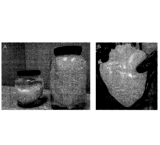
Organ clearing was observed within two hours of initiating perfusion.
Clear white color predominated 12 hours into perfusion. Decellularization was
terminated with the organ was white semi-transparent.
Example 10¨Transplantation of Decellularized Heart
Hearts from F344 rats were prepared by cannulating the aorta distal to
the Ao valve and ligating all other great vessels and pulmonary vessels except
the left branch of the pulmonary trunk (distal to its bifurcation) and the
inferior
vena cava (IVC). Decellularization was achieved using Langendorf retrograde
coronary perfusion and 2 liters of 1% SDS over 12-16 hours. The hearts were
then renatured with 35 mL of 1% Triton-X over 30-40 minutes, and then washed
with antibiotic and antifimgal-containing PBS for 72 hours. The IVC was
ligated before the transplantation.
A large (380 to 400 gram) RNU rat was prepared for reception of the
decellularized heart. A blunt-angled mosquito clamp was applied to both the
PVC and the abdominal Ao of the host animal to ensure isolation of areas of
anastomosis. The aorta of the decellularized heart was anastomosed to the host
abdominal aorta proximal and inferior to the renal branches using 8-0 silk
suture.
The left branch of the decellularized heart's pulmonary trunk was anastomosed
to the closest region of the host PVC to minimize physical stress on pulmonary
trunk.
After both vessels were sewn into the host animal, the clamp was
released and the decellularized heart filled with the host animal's blood. The
recipient animal's abdominal aortic pressure was observed visually in the
decellularized heart and aorta. The decellularized heart became distended and
red with blood. Bleeding was minimal at the site of anastomosis. Heparin was
administered 3 minutes after clamp release (initiation of perfusion), and the
heart
was photographed and positioned in the abdomen to minimize stress on the sites
27
CA 02618731 2008-02-11
WO 2007/025233
PCT/US2006/033415
of anastomosis. The abdomen was closed in sterile fashion and the animal
monitored for recovery. At 55 hours post-transplant, the a-nimal was
euthanized
and the decellularized heart was explanted for observation. The animals that
did
not receive heparin showed a large thrombosis in the LV upon dissection and
evaluation. Blood was also observed in coronary arteries in both the right and
left sides of the heart.
In other transplant experiments, the clamp was released after both vessels
were sewn into the host animal, and the decellularized heart filled with the
host
animal's blood. The recipient animal's abdominal aortic pressure was observed
visually in the decellularized heart and aorta. The decellularized heart
became
distended and red, and bleeding was minimal at the site of anastomosis.
Heparin
was administered (3000 IU) by IP injection 3 minutes after clamp release
(initiation of perfusion). The heart was photographed and positioned in the
abdomen to minimize stress on the sites of anastomosis. The abdomen was
closed in sterile fashion and the animal monitored for recovery. The animal
was
found dead from hemorrhage at approximately 48 hours after transplantation.
Transplantation time is currently in the 55 to 70 minute range.
Section C. Recellularization
Example 1¨Recellularization of Cardiac ECM Slices
To evaluate biocompatibility of decellularised ECM, 1 mm thick slices of
one decellularised heart were cultured with myogenic and endothelial cell
lines.
2 x 10⁵ rat skeletal myoblasts, C2C12 mouse myoblasts, human umbilical cord
endothelial cells (HUVECs), and bovine pulmonary endothelial cells (BPEC)
were seeded onto tissue sections and co-cultured under standard conditions for
7
days. Myogenic cells migrated through and expanded within the ECM and
aligned with the original fiber orientation. These myogenic cells showed
increased proliferation and fully re-populated large portions of the ECM
slice.
Endothelial cell lines showed a less invasive growth pattern, forming a
monolayer on the graft surface. There were no detectable antiproliferative
effects under these conditions.
28
CA 02618731 2008-02-11
WO 2007/025233
PCT/US2006/033415
Example 2¨Recellularisation of Cardiac ECM by Coronary Perfusion
To determine the efficiency of seeding regenerative cells onto and into
decellularised cardiac ECM by coronary perfusion, a decellularized heart was
transferred to an organ chamber and continuously perfused with oxygenised cell
culture media under cell culture conditions (5% CO2, 60% humiditiy, 37 C). 120
x 106 PKH labelled HUVECs (suspended in 50 ml of endothelial cell growth
media) were infused at 40 cm H20 coronary perfusion pressure. Coronary
effluent was saved and cells were counted. The effluent was then recirculated
and perfused again to deliver a maximum number of cells. Recirculation was
repeated two times. After the third passage, approximately 90 x 106 cells were
retained within the heart. The heart was continuously perfused with 500 ml of
recirculating oxygenised endothelial cell culture media for 120 hours. The
heart
was then removed and embedded for cryosectioning. HUVECs were confined to
arterial and venous residues throughout the heart, but were not yet completely
dispersed throughout the extravascular ECM.
Example 3¨Recellularization of a Decellularized Rat Heart with Neonatal Rat
Heart Cells
Isolation and preparation of rat neonatal cardiocytes. On day one, eight
to ten SPF Fisher-344 neonatal pups, aged 1-3 days (Harlan Labs, Indianapolis,
IN), were sedated with 5% inhaled Isoflurane (Abbott Laboratories, North
Chicago, IL), sprayed with 70% Et0H, and a rapid sternotomy was performed in
sterile fashion. Hearts were excised and placed immediately into 50m1 conical
tube on ice containing HBSS; Reagent #1 from a neonatal cardiomyocyte
isolation system (Worthington Biochemical Corporation, Lakewood, NJ).
Supernatant was removed and whole hearts were washed once with cold HBSS
by vigorous swirling. Hearts were transferred to a 100 mm culture dish
containing 5m1 cold HBSS, the connective tissue was removed, and remaining
tissue was minced into pieces <1 mm2. Additional HBSS was added to bring
total plate volume to 9 ml, to which 1 ml Trypsin (Reagent #2, Worthington
kit)
was added to give a final concentration of 50 g/ml. Plates were incubated
overnight in a 5 C cooler.
On day two, the plates were removed from the cooler and placed in a
sterile hood on ice. Tissue and trypsin-containing buffer were transferred to
50
29
CA 02618731 2008-02-11
WO 2007/025233
PCT/US2006/033415
ml conical tubes on ice using wide-mouth pipettes. Trypsin Inhibitor (Reagent
#3) was reconstituted with 1 ml HBSS (Reagent #1) and added to the 50 ml
conical tube and gently mixed. The tissue was oxygenated for 60-90 seconds by
passing air over the surface of the liquid. The tissue was then warmed to 37 C
and collagenase (300 units/m1) reconstituted with 5 ml Leibovitz L-15 was
added
slowly. The tissue was placed in a warm (37 C) shaker bath for 45 minutes.
Next, the tissue was titrated ten times using a 10 ml pipet to release the
cells (3
mls per second) and then strained through a 0.22 pm filter. The tissue was
washed with an 5 additional mls of L-15 media, titrated a second time, and
collected in the same 50 ml conical tube. The solution of cells was then
incubated at room temperature for 20 minutes, and spun at 50 xg for five
minutes
to pellet the cells. The supernatant was gently removed and the cells were
resuspended in the desired volume using Neonatal-Cardiomyocyte Media.
Media and Solutions. All media were sterile filtered and stored in the
dark in 5 C coolers. Worthington Isolation Kit contains a suggested media,
Leibovitz L-15, for culture. This media was used for Day Two of the tissue
processing only. For plating, an alternate calcium-containing media was used,
which is described herein. Worthington Leibovitz L-15 Media: Leibovitz media
powder was reconstituted using 1 L cell-culture grade water. Leibovitz L-15
media contains 140 mg/ml CaC1, 93.68 mg/ml MgCl, and 97.67 mg/ml MgS.
Neonatal-Cardiomyocyte Media: Iscove's Modified Dulbecco's Medium
(Gibco, Cat. No. 12440-053) was supplemented with 10% Fetal Bovine Serum
(HyClone), 100 U/ml penicillin-G (Gibco), 100 U/ml streptomycin (Gibco), 2
mmol/L L-glutamine (Invitrogen), and 0.1 mmol/L 2-mercaptoethanol (Gibco,
Cat. No. 21985-023) and sterile filtered before use. Amphotericine-B was added
as needed (0.25 pg/ml final concentration). This media was enhanced with 1.2
mM CaC1 (Fisher Scientific, Cat. No. C614-500) and 0.8 mM MgC1 (Sigma,
Cat. No. M-0250).
In Vitro Culture Analysis of Recellularization. As a step towards
creating a bioartificial heart, the isolated ECM was recellularized with
neonatal
heart-derived cells. Completely decellularized hearts (made as described
herein)
were injected with a combination of 50 x 106 freshly isolated rat neonatal
cardiomyocytes, fibrocytes, endothelial and smooth muscle cells. The heart
tissue was then sliced and the slices were cultured in vitro to test the
CA 02618731 2008-02-11
WO 2007/025233 PCT/US2006/033415
biocompatibility of the decellularized ECM and the ability of the resulting
constructs to develop into myocardium rings.
Minimal contractions within the resulting rings were observed
microscopically after 24 hours, demonstrating that the transplanted cells were
able to attach and engraft on the decellularized ECM. Microscopically, cells
oriented along the ECM fiber direction. Immunofluorescence staining
confirmed the survival and engraftment of cardiomyocytes expressing cardiac
myosin heavy chain. Within four days, clusters of contracting cell patches
were
observed on the decellularized matrix, which progressed to synchronously
contracting tissue rings by day 8.
At day 10, these rings were mounted between two rods to measure
contractile force under different preload conditions. The rings could be
electrically paced up to a frequency of 4 Hz and created contractile force of
up to
3 mN under a preload of up to 0.65 g. Thus, with this in vitro tissue culture
approach of recellularization, contractile tissue was obtained that generated
an
equally effective force as that generated by optimized engineered heart tissue
rings using artificial ECM constructs.
Recellularization of a Decellularized Heart via Perfusion. Recellularized
(50 x 106 freshly isolated rat neonatal cardiomyocytes, fibrocytes,
endothelial
and smooth muscle cells) scaffolds were mounted in a perfusable bioreactor
(n=10) that simulated rat cardiac physiology including pulsatile left
ventricular
distension with gradually increasing preload and afterload (day 1: preload 4-
12 '
mmHg, afterload 3-7 mmHg), pulsatile coronary flow (day 1: 7 ml/min), and
electric stimulation (day 2: 1 Hz) under sterile cardiac tissue culture
conditions
(5% CO2, 60% H20, 37 C). Perfused organ culture was maintained for one to
four weeks. Pressures, flows and EKG were recorded for 30 seconds every 15
minutes throughout the entire culture period. Videos of the nascent bio
artificial
hearts were recorded at days four, six and ten after cell seeding.
At day 10 after cell seeding, a more in-depth functional assessment was
performed including insertion of a pressure probe into the left ventricle to
record
left ventricular pressure (LVP) and video recording of wall motion as the
stimulation frequency was gradually increased from 0.1 Hz to 10 Hz and
performed pharmacological stimulation with phenylephrine (PE). The
recellularized heart showed contractile response to single paces with
31
CA 02618731 2008-02-11
WO 2007/025233
PCT/US2006/033415
spontaneous contractions following the paced contractions with corresponding
increases in LVP. After a single pace, the heart showed three spontaneous
contractions and then converted to a fibrillatory state. Similar to the
stimulated
contractions, spontaneous depolarizations caused a corresponding increase in
LVP and a recordable QRS complex possibly indicating the formation of a
developing stable conduction pattern.
Once stimulation frequency was increased to 0.4 Hz, an average of two
spontaneous contractions occurred after each induced contraction; at a pacing
frequency up to 1 Hz, only one spontaneous contraction occurred; and at a
pacing frequency of 5 Hz, no spontaneous contractions occurred. Maximum
capture rate was 5 Hz, which is consistent with a refractory period of 250 ms
for
mature myocardium. After perfusion with 100 AM of PE, regular spontaneous
depolarizations occurred at a frequency of 1.7 Hz and were coupled with
corresponding increases in LVP.
Histological analysis at day 10 revealed cell dispersion and engraftment
throughout the entire thickness of the left ventricular wall (0.5-1.2 mm).
Cardiomyocytes aligned with the ventricular fiber direction and formed areas
of
dense, organized grafts resembling mature myocardium and less dense immature
grafts similar to developing myocardium. Immunofluorescence staining for
cardiac myosin heavy chain confirmed the cardiomyocyte phenotype. A high
capillary density was maintained throughout the newly developed myocardium
with an average distance between capillaries of approximately 20 Arn, which is
similar to that reported for mature rat myocardium. Endothelial cell phenotype
was confirmed by immunofluorescent staining for vonWillebrand Factor (vWF).
Cell viability was maintained throughout the entire graft thickness,
indicating
sufficient oxygen and nutrient supply through coronary perfusion.
OTHER EMBODIMENTS
It is to be understood that while the invention has been described in
conjunction with the detailed description thereof, the foregoing description
is
intended to illustrate and not limit the scope of the invention, which is
defined
by the scope of the appended claims. Other aspects, advantages, and
modifications are within the scope of the following claims.
32