Note: Descriptions are shown in the official language in which they were submitted.
CA 02618933 2013-09-05
NEURONAL AVALANCHE ASSAY
CROSS REFERENCE TO RELATED PATENT APPLICATIONS
[01.]
BACKGROUND OF THE INVENTION
[02.] Transient formation of synchrony might serve as an integrative mechanism
to
bind neurons into coherent cell assemblies that are fundamental to cortical
function. Such synchronization has been observed during visual stimulus
discrimination, cognitive categorization tasks, potential motor command
signals,
and is similar to internally generated synchrony. However, understanding of
the
dynamical processes that underlie such fast and selective synchronization has
been
largely limited to computational models rather than biological networks. In
these
models, stable and spatially selective propagation of synchrony without
distortion, .
decay or explosion can only occur when key parameters are set in a narrow
range,
particularly when synchronization emerges locally within large networks. These
difficulties persist even when the constraints on synchronization are lowered
to
the propagation of transient firing rate increases, calling into question the
ability
of biological cortical networks to stably propagate synchrony.
[03.] There is a need for methods and systems for analyzing properties of
propagation within 'neuronal avalanches' and utilizing the ability of cortical
networks to support the propagation of precise patterns of synchrony within
multiple cortical areas and across multiple cortical columns.
SUMMARY OF THE INVENTION
[04.] Provided are systems and methods for determining a cognitive enhancement
and/or anti-epileptic effect comprising detecting synchronized neuronal
activity in
neuronal tissue, monitoring spreading of the synchronized neuronal activity,
determining a parameter indicative of the closeness of the synchronized
neuronal
activity to the critical state, and comparing the parameter to a predetermined
value.
1
CA 02618933 2008-02-12
WO 2007/022208 PCT/US2006/031884
1Aurttier provided are systems and methods for determining a cognitive
enhancement and/or anti-epileptic effect comprising detecting synchronized
neuronal activity in neuronal tissue, monitoring spreading of the synchronized
neuronal activity, determining a slope of a size distribution of the
synchronized
neuronal activity, comparing the slope of the size distribution to a threshold
slope, ,
determining a ratio of successively propagated synchronized neuronal activity,
and comparing the ratio to a threshold ratio.
[06.] Still further provided are systems and methods for screening
compositions for
a cognitive enhancement and/or anti-epileptic effect comprising applying a
composition to neuronal tissue, measuring propagated synchronized activity in
the ,
neuronal tissue, determining a parameter indicative of the closeness of the
synchronized neuronal activity to the critical state, and comparing the
parameter
to a predetermined value.
[071 Additional advantages of the invention will be set forth in part in the
description which follows or may be learned by practice of the invention. The
advantages of the invention will be realized and attained by means of the
elements
and combinations particularly pointed out in the appended claims. It is to be
understood that both the foregoing general description and the following
detailed
, description are exemplary and explanatory only and are not restrictive of
the
invention, as claimed.
BRIEF DESCRIPTION OF THE DRAWINGS
[08.] The accompanying drawings, which are incorporated in and constitute a
part
of this specification, illustrate embodiments of the invention and together
with the
description, serve to explain the principles of the invention. = =
[09.] Figure 1 illustrates an exemplary operating environment.
[010.] Figure 2 A and B show an example of continuous extracellular signals
recorded with a microelectrode array in an acute brain slice from rat medial
prefrontal cortex.
[011.] Figure 2 C is a raster plot of nLFP activity.
[012.] Figure 2 D and E illustrate avalanche size distributions.
[013.] Figures 3 A-C illustrate the spatiotemporal organization of nLFPs
induced by
co-application of dopamine and NMDA.
2
CA 02618933 2008-02-12
WO 2007/022208
PCT/US2006/031884
[014.] Figures 4 A-D illustrate the relationship of log(mod) = 0 for three
different
experimental preparations.
[015.] Figures 5 A-H illustrate an example of family clustering to determine
the slope
'
[016.] Figure 6 is a flow diagram illustrating exemplary steps of a provided
method.
[017.] Figures 7 A-F illustrate an inverted-U profile for the induction of
spontaneous
LFP activity by dopamine in acute slices of rat mPFC in the presence of NMDA.
[018.] Figures 8 A-C illustrate an inverted-U pharmacological profile for
avalanche
induction by dopamine.
[019.] Figures 9 A-E illustrate dopamine Di receptor stimulation induces
neuronal
avalanches via an inverted-U shaped pharmacological profile.
[020.] Figures 10 A-E illustrate that avalanche induction requires co-
activation of the
NMDA and dopamine D1 receptor.
[021.] Figures 11A-E illustrate co-stimulation of the dopamine D1 and NMDA
'
= receptor induces spontaneous avalanches predominantly in superficial
layers of
mPFC.
[022.1 Figures 12 A-D illustrate neuronal avalanche recurrence depends on
intact fast
synaptic inhibition and differs from disinhibited spontaneous activity.
[023.] Figures 13 A-G illustrate that spontaneous synchronized activity in
vivo
organizes into neuronal avalanches.
[024.] Figures 14 A-C illustrate nLFPs within an avalanche maintain the
waveform
of the initial event.
[025.] Figures 15 A-B illustrate spatial and temporal influence of avalanches
are
= independent of initiating LFP area.
[026.] Figures 16 A-F illustrate GABAA inhibition maintains the fidelity of
transmission of synchrony.
[027.] Figures 17 A-D illustrate spontaneous LFP bursts composed of 6/0 and 7-
frequency oscillations occur at the time of superficial layer differentiation
in
cortex in vitro.
[028.] Figures 18 A-D show that 7-oscillations reflect propagated waves within
superficial cortical layers.
[029.] Figures 19 A-E illustrate 7- and 0 oscillations are composed of
neuronal
avalanches characterized by a power law with slope a= -1.5.
[030.] Figures 20 A-C illustrate 7- oscillations reflect periods of high
coherence that
3
CA 02618933 2008-02-12
WO 2007/022208
PCT/US2006/031884
facilitate large avalanches.
[031.] Figures 21 A-B demonstrate balanced action of dopamine D 1/D2 receptor
activation on the formation of neuronal avalanche and 7-oscillation formation
in
cortical networks.
[032.] Figures 22 A-D demonstrate that 7¨oscillations originate within
cortical
superficial layers and do not depend on acute inputs from the VTA.
[033.] Figures 23 A-E demonstrate that 7-oscillations composed of neuronal
avalanches during early development depend on intact fast GABAA -mediated
synaptic inhibition.
[034.] Figures 24 A-E illustrate the slope and shape of avalanche size
distributions
provide a precise and quantitative measure for the sub-critical, critical, and
epileptic state in cortical networks.
[035.] Figures 25 A-E illustrate the atypical neuroleptic clozapine induces
epileptic,
neuronal avalanche activity in acute slices of adult rat mPFC.
, [036.] Figures 26 A-C illustrate avalanche induction is sub-optimal when
muscarinergic receptors are blocked in mPFC of aged rats (>2 months).
DETAILED DESCRIPTION OF THE INVENTION
[037.] Before the present methods and systems are disclosed and described, it
is to be
understood that this invention is not limited to specific synthetic methods,
specific
components, or to particular compositions, as such may, of course, vary. It is
also
to be understood that the terminology used herein is for the purpose of
describing
particular embodiments only and is not intended to be limiting.
= [038.] As used in the specification and the appended claims, the singular
forms "a,"
"an" and "the" include plural referents unless the context clearly dictates
otherwise. Ranges may be expressed herein as from "about" one particular
value,
and/or to "about" another particular value. When such a range is expressed,
another embodiment includes from the one particular value and/or to the other
particular value. Similarly, when values are expressed as approximations, by
use
of the antecedent "about," it will be understood that the particular value
forms
another embodiment. It will be further understood that the endpoints of each
of
the ranges are significant both in relation to the other endpoint, and
independently
of the other endpoint.
4
CA 02618933 2008-02-12
WO 2007/022208 PCT/US2006/031884
[039.] "Optional" or "optionally" means that the subsequently described event
or
circumstance may or may not occur, and that the description includes instances
where said event or circumstance occurs and instances where it does not.
[040.] The present invention may be understood more readily by reference to
the
following detailed description of preferred embodiments of the invention and
the
Examples included therein and to the Figures and their previous and following
description.
[041.] Provided herein in are methods and systems for performing a Neuronal
Avalanche Assay (NAS-assay). The NAS-assay uses the spatial distribution of
synchronized activity, in neuronal tissue. For exemplary purposes, the
description =
is directed toward the NAS-assay using local field potentials (LFP). In the
LFP,
the activity of a single neuron is barely detectable, however, if many neurons
synchronize their activities, the LFP is large enough to be registered by a
local
recording device, in this case, the microelectrode. As LFPs propagate along an
array of microelectrodes, the neuronal activity can be analyzed for neuronal
avalanches.
[042.] However, any method with which synchronized neuronal activity can be
detected locally in the living brain and which allows for the monitoring of
the
spread of synchronized activity, can be used in the NAS-assay. The NAS-assay,
in its principle design, is not limited to the use of LFPs only.
[043.] An example is the measurement of local synchronized activity using
Magentoencephalography (MEG). In the MEG, the tiny magnetic currents
induced by a single neuron are not detectable, however, if many neurons
synchronize their activities, the change in the magnetic field is measurable
with
high spatial (-1 mm) and temporal resolution (-1 ms). MEG is therefore very
similar to the LFP measurements in monitoring the spread of synchronized
neuronal activity in living tissue.
[044.] The NAS assay can be constructed from common forms of synchronized
activities, by way of example and not limitation, coherent delta/theta- (2- 8
Hz)
and gamma-(>25 Hz) oscillations measured from superficial cortical layers.
Such
oscillations are readily revealed in Electroencephalogram (EEG) measurements.
This allows the use of an EEG as a neuronal activity detector for the NAS-
assay.
[045.] The NAS-assay can utilize MEG, EEG, and the like. Any technique that
allows for the monitoring of the propagation of synchronized neuronal
activities
CA 02618933 2008-02-12
WO 2007/022208
PCT/US2006/031884
with high spatial (-1rnm) and temporal (-1 ms) resolution can be used.
I. Exemplary System
[046.] One skilled in the art will appreciate that what follows is a
functional
description and that the respective functions can be performed by software,
hardware, or a combination of software and hardware.
[047.] FIG. 1 is a block diagram illustrating an exemplary operating
environment for
performing the disclosed methods. This exemplary operating environment is only
an example of an operating environment and is not intended to suggest any
limitation as to the scope of use or functionality of operating environment
architecture. Neither should the operating environment be interpreted as
having
any dependency or requirement relating to any one or combination of components
, illustrated in the exemplary operating environment.
[048.] The systems and methods of the present invention can be operational
with
numerous other general purpose or special purpose computing system
environments or configurations. Examples of well known computing systems,
environments, and/or configurations that can be suitable for use with the
systems
and methods comprise, but are not limited to, personal computers, server
computers, laptop .devices, and multiprocessor systems. Additional examples
comprise set top boxes, programmable consumer electronics, network PCs,
minicomputers, mainframe computers, distributed computing environments that
comprise any of the above systems or devices, and the like.
[049.] In another aspect, the systems and methods of the present invention can
be
described in the general context of computer instructions, such as program
modules, being executed by a computer. Generally, program modules comprise
routines, programs, objects, components, data structures, etc. that perform
particular tasks or implement particular abstract data types. The systems and
methods of the present invention can also be practiced in distributed
computing
environments where tasks are performed by remote processing devices that are
linked through a communications network. In a distributed computing
environment, program modules can be located in both local and remote computer
storage media including memory storage devices.
[050.] Further, one skilled in the art will appreciate that the systems and
methods
disclosed herein can be implemented via a general-purpose computing device in
the form of a computer 101. The components of the computer 101 can comprise,
6
CA 02618933 2008-02-12
WO 2007/022208
PCT/US2006/031884
but are not limited to, one or more processors or processing units 103, a
system
memory 112, and a system bus 113 that couples various system components
including the processor 103 to the system memory 112.
[051.] The system bus 113 represents one or more of several possible types of
bus
structures, including a memory bus or memory controller, a peripheral bus, an
accelerated graphics port, and a processor or local bus using any of a variety
of
bus architectures. By way of example, such architectures can comprise an
,Industry Standard Architecture (ISA) bus, a Micro Channel Architecture (MCA)
=
bus, an Enhanced ISA (EISA) bus, a Video Electronics Standards Association
(VESA) local bus, an Accelerated Graphics Port (AGP)'bus, and a Peripheral
Component Interconnects (PC1) bus also known as a Mezzanine bus. The bus 113,
and all buses specified in this description can also be implemented over a
wired or
wireless network connection and each of the subsystems, including the
processor
103, a mass storage device 104, an operating system 105, NAS software 106,
neuronal data 107, a network adapter 108, system memory 112, an Input/Output
Interface 110, a display adapter 109, a display device 111, and a human
machine
interface 102, can be contained within one or more remote computing devices
114a,b,c at physically separate locations, connected through buses of this
form, in
effect implementing a fully distributed system.
[052.] The computer 101 typically comprises a variety of computer readable
media.
Exemplary readable media can be any available media that is accessible by the
computer 101 and comprises, for example and not meant to be limiting, both
volatile and non-volatile media, removable and non7removable media. The
system memory 112 comprises computer readable media in the form of volatile
memory, such as random access memory (RAM), and/or non-volatile memory,
such as read only memory (ROM). The system memory 112 typically contains
data such as neuronal data 107 and/or program modules such as operating system
105 and NAS software 106 that are immediately accessible to and/or are
presently
operated on by the processing unit 103.
[053.] In another aspect, the computer 101 can also comprise other
removable/non-
removable, volatile/non-volatile computer storage media. By way of example,
FIG. 1 illustrates a mass storage device 104 which can provide non-volatile
storage of computer code, computer readable instructions, data structures,
program modules, and other data for the computer 101. For example and not
7
CA 02618933 2008-02-12
WO 2007/022208
PCT/US2006/031884
meant to be limiting, a mass storage device 104 can be a hard disk, a
removable
magnetic disk, a removable optical disk, magnetic cassettes or other magnetic
storage devices, flash memory cards, CD-ROM, digital versatile disks (DVD) or
other optical storage, random access memories (RAM), read only memories
(ROM), electrically erasable programmable read-only memory (EEPROM), and
the like. =
[054.] Optionally, any number of program modules can be stored on the mass
storage
device 104, including by way of example, an operating system 105 and NAS
software 106. Each of the operating system 105 and NAS software 106 (or some
combination thereof) can comprise elements of the programming and the NAS
software 106. Neuronal data 107 can also be stored on the mass storage device
104. Neuronal data 107 can be stored in any of one or more databases known in
the art. Examples of such databases comprise, DB28, Microsofte.Access,
Microsoft SQL Server, Oracle , mySQL, PostgreSQL, and the like. The
databases can be centralized or distributed across multiple systems.
[055.] In another aspect, the user can enter commands and information into the
computer 101 via an input device (not shown). Examples of such input devices
comprise, but are not limited to, a keyboard, pointing device (e.g., a
"mouse"), a
microphone, a joystick, a scanner, and the like. These and other input devices
can
-
be connected to the processing unit 103 via a human machine interface 102 that
is
coupled to the system bus 113, but can be connected by other interface and bus
= structures, such as a parallel port, game port, an IEEE 1394 Port (also
known as a
Firewire port), a serial port, or a universal serial bus (USB).
[056.] In yet another aspect of the present invention, a display device 111
can also be
connected to the system bus 113 via an interface, such as a display adapter
109. It
= is contemplated that the computer 101 can have more than one display
adapter
= 109 and the computer 101 can have more than one display device 111. For
example, a display device can be a monitor, an LCD (Liquid Crystal Display),
or a
projector. In addition to the display device 111, other output peripheral
devices
can comprise components such as speakers (not shown) and a printer (not shown)
which can be connected to the computer 101 via Input/Output Interface 110.
[057.] A neuronal activity detector 116 can communicate with computer 101 via
Input/Output Interface 110 or across a local or remote network. In one aspect,
users utilize a neuronal activity detector that is capable of collecting
neuronal
8
CA 02618933 2008-02-12
WO 2007/022208
PCT/US2006/031884
data. It will be appreciated that the neuronal activity detector 116 can be
any type
of neuronal activity detector, for example and not meant to be limiting, a
micro electrode array (to record LFFs and single/multi-unit activity), a
surface
electrode system (to record the EEG), a charge-coupled device camera (CCD) or
photodiode array (to record activity-dependent fluorescence changes), a
magnetometer type SQUID (superconducting quantum interference device) sensor
(to record the MEG), and the like. In another aspect, the neuronal activity
r detector 116 can be an independent stand alone device, or can be
integrated into
another device. Optionally, the communication with computer 101 via
Input/Output Interface 110 can be via a wired or wireless connection.
[058.] The computer 101 can operate in a networked .environment using logical
connections to one or more remote computing devices 114a,b,c. By way of
example, a remote computing device can be a personal computer, portable
computer, a server, a router, a network computer, a peer device or other
common
network node, and so on. Logical connections between the computer 101 and a
remote computing device 114a,b,c can be made via a local area network (LAN)
and a general wide area network (WAN). Such network connections can be
through a network adapter 108. A network adapter 108 can be implemented in
both wired and wireless environments. Such networking environments are
conventional and commonplace in offices, enterprise-wide computer networks,
intranets, and the Internet 115. V V =
[059.] For purposes of illustration, application programs and other executable
program components such as the operating system 105 are illustrated herein as
' discrete blocks, although it is recognized that such programs and
components
reside at various times in different storage components of the computing
device
101, and are executed by the data processor(s). of the computer. An
implementation of NAS software 106 can be stored on or transmitted across some
form of computer readable media. Computer readable media can be any available
media that can be accessed by a computer. By way of example and not meant to
be limiting, computer readable media can comprise "computer storage media" and
"communications media." "Computer storage media" comprise volatile and non-
volatile, removable and non-removable media implemented in any method or
technology for storage of information such as computer readable instructions,
data
structures, program modules, or other data. Exemplary computer storage media
9
CA 02618933 2008-02-12
WO 2007/022208
PCT/US2006/031884
comprises, but is not limited to, RAM, ROM, EEPROM, flash memory or other
memory technology, CD-ROM, digital versatile disks (DVD) or other optical
storage, magnetic cassettes, magnetic tape, magnetic disk storage or other
magnetic storage devices, or any other medium which can be used to store the
desired information and which can be accessed by a computer.
[060.] The methods and systems can employ Artificial Intelligence techniques
such
as machine learning and iterative learning. Examples of such techniques
include,
but are not limited to, expert systems, case based reasoning, Bayesian
networks,
behavior based Al, neural networks, fuzzy systems, evolutionary computation
(e.g. genetic algorithms), swarm intelligence (e.g. ant, algorithms), and
hybrid
intelligent systems (e.g. Expert inference rules generated through a neural
network
or production rules from statistical learning).
[061.] The processing of the disclosed systems and methods of the present
invention
can be performed by software components. The disclosed systems. and methods
can be described in the general context of computer-executable instructions,
such
as program modules, being executed by one or more computers or other devices.
Generally, program modules comprise computer code, routines, programs,
objects, components, data structures, etc. that perform particular tasks or
implement particular abstract data types. The disclosed methods can also be
practiced in grid-based and distributed computing environments where tasks are
performed by remote processing devices that are linked through a
communications
network. In a distributed computing environment, program modules can be
located in both local and remote computer storage media including memory
storage devices.
[0624 The NAS Software 106 allows for the study of neuronal avalanches and
includes many analysis features. NAS Software 106 allows for the calculation
of
alpha (a), the slope of the avalanche size distribution, and sigma (a), the
branching parameter at the correct temporal resolution (Atavg). Avalanche
calculation controls set the parameters for concatenating LFPs into
avalanches. A
multi-function control window contains functions that extract the avalanche
parameters a and a at corresponding Atavg. For visual control, avalanche size
distributions in log-log coordinates can be generated and displayed. Cross-
correlation plots used to calculate Atavg can also be displayed at various
temporal
resolutions. Additional features relate to the identification and labeling of
CA 02618933 2008-02-12
WO 2007/022208
PCT/US2006/031884
recording locations to superficial cortical layers in which avalanches occur.
For
example, the NAS Software 106 allows for the topological identification of
electrode positions on a microelectrode array with respect to brain region and
cortical layer. NAS Software 106 allows for the storage of spatial
information, e.g.
images, and miscellaneous data specific to an experimental configuration.
[063.] NAS Software 106 can analyze the similarity in spatiotemporal
organization
between neuronal avalanches. The spatiotemporal organization of avalanche is
highly diverse and the diversity can be used to further evaluate the quality
of the
data and to quantify the critical state in the cortical network. More
specifically, the
size distribution of significant avalanche families reveals a heavy-tail in
family
sizes that forms a power law with slope gamma (7): Shuffle and cluster
controls
allow for the generation of shuffled data sets and statistical evaluation of
family
significance and calculation of Several additional features allow for a
detailed !.
examination of avalanche similarity on which the family size distribution is
based.
An avalanche generation tree can be generated that represents the generational
relationship between avalanches based on similarity. An avalanche similarity
matrix can be generated that contains the similarity index for all possible
pair wise
comparisons between avalanches. A multi-function control window for cluster
analysis contains functions for studying the spatiotemporal organization of
avalanches, for example, displaying a family frequency distribution plot to
derive
[064.] NAS Software 106 allows for a detailed pair-wise comparison between
individual avalanches selected from the similarity matrix. The spatiotemporal
pattern of an avalanche can be indicated by a specific color representing
active
electrodes on successive 8x8 matrices for two avalanches simultaneously in
order '
to study similarity features in further detail between two neuronal
avalanches.
.= [065.] NAS Software 106 allows for visualization and editing of the
temporal
organization of neuronal avalanches. This can play a role when judging the
quality
- of recorded data. An overview plot of LFP activity can be generated
that displays
the occurrence of LFPs during an experiment. A zoom view can display LFP
occurrence for the temporal duration indicated by a colored rectangle in an
overview plot. This allows for a detailed examination of avalanches and can be
used to clean data sets from spurious noise. It also allows for the indication
of
avalanche extent and precise labeling of individual avalanches within a data
set
11
CA 02618933 2008-02-12
WO 2007/022208
PCT/US2006/031884
with respect to rank of occurrence in time, corresponding family and order
within
a family. For evoked activity, this feature can display identified stimuli and
corresponding evoked avalanches. Family controls can display the type and
occurrence of families over time. The family control and avalanche zoom view
can be aligned in time for precise comparison.
IL Exemplary Methods
[066.] The methods will be described herein as applied to the use of a planar
microelectrode array as a neuronal activity detector. Spontaneous neuronal
activity can be studied with planar microelectrode arrays that contain, for
example, 60 electrodes arranged on an 8x8 matrix at an interelectrode distance
of
200 pm (30 ttm electrode diameter; comer electrodes missing); and oriented
such
that cortical layers are in parallel with electrode rows.
A. Data Acquisition
[067.] Continuous extracellular signals v(t) can be recorded between 0-.1 Hz
and
3000 Hz, low-pass filtered at 200 Hz (Butterfly- filter, Multichannel
Systems),
digitised at a rate of 1 kHz ( At =1 ms) and stored. For each electrode k
(k =[1,tzeiecl,neke= 60), the electrode noise of v(t) can be estimated by
calculating the mean and standard deviation (SD) of v(t) from the total
duration of
the recording, Tot, which can range, for example, between 40 ¨ 60 min. FIG.
2A,B shows an example of continuous extracellular signals recorded with a
microelectrode array in an acute brain slice from rat medial prefrontal
cortex.
Negative LFP peaks (nLFPs) at electrode k can be detected by negative
threshold
crossing at -3 SD of electrode noise and can be characterized by the time and
absolute amplitude of the negative LFP peak, Ak (ti), before v(t) returned to
threshold. More precisely, at a given temporal resolution At , nLFPs at
electrode
k can be represented by a vector of length tit., where n,, is an integer value
for
which Tto, = n.- At. This vector can contain zeros, except for positions ti =
i = At,
i E [1,...,nmax] with values Ak (ti) in V. The resulting raster plot of nLFP
activity
is shown as an example in FIG. 2C.
B. Definition of neuronal avalanches
[068.] A neuronal avalanche is a sequence of consecutive time bins of width At
with
at least one nLFP, which is preceded and terminated by at least one time bin
with
12
CA 02618933 2008-02-12
WO 2007/022208
PCT/US2006/031884
no activity (FIG. 3A). The absence of activity for a period of At thus
indicates
the end of an avalanche. If the decision of whether an avalanche has ended is
made too early (& too short), avalanches will be terminated prematurely; if
the
At chosen is too long, avalanches will be falsely concatenated. If avalanches
did
simply propagate like a wave, an approximation for At (Atan ) could be
obtained
by averaging the time between one nLFP at one electrode and the next nLFP at
neighbouring electrodes only. Because nLFPs in avalanches occur in irregular
patterns across electrodes on the array, a pair wise approximation can be used
in
order to assess the average time that is required for iiLFPs to propagate
between
electrodes.
[069.] In order to calculate Atavg , the distribution of time intervals T for
successive
nLFPs on the array can be obtained. Starting with the first nLFP, e.g. Ak(ti)
on
electrode k at time ti, the next occurrence of an nLFP on the array can be
searched for, e.g. AO on electrode 1 at time tj , and calculated the time
interval , where in= (ti ¨01 At . This process can be repeated for all
occurrences of nLFPs on electrode k and for all electrodes. The resulting
values
can be combined into one density distribution P(7), which captures how often
successive nLFPs occurred with a particular delay in = At on the array
irrespective
of their spatial location. Consequently, the average value of T provides an
approximation for Atavg , the average time to wait before making a decision
whether an nLFP propagated on the array. However, this interval distribution
is
highly skewed, particularly when one compares the last nLFP with the first
nLFP
in successive avalanches that are separated by long times. In order to exclude
time intervals from successive nLFPs that are barely correlated, a cut-off
time
yinax can be calculated for which the average crosscorrelation function (ccf)
for
pair wise electrode comparisons Red (r) had decayed to negligible values. The
ccf
between electrodes k, 1 (R (v)) (T)) can be calculated as
13
CA 02618933 2008-02-12
WO 2007/022208
PCT/US2006/031884
nimax
A kup (in = At + A'ILFp (m = At)
Rcke,fi Mmax m=1 (1)
a (A''11 Fp ).5(ZILFP
at At = lms for r e [-100,100], where in is an integer value up to
mmax= At + <70t , A'k (t) = Ac (t 1)¨ E(Ak ) , E(.) is the expected value for
Ak(ti) , and o.2(.) is the variance. Finally, the population ccf (Rcof (T))
can be
calculated as
2 no. elec
R Cef (r) = _______________________ E E (r) (2)
n elec (nelec k1 1k+1
[0701 The estimate of the average time between successive, correlated nLFPs on
the
array, i.e. Aran , can be obtained by integrating the density distribution of
intervals for each network up to v.,
npAt=rma.
APavg (Tinax ) = E Tm.AtP(7 (3)
m=1
[0711 Because the exemplary maximal sampling rate was 1 kHz, the actual &an to
calculate avalanches can be taken as the nearest multiple a At =lms. In short,
after Atavg was calculated for a particular network and experimental
condition, the
extracellular signals v(t) can be re-sampled at the new temporal resolution of
Atavg . Time and amplitude of nLFP peaks can be extracted as Ak(ti), where
= i = Atavg i E [1, n' .x] and Ti.t = n'. -Atavg Avalaliches can be determined
on
the downsampled data set at bin width Atavg=
[072.] FIG. 3 illustrates the derivation af neuronal avalanches at the correct
temporal
resolution Atavg using avalanche activity induced by co-application of
dopamine
and NMDA in an acute medial prefrontal cortex slice from an adult rat. A,
Neuronal avalanches are defined on a sequence of consecutively active time
bins
of width Atavg bracketed by at least one time bin with no activity. Sketch of
an
14
CA 02618933 2008-02-12
WO 2007/022208
PCT/US2006/031884
avalanche of size soe = 3 electrodes, size sup = 140 V, and lifetime in = 3
Atavg
on a 2x2 electrode array (absolute nLFP amplitudes Ak (ti) indicated by dot
diameter and numbers in V). B, nLFPs at different electrodes are strongly
clustered in time as demonstrated by the average crosscorrelation function
Rccf.
Left: single slice; average from 595 pair wise cross correlations. Right:
Average
from all n =6 acute rat slices encompassing 6824 correlation functions (30 M
dopamine and 3 M NMDA). Note that Reef decays to negligible levels within
100 ms. r maa is indicated by arrows for which R ca dropped below 10-4.
Derivation of average bin width Atavg for neuronal avalanche analysis.
Corresponding change in average inter-nLFP interval (thick line; right axis)
from
all electrode pairs, i.e. Atavg as a function of upper cut-off for integration
time
r max . The inter-nLFP interval distribution is plotted on the left axis (thin
line). For
the single slice (left), Rcef (rmax) <104 for T x = 75 ms, and Atavg (rmax)=-
2.7 ms.
For all slices (right), R ccf (v ax) <1 co for z. = 60 ms and At avg(r =
2.7 ms
(mean S.E.M.). Note plateau reached for Atavg at r . (arrows). In general,
At'avg varies very little for za. between 50¨ 100 ms for single networks as
well as
for the population of networks. Dotted line indicates read-out of Atavg .
C. Avalanche size distributions
[073.] Avalanche sizes can be calculated in two different ways (see also FIG.
3A).
By taking into account nLFP peak amplitudes 4, the avalanche size supAvali can
be
calculated by summing up 4 on active electrodes for the lifetime TAvalj = m
Atavg
of Aval defined as the number of bins in of width At avg that were occupied by
avalanche Avali that started at time ti and stopped at time ti + (in ¨1) = At
avg.
n elm
Aval=
SLFP Ellt4:+m=Ata, = (4)
i=0 k=1
[074.] For the density distribution ofsup , the range in sizes sup was covered
by 100
bins that increased logarithmically from 3 ¨ 300 V, which results in
equidistant
CA 02618933 2008-02-12
WO 2007/022208
PCT/US2006/031884
sampling of the data in logarithmic coordinates. Avalanche sizes can also be
calculated as the number of active electrodes within an avalanche, seie ,
using (4),
but setting all non-zero 4 values to I. For the density distribution ofsele ,
linear
= binning from 1 ¨ 100 can be used. In the critical state, the distribution
of
avalanche sizes forms a power law with slope a = -3/2. From the experimental
data, the exponent a of the power law represents the slope of the log-log
transformed size distribution and can be estimated using linear regression
analysis. Estimating a is not limited to regression analysis only. For
example, a
can be estimated using a maximum likelihood estimation
-1
"
¨a=l+n[Eln N(si) (5)
N(snin)
where N(si) represents the number of avalanches of size s, N(smin) represents
the
number of minimal avalanche size snin measured, and n represents the number of
size categories. Alternatively, a can be estimated from the cumulative size
distribution, which forms a slope of a+1. Because no significant differences
exist
between estimates of a based on sup (au) or (s6e) (aek), slope values can be
given as aLn, unless a particular emphasis is placed on the area an avalanche
= covers on the array. Average avalanche size distributions can be plotted
as mean
S.E.M. In FIG. 2D,E the avalanche size distributions obtained at Atavg for
sup and (seie) are plotted in log-log coordinates. Both distributions reveal a
power law with slope aup and aeie of --1.5 as determined by linear regression
(exact numbers and regression line are given in the figure; average from n =6
slices at 30 AM dopamine and 3 AM NMDA).
D. Avalanche branching parameter
[075.] The branching parameter a can be used to describe the balance of
propagated
synchronized activity in cortical tissue. The general definition of a refers
to the
ratio between successive generations, for example the average number of
descendants from one ancestor. When a= -3/2, the neuronal tissue is critical
and
correspondingly, a =1. The branching parameter a can be defined in binary and
analog terms.
16
CA 02618933 2008-02-12
WO 2007/022208
PCT/US2006/031884
LU/O.j In the binary case, a is defined as the average ratio of electrodes
activated in
time bin Atii+i (descendants; lid), divided by the number of electrodes active
in
time bin it (ancestor; na ). Mathematically, the average branching parameter a
for the electrode array in the case of one ancestor electrode (na =1) is
simply
given by
= E d = p(d), (6)
d=0
where d is the number of electrode descendants, p(d) is the probability of
observing d descendants, and nmd, is the maximal number of active electrodes.
Note that formula (6) does not describe a probability density and
theoretically
a can take any value 0. In the binary case, ois best estimated from the first
and
second time bin of an avalanche. Although strictly speaking, a is only defined
for one ancestor, a can also be estimated when ther'e are multiple ancestors.
Under these conditions, d is given by
\
d = round- (7)
\ a I
where ila is the number of electrode ancestors observed in the first time bin
and nd
is the number of active electrodes in the second time bin of an avalanche and
round is the rounding operation to the nearest integer. The likelihood of
observing d descendants can be approximated by:
nzatd nm ¨1 (8)ax
p(d) E
avalanches liZaj \nmax n a
where flak/ is the total number of electrode ancestors in all avalanches when
nd
descendants were observed, nza is the total number of ancestors observed in
all
avalanches, and nmax ¨1
is a factor that provided an approximate correction
a)
for the reduced number of electrodes available in the next time bin because of
= electrode refractoriness. Note that the branching parameter is not
defined for zero
ancestors and thus does not provide information about the initiation of
bursts. In
cases where there is only one ancestor, formula (8) is equivalent to (6).
In the analog case, the branching parameter a includes analog information
about
the LFP, e.g. its negative peak value (nLFP amplitude) or the nLFP area
17
CA 02618933 2008-02-12
WO 2007/022208
PCT/US2006/031884
(integrated LFP amplitude from crossing negative threshold to return to
threshold).
. [077.] For the analog calculation, each nLFP in an avalanche is
normalized to the
amplitude or area of the first nLFP in the avalanche. For each time bin, the
corresponding nLFP distributions from all avalanches can be combined. The
succession of nLFP distributions during the life time of the avalanche then
approximates the branching parameter o. More specifically, if the mode (mod)
of the nLFP distributions equals 1 for each time bin, nLFPs do not grow nor do
they decay within an avalanche, which is equivalent to the binary case of o-=
1.
µ, Because the distribution of ratios is better expressed in log
values, in the analog
case, one can state log(1) = 0. Thus, log(mod) = 0 demonstrates that the
tissue is
critical.
[078.] An example of such a calculation is shown in FIG. 4, in which for three
different experimental preparations this relationship of log(mod) = 0 is
demonstrated. For spontaneous activity in mature cortex slice cultures (FIG.
4,
left), the acute mPFC slice from adult rats (FIG. 4, middle; cp. FIG. 2A), and
the
awake macaque monkey (FIG. 4, right) nLFP area varies widely as shown in the
cumulative plots (FIG. 4A). However, nLFPs normalized to the corresponding
first nLFP in each avalanche are centered at 0 demonstrating that within an
avalanche, LFPs do not grow nor do they decay (FIG. 4B). This relationship is
clearly demonstrated when plotting log(mode) for successive times within an
avalanche (horizontal line at y 0 in FIG. 4C). This relationship could not
have
occurred by chance; when nLFPs are randomly drawn from the population
distribution for successive time steps within avalanches, the log(mod)= 0
relationship is destroyed (in FIG. 4D).
E. Cluster analysis of avalanche activity
[079.] Diverse avalanches can be grouped into families with similar
spatiotemporal
patterns. The grouping into families allows the determination Of a family size
distribution. In the critical state, the family size distribution reveals a
heavy-tail
with a slope gamma (7) around ¨2.4 (FIG. 5F). This value of 'y can be used as
a
measure to characterize the the critical state in the neuronal tissue.
18
CA 02618933 2008-02-12
WO 2007/022208
PCT/US2006/031884
[080.] A framei can be defined as the 8x8 matrix of Ak (ti) at time ti at
temporal
resolution Atavg . In this case, the microelectrode array has a layout of 8x8.
Because, in the example provided, corner electrodes on the microelectrode
array
were absent, a frame constituted a single pattern in a 60-dimensional space.
An
avalanche Aval j of lifetime TAvali = in = At comprised of in frames and
represented
a vector in a in = 60 dimensional space. The similarity Sim between two
avalanches Avali and Aval j in this space can be calculated using a
correlation
measure, which takes into account nLFP times as well as nLFP amplitudes. More
precisely
160 (Aval= Aval)j ¨ E (Av ali)E (Av al j)
Sim(Aval Avalj) = 111. (5)
a(Aval)c-(Aval)
. where E(.) is the expected value, o-(.) is the standard deviation for
individual
vector values respectively, and Aval = Aval is the vector dot product. The
similarity can range from ¨1 (perfectly anti-similar) to +1 (identical). The
similarity measure can be expanded in order to measure similarities between
avalanches with different lifetimes T Avals = i TAvalj =
In the latter case, the
avalanche Aval . with shorter lifetime can be shifted by up to mi ¨ mj steps.
For
, each shift, the similarity can be calculated from the intersection of frames
between
the two avalanches and the final similarity can be assigned from the shift
resulting
in the highest similarity value. When combined into a similarity matrix, the
diagonal of this matrix can indicate self-similarity, which has the maximal
value
of one (FIG. 5A). ,
[081.] Surrogate data sets with identical avalanche size and lifetime
distributions can
be obtained with 'paired-shuffling'. This shuffling method switches electrode
assignments between randomly selected pairs of nLFPs recorded throughout the
experiment on the array (FIG. 5D). It maintains the average rate of nLFPs at
each
electrode, the exact lifetime distribution, as well as the event size
distribution and
reduces type II errors (identifying similar avalanches when they could have
19
CA 02618933 2008-02-12
WO 2007/022208
PCT/US2006/031884
occurred by chance). Each experimental data set can be compared against 100
surrogate data sets.
[082.] Significance of similarities between avalanches can be determined using
surrogate data. A similarity distribution can be constructed from pair wise
similarities between avalanches from 100 surrogate data sets. A Type I error
of
x% can be represented by finding the similarity threshold for which x% of
similarities from the surrogate data sets were above this threshold.
[083.] Significant avalanche families can be identified by comparing the
summed
similarity among members within a family to that of surrogate data. However,
this procedure might miss families with few members, i.e. small family size n,
'whose members have high similarity, or large families with members of
relatively
low similarity. Therefore, the average similarity among avalanches in a family
simaFvargn
) can be compared to that from surrogate data. For example, the expected
distribution of family size n and corresponding SimaFT can be obtained from
100
surrogate data sets. As can be seen in FIG. 5E, the incidence of small
families
with high Sim:: and large families with relatively low SimaT is much lower in
the surrogate data sets (white) compared to the original data set (grey).
Families
that occupied the parameter space outside the area taken up by 100 surrogate
data
sets can be considered significant. Thus, a significant family of size n in
the
original data set had either a higher Sim than all surrogate families of size
72, or
alternatively, all surrogate families had smaller sizes.
[084.] The recurrence of family sizes > 10 beyond chance can be calculated and
plotted as a function of dopamine concentration. First, for a given type I
error,
family size distributions can be calculated from the original data and
corresponding 100 shuffled data sets. Then, the accumulated probability for
families > size 10 can be calculated from each distribution. The ratio of
these
probabilities can provide an estimate of the encounter of a family size > 10
beyond chance in the original data. The procedure can be repeated for
different
type I errors 0.1, 0.5, 1, 3, 5%. Values for different type I errors can be
averaged
and expressed in percentages. In general, the slope y of the family size
distribution is relatively robust for different type I errors (FIG. 5G).
CA 02618933 2008-02-12
WO 2007/022208
PCT/US2006/031884
[085.] In FIG. 5, an example using the family clustering to determine the
slope y is
demonstrated. The data are based on avalanche activity recorded from the
experiments shown in FIG. 2 (acute rat inPFC slice, 30 AM dopamine and 3 M
NMDA). Spontaneous retrieval of avalanches is demonstrated to peak at
moderate dopamine receptor stimulation and to be organized according to a
power
law with slope of-2.4 0.1. A, Sorted similarity matrix for 627 neuronal
avalanches with lifetimes ranging from 2 ¨ 9 times. Pairs of avalanches with
high
similarity are indicated by dark blue. The open red squares along the matrix
diagonal indicate significant families with similar neuronal avalanche members
(type I = 5%; single experiment from FIG. 1A). B, Average spatiotemporal
activation pattern for 4 significant families with at least 8 members from A.
For
each family, the average probability for an electrode on the 8x8 array to be
active
at time t (grayscaley is plotted for a period of around the main time of
activity. C,
Spontaneous recurrence of families (squares) is irregular and intermingled
with
other families (type I = 5%; 34 families with lifetime = Atavg and > 2
members
shown; data from A). D, Surrogate data were constructed by pair wise shuffling
of two electrodes in different bins. This maintains the lifetime as well as
area
distribution of avalanches, but destroys the spatiotemporal organization of
activity
within an avalanche. E, Scatter plot of family size n and average similarity
within
a family for original (gray) and shuffled (red) data. Significant families lie
outside
the boundary of the area covered by families from shuffled data sets
algorithm. F,
Family sizes n> 3 are distributed according to a power law , 7= ¨2.4 (broken
line). Average family size distribution (n = 6Y, averaged over 6 different
type I
errors (gray circles; mean S.E.M.; type I error= 0.1, 0.3, 0.5, 1, 3, 5%; 30
M
dopamine and 3 M NMDA; n =6 experiments). Families occur significantly
more often than expected by chance as demonstrated by the expected family size
distribution from 100 shuffled data sets per experiment for all type I errors
(red
line; mean S.E.M.). G, The slope yis independent of the type I error.
Corresponding slope analysis from F for different type I errors. II,
Recurrence of
avalanche families follows an inverted-U shaped profile reaching ¨500% higher
levels than expected by chance (average for family sizes >6 from all networks
for
each concentration of dopamine combined with 3 M NMDA).
E. Detailed Exemplary Embodiment
21
CA 02618933 2008-02-12
WO 2007/022208
PCT/US2006/031884
[086.] To determine if a cortical network is in the critical state,
spontaneous/evoked
local field potential activity in superficial cortical layers can be recorded
and
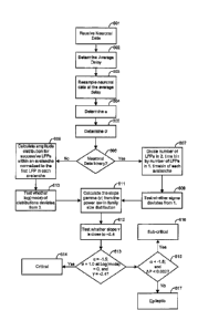
analyzed. In one aspect, as shown in FIG. 6, extraction of the avalanche
statistics
can determine if a cortical network is in the critical state. At block 601,
neuronal
data can be received. At block 602, the average delay between successive LFPs
in
the network at highest temporal resolution can be determined (Atavg). This
determination can be made, for example, using formulas 1 ¨3. At block 603, the
data can be resampled at the average delay (Atavg).
[087.] At block 604, a distribution of neuronal avalanche sizes can be
calculated
from binary or analog data using formula 4. Alpha (a) can be determined, for
example, from the power law slope using linear regression.
[088.] At block 605, the branching parameter sigma (a) can be determined. A
determination can be made as to whether the data is binary or analog at block
606.
If the neuronal data is binary, a can be determined using formula 6 ¨ 9 at
block
607. Then, at block 608, a test can be performed to determine if sigma
deviates
from 1. If the neuronal data is analog> amplitude distributions can be
calculated
for successive LFPs within an avalanche normalized to the first LFP in each
avalanche, as described in herein, at block 609. Then, at block 610, a test
can be
performed to determine whether log(mode) of distributions deviates from 0.
[089.] Then at block 611, the slope gamma (7) can be calculated from the power
law
in family size distribution. A test can be performed to determine whether
slope -y
is close to ¨2.4 at block 612. At block 613, it can be determined if a = -1.5
supported by a = 1.0 at log(mode) = 0 and -y = -2.4. If that determination is
met,
declare the network as critical at block 614. If that determination is not
met,
proceed to block 615 to determine if a< -1.5, < 1.0, log(mode) c 0, and AP <
0.002. If that determination is met, declare the network sub-critical at block
616.
If the determination is not met, in other words, if a < -1.5, log(mode) > 0,
and AP
> 0.002, declare the network epileptic at block 617.
[090.] In another aspect, provided is a method for determining a cognitive
enhancement and/or anti-epileptic effect comprising detecting synchronized
neuronal activity in neuronal tissue, monitoring spreading of the synchronized
neuronal activity, determining a parameter indicative of the closeness of the
22
CA 02618933 2008-02-12
WO 2007/022208
PCT/US2006/031884
synchronized neuronal activity to the critical state, and comparing the
parameter
to a predetermined value.
[091.] The parameter can be a slope of a size distribution of the synchronized
neuronal activity and the predetermined value can be -3/2. The parameter can
be a
branching ratio of successively propagated synchronized neuronal activity and
the
predetermined value can be 1 or log(1) = 0. If the slope is equal to the -3/2,
the
effect is optimal. If the determined slope is steeper than -3/2, the effect is
sub-
optimal. If the branching ratio is equal to 1, the effect is optimal. If the
branching
ratio is smaller or larger than 1 or deviates from log(1) = 0, the effect is
sub-
optimal.
[092.] The step of detecting synchronized neuronal activity can utilize, for
example,
a micro-electrode array, magnetoencephalograph, electroencephalograph, imaging
with fluorescent probes, and the like.
[093.] The synchronized neuronal activity can be, for example, local field
potentials,
magnetic currents, fluorescent probes, neuronal action potentials recorded as
extracellular single or multi-unit activity, and the like.
[094.]. The method can further comprise administering a composition suspected
of
having a cognitive enhancement and/or anti-epileptic effect to the neuronal
tissue.
[095.] The composition effect can be, for example, dopaminergic,
glutamatergic,
GABAergic, cholinergic, serotonergic, noradrenergic, and the like.
[096.] In yet another aspect, provided is a method for determining a cognitive
enhancement and/or anti-epileptic effect comprising detecting synchronized
neuronal activity in neuronal tissue, monitoring Spreading of the synchronized
neuronal activity, determining a slope of a size distribution of the
synchronized
neuronal activity, comparing the slope of the size distribution to a threshold
slope,
determining a ratio of successively propagated synchronized neuronal activity,
and comparing the ratio to a threshold ratio.
[097.] The threshold slope can be -3/2 and the threshold branching ratio can
be 1. If
the determined slope is equal to the threshold slope or threshold branching
ratio,
the effect is optimal. If the determined slope is steeper than the threshold
slope,
the effect is sub-optimal. If the determined branching ratio is equal to 1 or
log(1) --
0, the effect is optimal. If the determined branching ratio is smaller or
larger than
1 or log(1) = 0, the effect is sub-optimal.
23
CA 02618933 2008-02-12
WO 2007/022208
PCT/US2006/031884
[098.] The step of detecting synchronized neuronal activity can utilize, for
example,
a micro-electrode array, magnetoencephalograph, electroencephalograph, imaging
with fluorescent probes, and the like.
[099.] The synchronized neuronal activity can be, for example, local field
potentials,
magnetic currents, single or multi-unit activity, fluorescent probes, and the
like.
[0100.] The method can further comprise administering a composition suspected
of
having a cognitive enhancement and/or anti-epileptic effect to the neuronal
tissue.
[0101.] The composition effect can be, for example, dopaminergic,
glutamatergic,
GABAergic, cholinergic, serotonergic, noradrenergic, and the like.
[0102.]In a further aspect, provided is a method for screening compositions
for a
cognitive enhancement and/or anti-epileptic effect comprising applying a
composition to neuronal tissue, measuring propagated synchronized activity in
the
neuronal tissue, determining a parameter indicative of the closeness of the
synchronized neuronal activity to the critical state, and comparing the
parameter
to a predetermined value.
[0103.]The step of detecting synchronized neuronal activity can utilize, for
example,
a micro-electrode array, magnetoencephalo graph, electroencephalograph,
magnetic resonance imaging, imaging with fluorescent probes, and the like.
[0104.]The synchronized neuronal activity can be, for example, local field
potentials,
magnetic currents, single or multi-unit activity, fluorescent probes, and the
like.
The composition effect can be, for example, dopaminergic, glutamatergic,
GABAergic, cholinergic, serotonergic, noradrenergic, and the like.
[0105.] The parameter can be a slope of a size distribution of the
synchronized
, neuronal activity and the predetermined value can be -3/2. If the determined
slope
is equal to the threshold slope or threshold ratio, the effect is optimal. If
the
determined slope is steeper than the threshold slope, the effect is sub-
optimal.
[0106.] The parameter can be a branching ratio of successively propagated
synchronized neuronal activity and the predetermined value can be 1. If the
determined branching ratio is equal to 1 or log(1) = 0, the effect is optimal.
If the
determined branching ratio is smaller or larger than 1 or log(1) = 0, the
effect is
sub-optimal.
III. Examples
[0107.]The following examples are put forth so as to provide those of ordinary
skill in
the art with a complete disclosure and description of how the compounds,
24
CA 02618933 2008-02-12
WO 2007/022208
PCT/US2006/031884
compositions, articles, devices and/or methods claimed herein are made and
evaluated, and are intended to be purely exemplary of the invention and are
not
intended to limit the scope of what the inventors regard as their invention.
Efforts
have been made to ensure accuracy with respect to numbers (e.g., amounts,
temperature, etc.), but some errors and deviations should be accounted for.
Unless
indicated otherwise, parts are parts by weight, temperature is in C or is at
ambient
temperature, and pressure is at or near atmospheric.
' A. Example 1
[0108.1Example 1 demonstrates that dopamine, a neurotransmitter involved in
numerous cognitive and behavioural tasks, together with glutamate, the main
excitatory neurotransmitter in the cortex, regulate the critical state in
superficial
layers of cortex.
' i. Moderate dopamine receptor stimulation maximizes recurrence and
distance of spatial correlations in neuronal avalanches.
[0109.]Acute coronal slices of medial prefrontal cortex (rnPFC) were taken
from
adult rats and placed on planar microelectrode arrays. Extracellular neuronal
activity was recorded simultaneously from superficial and deep layers of mPFC
and up to 1.8 mm along layers (FIG. 2A,B and FIG.7). While slices were not
=
spontaneously active in normal ACSF, bath-application of dopamine in
combination with 3 AM of the glutamate N-Methyl-D-Aspartate (NMDA)-receptor
= agonist NMDA induced spontaneous extracellular activity that increased
over the
course of ¨30 mm after which the activity slowly tapered off (FIG. 7A ¨ C).
The
activity at single electrodes was composed of individual LFP events
characterized
by a sharp (10 ¨ 50-ms) negative peak followed by a brief positive deflection.
These nLFPs revealed an inverted-U dependence on the dopamine concentration;
the mean nLFP peak, the rate of nLFPs per electrode, as well as the total
activity,
Atot , i.e. sum of nLFP peak amplitudes, were maximal at moderate
concentration
of dopamine (30, AM) and were significantly reduced at dopamine concentrations
higher or lower than 30 AM (FIG. 7B F; peak: DF4,21--- 14.7; p = 0.005; rate:
DF4,21= 9.7; p = 0.046; Atot : DF4,21 = 15.0; p = 0.005). In contrast, the
number of
CA 02618933 2008-02-12
WO 2007/022208
PCT/US2006/031884
active electrodes (33 4 electrodes) and the duration of spontaneous activity
(43
3 min) were similar for each condition (n = 26 slices; p > 0.05).
[0110.]FIG. 7 illustrates an inverted-U profile for the induction of
spontaneous LFP
activity by dopamine in acute slices of rat mPFC in the presence of NMDA. A,
Bath-application of 30 AM dopamine and 3 AM NMDA gives rise to spontaneous
nLFPs characterized by sharp negative peaks followed by a transient
depolarization (acute rat mPFC slice, 24 most active electrodes shown; cp.
FIG.2B). B, Representative raster displays of nLFP activity on the
microelectrode
array for 5 different concentrations of dopamine when combined with 3 AM
NMDA. Dots represent times of negative nLFP peaks. Drugs were applied at t =
0 and were present throughout the experiment. Arrow indicates time period
shown in A. C, Corresponding average time course of spontaneous nLFP activity
as a function of the dopamine concentration when co-applied with 3 AM NMDA
(number of experiments given in brackets). Highest activity levels were
obtained
with 30 AM dopamine. D ¨ F, Corresponding dose-response relationship for
average negative nLFP peak, nLFP rate at single electrodes, and total activity
Aloe
' on the array.
[0111.] Since it was determined that activity at single cortical sites was
dependent on
the dopamine concentration, it was determined whether a similar relationship
governed the spatiotemporal organization of nLFPs on the array. When first
studying the temporal organization only, it was evident from visual inspection
of
the spontaneous activity on the array, nLFPs were highly clustered across
electrodes. This was quantified using crosscorrelation analysis for pairs of
electrodes. The average crosscorrelation Rcer (formula 2; cp. FIG. 3B) peaked
significantly and decayed to negligible values within ¨100 ms, again, without
indication of strong oscillatory activity. Thus, spontaneous activity in the
cortical
slices was composed of irregularly occurring spatiotemporal nLFP clusters.
[0112.]These clusters suggested that the spontaneous activity might be
composed of
neuronal avalanches as described previously in organotypic cultures and acute
slices from somato sensory cortex. When analyzing the activity for neuronal
avalanche organization, it was found that only at 30 pM dopamine, at which
nLFP
occurrence was highest, that the distribution, of concatenated nLFPs revealed
a
26
CA 02618933 2008-02-12
WO 2007/022208
PCT/US2006/031884
power law in cluster sizes with slope of a= ¨1.5 (FIG. 8, cp. FIG. 2D,E). This
finding held true whether a was calculated by summing nLFP amplitudes within
an avalanche (aup = ¨1.50 0.03; sup = 6 ¨ 300 IN; R = ¨0.99), or when
counting the number of active electrodes in an avalanche (aele= ¨1.47 0.03;
soe = 1 ¨ 18 electrodes; R = ¨0.996; FIG. 2D,E). At this concentration, the
optimal bin width Atavg was 2.7 0.2 ms (FIG. 3C), which translated into a
= propagation velocity of ¨74 mm/s for nLFPs on the array at an inter-
electrode
distance of 200m.
= [0113.]At dopamine concentrations lower or higher than 30 M, the
distribution of '
concatenated nLFPs revealed a cluster size distribution with a steeper slope a
close to ¨2 that was significantly different from ¨1.5 (FIG. 8A,B; DF5,215 =
156.8,
p 0.0005). These differences could not have resulted from
differences in Atõõõ,
which was similar for all dopamine concentrations tested (DF4,21= 0.98; p =
0.44;
2.7 0.1 ms average for all n = 26 experiments). Similarly, the distributions
of
cluster sizes obtained for different dopamine concentrations did not change
much
in shape as indicated by the high regression coefficient R for all conditions
(R =-
0.96 ¨ 0.99). Finally, in accordance with the finding on nLFP rate for single
electrodes, the avalanche rate was also maximal at moderate dopamine
concentrations, although not statistically significant (FIG. 8C; DF4,21 = 7.0;
P =
0.13).
[0114.}FIG. 8 illustrates an inverted-U pharmacological profile for avalanche
induction by dopamine. A,B, An inverted-U shaped pharmacological profile for
the slope a characterizes neuronal avalanche induction by dopamine in the
presence of NMDA. The maximal slope of a = ¨1.5 is reached at a concentration
of 30 AM dopamine (mean R = -0.92; regression taken from size = 4 ¨ 200 AM; p
<0.0005). The slope a is significantly more negative at lower and higher
concentrations of dopamine (mean S.E.M.). Numbers in brackets give the
number of experiments for each condition. C, Corresponding dose-response
relationship for avalanche rate.
[0115.]Provided is a precise and quantitative description of an inverted-U
shaped
pharmacological profile for NMDA-dopamine interaction at the network level.
Moderate NMDA and dopamine receptor stimulation induces avalanches with a
27
CA 02618933 2008-02-12
WO 2007/022208 PCT/US2006/031884
maximal slope of a = ¨1.5 in avalanche size distribution and maximizes the
spontaneous formation and retrieval of avalanches. A slope of a = ¨1.5 is the
maximal slope attainable within the wide range of dopamine concentrations
tested. This inverted-U profile for a has important implications for the
formation
of spatiotemporal patterns in superficial cortex layers. Because aup and
a eie quantify the occurrence of large avalanches relative to smaller
avalanches,
a provides a direct measure of the number and the extent of spatial
correlations
formed within the network. More specifically, at 30 AM dopamine, the maximal
, area sele within the power law regime was ¨18 electrodes (FIG. 2E), which
is
equivalent to a spatial extent of about 850 x 850 larn2. Accordingly, a slope
, smaller than ¨1.5 indicates a relative reduction in large nLFP clusters,
i.e. long-
range correlations that link distant sites in the network.
{0116.}The neuromodulator dopamine moves the network into the optimal state in
line with an increase in overall activity. Several aspects of the recurrence
rate .=
inside and outside the optimal state as measured deserve particular attention.
First, only the largest nLFPs will be recorded with planar microelectrodes
from
the bottom surface of the slice. While this does not affect much the estimate
of
the power law slope, it grossly underestimates the absolute rate of
avalanches.
For comparison, neuronal avalanche sizes range from 4 ¨ 4000 V in an
organotypic cortex slice culture, where electrodes are directly adjacent to
active
neuronal tissue. By measuring only the range of the largest avalanches, e.g.
from
400 ¨ 4000 V, which comprises about 6% of all avalanches, one would
underestimate avalanche rate by about 94%. Second, the inverted-U profile
leads
to a relatively sharp drop in large avalanches. For example, at a slope value
of ¨2
outside the optimal state, the recurrence of avalanches that are 100 times
larger for
any given avalanche size has dropped by a factor of 10 compared to a slope of
¨
1.5, which is in addition to a strong reduction in spontaneous avalanche
recurrence. In conclusion, sub-optimal dopamine-NMDA interaction results in a
drastic decrease of avalanche recurrence as well as a decrease in spatial
correlation when avalanches recur.
Supralinear dopamine D1 and NMDA receptor
interaction mediates the inverted-U profile of avalanche
induction
28
CA 02618933 2008-02-12
WO 2007/022208
PCT/US2006/031884
[0117.]PFC functions, e.g. working memory, depend in an inverted-U profile on
the
partial dopamine DI receptor agonist (+/-)-1-phenyl-2,3,4,5-tetrahydro-(111)-3-
benzazepine-7,8-diol hydrochloride (SKF38393). Similar to the dependence
found for dopamine, SKF38393 induced neuronal avalanches by means of an
inverted-U profile in the mPFC slices. Avalanches were robustly induced by
bath-application of 3 M NMDA and 3 M SKF38393 (FIG. 9A; n = 11). At 3
M of the agonist, the Atan as well as the slope a was not significantly
different
from avalanche induction using 30 M dopamine (FIG. 9B, C; Atan = 3.3 0.2;
aup = ¨1.52 0.06; p> 0.05). Importantly, the slope of a ¨ ¨1.5 was achieved
at
a significantly reduced level of total activitycompared to dopamine (p =
0.007).
This difference did not result from differenoes in avalanche rate (p = 0.42),
but
rather from the reduced average nLFP peak amplitude in the presence of the
agonist (6.62 0.01 mV; p = 0.001). For concentrations of SKF38393 higher or
lower than 3 M, the slope a changed to steeper values than ¨1.5 (FIG. 9C;
DF3,206= 39.1, p = 0.0005) and avalanche rate and nLFP activity decreased
(FIG.
9D,E; rate: DF3,31= 11.09, p = 0.012; A. : DF3,31= 10.1, p = 0.018).
[0118.]FIG. 9 illustrates dopamine DI receptor stimulation induces neuronal
avalanches via an inverted-U shaped pharmacological profile. A, Average time
course of spontaneous nLFP activity for four concentrations of the dopamine D1
receptor agonist SKF38393 ranging from 0.3 ¨ 300 M bath-applied in
combination with 3 M NMDA (number of experiments in brackets). Bottom:
Raster plot of nLFPs for a single, representative network at 3 M of the
agonist.
B, Corresponding distribution in avalanche sizes sup (mean S.E.M.). C, The
DI agonist SKF38393, when co-applied with 3 M NMDA, mimics the inverted-
U shaped pharmacological profile for the slope a obtained with dopamine. The
slope is maximal at 3 pM of SKF38393 and decreases at lower and higher
concentrations (mean R = ¨0.92; size = 4 ¨ 200 V; p <0.0005). Numbers in
brackets give the number of experiments for each condition. D, Corresponding
dose-response relationship for avalanche rate and (E) total activity.
[0119.]In accordance with the co-dependence of PFC functions on NMDA and
dopamine DI receptor stimulation, co-stimulation of the dopamine D1 and NMDA
receptor was required for avalanche induction. Bath-application of 3 M
29
CA 02618933 2008-02-12
WO 2007/022208
PCT/US2006/031884
SKF38393 alone did not induce nLFI's or avalanches, however, avalanches were
rescued when 3 pcM NMDA was added (au= ¨1.55 0.07; R = ¨0.95; FIG.
10A,B). At a concentration of 10 AM, the dopamine DI receptor antagonist RN-
7-chloro-8-hydroxy-3-methyl-l-pheny1-2,3,4,5-tetrahydro-1H-3-benzazepine
hydrochloride (SCH23390) blocks numerous dopamine and D1-mediated effects
in PFC at the single neuron level in vitro. Accordingly, bath-application of 3
AM
NMDA alone induced negligible nLFP and avalanche activity in the presence of
SCH23390 (10 M; FIG. 10C; n = 7). Finally, avalanche induction was
completely blocked by 10 ,M SCH23390 in the presence of 30 M dopamine and
NMDA, suggesting that it is indeed the dopamine D1- receptor which is crucial
for
avalanche induction (FIG. 10D,E; n = 8; aup = ¨1.55 0.06 for washout; R = ¨
0.98).
_ [01201 FIG. 10 illustrates that avalanche induction requires co-
activation of the
NMDA and dopamine D1 receptor. A, Dopamine D1 receptor stimulation with
SKF38393. (3 M) alone does not induce avalanches, but avalanche activity is
rescued by additional application of 3 ptM NMDA. Bottom: Raster plot of nLFPs
for a single, representative network. B, Corresponding avalanche size
distribution
for experiments shown in C (solid line: linear regression). C, Bath-
application of
3 ttM NMDA does not induce neuronal avalanches when the dopamine Di
receptor is blocked. D, The dopamine D1 receptor antagonist SCH23390 (10 M)
prevents induction of neuronal avalanches by 30. AM dopamine and 3 ttIVI NMDA.
Avalanche activity is rescued upon washout of SCH23390. Bottom: Raster plot of
riLFPs for a single, representative network. E, Corresponding avalanche size
distribution for recovery (solid line: linear regression).
[0121.] This control of neuronal avalanche formation provides a coherent
network
level representation for many robust actions of dopamine and NMDA at the
single
cell level in PFC. First, D1 receptor stimulation, through up-regulation of
NMDA
responses, increases the overall excitability of the cortical network, which
is in
line with the increase in spontaneous avalanches observed when 3 pcM NMDA is
co-applied with a dopamine D1 receptor agonist, but not otherwise. Second, D1
receptor stimulation, by reducing intrinsic potassium currents, allows neurons
to
respond faster to synaptic inputs, which is in accordance with the fast,
propagation
of neuronal activity that constitutes an avalanche. Third, dopamine D1
receptor
CA 02618933 2008-02-12
WO 2007/022208
PCT/US2006/031884
stimulation does not inhibit, but instead facilitates fast glutamatergic
transmission
in cortical pyramidal neurons, which supports the fast successive activation
of
neurons that underlies avalanche formation. Finally, dopamine DI receptor
stimulation depolarises cortical intemeurons and strengthens synaptic coupling
between pyramidal neuron input and interneuron spiking. This effect of
dopamine
increases the amplitude and temporal precision of inhibition in cortex in
support
of the formation of synchronized activity in the network. This interpretation
is in
line with the ability of dopamine and NMDA to induce sharp nLFPs indicative of
synchronized activity, which were blocked when fast synaptic inhibition was
reduced. How precisely fast inhibition contributes to avalanche formation can
require the identification of the cell types and intracellular activity with
respect to
nLFP generation.
[0122.] The concentration dependence of avalanche formation also agrees well
with
_
the effects of dopamine and NMDA at the single cell level. Optimal avalanche
induction and pattern retrieval can be achieved at 30 AM of dopamine, a
concentration at which dopamine robustly depolarises PFC fast-spiking
intemeurons and modulates GABAergic and glutamatergic inputs to PFC
pyramidal neurons in addition to affecting other single neuron properties. The
reduction of avalanches at higher concentrations of dopamine and SKF38393 is
consistent with the reduction of NMDA evoked currents and postsynaptic
potentials in pyramidal neurons at high concentrations of dopamine and a
dopamine Dl-agonist. It is also in line with the finding that local pressure
ejection of DA (100 p.M ¨ 10 mM) or SKF38393 (100 AM) decreases the
reliability and amplitudes of excitatory transmission in prefrontal circuits,
which
should reduce the formation and size of avalanches. Similarly, the induction
of
avalanches at low concentrations of NMDA and dopamine DI-receptor
stimulation supports the facilitation of NMDA currents at low concentrations
of
dopamine and SK1F38393 (<10 AM; and the supralinear increase in PFC neuron
excitability at these concentrations.
Neuronal avalanches are localized to superficial cortex
layers
[01231 While in principle, synchronized activity can form in any cortical
layer,
however, provided are experiments that precisely demonstrate that neuronal
avalanches are localized to superficial cortical layers only. Thus, the
localization
31
CA 02618933 2008-02-12
WO 2007/022208
PCT/US2006/031884
of recorded synchronized activity to superficial layers is an important aspect
01 the
NAS-assay.
[01241Although the microelectrode array covered all layers of mPFC, the
occurrence
of nLFPs was restricted to a smaller region on the array. When comparing light
microscopic images of slices taken during recording with subsequent anatomical
reconstruction of cortical layers based on Nissl stains (FIG. 11), it became
clear
that avalanche activity induced by co-application of 30 M dopamine and 3 M
= NMDA was strongest in superficial layers located at a cortical
depth
ranging from 200 ¨ 900 pm (FIG. 11A - C). In contrast, activity was
significantly
less in deep layers 'V/VI (p <0.05-; ANOVA; 30 AM dopamine and 3 M NMDA).
While there was also some variation in nLFP activity along superficial layers,
these variations were relatively small compared to differences encountered
between superficial and deep layers. The finding that avalanches are localized
to
superficial cortical layers was also confirmed for a cortical area different
than the
mPFC. Co-application of the dopamine D1 receptor agonist SKF38393 (3 M )
and NMDA (3 ) also induced neuronal avalanches primarily in
superficial
layers of acute somatosensory cortex slices from adult rats (FIG. 11D,E).
[0125.1 FIG. 11 illustrates co-stimulation of the dopamine D1 and NMDA
receptor
induces spontaneous avalanches predominantly in superficial layers of mPFC. A,
Overplot of a Nissl stained mPFC slice and nLFP density on the multielectrode
array. B, Stacked montage of nLFP density at corresponding recording locations
(bregma coordinates +4.2 to +3.1; light microscopic picture of the acute slice
and
multielectrode array position during recordings). Lines indicate electrode
contacts. C, Average nLFP density as a function of cortical depth (n = 6
experiments; 30 M dopamine and 3 M NMDA). D, Overplot of a Nissl stained
somatosensory acute rat slice and nLFP density on the microelectrode array
(avalanches were induced by 3 M SKF38393 and 3 M NMDA). Inset: light
microscopic image during recording. E, Average nLFP density is highest in
superficial layers IPIII (n = 6 somatosensory slices). Roman letters: layers;
broken
lines: average depths of layer borders
iv. A power law governs the spontaneous recurrence of
avalanche families in PFC
[0126.1Provided is a quantitative and robust relationship for the highly
diverse, yet
stably recurring avalanche families in superficial layers. The recurrence of
32
CA 02618933 2008-02-12
WO 2007/022208 PCT/US2006/031884
spontaneous spatiotemporal patterns reflects the organization of the
underlying
neuronal network and, in the presence of dopamine, regulates network
connectivity. Avalanche recurrence in superficial layers represents preferred
synaptic pathways that have been formed in the prefrontal cortex network
thereby
contributing to integrative and associative aspects of PFC functions.
[0127]Using an improved cluster algorithm, neuronal avalanches were grouped
into
families based on their similarities (FIG. 5). FIG. 5A shows how the
spatiotemporal patterns of avalanches, when compared with each other, give
rise
to a sorted similarity matrix. In this matrix, avalanches with high
similarities were
grouped into families located along the matrix diagonal. When averaging
avalanche patterns from individual families, different average spatiotemporal
activity patterns were obtained that indicated the likelihood of a particular
electrode being active when a family was activated (FIG. 5B).
[0128.] The recurrence of a family was irregular and spread out in time
intermingled
with recurrences from other families (FIG. 5C). In order to better understand
the
organization of avalanche recurrences, i.e. family sizes n, family size
distributions
were plotted in double logarithmic coordinates (30 ,M dopamine and 3 AM
NMDA; n =6 slices). This revealed a power law-like distribution P(n) cc nY for
significant families (size n >2) with slope 7= ¨2.44 0.01 (FIG. 5F; Rorg = ¨
0.964 0.01; n = 6 slices). The slope 7, measured for all significant
families, was
robust for a wide range of different type I errors (FIG. 5G; 0.1 % < type I <
10%).
[0129.] The statistical significance of a family recurrence was based on 100
surrogate
data sets obtained with pair wise shuffling (FIG. 5D¨ F). The analysis for the
corresponding surrogate data sets demonstrated the absence Of large families
as ir
seen in the original data. Instead, the family size distribution for surrogate
data
'decayed exponentially according to P(n)= 0.21e 34" (R = ¨0.998), resulting in
a
downward pointing curve when plotted in double-logarithmic coordinates (FIG.
5F).
[0130.]When comparing different concentrations of dopamine, an inverted-U
shape
profile characterized the likelihood that a family recurred beyond chance
(FIG.
511). In the presence of 3 AM NMDA, an optimal concentration of 30 p,M of
dopamine increased the likelihood of family recurrence by about ¨500% beyond
chance level (DF4,21= 15.1; p = 0.0017).
33
CA 02618933 2008-02-12
WO 2007/022208
PCT/US2006/031884
xii. Spontaneous recurrence of neuronal avalanches
requires intact fast GABAergic synaptic transmission
[01311 The formation of sharp negative nLFP peaks and the fast propagation of
activity, as identified in an avalanche, at first seem at odds with the slow
action of
dopamine and long-lasting NMDA currents. It was determined that while
dopamine and NMDA receptor stimulation are necessary for avalanches to
emerge, in addition, fast synaptic inhibition might be required to support the
formation of synchronized activity, as has been proposed from modelling
studies.
[01321 Indeed, when fast synaptic GABAergic transmission was reduced in the
mPFC
slice by adding 10 M picrotoxin, nLFP and avalanche activity ceased (FIG.
-. 12A). Furthermore, picrotoxin prevented spontaneous avalanches induction
' altogether when co-applied with 3 1AM NMDA and 3 jAM SKF38393 (n =6; FIG.
12B), further demonstrating that spontaneous recurrence of neuronal avalanches
required fast synaptic inhibition. Finally, spontaneous activity induced by
bath-
application of picrotoxin alone (10 ttM) was significantly less compared to
that
achieved by 30 1.1M dopamine and 3 A/I NMDA (FIG. 12C; k: 5033 719 V;
nLFP rate: 0.01 0.001 Hz; avalanche rate: 0.18 0.02 Hz; duration: 28 2
min;
all p <0.05; n = 6). Importantly, the initial slope a of the disinhibited
activity
was smaller than ¨1.5 (aele= ¨1.97 0.02; soe =1¨ 5 ; aup = ¨1.9 0.08;
SLFP 15 ¨85; p = 0.001). Similarly, the maximal time difference between
correlated electrodes on the array dropped significantly to r max= 30.5 7.2
(p =
0.012), with a corresponding slight reduction in the Atavg to 2.3 0.2 (p =
0.07).
[0133.1FIG. 12 illustrates neuronal avalanche recurrence depends on intact
fast
synaptic inhibition and differs from disinhibited spontaneous activity. A,
Neuronal avalanche activity sustained by 30 AM dopamine and 3 /.LM NMDA
ceases when the GABAA-antagonist picrotoxin (PTX; 10 M) is added. Solid line:
Time course of activity in the absence of the antagonist taken from FIG. 1C
for
comparison. Bottom: Raster plot of nLFPs for a single, representative network.
B,
PTX co-applied with 3 LM SKF38393 and 3 AM NMDA prevents induction of
neuronal avalanches. Numbers in brackets indicate numbers of experiments.
Note difference in the y scale compared to A. C, Spontaneous disinhibited,
i.e.
epileptic activity induced by bath-application of PTX alone (n = 6 slices)
consists
of relatively few nLFPs that are correlated for about 50 ms on the array
(Inset:
34
CA 02618933 2008-02-12
WO 2007/022208
PCT/US2006/031884
population ccf from n = 6 experiments). D, Corresponding avalanche size
distribution for disinhibited activity. The distributions based on see (filled
circles) or sup (open circles) have an initial slope a close to ¨2 and reveal
a
slight increase (arrows) in the probability for large avalanches (broken lines
have
slope of a = ¨2 ).
v. Neuronal avalanches differ from other types of
spontaneous cortical activity
[0134.] The spontaneous avalanches described herein differ profoundly from
other
types of spontaneous activity reported in acutely isolated cortex
preparations. The
localization of avalanches to mainly superficial layers, their fast
propagation ,
velocity, and non-oscillatory temporal organization, all differ from the slow
oscillation in cortex slices that is induced through a rise in extracellular
potassium
concentration. The diverse range of spatiotemporal patterns also separates
neuronal avalanches from the spiral wave-like activity found in the
disinhibited !
cortex slice. In contrast to spontaneous activity patterns reported in young
mouse
cortex slices, which include infrag-ranular layers and are suppressed by low
concentrations of dopamine or the D1 receptor agonist SKF38393, neuronal
avalanches are induced by dopamine D l-receptor stimulation. Finally, the
neuronal avalanches induced by optimal dopamine-NMDA interaction are
different from epileptic activity. Spontaneous activity in disinhibited
neuronal
network preparations are characterized by slow oscillations < 0.5 Hz, which
were
absent in the activity induced by dopamine and NMDA. Spontaneous activity in
disinhibited cortex slice cultures is characterized by a bimodal avalanche
size
distribution with an initial slope a < ¨1.5 . Similarly, in the acute slice,
picrotoxin
alone induced nLFPs at lower frequency, with a reduced overall duration, a
slightly higher propagation speed as indicated by the reduction in Atavg ,
and,
importantly, with a power law slope a that is smaller than ¨1.5 when compared
to
optimal NMDA-dopamine mediated stimulation. In conclusion, while an average
propagation velocity of ¨70 inm/s approaches the propagation speed for evoked
and spontaneous activity in a disinhibited cortex slice (70 ¨ 90 mm/s),
neuronal
avalanches represent a condition of fast propagation of activity in the
presence of
intact inhibition.
vi. Acute slice preparation and signal processing
CA 02618933 2008-02-12
WO 2007/022208
PCT/US2006/031884
[0135.]Brain slices from rat 8 ¨ 10 weeks old that included the mPFC were cut
at a
thickness of 400 Am in chilled ACSF containing (in mM) 124 NaC1, 0.3
NaH2PO4, 3.5 KC1, 1.2 CaC12, 1 MgSO4, 26.2 NaHCO3, 10 D-glucose, and 5UtM
D,L-2-amino-5-phosphonovalerate (AP5, Sigma, St. Louis, MO, USA) saturated
with 95% 02 and 5% CO2 (310 5 mOsm). The slices were coronally in
successive order and stored submerged for 1 ¨2 his at room temperature in ACSF
without AP5. For recording, slices were transferred onto planar microelectrode
arrays (Multichannel Systems, Germany) and allowed to attach to the array in a
mixture of 25 p1 chicken plasma and bovine thrombin (1,000 NIH units/0.75 ml;
Sigma) under high carbogen conditions (95% 02 and 5% CO2) for about 8 min,
after which they were submerged in ACSF at 32.5 0.5 C saturated with
carbogen at a flow rate of 3 ¨ 4 ml/min. For pharmacological tests, the
glutamate
receptor agonist NMDA, the dopamine D1 receptor agonist (+/-)-1-pheny1-2,3,4,5-
tetrahydro-(1H)-3-benzazepine-7,8-diol hydrochloride (SKF38393), the DI
receptor antagonist (R(+)-7-chloro-8-hydroxy-3-methy1-1-pheny1-2,3,4,5-
tetrahydro-1H-3-benzazepine hydrochloride (SCH23390), and picrotoxin (all
Sigma) were freshly dissolved in ACSF and bath-applied. Dopamine was applied
in combination with ascorbic acid (0.01%) to slow drug oxidation.
vii. Anatomical reconstruction of electrode positions
[0136.1For identifying electrode locations on the array with respect to
cortical layers,
comparative photographs of Nissl stained sections were matched with light
microscopic pictures from the acute slice recordings after con' ection of
shrinkage
from fixation. Density plots of nLFP activity were obtained by summing nLFPs
on each electrode for the duration of the recording, normalized by the most
active
electrode. For density distributions across cortical layers, nLFP amplitudes
were
summed for each electrode and normalized to maximal activity per electrode on
the array. Summing up row activity resulted in a density function across
cortical
layers, which could range from 0 (no activity at any corresponding row
electrode)
to 8 (each electrode in one row with maximal activity of 100 %). Each density
function was expressed in absolute coordinates for cortical depth with the
position
of the array taken into account. Population density distributions were
obtained by
spatially re-sampling data from different experiments at 200 pm. Borders for
cortical layers were obtained separately for each mPFC slice and averaged.
36
CA 02618933 2008-02-12
WO 2007/022208
PCT/US2006/031884
viii. Statistical Analysis
[0137.] All data are presented as mean S.E.M., if not stated otherwise. The
non-
parametric Kruska-Wallis H -test was used to test for significant differences
in
mean values at n >2 drug concentrations with the non-parametric Mann-Whitney
U as a post-hoc test, if not stated otherwise. Differences between two slope
values
were tested by using two-tailed Student's t statistic, whereas multiple
comparisons
between >2 slope values were analyzed with an analysis of covariance followed
by a post-hoc Tukey test. Differences in nLFP activity between layers were
analyzed using ANOVA.
B. Example 2
[0138.]Microelectrode array recordings of ongoing or spontaneous local field
potential (LFP) activity in vivo from the primary motor cortex of two awake
macaque monkeys sitting quietly in darkness were used (FIG. 13A), and in vitro
from rat medial prefrontal and somatosensory cortices using both organotypic
slice culture and acute slice preparations (N=6 cultures, 18 slices). The
array
configurations covered a range of scales, spanning ¨2 mm2 of cortex in vitro
and
64 and 36 mm2 of cortex in vivo for each of the two monkeys respectively. FIG.
13B shows a representative segment of an multielectrode array recording from
one monkey where the traces at each electrode reflect the LFP activity typical
for
the awake state. Negative components of the LFP were extracted by peak
detection using a threshold at ¨3SD. These extracted negative LFPs (nLFPs)
were
typically composed of a single negative peak, sometimes followed by a positive
component (FIG. 13C). For both monkeys, about 70 ¨ 90% of the units recorded
simultaneously were significantly correlated with the nLFP, supporting the
idea
that negative LFP peaks represent local synchronized spiking. nLFPs were then
grouped based on their likelihood of belonging to a single propagated event.
This
grouping was done by concatenating successive time bins of width At that
contained at least one nLFP peak until a time bin without an nLFP peak was
encountered (FIG. 130). The appropriate choice of At is the value that
represents
the average time taken to propagate across the average inter-electrode
distance
( Atavs ), for which both the number of missed and inappropriate
concatenations are
likely to be minimized.
37
CA 02618933 2008-02-12
WO 2007/022208
PCT/US2006/031884
[0139.]FIG. 13 illustrates that spontaneous synchronized activity in vivo
organizes
into neuronal avalanches. A. Positioning of rnicroelectrode arrays in primary
motor cortex of awake monkeys (monkey 1 left, monkey 2 right). Filled circles
indicate channels recorded. B. Segment of simultaneous LFP recordings in
monkey 2. C. Average LFP time course within 100 ms of the occurrence of
simultaneously recorded unit activity shows a significant negative peak
corresponding to unit occurrence. Black lines indicate 99% confidence
intervals.
LFPs are scaled in multiples of SD. Bars indicate the percentage of units that
were
associated with a significant negative LFP peak (148 units in monkey 1 and 68
units in monkey 2). D. Top: Raster showing occurrence of negative LFP peaks
' from each of 16 channels in monkey 2 over four seconds. Bottom: expanded
raster
showing concatenation of nLFPs into avalanches using a bin width of Atan
(here,
24 ms). Bins are concatenated when they are preceded and followed by at least
one bin with no activity. E. Avalanche size distributions based on binning
with
Atavg for both monkeys. The probability of occurrence P(size) is relatively
high for
small neuronal avalanches, with systematically decreasing probability for
larger
ones. This probability follows a power law with an exponent (slope on the log-
log
scale, as shown here) close to the in vitro average of -1.5 (shown with
threshold
of SD = -3 for monkey 1 and average of SD = -2 and SD = -3 for monkey 2). F.
The power law behavior was preserved for any choice of At, as shown previously
in vitro. Similarly, the exponent a varied with At as a power law according to
a cc (At) (relationship shown here on a log-log scale). G. The power law
behavior in vivo was robust to various choices of threshold, converging to a
common exponent a for different choices of threshold in multiples of SD(inset,
black) despite a significant drop in the number of peaks detected (inset,
red).
[0140.] Atavg was estimated by computing the average interval between nLFP-
peak
occurrences over the range of intervals that were correlated between
electrodes
(see formula 1 ¨ 3) . Atavg differed somewhat between networks giving rise to
mean velocities between 30 and 70 nun/s in all in vitro networks and in vivo
in
monkey 2. This range was similar to other measurements of propagation
velocities
in cortical tissue in vivo and in vitro. Monkey 1 however, differed from
monkey 2
in having a mean velocity of ¨200 minis. The faster speed may relate to the
38
CA 02618933 2008-02-12
WO 2007/022208
PCT/US2006/031884
presence ot a strong oscillation in monkey 1. Some oscillations have been
shown
to propagate at more rapid velocities of 200-500 mm/s.
[0141.] When nLFPs from awake-monkey recordings were grouped as described
above, they carried the spatiotemporal signature of the 'neuronal avalanches'
that
have been described previously in acute slice and organotypic culture from
cortex.
First, at a bin width of Atavg , the spatial size of these groups (i.e., the
number of
electrodes engaged) distributed according to a power law with an exponent,
a, close to the in vitro average of ¨1.5 (-1.4 in monkey 1, 4.65 in monkey 2;
FIG. 13E). Second, the power law was preserved for different choices of At
(FIG.
13F). Third, its exponent a scaled with At as a power law, a cc (At)fi (0 =
0.1610.01 in vitro, 0.2310.02 monkey 1, 1710.01 monkey 2). As a further
demonstration of the robustness of this phenomenon in vivo (FIG. 13G) this
behavior was insensitive to the choice of threshold despite a progressive and
significant decrease in the number of peak detections( SD from the mean;
shown here at At =2 ms, the resolution of the in vivo recordings).
[0142.] The emergence of the power law distribution indicates an important
principle
of the organization of synchronized activity in the cortex, namely the
existence of
long-range spatiotemporal correlations that allows the formation of a large
diversity of pattern sizes in a 'scale invariant' manner. If nLFPs were
randomly
organized in space and time, the grouping would result in a simple
exponentially
decaying size distribution, where the occurrence of large groups would be
extremely rare. The dynamics are also distinct from epileptiform activity
studied
in disinhibited slices23, which is characterized by a preponderance of large
groups
and thus lacks the diversity of group sizes demonstrated by the power law.
[0143.]Each neuronal avalanche represents a naturally occurring spatiotemporal
pattern of local synchrony. This allowed the study of how successive
activation of
synchrony at various spatial locations takes place in the cortex in the
context of
pattern formation. The properties of nLFP propagation within an avalanche were
examined on a millisecond time scale (1 ms in vitro and 2 ms in vivo, the
temporal
resolution of the recordings). For this analysis it was assumed that, in the
aggregate, the temporal order of detected nLFP peaks approximates the temporal
path of the avalanche.
39
CA 02618933 2008-02-12
WO 2007/022208
PCT/US2006/031884
[0144.]Provided is a precise analysis and demonstration that the branching
parameter
sigma is log(mode) = 0 in vitro and in vivo in the critical state, i.e. when a
= -1.5.
The propagation of LFPs was analysed for systematic decay or explosion within
an avalanche. For this purpose, nLFP areas were calculated by integrating the
LFP
from the first crossing of the baseline before the peak (peak ..3 SD) to the
first
crossing after the peak. The nLFP areas measured in each network spanned a
fold range in all cases, although each in vivo network had different ranges
owing
to different signal to noise characteristics (FIG. 4A). To study the
progressive
changes in event size within an avalanche, the area of the nLFPs with peaks
that
occurred in each subsequent time bin was normalized to the area of the first
nLFP
in that same avalanche. To linearize the distribution of ratios, distributions
were ,
plotted on a log2 scale. As shown in the representative cultured slice, acute
slice
and in vivo networks in FIG. 4B, the distributions of these normalized values
yielded similar symmetric distributions centered at 0 (log2(1)) for up to 20
successive time steps demonstrating the absence of a progressive spatial
dissipation, decay or growth of activity.
[0145.] The average of the modes of these distributions for all cultured
(left) and acute
(middle) slice networks and the modes for each individual in vivo network
(right)
are shown in FIG. 4C, indicating that the initial event size tends to be
preserved at
all steps. It was then determined whether this could have occurred by chance,
by
asking what random ordered pairings of nLFPs from different electrodes would
look like. Specifically, the rarely occurring large events were focused on,
which
represented only 10% of the total nLFP pool in each network (i.e. ._.90th
percentile
.;
in nLFP area). For these events, the expected distribution from random
pairings
was significantly left-shifted, reflecting the high probability of
subsequently
drawing a small event. In contrast, the distribution obtained from within
avalanches showed a dominant peak at 0 (i.e. log21) in each culture, acute
slice
and in vivo network (p<10-io by Kolmogorov-Smirnov (K-S) test for all
networks).
[0146.] FIG. 4 nLFPs within an avalanche maintain the initial event size, i.e.
reveal a
branching parameter of log(mode) =0. A. Cumulative distribution of all nLFP
areas for cultured (left) and acute (middle) slices and in vivo (right). Areas
spanned a wide range (-8 fold culture (right), ¨3 fold slice, >5 fold in vivo)
but
tended to cluster at the lower values. Insets: nLFPs of varying areas within
the
range indicated by the arrows. B. Distributions (from representative networks)
of
CA 02618933 2008-02-12
WO 2007/022208
PCT/US2006/031884
nLI-T areas at each successive time step normalized to the 1st nLFP and
linearized
on a log2 scale. Data show a 0-centered symmetric distribution that is
preserved at
all steps. C. The mode of the distributions shown in B for all cultured and
acute
slice networks (mean SEM) and each of two in vivo networks shows the tendency
for constant event size over all time steps in an avalanche. D. Normalized
area
=
distributions (as in B) restricted to rarely occurring avalanches with large
initial
LFPs (areas .90th percentile) had a dominant peak at 0, significantly
different
from the distribution obtained from random ordered pairings (gray) (p<10-10-by
K-S test).
[0147.1This finding prompted the question of whether there was a more precise
preservation of nLFP waveform, perhaps reflecting a precise spatiotemporal
pattern of synchronized activity within the local field. Indeed, overlays of
nLFPs
within an avalanche (examples in FIG. 14B), demonstrated a similar pattern at
each participating electrode for avalanches composed of nLFPs of all sizes and
spanning varying numbers of electrodes. This would not be surprising if the
nLFP
waveforms were homogeneous overall. Thus, as a rigorous test of this
similarity,
the total deviations in the waveform between the la nLFP and the nth nLFP were
compared (n=2, 4 and 8) within an avalanche and between avalanches ( E shaded
area, FIG. 14A) as a percentage of the area of the 1st nLFP. To exclude
differences arising due to differences in the area of nLFPs, avalanches were
binned according to the area of the initial nLFP in an avalanche, and the
between-
avalanche comparisons were made for area-matched nLFPs in each bin. FIG. 14C
,shows distributions constructed by pooling the deviations from all the area-
binned
comparisons. In both cultured and acute slices, the mean within-avalanche
deviations were ¨8-fold smaller than the between avalanche deviations for area
matched comparisons (p<0.0001 by t-test for mean difference in each area bin,
p<10-ioo by K-S test for the difference between distributions shown in FIG.
14C).
In vivo, the mean difference was ¨2.5 fold (p<0.01 each area bin by t-test;
p<10-io
by K-S test for the difference between distributions shown in FIG. 14C, both
networks).
[0148.1 This difference was far lower than in the in vitro preparations, owing
perhaps
to the greater distances separating electrodes, the higher rate of activity
overall,
and interactions with distant brain regions, all of which would lead to a
higher
incidence of misgrouping of propagated events. Within an avalanche, deviations
41
CA 02618933 2008-02-12
WO 2007/022208
PCT/US2006/031884
increased slightly for nLFPs occurring further in time from the first event
(culture:
p<0.001, n>1000, acute slice: p<0.01, n>100, in vivo p<10, n>500 between 2nd
and 8th nLFP comparisons by K-S test), but were in all cases many-fold smaller
from their corresponding betweenavalanche comparisons. This large difference
indicates that despite the large diversity of waveforms, the cortical network
is
capable of maintaining a precise memory of a waveform as it propagates,
perhaps
indicative of the preservation of a particular spatiotemporal sequence of
synchronized spikes.
[0149111 was then determined how the size of the initiating event affected the
spatial
extent of an avalanche (as measured by the number of electrodes engaged; FIG.
15A). One could imagine a scenario in which avalanches with smaller initial
nLFPs would tend to die out faster without propagating far, as shown in models
of
synfire chains, whereas those with large initiating nLFPs (presumably
representing many more synchronized spikes within the same local field) would
tend to display greater spatial spreads across the cortex. To test this
hypothesis,
avalanches were divided into three groups based on the initial nLFP area and
compared the dynamics of each of these groups. It was found that there was no
significant difference in the distributions of avalanche sizes (i.e., the
number of
electrodes engaged) between any of the groups for each network (p>0.5 for
difference between these slopes on log-log scale). Both in vivo and in vitro,
the
size distributions followed a power law with slope ¨1.5, similar to the
overall
distribution that has been previously describedis. Similarly, when one
compared
the time taken for all participating electrodes in an avalanche to become
engaged
' (i.e., the interval between the 'first and last peaks), once again there
was no
difference between any of these groups (p>0.5, culture; p>0.2, slice; p>0.2 in
vivo
for all comparisons by K-S test) (FIG. 15I3). Thus small groups and large
groups
were equally capable of rapidly engaging groups across long distances.
. [01501 The balance of excitation and inhibition is crucial for cortical
function and is
maintained under different levels of activity. Inhibition can synchronize
activity
and control the probability and temporal precision of spikes. Therefore the
role of
inhibition in maintaining the features of neuronal avalanches that described
herein
was tested. To do so, the cultured networks were used, which allowed for
stable
recordings over long periods under controlled conditions.
42
CA 02618933 2008-02-12
WO 2007/022208 PCT/US2006/031884
[0151.] An experimental demonstration is provided that in the epileptic, i.e.
disinhibited state, the branching parameter sigma follows log(mod) > 0. In the
presence of a low concentration (5 M) of the GABAA antagonist picrotoxin (PTX;
N=4 cultures), the average rate of spontaneous nLFPs decreased by 80 23%.
Within avalanches, there was a consistent trend towards larger nLFPs relative
to
the initial nLFP as demonstrated by the increasingly right-skewed distribution
in
FIG. 16A. In contrast to control cultures, both the mode and mean of these
distributions increased with each successive time step (FIG. 16B). Whereas
under
normal conditions, the likelihood to be larger than the nLFP in the
immediately
preceding time step (P(1og2(Area)Norm to previous > 0)) was exactly 0.5; when
inhibition was reduced, i.e. under epileptic conditions, there was a
significant and =
consistent increase in this probability, to ¨0.58, at every time step
considered
(FIG. 16C,D). An even more profound effect of reduced inhibition was evident
in
the precision with which nLFP waveforms were maintained. The application of
PTX increased the deviations in nLFP waveforms relative to the initial nLFP by
µ-3.5-fold in the first time step alone, an increase that grew to ¨6-fold
within 8
time steps (p<0.0001; FIG. 16E,F). These deviations were clearly visible in
the
larger variety of waveforms within any avalanche (FIG. 16E, cf FIG. 14B). The
active preservation of waveform by fast inhibition argues against the
suggestion
that similar waveforms measured at different electrodes reflect either
activity from
the Same local neuronal group or a stationary synchronization across the
network.
In fact, it argues strongly for the propagation of synchronized activity,
which
under normal conditions obeys the rule of a = 1, or log(mode) = 0.
[0152.]These findings identify three active roles for fast GABAA mediated
inhibition
at the network level that could not have been predicted by single-cell
studies: (1)
spontaneously generating local synchronized activity, i.e., nLFPs, (2)
maintaining
a balance between the probability of increasing or decreasing in size during
propagation, and (3) preserving the temporal characteristics of the initial
local
event during propagation of synchrony.
[0153.]In summary, evidence has been provided that the organization of
spontaneous
synchronous activity into neuronal avalanches is a robust feature of cortex
that is
present in vivo in the awake state and in in vitro preparations where key
aspects of
the cortical architecture, such as cortical layers, are preserved.
43
CA 02618933 2008-02-12
WO 2007/022208
PCT/US2006/031884
[01541Neuronal avalanches are diverse spatiotemporal patterns whose sizes are
distributed according to a power law. This indicates that avalanches occur at
all
spatial scales, a property that is further reflected in the robustness of this
organization to the number of electrodes and interelectrode distance used.
Avalanches spanned a maximal cortical distance of up to 1.7 mm in vitro and
11.2
mm in vivo, a spatial extent that reaches well beyond a single cortical
column27.
Remarkably, within an avalanche, synchrony translated with high fidelity in
space, i.e., preserving the precise waveform at different locations, before it
disappeared abruptly. This feature was highly dependent on fast inhibition.
[0155.]Smaller nLFPs were equally likely to propagate over long cortical
distances as
large ones. This demonstrates that the cortex robustly supports the lossless
propagation of synchrony not just without spatial dissipation, decay or
explosion
but also without destroying its precise temporal properties.
[0156.1It is now well established that the coherent spatiotemporal structure
of
spontaneous activity reflects the rich functional organization of the cortical
network and profoundly affects cortical input processing. Importantly,
spontaneous transient synchronizations at multiple cortical sites, when
preceding
behaviorally relevant periods, significantly increase network responses and
improve behavioral outcome, e.g. reaction times. This improvement in network
performance is thought to result from the spontaneous grouping of synchronized
neurons into cell assemblies, thereby selectively increasing responsiveness,
of
target neurons involved in stimulus processing. Theory has suggested stable
and
selective propagation of synchrony as a solution to the formation of these
cell
assemblies in the cortex. Similarly, the transient synchronizations observed
in
LFPs and EEGs have been previously analyzed in the framework of 'phase
synchronization', whereby synchrony transiently occurs at multiple sites in a
phase-locked manner with precise time lags, but not necessarily preservation
of
waveforms, suggesting a role for this 'phase synchronization' in cognitive
function. The findings presented here provide new insights into how
synchronized
activity manifests in cortex and new guidance for the theoretical and
experimental
exploration of cell assemblies using synchrony. On the one hand, the
successive
induction of distant, synchronized events is constrained by the preservation
of the
properties of the local synchronized event, in this case its waveform,
indicating
not only a precise transient phase locking with time lags corresponding to
fast
44
CA 02618933 2008-02-12
WO 2007/022208
PCT/US2006/031884
propagation but also a preservation of all other parameters such as amplitude
and
total charge. On the other hand, the event is conferred with many degrees of
freedom by virtue of the independence of propagation success, as measured by
the
spatial distance spanned, from the properties of the waveform. Significantly,
this
lossless transmission creates a memory of these properties during the
propagation
process that extends over several tens of milliseconds. This process of
transmission binds distant regions of the cortex, both temporally and by
virtue of
common features, suggesting an important role in information transmission and
associative processing of the neocortex.
[0157.] Organotypic cultures from slices of rat cortex were prepared in
accordance
with NMI guidelines. Coronal sections from rat brains (Sprague Dawley, Taconic
Farms, MD, USA) at postnatal day 0-2 were cut at 350 jim thickness. A coronal
slice section containing dorsal or dorsolateral cortex '(-1.5 mm deep and ¨2-3
mm
wide) was positioned on a multi-electrode array (Multichannelsystems,
Reutlingen, Germany). The array covered ¨50 ¨70 % of the cortical tissue with
the bottom row aligned to infragranular layers and the upper rows extending
beyond layer I. This arrangement allowed for the dorsal expansion of the
cortical
slice on the multielectrode array, which results from the development of
superficial layers during the first weeks postnatal in rat cortex. The slices
were
attached to the pre-cleaned, poly-D-lysine coated multielectrode array-surface
by
coagulation with 15 p1 chicken plasma and 15 p1 of bovine thrombin (Sigma St.
Louis, MO, USA), after which cultures were grown at 35.5 C in normal
atmosphere in a culture medium consisting of 50% Basal Medium Eagle, 25%
Hanks Balanced Salt Solution and 25% horse serum, 0.5% glucose, and 0.5 mM
L-glutamine (all Gibco, Grand Island, NY, USA). Photographs taken at 1 ¨3 DIV
and after recording sessions confirmed the position of recording electrodes in
the
cortical tissue and organotypic cortex slice culture.
[0158.] A custom-built incubator was used in order to allow for long-term
pharmacological experiments and repetitive, sterile recording sessions at
different
developmental stages under identical recording and culturing conditions. The
chamber design and preparation of the multielectrode array for recordings has
been described in more detail previously. In short, each multielectrode array
consisted of a square glass plate (5 x 5 cm, 1 mm thickness) with a square
array of
CA 02618933 2008-02-12
WO 2007/022208
PCT/US2006/031884
60 microelectrodes made of titanium nitride (8 x 8 grid with the corners
missing)
at its center (Egert et al., 1998). Titanium nitride electrodes were flat and
disk-
shaped with a diameter of 30 pt,m, an inter electrode spacing of 200 pm, and
were
attached to gold leads that ended at the edge of the glass square and served
as
contacts for the amplifier head stage. A circular glass well (-10 mm diameter)
with a Teflon cap was centered and cured to the glass plate by Sylgard
functioned as a tight-sealed, sterile culture chamber.
[01591Multielectrode arrays were placed onto storage trays inside the
incubator and
gently rocked. The temperature inside incubator was maintained at 35 0.5 C.
For optimal culture survival and culture growth a rocking trajectory was used
that
approximated a sinusoidal function (-400 s cycle time). with brief halts of
¨10 s at
the steepest angle before reversing directions. At the points of direction
reversal,
cultures were exposed to the inner chamber atmosphere. Through this
arrangement, the culture medium was in constant motion, which resulted in
superior growth of cultures for up to 2 months. After 3 and 27 days in vitro
(DIV), 10 ml of mitosis inhibitor was added for 24 hr (4.4 mM cytosine-5-b-
arabino-furanosid, 4.4 mM uridine and 4.4 mM 5-fluorodeoxyuridine; calculated
to final concentration; all Sigma). Medium was changed every 3-5 days. No
antibiotics were used throughout cultivation and recording periods.
[0160./For recordings, individual multielectrode arrays were placed into the
recording
head stage (MultiCharmelSystems, Inc.), which was affixed to a second tray
within the incubator and had the exact same motion as the primary storage
tray.
This allowed recording in culture medium under conditions identical to growth
conditions. The exchange between recording and storage location for each
multielectrode array took few seconds and was done without interruption of the
rocking cycle. Activity occurred spontaneously in organotypic cortex cultures
and
was recorded up to 5 hrs giving rise to >10,000 avalanches per network.
[01611 Acute mPFC slices from adult rats were prepared as described (see
above).
Activity in acute slices was elicited by bath application of a combination of
3 AM
N-Methyl-D-Aspartate(NMDA) and 3 ptM of the dopamine Dl-receptor agonist
(+/-)-1-pheny1-2,3,4,5-tetrahydro-(1H)-3-benzazepine-7,8-diol hydrochloride
(SKF38393) and persisted up to lhr giving rise to >1000 avalanches per
network.
[0162.]. Overall, negative local field potentials (nLFPs) occurred at 82 5%
of the
electrodes in the acute slices and 88 5% of the electrodes in cultures. Data
was
46
CA 02618933 2008-02-12
WO 2007/022208
PCT/US2006/031884
sampled at 1 kHz and low pass filtered at 50 Hz. To look at the effects of
inhibition, 5 AM picrotoxin was added directly into the medium of the cultures
after 2 to 5 hours of baseline recording and recordings were continued for 2to
5
hours.
[0163.]In vivo recordings were carried out in two awake adult macaque monkeys
sitting quietly in a dark room and chronically implanted with arrays of 30 mm
diameter monopolar tungsten electrodes (1 MO impedance) that were 1.5 mm in
length. LFP and unit activity was recorded simultaneously from 16 electrodes
in
monkey 1 and 32 electrodes in monkey 2 placed 1 mm and 2 mm apart along the
two axes (filled circles, FIG. 13A). Recordings were for 43 minutes in monkey
1
giving rise to ¨6,000 avalanches and 47 minutes in monkey 2 giving rise to
¨5,000 avalanches. Data was sampled at 0.5 kHz and band pass filtered between
1
and 100 Hz for LFP analysis. At some electrodes, a large peak was observed in
the power spectrum at the AC frequency of 60 Hz which was removed by filtering
out the band corresponding to 60 0.05 Hz. Negative local field potentials
(nLFPs)
were detected at all electrodes in both monkeys. Units were extracted using
the
spike sorting algorithm developed by Plexon (Plexon Inc, www.plexoninc.com)
[0164.]In the in vitro networks, LFPs whose negative peaks were below -3SD of
the
noise histogram were extracted at each electrode for farther analysis. In
vivo, due
to the absence of a clear baseline or noise fluctuations, the detection
threshold for
negative LFP peaks at each electrode was determined as -3SD of the overall
activity profile after excluding segments that exhibited high amplitude, slow-
wave
activity. These excluded slow-wave segments were defined as having 90% of the
power between 1 and 10 Hz and were identified by calculating the power spectra
within a sliding window of 2 s duration at successive time intervals of 1.5 s.
Segments of the recordings where this slow-wave activity was present on > 50%
of the channels were excluded from all subsequent analysis (-5-10% of the
recorded data).
[0165.] To check to what extent unit activity corresponded to negative LFP
peaks, the
portions of the LFPs within 100ms of unit occurrences were averaged. The same
procedure was performed for a randomized time series of unit activity. The
confidence interval for negative peak significance was taken to be 2.58 SD
(p>0.99) of the distribution of LFPs around the time-randomized units. LFPs
with
negative peaks beyond the confidence interval were considered significant. To
47
CA 02618933 2008-02-12
WO 2007/022208
PCT/US2006/031884
assess to what extent negative LFP peaks correlated with unit activity the
distribution of unit occurrences. within 100ms of the negative LFP peaks was
calculated. Two additional arrays placed in the premotor cortex were used for
monkey 2 for this analysis.
[01661 In each network, both in vivo and in vitro, cross correlations between
nLFP
peak occurrences were calculated between each electrode pair as previously
described. The average cross correlation across all pairs was then used to
determine an inter-event interval (IEI) cut-off by visually selecting a range
of
values where the cross correlation had decayed close to zero. In the case of
monkey 1, due to the presence of an oscillation, the cut-off was chosen as the
first
minimum. Atavg was then calculated as the average TM interval for those values
below the cut-off. This calculation yielded values of 3.0 0.2 ms for the
acute
slices, 5.8 0.2 ms for cultures and 5.51.0 ms and 26.8 6.9 ms for each of
monkeys 1 and 2 respectively. These average values were then rounded to the
nearest integer for the in vitro networks and to the nearest multiple of 2 for
the in
vivo networks (since the data was acquired at 1 and 2 ms resolution
respectively).
[01671 Within avalanches, nLFPs were compared at the recorded 1 ms or 2 ma
resolution. nLFP order within avalanches, calculated by both start time and
peak
time, were compared and found to be >80% similar. Peak time was considered
More accurate due to the better resolution beyond the noise and used to
determine
temporal order. To calculate deviations in the waveforms of nLFPs, nLFPs were
peak aligned and the deviations calculated between the first start and last
end of
the negative deflections. All analyses, of deviations in area and waveform
were
carried out within avalanches where there was only one nLFP peak in the first
millisecond time bin.
[0168.]Between-avalanche comparisons were made by first grouping avalanches by
the area of the 1st nLFP and then shifting the nth nLFPs in these groups. of
avalanches by 5 places; i.e. the 1st nLFP of avalanche i was compared to the
nth
nLFP of avalanche i+5. This was repeated for various shifts with no
difference.
Area-wise binning restricted differences attributed to area in the between
avalanche comparison to a maximum of 5%. For comparisons of size distributions
and peak-to-peak times of different LFP sizes, size classes were defmed based
on
three equally sized groups (i.e. 1st third of nLFPs, 2nd third of nLFPs and
3rd
48
CA 02618933 2008-02-12
WO 2007/022208
PCT/US2006/031884
third of nLFPs). While this did not yield an equal spacing of size, it
provided an
equal number of data points in each group for better distribution statistics.
C. Example 3 ¨ Oscillatory
[0169.]The NAS assay can be constructed based on oscillatory activity, i.e.
gamma
oscillations measured from superficial cortical layers. This allows the use of
an
Electroencephalogram (EEG) as a neuronal activity detector. Gamma-oscillations
are readily revealed in EEG measurements.
[0170.] Synchronized neuronal activity at millisecond precision such as 7-
oscillations
is 'considered to be important for higher cortical functions such as feature
binding,
selective attention, and consciousness. 7-band oscillations are found early on
during development, e.g. as human infants develop feature-binding
capabilities,
and their beneficial roles are clearly demonstrated in adults, where 7-
frequency
power is positively correlated with the difficulty level during working memory
tasks. On the other hand, an increase in 7-synchrony has been associated with
_ positive and a decrease with negative symptoms of schizophrenia
raising the
question whether there is an optimal regime in which 7-oscillations occur in
the
cortex and how this regime is regulated. Disclosed herein is a previously
unrecognized relationship between 7-oscillations and neuronal avalanches that
can
be used to determine the optimally balanced network state capable to support 7-
oscillations.
[0171.] 7-oscillations allow for the fast synchronization of neuronal
activities over
long distances with millisecond precision. Such synchronization can be
beneficial
under certain circumstances, e.g. linking distant cortical sites for feature
findings,
however, too much synchronization can lock the cortical network into a
redundant
dynamic, which would be detrimental for information processing. Neuronal
avalanches that distribute in sizes according to a power law with a -3/2, were
originally described as a non-oscillatory mode of synchronization that occurs
at
the millisecond level. The fast propagation of synchrony over long cortical
distances are features that neuronal avalanches share with 7-oscillations.
Furthermore, neuronal avalanches, which often last no longer than 20 ms, fit
into a
single 7-oscillation cycle. Evidence is provided for the co-existence between
7-
oscillations and avalanches. An optimization function is derived for neuronal
avalanches in oscillatory networks, which in turn allows for the understanding
of
optimal 7-oscillations formation in cortical networks.
49
CA 02618933 2008-02-12
WO 2007/022208 PCT/US2006/031884
[0172.]In order to develop an experimental paradigm to study the relationship
between 7-oscillations and neuronal avalanches, the time of development of
superficial cortical layers was focused upon. First, 7-oscillations as well as
neuronal avalanches originate in superficial cortical layers. Second, dopamine
plays a neurotrophic as well as acute role in regulating both dynamics. 7-
oscillations and neuronal avalanches require fast GABAA-mediated synaptic
inhibition and dopamine provides neurotrophic action to interneurons in
superficial cortical layers, whereas the acute induction of neuronal
avalanches
depend critically on the dopamine Dl-receptor. Therefore an immature cortex
= slice was cultured. at postnatal day 2 with a section of the midbrain,
which
contains the ventral-teDnental area (VTA). In this co-culture, superficial
cortical ,
= ' layers will develop during the second week postnatal in vitro in
line with the time
, course of postnatal development in vivo. Around this time during
development,
the VTA establishes dopaminergic inputs to the cortical network.
[0173.]This in vitro system gave rise to spontaneous 7-oscillations that were
organized into neuronal avalanches. The avalanche size distribution revealed a
r power law slope of a= - 1.5. This optimized propagation of synchrony
during 7-
oscillations in superficial cortical layers was maintained through balanced
dopamine D1/D2 receptor activation intrinsic to superficial layers and was
critically dependent on GABAA-mediated synaptic transmission (FIG. 17-23).
, [0174.] Cortical extracellular neuronal activity was recorded in
organotypic cortex-
VTA cultures using 8 x 8 multielectrode arrays (FIG. 17A). The activity in the
cortical tissue started as isolated single unit activity, which later formed
bursts of
multi-unit activity in the form of oscillations (FIG. 17B). Within the first 9
2
days in vitro (DIV; mean = 8.33 days, range 7-11 days, n = 9 networks)
cultures
showed strong oscillatory field potentials, which activity persisted up to 16
¨ 22
DIV. The appearance of oscillatory activity ranged from closely placed
isolated
LFPs to frank sinusoidal waves lasting from 50 ms to 500 ms. The amplitude of
activity in cycles showed an initially increasing and then decreasing trend
with
maximal amplitudes during the middle period the oscillatory burst. LFP
amplitudes varied from -10 V to -1200 V (mean = -38.2 3.4 V) and varied
between cultures -10.68 0.53 V to -223.6 19.25 V (mean high value).
[0175.]Frequency domain analysis of LFP waveforms revealed that activity
periods
contained two main frequency bands (FIG. 17C,D). The lower frequency
CA 02618933 2008-02-12
WO 2007/022208
PCT/US2006/031884
component had a peak in the range of 2 - 16 Hz (mean = 7.5 1.4 Hz) and the
higher component had a peak ranging from 40 to 105 Hz (mean =77 8.8 Hz).
Power in the waveforms revealed significant difference between the mean power
values in the noise and oscillations in the 7-range frequencies (e.g. noise:
1.0003
0.94 072, signal: 118.48 38.41 pV2, p = 0.01).
[01761-FIG. 17 illustrates spontaneous LFP bursts composed of 6/0 and 7-
frequency
oscillations occur at the time of superficial layer differentiation in cortex
in vitro.
A. Development of the organotypic cortex¨ventral tegmental area (VTA) culture
on the multielectrode array. Left: Position and orientation of the acute
coronal
cortex and VTA slice for culturing (WM: White mater; I, V/V/: layers land VN
=
respectively). Light microscopic picture of a co-culture at 1 day in vitro
(DIV;
.middle) and at 12 DIV (right). The position of the cortical slice on the
multielectrode array (open square) allowed for LFP recordings from 60 sites
across and as well as along cortical layers. B. Spontaneous bursts of
oscillatory
LFPs emerge at around 8 DIV and persist throughout the second week of
culturing, i.e. ¨8-16 days postnatal. Single LFP bursts at different times of
development (same culture). Spontaneous activity before LFP oscillations is
characterized by clustered multi-unit activity (inset at DIV6). C. Bursts are
composed of a 0- and -y-oscillation at 4 Hz and 80 Hz respectively (Average
power spectrum from in a single network; mean SD). Red line: Corresponding
power spectrum from extracellular activity before burst onset, i.e. noise. D.
, Summary plot of peak amplitudes vs. peak 6/0 and 7-frequencies (n = 11
cultures).
[0177.] Organotypic co-cultures were prepared from the frontal cortex (FC) and
ventral-tegrnental area (VTA) isolated from postnatal day 1 ¨2 (PND) old rats.
The VTA slices were cut at a thickness of 500 pm and cortex at 350 um. The
culture was done with cortex placed on multielectrode arrays (Multichannel
Systems, Reutlingen, Germany) with electrode diameter of 30 pm and
interelectrode distance of 200 p.m. Cultivation and recording was done in a
sterile
chamber on the microelectrode array as described above. The culture was kept
within the incubator with the rocking movement (-0.005 Hz) throughout the
recording. Spontaneous extracellular signals were recorded at a sampling
frequency of 4 kHz with 1 kHz low pass filtering continuously for an average
of 4
lhrs. The temperature inside incubator was maintained at 35 0.5 C.
51
CA 02618933 2008-02-12
WO 2007/022208
PCT/US2006/031884
[0178.]Neuronal oscillatory activity was analyzed by calculating the power
spectrum
from periods of prominent LFP activity, i.e. LFP bursts using a discrete Fast
Fourier Transform in Matlab (Mathworks). Periods of prominent LFP activity
were identified by negative threshold crossing at -3SD from electrode noise.
When calculating the average power spectrum for an activity period on the
array,
the duration for selecting activity periods was taken as the average duration
of
individual oscillatory activity in each recording session. While taking
activity
regions care was taken to avoid recalculation of spectra from the same time
period
in same electrode. The power spectrum of noise for each electrode was
separately
calculated from 2Cr separate recording regions where no activity was present
and
the power in the noise was subtracted from that in the signal. The selected
signal
region was filtered with a Harming window (a cosine window defined by : w(k +
1) = 0.5 * (1 ¨ cos(2 * pi * (k / (n - 1)))), k = 0,1,2....n-1) before
calculating the
power spectrum. The power spectrum of each activity period was calculated and
averaged from all the active electrodes in each activity period. Then the
power
spectrum was averaged over the entire 4 hr recording.
[0179.] Experimental evidence is provided that the cortical 6/0 and 7-
frequencies in
the in vitro co-culture system are also dominant in superficial cortical
layers,
which is similar to the location found for neuronal avalanches in adult rat
cortex.
To locate the origin of oscillatory activity within the layered cortical
cultures, a
two dimensional current source density (CSD) method was employed. Only
networks where the multidectrode array covered the complete extent of the
cortical culture, i.e. across all layers, were used for this analysis (FIG.
18A,B; n =
cultures). CSD revealed that current sinks, which are indicative of
synchronized inward currents, e.g. synaptic inputs and action potentials, were
strongest in superficial cortical layers. The mean sink strength was at 200 gm
from the dorsal border of the cortex was -1.34 0:5 V/pm2 compared to that of
-
0.05 0.23 V/gm2 at 600 tm distance (FIG. 18B,D). During a burst, sinks and
sources alternated within superficial layers with gamma oscillation cycles
(FIG.
18A,C).
[0180.] FIG. 18 shows that 7-oscillations reflect propagated waves within
superficial
cortical layers. A. Simultaneous recording of oscillatory LFPs during one
burst
period (8x8 electrode configuration at 200 iam interelectrode distance). B. 7-
oscillations are located in superficial cortical layers Wm (-100 ¨ 300 pm
distance
52
CA 02618933 2008-02-12
WO 2007/022208
PCT/US2006/031884
from the upper cortical border). 2-D CSD along the depth of the cortical
culture
reveals prominent sinks alternating with sources for 3 7-cycles (blue in A).
C.
Within superficial layers, current sinks that constitute 7-oscillations
propagate at
¨200 pm/ms (slope of dotted line). 2-D CSD along superficial layers of the
cortical culture for 3 7-cycles (red in A). D. Summary of the CSD profile
along
the cortical culture depth for 7 and U LFP activity respectively (Average from
n =
7 cultures plotted for positive (peak) and negative (trough) within one
cycle).
[0181.]At every time point, the CSD was calculated as the negative Laplacian
of local
field potential C__) on the multi-electrode array as given by
a2,7,
CSD Y' Y' (i)
a2x2 a2y2
which is approximated in the formula
q3x+i q)x-4,y ci)x,y-1 __
CSD (11)
(Az)2
where x and y coordinate represent the rows and column positions on the
multielectrode array and Az represents the inter-electrode distance which is
the
same in both x and y directions. This formula allows the calculation of the
CSD at
all inner electrodes of the multielectrode array. To identify the main source-
sink
distribution during one oscillation cycle, the CSD profile was first analyzed
at the
positive peak of the mean oscillatory activity. First, LFPs from all
electrodes were
averaged for one oscillatory activity burst. Then, the CSD was calculated at
the
time of the 5 highest LFP peaks within a burst and averaged. This procedure
was
repeated for a total of 150 bursts from each network and for different
networks.
The average CSD across layers was obtained using one column of the 2DCSD
profile and averaged over all networks. This procedure was repeated for the
negative peak of the oscillation resulting in two estimates of the sink-source
distribution across layers during one oscillation cycle. The spatio-temporal
CSD
as obtained by the population averaging at two extremes of an oscillation, was
further confirmed by calculating CSD for successive time frames during one
oscillation burst. The temporal progress of sink and source variation in the
array
was analyzed by estimating CSDs in every time frame of a burst period. The
sink
source variation between different layers and across the layers was visually
assessed.
53
CA 02618933 2008-02-12
WO 2007/022208
PCT/US2006/031884
[0182.] To test the hypothesis, that neuronal avalanches might persist in the
presence
of oscillatory activity, the signals from all channels were analyzed for LFPs
occurring with a peak 3xSD above noise (FIG. 19). This yielded a raster with
time
of occurrence and peak amplitude of LFPs (FIG. 19B,C). The data was binned at
Atavg obtained from cross correlation analysis. Analysis of the local field
potentials, which constituted the oscillations yielded results indicating that
even in
the presence of the oscillatory activity the network maintains the critical
state
dynamics. Plotting the probability of occurrence of avalanches of different
sizes
against avalanches in log-log coordinates of different sizes yielded a curve
with
slope a= -1.5 (FIG. 19D,E; mean = -1.53 0.02, n = 9).
[0183.] Evidence is provided that the two dominant oscillations present in the
spontaneous network activity, i.e. 6/0- and 7-frequencies, contribute
differently to
the maintenance of the critical state, i.e. slope a= -1.5. Temporal filtering
was
used to separate the two main frequency components in the original signal
(FIG.
20A) and each frequency band containing one of the dominant frequencies was
reanalyzed for the presence of the critical state. Both filtered signals
demonstrated
a marked deviation from the critical state regime, i.e. power law slope a= -
3/2.
The avalanches formed by the 7-oscillations alone, showed a shallower slope in
the avalanche size distribution (FIG. 20B,C; et= - 1.35 0.05, n = 6
networks)
suggesting that 7-oscillations facilitate the occurrence of large avalanches.
On the
other hand, the 5/0- frequency band supported snaaller avalanche sizes
resulting in
a steeper slope in avalanche sizes (FIG. 20B,C; a= -1.63 0.04, n = 6
networks).
Slopes between WO- and 7-frequencies were significantly different (p = 0.01, n
=
6).
[0184.JThe fact that 7-oscillations favor larger avalanches suggests that they
have a
role in extensive spread of neuronal activity thereby facilitating the
information
transmission across large distances in neuronal networks. At the same time the
5/0- frequency components compensates for excessive avalanche sizes, thereby
maintaining the critical network dynamics.
[0185.]Experimental evidence is provided that the critical state during
development is
maintained through a balanced D1/D2- receptor activation. Bath application of
the dopamine Dl-receptor antagonist SCH23390 (10 _M) significantly reduced
the power of dominant frequencies between 40¨ 120 Hz by more than 50% (FIG.
54
CA 02618933 2008-02-12
WO 2007/022208
PCT/US2006/031884
21Aal a3;p = 0.003; n = 5), whereas it tended to affect 7-frequency components
to a lesser extent (FIG. 21Aa2; p = 0.45, n = 5). The demonstration that large
avalanches are primarily supported in the 7-frequency range, suggested that
the
decrease in 7-frequency should be paralleled by a steeper power law slope a in
the
avalanche size distribution, i.e. reduction in large avalanches. Indeed,
SCH23390
also significantly decreased the slope to a= -1.60 0.04, significant
different
from the critical value of a= -3/2 (FIG. 21Aa4,5;p = 0.02, n = 5). The
dopamine Dl-receptor receptor blockade also reduced the peak frequency in the
high frequency band, which shifted by 59 1% from its original value (p =
0.01;
cp. FIG. 21Aa2). Conversely, blockade of the dopamine D2- receptor using 10
/LM sulpiride (FIG. 21B) doubled the power of 7-activity (p = 0.02) without a
change in frequency (FIG. 21Bb1-3;p = 0.02; n= 5). (cp. FIG. 21Bb2). As
expected, the increase in 7-oscillation activity was paralleled by a more
shallow
power law slope in the avalanche size distribution, i.e. a significant
increase in a
to -1.37 0.02.
[0186.]FIG 21 demonstrates balanced action of dopamine D1/D2 receptor
activation
on the formation of neuronal avalanche and 7-oscillation formation in cortical
networks. A. Blockade of the dopamine 131 receptor reduces g -oscillations and
the power law slope a. (al) Example bursts before (control), during drug
application (10 pcM SCH23390), and after (wash). (a2) SCH23390 reduces the
power in the 7-frequency spectrum (gray area; single culture). (a3) Summary
for
the relative drop in 7-frequency power for all 5 cultures (*p <0.005). (a4)
SCH23390 results in a steeper power law slope in avalanche size distribution
(a5)
Summary for the decrease in a for all 5 cultures (*p <0.005). B. Blockade of
the
dopamine D2 receptor increases 7-oscillations and the power law slope a. (bl)
Example bursts before (control), during drug application (10 AM Sulpiride),
and
after (wash). (b2) Sulpiride increases the power in the 7-frequency spectrum
(gray area; single culture). (b3) Summary for the relative increase in 7-
frequency power for all 5 cultures (*p <0.005). (b4) Sulpiride results in an
increase of the power law slope in avalanche size distribution. (b5) Summary
in
increase in a for all 5 cultures (*p < 0.005).
[0187.]The dopamine DI receptor antagonist R(+)-7-Chloro-8-hydroxy-3-methy1-1-
phenyl- 2,3,4,5-tetrahydro-1H-3-benzazepine hydrochloride (SCH23390; Sigma-
Aldrich) was first dissolved in Artificial Cerebrospinal Fluid (AC SF) at a
CA 02618933 2008-02-12
WO 2007/022208
PCT/US2006/031884
concentration of 2 mM. About 3 ILl of the stock solution was added to Me ¨OUU
of culture media in the multielectrode array chamber to reach a final
concentration
of 10 M. Similarly, the dopamine D2 receptor antagonist sulpiride (RBI) was
first
dissolved in DMSO and then added to the multielectrode array chamber to reach
a
final concentration of 10 M. The concentration of DMSO in the working
solution
was limited to 0.01%. Each addition of the drug was followed by 4 hours of
continuous recording, after which the culture was washed by replacing the
culture
medium with 50% of conditioned media (collected from the same culture the day
before drug application) and 50% of fresh, unconditioned medium, followed by 4
¨ 8 hrs of recording.
[0188-1 These findings demonstrate that the dopamine DI and D2 receptor
activation
has opposing effects on the formation of neuronal avalanche and 7-
oscillations.
The balanced activation of both receptors stabilizes the cortical network in
the
2 critical state, characterized by a power law slope a= ¨3/2. This is
a means by
which the system can maintain critical dynamics while retaining its capacity
for
generating larger avalanches with extensive spatiotemporal spread
[01891 Experimental evidence is provided that the oscillatory activity in
cortical
networks in the critical state does dependent on direct neuronal inputs from
VTA.
Neuronal connections between the cortex and VTA culture were cut using a micro
scalpel (FIG. 22). Under visual control using a stereoscope, a microrazor
blade
was placed perpendicular to the surface of the multielectrode array along the
border between cortex and VTA and slight pressure was applied. This resulted
in a
clear opening of the plasma-thrombin coat exposing the multielectrode array
surface along a gap of several millimeter length and'-4 mm width. Visual
inspection of this gap devoid of any neuronal tissue, tissue bridges between
the
cultures and the clear exposure of the multielectrode array surface was used
as a
control to ensure mechanical VTA separation from the cortical culture. The
electrical activity in the cortical region was recorded immediately prior and
after
separation of the two tissue areas. In order to address whether direct
synaptic
inputs from VTA are responsible for spontaneous 7-oscillation bursts, cortical
activity was recorded before and immediately after removal of VTA inputs in
the
co-culture. The acute lesion did not affect oscillations or critical dynamics
in the
network (FIG. 22) demonstrating that the fast 7-oscillation activity
originated
within cortical superficial layers.
56
CA 02618933 2008-02-12
WO 2007/022208
PCT/US2006/031884
[0190.] FIG. 22 demonstrates that 7-oscillations originate within cortical
superficial
layers and do not depend on acute inputs from the VTA. A. Cortex-VTA co-
culture before (left) and after an acute lesion of VTA inputs (right; broken
yellow
line). Square: MBA position; Filled dot: Cortical recording location of
spontaneous single LFP burst before (bottom left) and after (bottom right) the
acute lesion. B. Neuronal avalanche size distributions are not affected by the
acute removal of VTA inputs (single network). C. The avalanche size slope a = -
1.5 is preserved after the acute lesion, so is the average peak frequency of
the g -
oscillation (D). (n = 3 experiments).
[0191.] Avalanches based on non-oscillatory extracellular activity reveal a
slope in
size distribution steeper than -3/2 when inhibition is reduced. In line with
these
findings, avalanches formed during 510- and 7-oscillations also change their
slope
when GABAA-mediated synaptic inhibition is reduced (FIG. 23). Bath-
application of 5 AM picrotoxin greatly reduced 7-oscillations by more than 50%
(FIG. 23A,B) and periods of activity changed from a sinusoidal-like
oscillation to
a sequential activation of isolated, ictal spikes, typical for epileptic
activity (FIG.
23A). Inhibition of GABAA inputs can reduce the frequency of oscillatory
activity in the 7-range. Avalanche sizes distribution in the presence of
picrotoxin
were almost bimodal (FIG. 230) distribution as described previously for mature
slices and mature cortical cultures.
[0192.] FIG. 23 demonstrates that 7-oscillations composed of neuronal
avalanches
during early development depend on intact fast GABAA -mediated synaptic
inhibition. A. Example bursts before (control), during drug application (10 AM
, picrotoxin; PTX), and after (wash). B. PTX significantly decreases the power
in
the 7-frequency spectrum (gray area; single culture) and increases the power
in
the 518 -frequency range. C. Summary for the relative decrease in 7-frequency
power for all 5 cultures (*p <0.005; n = 3 cultures). D. PTX changes the
avalanche size distribution as reported previously for adult mature cultures
and
slices. E. PTX increases the average burst duration in the network (*p <0.05;
n
= 3 cultures).
D. Example 4- anti-epileptic measures using avalanche size distributions
[01-93.] The NAS assay can be used to study the anti-epileptic property of a
drug. In
epileptic discharge, the balance of excitation and inhibition is disturbed. In
order
to demonstrate that the NAS-assay can be used to study anti-epileptic
propensities
57
CA 02618933 2008-02-12
WO 2007/022208
PCT/US2006/031884
of compositions, experimental evidence is provided that a perturbation of
either
excitatory transmission or inhibitory transmission moves the tissue out of the
critical state.
[0194.]Provided is a precise quantitative framework to determine the
perturbance in
excitatory or inhibitory balance.
[0195.] Organotypic cortex cultures from rat were grown for 3 - 4 weeks on an
8x8
microelectrode array in a custom-designed incubator as described above.
Spontaneous activity in the form of local field potentials was recorded for up
to 5
hrs and processed for neuronal avalanches (n = 6). Under normal conditions,
avalanche sizes sLFP were distributed according to a power law with a slope a
= -
1.5, which demonstrates that these networks were in the 'critical' state as
reported
previously (FIG. 24A).
[01961A reduction in excitatory synaptic transmission in the critical state is
expected
to increase propagation failure thus reducing avalanche sizes. While an
overall
decrease in avalanche sizes would shift the size distribution to the left with
no
change in a, a decrease in large avalanches solely would change the cut-off of
the
distribution, again with no change in a. Alternatively, a reduction in
excitation
could affect avalanches of varying sizes differently thereby changing the
slope a.
Indeed, when the glutamate AMPA receptor antagonist DNQX (3 AM) was bath-
applied to cultured networks in the critical state, the power law in avalanche
size
was largely preserved, however, its slope a decreased close to -2, which was
significantly smaller than -1.5 (Table 1; P <0.05). Thus, when the network is
less
excitable, larger avalanches are increasingly less likely to emerge (FIG.
24B).
, [01971 According to the previous results, if a network in the critical state
is made
slightly more excitable, by e.g. a modest reduction in fast GABAergic synaptic
inhibition, activity should spread more efficiently thus increasing the
likelihood
for larger avalanche sizes. Again, this could have various effects on the
avalanche
size distribution including a change in a to a value greater than -1.5. Bath-
application of a relatively low dose of the GABAA-antagonist picrotoxin (2 ¨3
M), however, did not further increase the slope a, but instead changed the
power
law to a bimodal distribution with an initial slope a = -1.8, which again was
smaller than ¨1.5 in accordance with previous findings (FIG. 24C). The bimodal
size distribution indicates the presence of a size threshold in the system
that is
avalanches tend to be either small and local or very large and engage the
whole
58
CA 02618933 2008-02-12
WO 2007/022208 PCT/US2006/031884
network (FIG. 24C, arrow), a feature of epileptic activity, which is in line
with
the finding that blocking fast GABAA synaptic transmission induces strong
epileptic activity in cortical networks. The preference for large avalanches
is
easily quantified as the likelihood of observing large avalanches beyond what
is
predicted from the linear regression analysis (APmax, FIG. 24B,C).
[0198.1 It is sometimes more convenient to express avalanches sizes in number
of
electrode (sek), particularly when the detection of an LFP is not a problem,
but
when the derivation of its exact amplitude is difficult because of non-linear
microelectrode characteristics and/or non-optimal placement of electrodes
within
the neuronal tissue. For example, planar microelectrode recordings in acute
slices
are highly distorted in amplitude due to the relatively large distance between
active sites in the slice and the surface placement of the electrodes. Results
did
not differ when avalanche size distributions were based on sup), or seie (FIG.
24). I
[0199.] FIG 24 illustrates the slope and shape of avalanche size distributions
provide
, precise and quantitative measure for the sub-critical, critical, and
epileptic state in
cortical networks. A. In the critical state, a slope a = -1.5 characterizes
the power
law in avalanche size distribution sup (left) and seie (right) (broken line;
mean
s.e.m; 6 cortical cultures). B. In the subcritical state, in which fast
excitatory
transmission is slightly reduced by the AMPA glutamate receptor antagonist
DNQX (3 p.M), the power law in avalanche size distribution changes to a slope
a
¨ ¨2 (broken line; mean s.e.m; 6 cortical cultures). C. In the epileptic
state, in
which fast inhibition is slightly reduced with the GABAA-antagonist picrotoxin
(3
itM), the avalanche size distribution becomes bimodal with an initial slope a
¨ ¨2
(broken line). Note that the over-representation of large avalanches (arrow)
is
about ¨10 times higher in the epileptic state compared to the sub-critical
state for
Sup (left) as well as sele (right; gray regimes). Left: sup. right: soe.
Summary of
state identification for sub-critical, critical, and epileptic states. D. A
slope in a =
-1.5 indicates the critical state and is the largest slope that can be
obtained in
cortical avalanche dynamics. E. A large positive deviation for large
avalanches
from the power law indicates epileptic activity, while a small positive
deviation
indicates the sub-critical and critical state respectively.
[02001 Taken together, the two parameters a and AP. allow for an easy and
robust
classifications of the critical, sub-critical, and epileptic state in cortical
networks.
First, a maximal slope value of a= ¨1.5 separates the critical regime from non-
59
CA 02618933 2008-02-12
WO 2007/022208
PCT/US2006/031884
critical regimes (FIG 24D). Second, a large value for AP.,, separates the sub-
critical and critical regime from the epileptic regime (FIG 24E).
E. Example 5 ¨ a drug's epileptogenic potential
[0201.] The NAS assay can be used to study the epileptogenic potential of a
drug or
composition. The atypical neuroleptic clozapine preferentially releases
dopamine
in the medial prefrontal cortex (mPFC) of rat and rhesus monkey, shows agonist
action at the NMDA receptor, and at the single neuron level, enhances NMDA
currents. The drug can be considered a candidate to elicit neuronal avalanches
in
prefrontal cortex slices through dopamine-NMDA interaction.
[0202.]The NAS-assay demonstrates that the atypical neuroleptic clozapine at a
concentration of 100 nM, which is within the therapeutically advised regime
and
slightly lower than maximal receptor occupancy in humans, induces neuronal
avalanches in prefrontal cortex slices, but the resulting dynamics is
epileptic.
Thus, the in vitro NAS screen successfully captures two main feature of
clozapine
- action in vivo: the drug's potential to affect prefrontal cortex activity
and its
epileptogenic potential in schizophrenic patients.
[0203.]Clozapine, when bath-applied at 100 nI\4, was highly effective in
inducing
neuronal avalanches in acute slices from rat medial prefrontal cortex. Within
minutes, LFP activity in the form of negative population spikes occurred for a
total duration of ¨40 min (FIG. 25A,B). As reported for avalanches induced by
dopamine and NMDA (see above), clozapine induced LFP activity in superficial
cortical layers only (FIG. 25C). Furthermore, when LFPs were processed for
neuronal avalanches at Atavg'' 1 ms, the corresponding avalanche size
distribution
for sup was characterized by an initial slope aup = -2.00 0.13, which was
not
critical (R = 0.965; n = 49; range 5 ¨60 AV). Similarly, the distribution of
avalanche size seie based on electrode count revealed a slope (Yee = -2.07
0.08,
which also was not critical. Furthermore, the size distributions clearly
revealed a
postive value of AP,,,a,, = 0.006, which identified the neuronal avalanche
dynamics
induced by clozapine as epileptic (FIG. 4D), in line with the high
epileptogenic
potential of clozapine in human studies.
[0204.] FIG. 25 illustrates the atypical neuroleptic clozapine induces
epileptic,
neuronal avalanche activity in acute slices of adult rat mPFC. A, Bath-
application
of 100 nM clozapine gives rise to spontaneous LFP activity characterized by
the
irregular occurrence of negative population spikes clustered across electrodes
CA 02618933 2008-02-12
WO 2007/022208
PCT/US2006/031884
(Drug was applied at t =0 and was present throughout the experiment. B, Full
raster display of LFP activity on the multi-electrode array (linear order of
electrodes). Each dot represents the time of a negative LFP peak. C,
Corresponding light-microscopic image of the acute coronal slice showing the
position of the 8x8 electrode array on the mPFC and the normalized LFP density
per electrode (dot diameter). D, E, An initial slope a much steeper than ¨1.5
characterizes the distribution in avalanche sizes sup and sek in mPFC slices
induced by clozapine. A clear bimodal shape with a preference of larger
avalanches (arrow) demonstrates the epileptic character of the activity
induced by
clozapine.
F. Example 6¨ the effect of anti-muscarinergic agents on the critical state.
[0205.]Cholinergic function has been shown to be important for cognitive
cortical
processing. Disturbances of the cholinergic system can have serious cognitive
deficits as seen in e.g. Alzheimer's disease. The Neuronal avalanche size
(NAS)-
assay allows for the measurement of how cholinergic drugs optimize network
function (see below) and will be particularly useful to screen for non-
epileptogenic, cognitive enhancers in Alzheimer's disease.
[0206.] Previous experiments demonstrated in rat medial prefrontal cortex that
bath-
application of 3 1.1M NMDA and 3 tiM of the dopamine Dl-receptor agonist
SKF38393 induces neuronal avalanches with an optimal power law slope of-1.5
in the avalanche size distribution. Here experimental evidence is provided,
which
shows that this optimal network performance is not achieved when, in addition,
muscarinergic receptors are blocked in the cortex, using the broadband
muscarinergic antagonist atropine (10 pcM; n = 6). While atropine did not
block
neuronal avalanche induction (FIG. 26), it reduced the power law slope to crup
=
¨1.696 0.045, which was significantly from control (aup = ¨1.499 0.005 ; 3
jLMNMDA and 3 AM SKF38393; n = 11; t = -8.4224, v = 108, p 0.005).
[0207.]FIG. 26 illustrates avalanche induction is sub-optimal when
muscarinergic
receptors are blocked in InPFC of aged rats (>2 months). A, Blockade of
muscarinergic receptors with atropine (10 M) does not prevent avalanche
activity. B, Corresponding power law in avalanche event size distribution for
the
experiments shown in A. C, The power law slope is significantly different from
the optimal value of ¨1.5 when muscarinergic receptors are blocked (P <0.005).
61
CA 02618933 2013-09-05
[Q208.] While this invention has been described in connection with preferred
embodiments and specific examples, it is not intended that the scope of the
invention be limited to the particular embodiments set forth, as the
embodiments
herein are intended in all respects to be. illustrative rather than
restrictive.
[0209 .)Unless otherwise expressly stated, it is in no way intended that any
method set
forth herein be construed as requiring that its steps be performed in a
specific
order. Accordingly, where a method claim does not actually recite an order to
be
followed by its steps or it is not otherwise specifically stated in the claims
or
descriptions that the steps are to be limited to a specific order, it is no
way
intended that an order be inferred, in any respect. This holds for any
possible non-
express basis for interpretation, including: matters of logic with respect to
arrangement of steps or operational flow; plain meaning derived from
grammatical organization or punctuation; the number or type of embodiments
described in the specification.
62