Note: Descriptions are shown in the official language in which they were submitted.
CA 02619469 2013-03-19
. .
COMPOSITE BONE GRAFT CEMENT COMPRISING CALCIUM SULFATE
DIHYDRATE AND BRUSHITE
FIELD OF THE INVENTION
The invention is directed to a particulate composition adapted for forming a
bone graft
substitute cement upon mixing with an aqueous solution, a bone graft
substitute cement made
therefrom, a bone graft substitute kit comprising the particulate composition,
methods of
making and using the particulate composition, and articles made from the bone
graft substitute
cement.
BACKGROUND OF THE INVENTION
Defects in bone structure arise in a variety of circumstances, such as trauma,
disease,
and surgery. There is a need for effective repair of bone defects in various
surgical fields,
including maxillo-craniofacial, periodontics, and orthopedics. Numerous
natural and synthetic
materials and compositions have been used to stimulate healing at the site of
a bone defect. As
with compositions used to repair other types of tissue, the biological and
mechanical properties
of a bone repair material are critical in determining the effectiveness and
suitability of the
material in any particular application.
After blood, bone is the second most commonly transplanted material.
Autologous
cancellous bone has long been considered the most effective bone repair
material, since it is
both osteoinductive and non-immunogenic. However, adequate quantities of
autologous
cancellous bone are not available under all circumstances, and donor site
morbidity and trauma
are serious drawbacks to this approach. The use of allograft bone avoids the
problem of
creating a second surgical site in the patient, but suffers from some
disadvantages of its own.
For instance, allograft bone typically has a lower osteogenic capacity than
autograft bone, a
higher resorption rate, creates less revascularization at the site of the bone
defect, and typically
results in a greater immunogenic response. The transfer of certain diseases is
also a danger
when using allografts.
To avoid the problems associated with autograft and allograft bone,
considerable
research has been conducted in the area of synthetic bone substitute
-1-
CA 02619469 2008-02-14
WO 2007/030616
PCT/US2006/034854
materials that can be used in lieu of natural bone. For example, various
compositions
and materials comprising demineralized bone matrix, calcium phosphate, and
calcium
sulfate have been proposed.
Cements comprising calcium sulfate have a long history of use as bone graft
substitutes. Modern surgical grade calcium sulfate cements offer high initial
strength,
good handling properties, and are consistently replaced by bone in many
applications.
However, calcium sulfate cements are characterized by relatively rapid
resorption by
the body, which can be undesirable in certain applications.
Hydroxyapatite is one of the most commonly used calcium phosphates in bone
graft materials. Its structure is similar to the mineral phase of bone and it
exhibits
excellent biocompatibility. However, hydroxyapatite has an extremely slow
resorption rate that may be unsuitable in certain applications. Other calcium
phosphate materials have also been used in the art, such as 13-tricalcium
phosphate,
which exhibits a faster resorption rate than hydroxyapatite, but has less
mechanical
strength. Certain calcium phosphate materials that set in situ have also been
attempted, such as mixtures of tetracalcium phosphate and dicalcium phosphate
anhydrate or dihydrate, which react to form hydroxyapatite when mixed with an
aqueous solution.
The presently available synthetic bone repair materials do not present ideal
functional characteristics for all bone graft applications. As noted above,
some
compositions exhibit a resorption rate that is either too slow or too rapid.
Further,
many bone graft cements are difficult to implant because they fail to set or
cannot be
injected. Other drawbacks are inadequate strength and difficulty in adding
biologically active substances for controlled release. For these reasons,
there remains
a need in the art for bone graft cement compositions that combine a desirable
resorption rate with high mechanical strength, ease of handling, and
osteoconductivity.
BRIEF SUMMARY OF THE INVENTION
The present invention provides a particulate composition adapted for forming
a bone graft substitute cement upon mixing with an aqueous solution, as well
as a
hardened bone graft substitute cement made therefrom. The invention also
relates to
kits comprising the particulate composition, and methods of making and using
the
-2-
CA 02619469 2008-02-14
WO 2007/030616
PCT/US2006/034854
composition. The particulate composition of the invention comprises a calcium
sulfate hemihydrate powder in combination with a brushite-forming calcium
phosphate mixture. Upon mixing the particulate composition with an aqueous
mixing
solution, a hardened biphasic cement comprising brushite and calcium sulfate
dihydrate is formed. The calcium sulfate dihydrate provides good mechanical
strength and, due to its relatively fast resorption rate, is rapidly replaced
with bone
tissue in the resulting cement, while the brushite serves to reduce the
overall
resorption rate of the cement as compared to a cement composition solely
comprising
calcium sulfate dihydrate. Certain embodiments of the bone substitute cement
of the
invention exhibit high mechanical strength, such as high compressive strength
and
diametral tensile strength, set into a hardened composition within a
reasonable period
of time, facilitate development of high quality bone at the site of the bone
defect, and
exhibit acceptable handling characteristics.
In one aspect, the invention provides a particulate composition comprising a
mixture of a calcium sulfate hemihydrate powder having a bimodal particle
distribution and a median particle size of about 5 to about 20 microns, and a
brushite-
forming calcium phosphate composition. The brushite-forming calcium phosphate
mixture comprises monocalcium phosphate monohydrate powder and a 13-tricalcium
phosphate powder. The f3-tricalcium phosphate powder has a median particle
size of
less than about 20 microns. The calcium sulfate hemihydrate powder is present
at a
concentration of at least about 50 weight percent based on the total weight of
the
particulate composition, more preferably at least about 70 weight percent, and
most
preferably at least about 75 weight percent. The brushite-forming calcium
phosphate
composition is typically present at a concentration of about 3 to about 30
weight
percent based on the total weight of the particulate composition.
The I3-tricalcium phosphate powder portion of the particulate composition
preferably has a bimodal particle size distribution characterized by about 30
to about
70 volume percent of particles having a mode of about 2.0 to about 6.0 microns
and
about 30 to about 70 volume percent of particles having a mode of about 40 to
about
70 microns based on the total volume of the p-tricalcium phosphate powder. In
another embodiment, the bimodal particle size distribution comprises about 50
to
about 65 volume percent of particles having a mode of about 4.0 to about 5.5
microns
-3-
CA 02619469 2008-02-14
WO 2007/030616
PCT/US2006/034854
and about 35 to about 50 volume percent of particles having a mode of about 60
to
about 70 microns based on the total volume of the 13-tricalcium phosphate
powder.
The calcium sulfate hemihydrate portion of the particulate composition
preferably comprises a-calcium sulfate hemihydrate, and the bimodal particle
distribution preferably comprises about 30 to about 60 volume percent of
particles
having a mode of about 1.0 to about 3.0 microns, and about 40 to about 70
volume
percent of particles having a mode of about 20 to about 30 microns, based on
the total
volume of the calcium sulfate hemihydrate powder.
The particulate composition mixture may further comprise 13-tricalcium
phosphate granules having a median particle size of at least about 75 microns,
such as
about 75 to about 1,000 microns. The f3-tricalcium phosphate granules are
typically
present at a concentration of up to about 20 weight percent based on the total
weight
of the particulate composition, and more preferably at a concentration of up
to about
12 weight percent.
The particulate composition may comprise further additives, such as an
accelerant adapted for accelerating the conversion of calcium sulfate
hemihydrate to
calcium sulfate dihydrate. An example of such an accelerant is sucrose-coated
calcium sulfate dihydrate particles. Further, the composition may comprise a
biologically active agent, such as cancellous bone chips, growth factors,
antibiotics,
pesticides, chemotherapeutic agents, antivirals, analgesics, anti-inflammatory
agents,
and osteoinductive or osteoconductive materials. Demineralized bone matrix is
one
preferred biologically active agent.
In one embodiment, the particulate composition of the invention sets to a
hardened mass upon mixing with an aqueous solution in about 3 to about 25
minutes.
Thus, in another aspect of the invention, a bone graft substitute cement is
provided,
the cement comprising the paste formed by mixing the particulate composition
of the
invention with an aqueous solution. The bone graft substitute cement can
comprise p-
tricalcium phosphate granules (if present) and a reaction product formed by
mixing a
particulate composition of the invention with an aqueous solution, the
reaction
product comprising calcium sulfate dihydrate and brushite. The bone graft
substitute
cement can be cast in a predetermined shape, such as pellets, granules,
wedges,
blocks, and disks, molded into a desired shape at the time of application, or
simply
injected or otherwise delivered to the site of a bone defect without prior
molding or
-4-
CA 02619469 2008-02-14
WO 2007/030616
PCT/US2006/034854
shaping. The cement of the invention can also be incorporated into any of
various
orthopedic implant devices, typically being applied in the form of outer
coatings or as
filling material in porous outer layers of such devices in order to facilitate
bone
ingrowth in the area of the implanted device.
The hardened bone graft substitute cement preferably exhibits certain
mechanical strength characteristics, such as a diametral tensile strength of
at least
about 4 MPa after curing for one hour in ambient air following mixing of the
particulate composition with an aqueous solution, more preferably a diametral
tensile
strength of at least about 5 MPa, most preferably at least about 6 MPa.
Further,
preferred embodiments of the bone graft substitute cement exhibit a diametral
tensile
strength of at least about 8 MPa after curing for 24 hours in ambient air
following
mixing of the particulate composition with an aqueous solution, more
preferably a
diametral tensile strength of at least about 9 MPa after curing for 24 hours,
and most
preferably at least about 10 MPa.
Preferred embodiments of the bone graft substitute cement also exhibit a high
level of compressive strength, such as a compressive strength of at least
about 15 MPa
after curing for one hour in ambient air following mixing of the particulate
composition with an aqueous solution, more preferably a compressive strength
of at
least about 40 MPa. Further, preferred embodiments of the bone graft
substitute
cement will exhibit a compressive strength of at least about 50 MPa after
curing for
24 hours in ambient air following mixing of the particulate composition with
an
aqueous solution, more preferably a compressive strength of at least about 80
MPa.
Preferred embodiments of the bone graft substitute cement also exhibit an
average dissolution rate, expressed as an average percentage of weight loss
per day,
that is at least about 25% lower than the average dissolution rate of a cement
formed
using a particulate composition consisting of calcium sulfate, the average
dissolution
rate measured by immersion of a 4.8 mm OD pellet having a length of 3.3 mm in
distilled water at 37 C. More preferably, the average dissolution rate is at
least about
30% lower or at least about 35% lower.
In yet another aspect, the present invention provides a bone graft substitute
kit,
comprising at least one container enclosing the particulate composition
according to
the invention, a separate container enclosing a sterile aqueous solution, and
a written
instruction set describing a method of using the kit. The bone graft
substitute kit may
-5-
CA 02619469 2008-02-14
WO 2007/030616
PCT/US2006/034854
further comprise a mixing apparatus for mixing the aqueous solution with the
particulate composition, and a device for delivering the bone graft substitute
cement
to the site of a bone defect, such as an injection device (e.g., a syringe).
In a further aspect of the invention, a method for treating a bone defect is
provided. The method comprising applying the above-described bone graft
substitute
cement to the site of the bone defect. As noted above, the bone graft
substitute
cement can be administered in the form of a precast molded form, molded
immediately prior to administration into the desired shaped based on the size
and
shape of the bone defect, or administered using an injection device or other
means of
delivering the composition directly to the bone defect without prior molding.
In a still further aspect of the invention, a method of forming the
particulate
composition of the invention is provided. The method typically comprises
mixing or
blending each powder or granule component of the particulate composition in
order to
form a homogenous mixture. Thus, in one embodiment, the method of forming the
particulate composition comprises mixing the f3-tricalcium phosphate powder,
the
calcium sulfate hemihydrate powder (which can be optionally accelerated by the
addition of an accelerant as noted above), monocalcium phosphate monohydrate
powder, and 13-tricalcium phosphate granules (if present). The mixing of the
various
powder or granular ingredients preferably occurs immediately prior to mixing
of the
particulate composition with the aqueous solution.
The aqueous solution mixed with the particulate composition in order to form
the setting cement preferably comprises sterile water, and may include at
least one
carboxylic acid therein. For example, the carboxylic acid can be glycolic acid
or
other hydroxy carboxylic acids. Preferably, the acid is neutralized to a
neutral pH of
approximately 6.5-7.5.
In another aspect of the invention, methods, compositions, and kits are
provided for enhancing the storage stability of the components of the bone
graft
substitute composition of the invention. In one embodiment, the brushite-
forming
calcium phosphate materials (i.e., fl-tricalcium phosphate powder and
monocalcium
phosphate monohydrate powder) are either stored separately prior to
preparation of
the bone graft substitute cement (e.g., placed in separate containers in a
kit) or
hermetically packaged in a completely dry environment in order to prevent
reaction of
the two calcium phosphate compounds. In another embodiment, the organic
-6-
CA 02619469 2008-02-14
WO 2007/030616
PCT/US2006/034854
carboxylic acid component discussed above in connection with the aqueous
mixing
solution is packaged as a crystalline powder (e.g., in neutralized salt form
such as an
alkali metal salt) with the remaining particulate components of the kit rather
than in
solution. Using the acid component in powder form avoids degradation of the
acid
upon sterilization of the composition with gamma radiation, which can lead to
undesirable increases in the setting time of the bone graft substitute cement
of the
invention,
Thus, in one embodiment, the invention provides a method for improving the
storage stability of a kit comprising a particulate composition and an aqueous
solution
adapted for forming a bone graft substitute cement upon mixing, wherein the
kit
includes calcium phosphate powders reactive to form brushite in the presence
of water
and a carboxylic acid, the method comprising: i) packaging a monocalcium
phosphate
monohydrate powder and a P-tricalcium phosphate powder in separate containers
in
the kit; and ii) packaging the carboxylic acid in the kit either in the form
of a
crystalline powder or dissolved in the aqueous solution, with the proviso that
when
the carboxylic acid is dissolved in the aqueous solution, it is added to the
solution
after radiation sterilization of the aqueous solution. The kit may further
comprise
calcium sulfate hemihydrate powder, and the method may further comprise
packaging
the calcium sulfate hemihydrate powder in a separate container, or in
admixture with
one or both of the monocalcium phosphate monohydrate powder and the P-
tricalcium
phosphate powder. The method will typically further comprise irradiating the
components of the kit with gamma radiation for sterilization.
Exemplary neutralized salts of carboxylic acids that can be utilized as the
carboxylic acid powder include sodium glycolate, potassium glycolate, sodium
lactate, and potassium lactate. The carboxylic acid crystalline powder is
typically
packaged separately in a container or packaged in the container containing the
monocalcium phosphate monohydrate powder or in the container containing the 13-
tricalcium phosphate powder.
In another embodiment of the invention, a bone graft substitute kit is
provided,
comprising: i) a first container enclosing a monocalcium phosphate monohydrate
powder; ii) a second container enclosing a p-tricalcium phosphate powder; iii)
a
calcium sulfate hemihydrate powder enclosed within a separate container or
admixed
with one or both of the monocalcium phosphate monohydrate powder and the 13-
-7-
CA 02619469 2008-02-14
WO 2007/030616
PCT/US2006/034854
tricalcium phosphate powder; iv) an aqueous solution enclosed within a
separate
container; and v) a carboxylic acid dissolved within the aqueous solution or
present in
the form of a crystalline powder, the carboxylic acid crystalline powder being
enclosed within a separate container or admixed with any one or more of the
monocalcium phosphate monohydrate powder, the 13-tricalcium phosphate powder,
and the calcium sulfate hemihydrate powder, with the proviso that when the
carboxylic acid is dissolved in the aqueous solution, it is added to the
solution after
radiation sterilization of the aqueous solution. In certain embodiments, the
carboxylic
acid crystalline powder is enclosed within a separate container such that the
carboxylic acid crystalline powder can be reconstituted by admixture with the
aqueous solution prior to mixing the aqueous solution with one or more of the
monocalcium phosphate monohydrate powder, the P-tricalcium phosphate powder,
and the calcium sulfate hemihydrate powder.
The calcium sulfate hemihydrate powder may further include, in admixture, an
accelerant adapted for accelerating the conversion of calcium sulfate
hemihydrate to
calcium sulfate dihydrate. Additionally, the kit may further include f3-
tricalcium
phosphate granules in a separate container or in admixture with one or more of
the
monocalcium phosphate monohydrate powder, the I3-tricalcium phosphate powder,
and the calcium sulfate hemihydrate powder. A biologically active agent can
also be
included in the kit and enclosed within a separate container or admixed with
any one
or more of the monocalcium phosphate monohydrate powder, the P-tricalcium
phosphate powder, and the calcium sulfate hemihydrate powder.
In yet another embodiment, a bone graft substitute kit is provided,
comprising:
i) a first container enclosing a monocalcium phosphate monohydrate powder; ii)
a
second container enclosing a 13-tricalcium phosphate powder having a median
particle
size of less than about 20 microns; iii) an a-calcium sulfate hemihydrate
powder
enclosed within a separate container or admixed with the P-tricalcium
phosphate
powder in the second container, the a-calcium sulfate hemihydrate powder
having a
bimodal particle distribution and a median particle size of about 5 to about
20
microns; iv) an aqueous solution enclosed within a separate container; v) a
carboxylic
acid in the form of a crystalline powder, the carboxylic acid crystalline
powder being
enclosed within a separate container, wherein the carboxylic acid is in the
form of a
neutralized alkali metal salt; vi) an accelerant adapted for accelerating the
conversion
-8-
CA 02619469 2008-02-14
WO 2007/030616
PCT/US2006/034854
of calcium sulfate hemihydrate to calcium sulfate dihydrate in admixture with
the a-
calcium sulfate hemihydrate powder; and vii) P-tricalcium phosphate granules
in a
separate container or in admixture with one or both of the P-tricalcium
phosphate
powder and the calcium sulfate hemihydrate powder, wherein the granules have a
median particle size of at least about 75 microns.
BRIEF DESCRIPTION OF THE DRAWINGS
Having thus described the invention in general terms, reference will now be
made to the accompanying drawings, wherein:
Fig. 1 graphically illustrates the concept of a bimodal particle size
distribution
plot based on high resolution laser diffraction;
Figs. 2a, 2b, and 2c provide several views of an exemplary diametral tensile
strength specimen mold;
Fig. 3 graphically illustrates a comparison of diametral tensile strength of a
bone graft cement according to the invention and a commercially available
calcium
sulfate cement;
Fig. 4 graphically illustrates the in vitro dissolution properties of two bone
graft cements according to the invention as compared to a commercially
available
calcium sulfate cement; and
Fig. 5 graphically illustrates titration curves for solutions made using non-
irradiated and gamma irradiated crystalline glycolic acid.
DETAILED DESCRIPTION OF THE INVENTION
The present inventions now will be described more fully hereinafter with
reference to the accompanying drawings. The invention may be embodied in many
different forms and should not be construed as limited to the embodiments set
forth
herein; rather, these embodiments are provided so that this disclosure will
satisfy
applicable legal requirements. As used in this specification and the claims,
the
singular forms "a," "an," and "the" include plural referents unless the
context clearly
dictates otherwise.
The present invention provides a particulate composition useful as a bone
graft
substitute cement that hardens or sets upon mixing with an aqueous solution.
The
particulate composition includes a calcium sulfate hemihydrate (hereinafter
"CSH")
-9-
CA 02619469 2008-02-14
WO 2007/030616
PCT/US2006/034854
powder and a brushite-forming calcium phosphate mixture comprising monocalcium
phosphate monohydrate (hereinafter "MCPM") powder and a 13-tricalcium
phosphate
(hereinafter "f3-TCP") powder.
Use of the particulate composition of the invention produces a bone graft
substitute cement comprising calcium sulfate dihydrate (hereinafter "CSD"),
which is
the product of the reaction between CSH and water. The CSD component of the
cement confers good mechanical strength to the cement, stimulates bone growth,
and
provides a relatively fast resorption rate in vivo, such that a porous
structure in the
cement is quickly created upon implantation. Thus, the CSD component of the
cement can be rapidly replaced with bone tissue ingrowth into the implant
site.
The two calcium phosphate components react to form brushite upon mixing
with an aqueous solution. The presence of the brushite in the cement slows the
resorption rate of the bone graft substitute cement as compared to a cement
comprising CSD only. Thus, the biphasic bone graft substitute cement of the
invention provides a dual resorption rate defined by the CSD component and the
brushite component.
In addition to a slower resorption rate, embodiments of the particulate
composition of the invention can provide a bone graft substitute cement that
exhibits
high mechanical strength, good handling characteristics, and a reasonable
setting
time. Additionally, certain embodiments of the bone graft substitute cement of
the
invention are capable of producing high quality bone when used to treat bone
defects.
The CSH powder used in the present invention preferably has a bimodal
particle distribution. As understood in the art, a bimodal particle
distribution refers to
a particle distribution characterized by two peaks in a plot of particle size
vs. the
volume percentage of particles of each size. Figure 1 illustrates an exemplary
bimodal particle size distribution plot. In a preferred embodiment, the
bimodal
particle distribution of the CSH powder is characterized by about 30 to about
60
volume percent of particles having a mode of about 1.0 to about 3.0 microns
and
about 40 to about 70 volume percent of particles having a mode of about 20 to
about
30 microns, based on the total volume of the CSH powder. In yet another
embodiment, the bimodal particle distribution comprises about 40 to about 60
volume
percent of particles having a mode of about 1.0 to about 2.0 microns and about
40 to
about 60 volume percent of particles having a mode of about 20 to about 25
microns.
-10-
CA 02619469 2013-03-19
. =
The median particle size of the CSH powder is preferably about 5 to about 20
microns, more
preferably about 8 to about 15 microns, and most preferably about 10 to about
15 microns.
As used herein, "median particle size" refers to the particle size that
divides a
population of particles in half such that half of the volume of particles in
the population is
above the median size and half is below. Median particle size is measured
using linear
interpolation of data acquired through a high resolution laser diffraction
method. More
specifically, the laser diffraction method is performed with parallel light
with a constant
frequency of 632.8 nanometers and which exhibits 5 milliwatts of power.
Measurements of
laser diffraction are acquired through a 32 channel detector array. Particle
delivery to
measurement system is performed through a relatively constant mass flow rate
using an
optimum dispersing media such as air flow creating a -3.5 bar gauge pressure.
A commercially
available machine for laser-diffraction particle analysis is the OASIS
(Sympatec; Clausthal-
Zellerfeld, Germany) dispersing unit. The OASISTM system is used in the dry
mode via the
VIBRI model HDD200 and RODOS M. The VIBRI model is used with a 75% feed rate
and
3.0 mm gap. The -3.5 bar gauge pressure is produced through a 4 mm injector.
For measuring
particle size of calcium sulfate hemihydrate, the R2 lens (0.25/0.45
........... 87.5um) is preferred,
and for tricalcium phosphate components, the R4 lens (0.5/1.8
.................. 350um) is preferred (both
also from Sympatec).
The particulate composition in the invention preferably comprises a CSH powder
in an
amount of at least about 50 weight percent based on the total weight of the
particulate
composition, more preferably at least about 70 weight percent, and most
preferably at least
about 75 weight percent. In certain embodiments, the CSH powder is present in
an amount of
at least about 80 weight percent, at least about 85 weight percent, or at
least about 90 weight
percent. Typically, the CSH powder is present in an amount of about 70 weight
percent to
about 99 weight percent, more preferably about 70 weight percent to about 90
weight percent.
The CSH is preferably a-calcium sulfate hemihydrate, which exhibits higher
mechanical strength as compared to the beta form upon setting to form CSD. The
CSH portion
of the particulate composition is important for providing mechanical strength
to the resulting
bone graft substitute cement, as well as contributing to the ability to set or
harden in a relatively
short period of time. As is known in the art,
-11-
CA 02619469 2013-03-19
CSH has the formula CaSO4.V2H20, and will react with water to form calcium
sulfate dihydrate
(CaSO4.2H20). It is believed that the presence of CSD in the bone graft
substitute cement of
the invention contributes to rapid regeneration of bone tissue at the site of
the bone defect.
CSH powder can be formed by dehydration of the dihydrate form by heating.
Depending on the method of heating, the alpha or beta form is obtained. The
two forms exhibit
crystallographic and particle morphology differences. The preferred alpha
form, which has a
higher density, is typically characterized by large, hexagonal shaped rod-like
primary crystals
that are compact and well formed with sharp edges.
In a preferred embodiment, the CSH powder is made by the process disclosed in
U.S.
Pat. No. 2,616,789. The process involves immersion of calcium sulfate
dihydrate in a solution
of water and an inorganic salt. Preferred salts include magnesium chloride,
calcium chloride,
and sodium chloride. However, other inorganic salts can be used without
departing from the
invention, such as ammonium chloride, ammonium bromide, ammonium iodide,
ammonium
nitrate, ammonium sulfate, calcium bromide, calcium iodide, calcium nitrate,
magnesium
bromide, magnesium iodide, magnesium nitrate, sodium bromide, sodium iodide,
sodium
nitrate, potassium chloride, potassium bromide, potassium iodide, potassium
nitrate, cesium
chloride, cesium nitrate, cesium sulfate, zinc chloride, zinc bromide, zinc
iodide, zinc nitrate,
zinc sulfate, cupric chloride, cupric bromide, cupric nitrate, cupric sulfate,
and mixtures
thereof. Preferred salts are biocompatible, and any of the salts can be used
in their anhydrous
or hydrate forms. Reference to the salt is intended to encompass both
anhydrous and hydrate
forms. The calcium sulfate dihydrate and the solution are heated to
substantially the boiling
point at atmospheric pressure until a substantial portion of the calcium
sulfate dihydrate is
converted to CSH. The resulting CSH has a different crystalline structure than
CSH produced
by other hydrothermal processes and has a lower water-carrying capacity after
being milled. In
particular, the crystalline structure of the CSH made according to this method
is characterized
by thick, stubby, rod-like crystals.
In one embodiment, the CSH powder further includes an accelerant capable of
accelerating the conversion of CSH to the dihydrate form, thereby causing the
bone graft
substitute cement made therefrom to set more quickly. Although not wishing to
-12-
CA 02619469 2013-03-19
=
be bound by a theory of operation, it is believed that the accelerant
particles act as
crystallization nucleation sites for the conversion of CSH to calcium sulfate
dihydrate.
Examples of accelerants include calcium sulfate dihydrate, potassium sulfate,
sodium sulfate,
or other ionic salts. A preferred accelerant is calcium sulfate dihydrate
crystals (available from
U.S. Gypsum) coated with sucrose (available from VWR Scientific Products). A
process of
stabilizing the dihydrate crystals by coating with sucrose is described in
U.S. Pat. No.
3,573,947. The accelerant is typically present in an amount of up to about 1.0
weight percent,
based on the total weight of the particulate composition. In some embodiments,
the particulate
composition includes between about 0.001 and about 0.5 weight percent of the
accelerant, more
typically between about 0.01 and about 0.3 weight percent. Mixtures of two or
more
accelerants can be used.
The calcium phosphate portion of the particulate composition of the invention
comprises a MCPM powder (Ca(H2PO4)2.H20) and a 3-TCP powder (Ca3(PO4)2). As
understood in the art, the main reaction product of MCPM and P-TCP is
brushite, otherwise
known as dicalcium phosphate dihydrate (CaHPO4.2H20) (DCPD). The brushite-
forming
powders may also participate in other reactions that would result in the
formation of certain
calcium phosphates with a greater thermodynamic stability than DCPD, such as
hydroxyapatite, octacalcium phosphate, and the like. A certain amount of the 3-
TCP powder
may also remain unreacted in the cement.
The 3-TCP powder preferably has a median particle size of less than about 20
microns,
and more preferably a median particle size of less than about 18 microns, and
most preferably a
median particle size of less than about 15 microns. Typically the P-TCP powder
will have a
median particle size of about 10 microns to about 20 microns. The size of the
P-TCP powder
may affect the amount of brushite formed in the bone graft substitute cement.
It is believed that
smaller particle sizes of 3-TCP will result in an increased rate of brushite
formation, and larger
particle sizes will result in a lower rate of brushite formation. It is
typically preferred to use
smaller P-TCP particles in order to increase the brushite-forming reaction
rate.
The P-TCP powder portion of the particulate composition preferably has a
bimodal
particle size distribution characterized by about 30 to about 70 volume
percent of particles
having a mode of about 2.0 to about 6.0 microns and about 30 to about 70
volume percent of
particles having a mode of about 40 to about 70 microns
-13-
CA 02619469 2008-02-14
WO 2007/030616
PCT/US2006/034854
based on the total volume of the P-tricalcium phosphate powder. In one
embodiment,
the p-TCP powder has a bimodal particle size distribution characterized by
about 50
to about 65 volume percent of particles having a mode of about 4.0 to about
5.5
microns and about 35 to about 50 volume percent of particles having a mode of
about
60 to about 70 microns based on the total volume of the f3-tricalcium
phosphate
powder.
The MCPM powder is relatively soluble in water, which means particle size is
relatively unimportant. Typically, the MCPM powder will have a particle size
of less
than about 350 microns; however, other particles size could be utilized
without
departing from the invention. As would be understood, MCPM is the hydrate form
of
monocalcium phosphate (MCP). As used herein, reference to MCPM is intended to
encompass MCP, which is simply the anhydrous form of MCPM that releases the
same number of calcium and phosphoric acid ions in solution. However, if MCP
is
used in place of MCPM, the amount of water used to form the bone graft
substitute
cement would need to be increased to account for the water molecule missing
from
MCP (if it is desired to produce precisely the same dissolution product as
formed
when using MCPM).
As noted above, the brushite component of the bone graft substitute cement of
the invention serves to slow the in vivo resorption of the bone graft
substitute cement
as compared to a calcium sulfate cement. In turn, the slower resorption rate
may
enable the bone graft substitute cement to provide structural support at the
site of the
bone defect for longer periods of time, which can aid the healing process in
certain
applications. Although not bound by any particular theory of operation, it is
believed
that the bone graft substitute cement of the invention will become a highly
porous
matrix of calcium phosphate material after being administered in vivo due to
the
relatively quick resorption of the calcium sulfate component of the mixture.
The
remaining porous matrix of calcium phosphate provides excellent scaffolding
for bone
ingrowth during the natural healing process.
The amount of MCPM powder and P-TCP powder present in the particulate
composition can vary and depends primarily on the amount of brushite desired
in the
bone graft substitute cement. The brushite-forming calcium phosphate
composition
(i.e., the combined amount of MCPM and P-TCP powders) will typically be
present at
a concentration of about 3 to about 30 weight percent based on the total
weight of the
-14-
CA 02619469 2008-02-14
WO 2007/030616
PCT/US2006/034854
particulate composition, more preferably about 10 to about 20 weight percent,
most
preferably about 15 weight percent. The relative amounts of MCPM and P-TCP can
be selected based on their equimolar, stoichiometric relationship in the
brushite-
forming reaction. In one embodiment, the MCPM powder is present at a
concentration of about 3 to about 7 weight percent, based on the total weight
of the
particulate composition, and the P-TCP is present in an amount of about 3.72
to about
8.67 weight percent.
It has been discovered that the MCPM and P-TCP powders can react
prematurely during storage in the presence of residual moisture to form
brushite
.. and/or monetite, an undesirable anhydrous analog of brushite. Thus, storage
of the
brushite-forming calcium phosphate powders together in a homogenous mixture
can
result in reduction in the amount of brushite produced upon mixing the
particulate
composition with the aqueous mixing solution to form the bone graft substitute
cement, which in turn, can alter the properties of the bone graft substitute
cement in
.. an undesirable manner. As a result, in a preferred embodiment, the two
calcium
phosphate components are either packaged together in a dry environment and
hermetically sealed against moisture invasion during storage or are packaged
separately during storage. In one embodiment, the two calcium phosphate
powders
are packaged separately, wherein each powder is either packaged alone with no
other
.. components of the particulate composition of the invention or in admixture
with one
or more of the remaining components (e.g., the CSH powder).
In certain embodiments, the particulate composition of the invention will also
include a plurality ofp-TCP granules having a median particle size greater
than the
median particle size of the P-TCP powder. The 13-TCP granules typically have a
.. median particle size of about 75 to about 1,000 microns, more preferably
about 100 to
about 400 microns, and most preferably about 180 to about 240 microns. The
granules serve to further reduce the resorption rate of the bone graft
substitute cement
and contribute to scaffold formation. The f3-TCP granules are typically
present at a
concentration of up to about 20 weight percent, based on the total weight of
the
.. particulate composition, more preferably up to about 15 weight percent
based on the
total weight of the composition, and most preferably up to about 12 weight
percent.
In one preferred embodiment, the 13-TCP granules are present at a
concentration of
about 8 to about 12 weight percent. The (3-TCP granules can provide a
relatively inert
-15-
CA 02619469 2008-02-14
WO 2007/030616
PCT/US2006/034854
third phase in the final cement that exhibits an even slower resorption rate
than the
brushite formed by reaction of the MCPM and the P-TCP powder. Thus, the
presence
of the granules can further alter the resorption profile of the resulting bone
graft
substitute cement.
Both the p-TCP granules and the P-TCP powder used in the present invention
can be formed using a commercially available P-TCP powder as a starting
material,
such as P-TCP powder available from Plasma Biotal Ltd. (Derbyshire, UK). In
one
embodiment, the P-TCP components of the particulate composition are formed by
first wet milling a commercially available f3-TCP powder in a ball mill to a
median
particle size of less than 1.0 micron and then draining the resulting slurry
through a
strainer to remove the milling media. Thereafter, the solid cake of P-TCP can
be
separated from any remaining liquid components using any of a variety of
techniques
known in the art, such as centrifuging, gravity separation, filter pressing,
evaporation,
and the like. The dry cake is then processed through a series of sieves in
order to
produce two separate P-TCP components having different median particle sizes.
The
dried cake of P-TCP is typically milled either during or prior to sieving in
order to
fragment the cake. In one preferred embodiment, the system of sieves produces
a 13-
TCP component having a particle size range of about 125 to about 355 microns
in a
green (i.e., unfired) state and another P-TCP component having a particle size
range
of about 75 to about 355 microns in a green state. Thereafter, the two P-TCP
components are sintered, and thereby densified, by heat treatment in a
furnace. In one
embodiment, the furnace treatment involves heating the P-TCP powder components
on an alumina plate at a temperature of about 1100-1200 C for about three
hours. It
is typical to ramp the temperature up to the desired sintering temperature and
ramp the
temperature back down during the cooling period at a rate no greater than
about 5-6 C
per minute.
Following the sintering process, the densified P-TCP granules having had a
green state particle size of about 125 to about 355 microns can be used as the
granule
component of the particulate composition. The sintered f3-TCP component having
had a green (i.e., unfired) state particle size of about 75 to about 355
microns can be
dry milled in a ball mill for approximately one to four hours in order to form
the P-
TCP powder having a median particle size of less than about 20 microns, which
can
then be used in the particulate composition as described above.
-16-
CA 02619469 2008-02-14
WO 2007/030616
PCT/US2006/034854
The aqueous component that is mixed with the particulate composition of the
invention is selected in order to provide the composition with a desired
consistency
and hardening or setting time. Typically, the aqueous solution is provided in
an
amount necessary to achieve a liquid to powder mass ratio (LIP) of at least
about 0.2,
more preferably at least about 0.21, and most preferably at least about 0.23.
A
preferred LIP ratio range is about 0.2 to about 0.3, more preferably about 0.2
to about
0.25.
Examples of suitable aqueous components include water (e.g., sterile water)
and solutions thereof, optionally including one or more additives selected
from the
group consisting of sodium chloride, potassium chloride, sodium sulfate,
potassium
sulfate, EDTA, ammonium sulfate, ammonium acetate, and sodium acetate. In one
preferred embodiment, the aqueous mixing solution used is a saline solution or
a
phosphate buffered saline solution. An exemplary aqueous solution is 0.9% NaC1
saline solution available from Baxter International (Deerfield, IL) and
others.
In one embodiment, the aqueous solution further includes one or more organic
or inorganic carboxylic acid-containing compounds (hereinafter carboxylic
acids or
carboxylic acid compounds) which may or may not contain a hydroxyl group on
the
alpha carbon, optionally titrated to a neutral pH using a suitable base (e.g.,
neutralized
to a pH of about 6.5 to about 7.5 using an alkali metal base such as sodium
hydroxide
or potassium hydroxide), which can alter water demand, flowability, and/or
viscosity
of the bone graft substitute cement composition upon mixing. Exemplary
carboxylic
acids include glycolic acid and lactic acid. Preferred carboxylic acids have a
single
carboxylic acid group, from 1 to about 10 total carbon atoms (e.g., 1, 2, 3,
4, 5, 6, 7, 8,
9, or 10 carbon atoms including the carbonyl carbon), and 0-5 hydroxyl groups
(e.g.,
0, 1, 2, 3, 4, or 5) attached to the carbon chain. In one embodiment, the
mixing
solution is a 0.6M solution of glycolic acid neutralized to a pH of 7.0 using
NaOH.
Reference to the carboxylic acid compound herein encompasses both the free
acid and
salt forms.
It has been discovered, as set forth in Example 3, that the presence of the
carboxylic acid component in the aqueous solution prior to gamma radiation
sterilization can lead to inconsistent bone graft substitute cement
properties, such as
"drift" in cement setting time, due to degradation of the acid resulting from
the
radiation exposure. Thus, in one preferred embodiment, the carboxylic acid
-17-
CA 02619469 2008-02-14
WO 2007/030616
PCT/US2006/034854
compound discussed above in connection with the aqueous mixing solution is
packaged as a crystalline powder (e.g., in free acid or salt form) with the
remaining
particulate components of the kit, either in admixture with one or more other
powder
components or in a separate container, rather than in solution. Using the acid
component in powder form avoids degradation of the acid upon sterilization of
the
composition with gamma radiation. Alternatively, the carboxylic acid component
is
added to the aqueous solution after the solution is sterilized by radiation so
that the
carboxylic acid is not exposed to sterilizing radiation while in solution.
In one embodiment, the carboxylic acid for use in the invention is neutralized
to a pH of about 6.5 to about 7.5 in solution using, for example, an alkali
metal base
as noted above, and then isolated as a crystalline powder by evaporation of
the solvent
(e.g., water). The crystalline powder is typically isolated in a salt form,
such as an
alkali metal salt form (e.g., lithium, sodium, or potassium salts). Exemplary
dry
crystalline powders of a carboxylic acid, in salt form, for use in the
invention include
sodium glycolate, potassium glycolate, sodium lactate, and potassium lactate.
The
powdered carboxylic acid salt can be added to any of the other powder
ingredients
that together form the particulate portion of the bone graft substitute
cement, such as
the CSH component or either of the calcium phosphate components. However, in
certain embodiments, the powdered carboxylic acid is stored in a separate
container
so that it can be reconstituted with the aqueous solution prior to mixing the
solution
with the remaining particulate components of the composition.
The bone graft substitute cement of the invention can further include other
additives known in the art. The additives can be added as a solid or liquid to
either
the particulate composition of the invention or the aqueous mixing solution.
One
example of an additive for the calcium sulfate composition is a plasticizer
designed to
alter the consistency and setting time of the composition. Such a plasticizing
ingredient can retard the setting of calcium sulfate hemihydrate pastes,
thereby
increasing the time it takes for the composition to set following mixing with
an
aqueous solution. Exemplary plasticizers include glycerol and other polyols,
vinyl
alcohol, stearic acid, hyaluronic acid, cellulose derivatives and mixtures
thereof.
Alkyl celluloses are particularly preferred as the plasticizer ingredient.
Exemplary
alkyl celluloses include methylhydroxypropylcellulose, methylcellulose,
ethylcellulose, hydroxyethylcellulose, hydroxypropylcellulose,
-18-
CA 02619469 2013-03-19
hydroxypropylmethylcellulose, carboxymethylcellulose, cellulose acetate
butyrate, and mixtures
or salts thereof
Exemplary additives also include biologically active agents. As used herein,
the term
"biologically active agent" is directed to any agent, drug, compound,
composition of matter or
.. mixture that provides some pharmacologic affect that can be demonstrated in
vivo or in vitro.
Examples of biologically active agents include, but are not limited to,
peptides, proteins,
enzymes, small molecule drugs, dyes, lipids, nucleosides, oligonucleotides,
polynucleotides,
nucleic acids, cells, viruses, liposomes, microparticles, and micelles. It
includes agents that
produce a localized or systemic effect in a patient.
Particularly preferred classes of biologically active agents include
osteoinductive or
osteoconductive materials, antibiotics, chemotherapeutic agents, pesticides
(e.g., antifungal
agents and antiparasitic agents), antivirals, anti-inflammatory agents, and
analgesics. Exemplary
antibiotics include ciprofloxacin, tetracycline, oxytetracycline,
chlorotetracycline,
cephalosporins, aminoglycocides (e.g., tobramycin, kanamycin, neomycin,
erithromycin,
.. vancomycin, gentamycin, and streptomycin), bacitracin, rifampicin, N-
dimethylrifampicin,
chloromycetin, and derivatives thereof Exemplary chemotherapeutic agents
include cis-
platinum, 5-fluorouracil (5-FU), taxol and/or taxotere, ifosfamide,
methotrexate, and doxorubicin
hydrochloride. Exemplary analgesics include lidocaine hydrochloride,
bipivacaine and non-
steroidal anti-inflammatory drugs such as ketorolac tromethamine. Exemplary
antivirals include
.. gangcyclovir, zidovudine, amantidine, vidarabine, ribaravin, trifluridine,
acyclovir,
dideoxyuridine, antibodies to viral components or gene products, cytokines,
and interleukins. An
exemplary antiparasitic agent is pentamidine. Exemplary anti-inflammatory
agents include a-1-
anti-trypsin and a-l-antichymotrypsin.
Useful antifungal agents include diflucan, ketaconizole, nystatin,
griseofulvin,
.. mycostatin, miconazole and its derivatives as described in U.S. Pat. No.
3,717,655, bisdiguanides
such as chlorhexidine; and more particularly quaternary ammonium compounds
such as
domiphen bromide, domiphen chloride, domiphen fluoride, benzalkonium chloride,
cetyl
pyridinium chloride, dequalinium chloride, the cis isomer of 1-(3-chlorally1)-
3,5,7-triaza-l-
azoniaadamantane chloride (available commercially from the Dow
-19-
CA 02619469 2013-03-19
. -
Chemical Company under the trademark Dowicil 200) and its analogues as
described in U.S.
Pat. No. 3,228,828, cetyl trimethyl ammonium bromide as well as benzethonium
chloride and
methylbenzethonium chloride such as described in U.S. Pat. Nos. 2,170,111;
2,115,250; and
2,229,024, the carbanilides and salicylanilides such 3,4,4'-
trichlorocarbanilide, and 3,4,5-
tribromosalicylanilide; the hydroxydiphenyls such as dichlorophene,
tetrachlorophene,
hexachlorophene, and 2,4,4'-trichloro-2'-hydroxydiphenylether; and
organometallic and
halogen antiseptics such as sinc pyrithione, silver sulfadiazone, silver
uracil, iodine, and the
iodophores derived from non-ionic surface active agents such as described in
U.S. Pat. Nos.
2,710,277 and 2,977,315, and from polyvinylpyrrolidone such as described in
U.S. Pat. Nos.
2,706,701, 2,826,532 and 2,900,305.
As used herein, the term "growth factors" encompasses any cellular product
that
modulates the growth or differentiation of other cells, particularly
connective tissue progenitor
cells. The growth factors that may be used in accordance with the present
invention include,
but are not limited to, fibroblast growth factors (e.g., FGF-1, FGF-2, FGF-4);
platelet-derived
growth factor (PDGF) including PDGF-AB, PDGF-BB and PDGF-AA; bone morphogenic
proteins (BMPs) such as any of BMP-1 to BMP-18; osteogenic proteins (e.g., OP-
1, OP-2, or
OP-3); transforming growth factor-a, transforming growth factor-13 (e.g., pl,
132, or P3); LIM
mineralization proteins (LMPs); osteoid-inducing factor (0IF); angiogenin(s);
endothelins;
growth differentiation factors (GDF's); ADMP-1; endothelins; hepatocyte growth
factor and
keratinocyte growth factor; osteogenin (bone morphogenetic protein-3); heparin-
binding
growth factors (HBGFs) such as HBGF-1 and HBGF-2; the hedgehog family of
proteins
including indian, sonic, and desert hedgehog; interleukins (IL) including IL-1
thru -6; colony-
stimulating factors (CSF) including CSF-1, G-CSF, and GM-CSF; epithelial
growth factors
(EGFs); and insulin-like growth factors (e.g., IGF-I and ¨II); demineralized
bone matrix
(DBM); cytokines; osteopontin; and osteonectin, including any isoforms of the
above proteins.
Particulate DBM is a preferred osteoinductive additive.
-20-
CA 02619469 2013-03-19
. =
The biologically active agent may also be an antibody. Suitable antibodies,
include by
way of example, STRO-1, SH-2, SH-3, SH-4, SB-10, SB-20, and antibodies to
alkaline
phosphatase. Such antibodies are described in Haynesworth et al., Bone (1992),
13:69-80;
Bruder, S. et al., Trans Ortho Res Soc (1996), 21:574; Haynesworth, S. E., et
al., Bone (1992),
13:69-80; Stewart, K., et al, J Bone Miner Res (1996), 11(Suppl.):S142;
Flemming J E, et al.,
in "Embryonic Human Skin. Developmental Dynamics," 212:119-132, (1998); and
Bruder S P,
et al., Bone (1997), 21(3): 225-235.
Other examples of biologically active agents include bone marrow aspirate,
platelet
concentrate, blood, allograft bone, cancellous bone chips, synthetically
derived or naturally
derived chips of minerals such as calcium phosphate or calcium carbonate,
mesenchymal stem
cells, and chunks, shards, and/or pellets of calcium sulfate.
A bone graft substitute cement according to the invention can be formed by
mixing the
particulate composition with the aqueous solution using manual or mechanical
mixing
techniques and apparatus known in the art. It is preferred to mix the
components of the cement
at atmospheric pressure or below (e.g., under vacuum) and at a temperature
that will not result
in freezing of the aqueous component of the mixture or significant
evaporation. Following
mixing, the homogenous composition typically has a paste-like consistency,
although the
viscosity and flowability of the mixture can vary depending on the additives
therein. The bone
graft substitute cement material can be transferred to a delivery device, such
as a syringe, and
injected into a target site, for example, to fill in cracks or voids of a bone
defect. In some
embodiments, the material can be injected through an 11 to 16-gauge needle up
to, for example,
10 cm long.
The bone graft substitute cements of the invention will generally set, as
defined by the
Vicat needle drop test set forth below, in about 3 to about 25 minutes, more
preferably about 10
to about 20 minutes. The bone graft substitute cement material of the
invention will typically
reach a hardness comparable to or greater than bone within about 30 to about
60 minutes.
Setting of the material can occur in a variety of environments, including air,
water, in vivo, and
under any number of in vitro conditions.
-21-
CA 02619469 2008-02-14
WO 2007/030616
PCT/US2006/034854
The hardened bone graft substitute cement preferably exhibits certain
mechanical strength properties, particularly as characterized by diametral
tensile
strength and compressive strength. Preferred embodiments of the cement exhibit
a
diametral tensile strength of at least about 4 MPa after curing for one hour
in ambient
air following mixing of the particulate composition with an aqueous solution,
more
preferably a diametral tensile strength of at least about 5 MPa, most
preferably at least
about 6 MPa. Further, preferred embodiments of the bone graft substitute
cement
exhibit a diametral tensile strength of at least about 8 MPa after curing for
24 hours in
ambient air following mixing of the particulate composition with an aqueous
solution,
more preferably a diametral tensile strength of at least about 9 MPa after
curing for 24
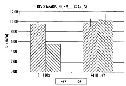
hours, and most preferably at least about 10 MPa.
Preferred embodiments of the bone graft substitute cement also exhibit a high
level of compressive strength, such as a compressive strength of at least
about 15 MPa
after curing for one hour in ambient air following mixing of the particulate
composition with an aqueous solution, more preferably a compressive strength
of at
least about 40 MPa. Further, preferred embodiments of the bone graft
substitute
cement will exhibit a compressive strength of at least about 50 MPa after
curing for
24 hours in ambient air following mixing of the particulate composition with
an
aqueous solution, more preferably a compressive strength of at least about 80
MPa.
The bone graft substitute cement of the invention will also exhibit a
dissolution rate that is significantly slower than a comparable bone graft
substitute
cement made substantially entirely of calcium sulfate. In certain preferred
embodiments, the cement of the invention exhibits an average dissolution rate,
expressed as an average percentage of weight loss per day, that is at least
about 25%
lower than the average dissolution rate of a cement formed using a particulate
composition consisting of calcium sulfate, the average dissolution rate
measured by
immersion of a 4.8 mm OD pellet having a length of 3.3 mm in distilled water
at 37 C
as described in greater detail below. More preferably, the bone graft
substitute
cement of the invention has an average dissolution rate that is at least about
30%
lower than a calcium sulfate cement, most preferably at least about 35% lower,
and in
some embodiments, as much as 40% lower or more. A preferred range of
dissolution,
expressed as an average percentage of weight loss per day measured using the
test
procedure set forth below, is about 5% to about 15%, more preferably about 7%
to
-22-
CA 02619469 2008-02-14
WO 2007/030616
PCT/US2006/034854
about 13%. Average dissolution rates stated are determined by linear
regression of %
weight loss per day using data from days 0, 1, 2, 3, and 4 determined using
the
procedure set forth below.
The present invention also provides a bone graft substitute kit comprising the
particulate composition of the invention. Typically, the kit comprises one or
more
containers enclosing the particulate composition as described above and a
separate
container enclosing a sterile aqueous solution. The kit will typically contain
a written
instruction set describing a method of using the kit. In addition, the bone-
graft
substitute kit of the invention will preferably comprise an apparatus for
mixing the
particulate composition with the aqueous solution in order to form the bone
graft
cement, such as a vacuum mixing apparatus. Additionally, the kit will
typically
include a device for delivering the bone graft cement to the site of the bone
defect,
such as an injection device (e.g., a needle and syringe). The particular
composition
and the sterile aqueous solution will typically be sterilized by irradiation
prior to
packaging in the kit.
As noted previously, in certain embodiments, the kit of the invention will
separate the two calcium phosphate powder components into different containers
to
avoid reaction during storage. There are a number of packaging configurations
that
can accomplish this goal. For example, in one embodiment, the kit includes one
container for CSH powder, one container for P-TCP powder, and one container
for
MCPM powder. In another embodiment, the kit includes two containers for the
particulate composition, one including P-TCP powder and a portion of the CSH
component and a second containing MCPM powder and a portion of the CSH
component. In yet another embodiment, the MCPM powder is packaged in a
separate
container by itself, and the P-TCP powder and the CSH powder are packaged
together. In a still further embodiment, the P-TCP powder is packaged in a
separate
container by itself, and the MCPM powder and the CSH powder are packaged
together. In any of the above embodiments, any of the powder containers can
further
include the crystalline powder of the carboxylic acid salt component and/or
the P-TCP
granules, or those components could be packaged separately in their own
containers.
When present, the accelerator adapted to accelerate conversion of CSH to CSD
is
typically in admixture with the CSH powder. In one preferred embodiment, the
kit
comprises one container enclosing the MCPM powder, and a second container
-23-
CA 02619469 2008-02-14
WO 2007/030616
PCT/US2006/034854
enclosing the remaining particulate ingredients in admixture, such as one or
more of
the CSH powder, the CSH accelerator, the 13-TCP powder, the 13-TCP granules,
and
the carboxylic acid crystalline powder.
In one preferred embodiment, the powdered form of the carboxylic acid is
packaged separately so that it can be reconstituted in the aqueous solution,
if desired,
prior to mixing the solution with the remaining particulate components.
However, as
noted previously, the aqueous solution of the kit may also contain the
carboxylic acid
component in solution form if the carboxylic acid is added after radiation
sterilization
of the aqueous component of the kit.
It can be important to utilize all of the aqueous solution packaged in the kit
in
order to ensure that consistent setting times are achieved. In one embodiment,
the
aqueous solution is packaged in a highly hydrophobic container, such as a
glass
syringe or other glass container, that is less prone to retention of residual
solution in
amounts that will cause changes in the performance characteristics of the bone
graft
substitute cement.
The present invention also provides a method for treating a bone defect. The
method of the invention involves applying a bone graft substitute cement as
described
above to the site of the bone defect. The bone graft substitute cement can be
applied
in flowable form following mixing of the particulate composition with the
aqueous
solution, such as through an injection device, prior to setting of the
composition.
Alternatively, the bone graft substitute cement can be used in a precast
hardened
form, wherein the cement is provided in predetermined shapes such as pellets,
granules, wedges, blocks, or disks, or used in the form of randomly-shaped
shards
created by mechanically breaking a cement mass into smaller pieces. In a
further
embodiment, the clinician can form the bone graft cement mixture and manually
mold
the mixture into a desired shape, such as the shape needed to fill a
particular bone
defect, prior to application.
In another embodiment, the bone graft substitute cement of the invention can
be incorporated into an orthopedic implant, such as any of the various devices
adapted
for joint replacement. The bone graft substitute cement is typically
incorporated into
such devices as an outer coating or as a filling material within the pores of
a porous
outer component of the device. In this embodiment, the bone graft substitute
cement
facilitates bone ingrowth in the area surrounding the implanted device.
Exemplary
-24-
CA 02619469 2013-03-19
. ,
orthopedic implants include knee replacement devices (e.g., constrained or non-
constrained
knee implant devices, hinged knee devices, metallic plateau knee devices, and
patellar devices),
hip replacement devices (e.g., acetabular components and femoral components),
elbow
replacement devices (e.g., constrained, semi-constrained, and non-constrained
devices), upper
femoral devices, upper humeral devices, wrist replacement devices (e.g., semi-
constrained 2-
and 3-part articulation devices), shoulder devices, passive tendon devices,
spinal devices (e.g.,
thoracolumbar spinal fixation devices, cervical spinal fixation devices, and
spinal fusion cages),
finger/toe devices, and diaphysis devices.
The present invention will be further illustrated by the following non-
limiting example.
EXPERIMENTAL
Example 1 illustrates in vivo use of a bone graft substitute cement of the
invention, and
particularly describes the reduced resorption rate (as compared to a calcium
sulfate
composition), good mechanical properties, and acceptable setting times
exhibited by the
inventive composition. Example 2 illustrates the ability of an embodiment of
the inventive
composition increase the amount, strength, and stiffness of restored bone as
compared to use of
conventional CaSat pellets. Example 3 demonstrates the degradation effect of
gamma
radiation on glycolic acid in solution, and the effect of such degradation on
setting times of the
bone graft substitute cement. Example 4 demonstrates that placement of a
glycolic acid salt
form in the particulate composition reduces the effect of radiation on the
performance of the
bone graft substitute cement without sacrificing other advantageous
properties, such as certain
handling and mechanical strength properties.
Setting Time Measurement
Setting times can be measured using a Vicat needle that is 1 mm in diameter, 5
cm long,
and which possesses a total weight of 300 g, all per ASTM C-472. The sample
being tested
should be mixed in a manner that a homogeneous, flowable paste is created. The
sample size
for the Vicat needle drop test is about 3 cc to about 5 cc of material tapped
down to a cake in an
approximately 20 mL polyethylene cup; the sample shall be handled such that no
agitation is
inflicted upon the material 1 minute after the aqueous solution
-25-
CA 02619469 2008-02-14
WO 2007/030616
PCT/US2006/034854
contacts the particulate composition other than the dropping and removal of
the Vicat
needle. The cup should be of such dimensions that the cake is a short, flat
cylinder
measuring about 1/4" to about 3/8" in height.
Set time according to the Vicat needle drop test is defined as the amount of
time elapsed between the time the aqueous solution contacts the particulate
composition and the time the Vicat needle will not pass through 50% of the
height of
a cement sample upon being dropped from the upper surface of the sample. The
needle is allowed to fall under its own weight, under gravity alone, through a
line
perpendicular to the top and bottom, flat faces of the cylinder-shaped sample
cake.
The needle is dropped every 30 seconds after the first drop. The needle shall
not be
dropped more than 6 times during the duration of the test. If after the 6th
drop the
needle continues to pass through more that 50% of the height of the sample,
the test
must be repeated with fresh material; a new, clean cup; and a clean Vicat
needle free
of debris, especially that which is left behind from previous tests. Cups,
mixing
equipment, and material transfer equipment should not be reused. All materials
and
equipment used during testing should be between 21-27 C and exposed to an
environment with a relative humidity between 20-50%.
Compression Strength Measurement
Compression strength of the material is determined through the following test
methodology. Specimens are cast to size per ASTM F451 (6 mm outer diameter x
12
mm in length), which is incorporated by reference in its entirety, utilizing a
stainless
steel split mold with a capacity of eight specimens.
The split mold is placed on a glass plate with the cylindrical voids, specimen
slots, standing upright. The material is mixed and then loaded into a device
for
delivery of the material into the slots such that a back filling method can be
utilized; a
syringe with a jamshidi-type needle is commonly used. Each specimen slot is
filled
from bottom to top in a back filling manner. It is customary to excessively
fill the
mold such that excess material extrudes out above the dimensions of the split
molds,
this assures displacement of any air entrapped within the specimen slots. It
may be
necessary to hold mold down on to glass plate during casting to prevent
material from
extruding out of the bottom of the specimen slots, between the glass plate and
mold.
Upon filling each specimen slot another glass plate is pushed by hand onto the
excess material located on the top of the mold, producing a thin sheet of
flashing
-26-
, CA 02619469 2013-03-19
_=_
across the tops of the specimens and split mold itself. This glass plate is of
a size which does
not produce an excessive compressive force or a pressurized environment in
which the material
cures. All specimens are cast and flashing is created within 2 minutes of the
aqueous solution
coming into contact with the particulate component.
The specimens are demolded 30 minutes after the aqueous solution has come into
contact with the particulate component. First the flashing is removed from
both sides of the
split mold containing the faces of the specimens; regardless of holding mold
against the lower
glass plate upon casting, a thin film of flashing is created on the lower
surface of the mold.
Commonly, a razor blade is used to scrape off the flashing and in doing so
create smooth faces
on the specimens. The split mold is separated and the specimens are removed.
All specimens
should be removed within 32 minutes of the aqueous solution coming into
contact of the
particulate component. Upon removal of the specimens, they should be allowed
to continue
curing in air at room condition (21-27 C; 20-50% relative humidity) until time
of testing.
Testing of the material is performed at a predetermined time after the aqueous
solution
has come into contact with the particulate component. Commonly, testing is
performed at 1 hr
and 24 hrs. Testing is performed on a compression test fixture per ASTM D695.
The
compression test fixture is placed on a mechanical test frame capable of
displacement control
and monitoring of displacement and force through data acquisition running at
50 Hz or faster.
The specimens are tested individually on the compression test frame. The
specimens
are placed between the platens in a manner such that the cylinder faces are
positioned against
the platens. The compression test frame containing the specimen is loaded in
compression at a
rate of 0.333 mm/sec until failure. Force and displacement are monitored
throughout the test,
and maximum force at failure is noted. Proper failure will result in a
fracture across the height
of the specimen. The maximum compression force at failure is noted. Failure is
defined as a
sudden drop in load, deviation of the loading curve from the initial slope
created by the loading
of the specimen, and/or the force noted upon visual failure of the specimen.
The compression strength in MPa is then calculated as followed: (Pmax)/(n*R2);
where
Pmax is the load at failure in Newtons, TC is approximately 3.14, and R is the
radius of the
specimen in mm (3).
-27-
CA 02619469 2008-02-14
WO 2007/030616
PCT/US2006/034854
It is crucial when performing compression strength specimen preparation that
all equipment used is clean of all debris, especially that of the cured
material of
interest.
Diametral Tensile Strength Measurement
The diametral tensile strength is determined through the following test
methodology. A 1" cube of 10 lb/ft3 closed-cell polyurethane foam (available
as Last-
A-Foam from General Plastics Manufacturing Company, Tacoma, WA) with an
approximately 5/8 in. (15.8 mm) outer diameter cylindrical void and notches
for side
removal is used as the specimen mold. The approximately 5/8 in. outer diameter
cylindrical void is created by drilling perpendicularly through opposite faces
of the
cube in one depression of a drill press utilizing a 5/8 in. drill bit. The
void runs the
entire length of the cube and is centered such that both opposite, drilled
faces share
the same center as the circular voids created in them from the drilling. Two
opposite
sides from the remaining four full sides are designated to become the open
sides of
the final specimen; these sides will be removed via the notches. These sides
are
notched, two notches per side, in a manner such that they can be removed
immediately prior to testing and not affect the sample integrity. The notches
shall run
the entire length of the cube and be separated in a manner that upon removal
>50% of
the height of the specimen is exposed. Commonly the notches are created using
an
upright band saw. Figures 2a-2c illustrate an exemplary diametral tensile test
mold
20. Fig. 2a provides a top and bottom view of the mold 20. Fig. 2b provides a
side
view of the mold 20. Fig. 2c provides a front and rear view of the mold 20 and
shows
a 16 mm outer diameter cylindrical void 30 therein.
The material to be tested is mixed to a homogeneous paste and loaded into a
device suitable for injection of the paste into the 16 mm outer diameter
cylindrical
void. Commonly a 30 cc syringe with a 1 cm opening is used for this. The mold
is
held by hand using the thumb and middle finger positioned on the opposite,
notched
sides. The index finger of the hand used to hold the mold is positioned over
one of
the circular openings. The material is then injected into the void from the
opposite
side of the void from the index finger; the entire face of the syringe
exhibiting the 1
cm opening is lightly pushed up against the circular opening of the mold. Upon
injection of the material into the mold, pressure will be felt on the index
finger
covering the back opening from the ejected material. The index finger is
slowly
-28-
CA 02619469 2008-02-14
WO 2007/030616
PCT/US2006/034854
removed while filling continues, allowing the paste to flow out of the rear of
the mold
in an extrusion with the same 16 mm outer diameter as the void. The syringe is
slowly backed out from the front opening while back filling of paste is
performed
through further ejection from the syringe until the entire void is filled and
excess
material is located outside the dimensions of the original cube of foam. The
front and
rear sides of the specimen are wiped smooth, flush with the front and rear
sides of the
mold using a spatula. All specimens to be tested should be made within 2
minutes
from the start of mixing, defined by the aqueous solution coming into contact
with the
particulate composition.
The specimens are allowed to cure horizontally in air in the mold with the
front and rear sides of the mold exposed to air at room conditions (21-27 C;
20-50%
relative humidity) for a predetermined amount of time, normally 1 hr or 24
hrs. This
predetermined amount of time begins at the time at which the aqueous solution
comes
into contact with the particulate composition at the beginning of the mixing
process.
Testing is performed on a mechanical test frame capable of displacement
control and of monitoring displacement and force through data acquisition
running at
Hz or faster. The sides of the specimen mold are removed immediately prior to
testing; only the areas between the notches are removed.
Removal of the sides is normally performed with a knife. The top and bottom
20 of the mold are held between two fingers with slight pressure to prevent
specimen
surface-to-mold interface damage. The knife blade is placed into one of the
notches
and then twisted to break the area between the notches free; this is repeated
for the
other side in the same manner. The tops and bottom of the molds are left in
place to
hold the specimen and prevent shear stresses on the surface. The specimen is
placed
between two flat, parallel platens; one of which is free to swivel to allow
alignment
with the loading train. The swiveling platen assures an equally distributed
load across
the specimen contact points. The specimen is loaded transversely at a rate of
5
mm/minute until failure. Proper failure will result in a vertical fracture
completely
through the length of the specimen. The maximum force at failure is noted.
A loading curve of force versus displacement is created to determine the
maximum force at failure, in which displacement and force are positive values.
The
first part of the loading curve shows the loading of the foam followed by its
compression. The compression of the foam portion will be evident by continued
-29-
CA 02619469 2008-02-14
WO 2007/030616
PCT/US2006/034854
displacement with no substantial increase in force; this can also be seen
visually
during the test. After the foam is completely compressed, the force will begin
to rise
again, creating an increasing slope on the loading curve followed by a
constant slope
as the load is transferred to the specimen. The increasing slope is commonly
known
as a "toe in". Failure is defined as a sudden drop in load, a decrease in the
slope of
the loading curve after the constant slope from specimen loading has been
established,
and/or the force noted upon visual failure of the specimen while the test is
running.
The diametral tensile strength in MPa is then calculated as followed:
(2*Pmax)/( 2 *L*H); where Pmax is the load at failure in Newtons, 7E is
approximately equal to 3.14, L is the length of the specimen in mm (25.4), and
H is
the height of the specimen in mm (16). Specimens are disqualified for
diametral
tensile strengths if any one or more of the following occur: fracture is not
vertical,
facture does not completely run the length of the specimen, length of the
specimen
fails, or voids in the material are seen on the fractured walls of the
specimen.
It is crucial when performing diametral tensile strength specimen preparation
that all equipment used is clean of all debris, especially that of the cured
material of
interest.
Dissolution Rate Measurement
Dissolution rate of the material is determined through the following
methodology. Specimens are cast in silicone molds to a size of 4.8 mm outer
diameter and 3.3 mm tall cylinders. A 3.3 mm thick sheet of silicone
containing
cylindrical voids is used as a mold. Cylindrical voids are 4.8 mm in outer
diameter
and 3.3 mm tall, and orientated such that the circular faces of the void are
parallel and
in the same plane as the surfaces of the silicone sheet.
A thin sheet of polyethylene is laid on a table. A polyethylene mesh is placed
on top of the polyethylene sheet; sheet and mesh are of same dimensions
(excluding
thickness) and positioned such that the mesh masks the sheet from the top.
Next a
silicone mold of smaller dimensions is placed on top of the mesh (excluding
thickness). No part of the mold hangs off the edge of the mesh or sheet.
The material to be tested is then mixed together to form a homogeneous paste.
The paste is then wiped across the top of the mold using a spatula in a manner
that the
voids are packed with the material. The mesh will allow air to be displaced
out of the
void as the mold is filled. Several wipes are performed to assure that
material has
-30-
CA 02619469 2008-02-14
WO 2007/030616
PCT/US2006/034854
completely penetrated to bottom of the mold and extruded out through the mesh
and
onto the lower polyethylene sheet. A final wipe with the spatula across the
top of the
mold is performed to remove the majority of excess material and produce smooth
top
faces for the specimens.
Another polyethylene sheet of the same dimensions of the as the first is then
placed across the top of the mold, such that it completely covers the top of
the mold.
This sheet is then gently pressed against the mold using a finger in a gentle
rubbing
motion. An intimate contact between the top polyethylene sheet and the
specimens is
created.
The entire system, sheet, mesh, mold, and sheet, is then picked up as a whole
and flipped over in a manner such that the original top is now facing down.
The
system is held by hand and slapped repeatedly onto table in a manner such that
any air
entrapped in the molds will be displaced out by the material; slapping of the
system
should not be excessive in force or repetitions. Upon removal of the majority
of the
air the system is returned to table in the upside down orientation, sheet and
mesh side
up. The top polyethylene sheet, originally the bottom, and mesh are removed
and the
spatula is again used to wipe material into voids in the tops (previously
bottoms) of
the specimens created from air removal. A final wipe with the spatula across
the top
of the mold is performed to remove the majority of excess material. The sheet
(no
mesh) is returned to the top of the mold. The sheet is then pressed against
the mold
using a finger in a gentle rubbing motion. An intimate contact between the top
and
bottom polyethylene sheet and the specimens has now been created.
The specimens are left in the mold to cure for a minimum of 8 hrs after the
second polyethylene sheet has been placed in direct contact with the specimens
and
mold (no mesh). After at least 8 hrs have passed, the specimens are demolded
by
hand. Any flash remaining attached to pellet faces are removed by rolling
specimen
between fingers. All defective specimens are disqualified from the test and
discarded.
A defective specimen is defined as a specimen not exhibiting a cylindrical
shape,
which could be caused by entrapped air, defects created upon demolding, and/or
physical damage to the specimen itself.
All specimens which are not defective are spread across a stainless steel pan
in
a monolayer. The pan and specimens are then dried in an oven at 40 C for a
-31-
CA 02619469 2008-02-14
WO 2007/030616
PCT/US2006/034854
minimum of 4 hrs, and then removed from oven and allowed to cool for 30
minutes in
room conditions (21-27 C; 20-50% relative humidity).
From the specimens created, five (5) specimens are arbitrarily chosen to be
used for dissolution testing. Each specimen chosen is paired with a clean
cylindrical
fritted glass extraction thimble of the following dimensions: 90.25 mm overall
height,
4 mm fritted glass base (40-60 micron pores) located 80 mm from top of
thimble, 25
mm outer diameter, and 22 mm inner diameter. The mass of each extraction
thimble
is measured (0.01 mg) and noted. The mass of each specimen in measured (0.01
mg)
and noted. A polyethylene bottle (300 mL) is designated to each pair (specimen
and
thimble). The bottle has dimensions that allow thimble and specimen to easily
be
placed in and removed from bottle and upon filling with 275 mL of water will
create a
column of water that is taller than the thimble. The bottle is filled with 275
mL of
distilled water at room temperature (21-27 C). The specimen is placed into its
corresponding thimble and the thimble is lowered into the bottle; care is
taken to keep
any part of the material from escaping from the thimble. The bottle is capped
and
placed into a water bath at 37 C with no agitation and the time is noted.
24 hrs after the specimen has been in the water, the thimble containing the
specimen is retrieved. The water is allowed to drain out of the thimble
through the
fitted glass base. The thimble containing the specimen is then dried for 4 hrs
in a
40 C oven or until completely dried (determined gravimetrically). The thimble
containing the specimen is then allowed to cool down for 30 minutes at room
conditions (21-27 C; 20-50% relative humidity).
The thimble-containing the pellet is then weighed to an accuracy of 0.01 mg.
Subtracting the known empty thimble mass from the mass of the combination will
result in the mass of the specimen alone. Subtracting this mass from the
initial
specimen mass will produce the mass lost to dissolution. This mass lost can be
divided by the specimen initial mass and the product of that multiplied by 100
will
result in the % mass lost from dissolution.
At this point the thimble containing the pellet is returned to the bottle
containing fresh distilled water (275 mL) at room temperature (21-27 C), and
the
bottle is capped and returned to the water bath. After 24 hrs the drying and
weighing
process is repeated. These actions are repeated with fresh distilled water
after every
24 hr soak until the test is terminated or the material completely dissolves.
-32-
CA 02619469 2013-03-19
EXAMPLE 1
Diametral tensile strength, dissolution properties, and in vivo evaluation of
new bone
ingrowth and residual material of bone graft cements of the invention were
compared to a
commercially available calcium sulfate material. The experimental group for
all experiments
was an embodiment of the current invention including a cement consisting of
74.906 weight
percent calcium sulfate hemihydrate, 0.094 weight percent accelerator (sucrose
coated calcium
sulfate dihydrate), 6.7 weight percent monocalcium phosphate monohydrate, 8.3
weight percent
beta tricalcium phosphate powder, 10 weight percent beta tricalcium phosphate
granules, and
an aqueous solution of 0.6 molar glycolic acid neutralized to a pH of 7.00
with 10 normal
sodium hydroxide solution (hereinafter "SR"). MIIG X3 Bone Graft Substitute
(hereinafter
"X3") (Wright Medical, Arlington, TN) calcium sulfate was used as a control
for all
experiments. The SR material was formulated to set in 14-19 minutes, whereas
the X3 material
was formulated to set in 7-10 minutes.
An intermediate resorbing calcium sulfate, calcium phosphate composite cement
was
also evaluated in this study. Dissolution properties, compression strength,
and in vivo
evaluation of new bone ingrowth and residual material were evaluated for this
material. This
material is also an embodiment of the present invention and comprised 84.999
weight percent
calcium sulfate hemihydrate, 6.7 weight percent monocalcium phosphate
monohydrate, 8.3
weight percent beta tricalcium phosphate powder, 0.0013 weight percent
accelerator (sucrose
coated calcium sulfate dihydrate), and an aqueous component of water. This
intermediate
material was formulated to set in 11-16 minutes.
Compression strength was measured on vacuum mixed specimens cast in the manner
set
forth above. Specimens (n=6) were cured for 1 hr in ambient air. Specimens
(n=3) were cured
for 24 hrs in ambient air. The specimens were loaded lengthwise using a MTS
858 BionixTM
test system at a constant rate of 0.333 mm/sec. The compression strength in
MPa was
calculated using the formula (Pmax)/( eR2).
Diametral tensile strength (DTS) was measured on vacuum mixed specimens cast
in the
manner set forth above. The sides of the foam blocks were removed prior to
testing.
Specimens (n=4) were cured for 1 and 24 hrs in ambient air at room
temperature. The
specimens were transversely loaded to failure in compression using
-33-
CA 02619469 2008-02-14
WO 2007/030616
PCT/US2006/034854
a MTS 858 Bionix test system at a constant rate of 5 mm/min. DTS was
calculated
from the formula DTS = (2*Pmax)/( 7C*L*H).
Dissolution tests were performed on 4.8 mm OD X 3.3 mm cylindrical pellets
(n=5). Specimens were placed in 275 mL of distilled water at 37 C. Solutions
were
changed daily. Specimens were dried and weighed daily for first 30 days and
every 5
days thereafter until a residual mass of <5% was achieved. X-Ray diffraction
(XRD)
was used to identify the residual material.
Results:
Fig. 3 shows the DTS results. One-way ANOVA were performed using JMP
software (SAS, Cary, NC). A significant difference was seen between 1 and 24
hr
cure times for SR cured in air (p<0.001) and no difference for the X3
(p=0.508). It is
apparent from the air cured data that the SR reaction is incomplete at 1 hr
while the
X3 setting reaction is essentially complete. This result was expected based on
the
differences in setting time.
Average, maximum, and minimum compression strength values for the
intermediate material were determined. The 1 hr cure time data produced an
average
strength of 19.4 MPa, a minimum of 16.2 MPa, and a maximum of 21.4 MPa. The 24
hr cure data produced an average strength of 69.9 MPa, a minimum of 61.4 MPa,
and
a maximum of 77.3 MPa.
Dissolution results are shown in Fig. 4. Linear regression of days 0 through 4
of the curves were used to estimate the dissolution rates. The average SR rate
was
10.7%/day, while the X3 rate was 17.8%/day. The average rate for the
intermediate
material was 13.5%/day. Following dissolution of 95% of the bone graft
substitute
cement material, XRD of residual SR material showed it to be beta tricalcium
phosphate, a known bioresorbable and osteoconductive material.
A 6-week in vivo pilot study was conducted under an Institutional Animal
Care and Use Committee (IACUC) approved protocol. In each of 3 dogs, two
defects
measuring 9 mm X 15 mm were created in each proximal humerus. Each site was
filled with either an injected bolus of SR (1-1.5 cc), 4.8 mm OD X 3.3 mm
pellets of
SR, 4.8 mm OD X 3.3 mm pellets of X3, or an injected bolus of the intermediate
resorbing calcium sulfate, calcium phosphate composite cement. Implants were
sterilized with gamma radiation. Each dog received one implant of each
material.
Healing of the defects and resorption of the pellets and boluses were assessed
from
-34-
CA 02619469 2008-02-14
WO 2007/030616
PCT/US2006/034854
radiographs obtained after 0, 2, and 4 weeks and contact radiographs after 6
weeks.
New bone formation and residual implanted material in the defects were
evaluated
using light microscopy of undecalcified, plastic embedded histological
sections
stained with basic fuchsin and toluidine blue. Area fraction of new bone and
residual
material in the defects were determined using histomorphometry.
In the in vivo study, the radiographic and histologic data indicated that both
types of pellets and boluses were replaced with newly formed osteoid, woven,
and
lamellar bone that had formed in concentric lamellae at the previous implant
sites. At
6 weeks, area fraction of new bone formation was 35.9 6.1% for defects
implanted
with SR pellets and 26.7 10.0% for defects implanted with X3 pellets. At 6
weeks,
the majority of the implanted pellet materials had resorbed, but there was
slightly
more residual implant material in SR pellet defects compared to the X3 pellet
defects.
For the SR bolus implants new bone formation was 15.65.6% with 29.9 11.9%
residual implant material. For the bolus of intermediate resorbing calcium
sulfate,
calcium phosphate composite cement, new bone formation was 23.4 7.1% with
19.3 8.0% residual implant material. Smaller fractions of new bone formation
can be
expected for bolus materials at early time due to larger percentages of
residual
material and smaller surface area to implant volume ratios when compared to
that of
pellets.
The composite cement of the invention demonstrated consistent setting and
strength characteristics similar to those of the control. The goal of slowing
down the
dissolution rate was achieved, and the early in vivo bone growth was
equivalent or
superior to the pure calcium sulfate control.
EXAMPLE 2
Materials and Method:
Under an IACUC-approved protocol, 10 skeletally mature, male dogs (25-32
kgs) had a critical-size, axial medullary defect (13mm dia X 50mm) created
bilaterally in the proximal humerus and were studied for 13 (n=5) and 26 (n=5)
weeks. The defect in one humerus was injected with 6 cc of the test material
(SR
cement according to Example 1). An identical defect in the contralateral
humerus
received an equal volume of Ca504 pellets (OSEOSET pellets, Wright Medical).
Radiographs were obtained at 0, 2, 6, 13 and 26 weeks. Transverse,
undecalcified
-35-
CA 02619469 2008-02-14
WO 2007/030616
PCT/US2006/034854
stained sections of the bones were prepared. The area fractions of new bone
and
implanted residual materials in the defects were quantified using standard
point-
counting techniques. The sections were also examined using high-resolution
contact
radiographs. The yield strength and modulus of an 8mm dia. X 20mm test
cylinder
cored from the midlevel of each defect was determined in unconfined, uniaxial
compression tests at a crosshead speed of 0.5 mmimin. The histomorphometric
and
biomechanical data were analyzed using the Friedman and Mann-Whitney tests.
Data
are presented as the mean and standard deviation.
Results:
The clinical and postmortem radiographs revealed markedly different rates of
resorption of the bone graft substitutes and replacement with bone in the
defects.
Resorption of the CaSO4 pellets was apparent beginning at 2 weeks and
substantially
complete by 6 weeks. There was slower resorption of the SR cement, also
beginning
at 2 weeks, but some cement persisted at 26 weeks.
In all of the stained histological sections, there was restoration of the
defects
by bone and marrow with only focal areas of fibrous tissue and relatively low
volumes of residual implanted material. The area fraction of new mineralized
bone at
13 weeks was 2-fold greater in defects treated with SR cement (39.4+4.7%)
compared
to defects treated with conventional CaSO4 pellets (17.3+4.3%) (p=0.025). At
26
weeks, the bone had remodeled to a more normal architecture, but there was
still more
bone in defects treated with cement (18.0+3.4%) compared to pellets
(11.2+2.6%)
(p=0.025).
Residual matrix and B-TCP granules were incorporated into bone trabeculae.
Surfaces of the materials not covered by bone appeared to be undergoing
remodeling
by osteoclast-like cells, some of which contained minute particles. The area
fraction
of residual matrix was greater in the cement-treated defects at 13 weeks
(2.9+2.8%)
and at 26 weeks (0.6+0.8%) compared to pellet-treated defects (0.0% at 13 and
26
weeks) (p=.025 and .083, respectively). Residual matrix decreased with time in
the
cement-treated defects (p=.047). The area fraction of residual B-TCP granules
also
decreased from 13 weeks (3.6+1.0%) to 26 weeks (0.8+1.4%) (p=.016). The
maximum dimension of the B-TCP granules decreased from 348+13 [tin at 13 weeks
to 296+29m at 26 weeks (p=.008).
-36-
CA 02619469 2008-02-14
WO 2007/030616
PCT/US2006/034854
Cored bone samples from defects treated with the cement were considerably
stronger and stiffer than those treated with CaSO4pellets at both 13 and 26
weeks
(Table 1 below). For comparison, similar cored trabecular bone specimens from
8
normal proximal humeri had a yield strength of 1.4+0.66 MPa and a modulus of
117+72 MPa.
Table 1
Time (wks) SR Cement
CaSO4Pellets
Yield Strength (MPa) 13 5.3 (2.6)* .90 (.44)
Yield Strength (MPa) 26 2.2 (.41)** .47 (.46)
Modulus (MPa) 13 283 (217) 40.8 (35.6)
Modulus (MPa) 26 150 (73)* 15.8 (23.6)
*p = 0.025, **p=.046, different from pellets
Conclusion:
Several Ca-based materials with different resorption rates were successfully
combined to produce a cement with a tailored, slower resorption profile. In
this
cement, the majority of the calcium sulfate and dicalcium phosphate dihydrate
matrix
resorbs early, promoting bone formation deep into the bolus of cement, while
the
distributed13-TCP granules provide a scaffold, incorporate into new bone, and
are
then more slowly resorbed. The engineered cement increased the amount,
strength
and stiffness of restored bone when compared to conventional CaSO4 pellets
after 13
and 26 weeks. This cement holds promise for clinical applications where a
strong,
injectable and highly biocompatible bone graft substitute would be
advantageous.
EXAMPLE 3
Materials and Method:
250 mL of mixing solution, 0.6M glycolic acid neutralized with sodium
hydroxide, was created and the pH was noted with a calibrated pH meter. The
solution was made using crystalline glycolic acid (Alfa Aesar Part # A12511;
Ward
Hill, MA), lON sodium hydroxide solution (EMD Chemicals Part # SX0607N-6;
Darmstadt, Germany), and USP water for irrigation (Baxter Healthcare
Corporation
Part # 2F7112; Deerfield, IL).
-37-
CA 02619469 2008-02-14
WO 2007/030616
PCT/US2006/034854
The solution was then divided into two ¨125mL aliquots and then individually
rebottled. One of the bottles was sent out for bulk gamma radiation
sterilization, 25-
32 kGy dose, and the other was retained as an unsterilized control. Upon
return of the
sterilized solution the pH of both the sterilized and the non-sterilized
solutions were
checked with a calibrated pH meter and noted.
A single lot of SR powder of the type utilized in Example 1 was used in this
study to avoid lot-to-lot variability in set time and injection forces.
Three vials were filled with 6.9 mL of the unsterilized solution and coupled
with three vials of unsterilized SR powder containing 30 g per vial. This
group served
as a control.
Another group was made to represent the option of aseptic filling of the
individual units of neutralized glycolic acid. This group consisted of three
vials of 6.9
mL of glycolic acid filled out of the 125 mL of bulk sterilized solution and
three vials
of SR powder filled to 30 g. The powder vials were sent out for gamma
radiation
sterilization. This represents sterilization of the bulk solution followed by
aseptic
filling and coupling in a kit containing the already sterilized unit of
powder.
The third and final group represents a preferred manufacturing situation:
gamma radiation sterilization of the bulk solution followed by gamma radiation
sterilization of the individual units. Three vials of solution were filled to
6.9 mL with
the sterilized bulk solution. Another three vials were filled with 30 g of the
SR
powder. All six of these vials were sent out to sterilization. This represents
filling the
solution from a bulk sterilized solution, packaging kits containing
unsterilized powder
with bulk sterilized solution, and then sending the kit out for a final
sterilization.
Upon return of all the groups the following testing was performed. All
solutions, including the remainder of the bulk solution were checked for pH
with a
calibrated pH meter and noted. The nine sets of units (three units of
unsterilized
solution and unsterilized powder, three units of one time bulk sterilized
solution and
unit sterilized powder, and three units of two times sterilized solution (once
in bulk
followed by once as a unit) and one time unit sterilized powder) were mixed to
form a
homogeneous paste under vacuum. Set times of approx. 'A in. thick aliquot of
paste in
a 25 mL plastic cup were determined through the use of a 300 g Vicat needle.
Injection force from a 3 cc syringe attached to a 6 cm 11 gauge non-tapered,
ported
jamshidi type needle was determined at 3 and 5 minutes after the powder and
-38-
CA 02619469 2008-02-14
WO 2007/030616
PCT/US2006/034854
solutions had come into contact with one another. Injection forces are
reported as
forces seen at 15 mm of plunger displacement being displaced at 4.4 mm/sec.
Injection testing was performed using a materials test frame in displacement
control,
and data acquisition was taken at 50 Hz of force and displacement.
Results:
pH drift was seen for all solutions. Results were consistent within a group
although the two time sterilized solution produced a pH different from that of
the
control and one time sterilized group. Specifically, the two times sterilized
solution
produced an average pH of about 6.3, while the other solution groups exhibited
a pH
of about 5.5.
Forces of injection for all groups were the same. At the 3 minute time point
the injection force was about 25 N, and for the 5 minute time point the
injection force
was about 40 N.
The set time for the unsterilized and one time sterilized group were
consistently around 18.5 minutes, except for one unit of the one time
sterilized group
which was at 19.75 minutes. Set time measurements for the two times solution
sterilized group and one time powder sterilized had consistently shifted to
about 22
minutes.
Conclusion:
The pH and set time shifts in the two times sterilized solution group shows
degradation of the neutralized glycolic acid solution through gamma radiation
sterilization. Although the effects were not pronounced in the one-time
sterilized
solution, degradation must have occurred in that group as radiation
degradation is an
additive process.
EXAMPLE 4
Materials and Method:
First, the effect of gamma sterilization of crystalline glycolic acid (GA) on
the
material's acid-base titration curve using a stock solution of 0.6M sodium
hydroxide
(NaOH) was examined. Then, physical property comparisons were made between
radiation sterilized samples of a cement powder with solid sodium glycolate
(Na-GA)
blended into the precursor powder and a non-irradiated material. Diametral
tensile
strength, injection force, Vicat set time, and morphological (SEM) comparisons
of the
-39-
CA 02619469 2008-02-14
WO 2007/030616
PCT/US2006/034854
set cements from each configuration were made. Additionally, Vicat set time
comparisons were made between unsterilized samples of each product
configuration.
Approximately 50 g of GA (GLYPURE available from Dupont) was
subjected to gamma radiation sterilization (25-32 kGy dosage). Two ¨1M
solutions
of GA were created at equal volumes, one with the gamma irradiated GA and the
other with non-irradiated GA of the same manufacturing lot. In order to avoid
loss of
material during liquid transfers and from evaporation, the solutions were made
immediately prior to being used by dissolving 3.803 g of GA with 50.000 g of
DI
water in a 250 mL beaker.
A 500mL 0.6M NaOH stock solution was created by diluting 30 mL of the
10N NaOH with DI water in a 500 mL volumetric flask. This stock solution was
used
as the titrant for both GA solutions.
A 50 mL burette (0.1 mL increments) equipped with a stopcock was used to
dispense the NaOH stock solution in various increments directly into the 250
mL
beakers containing the ¨1M GA solutions. During titration the GA solutions
were
stirred using a polytetraflouroethylene coated magnetic stir bar and plate.
The volume
of NaOH stock dispensed was monitored and recorded through the titrations. The
pH
of the GA solution was also monitored and recorded with each increment of NaOH
stock added. pH measurements were determined through use of a pH meter (VWR
Scientific; Model 8000) and electrode (VWR Scientific, P/N 14002-780)
calibrated
between pH = 4.00 and 7.00 using standard buffer solutions (VWR Scientific,
P/N
34170-130 and 34170-127, respectively). Titration was carried out until
minimal
changes of pH in the alkaline range were seen with consecutive additions of
the stock
solution. Titration curves (pH of GA solution vs. mL 0.6M NaOH) were plotted
and
comparisons were made to detect effects of gamma irradiation on crystalline
GA.
A 300 g batch of an SR material as described in Example 1 (Configuration 1
with NA-GA in solution) was blended for 20 min in a 1 qt acrylic V-shell using
a
60Hz P-K Twin-Shell Yoke blender (Patterson-Kelley Co.; East Stroudsburg, PA).
All pastes created with Configuration 1 were produced using 0.6M Na-GA
solution at
a liquid weight to powder weight ratio (L/P) value of 0.23.
Twenty-five (25) 15 cc injectable kits of a modified SR material
(Configuration 2 comprising 1.290 wt% of <441m Na-GA powder) were prepared
(35.00 g 0.01 g powder and 7.59 g 0.01g sterile water for irrigation) from
a
-40-
CA 02619469 2008-02-14
WO 2007/030616
PCT/US2006/034854
1013.071 g batch blended for 20 min in a 2 qt stainless V-shell using a 60Hz P-
K
Twin-Shell Yoke blender. The water was overfilled by 0.10 g to account for
solution
loss in the vial during transfer. The kits were subjected to gamma radiation
sterilization (25-32 kGy dosage). Four of these kits were used for this study.
The LIP value for Configuration 2 is 0.214. The difference in LIP values for
the two configurations is due to the movement of the Na-GA from the solution
to the
powder.
Results:
The results of the Vicat set time showed Configuration 2 to have shifted the
Vicat set time out by a small amount. Other than the location of the Na-GA
within
the two configurations, the only other variable is that the Configuration 2
kits were
irradiated, while the Configuration 1 materials were not. To address these two
variables, Vicat set time of two additional samples for each configuration
were taken;
however, the Configuration 2 samples were not subjected to sterilization.
Two 35 g units of Configuration 1 powder were tested for Vicat set time. The
entire mix was transferred to a 50 mL polystyrene beaker cup (VWR Scientific
P/N
13916-015); the paste was leveled and major air pockets were removed through
the
gentle tapping of the cup on a table. Vicat set time was determined through
the same
method as performed above on both samples.
Two units of Configuration 2 powder were tested, and the entire mix was used
to determine Vicat set times as performed in the previous paragraph. One of
the
mixes was performed with 30 g of powder due to lack of material.
The new data obtained for Configuration 1 was combined with results from
the previous Vicat testing since there was no difference in the treatment of
the
specimens other than volume. The new data for Configuration 2 was used
independently to compare against the Configuration 1 results.
Figure 5 shows the overlaid curves of the titrations of the 1M GA solutions
produced from crystalline GA with and without gamma sterilization. The
resulting
curves are indistinguishable. As noted in Example 3, solutions of Na-GA used
in the
manufacturing of Configuration 1 kits displayed a pH shift post gamma
radiation
sterilization. However, this change in pH was not seen for a solution created
with GA
gamma irradiated in the crystalline form. This result is indicative that
degradation via
gamma irradiation of the glycolate ion is greatly, if not completely,
alleviated by
-41-
CA 02619469 2008-02-14
WO 2007/030616
PCT/US2006/034854
exposure in the crystalline form. This is strong evidence that the crystalline
Na-GA
component in Configuration 2 will also be less affected by gamma irradiation.
Table 2 below shows the average results from the 24 hr dry DTS testing of
each configuration. Both configurations exhibited DTS values close to 9 MPa
with
less than a 10% coefficient of variance within each group. Although
Configuration 2
exhibited a slightly higher average strength value of 9.29 MPa, the difference
between
the two configurations was not statistically significant (p=0.25). The
observed
difference can be attributed to error inherent of the test methodology. These
results
show that the final set cement of both configurations exhibit the same
mechanical
strengths.
Table 2
24 hr DTS (MPa), n=6
Configuration Avg. [SD]
1 8.80 [0.62]
2 9.29 [0.75]
Table 3 below shows the average results from the four day dissolution test of
each configuration. The two configurations exhibited almost identical
dissolution
results with average weight percent remaining values of 63% after four days.
The
similarity in the measurements shown for each configuration is further
justification of
both systems resulting in the same reaction chemistries and extent of
reactions.
Table 3
4 Day Dissolution (wt% remaining), n=5
Configuration Avg. [SD]
1 63.24 [3.72]
2 62.55 [1.94]
SEM micrographs of typical features seen throughout the bulk of the set
cements taken of the fracture surface of a DTS specimen made from each of the
-42-
CA 02619469 2008-02-14
WO 2007/030616
PCT/US2006/034854
configurations were reviewed. The final products of each configuration are
substantially identical based on this microscopic evaluation.
Table 4 below shows the average injection force and Vicat set time results for
each configuration. Both configurations exhibit very similar injection force
results
with averages differing by less than 10 N, which is less than 3% of the
overall
average. The coefficients of variance for both measurements are below 6%,
demonstrating good reproducibility in the methodology. The average injection
force
of Configuration 2, 336.9 N, was slightly (2.6%) lower than that of
Configuration 1.
These results show equivalence in the viscosities and flow characteristics of
pastes
made from both configurations.
Table 4
Injection Force and Vicat Set Time, n=3
Configuration Avg. Inj. Force (N) [SD] Avg. Set Time (mm:ss) [SD]
1 346.0 [19.6] 15:00 [00:30]
2 336.9{13.4] 17:40[01:26]
The two configurations did exhibit a difference in the Vicat set time
measurements. The average Vicat set time for Configuration 2 was 17:40
(mm:ss),
which is 2:40 longer than that seen for Configuration 1. With a standard
deviation of
30 sec, the Configuration 1 measurements resulted in a very tight data spread
in
comparison to the Configuration 2 data, the standard deviation of which was
1:26.
There is clearly a difference between the Vicat set time of Configuration 1
and
gamma sterilized Configuration 2.
In order to address the shift in Vicat set time shift seen for the irradiated
Configuration 2 kits, two additional Vicat set time measurements were taken of
each
configuration. The Configuration 2 powder retained prior to sterilization was
used to
determine if the shift was induced by the radiation or from the relocation of
the Na-
GA. Table 5 below shows the average Vicat set time results for the two
configurations. Results presented for Configuration 1 are the combined results
from
the two additional units as well as the three measurements presented above.
-43-
CA 02619469 2008-02-14
WO 2007/030616
PCT/US2006/034854
Table 5
Vicat Set Time for Unsterilized Configurations, n=5
Config. 1; n=2 Config. 2
Configuration Avg. Set Time (mm:ss) [SD]
1 14:18 [01:02]
2 14:45 [00:21]
In this scenario, the Vicat set times of each configuration matched up very
nicely with the difference between the averages being under 30 s, unlike what
was
seen above for the irradiated Configuration 2 data. This shows that the
reaction
kinetics for the two configurations result in very similar Vicat set times,
and further
demonstrates equivalence between the two configurations. The shift in Vicat
set time
seen in the data presented earlier was the result of gamma irradiation and not
differences between the two configurations.
The observation that gamma irradiation induces a shift of Vicat set for
Configuration 2 was not unexpected. This observation is consistent with
Example 3,
where a Configuration 1 type blend of powder showed an increasing average
Vicat set
time with consecutive doses of gamma irradiation of the Na-GA solution, in the
same
dose range.
Conclusion:
No statistically significant differences between DTS, dissolution, and
injection
force values were observed between the two product configurations. A
statistical
difference in the Vicat set time values was observed when the irradiated
Configuration 2 data was evaluated (p-value = 0.04), but no difference was
seen when
the analysis was performed with the unsterilized Configuration 2 data (p-value
=
0.59). This difference can not be blamed on the configuration changes as the
second
Vicat set time comparison would have resulted in a significant difference as
well if
relocation of the No-GA was the cause. Thus, this study shows chemical,
physical,
mechanical, and morphological equivalence between the two configurations in
both
the paste and set cement forms.
Many modifications and other embodiments of the inventions set forth herein
will come to mind to one skilled in the art to which these inventions pertain
having
the benefit of the teachings presented in the foregoing descriptions and the
associated
-44-
CA 02619469 2008-02-14
WO 2007/030616
PCT/US2006/034854
drawings. Therefore, it is to be understood that the inventions are not to be
limited to
the specific embodiments disclosed and that modifications and other
embodiments are
intended to be included within the scope of the appended claims. Although
specific
terms are employed herein, they are used in a generic and descriptive sense
only and
not for purposes of limitation.
-45-