Note: Descriptions are shown in the official language in which they were submitted.
CA 02619725 2014-07-30
SINGLE CHAIN ANTIBODIES AGAINST g-AMYLOID PEPTIDE
BACKGROUND OF THE INVENTION
Field of the Invention
[0002] The present invention relates to antibodies for
treating Alzheimer's patients.
Description of the Related Art
[0003] Beta amyloid peptide (App), the product of processed
amyloid precursor protein, is accumulated in the brains of
Alzheimer patients for reasons that have not been elucidated yet.
Immunotherapy towards Agp is one approach researchers are using
in looking for a relief or a cure for this disease. Antibodies
generated towards the N-terminus of the beta amyloid peptide
prevented the fibrilization of Agp peptide in vitro, and
significantly reduced the cytotoxic effects of APp fibrils on
PCI2 cells (Frenkel et al., 2000). Also, immunization with Ag of
transgenic mice that express the human amyloid precursor protein
(APP) and exhibit Alzheimer's-like pathology, demonstrated that
the mice developed humoral reaction towards the antigen, and
exhibited a significant reduction in plaque load compared to
controls (Schenk et al., 1999). Moreover, in these mice, active
clearance of amyloid by microglial cells was noted. Bacskai et
al. (2002) reported that FITC-labeled F(ab)2 fragments of
monoclonal anti-Ag antibody or full-length antibody led to the
clearance of 4596 of the amyloid deposits in 18-month-old
CA 02619725 2014-07-30
transgenic mice within 3 days. These results suggest that
immunotherapy has the potential to either delay the generation of
or reduce one of the major hallmarks of Alzheimer's pathology,
i.e., AO plaques.
[0004] Citation of any document herein is not intended as an
admission that such document is pertinent prior art, or
considered material to the patentability of any claim of the
present application. Any statement as to content or a date of
any document is based on the information available to applicant
at the time of filing and does not constitute an admission as to
the correctness of such a statement.
SUMMARY OF THE INVENTION
[0005] The present invention provides single chain antibodies
against fl-amyloid peptide, which include the complementarity-
determining regions of the heavy antibody chain and/or light
antibody chain, preferably single chain (scFv) or single domain
antibodies, and nucleic acid molecules and vectors encoding these
anti-App antibodies. Also provided are a host cell transformed
with the nucleic acid molecule encoding the antibody against 0-
amyloid peptide and a method for producing and isolating such an
antibody.
[0006] The present invention further provides a composition
and a method for inhibiting or treating Alzheimer's disease by
administering an anti-Aflp antibody of the present invention to a
patient in need thereof.
- 2 -
CA 02619725 2014-07-30
[0006a] In one aspect, there is provided a single chain
(scFv) antibody against p-amyloid peptide, comprising the
complementarity-determining regions (CDRs) sequences of: (1) SEQ
ID NOs: 10, 12, 14, 16, 18 and 20; or (2) SEQ ID NOs: 22, 24,
26, 28, 30 and 32.
[0006b] In another aspect, there is provided a single domain
antibody, comprising the complementarity-determining regions
(CDRs) sequences of SEQ ID NOs: 10, 12 and 14.
BRIEF DESCRIPTION OF THE DRAWINGS
[0007] Figure 1 shows the sequences of the linker forward
(SEQ ID NO:1) and reverse (SEQ ID NO:2) primers, the linker
sequence (SEQ ID NO:3) and the linker peptide sequence (SEQ ID
NO: 4)
- 2a -
CA 02619725 2008-02-19
WO 2007/022416 PCT/US2006/032319
[0008] Figure 2 is a schematic diagram of an outline of the
procedure for heavy (H) and light (L) chain synthesis and their
connection with the linker sequence.
[0009] Figures 3A-3D show the nucleotide and amino acid
sequences of RCK37 (SEQ ID NOs:5 and 6; Fig. 3A) and RCK22 (SEQ
ID NOs:7 and 8; Fig. 3C) along with their CDR nucleotide and
amino acid sequences (SEQ ID NOs:9-32; Figs. 3B and 3D).
[0010] Figure 4 is a graph of the results of an ELISA assay
demonstrating the binding of Q-RCK37, Q-RCK22 and Q-helper to
three different antigens which all harbor the EFRH (SEQ ID NO:33)
epitope. The results are depicted as OD measurements at 492 nm.
[0011] Figures 5A-5C show amyloid plaque stained brain
sections of hAPP tg mice where Fig. 5A is staining with Q-RCK22,
Fig 5B is staining with Q-RCK37, and Fig. 5C is staining with Q-
helper (no ScFv).
[0012] Figure 6 is a graph of the results of an ELISA assay
demonstrating the binding of soluble MBP-ScFv to P-amyloid
peptide and the binding of MBP alone as a control.
[0013] Figures 7A-7D are graphs showing the results of the
ELISA assay performed with constant P-amyloid concentrations and
elevated 37H or 37L concentrations. The binding of different
concentrations of 37H (Fig. 7A) and 37L (Fig. 7B) to 10 gM Agp 1-
40 and the binding of the 12.5 gM 37H (Fig. 7C) and 37L (Fig. 7D)
to different concentrations of APp 1-40 are shown.
[0014] Figures &A and 8B show the binding of 37H to P-amyloid
fibrils in solution and their partial disaggregation. In Fig.
&A, the Tht emission values at 485 nm as a function of antibody
added to the pre-aggregated 0-amyloid are shown. The same
results are shown in Fig. 8B except that the results are depicted
relative to the level of .A./3p 1-40 Tht emission which is
considered 100% aggregated.
- 3 -
CA 02619725 2008-02-19
WO 2007/022416 PCT/US2006/032319
[0015] Figure 9 shows Tapir staining of amyloid plaques in a
PDAPP mouse brain section by 37H nanobody.
[0016] Figure 10 is a graph showing amyloid load as determined
by Thioflavin S staining. Data are presented as the average
scores of all mice in each group: n=12 in the scFv treated groups
and n=11 in the control group.
[0017] Figure 11 is a graph showing amyloid load determined by
staining with 21F12, an anti-Ab 1-42 antibody. Data are presented
as the average scores of all mice in each group: n=12 in the scFv
treated groups and n=11 in the control group.
DETAILED DESCRIPTION OF THE INVENTION
[0018] Primers that can be used to amplify mouse antibody
variable heavy and light chains as well as primers to connect
these chains by a peptide linker and primers to amplify the
assembled antibodies were generated. These primers were used to
generate a scFv library from the spleens of immunized mice and
from hybridoma 196 that expressed anti-APP antibody. Novel scFv
antibodies were isolated from these sources and designated as
RCK37 and RCK22, respectively. These antibodies when displayed on
filamentous phage or as a soluble protein molecule, stabilized by
the maltose binding protein, can prevent the fibrilization of APp
1-40 and can disaggregate APp 1-40 fibrils generated in vitro.
They also stained amyloid neuritic plaques on slices of
transgenic mice. Single domain antibodies with only the heavy or
light chain of RCK37 were generated and were also shown to
prevent the fibrilization of Agp 1-40 and can disaggregate Agp
fibrils generated in vivo.
[0019] The present invention provides an antibody against the
3-amyloid peptide. The anti-App antibody according to the
present invention contains the complementarity-determining
regions (CDRs) of an antibody heavy chain or light chain, where
- 4 -
CA 02619725 2008-02-19
WO 2007/022416 PCT/US2006/032319
the CDRs are the hypervariable regions of an antibody or
immunoglobulin molecule that recognize and bind an epitope. The
CDRs contained by the anti-App antibody according to the present
invention are a combination of CDR1, CDR2, and CDR3 sequences
selected from: (1) SEQ ID NOs:10, 12 and 14 (heavy chain CDRs of
RCK37); (2) SEQ ID NOs: 16, 18 and 20 (light chain CDRs of
RCK37); (3) SEQ ID NOs: 22, 24 and 26 (heavy chain CDRs of
RCK22); and (4) SEQ ID NOs: 28, 30 and 32 (light chain CDRs of
RCK22).
[0020] Preferably, the anti-Agp antibody of the present
invention is a single chain (scFv) antibody or a single domain
(heavy or light chain of Fv) antibody. When the antibody is a
single domain antibody, it has the combination of CDR sequences
of (1), (2), (3) or (4) above and when the antibody is a scFv
antibody, it has the combination of CDR sequences of (1) plus (2)
or (3) plus (4). Most preferably, the single chain scFv antibody
according to the present invention has the amino acid sequence of
either SEQ ID NO:6 (RCK37) or SEQ ID NO:8 (RCK22)- and the single
domain antibody according to the present invention has the amino
acid sequence corresponding to residues 1 to 124 or residues 140
to 247 of SEQ ID NO:6.
[0021] The present invention is also directed to a nucleic
acid molecule which encodes the anti-App antibody of the present
invention. Preferably, the nucleic acid molecule includes the
nucleotide sequence of either SEQ ID NO:5 or SEQ ID NO:7 when the
nucleic acid molecule encodes a single chain scFv antibody or
includes nucleotides 1 to 372 or 418 to 741 of SEQ ID NO:5 when
the nucleic acid molecule encodes a single domain antibody.
[0022] The present invention further provides a vector which
contains the nucleic acid molecule encoding the anti-App antibody
of the present invention, preferably as an expression vector
capable of expressing the anti-App antibody in a host cell.
- 5 -
CA 02619725 2014-07-30
[0023] Further aspects of the present invention include a host
cell transformed with the vector of the present invention and a
method for producing and isolating the anti-App antibody of the
present invention. This method of production involves culturing
the transformed host cell to express and produce an anti-Agp
antibody and then isolating the produced anti-App antibody from
the cell culture.
[0024] A composition, preferably a pharmaceutical composition
which contains an effective amount of the anti-APp antibody of
the present invention and a pharmaceutically acceptable carrier,
diluent, excipient or auxiliary agent, is also provided by the
present invention.
[0025] Finally, the present invention also further provides a
method for inhibiting or treating Alzheimer's disease by
administering, preferably intranasally, the anti-Agp antibody of
the present invention by passive immunization to a patient in
need thereof to inhibit or treat Alzheimer's disease. Antibodies
and methods for passive immunization against Alzheimer's disease
and other diseases or disorders characterized by amyloid
aggregation are well known in the art. See for example WO
99/27944 and U.S. Patent 5,688,651.
[0026] Having now generally described the invention, the same
will be more readily understood through reference to the
following examples which are provided by way of illustration and
is not intended to be limiting of the present invention.
EXAMPLE 1
ScPir Antibodies Against g-Amyloid Peptide
[0027] In the study in this example, mice were immunized with
MA2-(EFRH)2 and an ScFv library displayed on filamentous phage.
MAP is an abbreviation fo rthe multiple antigen peptide
- 6 -
CA 02619725 2014-07-30
presentation disclosed in WO 2003/076455. Anti-EFRH antibodies
were selected by biopanning. Also, a single chain antibody was
generated from Hybridoma 196 that express antibodies against EFRH
(SEQ ID NO:33). The best binding ScFv antibodies were either
displayed on filamentous phage or produced as soluble MEP
(maltose binding site)- fused antibodies and used for further
investigation.
MATERIALS AND METHODS
mRNA extraction
[0028] Mice were immunized with MAP-EFRH2 and developed high
titer of antibodies to the EFRH (SEQ ID NO:3) epitope. The
spleens of 5 mice were excised, cut into small pieces and
TM
homogenized in RNA extraction reagent (tri-reagent, Biological
Industries, Kibbutz Mishmar Haemek, Israel). RNA was extracted
according to the manufacturer's instructions, precipitated and
TM
suspended in dH2O. mRNA was extracted using Macs mRNA isolation
kit (Miltenyi Biotec, Bergisch Gladbach, Germany).
[0029] RNA and mRNA were also extracted from hybridoma cells
that expressed monoclonal antibodies against the EFRH (SEQ ID
NO:33) epitope using the same procedures as above.
Primer design
[0030] Mouse variable heavy and light chains sequence-
alignments were used from the interface of the Kabat data bank
(kabatdatabase.com) and the Antibody Group (ibt.unam.mx/vir).
Degenerated primers were designed according to consensus amino
acids in the amino- and carboxy- termini of the variable heavy
and light chains (Table 1). The sequences of the forward and
reverse primers of the heavy and light chains were used to design
the other primers. Linker primers to join the heavy and light
chains were designed with an NheI site. The linker-rev primer
- 7 -
CA 02619725 2008-02-19
WO 2007/022416 PCT/US2006/032319
contains at its 5' end about half of the peptide-linker-encoding
sequence while the second half-linker sequence is contained in
primer linker-fwd. The two primers overlap at 24 nucleotides out
of the 48 nucleotides that encode the linker peptide (Figure 1).
Primer NcoI-fwd contains at its 5' an NcoI restriction site and
degenerated sequence of the 5' end of the heavy chain. Primer
NotI-rev contains the sequence for NotI nuclease and the
degenerated sequence of the 3' end of the light chain.
- 8 -
CA 02619725 2008-02-19
WO 2007/022416 PCT/US2006/032319
Table 1
Library primers
VH-fwd (23 mer'
5'- SANRTBMARYTKSWGSAGYCWGG-3' (SEQ ID NO:34)
VH-rev 24 mer
5'- TGMRGAGACNGTGASHRDVGTBCC-3' (SEQ ID NO:35)
VK-fwd 23 mer
5'- GANRTYKTGMTSACVCARDCTMC-3' (SEQ ID NO:36)
VK-rev 24 mer
5'- MCGWTTBAKYTCCARSTTKGTSCC-3' (SEQ ID NO:37)
Linker primer-fwd (47 mer)
5'- TCAGGGGGAGGTGCTAGCGGTGGCGGAGGCTCTGAIRTYKTGMTSACICA-3'
(SEQ ID NO:1)
Linker primer-rev (47 mer)
5'-GCCACCGCTAGCACCTCCCCCTGATCCGCCTCCACCTGMRGAGACIGTGASIRIIGT-3'
(SEQ ID NO:2)
NcoI primer (fwd) (30 mer)
5'- CATGCCATGGCTSANRTBMARYTKSWGSAG-3' (SEQ ID NO:38)
NotI primer (rev) (33 mer)
5'- ATAGAATGCGGCCGCMCGWTTBAKYTCCARSTT-3' (SEQ ID NO:39)
Glossary
B=C or G or T
D=AorGorT
H =A or C or T
K=GorT
M=AorC
N = A or C or G or T
R=AorG
S= C or G
V= A or C or G
W=AorT
Y=CorT
I = inosine
- 9 -
CA 02619725 2014-07-30
Library construction
[0031] All the procedures described below have been carried
out separately with spleen mRNA and with hybridoma mRNA.
Synthesis of the first strand cDNAs of the heavy and light chains
variable domains were carried out with primers linker-rev and
TM
Not-rev, respectively, using RevertAid synthesis kit (Fermentas,
Vilnius, Lithuania) (see procedure outline in Figure 2). Primer
NcoI-fwd was added to the VH reaction and primer Linker-fwd was
added to the VK reaction. Amplification of the heavy and light
chains, each with an extended linker-half was carried out using
TM
AmpliTaq DNA polymerase (Perkin Elmer). The heavy and light
chains were gel purified and digested with NheI followed by
digestion of VI/ fragments with NcoI and VK fragments with NotI
(these two sites are contained in the 5 region of primers NcoI-
fwd and NotI-rev, respectively). For the spleen library, 0.8 g
of each of the digested chains were combined with 1 jig pCANTABSE
(Amersham BioSciences, Piscataway, NJ) vector that was modified
to contain an NcoI site and was linearized by NcoI and NotI
digestion. For the hybridoma library, 50 ng of each fragment and
vector were ligated. Tri-partite ligations were performed using
T4 DNA ligase for 2 hr at room temperature. The ligated mixtures
were transferred into E. coli TG1 cells by electroporation.
Transformants were selected on 2YT agar plates with 100 g/ml
carbenicillin for 12 hr at 30 C. The colonies were scraped with
disposable scrapers and suspended in 2YT liquid medium
supplemented with 100 jig/m1 carbenicillin. The slurries were
divided to aliquots and kept at -75 C. Two libraries were
constructed, one from the spleen mRNA and one much smaller from
=
hybridoma mRNA.
- 10 -
CA 02619725 2008-02-19
WO 2007/022416 PCT/US2006/032319
Biopanning
[0032] 25 cm2 flasks were coated with 20 g/ml avidin (Sigma)
in 0.1M NaHCO3, pH 9.0, overnight at 4 C. A biotinylated peptide
composed of amino acids 1-16 of the AP peptide at 1 g/ml was
applied to the coated flasks followed by blocking with 10% milk
powder in PBS at room temperature.
[0033] Phages were prepared from each library by diluting 50
1 aliquots into 10 ml 2YT with 100 g/ml carbenicillin and 0.2%
glucose. Bacteria were grown to late-log phase and infected with
1 x101 cfu of helper phage M13K07 (New England Biolabs, Beverly,
MA), for 30 minutes at 37 C. The infected bacteria were
centrifuged and the supernatant which contained non-infected
phages was discarded. The cells were suspended in fresh 2YT with
100 g/ml Carbenicillin, 50 g/ml Kanamycin and 0.2 mM IPTG, and
grown over night at 37 C. Phages were extracted as described
elsewhere, precipitated with PEG-NaC1 and suspended in 2 ml PBS
and 5% blocker. Phages were applied to the previously blocked
flasks and allowed to bind the peptide for 2 hrs at 37 C. Flasks
were stringently washed with PBST (0.1%-l% TWEEN 20) and PBS.
Late-log-phase growing TG1 cells were added and allowed to be
infected with phages that bound the antigen. Aliquotes of the
infected bacteria were diluted and plated on carbenicillin plates
to estimate the titer of bound phage, and the rest of bacteria
were used for further biopanning rounds.
[0034] Individual colonies that grew on selection plates were
used to produce phages that were retested for antigen binding (by
ELISA). Positive clones were kept for further analysis and as
possible candidates for vaccine application and soluble protein
production.
- 11 -
CA 02619725 2014-07-30
ELISA
[0035] Phages produced from individual bacteria were tested
for their ability to bind either the biotinylated 1-16 peptide or
TM
beta amyloid peptide 1-40. Wells of microtiter plates (Maxisorb,
Nunc) were coated with 20 g/ml avidin (Sigma) in 0.1 M NaHCO2 pH
9.0 over-night at 4 C.
[0036] The wells were blocked with 5% milk powder in PBS for 1
hr at 37 C. Phages displaying ScFv candidates were diluted to
1012/m1 in 1% blocking solution and applied to the wells for 1 hr
at 37 C. The wells were washed 3 times with PBST (0.05% TWEEN
20) and 3 times with PBS. Mouse anti-phage antibodies were
applied at 1 pg/m1 followed by a secondary anti-mouse horse-
radish peroxidase (HRP)-conjugated antibody. HRP activity was
detected at room temperature for 20 minutes using
Ortophenylenediamine (OPD) reagent and measured at 492 nm with
reference reading at 405 nm. Helper phage was used as a negative
control and anti beta-amyloid-peptide antibody for positive
control. For testing the binding of candidate phages to beta
amyloid peptide, soluble APP 1-40 (Sigma) was diluted to 10 g/ml
in sterile dH20 and 50 1 aliquotes were distributed in
microtiter plates and incubated over night at 37 C. The next
day, the plates were washed, blocked and the detection of
positive clones was carried out as described above.
Immunostaining
[0037] Phages displaying positive ScFvs were used to stain
paraffin embedded brain sections of hAPP transgenic mice that
contained amyloid plaques. Phages were diluted to 10127m1 in PBS
and were overlaid on sections that were previously blocked with
TM
Histamouse (Zymed/Invitrogen, Carlsbad, CA). Mouse anti-phage
antibodies were added at a concentration of 0.1 gg/m1 and polymer
- 12 -
CA 02619725 2014-07-30
(Zymed/Invitrogen) was used as a detecting tool. HRP reaction
was develop using DAB. The sections were visualized for plaque
staining using Leica DMLB microscope and Images were photographed
TM
with a ProGress C14 color video camera.
Soluble ScFv constructs and purification
[0038] Selected scFvs were removed by NcoI and NotI digestion
and cloned in pMAL vector (New England Biolabs) that was modified
to include an NcoI site in its multiple cloning site (MCS). This
translational fusion caused the cloned scFvs to form a single
protein with the maltose binding protein at their N termini. The
plasmids were transformed into E. coil strain BL21-trxB. Single
colonies were grown in 100 ml 2YT supplemented with 100 pg/m1
carbenicillin and 1% glucose, to mid-log phase (OD 0.5 at 600
nm). IPTG at 1 mM final concentration was added and the cells
kept growing for 3 hr at 32 C. The cells were harvested and
suspended in column buffer (20 mM Tris-HC1, pH 7.4; 200 mM NaCl;
1 mM EDTA) with Complete Mini EDTA free (protease
inhibitor)(Roche). They were sonicated and centrifuged. The
supernatant was collected and filtered through 0.45 mm filter.
The crude extract was loaded on manually packed amylose column
TM
and protein chromatography was carried out using Akta Prime
(Amersham Biosciences). MBP-fused scFvs and MBP alone were .
eluted into fraction collector using elution buffer (column
buffer plus 0.3 mM maltose).
RESULTS
Positive Phage-ScFv selection
[0039] The spleen ScFv library contained an estimated number
of 5x10⁵ clones, about 6% of which were self-ligated vector.
Four rounds of biopanning were performed. The first round
started with an approximate number of 3x10¹² phages and ended
- 13 -
CA 02619725 2008-02-19
WO 2007/022416 PCT/US2006/032319
with a calculated number of 485,000 cfu (based on plating of
diluted bacteria). At each additional panning cycle the phage
titer was about 10 fold lower and 270 Colonies from each panning
cycle were examined by ELISA and candidates that showed high
absorbance at 492 nm (>0.1 OD), compared to helper phage control
(0.003-0.005 OD), were analyzed by PCR using primers Si and S6 of
pCANTAB5E. Candidates which contained full length ScFv fragment
were also digested with NheI to verify correct assembly of the
heavy and light chains. A total of thirty final candidates were
sequenced. The best clone was designated Q-RCK37 and used in
immunostaining of AP plaques. The hybridoma library contained a
few thousand clones. One cycle of panning was performed and 30
colonies were digested and divided into 2 groups. One group
consisted of 28 identical clones and the other group of 2
identical clones. The latter differ in a small insertion in the
light chain. One out of the 28 identical clones was designated
Q-RCK22. The nucleotide and amino acid sequences of RCK37 and
RCK22 as well as their CDR sequences are shown in Figs. 3A-3D.
An ELISA analysis that compares its binding together with Q-RCK37
and helper phage as a control is shown in Fig. 4.
Immunohistology
[0040] Phages Q-RCK37, Q-RCK22 and helper phage as a control,
were used to stain plaques of transgenic mice that express
mutation V717F in APP and the Swedish mutation in the g-secretase
cleavage site on APP. These mice develop a high number of plaques
in their brains. Q-RCK22 stained mostly plaques. Q-RCK37 stained
plaques and what is suspected to be neurofibrillary tangles.
Most of the staining occurred in the cortex section of the brain
(Figs. 5A-5C).
- 14 -
CA 02619725 2008-02-19
PCT/US2006/032319
WO 2007/022416
Soluble ScFv
[0041] Previous studies demonstrated that the maltose binding
protein stabilizes foreign proteins when expressed in E. coil and
helps them to maintain being soluble in the bacterial cytoplasm
(Bach et al., 2001). RCK37 and RCK22 were fused to the MBP gene
and expressed in E. coil BL21-trxB strain which possesses a
thioredoxin-reductase mutation. This strain of bacteria
facilitates cytoplasmic disulfide-bond formation which increases
the fraction of properly folded protein. Following expression of
RCK37 and RCK22, RCK37 and RCK22 were purified by affinity
chromatography on amylose resin and were tested by ELISA for
binding to APP. Both soluble ScFvs bound APP were compared to
MBP alone, which served as a control (Figure 6).
EXAMPLE 2
Single Domain Antibodies Against P-Amyloid Peptide
[0042] In order to minimize the size of scFv37 antibody, the
heavy and light chains of scFv were cloned separately in a pET28
vector and each molecule was expressed in an E. coil BL21 lysS
strain. The proteins were extracted from inclusion bodies and
checked by ELISA for the binding of APP (1-40) that was
preaggregated overnight, and also for the binding of amino acids
1-16 of APP.
MATERIALS AND METHODS
Cloning
[0043] The heavy and light chain of RCK37 were synthesized and
amplified by PCR using the following primers in Table 2 below:
- 15 -
CA 02619725 2008-02-19
WO 2007/022416 PCT/US2006/032319
TABLE 2
HEAVY CHAIN
37H-fwd:
5'- CATATGGCTCATGTCCAG - 3' (SEQ ID NO:40)
37h-REV:
5'- CTCGAGTGCAGAGACGGTGAC -3' (SEQ ID NO:41)
LIGHT CHAIN
37L-fwd:
5'- CATATGGAGATAATGATAACGCAG -3' (SEQ ID NO :42)
37L -rev:
5'- CTCGAGGCGTTTCATCTCCAG - 3' (SEQ ID NO:43)
[0044] PCR was carried out with Qiagen enzyme and a scFv37-
carrying plasmid as a template DNA, using the following protocol:
initial denaturation, 2 min at 94 C, followed by 25 cycles of 30
sec denaturation at 94 C, annealing for 45 sec at 48 C; and 1 min
DNA synthesis at 72 C. The final products contained the net
nucleotide sequence of the heavy and light chains of RCK37. The
PCR fragments were cloned in pGEM-T vector (Promega, Madison,
WI), and the fragments were excised by NdeI and XhoI digestion
and cloned in pET28 vector (Novagen, Madison, WI) in which the
BglII-XhoI fragment was exchanged with the same fragment from
pET21. This exchange enables the heavy or light chains to be
fused to the His tag at the C-terminus only. The resulting
ligated DNA molecules were transferred to E. coil strain BL21
lysS (Novagen). For heavy or light chain production, cells were
grown in 1 liter culture at 37 C to late-mid log phase (0D600 =
0.6). IPTG induction was started at this stage, using 0.8 mM
IPTG overnight at 30 C. The next day, cells were centrifuged,
and inclusion bodies were prepared as described in Biotechniques,
Vol XI, No. 6, December 1991, pp 748-752. The purified heavy and
light chains were dialyzed overnight in PBS to exchange the
- 16 -
CA 02619725 2008-02-19
WO 2007/022416
PCT/US2006/032319
renaturation buffer in which they were suspended in the last step
of inclusion body extraction. Protein concentrations were
determined and the binding of the heavy and light chains to
APp 1-40 was determined by ELISA.
ELISA
100451 P-amyloid peptide 1-40, at increasing concentrations
(0-5 M) was applied to 96 well plate in duplicates of 50 1.i1 each
and incubated in 37 C overnight. Wells were washed and blocked
with 3% milk powder in PBS for 1 hr at 37 C. The 37H or 37L
molecules were applied at the constant concentration of 12.5 M,
and 10D5 (monoclonal antibody against the N terminus of Ag
peptide) was also applied as a control at 1 g/ml, for 1hr at
37 C. Detection of 37H or 37L was carried out with mouse anti-
His antibody and with anti-mouse antibody for 10D5, followed by
Goat-anti mouse antibody conjugated to HRP (horse radish
peroxidase) for all three antibodies. In another assay, a
constant concentration of 10 M g-amyloid peptide 1-40 was
applied to 96 well plate and the 37H or 37L molecules were
applied at different concentrations (from 3.125 M to 25 M). The
rest of the ELISA assay was performed as described above.
ThT
[0046) In vitro fibril formation of APp was measured by the
Thioflavin T (ThT) binding assay. ThT binds amyloid fibrils and
exhibits enhanced fluorescence emission at 485 nm upon excitation
at 435 p.m. Fluorescence intensity is correlated with the degree
of Agp fibril formation. Agp was solubilized in dH20 (pH 5.5) to
23.2 M, aliquoted into 20 1 samples in blocked tubes and
incubated in a 37 C humidified incubator for a week. For
disaggregation assay, 20 1 aliquote of 12.5 M 37H, 25 M 37H
- 17 -
CA 02619725 2008-02-19
PCT/US2006/032319
WO 2007/022416
or 1 M mAb 10D5 were added at this point to the Aflp aliquots,
mixed well and incubated together for an additional week. To
measure fluorescence as a measure of the amount of fibrils, 0.98
ml aliquots of 2 M ThT (in 50 mM glycine, pH 9.0) were added to
20 1 Agp preparations and read in an LSB -50 Perkin Elmer
spectorofluorimeter.
RESULTS
[0047] Figures 7A and 7B show the results of the ELISA assay
performed with constant P-amyloid concentrations and elevated 37H
or 37L concentrations. It demonstrates that both 37H and 37L
bind APp 1-40 (Abeta 1-40), however the binding of 37H is 5 times
stronger than the binding of 37L. The binding is linear at the
concentrations examined, suggesting a real affinity of the
antibody to 0-amyloid peptide. The 10 mM P-amyloid peptide used
was high enough to bind even the highest antibodies concentration
used.
[0048] The same linear reactivity is observed when the
concentrations of the antibodies are constant (12.5 M), while
the antigen's concentrations are titrated (Figs. 7C and 7D);
however, in this case, the binding is linear only up to 1.25 gM
APp. In the two other concentrations examined, 2.5 and 5 M, the
binding was slightly lower (not shown), suggesting that the
P-amyloid above 1.25 M is aggregated and the binding is reduced
as a result. This probably happened also in the assay presented
in Figs. 7A and 7E, but because 10 M AP were used, there were
enough linear fl-amyloid molecules to bind the antibodies that
were applied. The binding of antibody 10D5 is not shown in Figs.
7A-7D.
[0049] In Figs. 8A and 8B, the binding of 37H to P-amyloid
fibrils in solution and their partial disaggregation is shown.
Compared to the monoclonal antibody 10D5, the concentrations
- 18 -
CA 02619725 2008-02-19
PCT/US2006/032319
WO 2007/022416
needed to solubilized the AP fibrils are relatively higher, but
producing 37H is much easier and economical. Table 3 below
provides a summary of the relevant aggregation values and the
values of disaggregation.
Table 3
Percent Aggregation Percent Disaggregation
A beta only. 100 0
Plus 25 AM 37H 52.95 47.04459
Plus 12.5 AM 37H 62.29 30.71043
Plus 1 AM 10D5 61.35848357 38.64152
EXAMPLE 3
In Vivo Experiments
MATERIAL AND METHODS
TAPIR assay with nanobody 37H
[0050] Soluble nanobody 37H (single domain antibody) was used
to stain brain sections of a PDAPP transgenic mice. Paraffin
sections (5 Am) were deparaffinized by a series of xylenes,
hydrated with a gradient series of ethanol, and quenched by 3%
H202 in methanol and then blocked with Histamouse kit blocker A
(Zymed, USA). 37H protein was diluted to 10 Ag/ml PBS and
overlaid on the sections for 2 hr at room temperature. Mouse
anti-His tag-HRP conjugated antibodies were added at a
concentration of 0.1 mg/ml. Slides were developed with DAB
(Zymed).
Animals
[0051] Male and female PDAPP mice and non-Tg littermates were
used. The mice were genetically engineered by employing a
- 19 -
CA 02619725 2008-02-19
PCT/US2006/032319
WO 2007/022416
platelet-derived growth factor promoter driving a hgAPP minigene
encoding the 7171,4F mutation associated with familial AD. The
ambient temperature was maintained at 25 C+1 and a 12:12-h light-
dark cycle was maintained throughout the experiment. Food and
water were available ad libitum. Animal care, maintenance and
experimental procedures were according to the National Institutes
of Health Guide for the Care and Use of Laboratory Animals.
Immunization with phage-anti EFRH scFv
[0052] The administration of scFv-phage to the mice was
started when they were nine months old. Each mouse of the scFv-
phage group received 1011 phages per administration every 2 weeks
for 2 months and then once a month for a total of 14 applications
(administrations). Intranasal administration was done as follows:
briefly, the mouse was held firmly with its head pointing to the
ceiling, with one hand, and with the other hand, phages were
applied using a 100- 1 pipette with narrow sterile protected tips
that contained 20 1 of solution. Approximately 10 1 per nostril
were applied, in short intervals to ensure that the solution
wetted the mucosa of the nose. The scFv 22 and scFv 37 treated
groups included 12 mice rch. The PBS control group included 11
mice.
[0053] Phage 37 was also applied by intraperitoneally (IP)
method, 1011 phages in 300 1 were injected into each mouse
peritoneum. The number of administrations was similar to the
intranasal (IN) treatment. Five mice were included in this group.
Preparation of brain tissues
[0054] At the end of the experiment, the mice (20 months old)
were euthanized, using an overdose of a standard inhalation of
anesthesia (Isofluran , Baxter, USA). Next, their brains were
removed and the right hemispheres collected from each mouse were
- 20 -
CA 02619725 2014-07-30
fixed for 24 hr in 4% paraformaldehyde/PBS, transferred to PBS
(pH 7.4), and then immersed in 30% sucrose in PBS. When the
brains sank, they were frozen in Acetone-dry-ice bath. Serial
coronal sections (5 m), in an anterior'-to-posterior direction,
250 m apart from each other, were prepared in a cryostat for
histology. The brain pathology was examined after BEcE staining
and hemosiderin stainings were performed to detect any vascular
hemorrhage.
Quantitative analysis of amyloid plaque load
[0055] Two well-defined coronal sections at the levels of -1.6
and -3.6 from bregma were selected for thioflavin-S staining
(which stains dense plaques). Sections were hydrated, and stained
first with hematoxylin to quench autofluorescence, and then with
1% thioflavin-S for 3 minutes, followed by immersion in 1% acetic
acid for 20 minutes, then washed, cleared, and mounted. Images
from these sections were captured by a CCD color video camera
(ProgRes C14, Jenoptic, Jena, Germany) attached to a Leica DMLB
microscope (Leica, Germany) and analyzed with appropriate
software (Leica Qwin, Leica, Germany). The total amyloid dense
core load in plaques in the cortex and hippocampus was expressed
as the percentage of the area stained with thioflavin-S out of
the total area of the these areas in each section.
Staining with 21F12, an anti-An 1-42 antibody
[0056] This antibody stains soluble and fibrillar AP 1-42.
Slides with serial brain sections were washed in TBS and quenched
in 3% H202 in methanol for 15 min in room temp, followed by 3
washes in TBS. Denaturation was carried out in 90% formic acid
for 30 min. at room temp, followed by 3 washes in TBS. Tissue
TM
permeabilization was done by 0.3% Triton in TBS for 20 min at
room temp followed by 3 washes in TBS. Blocking was done with UV
-21-
CA 02619725 2008-02-19
WO 2007/022416 PCT/US2006/032319
block (LabVision) for 10 min at room temp and antibody 21F12 was
applied at 1:1000 in PBS for 1 hr at 37 C followed by overnight
incubation at 4 C and 3 washes, 5 min each, in TBS. Supes
Picture (Zymed, ready to use) was applied for 20 min at room temp
with gentle shaking followed by 4 TBS washes, each for 4 min.
Developing was done with DAB (Zymed) for 5 min and stopped with
multiple dH20 washes.
RESULTS
[0057] Figure 9 shows amyloid plaques of a PDAPP Tg mouse
stained with 37H nanobody (the heavy chain of scFv RCK37). Note
that the antibody stains plaques, fibrillar and soluble AP
deposits in the tissue. In heavily condensed plaques, the
nanobody stains the outskirts of the plaque that are less
condensed. Around some plaques, glial cells that also were
stained are visible, probably because they contain beta amyloid
deposits.
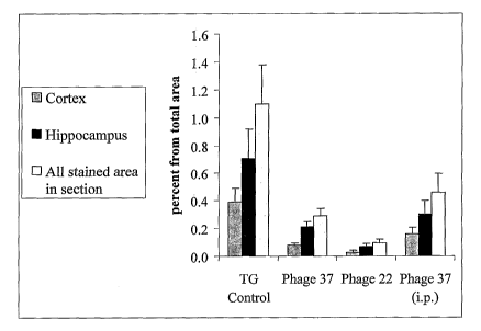
[0058] In Figure 10, a summary of the plaque load detected by
thioflavin S staining in the cortex and hippocampus of PDAPP mice
treated with phage 22 or phage 37, versus control mice, treated
with PBS only, is presented. Phage 37 reduced plaque load
significantly in both the cortex and hippocampus areas of mice
treated with it by intranasal application. Intraperitoneal (IP)
application of phage 37 did not show significant plaque load
reduction. Plaque load reduction in phage 22 treated mice was
much more dramatic both in the cortex and in the hippocampus,
compared to untreated controls.
[0059] Figure 3 shows a summary of the plaque load detected by
staining with antibody 21F12 in the cortex and hippocampus of
PDAPP mice treated with phage 22 or phage 37, versus control
mice, treated with PBS only. Here too, both phage 37 and phage 22
reduced plaque load significantly in both the cortex and
- 22 -
CA 02619725 2014-07-30
hippocampus areas of mice treated with these scFvs by intranasal
application. IP application of phage 37 did not show significant
plaque load reduction.
[0060] Both scFv 22 and scFv 37 significantly reduced the AP
load in treated mice brains, They affected the soluble and
fibrillary amyloid deposits (stained with 21F12 antibody) as well
as the dense amyloid plaques (stained by thioflavin S).
Furthermore, intranasal administration/application of the phage
scFVs is an effective way to introduce the scFv antibodies to the
mice brains, compared with the IP application of phage 37. While
IP administration of phage did show reduced plaque load, the
reduced plaque load was not as significant ashy intranasal
administration.
[0061] Having now fully described this invention, it will be
appreciated by those skilled in the art that the same can be
performed within a wide range of equivalent parameters,
concentrations, and conditions without departing from the spirit
and scope of the invention and without undue experimentation.
[0062] The scope of the claims should not be limited by
particular embodiments set forth herein, but should be construed
in a manner consistent with the specification as a whole.
- 23 -
CA 02619725 2014-07-30
[0064] Reference to known method steps, conventional methods
steps, known methods or conventional methods is not in any way an
admission that any aspect, description or embodiment of the
present invention is disclosed, taught or suggested in the
relevant art.
[0065] The foregoing description of the specific embodiments
will so fully reveal the general nature of the invention that
others can, by applying knowledge within the skill of the art
(including the contents of the references cited herein), readily
modify and/or adapt for various applications such specific
embodiments, without undue experimentation, without departing
from the general concept of the present invention. Therefore,
such adaptations and modifications are intended to be within the
meaning and range of equivalents of the disclosed embodiments,
based on the teaching and guidance presented herein. It is to be
understood that the phraseology or terminology herein is for the
purpose of description and not of limitation, such that the
terminology or phraseology of the present specification is to be
interpreted by the skilled artisan in light of the teachings and
guidance presented herein, in combination with the knowledge of
one of ordinary skill in the art.
[0066] Thus the expressions "means to..." and "means for...",
or any method step language, as may be found in the specification
above and/or in the claims below, followed by a functional
statement, are intended to define and cover whatever structural,
physical, chemical or electrical element or structure, or
whatever method step, which may now or in the future exist which
carries out the recited function, whether or not precisely
- 24 -
CA 02619725 2008-02-19
WO 2007/022416
PCT/US2006/032319
equivalent to the embodiment or embodiments disclosed in the
specification above, i.e., other means or steps for carrying out
the same functions can be used; and it is intended that such
expressions be given their broadest interpretation.
-25-
CA 02619725 2008-02-19
W02007/022416 PCT/US2006/032319
REFERENCES
Bach H, Mazor Y, Shaky S, Shoham-Lev A, Berdichevsky Y, Gutnick
DL, Benhar I. Escherichia coli maltose-binding protein as a
molecular chaperone for recombinant intracellular
cytoplasmic single-chain antibodies. J* Mbl Biol. (2001)
312(1):79-93
Bacskai BJ, Kajdasz ST, McLellan ME, Games D, Seubert P. Schenk
D, Hyman BT. 3. Non-Fc-mediated mechanisms are involved in
clearance of amyloid-beta in vivo by immunotherapy. J
Neurosci. 2002 Sep 15;22(18):7873
Frenkel, D., Solomon B. and Benhar I. Modulation of Alzheimer's
beta amylois neurotoxicity by site-directed single-chain
antibody. J. of Neuroimmunology (2000) 106:23-31.
Schenk D., Barbour R. Dunn W. et al. Immunization with amyloid-
beta attenuates Alzheimer-disease-like pathology in the
PDAPP mouse. Nature (1999) 400:173-177.
- 26 -