Note: Descriptions are shown in the official language in which they were submitted.
CA 02619810 2013-01-23
ULTRASOUND CATHETER CALIBRATION WITH ENHANCED ACCURACY
CROSS-REFERENCE TO RELATED APPLICATION
This application claims the benefit of U.S.
Provisional Patent Application 60/887,457, filed January
31, 2007.
FIELD OF THE INVENTION
The present invention relates generally to
ultrasound imaging systems, and specifically to devices
and methods for calibration of ultrasound probes.
BACKGROUND OF THE INVENTION
U.S. Patent Application Publication 2004/0254458 Al
describes apparatus and methods for calibrating a probe
having a position sensor and an ultrasonic transducer.
The apparatus includes a test fixture, which includes an
ultrasonic target disposed therein at a known position.
A computer receives a position signal generated by the
position sensor while the transducer is in alignment with
the ultrasonic target. The computer thus determines the
orientation of the probe in a frame of reference of the
test fixture and determines calibration data for the
probe responsive to the orientation of the probe.
Various methods are known in the art for calibrating
position sensors. For
example, U.S. Patents 6,266,551
and 6,370,411 describe methods and apparatus for
calibrating a probe comprising a magnetic position
sensor. The
calibration is used to measure and
compensate for variations in the positions, orientations
and gains of magnetic sensor coils in the probe. To
1
CA 02619810 2013-01-23
calibrate the probe, a mechanical jig holds the probe in
one or more predetermined positions and orientations, and
radiators generate known, substantially uniform magnetic
fields in the vicinity of the jig. Signals generated by
the coils are analyzed and used to produce calibration
data regarding the gains of the coils and deviations of
the coils from orthogonality.
Other methods for calibrating ultrasound imagers
with position sensors are also known in the art. For
example, U.S. Patent 6,138,495 describes a method and
apparatus for calibrating a position measuring component
on an imaging or scanning transducer with respect to the
scanning plane. Calibrations are performed by using a
calibrating device including an additional position
measuring component, such that during the calibration
process, the relative position of these position
measuring components can be calculated. Calibrations are
also performed by viewing targets in the scanning plane
that are at a known position with respect to the
additional position measuring component.
As another example, U.S. Patent 6,585,561 describes
a calibration unit for calibrating an ultrasound head.
The calibration unit is configured to receive the
ultrasound head in a known position and orientation with
respect to a reference portion of the calibration unit.
The calibration unit allows the calibration of a
coordinate system of markers associated with the
ultrasound device. Echoes
received from the reference
portion can be used to calibrate, for example, an offset
2
CA 02619810 2008-01-30
between the ultrasound head and the reference portion.
The calibration unit is preferably formed of a material
in which the sound velocity is known, such as a suitable
plastic with a hole having a diameter to receive the
ultrasound device. During calibration, echoes are
received from the interface of the bottom of the
calibration unit and the surrounding medium, which is
preferably air. The echo can be used to calculate an
offset from the ultrasound device head to the interface.
SUMMARY OF THE INVENTION
The embodiments of the present invention that are
disclosed hereinbelow describe improved systems and
methods for calibrating an ultrasonic imaging probe with
a position sensor. These
embodiments are useful
particularly in calibrating ultrasound catheters, which
include a transducer array and position sensor and are
adapted for imaging within body cavities, such as
chambers of the heart. The
principles of the present
invention may be applied, however, to a variety of
different types of probes, for both intra- and extra-
corporeal use.
In one embodiment, apparatus is provided for
calibration of a probe that includes a magnetic position
sensor and an acoustic imaging device. The
apparatus
comprises a rigid mechanical framework, which serves as
the frame of reference of calibration of the imaging
device relative to the position sensors. One or more
field generators, fixed to the framework, generate a
magnetic field of known spatial characteristics. An
acoustic target assembly is also fixed to the framework.
3
CA 02619810 2008-01-30
This assembly comprises a phantom coupled to a motion
mechanism, which moves the phantom in a known orbit
relative to the framework. A jig,
fixed to the
framework, holds the probe within the magnetic field, in
an orientation suitable for the imaging device to image
the phantom. In this configuration, a processor receives
position signals from the position sensor and image
signals from the imaging device, and processes the
signals in order to calibrate the coordinates of the
imaging device relative to the position sensor.
In another embodiment, an ultrasound phantom for
calibration of a probe comprises walls shaped so as to
define an interior space, which is wholly or partly
enclosed by the walls. The walls
comprise multiple
calibration targets at different, respective positions.
One or more field generators generate an energy field of
known spatial characteristics in a vicinity of the
phantom. The probe is inserted into the interior space
defined by the walls and is moved through multiple
locations and orientations. While the
probe is in the
interior space, a processor receives position signals
from the position sensor and image signals from the
imaging device, and processes the position and image
signals in order to calibrate coordinates of the imaging
device relative to the position sensor.
The present invention will be more fully understood
from the following detailed description of the
embodiments thereof, taken together with the drawings in
which:
4
CA 02619810 2008-01-30
. ,
BRIEF DESCRIPTION OF THE DRAWINGS
Fig. 1 is a schematic, pictorial illustration of a
catheter-based system for ultrasonic imaging, in
accordance with an embodiment of the present invention;
Fig. 2 is a schematic, pictorial illustration of a
system for calibration of an ultrasonic imaging catheter,
in accordance with an embodiment of the present
invention;
Figs. 3 and 4 are schematic side and top views of
the system of Fig. 2;
Fig. 5 is a schematic, pictorial illustration of an
acoustic target assembly, in accordance with an
embodiment of the present invention;
Fig. 6 is a schematic, pictorial illustration of an
ultrasound phantom, in accordance with an embodiment of
the present invention;
Figs. 7A and 7B are schematic representations of
ultrasound images of the phantom of Fig. 6, captured
using a probe in different, respective alignments, in
accordance with an embodiment of the present invention;
Fig. 8 is a schematic, pictorial illustration of an
ultrasound phantom inside a calibration tank, in
accordance with an embodiment of the present invention;
and
Fig. 9 is a schematic side view of a system for
calibration of an ultrasonic imaging catheter, in
accordance with an embodiment of the present invention.
DETAILED DESCRIPTION OF EMBODIMENTS
Fig. 1 is a schematic, pictorial illustration of an
ultrasonic imaging system 20 comprising an elongate
probe, such as a catheter 22, for insertion into the body
of a patient, in accordance with an embodiment of the
5
CA 02619810 2008-01-30
present invention. System 20
comprises a console 24,
which typically comprises a computer processor with
suitable signal processing and user interface circuits.
This console receives and processes signals from catheter
22, as described hereinbelow. Typically,
the console
enables a user to observe and regulate the functions of
catheter 20 and displays images that are formed using the
catheter. Catheter 20 typically includes a handle 26 for
controlling operation of the catheter by the user. The
handle or a connector coupling the catheter to console 24
may comprise a microcircuit for storing calibration data,
as described in the above-mentioned U.S. Patent
6,266,551, for example.
A distal end 28 of catheter 22 comprises an
ultrasound imaging device 32, which is used to produce
ultrasound images of the inside of the body. An
enlarged, cross-sectional view of distal end 28 is shown
in the inset in Fig. 1.
Ultrasound imaging device 32
typically comprises a phased array of transducers 34,
which is operated, as is known in the art, so as to
create a two-dimensional image "fan" 38 in the plane of
the scanning ultrasonic beam (referred to herein as the
"beam plane" or "image plane"), which contains the
longitudinal axis of the catheter (identified as the Z-
axis in the figures). The transducers receive ultrasonic
waves that are reflected from objects in the beam plane
and output signals in response to the reflected waves.
Typically, these signals are processed by console 24 in
order to form and display ultrasound images.
Alternatively or additionally, ultrasound transducers 34
may be used for other diagnostic purposes, such as
Doppler measurements, or for therapeutic uses.
6
CA 02619810 2013-01-23
Distal end 28 of catheter 22 further comprises a
position sensor 30, which generates signals that indicate
the position and orientation of the catheter within the
body. Based on
these position signals, console 24
determines the location and orientation of each fan image
captured by imaging device 32. The console is thus able
to determine the coordinates of objects appearing in the
fan image, as well as to combine multiple images captured
at different catheter positions.
Position sensor 30 is typically adjacent to imaging
device 32 in a fixed locational and orientational
relationship. In some
embodiments, the position sensor
comprises one or more coils, which produce signals in
response to a magnetic field generated by a field
generator outside the patient's body. The
signals are
analyzed by console 24 in order to determine position and
orientation coordinates of distal end 28. This sort of
magnetic position sensing is described in detail, for
example, in the above-mentioned U.S. Patent 6,266,551.
Other exemplary systems that combine ultrasonic imaging
with magnetic position sensing are described in U.S.
Patents 6,690,963, 6,716,166 and 6,773,402.
Alternatively, catheter 22 may comprise any other
suitable type of position sensor known in the art. For
example, position sensor 30 may comprise other types of
field sensing devices, such as a Hall Effect sensor.
Alternatively, sensor 30 may generate magnetic fields,
which are detected by sensing antennas outside the body.
Further alternatively, position sensor 30 may operate by
7
CA 02619810 2008-01-30
measuring impedance of the body to electrical signals or
by transmitting or receiving ultrasonic position signals.
The principles of the present invention are applicable to
substantially any position sensing technology that can be
implemented in a medical probe.
As shown in Fig. 1, due to physical constraints in
the construction of catheter 22, position sensor 30 and
ultrasound imaging device 32 are both located in catheter
22 at certain respective distances from the distal tip
of the catheter. (This
configuration of the position
sensor and imaging device is shown by way of example, and
the principles of the present invention may similarly be
applied to other arrangements of these elements,
including side-by-side arrangements.) The actual
position and orientation of fan 38 is computed by taking
into account the distance between the position sensor and
the ultrasound imaging device. It has
been found
empirically that because of deviations in the process of
manufacturing catheter 22, this distance typically varies
from one catheter to another. Furthermore, the axes of
the position sensor and of the ultrasonic transducer
array in imaging device 32 may not be precisely aligned
with the Z-axis or with one another other, thereby
introducing additional variation in determining the
orientation of fan 38.
These and other sources of alignment variation are
described in greater detail in the above-mentioned Patent
Application Publication US 2004/0254458 Al. If not
corrected, the alignment variation will cause errors in
determining the position coordinates of objects appearing
in image fan 38. Certain
methods for calibrating and
8
CA 02619810 2013-01-23
correcting for these alignment variations are described
in US 2004/0254458 Al, while other methods are described
in U.S. Patent Application Publication US 2007/0106156
Al. Other, enhanced systems and methods for calibration
are described hereinbelow.
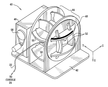
Reference is now made to Figs. 2-4, which
schematically illustrate a system 40 for calibration of
an ultrasonic imaging catheter, in accordance with an
embodiment of the present invention. Fig. 2 is a
pictorial illustration, while Figs. 3 and 4 are side and
top views, respectively. System 40 comprises a base 42,
which serves as a rigid mechanical framework for a set of
magnetic field generators 44 and an acoustic target
assembly 46. Various
types of field generators may be
used in this context. In this
embodiment, the field
generators comprise three pairs of Helmholtz coils 48, 50
and 52, each pair oriented along one of the X, Y and Z
axes.
Catheter 22 is inserted into a suitable jig 54 at
the center of field generators 44, with imaging device 32
facing toward target assembly 46. The
target assembly
comprises a phantom 56, which moves in a known orbit
relative to catheter 22 within the field of view of
imaging device 32, under control of a motion mechanism
58. Various types of phantoms and mechanisms may be used
in the target assembly. Some
particular examples are
shown in Figs. 5 and 6 and are described hereinbelow with
reference thereto.
9
CA 02619810 2008-01-30
The present embodiment addresses a number of
difficulties that exist in some prior ultrasound catheter
calibration approaches. For
example, wires, which are
used in many ultrasound calibration phantoms, are smaller
than the width of the ultrasound beam and therefore cause
artifacts that limit their observability in the
ultrasound image. In
addition, inclination of the
phantom relative to the ultrasound beam can cause
inaccuracy in determining the precise location at which
the wire crosses the beam. Other approaches use larger
phantoms scanned by the ultrasound beam while measuring
catheter position using an electromagnetic system. This
approach relies on the accuracy of the an electromagnetic
system, which is typically on the order of 1 mm.
In the present embodiment, the location readings of
position sensor 30 are made in proximity to the center of
a Helmholtz cell, with typical position accuracy of 0.1
mm, using gradient-calibrated electromagnetic fields
generated by the three pairs of Helmholtz coils 48, 50,
52.
(Optionally, the sensitivity of the sensor may first
be calibrated in a uniform magnetic field.) The
sensor
is placed in proximity to the center of the coils. The
coils in each pair are driven with currents running in
opposite directions, so that the electromagnetic field in
the center has nearly-constant gradient. Because
the
three pairs of Helmholtz coils are orthogonal to one
another, the three electromagnetic fields have gradients
in the three orthogonal directions.
Before calibrating catheter 22, the Helmholtz
electromagnetic fields are calibrated using a
mechanically-accurate sensor at known points in the
CA 02619810 2008-01-30
volume that will be used for calibrating the catheter
position sensor. The measured positions are referred to
a predefined mechanical origin, which is fixed in the
frame of reference of base 42. From these measurements,
the Helmholtz electromagnetic field is accurately mapped
as a function of location. When
catheter sensor 30 is
then placed in the calibrated volume, the position and
orientation of the sensor may be calculated to an
accuracy of 0.1 mm, which is typically much better than
the operational accuracy of the electromagnetic tracker
system used in actual operation of catheter 22. This
high accuracy is due to the high gradient present in the
Helmholtz chamber.
Fig. 5 is a schematic, pictorial illustration
showing details of acoustic target assembly 46, in
accordance with an embodiment of the present invention.
In this embodiment, motion mechanism 58 comprises a
motor, which drives a rotor 64 to move phantom 56 in a
fixed orbit via a linkage 66. Phantom 56 comprises lines
68 and 70 that cross the image plane of fan 38. Lines 68
and 70 are non-parallel to improve the estimation
calculation of the image coordinate system. In addition
to the set of lines, point sources 72 may be placed in
several locations on the phantom. These point sources,
for example, may take the form of protrusions on the
lines in the phantom, as shown in the figure. These
latter elements improve the accuracy of the calibration,
especially for parameters that are most affected by the
low resolution of the ultrasound image in the direction
perpendicular to the ultrasound beam plane.
11
CA 02619810 2008-01-30
During calibration, phantom 56 is moved in an
accurate orbit in front of imaging device 32, typically
in a circular orbit having an axis roughly parallel to
the axis of the array of transducers 34. Motion
mechanism 58 is built, as shown in Fig. 5, so that each
of lines 68 and 70 in the target cuts the ultrasound beam
at a low inclination during the entire orbit. (In
other
words, each line always remains parallel to its original
orientation.) Many images are captured in this manner at
different positions of the phantom in the orbit.
Optionally, a position sensor (not shown) may be fixed to
phantom 56, so as to enable electromagnetic registration
of the phantom in each image, relative to the fixture
base. The
positions of the lines intersecting the
ultrasound beam plane are extracted from the images. The
intersection points from all the images in the ultrasound
coordinate system are transformed to the corresponding
coordinates in the fixed frame of reference of base 42.
The image origin in the fixed coordinate frame is
defined by solving the following minimization expression:
2
argMin - Pi(az, el, rl, x0, yO, z0) )
{az , el,r1,x0, y0,z0}
Here 13.2: is the {col,row} measurement of the intersection
point of each of the lines (arranged in a predefined
order), and Pi(az, el, rl, x0, yO, z0) is an analytical function
of the line intersection with the ultrasound plane as a
function of the plane origin (x0,y0,z0) and orientation
coordinates (az,el,r1). The
minimization problem can be
12
CA 02619810 2008-01-30
solved using any suitable numerical or analytical method
known in the art.
Using the methods described above, both the image
origin of imaging device 32 and the electromagnetic
origin of position sensor 30 may be determined in six
dimensions (location and orientation) with high accuracy,
in the same, fixed frame of reference. The
relative
coordinates of the origins are used to compute the
calibration transformation between the electromagnetic
sensor coordinates and the ultrasound image coordinates.
The techniques described herein, including the use
of mechanically-accurate motion of a line phantom and
building the lines from large planes that are almost
invariant over the width of the ultrasound beam, improve
the accuracy with which the point of intersection between
the ultrasound beam and the phantom can be determined.
Furthermore, integrating the electromagnetic calibration
with the ultrasound phantom in a unified system provides
a robust basis for calibration, whose accuracy is
dependent only on the mechanical accuracy of the
calibration system. This
mechanical accuracy is
generally better than the accuracy of both the
electromagnetic position tracking and the ultrasound
image measurement. The use
of Helmholtz coil pairs to
calibrate the electromagnetic sensor also improves
accuracy, since the large electromagnetic gradient inside
the Helmholtz assembly provides better estimation of the
electromagnetic sensor position and orientation in
comparison with most other electromagnetic tracker
constructions (using single or multiple field
generators).
13
CA 02619810 2008-01-30
. .
Lines 68 and 70 in phantom 56 may be laid out in two
planes that form an arrowhead pointing toward the ULS
sensor (i.e., the planes meet along a line that is
perpendicular to the ultrasound fan plane). As a result,
the beam always reflects back diffusely from the lines,
giving a clear arrowhead shape in the ultrasound image.
The arrowhead shape is detected in the image, and the
arrowhead location is calculated from the intersection of
the two lines forming it.
Fig. 6 is a schematic, pictorial illustration of an
ultrasound phantom 80, which may be used in place of
lines 68 and 70, for example, in accordance with another
embodiment of the present invention.
Phantom 80
comprises an elongated piece 82 of triangular profile,
which is shaped so as to define two spatial planes, which
meet in a line, defining a sort of arrowhead shape. The
phantom is typically positioned in target assembly 46 so
that the line intersects the image plane of fan 38, with
the arrowhead pointing toward imaging device 32. Phantom
80 further comprises a cross-piece 84, with front edges
that are linear extensions of the planar surfaces of
triangular piece 82.
Cross-piece 84 thus defines an
alignment plane, which is perpendicular to the two
spatial planes of piece 82.
Figs. 7A and 7B are schematic representations of
ultrasound images 86 and 90 of phantom 80, captured using
imaging device 32 in different, respective alignments of
catheter 22, in accordance with an embodiment of the
present invention. These images show how catheter 22 may
be brought into precise alignment with phantom 80, such
14
CA 02619810 2008-01-30
. .
that fan 38 is parallel to and congruent with the plane
of cross-piece 84 (and thus perpendicular to the planar
surfaces of piece 82).
In image 86, reflection of the
ultrasound beam from the planes of piece 82 gives an
arrowhead shape 88 having a given length. In this image,
however, fan 38 is not aligned with cross-piece 84, and
the cross-piece is therefore not seen in the image.
In
image 90, however, the image plane is aligned with the
alignment plane defined by cross-piece 84. As a result,
the image contains an arrowhead shape 92 of increased
length relative to shape 88, due to the reflection of the
acoustic waves from the linear extensions of the planes
of piece 82 that are provided by cross-piece 84. Thus,
the operator of system 40 is able to determine that the
catheter is properly aligned in the system.
Additionally or alternatively, alignment of the
catheter may be based on a virtual three-dimensional
rigid body that is created by the motion of the phantom.
This approach enables registration to be carried out
automatically, with improved accuracy due to the
triangular profiles that overcome the blurring due to the
width of fan 38.
The shapes and configurations of the phantoms and
target assembly in the above figures are shown only by
way of example, and a wide variety of alternative shapes
and configurations may be used within the scope of the
present invention.
Possible variations include the
following:
= The shape, size and orientation of the phantom may be
varied.
CA 02619810 2008-01-30
= Curves, such as harmonic curves, may be used in place
of the lines in the phantom. (Examples include sine
curves, circular shapes, and curves of other types.)
= Different algorithms may be used for shape extraction
from the ultrasound image.
= Instead of extracting intersection points from each
image, the ultrasound image may be analyzed by matching
the image against a model of the response of the
phantom to the ultrasound pulse.
= The mechanical orbit that the phantom performs need not
be circular, but may rather have any shape that permits
enough information to be generated in order to make the
computations accurately.
= The phantom may be held stationary while the jig
holding the catheter moves. As a result, from
the
point of view of the calibration procedure, the phantom
can still be seen as describing a known orbit relative
to the probe.
Reference is now made to Figs. 8 and 9, which
schematically illustrate elements of a system 100 for
calibration of ultrasound imaging catheter 22, in
accordance with another embodiment of the present
invention. Fig. 8 is a pictorial illustration showing an
ultrasound phantom 104 inside a tank 102, while Fig. 9 is
a side view. During calibration, tank 102 is typically
filled with a suitable fluid, such as water, both inside
and outside phantom 104, but the fluid is omitted from
the interior of the phantom in Fig. 8 so that the details
of the phantom can be seen clearly in the figure.
Phantom 104 comprises walls shaped so as to define a
container, in this case a box. The walls
of the box
16
CA 02619810 2008-01-30
comprise multiple calibration targets 106, 108 at
different, predetermined locations. Typically, as shown
in the figure, the targets are located in different walls
and thus are oriented in different planes. The interior
space of the container has a shape and size sufficient to
permit catheter 22 to be inserted into the container and
moved through multiple locations and orientations, so as
to aim imaging device 32 at different targets in
different locations and orientations of the catheter. A
location pad 110 with one or more field generators 112,
such as electromagnetic coils, is placed adjacent to tank
102, and the coils are driven to generate an
electromagnetic energy field of known spatial
characteristics in the vicinity of phantom 104. The
arrangement of field generators shown in this figure will
not typically generate the sort of gradient-calibrated
field that is described above, but substantially any
suitable field geometry may be used in the present
embodiment.
While the probe is in the interior space of phantom
104, processor 24 receives position signals from position
sensor 30 in response to the electromagnetic field, and
image signals from imaging device 32 due to reflection of
acoustic waves from the phantom. The operator of system
100 freezes each image, marks the location of the target
that appears in the image, and identifies which one of
the targets it is. For each image, the magnetic position
sensing system determines location and orientation
coordinates of the catheter tip. The
annotated images
and the corresponding coordinates are used by processor
24 (or by a separate calibration processor) to calibrate
the linear and angular offset of the ultrasound
17
CA 02619810 2008-01-30
transducer array in the catheter relative to the position
sensor. Processor 24 processes the position and image
signals in order to calibrate coordinates of imaging
device 32 relative to position sensor 30.
The detailed calibration procedure may be carried
out as follows:
1. Calibrate the offset of the tip of catheter 22 relative
to position sensor 30 in a dedicated jig (as described
in the above-mentioned US 2004/0254458, for example).
2. Insert the catheter into a tube and rotate it while
acquiring position coordinates in order to estimate the
offset of the position sensor from the catheter axis.
3. Insert the catheter into the calibration bath and
connect it to processor 24, so that the processor
receives both position coordinates and ultrasound image
signals.
4. Acquire data points by capturing images of different
targets, as explained above. Each
data point
corresponds to one of the images and includes the
location of the target in the image (as marked by the
operator or determined automatically by the processor),
the actual, known spatial coordinates of the target,
and the position coordinates of the catheter, as
measured using the position sensor when the image was
captured.
18
CA 02619810 2008-01-30
, .
5. Calculate a calibration transformation matrix using the
set of data points. The estimation error of the matrix
may be computed in order to confirm that the
calibration is valid.
The use of a three-dimensional, multi-target
phantom, such as phantom 104, permits fast, convenient
calibration without requiring that the catheter be
constrained in a jig (except in step 1, above). Although
Figs. 8 and 9 show a certain particular phantom
configuration, any suitable three-dimensional arrangement
of targets may be used in like manner. For example, the
walls of the phantom may be arranged to define containers
of different shapes, or the walls may only partially
enclose the interior space that they define.
It will thus be appreciated that the embodiments
described above are cited by way of example, and that the
present invention is not limited to what has been
particularly shown and described hereinabove.
Rather,
the scope of the present invention includes both
combinations and subcombinations of the various features
described hereinabove, as well as variations and
modifications thereof which would occur to persons
skilled in the art upon reading the foregoing description
and which are not disclosed in the prior art.
19