Note: Descriptions are shown in the official language in which they were submitted.
CA 02619820 2008-02-19
WO 2007/024514 PCT/US2006/031427
TRANSEPTAL APPARATUS, SYSTEM ANB METHOD OF FORMING THE APPARATUS
Field of the Invention
The present invention relates generally to apparatus, systems, and
methods for use in a heart, more particularly to apparatus, systems, and
methods
for locating a PFO with a positioning device that can be configured to occlude
the PFO.
Backstround
The human heart is divided into four chambers. These include the right
atrium, the right ventricle, the left atrium, and the left ventricle. The
right atrium
and right ventricle are divided from the left atrium and left ventricle by a
muscular wall called the septum. The atrial septum is the wall separating the
atria, and the ventricular septum is the wall separating the ventricles.
Early in fetal development the two atria (i.e., left and right atriums) are a
single chamber. A wall or membranous structure develops from the superior
aspect of the atrial chamber and extends superiorly toward the base of the
atrial
chamber. This membrane is the septum primum (SP). As the SP seals to the
base of the chamber, it is dissolved away at the superior attachment, creating
a
passageway for blood to travel from the right atria to the left atria
(bypassing the
developing lungs). At about the same time, a second membrane develops from
the superior aspect of the right atrium and extends inferiorly. This membrane
is
the septum secundum (SS). It fuses with the SP along the walls of the atria,
but
does not extend to the base of the atria. The inferior portion of the SS is
named
the limbus. The two membranes forin a passage defined by thin tissue (SP) and
thick tissue (SS) that extends from the right atria to the left atria. This
passage is
named the foramen ovale. The portion of the SP that comprises the left side of
the foramen ovale is named the fossa ovalis. The limbus of the SS is distinct
from the fossa ovalis of the SP in that it is thicker and more muscular.
Upon birth blood must be diverted into the lungs of the newborn. One
event that enables this is an increase in pressure within the left atrium
relative to
the right atrium. This pressure reversal effectively closes the foramen ovale
and
1
CA 02619820 2008-02-19
WO 2007/024514 PCT/US2006/031427
eliminates the shunting of blood from right to left. In most people, the SP
and
SS membranes that form the passage of the shunt fuse and the passage is
eliminated. However, in a minority of people, these membranes do not fuse
effectively and the shunt remains sealed by pressure, but the passage remains
viable, or patent. This condition is named patent foramen ovale (PFO). In
unusual circumstances the pressure in the right atrium can exceed that in the
left
atrium, allowing passage of blood through the PFO. This would typically be
inconsequential, except when the venous (right atrial) blood contains
thrombotic
debris that is normally eliminated by thrombolytic mechanisms in the lungs. In
this case, a clot can travel to the left atria and become an embolic risk to
the
patient's health through myocardial infarction or stroke.
Brief Description of the Drawings
Figure 1 illustrates an embodiment of a right lateral view of the heart.
Figures 2A illustrates a positioning device according to one embodiment
of the present invention.
Figure 2B illustrates an elongate structure of the positioning device in a
first position according to one embodiment of the present invention.
Figure 2C illustrates the positioning device according to an additional
embodiment of the present invention.
Figure 2D illustrates the elongate structure of the positioning device in a
second position according to one embodiment of the present invention.
Figures 3A-3F illustrate extension members of the positioning device
according to various embodiments of the present invention.
Figures 4A-4C illustrate various embodiments of a system according to
the teachings of the present invention.
Figure 5A illustrates the system within the right atrium of the heart
according to an embodiment of the present invention.
Figures 5B-5C illustrate the positioning device seated on the limbus of
the septum secundum according to the teachings of the present invention.
Figure 5D provides an illustration of tightening the tissue defining the
passage according to the teachings of the present invention.
Figure 5E provides an illustration of piercing the thick and thin tissue and
of the passage according to the teachings of the present invention.
2
CA 02619820 2008-02-19
WO 2007/024514 PCT/US2006/031427
Detailed Description
Embodiments of the present invention are directed to methods, apparatus,
and systems for locating a PFO with a positioning device that can be
configured
to occlude the PFO. As will be discussed in more detail herein, a positioning
device on a delivery catheter can be seated on the septum secundum (SS) of the
atrial septum, e.g., seated on the limbus of the SS. Seating the positioning
device on the SS helps to locate the positioning device at a position on the
atrial
septum where two membranes, the SS and the septum primum (SP), lie parallel
to one another. This position makes possible the use of the various
embodiments
described herein to prepare a PFO for occlusion and to introduce various
components of the positioning device to the left atrium from the right atrium.
For example, in various embodiments, the SS or the SS and the SP can be
pierced with a piercing member that extends from an elongate structure of the
positioning device and into the left atrium.
In some embodiments, the positioning device can include extension
members that can be used to tighten thin tissue of the SP and/or thick tissue
of
the SS within the passage of a PFO prior to piercing those tissues. Thus, in
various embodiments, by manipulating components of the positioning device
(e.g., extension members and/or elongate structure and piercing member) thick
and/or thin tissue can be tightened and pierced.
In various embodiments, the positioning device can include an extension
member that can extend into the passage of the PFO while the elongate body of
the positioning device remains in the right atrium. The extension member
assures that the elongate body of the device is correctly oriented with
respect to
the passage of the PFO. This positioning mechanism assures correct alignment
for a piercing member contained within the elongate structure of the
positioning
device.
In various embodiments, the positioning device can be used as a
transeptal delivery device for introducing devices such as therapeutic and
diagnostic devices, solids, fluids, substances, and the like, from a first
heart
chamber to a second heart chamber (e.g., from right atrium to left atrium).
These and other embodiments of the present invention are discussed herein.
3
CA 02619820 2008-02-19
WO 2007/024514 PCT/US2006/031427
The Figures herein follow a numbering convention in which the first
digit or digits correspond to the drawing Figure number and the remaining
digits
identify an element or component in the drawing. Similar elements or
components between different figures may be identified by the use of similar
digits. For example, 110 may reference element "10" in Fig. 1, and a similar
element may be referenced as 210 in Fig. 2. As will be appreciated, elements
shown in the various embodiments herein can be added, exchanged, and/or
eliminated so as to provide a number of additional embodiments of the
positioning device according to the present invention.
In Figure 1, a right lateral view of the heart 100 is shown with an opened
right atrium 102. The heart 100 is divided Into four chambers, which are
referred to herein as the right atrium 102, a right ventricle, a left atrium
104 and
a left ventricle. Heart 100 also includes a septal wall 106 that divides the
four
chambers of the heart. The portion of the septal wall dividing the left and
right
atriums 102 and 104 is called the interatrial septum 108. The portion of the
septal wall 106 dividing the left and right ventricle is called the
ventricular
septum.
As shown in Figure 1, the fossa ovalis 110 is situated at the lower part of
the atrial septum 108, above and to the left of the orifice of the inferior
vena cava
112. The limbus 114 of the fossa ovalis 110 is the pronounced anterosuperior
margin of the fossa ovalis 110 within the right side (i.e., the right atrium
102) of
the interatrial septum 108. It represents the inferior margin of the SS during
fetal
life.
The passage 116 can be defined by surfaces of the SS (thick tissue) and
surfaces of the SP (thin tissue) and extends between the right and left
atriums
102 and 104. As used herein, the passage 116 is defined by surfaces of the SS
and SP and can be used interchangeably with a PFO. The thick tissue 118 forms
the right margin of the passage 116 and comprises the superior portion of the
interatrial septum 108. Thus, the thick tissue 118 is located adjacent the
limbus
114 and extends upward and rightward away from the limbus 114. The thin
tissue 120 forms the left margin of the passage 116 and comprises the inferior
portion of the interatrial septum 108 (i.e., below the thick tissue 118) and
4
CA 02619820 2008-02-19
WO 2007/024514 PCT/US2006/031427
extends upward and rightward substantially parallel to the thick tissue 118
and
toward the left atrium 104.
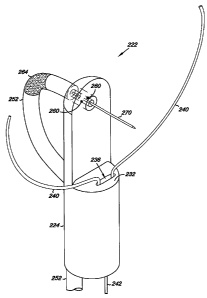
Figures 2A-2D illustrate various embodiments of the positioning device
222 that can be used to pierce thick and thin tissue according to the
teachings of
the present invention. Figure 2A provides an illustration of a positioning
device
222 according to the teachings of the present invention. As shown in Figure
2A,
positioning device 222 includes an elongate body 224 having a proximal end 226
and a distal end 228. The elongate body 224 includes a wall 230 that extends
from the distal end 228 toward the proximal end 226. In the embodiment shown
in Figure 2A, the wall 230 includes a planar surface. However, in various
embodiments, the wall 230 can include other types of surfaces. For example, in
some embodiments, the wall 230 can include non-planar surfaces such as a
convex surface or a concave surface.
The wall 230 extends toward the proximal end 226 to a ledge 232 that
extends away from the wall 230. In one embodiment, the ledge 232 extends
perpendicularly away from the wall 230 for a predetermined distance. The ledge
232 includes a planar surface whose outer edge defines a semi-circular shape.
As will be discussed herein, the ledge 232 of the positioning device 222
allows
the positioning device 222 to be seated on the limbus of the SS of a patient's
heart.
Since the size and shape of the limbus can vary from patient to patient,
the positioning device 222, including the wall 230 and the ledge 232 can
include
various shapes and sizes that can be based on the anatomical structures of a
patient's heart including the limbus of the SS. For example, in some
embodiments, the ledge 232 can have a surface defining various geometric
shapes and sizes, including, but not limited to, convex shapes, concave
shapes,
recessed shapes, and irregular shapes, among others. In addition, in some
embodiments, the ledge 232 can extend at various angles other than
perpendicular from the wall 230 of the elongate body 224.
The positioning device 222 includes a number of lumens that extend
various lengths within the positioning device 222. In one embodiment, a first
lumen 234 extends toward the ledge 232. As shown in Figure 2A, the first
lumen 234 extends toward ledge 232 and communicates with a ledge opening
236 defined by the surface of the ledge 232. In one embodiment, the first
lumen
CA 02619820 2008-02-19
WO 2007/024514 PCT/US2006/031427
234 and the ledge opening 236 can accommodate the movement of a component
positioned within the first lumen 234, as will be discussed herein.
As shown in Figure 2A, the first lumen 234 includes surfaces defining an
ovular cross-sectional shape. In various embodiments however, the first lumen
234 can include other cross-sectional shapes including, but not limited to,
circular and polygonal cross-sectional shapes. In various embodiments, the
cross-sectional shape of the first lumen 234 can be fonned to accommodate a
particular design of a component therein.
For example, in one embodiment, the component can be an extension
member 238. As shown in Figure 2A, the extension member 238 is extendably
positioned within the first lumen 234 toward the ledge 232 of the elongate
body
224. As used herein, an extendably positioned extension member 238 is an
extension methber having at least one arm 240 that can be moved within the
first
lumen 234 and through the ledge opening 236 such that the arm 240 extends
away from the ledge 232 of the elongate body 224. In various embodiments, the
arm 240 can extend away from the ledge 232 in various directions and in
various
planes, as will be discussed herein with respect to Figures 3A.
In various embodiments, the extension member 238 can include one or
more arms and one or more bases. For example, in some embodiments, the
extension member 238 can include two arms and two bases. And, in other
embodiments, the extension member can include a single arm and a single base,
as will be discussed herein with respect to Figures 3A-3F.
In the embodiments illustrated in Figures 2A and 2C, the extension
member 238 includes two arms 240 that diverge from a base 242. The two arms
240 extend away from the ledge 232 both longitudinally and radially when
moved through the ledge opening 236, as shown in Figure 2C.
In one embodiment, the arms 240 have a predefined shape in their
relaxed state, as illustrated in Figure 2C. When retracted within the first
lumen
234, the arms 240 elastically bend so as to be held in compression within the
first lumen 234. As the arms 240 extend from the first lumen 234, the arms 240
return towards their predefined shape. As will be discussed herein, as the
arms
240 return towards their predefined shape they can help to impart an expansion
6
CA 02619820 2008-02-19
WO 2007/024514 PCT/US2006/031427
force upon tissue forming defining the passage of a PFO in a manner that
stretches the tissue of the passage in different directions.
In various embodiments, the extension member can include a number of
cross-sectional shapes. Examples of cross-sectional shapes of the extension
member can include, but are not limited to, circular, ovular, and polygonal
cross-
sectional shapes, among others.
Examples of suitable materials for forming the extension member 238
can include, but are not limited to, metals, metal alloys, and/or polymer
materials. Specific examples of such materials can include shape memory
metals such as Nitinol having super elastic properties, linear elastic
properties,
and/or shape memory properties. Other examples can include shape memory
polymers. These materials can allow for forming and setting the predefined
shape in the arms 240 that can resiliently flex to be compressed within the
first
lumen 234 and then extend toward the predefined shape as the extension
member 238 extends from the first lumen 234.
The embodiments illustrated in Figures 3A-3F show examples of
extension members 338 having a variety of configurations. The embodiments
illustrated in Figures 3A-3F are not meant to limit the extension members, but
rather, to illustrate a few of the many types of extension member that are
contemplated by this disclosure.
As shown in Figures 3A and 3B, the extension member 338 includes a
single arm 340 and a single base 342. In some embodiments, the extension
member can include a number of arms and a number of bases. As shown in the
embodiments illustrated in Figures 3C and 3D, the extension member 338
includes two arms 340 and two bases 342. In these embodiments, each arm 340
includes a base 342.
When the extension member 338 extends from the ledge 332, the arm or
arms, depending upon the particular configuration of the extension member, can
extend away in a single plane. For example, the embodiment illustrated in
Figure 3E includes a side view of an extension member 338 having two arms
340 and a single base 342. The extension member 338 illustrated in Figure 3E
is
shown extending away from the ledge 332 of the elongate body 324 within the
same plane. Since the arms 340 extend away from the ledge 332 within the
7
CA 02619820 2008-02-19
WO 2007/024514 PCT/US2006/031427
same plane, and because a side view is illustrated in Figure 3E, only one arm
340
can be seen in Figure 3E.
In other embodiments, the extension member can extend away from the
ledge in a number of different planes. For example, the embodiment illustrated
in Figure 3F illustrates a side view of an extension member 338 having two
arms
340 and a single base 342. The extension member illustrated in Figure 3F is
shown extending away from the ledge 332 in two different planes. Since the
arms 340 are shown as extending away from the ledge 332 in two different
planes, and because a side view is illustrated in Figure 3F, the two arms 340
of
the extension member 338 can be seen in Figure 3F.
Referring again to Figure 2A, the positioning device 222 can include a
second lumen 244. In various embodiments, the second lumen 244 can extend
toward the distal end 228 of the elongate body 224. In the embodiment
illustrated in Figure 2A, the second lumen 244 extends toward the distal end
228
of the elongate body 224 to communicate with a channel 246. In this
embodiment, the length of the second lumen 244 is short relative to the first
lumen 234. In various embodiments however, the length of the second lumen
244 can be substantially longer as will be discussed herein.
The channel 246 is defined by the surface of the elongate body 224 and
extends longitudinally between the second lumen 244 and a third lumen 248.
The third lumen 248 extends from a wall opening 250, which is defined
by the surface of the wall 230. The third lumen 248 extends from the wall
opening 250 and tlirough the elongate body 234. In one embodiment, the third
lumen 248 extends through the elongate body to communicate with the channel
246, as discussed herein. In various embodiments, the third lumen 248 is
perpendicular relative to the second lumen.244 and the channel 246. However,
in some embodiments, the third lumen 248 can be angled other than
perpendicularly relative to the second lumen 244 and the channel 246. And, in
some embodiments, the third lumen can include curved surfaces that define a
rotation point, as will be discussed more fully herein.
In the embodiments described herein, the second lumen 244, the channel
246, and the third lumen 248 can form a contiguous conduit in which
components of the positioning device 222 can be positioned, extended, and/or
retracted. For example, one such component can include an elongate structure
8
CA 02619820 2008-02-19
WO 2007/024514 PCT/US2006/031427
252, as illustrated in Figures 2A-2D. The elongate structure 252 includes a
proximal end 254 and a distal end 256. The elongate structure 252 also
includes
a lumen 258 that extends longitudinally between the proximal end 254 and the
distal end 256 of the elongate structure 252. In various embodiments, the
elongate structure 252 can be extendably positioned within the second lumen
244 of the elongate body 224 toward the distal end 228 of the elongate body
224.
In such embodiments, the elongate structure 252 passes through the second
lumen 244, the channel 246, and to the third lutnen 248, as shown in Figure
2A.
In various embodiments, the elongate structure 252 can include a rotation
point 260 along which the distal end 256 of the elongate structure 252 can
rotate.
As shown in Figures 2A-2D, the rotation point 260 includes two pivots coupled
to an outer surface of the elongate structure 252. In turn, the pivots can be
rotatably coupled to surfaces defining the channel 246 proximal the distal end
228 of the elongate body 224. In an alternative embodiment, the rotation point
258 can be defined by surfaces of the third lumen 248. In the alternative
embodiment, the surfaces of the third lumen 248 can be formed to provide the
rotation point 260 along which the distal end 256 of the elongate structure
252
can rotate. In such an embodiment, the elongate structure 252 would not
require
pivots.
The elongate structure 252 can include a flexible portion 264. The
flexible portion 264 can be configured as a region of the elongate structure
252
that is more flexible as compared to other portions of the elongate structure
252.
For example, in some embodiments, the flexible portion 264 of the elongate
structure 252 can be formed of a flexible plastic and/or metal that can bend
without obstructing the lumen 258 of the elongate structure 252. A portion of
the elongate structure 252 extending from the flexible portion 264 toward the
proximal end 254 of the elongate structure 252 can be formed of a semi-
flexible
plastic and/or metal that can bend, but not as easily as the flexible portion
264.
And, a portion of the elongate structure 252 extending from the flexible
portion
264 toward the distal end 256 of the elongate structure can be formed of a
substantially rigid plastic and/or metal so as not to bend.
In the embodiments described herein, the rotation of the elongate
structure 252 is accompanied by a predetermined bend of the elongate structure
9
CA 02619820 2008-02-19
WO 2007/024514 PCT/US2006/031427
252. That is, the rotation occurs along the rotation point 260 and the
predetermined bend occurs along the flexible portion 264 of the elongate
structure 252.
The following description provides one example of the rotation and the
bending of the elongate structure 252. In Figures 2A and 2B the elongate
structure 252 is illustrated in a first position 266. In Figures 2C and 2D,
the
elongate structure 252 is illustrated in a second position 268. For ease of
illustration, Figures 2B and 2D illustrate the elongate structure 252
separated
from the elongate body 224 of the positioning device 222.
In the first position 266, the elongate structure 252 is extendably
positioned within the first lumen 234, the channel 246, and the third lumen
248
of the elongate body 224, as discussed herein. In the second position 268
(e.g.,
Figures 2C and 2D), the elongate structure 252 extends away from the channel
246. In addition, the second position 268 also illustrates the predetermined
bend
at the flexible portion 264, as well as the rotation of the elongate structure
along
the rotation point 260. As shown in Figure 2C, a portion of the elongate
structure 252 proximal to and at the distal end 256 is rotated substantially
90
degrees relative to the elongate body 224. As will be discussed herein,
rotating
the elongate structure substantially 90 degrees positions a piercing member
substantially perpendicular to the thick tissue (i.e., septum secundum).
However, in various embodiments, the elongate structure 252 can be rotated
more than 90 degrees and less than 90 degrees.
In one embodiment, the movement from the first position 266 to the
second position 268 can result from a compression force, indicated by arrow
262
in Figure 2B, applied to the elongate structure 252. As used herein, the
compression force is a force applied through the elongate structure 252 to
impart
compression on the rotation point 260 of the elongate structure 252. The
compression force can originate from the proximal end 254 of the elongate
structure 252 by a pushing force applied to the elongate structure at the
proximal
end 254 of the elongate structure 252.
To move from the first position 266,*as shown in Figure 2B, to the
second position 268, as shown in Figure 2D, the pushing force can be applied
by
CA 02619820 2008-02-19
WO 2007/024514 PCT/US2006/031427
a deployment shaft, as will be discussed herein, towards the proximal end 254
of
the elongate structure 252. Pushing force applied to the deployment shaft acts
on the pivots of the rotation point 260. As the compression force increases, a
result of increasing the pushing force at the proximal end 254, a column
strength
of the elongate structure is eventually overcome such that the flexible
portion
264 of the elongate structure 252 begins to bend relative the remainder of the
elongate structure 252. As the flexible portion 264 begins to bend, the
elongate
structure 252 begins to extend away from the channel 246 of the elongate body
224. As the elongate structure 252 extends away, the predetermined bend of the
flexible portion 264 begins to form as the distal end 256 of the elongate
structure
252 rotates along the rotation point 260 of the elongate structure 252 to the
second position 268.
At the second position 268, the distal end of the elongate structure is
positioned substantially 90 degrees relative to the elongate body 224 and is
temporarily locked in the second position 268. Locking the elongate structure
in
the second position 268 can include a number of methods. In one embodiment,
for example, the deployment shaft used to apply the pushing force can be
locked
to prevent it from backing away from the elongate structure, and thus
releasing
the pushing force acting on the elongate structure.
To move from the second position 268 to the first position 266, a pulling
force can be applied to the proximal end 254 of the elongate structure 252 to
pull
the elongate structure 252 from the second position 268 to the first position
266.
For example, in some embodiments, the pulling force can be the result of
pulling
the proximal end 254 of the elongate structure 252 with the deployment shaft,
as
will also be discussed herein.
As shown in Figure 2B, in various embodiments, a piercing member 270
can be slidably positioned within the lumen 258 of the elongate structure 252.
The piercing member 270 includes an elongate body 215 having a proximal and
a distal end 217 and 219. In various embodiments, the proximal end 217 can
include a structure that can penetrate tissue, such as a pointed, tapered,
etc.,
structure.
11
CA 02619820 2008-02-19
WO 2007/024514 PCT/US2006/031427
In various embodiinents, the piercing member 270 can be formed of a number of
materials such as metals, metal alloys, polymers, shape memory metals and
polymers, and others.
In various embodiments, the piercing member can includes a therapeutic
device 275. For example, in various embodiments, the distal end of the
piercing
member can be used to pierce the SS and SP to induce trauma to those tissues
such that when they are in contact and begin to heal, they can fasten to each
other to effectively occlude a PFO. In other embodiments, a therapeutic
device,
such as sutures, can be coupled to the piercing member 270 and the piercing
member can be used to pierce the SS and SP to stitch the tissues together to
effectively occlude a PFO.
In various embodiments, the piercing member can include a diagnostic
device 277. Such diagnostic devices can include pressure sensors, optical
sensors, oxygen sensors etc. In one embodiment, a pressure sensor 277 can be
coupled to the piercing member 270 and advanced into the left atrium to aid in
the measurement of pressure in the left atrium relative to the left ventricle
and
vice versa to help with diagnosing valve problems, e.g., a defective native or
artificial mitral valve. In such an embodiment, the piercing member 270 can be
advanced to the left ventricle via the mitral valve to determine pressure in
the
left ventricle.
In various embodiments, the piercing member 270 can be positioned
proximal the distal end 256 of the elongate structure 252, as shown in Figures
2A and 2B. In various embodiments, piercing member 270 can be moved within
the lumen 258 of.the elongate structure 252 such that a portion of the
piercing
member 270 moves through the wall opening 250 and away from the wall 230,
as shown in Figures 2C and 2D. As will be discussed below with respect to
Figures 5A-5E, the piercing member 270 can be used to pierce the thick tissue
of
the SS and thin tissue of the SP to access the left atrium from the right
atrium.
Figures 4A-4C illustrate various embodiments of a system 474 that
includes the positioning device 422 of the present invention. As shown in
Figures 4A and 4B, system 474 includes positioning device 422, as described
herein. System 474 also includes a catheter 476. The catheter 476 includes an
elongate body 478 having a proximal end 480 and a distal end 482. In various
embodiments, the positioning device 422 can be located between the proximal
12
CA 02619820 2008-02-19
WO 2007/024514 PCT/US2006/031427
end 480 and the distal end 482 of the catheter 476. The catheter 476 includes
lumen 484. In various embodiments, the lumen 484 can extend longitudinally
toward the distal end 482 of the catheter 476. In one embodiment, lumen 484
extends from the proximal end 480 to the distal end 482 of the catheter 476.
The catheter 476 can further include a guidewire lumen 486. The
guidewire lumen 486 can extend within the elongate body 478 of the catheter
476 from the proximal end 480 to the distal end 482 of the catheter 476. In
various embodiments, the guidewire lumen 486 can receive a guidewire for
positioning the catheter 476 and the positioning device 422 within a heart
chamber (e.g., a right atrium of a patient).
In various embodiments, the system 474 can include a sheath 490 having
proximal end 492 and a distal end 494. In some embodiments, the sheath 490
can be slidably positioned within the lumen 484 of the catheter 476. In one
embodiment, the positioning device 422 can be coupled to the sheath 490 at the
distal end 494 of the sheath 490. In such an embodiment, the sheath 490,
including the positioning device 422 coupled thereon, can be slidably
positioned
with the lumen 484 of the catheter 476 to deploy the positioning device 422
from
the catheter 476. In some embodiments, the positioning device can be slidably
positioned within the lumen of the catheter without the sheath, as will be
discussed below with respect to Figure 4C.
The sheath 490 includes a number of lumens extending between the
proximal end 492 and the distal end 494 of the sheath 490. As shown in Figures
4A and 4B, the sheath 490 includes a first lumen 496 and a second lumen 498.
In various embodiments, the catheter 476 and the sheath 490 can include
various
lumen designs, e.g.; coaxial, dual, triple; quadruple, etc., lumen designs. In
the
embodiment shown in Figures 4A and B, the catheter 476 includes a dual lumen
design (e.g., lumen 484 and guidewire lumen 486) and the sheath 490 includes
both a dual lumen design (e.g., first lumen 496 and second lumen 498) and a
coaxial lumen design (e.g., third lumen 405 within second lumen 498).
In various embodiments, the first and second lumens 496 and 498 can
house various components of the system 474 that move within the first and
second lumens 496 and 498. For example, the system 474 can include a number
of deployment shafts positioned within the first and second lumens 496 and
498.
13
CA 02619820 2008-02-19
WO 2007/024514 PCT/US2006/031427
The deployment shafts can be used to deploy the various components (e.g., the
elongate structure 252 shown in Figures 2B and 2D) of the positioning device
422 from the catheter 476. In one embodiment, the first lumen 496 of the
sheath
490 includes a first deployment shaft 401 therein. The first deployment shaft
401 can be positioned adjacent the base of the extension member, as discussed
herein. In such embodiments, the first deployment shaft 401 moves within the
first lumen 496 of the sheath 490 and the first lumen of the elongate body 424
to
extend (i.e., push) the extension member from the first lumen of the elongate
body 424 of the positionirig device 422.
The second.lumen 498 of the sheath 490 includes a second deployment
shaft 403 positioned therein. In various embodiments, the second deployment
shaft 403 can be positioned adjacent the proximal end of the elongate
structure,
as discussed herein. In such embodiments, the second deployment shaft 403
moves within the second lumen 498 of the sheath 490 and the second lumen of
the elongate body of the positioning device 422 to extend the elongate
structure
away from the channel of the elongate body 424, as discussed herein.
The sheath 490 can also include a third lumen 405. The third lumen 405
can include a third deployment shaft 407 positioned therein. In various
embodiments, the third deployment shaft 407 can be positioned adjacent the
piercing member, as discussed herein. In such an embodiment, the third
deployment shaft 407 moves within the third lumen 405 of the sheath 490 and
the lumen of the elongate structure to push the piercing member from the lumen
of the elongate structure, as discussed herein.
Figure 4C illustrates another embodiment of system 474. In the
embodiment illustrated in Figure 4C, the positioning device 422 is slidably
positioned within the lumen 484 of the catheter 476 without the sheath. In
this
embodiment, the catheter 476 includes a dual lumen design and the positioning
device includes lumens having both dual lumen and a coaxial lumen designs. As
shown in Figure 4C, the proximal end 426 of the positioning device 422 extends
from the lumen 484 at the proximal end 480 of the catheter 476. In the
embodiment illustrated in Figure 4C, the positioning device 422 can be
deployed
from the distal end 482 of the catheter 476 by applying a pushing force to the
proximal end 426 of the positioning device 422.
14
CA 02619820 2008-02-19
WO 2007/024514 PCT/US2006/031427
The embodiment illustrated in Figure 4C can include a number of
deployment shafts, as discussed herein. The deployment shafts can extend
within the various lumens of the elongate body 424 to deploy the various
components of the positioning device 422. For example, the first deployment
shaft 401 can be positioned within the first lumen 434 of the elongate body
424
and adjacent the base of the extension member, as discussed herein. In such
embodiments, the first deployment shaft 401 moves within the first lumen 434
of
the elongate body 424 to extend the extension member from the first lumen 434
of the elongate body 424.
Additionally, the second deployment shaft 403 can be positioned
adjacent the proximal end of the elongate structure, as discussed herein. In
such
embodiments, the second deployment shaft 403 moves within the second lumen
444 of the elongate body 424 to extend the elongate structure away from the
channel, as discussed herein.
The third deployment shaft 407 can be positioned adjacent the piercing
member, which is positioned within the lumen of the elongate structure, as
discussed herein. In such an embodiment, the third deployment shaft 407 moves
within the lumen 458 of the elongate structure 452 to extend the piercing
member from the lumen 458 of the elongate structure 452.
In an alternative embodiment, some components of the positioning
device do not include deployment shafts. In such an embodiment, various
components of the positioning device can be deployed from the elongate body of
the positioning device by manipulating the components themselves. For
example, the extension member and the elongate structure can include proximal
ends that extend out of the positioning device at the proximal end of the
positioning device. In this configuration, a surgeon can apply a pushing force
to
the proximal end of the extension member, for example, to extend the extension
member away from the ledge of the positioning device, as discussed herein.
The embodiments of the present invention further include methods for
forming the positioning device of the present invention, as discussed herein.
For
example, the elongate body of the positioning device can be formed from a
plastic and/or metal. The elongate body can include the proximal and distal
end.
In various embodiments, the wall can be formed that extends from the distal
end
toward the proximal end. A ledge can be formed that extends away from the
CA 02619820 2008-02-19
WO 2007/024514 PCT/US2006/031427
wall. In various embodiments, the ledge can extend away from the wall
perpendicularly or at other angles. In various embodiments, the ledge can be
formed to include a surface that defines the ledge opening. Similarly, the
wall
can be formed to include a surface that defines the wall opening.
In various embodiments, the elongate body of the positioning device can
be formed to include the first lumen, the second lumen, the channel, and the
third
lumen. In such embodiments, the second lumen can extend toward the distal end
of the elongate body. In one embodiment, the second lumen extends between
the proximal end of the elongate body and the channel.
In various embodiments, surfaces of the elongate body can be formed in
such a way as to define the channel. In various embodiments, the channel can
be
formed to extend longitudinally between the second lumen and the third lumen.
The third lumen can be formed such that it extends from the second channel and
through the elongate body. In such embodiments, the third lumen meets the
channel.
In various embodiments, the first lumen, the channel, and the third lumen
can formed to include a contiguous conduit in which components of the
positioning device can be positioned, extended, and/or retracted.
Forming the components of the positioning device can include forming
the extension member such that it is extendably positioned within the first
lumen
toward the distal end of the positioning device. Forming the extension member
can include positioning the extension member in the compressed state within
the
first lumen of the elongate body. Additionally, the elongate structure can be
formed such that it is extendably positioned within the second lumen toward
the
distal end of the positioning device. The elongate structure can be formed to
include a lumen that extends between the proximal end and the distal end of
the
elongate structure. In various embodiments, the piercing member can be formed
such that it is releasably positioned within the lumen of the elongate
structure
proximal the distal end of the elongate structure.
Various embodiments of the present invention can include methods to
locate a PFO and to access the left atrium from the right atrium by extending
a
piercing member through the tissue defining the PFO (i.e., the septum secundum
(SS) and the septum primum (SP). Methods can also include introducing
devices such as therapeutic and diagnostic devices, solids, fluids,
substances, and
16
CA 02619820 2008-02-19
WO 2007/024514 PCT/US2006/031427
the like, to the left atrium. Methods can also include preparing tissue
defining
the PFO for occlusion with the use of a closure device configured for use with
the positioning device.
Figures 5A-5F illustrate various method embodiments that can be
implemented to pierce the septum secundum (SP) and the septum primum (SP).
These method embodiments describe how to seat the positioning device
described herein on the limbus of the SS. In addition, these method
embodiments describe how to locate and manipulate the various components of
the positioning device described herein for piercing and occluding a PFO, as
well as to gain entry to the left atrium.
Figure 5A provides an illustration for accessing the right atrium of the
heart according to the present invention. Figures 5B and 5C provide
illustrations
for seating the positioning device on the limbus of the SS (thick tissue 518)
according to the present invention. Figure 5D provides an illustration of
tightening the tissue defining the passage according to the present invention.
Figure 5E provides an illustration of piercing the thick and thin tissue of
the
passage so as to gain entry to the left atrium (LA) according to the present
invention. Finally, Figure 5F provides an illustration of an occluded or
sealed
passage according to the present invention.
As the reader will appreciate, tightening tissue of the passage, as shown
in Figures 5D, can be implemented prior to piercing the passage, as shown in
Figures 5E and 5F. However, in some embodiments, the passage can be pierced
and occluded without tightening the tissue of the passage.
The embodiments illustrated in Figures 5C and 5D show the passage 516
(i.e., the patent foramen ovale), among other things. For purposes of
simplicity
however, the PFO, illustrated in Figures 5C and 5D, includes dark bands on the
upper portion, e.g., rightward portion, of the passage, which are labeled as
518.
The dark bands are intended to illustrate that the upper, e.g., rightward,
portion
of the passage 516 is formed substantially of thick tissue 518 and the lower,
leftward, portion of the passage 516 is formed of substantially thin tissue
520, as
discussed herein.
In addition, the passage 516 illustrated in Figures 5C and 5D is intended
to show, generally, a change in the shape of the passage 516. As discussed
herein, the change in the shape of the passage 516 is the result of the arms
540 of
17
CA 02619820 2008-02-19
WO 2007/024514 PCT/US2006/031427
the extension member 538 extending away from the ledge 532 so as to create the
expansion force on the inner surfaces of the tissue, as discussed herein.
Referring now to Figure 5A, the method for positioning the positioning
device within the right atrium 502 includes introducing the catheter 576 into
the
venous system of the patient using a minimally invasive percutaneous,
transluminal catheter based delivery system.
A unique aspect of the passage 516 is its location relative to the orifice of
the inferior vena cava 512. Since the passage 516 is located above and to the
left
of the orifice of the inferior vena cava 512, the positioning device 522 can
be
deployed upon entering the right atrium 502 from the orifice of the inferior
vena
cava 512. For example, a guidewire can be positioned within the venous system
aiid advanced to the right atrium 502 of a patient. In one embodiment, the
right
atrium 502 can be entered via the orifice of the inferior vena cava 512. The
catheter 576, including the positioning device 522, as described herein, can
be
positioned over the guidewire and the catheter 576 advanced so as to position
the
distal end 582 of the catheter 576 at or adjacent the septal wall 506 of right
atrium 502. Once positioned within the right atrium 502, the positioning
device
522 can be deployed from the catheter 576.
In one embodiment, radiopaque markers on the catheter 576 and/or the
positioning device 522 can be used to help positioning the positioning device
522 within the right atrium 502 and/or to seat the positioning device 522 on
the
limbus 514, as will be discussed herein. In addition, orientation and
visualization of the positioning device 522 and the various components of the
positioning device (e.g., elongate structure, piercing member, and extension
member) may be accomplished through the use of any combination of
echogenic, angioscopic, ultrasound, magnetic resonance imaging, and
fluoroscopic visualization techniques.
Referring now to Figures 5A-5C, seating the positioning device 522 on
the limbus 514 of the SS 518 can include positioning the elongate body 524
adjacent the limbus 514 of the SS 518. To do this, the deployed positioning
device 522 can be positioned against the septal wall 506 and slid along the
septal
wall 506 of the right atrium toward the interatrial septum 508. Because the
limbus 514 includes the pronounced anterosuperior margin of the fossa ovalis
510, the limbus 514 can catch the ledge 532 of the positioning device 522 as
the
18
CA 02619820 2008-02-19
WO 2007/024514 PCT/US2006/031427
positioning device 522 slides along the septal wall 506 to seat the
positioning
device on the limbus 514.
In various embodiments, seating the positioning device 522 on the
limbus 514 can help to locate and properly position the various components of
the positioning device 522. For example, seating the positioning device 522 on
the limbus 514 of the SS 18 can include locating the extension member 538 of
the positioning device 522 adjacent the passage 516, as shown in Figures 5B
and
5C. Locating the extension member 538 adjacent the passage 516 helps to
properly position the ledge opening 536 of the elongate body 524 such that the
extension member 538 can be extended into the passage 516 without being
obstructed by the limbus 514, as shown in Figures 5B and 5C.
In various embodiments, the method can include extending the extension
member 538 into the passage 516. In various embodiments, extending the
extension member into the passage 516 can include tightening tissue of the
passage 516, as shown in Figure 5D. In the native state, the tissues that
forni the
passage can include elevations and/or depressions along the length of the
passage and thus, the surfaces are generally not linear along the length of
the
passage. In various embodiments, tightening the tissue of the passage 516 can
help to provide for a substantially linear surface of the tissues of the
passage 516
relative to the surfaces that are not tightened. For example, once the
extension
member 538 has been extended from the elongate body 524, the arms 540 extend
away from the ledge 532 of the elongate body 524. When the arms 540 extend
away from the ledge 532, the arms 540 contact this tissue and create an
expansion force, as discussed herein, against internal surfaces of the passage
516. In one embodiment, the internal surfaces can include the thin tissue 520
of
the passage 516. In another embodiment, the internal surfaces can include thin
tissue 520 and thick tissue 518 of the passage 516. The expansion force acting
on the internal surfaces of the passage causes the tightening of the tissue of
the
passage so as to provide for substantially linear surfaces of the passage 516.
In various embodiments, tightening the tissue of the passage 516 can also
include stretching the tissue of the passage 516 in different directions, such
that
the thin tissue 520 of the passage 516 is urged toward the thdck tissue 518 of
the
passage 516, as shown in Figure 5D. In one embodiment, urging the thin tissue
520 toward the thick tissue 518 of the passage 516 can provide for a reduced
19
CA 02619820 2008-02-19
WO 2007/024514 PCT/US2006/031427
distance in which the piercing member 570 is extended from the elongate
structure 552 to pierce both the thick and thin tissues 518 and 520 of the
passage
516 as shown in Figures 5D and 5E. In addition, the extension member 538 can
assure that the elongate body 524 of the positioning device 522 is correctly
oriented with respect to the passage. This positioning mechanism assures
correct
alignment for the piercing member 570 as it pierces the thick and thin tissue
518
and 520 of the passage 516.
In various embodiments, piercing the tissue defining the passage 516 can
include positioning the elongate structure 552 of the positioning device
substantially perpendicular to the thick tissue 518 of the passage 516 as
shown in
Figures 5D and 5E. Positioning the elongate structure 552 can include pushing
the elongate structure 552 away from the channel 546 of the elongate body 524
using the first deployment shaft 501, as discussed herein with respect to
Figures
4A-4C. As the elongate structure 552 is pushed away from the channel 546, the
flexible portion forms the predetermined bend and the distal end 556 of the
elongate structure 552 rotates along the rotation point from the first
position to
the second position, as described herein with respect to Figures 2B-2D.
Positioning the elongate structure 552 substantially perpendicular to the
thick and thin tissue 518 and 520 can help to properly position the piercing
member 570 relative to the passage 516 such that the piercing member 570 can
be pushed through the passage 516 at substantially a right angle relative to
the
thick and thin tissues 518 and 520 as shown in Figures 5D and 5E. In various
embodiments, the rotation of the elongate structure 552 can rotate more than
90
degrees, and in other embodiments, the rotation of the elongate structure 552
can
rotate less than 90 degrees.
In some embodiments, pushing the piercing member 570 through the
thick tissue and the thin tissue 518 and 520 of passage 516 includes fastening
the
thick tissue and the thin tissue 518 and 520 to occlude the passage 516 of the
fossa ovalis 510 as shown in Figure 5F. As discussed herein, fastening the
thick
and thin tissue can involve traumatizing the thick and thin tissue by piercing
the
tissue so as to cause
While the present invention has been shown and described in detail
above, it will be clear to the person skilled in the art that changes and
CA 02619820 2008-02-19
WO 2007/024514 PCT/US2006/031427
modifications may be made without departing from the scope of the invention.
As such, that which is set forth in the foregoing description and accompanying
drawings is offered by way of illustration only and not as a limitation. The
actual
scope of the invention is intended to be defined by the following claims,
along
with the full range of equivalents to which 'such claims are entitled.
In addition, one of ordinary skill in the art will appreciate upon reading
and understanding this disclosure that other variations for the invention
described herein can be included within the scope of the present invention.
For
example, the catheter can be coated with a non-thrombogenic biocompatible
material, as are known or will be known.
In the foregoing Detailed Description, various features are grouped
together in several embodiments for the purpose of streamlining the
disclosure.
This method of disclosure is not to be interpreted as reflecting an intention
that
the embodiments of the invention require more features than are expressly
recited in each claim. Rather, as the following claims reflect, inventive
subject
matter lies in less than all features of a single disclosed embodiment. Thus,
the
following claims are hereby incorporated into the Detailed Description, with
each claim standing on its own as a separate embodiment.
21