Note: Descriptions are shown in the official language in which they were submitted.
CA 02619925 2008-02-20
WO 2007/030598 PCT/US2006/034824
WOUND DRESSING WITH VACUUM RESERVOIR
CROSS-REFERENCE TO RELATED APPLICATIONS
This patent application claims priority to and the benefit of U.S.
Provisional Patent Application No. 60/714,805, filed in the U.S. Patent and
Trademark
Office on September 7, 2006.
BACKGROUND
1. Technical Field
The present disclosure relates to an apparatus for treating an open wound,
and, more specifically, relates to a wound dressing that draws wound fluids
into a
vacuum reservoir to facilitate the wound healing process.
2. Description of Related Art
Wound closure involves the migration of epithelial and subcutaneous
tissue adjacent the wound towards the center of the wound until the wound
closes.
Unfortunately, closure is difficult with large wounds or wounds that have
become
infected. In such wounds, a zone of stasis (i.e. an area in which localized
swelling of
tissue restricts the flow of blood to the tissues) form.s near the surface of
the wound.
Without sufficient blood flow, the epithelial and subcutaneous tissues
surrounding the
wound not only receive diminished oxygen and nutrients, but, are also less
able to
successfully fight microbial infection and, thus, are less able to close the
wound naturally.
Such wounds have presented difficulties to medical personnel for many years.
1
CA 02619925 2008-02-20
WO 2007/030598 PCT/US2006/034824
Wound dressings have been used in the medical industry to protect and/or
facilitate healing of open wounds. One popular technique has been to use
negative
pressure therapy, which is also known as suction or vacuum therapy. A variety
of
negative pressure devices have been developed to allow excess wound fluids,
i.e.,
exudates to be removed while at the same time isolating the wound to protect
the wound
and, consequently, reduce recovery time. Various wound dressings have been
modified
to promote the healing of open wounds.
Issues that continually need to be addressed when using a wound dressing
include ease of use, efficiency of healing a wound, and the source of constant
or varying
negative pressure. Thus, there remains a need to constantly improve negative
pressure
wound dressings for open wounds.
SUMMARY
In one preferred embodiment, a wound dressing apparatus includes a
wound dressing member dimensioned for positioning relative to a wound bed. The
wound dressing member includes an internal vacuum reservoir and having a port
in
communication with the vacuum reservoir for applying subatmospheric pressure
to the
vacuum reservoir to facilitate removal of fluid from the wound bed. The wound
dressing
member includes an access door associated therewith and being selectively
movable
between a closed position substantially enclosing the vacuum reservoir and an
open
position permitting access to the vacuum reservoir.
2
CA 02619925 2008-02-20
WO 2007/030598 PCT/US2006/034824
The wound dressing member preferably includes a lower absorbent
member which is positionable adjacent the wound bed and an upper member. The
upper
member at least partially defines the vacuum reservoir. The access door is
mounted to
the upper member. The lower member may comprise a material selected from the
group
consisting of foams, nonwoven composite fabrics, cellulosic fabrics, super
absorbent
polymers, hydrogels and combinations thereof. The lower member also may
include at
least one of a medicament, an anti-infective agent, an antimicrobial, such as
polyhexamethylene biguanide (hereinafter, "PHMB"), antibiotics, analgesics,
healing
factors, vitainins, growth factors, debridement agents and nutrients. The
wound dressing
member may include an adhesive member which is adapted to be secured about the
wound bed to provide a seal between the wound dressing member and tissue
surrounding
the wound bed.
The wound dressing apparatus may further include a vacuum source in
fluid communication with the port. The vacuum source is adapted to supply
subatmospheric pressure in a range between about 20 mmHg and about 500 mmHg to
the
vacuum reservoir. The port may include valve means.
The wound dressing member may include a visual pressure indicator for
indicating a level of pressure within the vacuum reservoir. The preferred
visual pressure
indicator includes color indicia which correspond to a condition of the
subatmospheric
pressure within the vacuum reservoir. The preferred visual pressure indicator
includes a
position sensor. The visual pressure indicator may include circuit means and
visible
alarm means. The circuit means is adapted to actuate the visible alarm means
when the
position sensor detects a relative positioning of the top member of the wound
dressing
3
CA 02619925 2008-02-20
WO 2007/030598 PCT/US2006/034824
member to provide a visual indication of the condition of the subatmospheric
pressure
within the vacuum reservoir.
In another embodiment, a wound dressing apparatus includes a wound
dressing member dimensioned for positioning relative to a wound bed. The wound
dressing member including an internal vacuum reservoir and has a port in
communication
with the vacuum reservoir for applying subatmospheric pressure to the vacuum
reservoir
to facilitate removal of fluid from the wound bed and stimulate wound healing.
The
wound dressing member includes a visual pressure indicator associated
therewith for
indicating a level of pressure within the vacuum reservoir. The visual
pressure indicator
may include color indicia having a plurality of colors corresponding to a
condition of the
pressure witliin the vacuum reservoir. The wound dressing member includes a
lower
absorbent member positionable adjacent the wound bed and an upper member which
at
least partially defines the vacuum reservoir. At least one of the top member
and the
lower absorbent member has the visual pressure indicator mounted thereto. The
visual
pressure indicator may include an electronic position sensor. The visual
pressure
indicator may further include circuit means and visible alarm means. The
circuit means
is adapted to actuate the visible alarm means when the position sensor detects
a relative
positioning of the top member of the wound dressing member to provide a visual
indication of the condition of the subatmospheric pressure within the vacuum
reservoir.
BRIEF DESCRIPTION OF THE DRAWINGS
Various embodiments of the subject wound dressing are described herein
with reference to the drawings wherein:
4
CA 02619925 2008-02-20
WO 2007/030598 PCT/US2006/034824
Figure 1 is a side cross-sectional view of the wound dressing apparatus in
accordance with the principles of the present disclosure on a wound bed;
Figure 2 is a view similar to the view of Figure 1 illustrating the wound
dressing subjected to subatmospheric pressure;
Figure 3 is a top view of the wound dressing;
Figure 4 is a view similar to the view of Figure 2 illustrating the access
door in an open condition to permit access to the internal vacuum reservoir;
Figure 5 is a cross-sectional view taken along the lines 5-5 of Figure 3
illustrating the visual pressure indicator;
Figure 6 is a block diagram illustrating the components of the electronic
visual indicator device;
Figure 7 is a view similar to the view of Figure 1 illustrating an alternate
embodiment of the present disclosure;
Figure 8 is a view illustrating an alternate visual indicia arrangement of
the visual indicator device;
Figure 9 is a side cross-sectional view of an alternate wound dressing on a
wound bed and in the absence of a vacuum;
Figure 10 is a view similar to the view of Figure 9 illustrating the wound
dressing in a contracted condition when subjected to subatmospheric pressure;
and
CA 02619925 2008-02-20
WO 2007/030598 PCT/US2006/034824
Figure 11 is a top view of the wound dressing of Figure 9.
DESCRIPTION OF THE PREFERRED EMBODIMENTS
The composite wound dressing of the present disclosure promotes
healing of a wound via the use of a vacuum reservoir. The vacuum reservoir
subjects the
wound to vacuum or subatmospheric pressure to effectively draw wound fluid
including
liquid exudates from the wound bed without the continuous use of a vacuum
source or
pump. Hence, vacuum pressure can be applied once or in varying intervals
depending on
the nature and severity of the wound until the composite wound dressing is
saturated with
exudate or the wound is healed. If the wound dressing is saturated with
exudate and the
wound is not healed, certain and/or all layers of the composite wound dressing
can be
replaced and the process of applying subatmospheric pressure can be repeated.
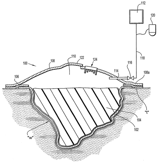
Referring now to Figures 1-3, the composite wound dressing 100 in
accordance with a preferred embodiment of the present disclosure is
illustrated in the
form of an article with multiple layers arranged in juxtaposed or superposed
relation.
The multiple layers include, but, are not limited to a lower or base layer
102, an
absorbent/packing layer 104, an adherent layer 106, and a top layer 108 which
includes
and/or defines the internal vacuum reservoir 110.
The base layer 102 is in direct contact with the wound bed "w". The base
layer 102 is typically porous allowing passage of subatmospheric pressure to
the wound
bed. In one preferred embodiment, the base layer includes a "non-adherent"
material.
"Non-adherent" as used herein refers to a material that does not adhere to
tissues in and
6
CA 02619925 2008-02-20
WO 2007/030598 PCT/US2006/034824
around the wound bed. "Porous" as used herein refers to a material which
contains
numerous small perforations or pores which allow wound fluids of all kinds to
pass
through the material to the dressing layers above. The passage of wound fluid
through
the porous material may be unidirectional such that wound exudate does not
flow back to
the wound bed. This direction flow feature could be in the form of directional
apertures
imparted into the material layer, a lamination of materials of different
absorption to the
base layer 102 or specific material selection that encourages directional
flow. Exemplary
materials used as the base layer 102 include a contact layer sold under the
trademark
XEROFLOW by Kendall Corp., a division of TycoHealthcare. In the alternative,
the
base layer 102 may include an adherent material.
In addition, agents such as hydrogels and medicaments could be bonded or
coated to the base layer 102 to reduce bioburden in the wound, promote healing
and
reduce pain associated with dressing changes or removal. Medicaments include,
for
example, antimicrobial agents, growth factors, antibiotics, analgesics,
debridement agents
and the like. Furthermore, when an analgesic is used, the analgesic could
include a
mechanism that would allow the release of that agent prior to dressing removal
or
change. Exemplary triggers of a release mechanism could be temperature change.
The layer proximal to the base layer 102 or composite structures making
the base layer 102 is the absorbent/packing layer 104. The absorbent/packing
layer 104
of the wound dressing 100 is intended to absorb and capture wound fluid and
exudates.
Exemplary absorbent materials include foams, nonwoven composite fabrics,
hydrogels,
cellulosic fabrics, super absorbent polymers, and combinations thereof.
Typically, the
absorbent/packing layer 104 can absorb up to about 100 cubic centimeters (cc)
or more of
7
CA 02619925 2008-02-20
WO 2007/030598 PCT/US2006/034824
wound fluid. Preferably, the absorbent material includes the antimicrobial
dressing sold
under the trademark KERLIX by Kendall Corp., a division of TycoHealthcare. In
one
preferred embodiment, the absorbent/paclcing layer 104 could be preformed or
shaped to
conform to varying shapes of the wound bed. Those skilled in the art will
recognize that
the absorbent/packing layer 104 can be formed in any suitable shape.
Absorbent/packing
layer 104 may include multiple layers. The only requirement as to shape is
that the
absorbent/packing layer 104 is suitable to treat a particular shape of the
wound.
Additionally, the absorbent/packing layer 104 could be treated with
medicaments. Medicaments include, for exainple, an anti-infective agent such
as an
antiseptic or other suitable antimicrobial or combination of antimicrobials,
polyhexamethylene biguanide (liereinafter, "PHMB"), antibiotics, analgesics,
healing
factors such as vitamins, growth factors, nutrients and the like, as well as a
flushing agent
such as isotonic saline solution.
With continued reference to Figures 1-3, the adherent layer 106 at least
encompasses the perimeter of the wound dressing 100 to surround the wound bed
to
provide a seal around the perimeter of the wound bed "w". For instance, the
sealing
mechanism may be any adhesive bonded to a layer that surrounds the wound bed
"w" or
an adhesive applied directly to the skin. The adhesive must provide acceptable
adhesion
to the tissue "t" surrounding the wound bed "w" skin, e.g., the periwound
area, and be
acceptable for use on skin without contact deterioration (for example, the
adhesive should
preferably be non-irritating and non-sensitizing.) The adhesive may be semi-
permeable
to permit the contacted skin to transmit moisture or may be impermeable.
Additionally,
the adhesive could be activated or de-activated by an external stimulus such
as heat or a
8
CA 02619925 2008-02-20
WO 2007/030598 PCT/US2006/034824
given fluid solution or chemical reaction. Adhesives include, for example, the
dressing
sold under the trademark ULTEC Hydrocolloid dressing by Kendall Corp., a
division of
TycoHealthcare.
The adherent layer 106 may also be in the form of an entire layer proximal
to the absorbent/packing layer 104 or preferably is annular or "donut shaped"
as shown.
Preferably, the adherent layer 106 is not bonded to the absorbent/packing
layer 104 to
allow for easy replacement of the absorbent/packing layer 104. In a preferred
embodiment, the adherent layer 106 is at least bonded to the periphery of the
base layer
102. In turn, the peripheral portion 108a of the top layer 108 may be bonded
to the
adherent layer 106 to provide a seal around the perimeter of the wound.
Alternatively,
the adherent layer 106 may be positioned on the peripheral portion 108a of the
top layer
108 and secured to the tissue "t" about the wound bed "w", and not bonded to
the base
layer 102. As a further alternative, the peripheral portion 108a of the top
layer 108 may
include an adhesive surface. It is anticipated that removable contact liners
may also be
used to protect the adhesive surface of the adherent layer 106 prior to use.
The top or upper layer 108 typically seals the top of the wound dressing
100 and helps maintain the appropriate vacuum level within the wound dressing
100. In
one preferred embodiment, the top layer 108 includes the flexible transparent
dressing
manufactured under the trademark POLYSKIN II by Kendall Corp., a division of
TycoHealthcare. POLYSKIN II is a transparent, semi-permeable material which
permits moisture and oxygen exchange with the wound site, and provides a
barrier to
microbes and fluid containment. In the alternative, the top layer 110 may be
9
CA 02619925 2008-02-20
WO 2007/030598 PCT/US2006/034824
impermeable. As a further alternative, the top layer 108 may include a
resilient, e.g.,
elastomeric, material in the shape, e.g., of a dome.
The top layer 108 defines a sealed or enclosed vacuum reservoir 110. The
vacuum reservoir 110 is preferably maintained at an appropriate vacuum level
for a
predetermined period of time sufficient to initiate or complete healing of the
wound bed
"w", i.e., to draw wound fluid and exudate away from the wound bed "w" while
subjecting the wound to subatmospheric pressure. The vacuum may be re-applied
as
needed to maintain a therapeutic effect. The vacuum may be continuous or
interinittent
as desired.
As best seen in Figure 1, the vacuum reservoir 110 is defined within the
dome of the top layer 108. As shown in Figure 2, once vacuum is applied, the
dome of
the top layer 108 is drawn downwardly toward the absorbent/packing layer 104
with the
vacuum or subatmospheric reservoir 110 created beneath the top layer 108.
Typically,
the top layer 108 includes a vacuum port or connector 114 in fluid
communication with
the vacuum reservoir 110. Preferably, the vacuum port 114 includes a one-way
valve
(shown schematically as reference numeral 116) which provides unidirectional
flow of
suction and may provide a means for allowing connection of the composite wound
dressing 100 to the vacuum source 112. The one way valve 116 may be
incorporated
within the vacuum port 114 or, alternatively, be "in line" with the vacuum
source 112. A
flexible tubing 118 is connected to the vacuum port 114 and the vacuum source
112. The
tubing 118 provides suction to the wound from the vacuum source 112 and
enables the
wound fluid to be transferred from the wound dressing 100. The tubing 118 may
be
fabricated from PVC, silicone based material or other flexible materials
(polymers). The
CA 02619925 2008-02-20
WO 2007/030598 PCT/US2006/034824
tubing 118 may optionally include a connection to a collection canister 120
for wound
drainage and debris. Hence, the vacuum source 112 can draw wound fluid through
the
composite wound dressing 100 and tubing 118 into the canister 120. In a
preferred
embodiment of the present disclosure, the canister 120 is portable so that the
patient will
have the freedom to move about rather than being confined to a fixed location.
The
canister 120 may also house an absorbent material to absorb wound fluid and
exudate.
The vacuum source 112 may apply vacuum to the wound by means such
as a manual pump as disclosed in commonly assigned U.S. Patent No. 5,549,584
to
Gross, the entire contents of which are hereby incorporated herein by
reference. In the
alternative, the vacuum source 112 may include an automated pump. Typically,
the
vacuum level is in a range between about 20 mmHg to about 500mmHg, more
preferably,
about 40 mmHg and about 125 mmHg. The automated pump may be a wall suction
apparatus such as those available in an acute or sub-acute care facility. The
automated
pump may be in the form of a portable pump. The portable pump may include a
small or
miniature pump that maintains or draws adequate and therapeutic vacuum levels.
In a
preferred embodiment, the pump is a portable, lightweight, battery operated,
suction
pump which attaches to the distal end of the tubing. Typically, the vacuum
source 112
has regulation means to apply the optimal vacuum pressure for healing the
wound.
~:., .
Furthermore, the vacuum source 11'~2 would preferably contain a mechanism to
detect a
leak in the system if the optimal vacuum pressure is not achieved. Preferably,
the
vacuum source 112 would also contain an indicator (not shown) to indicate when
the
optimal vacuum pressure is achieved. In the alternative, a hand pump in the
form of a
squeeze bulb or a foot pump may seive as the vacuum source 112.
11
CA 02619925 2008-02-20
WO 2007/030598 PCT/US2006/034824
Preferably, a pump is used as the vacuum source 112. Typical pumps
include diaphragm or voice coil activated styles that can deliver variable
vacuum up to 50
cc/minute.
With reference now to Figures 1-4, the top layer 108 may include an
access door 122 to provide access to the interior of the wound dressing 100
and/or the
wound bed "w". The door 122 could be a flap integrally formed with the top
layer 108 or
a separate component connected to the top layer 108 via a hinge or the like.
The door
122 is preferably resealable to maintain the integrity of the vacuum reservoir
110 and
provide a seal relative to the top layer 108. One suitable means for
releasably sealing the
door 122 includes a snap fit 'arrangement, tongue and groove arrangement, "zip
loco"
arrangement, adhesives, VELCRO , etc. The door 122 preferably provides access
to the
wound bed "w" to enable the clinician to monitor the status of the wound,
change the
absorbent/packing layer 104, or apply additional medical treatment to the
wound such as
antimicrobial agents, growth factors, debriders, or other wound healing agents
as needed.
Once the desired procedure is completed, the door 122 would be resealed
relative to the
top layer 108 to maintain the integrity of the vacuum reservoir 110. Figure 4
illustrates
the removal of the absorbent/packing layer 104 through the door 122 when the
door 122
is in an open position. As discussed, a new absorbent/packing layer 104
subsequently
may be introduced through the door 122 to absorb the exudates from the wound
bed "w".
With reference now to Figures 3 and 5, in conjunction with Figures 1-2,
the wound dressing 100 may include a visual indicator device 124 mounted to
the top
layer 108 to provide a visual indication of vacuum pressure within the wound
dressing
100. The visual indicator device 124 may include at least one electronic
position
12
CA 02619925 2008-02-20
WO 2007/030598 PCT/US2006/034824
indicator for detecting the relative position of the top layer 108 and the
wound bed, and,
thus the state of the vacuum within the wound dressing 100. In accordance with
this
embodiment, the top layer 108 may have a resilient characteristic either
through the
material of construction of the top layer 108 or through reinforcement (e.g.,
elastomeric)
members incorporated in the top layer 108.
In one embodiment depicted in Figure 6, the visual indicator device 124
includes at least one, preferably, three position-sensitive switches 126a-
126c, and a self-
powered electronic signaling module 128. The module 128 may include an
electronic
signaling module circuit board 130, a battery power source 132, at least one
transducer
including three light emitting diodes (LED) 134a-c and/or a loudspeaker 136
electrically
connected 138 to the circuit board 130. The LEDs 134a-c are color coded red,
yellow
and green respectively. The position-sensitive switches 126a-126c each may be
a
pressure sensor which electrically bridges contacts 140a, 140b of the visual
indicator
device 124.
In the embodiment shown, three position switches 126a-c are mounted to
top layer 108. The switches 126a-c include switch plates or contact arms
arranged as a
series of decreasing diameter annular coaxial elements. Alternatively, the
switches may
be linear in configuration depending downwardly from the top layer 108. The
switch
plate or contact arm of switches 126a-c are of predetermined length extending
downwardly from the top layer 108 within the vacuum reservoir 110. (Figures 4
and 5)
The contact arm of outer switch 126a of the pressure indicator 124 has the
greatest
length. The contact arm of the middle switch 126b has a length less than the
length of the
contact arm of the outer ring 126a. The contact arm of the inner switch 126c
has the
13
CA 02619925 2008-02-20
WO 2007/030598 PCT/US2006/034824
smallest length. The contacts 140a of switches 126a-c are integrated within
the contact
aims. Contacts 140b of the switches 126a-c may be disposed on the top surface
of the
absorbent/packing layer 104 or integrated within the absorbent/packing layer
104 in
general longitudinal alignment with their respective contact arms.
Alternatively, the
position sensor may be a magnetic proximity sensor. The self-powered
electronic
signaling module 130 may be any conventional modules adapted to emit light
and/or
audible sound etc. upon closing of the switch.
When the top layer 108 is drawn down by vacuum pressure within the
vacuum reservoir 110 toward its vacuum state of Figure 2, inner switch 126c is
in contact
with a respective coupler or contact 140b disposed within the
absorbent/packing layer
104 thereby energizing the visual indicator device 124 to illuminate the green
LED 134c
within the pressure indicator device mounted to the top layer 108. The green
light of the
LEDs 134c indicates a full vacuum condition of the vacuum reservoir 110. As
appreciated, switches 126a, 126b may also be in contact with their respective
couplers in
this vacuum condition of top layer 108. However, it is envisioned that circuit
board 130
will incorporate circuitry to override the electrical contact of these two
switches when
switch 126c is in contact with its respective coupler.
As vacuum pressure decreases and the dome of the top layer 108 begins to
assume its normal condition of Figure 1, through, e.g., the resilient
characteristic of the
top layer 108 discussed hereinabove, the inner switch 126c loses contact with
the
absorbent/packing layer 104 while the middle switch 126b maintains/establishes
electrical contact with its associated contact 140b. The electrical connection
of the
middle switch 126b results in illumination of the yellow LED 134b. The yellow
LED
14
CA 02619925 2008-02-20
WO 2007/030598 PCT/US2006/034824
134b represents a partial vacuum or marginal vacuum condition of the vacuum
reservoir
110. As the vacuum pressure further decreases and the top layer 108 moves
towards its
fully expanded or normal condition of Figure 1, the outer switch 126a is the
remaining
switch in contact with its associated contact 140b within the
absorbent/packing layer 104.
In this condition, the red LED 134a is energized and visible to the clinician
essentially
providing a warning that the vacuum within the vacuum reservoir has dissipated
or is
nearly dissipated (i.e., subatmospheric pressure is close to or no longer
present).
Consequently, the vacuum source 112 may be actuated either manually or
automatically
to reestablish the vacuum state within the vacuum reservoir 110. Further
vacuum loss
will result in the remaining switch 126c losing its contact where no lights
are visible to
the patient.
It is also envisioned that the circuit board 130 could be devoid of the
aforementioned override circuitry. As a result, in the full vacuum condition
of dressing
100, each of the green, yellow and red LEDs 134a-134c would be illuminated
while in
the partial vacuum state, the yellow and red LEDs 134a, 134b would be
illuminated and
in the warning state, the red LED 134a would be illuminated. It is further
envisioned that
the loudspeaker 136 could emit an audible alarm when any of the aforementioned
vacuum states are realized.
Figure 7 illustrates an alternate embodiment of the visual pressure
indicator 124. In accordance with this embodiment, the visual pressure
indicator 124 is
mounted to the absorbent/packing layer 104 or positioned on the
absorbent/packing layer
104 in juxtaposed relation. The top layer 108 is preferably transparent to
permit viewing
of the pressure indicator 124 through the top layer 108 and into the vacuum
reservoir 110.
CA 02619925 2008-02-20
WO 2007/030598 PCT/US2006/034824
The positioning of the visual pressure indicator 124 is reversed or arranged
on its back in
a manner where the respective switches 126a-c extend upwardly toward top layer
108.
The respective coupler or contacts 140b are incorporated within the top layer
108. In the
full vacuum state or condition of Figure 2, the contact 140b of the top layer
108 is in
contact with the switch 126c thereby energizing the LED 134c to display the
green color
to the clinician. During healing, as the vacuum reservoir begins to lose its
vacuum, the
top layer 108 moves towards its open condition of Figure 1, the inner switch
126c loses
its contact with its respective coupler 140b of the top layer 108 resulting in
electrical
contact with the yellow LED 134b indicating a partial vacuum condition of the
vacuum
reservoir 110. Continued loss of vacuum within the vacuum reservoir 110 causes
the
electrical contact of the coupler 140b witli the outer switch 126c. This
contact of the
LED 134c with the outer switch 126c is indicated by the presence of its red
color and
may correspond to a loss or near loss of vacuum within the vacuum reservoir
110 thus
proinpting the clinician to activate the vacuum source 112 or pursue other
clinical
measures.
Figure 8 illustrates another embodiment where the LEDs 134a-134c are
supplemented with additional visual indicia. The visual indicia may include
various
symbols which correspond to the state of the vacuum within the vacuum
reservoir 110.
When a desired level of vacuum is reached, the "smiley" symbol 152 would
illuminate
indicating an adequate vacuum state. A partial vacuum state would result in
the
illumination of the "caution triangle" symbol 154. A loss or near loss of
vacuum would
result in the illumination of the "octagon" symbol (no vacuum) 156. One
skilled in the
art will readily appreciate the design of electronic circuitry to achieve this
objective.
16
CA 02619925 2008-02-20
WO 2007/030598 PCT/US2006/034824
Figures 9-11 illustrate an alternate embodiment of the present disclosure.
In accordance with this embodiment, the visual indicator device 124 may
include a series
of rings 200,202,204 disposed on the underside of the top layer 108, e.g.,
printed on the
top layer 108, and arranged in concentric relation as shown. The rings include
outer ring
200, middle ring 202 and inner ring 204 and are color coded red, yellow and
green,
respectively. Each colored ring 200,202,204 is positioned to contact the
absorbent/packing layer 104 depending on the state or condition of the vacuum
within the
vacuum reservoir 110. Under full vacuum depicted in Figure 10, all the rings
200,202,204 would contact absorbent/packing layer 104 and thus be activated
and visible
through the top of the wound dressing. As the vacuum within the reservoir
dissipates or
is reduced, the top layer 110 pulls away from the absorbent/packing layer 104.
In one
embodiment with an elastomeric dome, the inner ring 204 first loses contact
followed by
the middle and outer rings, 202, 200, respectively, as the vacuum is reduced.
As each
colored ring 200,202,204 loses contact with absorbent/packing layer 104, the
respective
ring becomes less visible or not visible from above the wound dressing 100
thus
indicating to the clinician the condition of the vacuum within the vacuum
reservoir. The
rings 200,202,204 may incorporate electronic switches to be activated in the
manner
discussed hereinabove in connection with the embodiment of Figures 1-6.
Alternatively,
each ring 200,202,204 may incorporate an analytical test strip device which,
e.g., may
detect the presence of a predetermined analyte in the exudates contained in
the
absorbent/packing layer 104. Upon contact with the predetermined analyte, the
test strip
device of each ring 200,202,204 assumes a color such as red, yellow or green.
When a
respective ring 200,202,204 loses contact with the predetermined analyte, the
color of the
17
CA 02619925 2008-02-20
WO 2007/030598 PCT/US2006/034824
respective ring may fade or assume a neutral color. One exampled of a color
coded test
strip device suitable for use with the present disclosure is disclosed in U.S.
Patent No.
7,049,130 to Carroll et al., issued May 23, 2006, the entire contents of which
are
incorporated herein by reference. One skilled in the art may determine the
parameters
and characteristics of a test strip device to perform the objectives discussed
hereinabove.
In addition, the door 122 of the embodiment of Figures 9-11 is adapted to
pivot along hinge 208 to provide access to the vacuum reseivoir 110. The
opening of the
door 122 is disposed adjacent the periphery of the wound dressing 102 and thus
provides
a relatively large access opening upon pivoting or opening the door 122 along
the hinge
208. This facilitates removal and replacement of absorbent/packing layer 104.
It is further contemplated that the wound dressing apparatus may
incorporate external means or applications to stimulate tissue growth and/or
healing. For
example, an ultrasonic transducer may be incorporated into the wound dressing
apparatus
to impart mechanical energy for the treatinent of the tissue such as, for
instance, directing
thermal or vibratory energy on the wound area to stimulate healing and/or
further
encouraging exudates removal by vacuum and/or introducing various drugs into
the
human body through the skin. Other sensor types are also contemplated for
incorporation into the wound dressing apparatus including oxygen, chemical,
microbial
and/or temperature sensors. The detection of oxygen adjacent the wound area
would
assist the clinician in determining the status of wound healing. The presence
of an
elevated temperature may be indicative of an infection.
18
CA 02619925 2008-02-20
WO 2007/030598 PCT/US2006/034824
While the disclosure has been illustrated and described, it is not intended
to be limited to the details shown, since various modifications and
substitutions can be
made without departing in any way from the spirit of the present disclosure.
For
example, it is envisioned the subject matter of the commonly assigned patent
application
filed concurrently herewith under Express Mail Certificate No. EL985194525 US,
and
which claims priority to provisional application No. 60/714,812, filed on
September 6,
2006, and the subject matter of the commonly assigned patent application filed
concurrently herewith under Express Mail Certificate No. EV879103054 US, and
which
claims priority to provisional application No. 60/714,912, filed on September
7, 2006,
(the entire contents of each application being incorporated herein) may be
incorporated
into the present disclosure. As such, further modifications and equivalents of
the
invention herein disclosed can occur to persons skilled in the art using no
more than
routine experimentation, and all such modifications and equivalents are
believed to be
within the spirit and scope of the disclosure as defined by the following
claims.
19